Embed Size (px)
Citation preview

Dr Maha ELBeltagy
Dr. Maha ELBeltagy Assistant Professor of Anatomy
Faculty of Medicine
The University of Jordan
2018
Neuroanatomy
Development of the Central
Nervous System
Dr Maha ELBeltagy
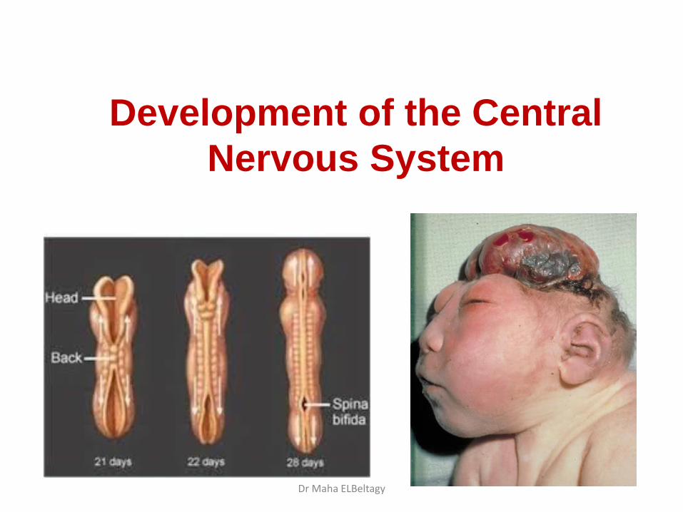
Development of the nervous system Development of the neural tube:
- At the beginning of the 3rd week an ectodermal thickening appears in the middle of
the trilaminar germ disc known as the neural plate.
- The neural plate invaginates to form a neural groove.
- The lips of the neural groove approach each other & fuse together transforming the groove into a neural tube with an anterior & posterior neuropores which are obliterated on day 25 & 27 respectively transforming the neural tube into a closed tube.
Dr Maha ELBeltagy
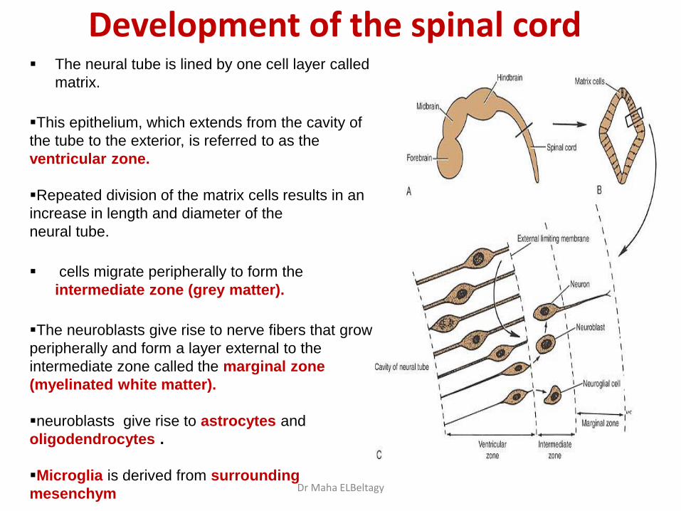
Development of the spinal cord The neural tube is lined by one cell layer called
matrix.
This epithelium, which extends from the cavity of
the tube to the exterior, is referred to as the
ventricular zone.
Repeated division of the matrix cells results in an
increase in length and diameter of the
neural tube.
cells migrate peripherally to form the
intermediate zone (grey matter).
The neuroblasts give rise to nerve fibers that grow
peripherally and form a layer external to the
intermediate zone called the marginal zone
(myelinated white matter). neuroblasts give rise to astrocytes and
oligodendrocytes .
Microglia is derived from surrounding
mesenchym
Dr Maha ELBeltagy
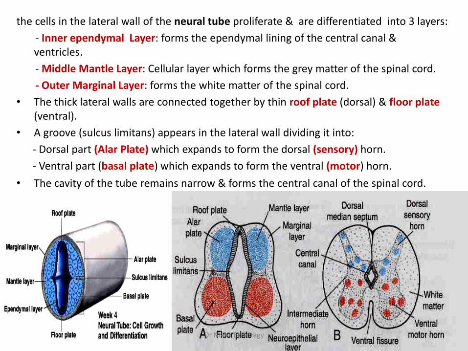
the cells in the lateral wall of the neural tube proliferate & are differentiated into 3 layers:
- Inner ependymal Layer: forms the ependymal lining of the central canal & ventricles.
- Middle Mantle Layer: Cellular layer which forms the grey matter of the spinal cord.
- Outer Marginal Layer: forms the white matter of the spinal cord.
• The thick lateral walls are connected together by thin roof plate (dorsal) & floor plate (ventral).
• A groove (sulcus limitans) appears in the lateral wall dividing it into:
- Dorsal part (Alar Plate) which expands to form the dorsal (sensory) horn.
- Ventral part (basal plate) which expands to form the ventral (motor) horn.
• The cavity of the tube remains narrow & forms the central canal of the spinal cord.
Dr Maha ELBeltagy

Development of sensory and motor roots
Dr Maha ELBeltagy
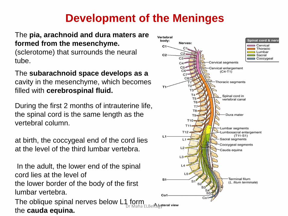
Development of the Meninges
The pia, arachnoid and dura maters are
formed from the mesenchyme.
(sclerotome) that surrounds the neural
tube.
The subarachnoid space develops as a
cavity in the mesenchyme, which becomes
filled with cerebrospinal fluid.
During the first 2 months of intrauterine life,
the spinal cord is the same length as the
vertebral column.
at birth, the coccygeal end of the cord lies
at the level of the third lumbar vertebra.
In the adult, the lower end of the spinal
cord lies at the level of
the lower border of the body of the first
lumbar vertebra.
The oblique spinal nerves below L1 form
the cauda equina. Dr Maha ELBeltagy
Dr Maha ELBeltagy

Congenital Malformations of spinal cord development 1) Spina bifida occulta: Absent vertebral arch with normal spinal cord. It
affects the lumbosacral area & is usually covered with hairy skin.
2) Spina bifida cystica:
- Meningocele: The meninges herniates through the spina bifida to form subcutaneous sac filled with CSF.
- Meningomyelocele: The spinal cord herniates through the meningocele.
- Myelocele (Rachischisis): Failure of obliteration of the neural tube.
Dr Maha ELBeltagy
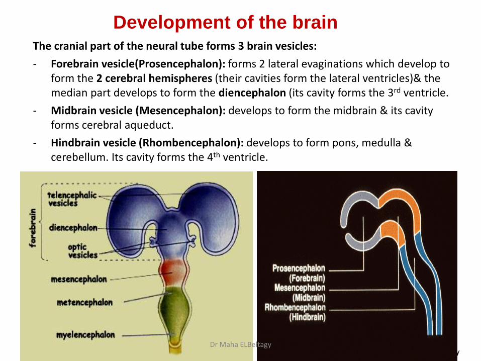
The cranial part of the neural tube forms 3 brain vesicles:
- Forebrain vesicle(Prosencephalon): forms 2 lateral evaginations which develop to form the 2 cerebral hemispheres (their cavities form the lateral ventricles)& the median part develops to form the diencephalon (its cavity forms the 3rd ventricle.
- Midbrain vesicle (Mesencephalon): develops to form the midbrain & its cavity forms cerebral aqueduct.
- Hindbrain vesicle (Rhombencephalon): develops to form pons, medulla & cerebellum. Its cavity forms the 4th ventricle.
Prof Yousry
Development of the brain
Dr Maha ELBeltagy
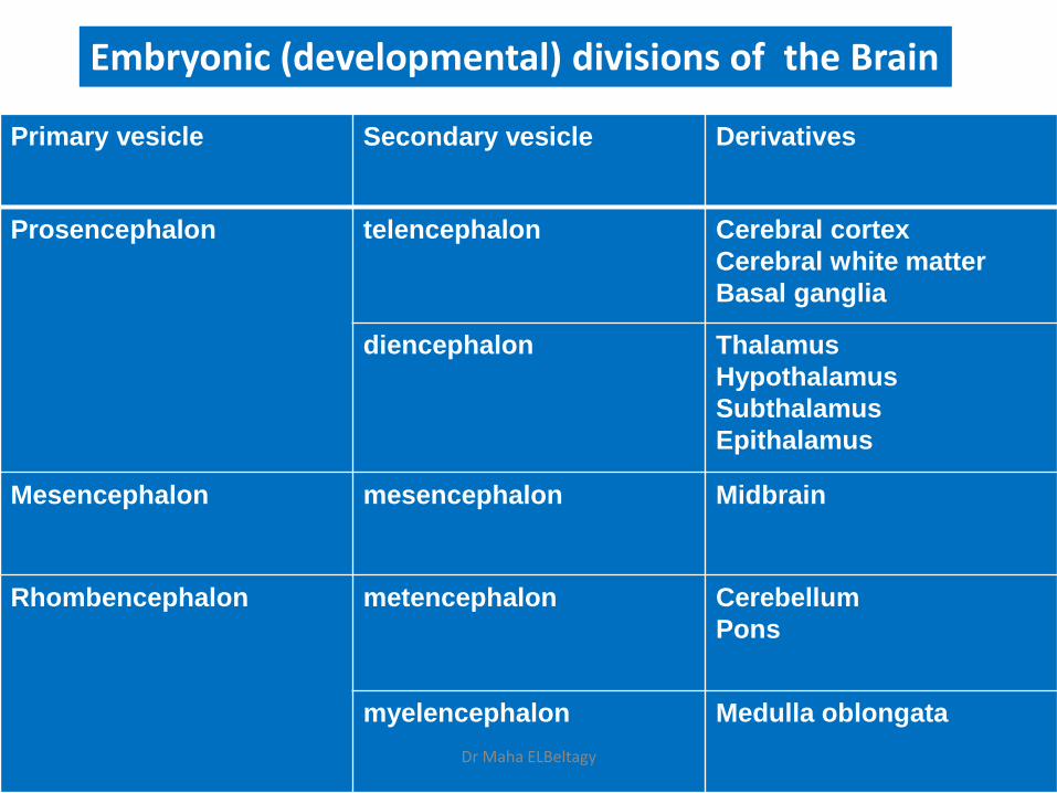
Embryonic (developmental) divisions of the Brain
Primary vesicle
Secondary vesicle
Derivatives
Prosencephalon
telencephalon
Cerebral cortex
Cerebral white matter
Basal ganglia
diencephalon Thalamus
Hypothalamus
Subthalamus
Epithalamus
Mesencephalon
mesencephalon Midbrain
Rhombencephalon metencephalon
Cerebellum
Pons
myelencephalon
Medulla oblongata
Dr Maha ELBeltagy

DEVELOPMENT OF THE MEDULLA OBLONGATA
• As in the development of the spinal cord the medulla will have an alar plate & a basal plate separated by a sulcus limitans & connected by a thin roof plate & a floor plate.
• The lateral walls move away from each other stretching the roof plate & enlarging its cavity which forms the 4th ventricle.
The alar plate forms the sensory nuclei of the medulla & the basal plate forms the motor nuclei. Between the fourth and fifth months, local resorptions of the roof plate
occur, forming lateral foramina of Luschka, and a median foramen of Magendie.
Dr Maha ELBeltagy
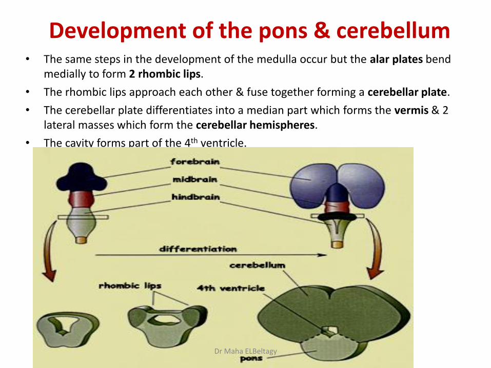
Development of the pons & cerebellum • The same steps in the development of the medulla occur but the alar plates bend
medially to form 2 rhombic lips.
• The rhombic lips approach each other & fuse together forming a cerebellar plate.
• The cerebellar plate differentiates into a median part which forms the vermis & 2 lateral masses which form the cerebellar hemispheres.
• The cavity forms part of the 4th ventricle.
Dr Maha ELBeltagy

Development of Midbrain • As in the development of the spinal cord & the medulla the midbrain will have an alar plate &
a basal plate separated by a sulcus limitans & connected by a thin roof plate & a floor plate.
• The alar plates develop to form the tectum which is divided by a vertical & transverse grooves into 4 colliculi.
• The basal plate forms the motor nuclei in the tegmentum of midbrain
• The marginal layer of the basal plate enlarges greatly to form the crus cerebri.
• It cavity remains narrow & forms the cerebral aqueduct.
Prof Yousry Dr Maha ELBeltagy

Development of the Diencephalon It develops from the median part of the forebrain. It consists of 2 lateral walls
connected by a roof plate & a floor plate, its cavity is called the 3rd ventricle.
The roof plate:
- Its anterior part forms the choroid plexus of the 3rd ventricle.
- Its posterior part forms the pineal body.
A hypothalamic sulcus appears in the lateral wall which separates the thalamus above from the hypothalamus below.
The floor plate forms the posterior lobe of the pituitary gland.
Dr Maha ELBeltagy
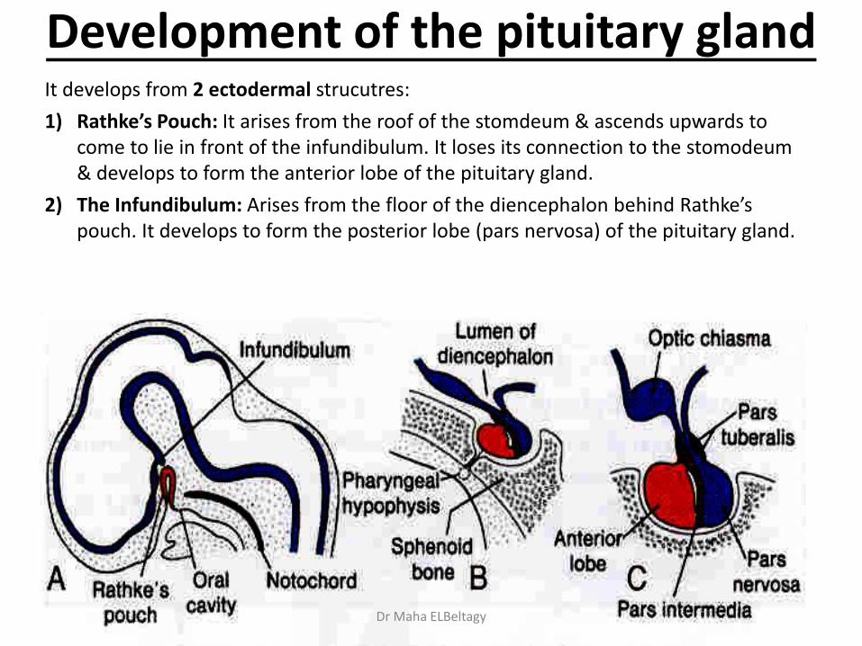
Development of the pituitary gland It develops from 2 ectodermal strucutres:
1) Rathke’s Pouch: It arises from the roof of the stomdeum & ascends upwards to come to lie in front of the infundibulum. It loses its connection to the stomodeum & develops to form the anterior lobe of the pituitary gland.
2) The Infundibulum: Arises from the floor of the diencephalon behind Rathke’s pouch. It develops to form the posterior lobe (pars nervosa) of the pituitary gland.
Dr Maha ELBeltagy

Development of the cerebral hemisphere The 2 cerebral hemispheres arise as 2 evaginations from the lateral wall of the
forebrain.
The cavity of each of them expands to form the lateral ventricle.
The wall of the hemisphere consists of 3 layers: ependymal, mantle & marginal.
The mantle layer at the base of the hemisphere forms the basal ganglia.
The hemispheres enlarge & overlaps the brain stem & cerebellum.
Dr Maha ELBeltagy
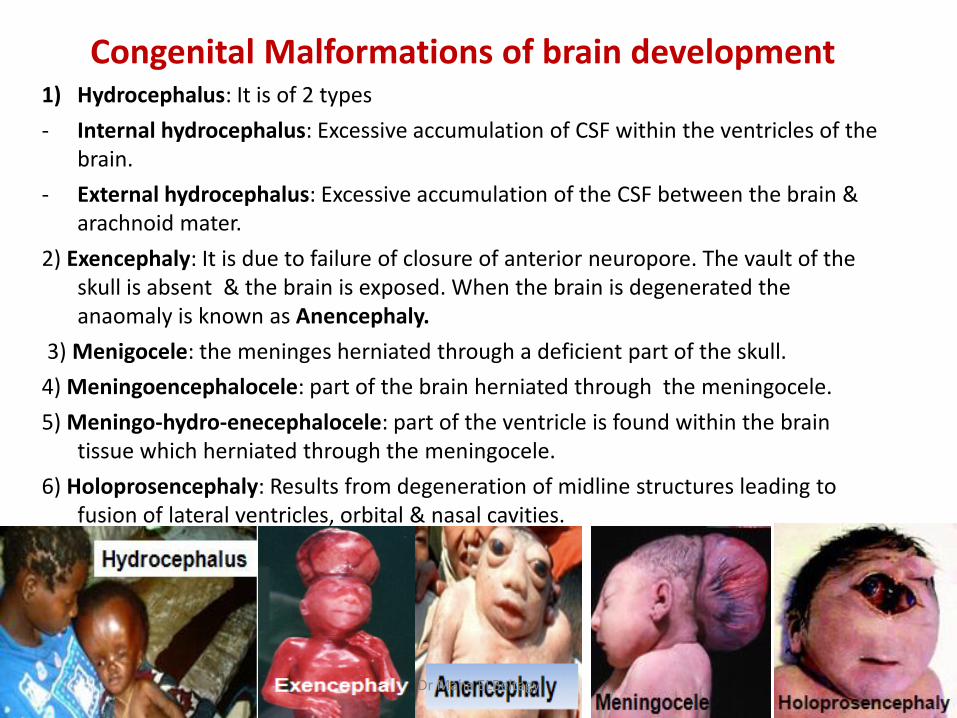
Congenital Malformations of brain development 1) Hydrocephalus: It is of 2 types
- Internal hydrocephalus: Excessive accumulation of CSF within the ventricles of the brain.
- External hydrocephalus: Excessive accumulation of the CSF between the brain & arachnoid mater.
2) Exencephaly: It is due to failure of closure of anterior neuropore. The vault of the skull is absent & the brain is exposed. When the brain is degenerated the anaomaly is known as Anencephaly.
3) Menigocele: the meninges herniated through a deficient part of the skull.
4) Meningoencephalocele: part of the brain herniated through the meningocele.
5) Meningo-hydro-enecephalocele: part of the ventricle is found within the brain tissue which herniated through the meningocele.
6) Holoprosencephaly: Results from degeneration of midline structures leading to fusion of lateral ventricles, orbital & nasal cavities.
Dr Maha ELBeltagy

THANK YOU
Dr Maha ELBeltagy