Embed Size (px)
Citation preview
Nc
Ca
b
c
a
ARRA
KCPWD
1
mo(cmai
BU
1d
Developmental Cognitive Neuroscience 1 (2011) 175–186
Contents lists available at ScienceDirect
Developmental Cognitive Neuroscience
journa l homepage: ht tp : / /www.e lsev ier .com/ locate /dcn
eural indices of improved attentional modulation over middlehildhood
. Wendelkena,∗,1, C.L. Baymb,1, A. Gazzaleyc, S.A. Bungea
Helen Wills Neuroscience Institute, University of California at Berkeley, United StatesDepartment of Psychology, University of Illinois at Urbana-Champaign, United StatesUniversity of California at San Francisco, United States
r t i c l e i n f o
rticle history:eceived 10 June 2010eceived in revised form 30 October 2010ccepted 1 November 2010
eywords:ognitive controlarahippocampal place areaorking memory
evelopment
a b s t r a c t
The ability to control the focus of attention relies on top-down modulation of cortical activ-ity in areas involved in stimulus processing, and this ability is critical for maintaining itemsin working memory in the presence of distraction. Prior research demonstrates that chil-dren are less capable of focusing attention, relative to adults, and that this ability developssignificantly during middle childhood. Here, using fMRI and a face/scene working mem-ory task adapted from Gazzaley et al. (2005a,b), we compared top-down modulation in 15children (aged 8–13) and 15 young adults (aged 19–26). Replicating prior results, in youngadults, attention to scenes modulated activity in the parahippocampal place area (PPA). Inaddition, modulation of PPA activity increased as a function of age in children. PPA activity
was also related to performance in this group, on the working memory task as well on atest of subsequent memory. Dorsolateral PFC also demonstrated increasing task-specificactivation, as a function of age, in children. The present findings support the idea that chil-dren’s reduced ability to maintain items in working memory, especially in the presenceof distraction, is driven by weaker top-down modulation of activity in areas involved instimulus processing.. Introduction
Working memory, the capacity to maintain andanipulate information, particularly in the presence
f distraction, depends critically on attentional controlBaddeley, 1998). In fact, the contents of working memory
an be conceptualized as active internal representationsaintained within the focus of attention (Cowan, 1995),nd this viewpoint is supported by neuroimaging stud-es that have identified a mechanistic overlap between
∗ Corresponding author at: Helen Wills Neuroscience Institute, 132arker Hall, University of California at Berkeley, Berkeley, CA 94720,nited States.
E-mail address: [email protected] (C. Wendelken).1 Shared first authorship.
878-9293/$ – see front matter © 2010 Elsevier Ltd. All rights reserved.oi:10.1016/j.dcn.2010.11.001
© 2010 Elsevier Ltd. All rights reserved.
attention and working memory (Awh and Jonides, 2001).The ability to control the focus of our attention allowsus to maintain in working memory those aspects of theenvironment that are relevant to our goals, and to devotefewer resources to processing environmental cues thatare not goal-relevant. This ability waxes and wanes overthe lifespan; indeed, in both children (Harnishfeger andBjorklund, 1994) and older adults (Hasher and Zacks, 1988),the ability to maintain relevant information in workingmemory is hindered by suboptimal selective attention.Prior research on the development of working memoryhas demonstrated that the ability to maintain items in the
absence of distraction, and the neural substrates of thisability, are established very early in childhood (Diamondet al., 1994). However, there is protracted development ofworking memory, into adolescence and even adulthood, asit involves large item loads, manipulation of items, or main-
l Cogniti
176 C. Wendelken et al. / Developmentatenance in the presence of distraction (Luna et al., 2004;Scherf et al., 2006; Geier et al., 2009). For recent reviewsof this literature, see Bunge and Wright (2007) and Lunaet al. (2010). Protracted development of attentional controlis likely to be a major cause of the protracted developmentof working memory.
There is strong neurophysiological evidence that atten-tional control is mediated by top-down (or goal-based)modulation of activity in brain regions involved in stim-ulus processing (Desimone and Duncan, 1995; Millerand Cohen, 2001). This evidence comes from single-cellphysiology (Moran and Desimone, 1985), electroen-cephalography (Hillyard et al., 1973), and neuroimaging(Corbetta et al., 1990; Pessoa et al., 2003). This body ofresearch has demonstrated that activity in cortical regionsinvolved in the representation of particular classes of stim-uli increases as a result of top-down shifts of attentiontoward the relevant stimulus class. This mechanism of top-down modulation supports working memory, as well asattention, by favoring the effective encoding of relevantinformation (Rainer et al., 1998; Ploner et al., 2001; Vogelet al., 2005). Numerous studies have implicated frontal andparietal regions in top-down attentional control (Pessoaet al., 2003). Lateral prefrontal cortex (PFC), in particular,has been linked to selective attention tasks where rele-vant and irrelevant stimuli compete for attention (Rossiet al., 2009; Iba and Sawaguchi, 2003; Everling et al.,2002).
Gazzaley, D’Esposito, and colleagues have previouslydemonstrated that younger adults exhibit enhanced acti-vation of relevant visual association areas in response tostimuli to be maintained in working memory, and reducedactivity in response to distracting stimuli (Gazzaley et al.,2005a,b). In addition, they have demonstrated the func-tional relevance of these activation changes for workingmemory performance. Their work has focused on the top-down modulation of activation in two brain regions: thefusiform face area (FFA) and the parahippocampal placearea (PPA). FFA has been identified as a region that is par-ticularly sensitive to the observation of faces (Kanwisheret al., 1997). PPA, by contrast, has been shown to be partic-ularly sensitive to the observation of buildings and outdoorscenes (Epstein and Kanwisher, 1998).
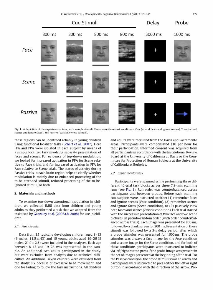
Gazzaley and colleagues sought to test whether acti-vation in these brain regions that process faces or sceneswould be modulated by instructions to attend to eitherof these stimuli. In the face-scene working memory task,participants were instructed to view a sequence of fourimages: two faces and two scenes. Following a delay, theyviewed a probe stimulus and indicated whether or not itmatched one of the target items. In the “Attend Face” con-dition, they were instructed to attend only to the faces, asthey would always receive a face stimulus as a probe. Like-wise, in the “Attend Scene” condition, participants wereinstructed to attend only to the scenes, as they wouldalways receive a scene stimulus as a probe. In the “Passive
View” condition, they were instructed to passively view thestimuli, as they would never be asked to make a recogni-tion memory judgment at the end of the trial, but ratherto respond to a leftward or rightward arrow stimulus bypressing one of two buttons.ve Neuroscience 1 (2011) 175–186
Within the right FFA, the instruction to attend to facesproduced increased activation during cue presentation rel-ative to passive viewing. Similarly, within the PPA, theinstruction to attend to scenes produced increased cue-related activation relative to passive viewing. Gazzaleyet al. argued that these effects result from top-down atten-tional enhancement. In the PPA, the researchers also foundevidence of reduced activation when participants wereasked to attend to the faces as compared with passive view-ing. Gazzaley et al. argued that these effects result fromtop-down attentional suppression. Further, they showed thata region in left lateral PFC was highly correlated withPPA during attention to scenes, and that the strength ofthis coupling correlated with the magnitude of attentionalmodulation in PPA, suggesting that this region biases activ-ity levels in PPA (Gazzaley et al., 2007).
In a follow-up study, Gazzaley and colleagues used thesame fMRI paradigm to examine selective attention in olderadults, aged 60–72 (Gazzaley et al., 2005a,b). Comparedto younger adults, older adults demonstrated diminishedmodulation of PPA. This difference was driven by reducedsuppression of activity associated with irrelevant infor-mation; specifically, older adults did not show reducedactivation of left PPA for “Attend Face” relative to “PassiveView” trials. Enhancement of activity associated with rel-evant information (“Attend Scene” > “Passive View”) wassimilar in older and younger adults. This inability to sup-press activation of left PPA was correlated with impairedperformance on the working memory task. These resultssupported the “inhibitory deficit hypothesis” (Hasher andZacks, 1988), suggesting that age-related decline in work-ing memory capacity may be driven specifically by declinein the ability to suppress irrelevant information.
Here, we sought to use the same fMRI paradigm to assessthe effectiveness of top-down attentional modulation inchildren, both in terms of its neural signature as well asits consequences for performance. There is a rich behav-ioral literature showing that children experience greaterinterference from distracters than do young adults, whichaffects their performance on tasks that present compet-ing stimuli (Bjorklund and Harnishfeger, 1990; Ordaz et al.,2010). Given the parallels between the behavioral find-ings for children and older adults, we posited that children,like older adults, would demonstrate reduced modulationof visual association areas. Previous studies have demon-strated that attentional control improves over the courseof middle childhood (Rueda et al., 2004), and that children,relative to adults, show reduced activation in frontal andposterior brain regions associated with attentional control(Konrad et al., 2005). Similar changes have been observedwith respect to working memory and interference con-trol, with age-related improvements in performance andchanges in brain activation, particularly in lateral PFC andposterior parietal cortex during task performance (Kwonet al., 2002; Olesen et al., 2007; Velanova et al., 2009; Lunaet al., 2010). However, it is not yet known whether children
exhibit reduced modulation of visual association areas rela-tive to young adults during performance of a task requiringselective attention to visual stimuli.Although FFA and PPA continue to mature throughchildhood and adolescence (Golarai et al., 2007, 2010),
C. Wendelken et al. / Developmental Cognitive Neuroscience 1 (2011) 175–186 177
F were ths
tuFafwtFPmti
2
datd
2
(mbpbcto
ig. 1. A depiction of the experimental task, with sample stimuli. Therecenes and ignore faces), and Passive (passively view stimuli).
hese regions can be identified reliably in young childrensing functional localizer tasks (Scherf et al., 2007). HereFA and PPA were isolated in each subject by means ofsimple localizer task involving separate presentation of
aces and scenes. For evidence of top-down modulation,e looked for increased activation in PPA for Scene rela-
ive to Face trials, and for increased activation in FFA forace relative to Scene trials. The status of activity duringassive trials in each brain region helps to clarify whetherodulation is mainly due to enhanced processing of the
o-be-attended stimuli, reduced processing of the to-be-gnored stimuli, or both.
. Materials and methods
To examine top-down attentional modulation in chil-ren, we collected fMRI data from children and youngdults as they performed a task that we adapted from theask used by Gazzaley et al. (2005a,b, 2008) for use in chil-ren.
.1. Participants
Data from 15 typically developing children aged 8–136 males, 11.5 ± .43) and 15 young adults aged 19–26 (8
ales, 21.9 ± 2.3) were included in the analyses. Each ageetween 8–13 and 19–26 was represented in the sam-
le. An additional two adults participated in the study,ut were excluded from analysis due to technical diffi-ulties. An additional seven children were excluded fromhe study: six because of excessive head movement, andne for failing to follow the task instructions. All childrenree task conditions: Face (attend faces and ignore scenes), Scene (attend
and adults were recruited from the Davis and Sacramentoareas. Participants were compensated $10 per hour fortheir participation. Informed consent was acquired fromall participants in accordance with the Institutional ReviewBoard at the University of California at Davis or the Com-mittee for Protection of Human Subjects at the Universityof California at Berkeley.
2.2. Experimental task
Participants were scanned while performing three dif-ferent 40-trial task blocks across three 7.8-min scanningruns (see Fig. 1). Run order was counterbalanced acrossparticipants and between groups. Before each scanningrun, subjects were instructed to either (1) remember facesand ignore scenes (Face condition), (2) remember scenesand ignore faces (Scene condition), or (3) passively viewboth faces and scenes (Passive condition). Each trial startedwith the successive presentation of two face and two scenepictures, in pseudo-random order (with order counterbal-anced across trials). Each image was presented for 800 ms,followed by a blank screen for 200 ms. Presentation of thesestimuli was followed by a 3-s delay period, after whicha probe stimulus was presented for 1600 ms. The probestimulus was always a face image for the Face conditionand a scene image for the Scene condition, and for both ofthese conditions participants were instructed to indicate
via left/right button press if the probe image was present inthe set of images presented at the beginning of the trial. Forthe Passive condition, the probe stimulus was an arrow andparticipants were instructed to press either the left or rightbutton in accordance with the direction of the arrow. Pre-
l Cogniti
178 C. Wendelken et al. / Developmentasentation of the probe stimulus was followed by a jitteredfixation period of between 2 and 10 s, with a distributiondetermined by an optimal sequencing program (optseq2)designed to maximize estimation efficiency (Dale, 1999).
The task used here was modified from a paradigm usedpreviously in adults (Gazzaley et al., 2005a,b). The delayperiod in this study was considerably shorter (3 s ratherthan 7 s) and the probe period was slightly longer (1.6 srather than 1 s). These modifications were intended to ren-der the working memory task easier for children, and tolimit the amount of time that they needed to stay still inthe scanner.
In addition to the main face-scene working memorytask, participants also performed a face-scene functionallocalizer task. The purpose of this task was to localize theFFA and PPA – or, more specifically, FFA and PPA voxelsinvolved in face or scene working memory – in each indi-vidual subject. In the localizer task, participants were pre-sented with alternating 20-s blocks of face or scene stimuli(10 blocks of each type) and were instructed to attend to thestimuli and press a button whenever the current stimulusrepeated the immediately preceding stimulus.
2.3. Long-term memory testing
Following completion of the fMRI task, participantswere given a surprise recognition memory test outsidethe scanner. The purpose of this was to test whether theattentional instructions influenced long-term retention ofto-be-remembered versus to-be-ignored items differentlyin adults and children. The test was comprised of 192images (96 faces and 96 scenes), half of which had beenseen during the fMRI session. Images were presented ona computer screen and participants were instructed torespond using a 4-point scale whether they had seen theimage previously and their confidence. Stimuli that hadbeen used as probes in the fMRI task were not used in thelong-term memory test. Previously viewed stimuli in thememory test were taken equally from each condition inthe fMRI task (Face, Scene, Passive).
2.4. Scan procedure
Imaging was performed using an 8-channel phased-array coil on a 3-T Siemens Trio MRI scanner (SiemensMedical Solutions, Erlangen, Germany) at the University ofCalifornia at Davis Imaging Research Center (Sacramento,CA). Children were introduced to the scanner environmentwith a mock scanner, where they were trained to lie still.Prior to fMRI data acquisition, all participants were pro-vided with explicit task instructions. Task stimuli werepresented using Presentation software (NeurobehavioralSystems, Inc.), and were projected to a screen, which par-ticipants could view from within the scanner by means of amirror. Participants responded via button box, held in theright hand. Inside the scanner, but prior to scanning, par-
ticipants practiced using the button box to respond to a setof sample problems.After acquisition of a T2 localizer scan, four functionalruns were collected (TR = 2000 ms, TE = 25 ms, 34 axialslices, no interslice-gap, 3.4 mm × 3.4 mm × 4 mm voxels,
ve Neuroscience 1 (2011) 175–186
flip angle = 90◦, field of view = 220 mm), including threeruns of the working memory task and one run for thelocalizer task. A gradient-echo echo-planar pulse Prospec-tive Acquisition Correction (3D-PACE) sequence was usedto minimize motion artifacts by prospectively adjust-ing scan parameters throughout a run on the basis ofreal-time assessment of head motion (Siemens MedicalSolutions) (Thesen et al., 2000). Four volumes from thestart of each functional scan were removed from analy-sis to account for magnetic field equilibration. Followingthe functional scans, high-resolution three-dimensional T1MPRAGE anatomical images were acquired.
2.5. fMRI data preprocessing and analysis
FMRI data were analyzed using SPM5 (WellcomeDepartment of Cognitive Neurology, London, UK). Func-tional volumes from each participant were correctedfor interleaved slice acquisition, and then were trans-lated using a rigid-body motion correction. Functionalimages were then normalized to an EPI template usinga 12-parameter affine transformation and resampled to3 mm × 3 mm × 4 mm voxels. The SPM EPI template hasbeen validated for use in normalization of brain volumes forchildren aged 6 and up (Burgund et al., 2002). After normal-ization, functional images were smoothed using an 8 mmfull-width at half maximum isotropic Gaussian kernel.
Statistical analyses were performed using the generallinear model in SPM5. For the localizer task, separate blockregressors for the face and scene blocks were convolvedwith SPMs canonical hemodynamic response function(HRF), and then fit to each subject’s data to obtain param-eter estimate (beta) images for each condition. Face andScene beta images were then contrasted, and these contrastimages submitted to group-level analysis, in order to iden-tify group maxima within right FFA (for Faces > Scenes) andwithin left and right PPA (for Scenes > Faces). For each sub-ject, the local maximum nearest the group FFA maximum(42, −51, −20) was identified as the center of a subject-specific FFA ROI. Similarly, the local maxima nearest thegroup PPA maxima at (−24, −48, −12) and (27, −51, −12)were identified as centers of subject-specific left and rightPPA ROIs. In all cases, subject-specific ROIs were definedas spheres that included the center voxel along with thesurrounding six voxels.
For the main task, data analysis was conducted using afinite impulse response (FIR) model in SPM5. This choicewas dictated by the need to isolate cue-related activation(in which visual stimuli were similar across conditions, butattentional demands differed) from probe period activation(in which the visual stimulus varied as a function of con-dition). The FIR model does not convolve the underlyingneural model with an HRF. Instead, it creates a series of stickfunctions for each event (spanning multiple timepoints)and then obtains a parameter estimate for each timepoint,for each condition. Our FIR model included 8 timepoints,
spanning the 16 s after the start of each correct trial. Inour examination of cue-related activation, we restrict ouranalysis to the third FIR timepoint (4–6 s), which shouldcorrespond to the peak of the BOLD response associatedwith the appearance of the cue stimulus, for children and
C. Wendelken et al. / Developmental Cognitive Neuroscience 1 (2011) 175–186 179
Table 1Accuracy and response time results for Adults and Children. Standard errors are shown in parentheses.
Group Accuracy (%) Response time (s)
ce
.7 (2)
.9 (4)
atasmdos
dmbafdtcgwti
tttiara(Bm
3
3
maCdvbwFsfS
Amr
Scene Passive Fa
Adults 91.1 (2) 99.7 (0) 87Children 77.5 (4) 99.6 (0) 75
dults (Richter and Richter, 2003). We also report activa-ion at the sixth timepoint as a proxy for probe-relatedctivation. The advantage of the FIR approach over thetandard convolution with a canonical HRF is that the esti-ate of cue-related activation (i.e. FIR timepoint 3), which
epends only on data obtained between 4 and 6 s post-nset, cannot be affected by activation induced by theubsequent probe, which appears at 7 s post-onset.
In addition to the task conditions, six parameters,escribing participants’ motion, were also included in theodel. The FIR model was used for both exploratory whole-
rain as well as ROI analyses. For single-subject voxelwisenalyses, beta images were obtained for each FIR timepoint,or each condition, and contrast images were formed asifferences between these beta images. For a given con-rast and timepoint of interest, appropriate single-subjectontrast images were submitted to a T-test to obtain aroup contrast image and associated T-map. These T-mapsere thresholded at p < .001 (uncorrected), with an extend
hreshold of k > 10 voxels, to produce cluster activationmages.
Functional ROIs of the FFA and PPA were obtained fromhe localizer task, as described above. Additional func-ional ROIs were obtained based on contrast activation atimepoint 3 of the FIR analysis for the face-scene work-ng memory task. For each ROI, data were averaged acrossll voxels in the region to produce a single timeseries peregion. These ROI timeseries were then submitted to FIRnalysis, as above, producing a single parameter estimatebeta value) per FIR timepoint per condition per region.eta values corresponding to the third timepoint were sub-itted to second-level analyses (ANOVAs) within SPSS.
. Results
.1. Behavioral performance
To assess participants’ performance on the workingemory task, we submitted accuracy scores (Table 1) to3 (condition: Face, Scene, or Passive) × 2 (group: Adult orhild) mixed analyses of variance (ANOVA). Overall, chil-ren performed worse than adults (F1,28 = 10.1, p = .004, 84%ersus 93% correct, respectively), though performance ofoth groups was at ceiling (>99%) for Passive trials. Thereere no significant differences in performance between
ace and Scene conditions, for either group. There wereignificant positive correlations between age and WM per-ormance in children for both the Face (r2 = .20, p < .05) and
cene (r2 = .43, p = .004) conditions (Fig. 2).Response times were analyzed in a similar manner.dults, with a mean response time of 820 ms, werearginally faster than children (p = .06), with a mean
esponse time of 890 ms. The group × condition interaction
Scene Passive Face
.94 (.03) .56 (.02) .95 (.04)1.03 (.04) .63 (.02) 1.02 (.03)
was not significant (F < 1). For both groups, responses toPassive trials were faster than responses to either Face orScene trials (p < .001).
To assess participants’ long-term memory, we submit-ted familiarity ratings (Table 2) to a 4 (condition: Attended,Passive, Ignored, or New) × 2 (group) mixed ANOVA, sepa-rately for scenes and faces. For scenes, the overall effectof condition was highly significant (F3,84 = 15.3, p < .001),but the group × condition interaction was not (p = .24).Both adults and children indicated the highest level offamiliarity with to-be-attended scenes, an intermediatelevel of familiarity with passively viewed scenes, and theleast familiarity with to-be-ignored scenes (i.e. scenes thathad been presented on Face trials). Both groups ratedto-be-attended and passively viewed scenes, but not to-be-ignored scenes, as significantly more familiar thannew scenes (adults: p’s < .001, children: p’s < .05). Notably,the effect of attention on scene memory (i.e. AttendedScene − Ignored Scene) was positively correlated with agein the child group (r2 = .32, p = .03), as were raw memoryscores for the Attended Scene (r2 = 40, p = .01) and Pas-sive Scene (r2 = 32, p = .03) conditions. Adults demonstrateda stronger effect of top-down enhancement (AttendedScene − Passive Scene) on scene memory than did children(p = .04), but there was no appreciable difference in theeffects of top-down suppression (Passive Scene − IgnoredScene).
For faces, there was again a significant overall effect ofcondition (F3,84 = 8.3, p < .001) and no interaction betweencondition and group (F < 1). Both children and adultsreported greater familiarity for to-be-attended faces thanfor passively viewed faces (adults: p = .006, children:p = .02) or ignored (adults: p = .13, children: p = .04). How-ever, for both groups, familiarity ratings for old faces werenot significantly different from familiarity ratings for newfaces, suggesting negligible long-term retention of the facestimuli (despite the fact that working memory accuracywas well above chance).
3.2. Whole-brain comparisons: attention to scenesversus attention to faces
Initial exploratory analyses focused on the contrastbetween the Scene and Face conditions. Our primary inter-est is in cue-related activation (i.e. FIR timepoint 3), though,as a point of comparison, we also report probe-related acti-vation (i.e. FIR timepoint 6).
During the cue period, bilateral PHG and bilateral pre-
cuneus were more active when participants were cued toattend to the scenes than the faces (Table 3A and Fig. 3A,left). There were no significant group differences withrespect to the Scene–Face contrast, as revealed by a whole-brain two-sample T-test. All of the regions activated by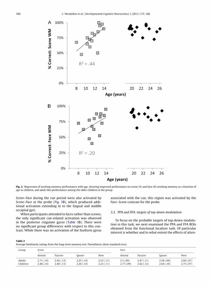
180 C. Wendelken et al. / Developmental Cognitive Neuroscience 1 (2011) 175–186
provedhe grou
Fig. 2. Regression of working memory performance with age, showing image in children, and adult-like performance among the older children in t
Scene–Face during the cue period were also activated byScene–Face at the probe (Fig. 3B), which produced addi-tional activation extending in to the lingual and middleoccipital gyri.
When participants attended to faces rather than scenes,
the only significant cue-related activation was observedin the posterior cingulate gyrus (Table 3B). There wereno significant group differences with respect to this con-trast. While there was no activation of the fusiform gyrusTable 2Average familiarity ratings from the long-term memory test. Parentheses show s
Group Scene
Attend Passive Ignore New
Adults 2.71 (.14) 2.45 (.13) 2.25 (.14) 2.23 (.11)Children 2.46 (.14) 2.40 (.13) 2.26 (.14) 2.23 (.11)
performance on scene (A) and face (B) working memory as a function ofp.
associated with the cue, this region was activated by theFace–Scene contrast for the probe.
3.3. PPA and FFA: targets of top-down modulation
To focus on the probable targets of top-down modula-tion in this task, we next examined the PPA and FFA ROIsobtained from the functional localizer task. Of particularinterest is whether and to what extent the effects of atten-
tandard error.
Face
Attend Passive Ignore New
2.1 (.09) 2.47 (.11) 2.58 (.09) 2.69 (.07)2.77 (.09) 2.62 (.12) 2.64 (.10) 2.75 (.07)
C. Wendelken et al. / Developmental Cognitive Neuroscience 1 (2011) 175–186 181
Table 3Activation clusters associated with Scene versus Face contrasts. All clusters that survive thresholding at p < .001 (uncorrected), with a 10-voxel extentthreshold, are reported. In addition, FWE-corrected p-values are reported for each cluster. PHG = parahippocampal gyrus, LG = lingual gyrus, SOG = superioroccipital gyrus, MOG = middle occipital gyrus, SPL = superior parietal lobe, MFG = middle frontal gyrus, IFJ = inferior frontal junction, MTG = middle temporalgyrus, and STG = superior temporal gyrus.
Region x, y, z Z (voxel) cluster size (# voxels) p-Value (corr. cluster)
(A) Scene > Face (cue) All SubjectsLeft PHG −27, −45, −16 4.73 103 .001Right PHG 27, −36, −16 3.79 51 .036Left precuneus, SPL −15, −69, 52 4.51 253 <.001Right SOG, MTG 24, −57, 24 4.43 213 < .001Left posterior cingulate −15, −54, 16 4.40 40 .08Left MFG −45, 33, 16 3.58 36 .11
(B) Face > Scene (cue) All SubjectsPosterior cingulate gyrus −3, −51, 28 3.92 92 .002Right STG 60, −51, 8 3.98 44 .06
(C) Scene > Passive (cue) All SubjectsLeft precuneus, SPL −15, −69, 52 5.60 684 <.001Right precuneus, SPL 20, −60, 56 5.51 660 <.001Left IFJ (MFG/precentral) −36, 0, 36 4.92 370 <.001Right IFJ (MFG/precentral) 39, 3, 36 4.67 302 <.001Left MOG/MTG/fusiform −48, −63, −4 4.51 133 .001Left MFG, IFG −36, 27, 24 4.61 44 .02
(D) Face > Passive (cue) All SubjectsLeft precuneus, SPL −24, −69, 32 4.58 248 <.001Right precuneus, SPL 27, −69, 40 4.04 207 <.001Left IFJ (precentral/MFG) −42, −3, 36 4.14 89 .005
508
ti
eeFF(sd(oTen
tvi((bva
vfaadto
Right IFJ (precentral/MFG) 39, 0, 40 4.8Left MOG/MTG/fusiform −51, −63, −4 5.0Right MTG/MOG/fusiform 48, −54, 8 4.4
ion on activity modulation, previously observed in adults,s present in children.
Within bilateral PPA (Fig. 4A), both children and adultsxhibited attentional modulation during the cue period,xhibiting greater activation for Scene trials than forace trials (Fig. 4B; adults: F2,28 = 11.9, p < .001; children:2,28 = 3.2, p = .05; 87% of adults (13/15) and 66% of children10/15) exhibited this pattern). Adults also demonstratedignificant enhancement (Scene > Passive), and childrenemonstrated marginal enhancement, but only in left PPAadults: F1,14 = 8.1, p = .01; children: F1,14 = 4.2, p = .06; 87%f adults versus 80% of children exhibited this pattern).here was no significant condition × group interaction inither left or right PPA (F’s < 1). Neither group exhibited sig-ificant attentional suppression (Passive > Face) of the PPA.
To investigate development of top-down modula-ion, we probed for correlations between PPA acti-ation and age in children. Significant age-relatedncreases were observed for both top-down modulationScene–Face; r2 = .28, p = .04) and top-down enhancementScene–Passive; r2 = .31, p = .03). These effects were driveny a highly significant age-related increase in PPA acti-ation for Scene trials (r2 = .56, p = .001) in the absencege-related changes for Face or Passive trials (Fig. 4C).
In children, in addition to correlating with age, PPA acti-ation on Scene trials also correlated with performance,or both scene working memory (r2 = .46, p = .006; Fig. 5A)
nd scene long-term memory (r2 = .49, p = .004; Fig. 5B). Indults, by contrast, although the trends were in the sameirection as in children, there were no significant correla-ions between PPA activation and either working memoryr long-term memory performance.202 <.001203 <.001136 <.001
As with PPA, investigation of the right FFA ROI con-firmed the success of the localizer task, insofar as therewas a large effect of condition (Face > Scene, Passive) associ-ated with the probe, in both children and adults. However,in the FFA, neither group demonstrated an effect of atten-tional modulation during presentation of the cue stimulus(adults: F2,26 < 1, children: F2,26 = 1.1). Nevertheless, foradults, though not for children, there was a significantcorrelation between face working memory performanceand FFA cue-related activation for the Face–Scene contrast(r2 = .21, p = .04), demonstrating that top-down modulationof FFA did occur, at least in the better-performing adultparticipants.
3.4. Sources of top-down modulation
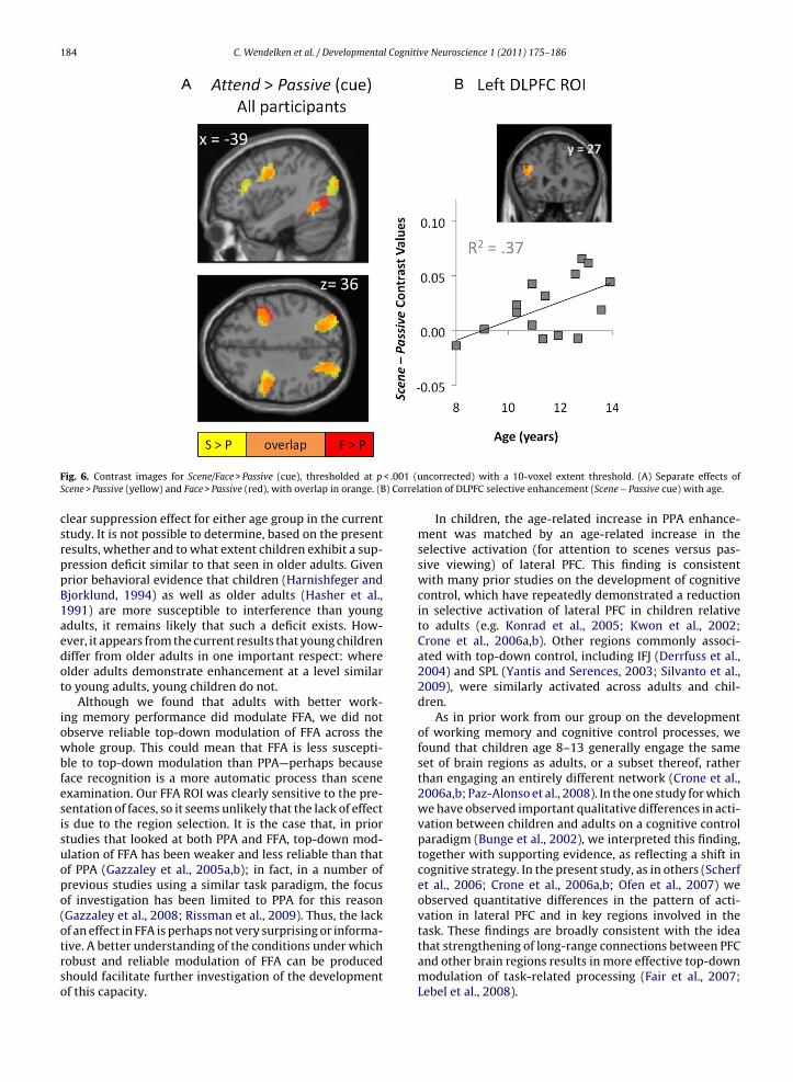
To identify additional regions that were modulated byattentional demands during the cue period, we examinedthe Scene > Passive and Face > Passive contrasts across allsubjects (Table 3C and D and Fig. 6A). Activation in bilat-eral inferior frontal junction (IFJ), and in bilateral SPL wasobserved for both contrasts. In addition, activation with aleft DLPFC peak (−36, 27, 24), spanning the inferior frontalsulcus, was observed for Scene > Passive across all subjects.
We next probed explicitly for differences betweenadults and children in top-down control by submittingScene > Passive and Face > Passive cue contrast images from
both groups to a voxel-wise, two-sample t-test. No sig-nificant clusters emerged from this analysis (we note,however, that when the threshold was lowered to p < .005,a cluster in left lateral PFC (−48, 18, 8) was observed forScene > Passive).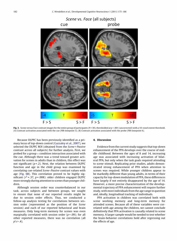
182 C. Wendelken et al. / Developmental Cognitive Neuroscience 1 (2011) 175–186
ts (N = 3trast act
Fig. 3. Scene versus Face contrast images for the entire group of participan(A) Contrast activation associated with the cue (FIR timepoint 3). (B) Con
Because DLPFC has been previously identified as a pri-mary locus of top-down control (Gazzaley et al., 2007), weselected the DLPFC ROI (obtained from the Scene > Passivecontrast across all subjects) for further analysis. First, weprobed for a group × condition interaction associated withthe cue. Although there was a trend toward greater acti-vation for scenes in adults than in children, this effect wasnot significant (p = .2). Next, the relation between DLPFCfunction and age in the child group was examined bycorrelating cue-related Scene–Passive contrast values withage (Fig. 6B). This correlation proved to be highly sig-nificant (r2 = .37, p = .008): older children engaged DLPFCmore strongly during attention to scenes than younger chil-dren.
Although session order was counterbalanced in ourtask, across subjects and between groups, we soughtto ensure that none of our reported results might bedue to session order effects. Thus, we conducted afollow-up analysis testing for correlations between ses-sion order (represented as the position of the Scene
session) and each of our reported behavioral and brainmeasures. Only long-term memory for scenes was evenmarginally correlated with session order (p = .09); for allother reported measures, there was no correlation (allp’s > .4).0), thresholded at p < .001 (uncorrected) with a 10-voxel extent threshold.ivation associated with the probe (FIR timepoint 6).
4. Discussion
Evidence from the current study suggests that top-downenhancement of the PPA develops over the course of mid-dle childhood. Between the ages of 8 and 14, increasingage was associated with increasing activation of bilat-eral PPA, but only when the task goals required attendingto scene stimuli. Replicating prior studies, adults demon-strated strong enhancement of PPA when attention toscenes was required. While younger children appear tobe markedly different than young adults, in terms of theircapacity for top-down modulation of PPA, these differenceshave largely if not entirely disappeared by the age of 14.However, a more precise characterization of the develop-mental trajectory of PPA enhancement will require furtherstudy, with more individuals from the age range in questionand, ideally, longitudinal tracking of individuals.
PPA activation in children was correlated both withscene working memory and long-term memory forattended scenes. Because all of these variables were cor-
related with age among the children, we cannot concludedefinitively that PPA activation is causally related to scenememory. A larger sample would be needed to test whetherthe brain-behavior correlations hold after regressing outthe effects of age.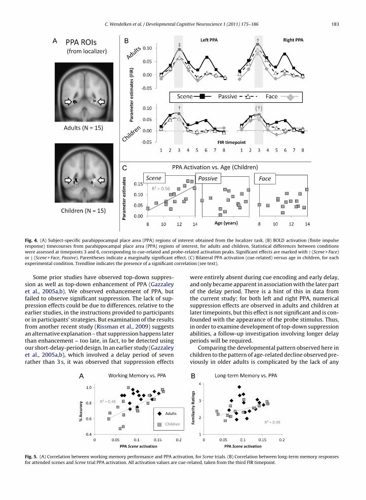
C. Wendelken et al. / Developmental Cognitive Neuroscience 1 (2011) 175–186 183
Fig. 4. (A) Subject-specific parahippocampal place area (PPA) regions of interest obtained from the localizer task. (B) BOLD activation (finite impulser of interw obe-relo effect. (e correlat
sefpeofatoer
Ff
esponse) timecourses from parahippocampal place area (PPA) regionsere assessed at timepoints 3 and 6, corresponding to cue-related and pr
r ‡ (Scene > Face, Passive). Parentheses indicate a marginally significantxperimental condition. Trendline indicates the presence of a significant
Some prior studies have observed top-down suppres-ion as well as top-down enhancement of PPA (Gazzaleyt al., 2005a,b). We observed enhancement of PPA, butailed to observe significant suppression. The lack of sup-ression effects could be due to differences, relative to thearlier studies, in the instructions provided to participantsr in participants’ strategies. But examination of the resultsrom another recent study (Rissman et al., 2009) suggests
n alternative explanation – that suppression happens laterhan enhancement – too late, in fact, to be detected usingur short-delay-period design. In an earlier study (Gazzaleyt al., 2005a,b), which involved a delay period of sevenather than 3 s, it was observed that suppression effectsig. 5. (A) Correlation between working memory performance and PPA activatioor attended scenes and Scene trial PPA activation. All activation values are cue-re
est, for adults and children. Statistical differences between conditionsated activation peaks. Significant effects are marked with † (Scene > Face)C) Bilateral PPA activation (cue-related) versus age in children, for eachion (see text).
were entirely absent during cue encoding and early delay,and only became apparent in association with the later partof the delay period. There is a hint of this in data fromthe current study; for both left and right PPA, numericalsuppression effects are observed in adults and children atlater timepoints, but this effect is not significant and is con-founded with the appearance of the probe stimulus. Thus,in order to examine development of top-down suppression
abilities, a follow-up investigation involving longer delayperiods will be required.Comparing the developmental pattern observed here inchildren to the pattern of age-related decline observed pre-viously in older adults is complicated by the lack of any
n, for Scene trials. (B) Correlation between long-term memory responseslated, taken from the third FIR timepoint.
184 C. Wendelken et al. / Developmental Cognitive Neuroscience 1 (2011) 175–186
< .001 (u) Correl
Fig. 6. Contrast images for Scene/Face > Passive (cue), thresholded at pScene > Passive (yellow) and Face > Passive (red), with overlap in orange. (B
clear suppression effect for either age group in the currentstudy. It is not possible to determine, based on the presentresults, whether and to what extent children exhibit a sup-pression deficit similar to that seen in older adults. Givenprior behavioral evidence that children (Harnishfeger andBjorklund, 1994) as well as older adults (Hasher et al.,1991) are more susceptible to interference than youngadults, it remains likely that such a deficit exists. How-ever, it appears from the current results that young childrendiffer from older adults in one important respect: whereolder adults demonstrate enhancement at a level similarto young adults, young children do not.
Although we found that adults with better work-ing memory performance did modulate FFA, we did notobserve reliable top-down modulation of FFA across thewhole group. This could mean that FFA is less suscepti-ble to top-down modulation than PPA—perhaps becauseface recognition is a more automatic process than sceneexamination. Our FFA ROI was clearly sensitive to the pre-sentation of faces, so it seems unlikely that the lack of effectis due to the region selection. It is the case that, in priorstudies that looked at both PPA and FFA, top-down mod-ulation of FFA has been weaker and less reliable than thatof PPA (Gazzaley et al., 2005a,b); in fact, in a number ofprevious studies using a similar task paradigm, the focusof investigation has been limited to PPA for this reason(Gazzaley et al., 2008; Rissman et al., 2009). Thus, the lack
of an effect in FFA is perhaps not very surprising or informa-tive. A better understanding of the conditions under whichrobust and reliable modulation of FFA can be producedshould facilitate further investigation of the developmentof this capacity.ncorrected) with a 10-voxel extent threshold. (A) Separate effects ofation of DLPFC selective enhancement (Scene − Passive cue) with age.
In children, the age-related increase in PPA enhance-ment was matched by an age-related increase in theselective activation (for attention to scenes versus pas-sive viewing) of lateral PFC. This finding is consistentwith many prior studies on the development of cognitivecontrol, which have repeatedly demonstrated a reductionin selective activation of lateral PFC in children relativeto adults (e.g. Konrad et al., 2005; Kwon et al., 2002;Crone et al., 2006a,b). Other regions commonly associ-ated with top-down control, including IFJ (Derrfuss et al.,2004) and SPL (Yantis and Serences, 2003; Silvanto et al.,2009), were similarly activated across adults and chil-dren.
As in prior work from our group on the developmentof working memory and cognitive control processes, wefound that children age 8–13 generally engage the sameset of brain regions as adults, or a subset thereof, ratherthan engaging an entirely different network (Crone et al.,2006a,b; Paz-Alonso et al., 2008). In the one study for whichwe have observed important qualitative differences in acti-vation between children and adults on a cognitive controlparadigm (Bunge et al., 2002), we interpreted this finding,together with supporting evidence, as reflecting a shift incognitive strategy. In the present study, as in others (Scherfet al., 2006; Crone et al., 2006a,b; Ofen et al., 2007) weobserved quantitative differences in the pattern of acti-vation in lateral PFC and in key regions involved in the
task. These findings are broadly consistent with the ideathat strengthening of long-range connections between PFCand other brain regions results in more effective top-downmodulation of task-related processing (Fair et al., 2007;Lebel et al., 2008).
l Cogniti
mCwoadcmictoedar
mopPwwstcoot
C
tt
A
MWtc
R
A
B
B
B
B
B
B
C. Wendelken et al. / Developmenta
Developmental changes in neural activation patternsay be related to concurrent changes in brain structure.
hanges in cortical thickness occur throughout childhood,ith different brain regions demonstrating different devel-
pmental trajectories (Shaw et al., 2008). DLPFC, whichppears to be a major locus of top-down modulation,emonstrates particularly delayed maturation in terms ofortical thickness (Gogtay et al., 2004). Over the course ofiddle childhood, myelination processes result in increas-
ng white matter volume (Giedd, 2004) and increasing tractoherence (Barnea-Goraly et al., 2005). It is highly likelyhat these white-matter changes have a positive impactn the potential for top-down modulation, and help toxplain the reduced modulation observed in younger chil-ren. Further investigation of the link between structuralnd functional development is an important goal for futureesearch.
In summary, the present results suggest that top-downodulation of PPA, mediated by lateral PFC, develops
ver the course of middle childhood. Attention-relatedrefrontal activation and the associated enhancement ofPA activation are reduced in younger children comparedith older children and young adults. Notably, althoughe are unable to draw firm conclusions about top-down
uppression in the current study, the observed reduc-ion of top-down enhancement in younger children alsoontrasts with the suppression-specific deficit previouslybserved in older adults, indicating that the developmentf top-down attentional modulation may follow a differentrajectory than its eventual decline.
onflict of interest statement
The authors declare that the research was conducted inhe absence of any commercial or financial relationshipshat could be construed as a potential conflict of interest.
cknowledgements
This study was funded by Program Project directed byark D’Esposito (P01 NS040813-06). We thank Samantharight, Natalie Repin, and Michael Rubens for their assis-
ance with data collection, and Chris Blais for substantiveomments.
eferences
wh, E., Jonides, J., 2001. Overlapping mechanisms of attention and spatialworking memory. Trends Cogn. Sci. 5, 119–126.
addeley, A., 1998. Working memory. C. R. Acad. Sci. III 321, 167–173.
arnea-Goraly, N., Menon, V., Eckert, M., Tamm, L., Bammer, R., Karchem-skiy, A., et al., 2005. White matter development during childhood andadolescence: a cross-sectional diffusion tensor imaging study. Cereb.Cortex 15, 1848–1854.
jorklund, D., Harnishfeger, K., 1990. The resource construct in cognitivedevelopment: diverse sources of evidence and a theory of inefficientinhibition. Dev. Rev. 10, 48–71.
unge, S.A., Dudukovic, N.M., Thomason, M.E., Vaidya, C.J., Gabrieli, J.D.E.,
2002. Immature frontal lobe contributions to cognitive control in chil-dren: evidence from fMRI. Neuron 33, 301–311.unge, S.A., Wright, S.B., 2007. Neurodevelopmental changes in workingmemory and cognitive control. Curr. Opin. Neurobiol. 17, 243–250.
urgund, E.D., Kang, H.C., Kelly, J.E., Buckner, R.L., Snyder, A.Z., Petersen,S.E., Schlaggar, B.L., 2002. The feasibility of a common stereotactic
ve Neuroscience 1 (2011) 175–186 185
space for children and adults in fMRI studies of development. Neu-roimage 17 (1), 184–200.
Corbetta, M., Miezin, F.M., Dobmeyer, S., Shulman, G.L., Petersen, S.E.,1990. Attentional modulation of neural processing of shape, color, andvelocity in humans. Science 248, 1556–1559.
Cowan, N., 1995. Sensory memory and its role in information processing.Electroencephalogr. Clin. Neurophysiol. Suppl. 44, 21–31.
Crone, E.A., Donohue, S.E., Honomichl, R., Wendelken, C., Bunge, S.A.,2006a. Brain regions mediating flexible rule use during development.J. Neurosci. 26, 11239–11247.
Crone, E.A., Wendelken, C., Donohue, S., van Leijenhorst, L., Bunge, S.A.,2006b. Neurocognitive development of the ability to manipulateinformation in working memory. Proc. Natl. Acad. Sci. U.S.A. 103,9315–9320.
Dale, A.M., 1999. Optimal experimental design for event-related fMRI.Hum. Brain Mapp. 8, 109–114.
Derrfuss, J., Brass, M., von Cramon, D.Y., 2004. Cognitive control in the pos-terior frontolateral cortex: evidence from common activations in taskcoordination, interference control, and working memory. Neuroimage23, 604–612.
Desimone, R., Duncan, J., 1995. Neural mechanisms of selective visualattention. Annu. Rev. Neurosci. 18, 193–222.
Diamond, A., Towle, C., Boyer, K., 1994. Young children’s performance on atask sensitive to the memory functions of the medial temporal lobe inadults – the delayed nonmatching-to-sample task – reveals problemsthat are due to non-memory-related task demands. Behav. Neurosci.108, 659–680.
Epstein, R., Kanwisher, N., 1998. A cortical representation of the localvisual environment. Nature 392, 598–601.
Everling, S., Tinsley, C.J., Gaffan, D., Duncan, J., 2002. Filtering of neuralsignals by focused attention in the monkey prefrontal cortex. Nat.Neurosci. 5, 671–676.
Fair, D., Dosenbach, N., Church, J., Cohen, A., Brahmbhatt, S., Miezin, F.,et al., 2007. Development of distinct control networks through segre-gation and integration. Proc. Natl. Acad. Sci. U.S.A. 104, 13507–13512.
Gazzaley, A., Clapp, W., Kelley, J., McEvoy, K., Knight, R., D’Esposito, M.,et al., 2008. Age-related top-down suppression deficit in the earlystages of cortical visual memory processing. Proc. Natl. Acad. Sci. U.S.A.105, 13122–13126.
Gazzaley, A., Cooney, J., McEvoy, K., Knight, R., D’Esposito, M., 2005a. Top-down enhancement and suppression of the magnitude and speed ofneural activity. J. Cogn. Neurosci. 17, 507–517.
Gazzaley, A., Cooney, J., Rissman, J., D’Esposito, M., 2005b. Top-down sup-pression deficit underlies working memory impairment in normalaging. Nat. Neurosci. 8, 1298–1300.
Gazzaley, A., Rissman, J., Cooney, J., Rutman, A., Seibert, T., Clapp, W., et al.,2007. Functional interactions between prefrontal and visual associa-tion cortex contribute to top-down modulation of visual processing.Cereb. Cortex, i125–i135.
Geier, C.F., Garver, K., Terwilliger, R., Luna, B., 2009. Development of work-ing memory maintenance. J. Neurophysiol. 101, 84–99.
Giedd, J.N., 2004. Structural magnetic resonance imaging of the adolescentbrain. Ann. N. Y. Acad. Sci. 1021, 77–85.
Gogtay, N., Giedd, J.N., Lusk, L., Hayashi, K.M., Greenstein, D., Vaituzis, A.C.,et al., 2004. Dynamic mapping of human cortical development duringchildhood through early adulthood. Proc. Natl. Acad. Sci. U.S.A. 101,8174–8179.
Golarai, G., Ghahremani, D.G., Whitfield-Gabrieli, S., Reiss, A., Eberhardt,J.L., Gabrieli, J.D.E., et al., 2007. Differential development of high-levelvisual cortex correlates with category-specific recognition memory.Nat. Neurosci. 10, 512–522.
Golarai, G., Liberman, A., Yoon, J.M.D., Grill-Spector, K., 2010. Differentialdevelopment of the ventral visual cortex extends through adoles-cence. Front. Hum. Neurosci. 3, 80.
Harnishfeger, K., Bjorklund, D., 1994. A developmental perspective onindividual differences in inhibition. Learn. Individ. Differ. 6, 331–355.
Hasher, L., Zacks, R., 1988. Working memory, comprehension, and aging:a review and a new view. Psychol. Learn. Motiv.: Adv. Res. Theory 22,193–225.
Hasher, L., Stoltzfus, E.R., Zacks, R.T., Rypma, B., 1991. Age and inhibition.J. Exp. Psychol. Learn. Mem. Cogn. 17, 163–169.
Hillyard, S.A., Hink, R.F., Schwent, V.L., Picton, T.W., 1973. Electrical signsof selective attention in the human brain. Science 182, 177–180.
Iba, M., Sawaguchi, T., 2003. Involvement of the dorsolateral prefrontalcortex of monkeys in visuospatial target selection. J. Neurophysiol.89, 587–599.
Kanwisher, N., McDermott, J., Chun, M.M., 1997. The fusiform face area: amodule in human extrastriate cortex specialized for face perception.J. Neurosci. 17, 4302–4311.
l Cogniti
186 C. Wendelken et al. / DevelopmentaKonrad, K., Neufang, S., Thiel, C.M., Specht, K., Hanisch, C., Fan, J., et al.,2005. Development of attentional networks: an fMRI study with chil-dren and adults. Neuroimage 28, 429–439.
Kwon, H., Reiss, A.L., Menon, V., 2002. Neural basis of protracted devel-opmental changes in visuo-spatial working memory. Proc. Natl. Acad.Sci. U.S.A. 99, 13336–13341.
Lebel, C., Walker, L., Leemans, A., Phillips, L., Beaulieu, C., 2008. Microstruc-tural maturation of the human brain from childhood to adulthood.Neuroimage 40, 1044–1055.
Luna, B., Garver, K.E., Urban, T.A., Lazar, N.A., Sweeney, J.A., 2004. Matu-ration of cognitive processes from late childhood to adulthood. ChildDev. 75, 1357–1372.
Luna, B., Padmanabhan, A., O’Hearn, K., 2010. What has fMRI told us aboutthe development of cognitive control through adolescence? BrainCogn. 72, 101–113.
Miller, E.K., Cohen, J.D., 2001. An integrative theory of prefrontal cortexfunction. Annu. Rev. Neurosci. 24, 167–202.
Moran, J., Desimone, R., 1985. Selective attention gates visual processingin the extrastriate cortex. Science 229, 782–784.
Ofen, N., Kao, Y.C., Sokol-Hessner, P., Kim, H., Whitfield-Gabrieli, S.,Gabrieli, J.D.E., et al., 2007. Development of the declarative memorysystem in the human brain. Nat. Neurosci. 10, 1198–1205.
Olesen, P.J., Macoveanu, J., Tegnér, J., Klingberg, T., 2007. Brain activityrelated to working memory and distraction in children and adults.Cereb. Cortex 17, 1047–1054.
Ordaz, S., Davis, S., Luna, B., 2010. Effects of response preparation on devel-opmental improvements in inhibitory control. Acta Psychol. (Amst.)134 (3), 253–263.
Paz-Alonso, P.M., Ghetti, S., Donohue, S.E., Goodman, G.S., Bunge, S.A.,2008. Neurodevelopmental correlates of true and false recognition.Cereb. Cortex 18, 2208–2216.
Pessoa, L., Kastner, S., Ungerleider, L.G., 2003. Neuroimaging studies of
attention: from modulation of sensory processing to top-down con-trol. J. Neurosci. 23, 3990–3998.Ploner, C.J., Ostendorf, F., Brandt, S.A., Gaymard, B.M., Rivaud-Péchoux,S., Ploner, M., et al., 2001. Behavioural relevance modulates accessto spatial working memory in humans. Eur. J. Neurosci. 13,357–363.
ve Neuroscience 1 (2011) 175–186
Rainer, G., Asaad, W.F., Miller, E.K., 1998. Selective representation of rele-vant information by neurons in the primate prefrontal cortex. Nature393, 577–579.
Richter, W., Richter, M., 2003. The shape of the fMRI BOLD response inchildren and adults changes systematically with age. Neuroimage 20,1122–1131.
Rissman, J., Gazzaley, A., D’Esposito, M., 2009. The effect of non-visualworking memory load on top-down modulation of visual processing.Neuropsychologia 47, 1637–1646.
Rossi, A.F., Pessoa, L., Desimone, R., Ungerleider, L.G., 2009. The prefrontalcortex and the executive control of attention. Exp. Brain Res. 192,489–497.
Rueda, M.R., Fan, J., McCandliss, B.D., Halparin, J.D., Gruber, D.B., Lercari,L.P., et al., 2004. Development of attentional networks in childhood.Neuropsychologia 42, 1029–1040.
Scherf, K.S., Behrmann, M., Humphreys, K., Luna, B., 2007. Visual category-selectivity for faces, places and objects emerges along differentdevelopmental trajectories. Dev. Sci. 10, F15–F30.
Scherf, K.S., Sweeney, J.A., Luna, B., 2006. Brain basis of developmen-tal change in visuospatial working memory. J. Cogn. Neurosci. 18,1045–1058.
Shaw, P., Kabani, N.J., Lerch, J.P., Eckstrand, K., Lenroot, R., Gogtay, N., et al.,2008. Neurodevelopmental trajectories of the human cerebral cortex.J. Neurosci. 28, 3586–3594.
Silvanto, J., Muggleton, N., Lavie, N., Walsh, V., 2009. The perceptual andfunctional consequences of parietal top-down modulation on thevisual cortex. Cereb. Cortex 19, 327–330.
Thesen, S., Heid, O., Mueller, E., Schad, L.R., 2000. Prospective acquisitioncorrection for head motion with image-based tracking for real-timefMRI. Magn. Reson. Med. 44 (3), 457–465.
Velanova, K., Wheeler, M.E., Luna, B., 2009. The maturation of taskset-related activation supports late developmental improvements in
inhibitory control. J. Neurosci. 29, 12558–12567.Vogel, E.K., McCollough, A.W., Machizawa, M.G., 2005. Neural measuresreveal individual differences in controlling access to working memory.Nature 438, 500–503.
Yantis, S., Serences, J.T., 2003. Cortical mechanisms of space-based andobject-based attentional control. Curr. Opin. Neurobiol. 13, 187–193.