Embed Size (px)
Citation preview
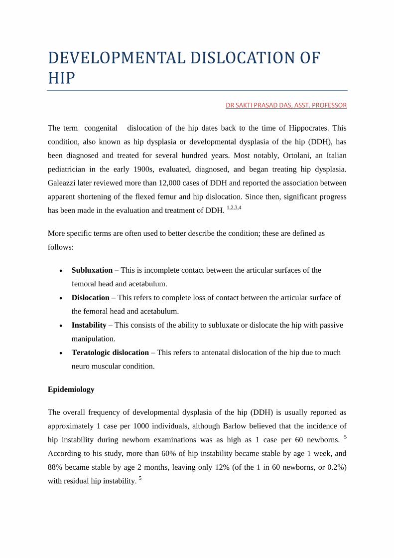
DEVELOPMENTAL DISLOCATION OF HIP
DR SAKTI PRASAD DAS, ASST. PROFESSOR
The term congenital dislocation of the hip dates back to the time of Hippocrates. This
condition, also known as hip dysplasia or developmental dysplasia of the hip (DDH), has
been diagnosed and treated for several hundred years. Most notably, Ortolani, an Italian
pediatrician in the early 1900s, evaluated, diagnosed, and began treating hip dysplasia.
Galeazzi later reviewed more than 12,000 cases of DDH and reported the association between
apparent shortening of the flexed femur and hip dislocation. Since then, significant progress
has been made in the evaluation and treatment of DDH. 1,2,3,4
More specific terms are often used to better describe the condition; these are defined as
follows:
Subluxation – This is incomplete contact between the articular surfaces of the
femoral head and acetabulum.
Dislocation – This refers to complete loss of contact between the articular surface of
the femoral head and acetabulum.
Instability – This consists of the ability to subluxate or dislocate the hip with passive
manipulation.
Teratologic dislocation – This refers to antenatal dislocation of the hip due to much
neuro muscular condition.
Epidemiology
The overall frequency of developmental dysplasia of the hip (DDH) is usually reported as
approximately 1 case per 1000 individuals, although Barlow believed that the incidence of
hip instability during newborn examinations was as high as 1 case per 60 newborns. 5
According to his study, more than 60% of hip instability became stable by age 1 week, and
88% became stable by age 2 months, leaving only 12% (of the 1 in 60 newborns, or 0.2%)
with residual hip instability. 5
Etiology
The etiology of hip dysplasia is not clear, but this condition does appear to be related to a
number of different factors. 6
One such factor is racial background; among Native Americans
and Laplanders, the prevalence of hip dysplasia is much higher (nearly 25-50 cases per 1000
persons) than other races, and the prevalence is very low among southern Chinese and black
populations. 7,8,9,10
An underlying genetic pre disposition also appears to exist in that a 10-fold
increase in the frequency of hip dysplasia occurs in children whose parents had
developmental dysplasia of the hip (DDH) compared with those whose parents did not. 11
Other factors possibly related to DDH include intrauterine positioning and sex, and some of
these are interrelated. Female sex, being the first-born child, and breech positioning are all
associated with an increased prevalence of DDH. An estimated 80% of persons with DDH
are female , 11
and the rate of breech positioning in children with DDH is approximately 20%
(compared with 2-4% in the general population) 13, 14
The prevalence of DDH in females born
in breech position has been estimated to be as high as 1 case in 15 persons in some studies. 15
Other musculoskeletal disorders of intrauterine malpositioning or crowding, such as
metatarsus adductus and torticolis have been reported to be associated with DDH, 16,17
Oligohydramnios ( less amniotic fluid) is also reported to be associated with an increased
prevalence of DDH. 18
The left hip is more commonly associated with DDH than the right,
and this is believed to be due to the common intrauterine position of the left hip against the
mother's sacrum, forcing it into an adducted position. 18
Children in cultures in which the
mother swaddles the baby, forcing the infant's hips to be adducted, also have a higher rate of
hip dysplasia. 19
Hip dysplasia can be associated with underlying neuromuscular disorders, such as cerebral
palsy, myelomeningocele, arthrogryposis , and Larsen syndrome, although these are not
usually considered DDH.
Pathophysiology
Developmental dysplasia of the hip (DDH) involves abnormal growth of the hip.
Ligamentous laxity is also believed to be associated with hip dysplasia, although this
association is less clear. DDH is not part of the classic description of disorders that are
associated with significant ligamentous laxity, such as Ehler’s – Danlon’s syndrome or
Marfan’s syndrome. Children often have ligamentous laxity at birth, yet their hips are not
usually unstable; in fact, it takes a great deal of effort to dislocate a child's hip. Therefore,
more than just ligamentous laxity may be required to result in DDH. At birth, white children
tend to have a shallow acetabulum. 20,21
; this may provide a susceptible period in which
abnormal positioning or a brief period of ligamentous laxity may result in hip instability.
However, this characteristic is not as true for children of black descent, who have a lower rate
of DDH. 10
Anatomy
The normal growth of the acetabulum depends on normal epiphyseal growth of the triradiate
cartilage and on the 3 ossification centers located within the acetabular portion of the pubis
(os acetabulum), ilium (acetabular epiphysis), and ischium. Additionally, normal growth of
the acetabulum depends on normal interstitial appositional growth within the acetabulum.
The presence of the spherical femoral head within the acetabulum is critical for stimulating
normal development of the acetabulum.
The anatomy of the dislocated hip, especially after several months, often includes formation
of a ridge called the neolimbus. Closed reduction is often unsuccessful at a later date,
secondary to various obstacles to reduction. These include adductor and psoas tendon
contraction, ligamentous teres, a transverse acetabular ligament, and pulvinar and capsular
constriction. With long-standing dislocations, interposition of the labrum can also interfere
with reduction.
Presentation
Early clinical manifestations of developmental dysplasia of the hip (DDH) are identified
during examination of the newborn. The classic examination finding is revealed with the
Ortolani maneuver; a palpable "clunk" is present when the hip is reduced in and out of the
acetabulum and over the neolimbus. A high-pitched "click" (as opposed to a clunk) in all
likelihood has little association with acetabular pathology. 22,23
Ortolani originally described
this clunk as occurring with either subluxation or reduction of the hip (in or out of the
acetabulum). More commonly, the Ortolani sign is referred to as a clunk, felt when the hip
reduces into the acetabulum, with the hip in abduction.
To perform this maneuver correctly, the patient must be relaxed. Only one hip is examined at
a time. The examiner's thumb is placed over the patient's inner thigh, and the index finger is
gently placed over the greater trochanter. The hip is abducted, and gentle pressure is placed
over the greater trochanter. In the presence of DDH, a clunk is felt when the hip is reduced.
The Ortolani maneuver should be performed gently, such that the fingertips do not blanch. 24
Barlow described another test for DDH that is performed with the hips in an adducted
position, in which slight gentle posterior pressure is applied to the hips. A clunk should be
felt as the hip subluxates out of the acetabulum. 5
The clinical examination for late DDH, when the child is aged 3-6 months, is quite different.
At this point, the hip, if dislocated, is often dislocated in a fixed position. 11
The Galeazzi sign
is a classic identifying sign for unilateral hip dislocation . This is performed with the patient
lying supine and the hips and knees flexed. The examination should demonstrate that one leg
appears shorter than the other. Although this finding is usually due to hip dislocation,
realizing that any limb-length discrepancy results in a positive Galeazzi sign is important.
Additional physical examination findings for late dislocation include asymmetry of the
gluteal thigh fold or labral skin folds, decreased abduction on the affected side, standing or
walking with external rotation, and leg-length inequality.
Bilateral dislocation of the hip, especially at a later age, can be quite difficult to diagnose.
This condition often manifests as a waddling gait with hyperlordosis. Many of the
aforementioned clues for a unilateral dislocated hip are not present, such as the Galeazzi sign,
asymmetrical thigh and skin folds, or asymmetrically decreased abduction. Careful
examination is needed, and a high level of suspicion is important.
Ultrasound and radiological examination
Mahan et al, of Children's Hospital in Boston, found that the screening strategy associated
with the highest probability of having a non arthritic hip at the age of 60 years was to screen
all neonates for hip dysplasia with a physical examination and to use ultrasonography
selectively for infants who are at high risk. 25
The use of ultrasound in a clinic for the selective screening of at risk hips has been of benefit.
Ultrasound can detect hip displacement in the clinically normal neonate and identify the hips
that need treatment. It has been shown to be very helpful in the early management of definite
hip displacement.
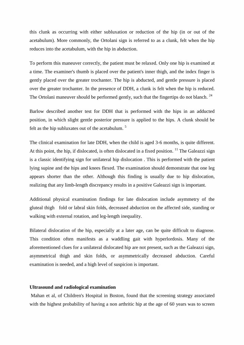
Ultrasound has been of help in treated cases with Pavlik’s harness. Location of the hip in the
harness can be recorded. Splintage can be abandoned if there is no concentric reduction. 26,
27,28
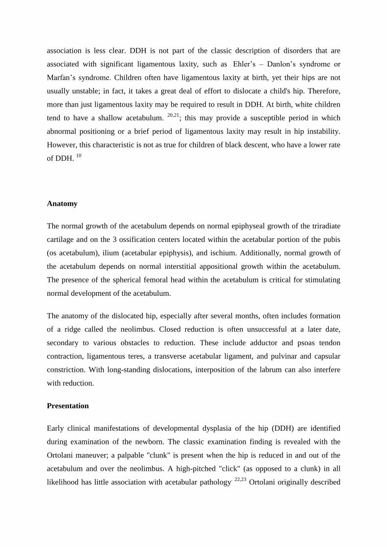
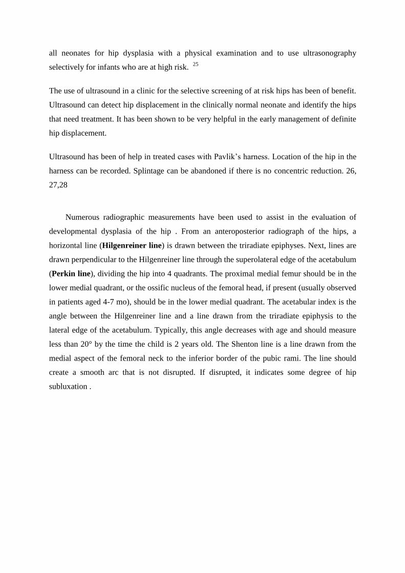
Numerous radiographic measurements have been used to assist in the evaluation of
developmental dysplasia of the hip . From an anteroposterior radiograph of the hips, a
horizontal line (Hilgenreiner line) is drawn between the triradiate epiphyses. Next, lines are
drawn perpendicular to the Hilgenreiner line through the superolateral edge of the acetabulum
(Perkin line), dividing the hip into 4 quadrants. The proximal medial femur should be in the
lower medial quadrant, or the ossific nucleus of the femoral head, if present (usually observed
in patients aged 4-7 mo), should be in the lower medial quadrant. The acetabular index is the
angle between the Hilgenreiner line and a line drawn from the triradiate epiphysis to the
lateral edge of the acetabulum. Typically, this angle decreases with age and should measure
less than 20° by the time the child is 2 years old. The Shenton line is a line drawn from the
medial aspect of the femoral neck to the inferior border of the pubic rami. The line should
create a smooth arc that is not disrupted. If disrupted, it indicates some degree of hip
subluxation .
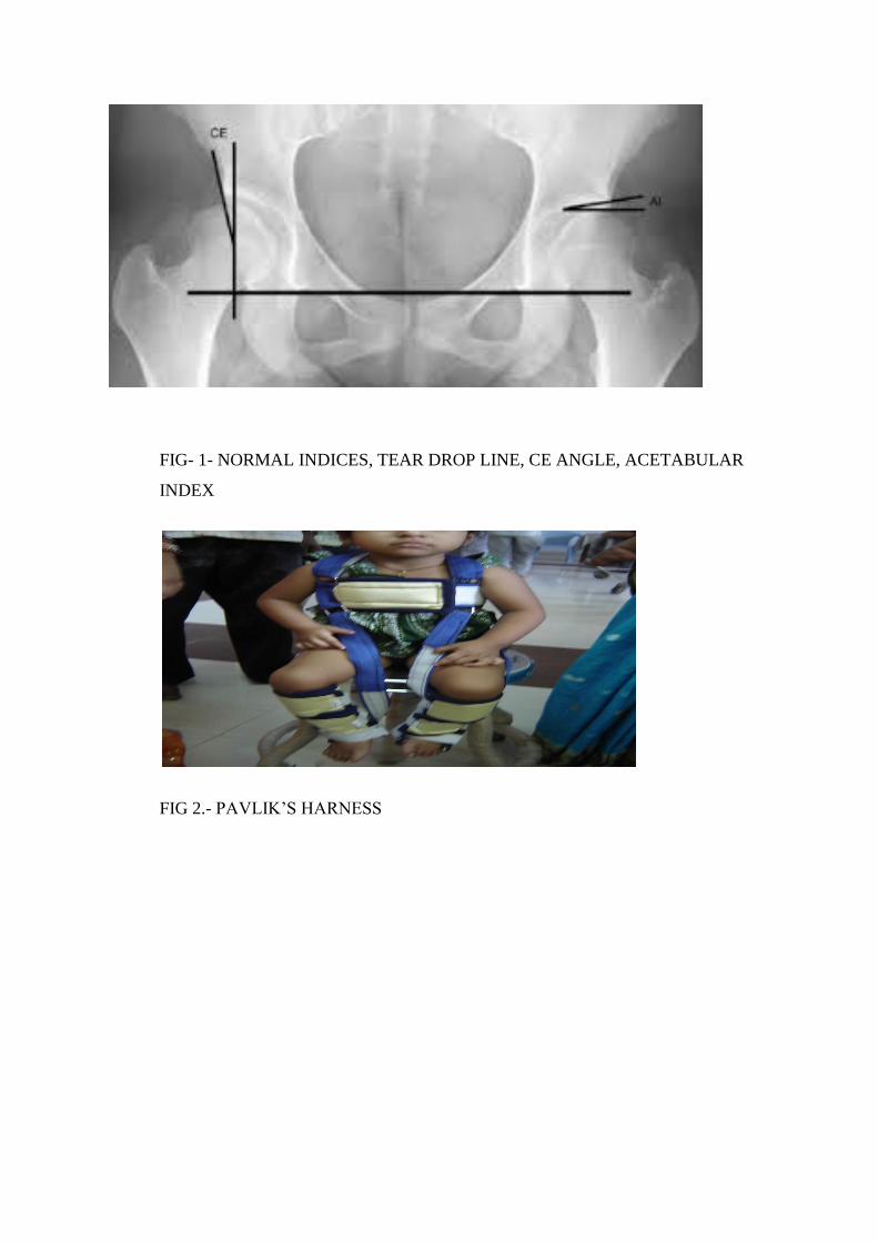
FIG- 1- NORMAL INDICES, TEAR DROP LINE, CE ANGLE, ACETABULAR
INDEX
FIG 2.- PAVLIK’S HARNESS

FIG 3- CLOSED REDUCTION AND ARTHROGRAPHY
FIG 4. – HIP SPICA AFTER CLOSED REDUCTION
Management
Indications for treatment depend on the patient's age and the success of the previous
techniques. Children younger than 6 months with instability upon examination are treated
with a form of bracing, usually a Pavlik’s harness. If this is not effective or if the hip
instability or dislocation is noted when the child is older than 6 months, closed reduction is
typically recommended, often with the administration of traction before the reduction. The
children after failure of conservative treatment or with late presentations are selected for
surgery.
Indications of surgery
Indications for surgery are met if the results of the surgery would be better than the results
of the natural progression of developmental dysplasia of the hip (DDH). 26
The natural history
of hip dysplasia depends, in part, on the severity of the disease, bilaterality, and whether or
not a false acetabulum is formed. 6,29,30
When the child is older than 1 years or with failure of the previous treatment, open reduction
is considered. If the patient is older , femoral shortening is performed instead of traction, with
additional varus applied to the femur, if necessary. A patient with residual acetabular
dysplasia who is older than one year may be treated with an acetabular procedure (Dega).
In neglected cases unilateral dislocations result in significant leg-length inequality, with a
gait disturbance and possibly associated hip and knee pain. In addition, Hip pain commonly
manifests as knee or anterior thigh pain due to the innervation of the hip joint (obturator and
femoral nerve distribution). Typically, true hip pain is identified as groin pain. The
development of a false acetabulum is associated with a poor outcome in approximately 75%
of patients. Bilateral hip dislocation in a patient without false acetabuli has a better overall
prognosis. In fact, a case was reported of a 74-year-old man with no history of hip or thigh
pain whose dislocated hips were only discovered shortly before his death. 31
Treatment for DDH that is diagnosed when the patient is a young adult can be considered for
residual DDH. Unfortunately, radiographic characterization of developmental dysplasia of
the hip that is severe enough to lead to early osteoarthrosis is difficult. A center-edge angle
less than 16º often has been used to predict early osteoarthrosis 30
but other authors have
found this measurement to be less reliable. 31,32
Subluxation, defined as a break in the
Shenton line, has been demonstrated to be associated with osteoarthrosis and decreased
function . 32
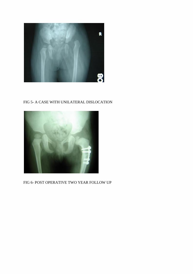
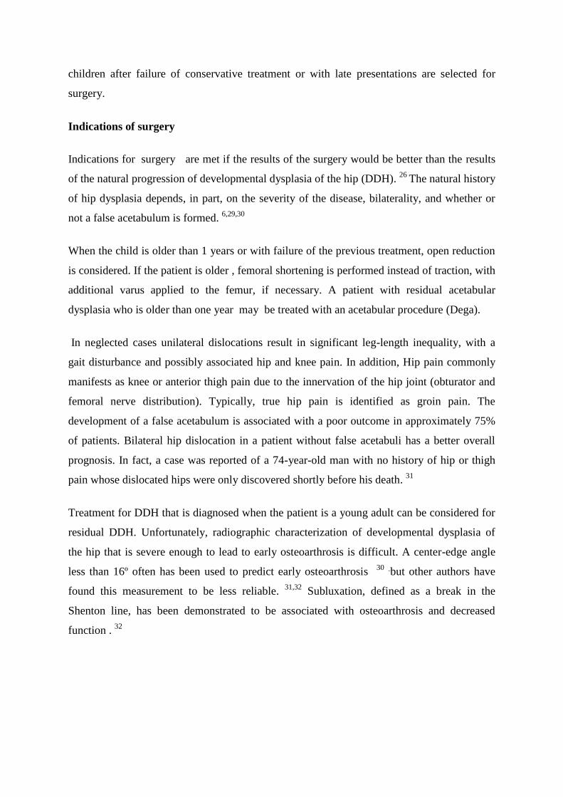
FIG 5- A CASE WITH UNILATERAL DISLOCATION
FIG 6- POST OPERATIVE TWO YEAR FOLLOW UP
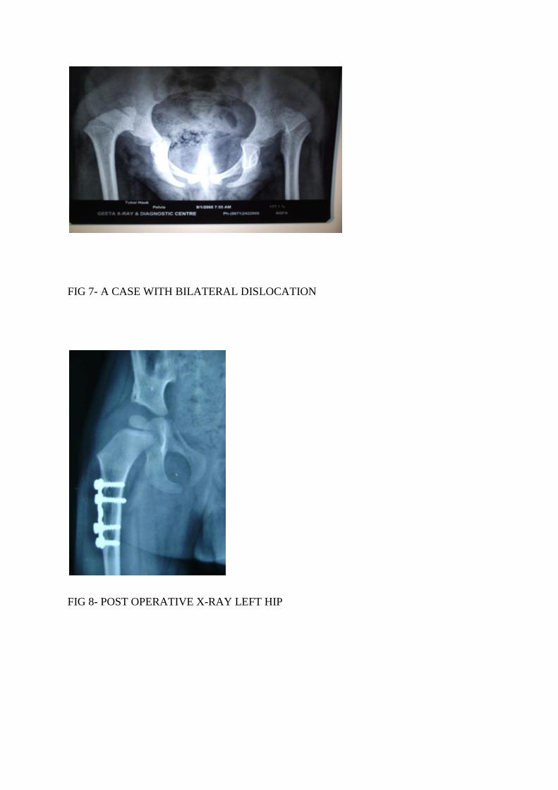
FIG 7- A CASE WITH BILATERAL DISLOCATION
FIG 8- POST OPERATIVE X-RAY LEFT HIP
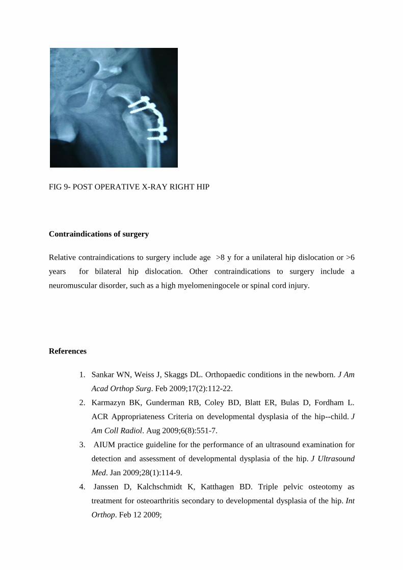
FIG 9- POST OPERATIVE X-RAY RIGHT HIP
Contraindications of surgery
Relative contraindications to surgery include age >8 y for a unilateral hip dislocation or >6
years for bilateral hip dislocation. Other contraindications to surgery include a
neuromuscular disorder, such as a high myelomeningocele or spinal cord injury.
References
1. Sankar WN, Weiss J, Skaggs DL. Orthopaedic conditions in the newborn. J Am
Acad Orthop Surg. Feb 2009;17(2):112-22.
2. Karmazyn BK, Gunderman RB, Coley BD, Blatt ER, Bulas D, Fordham L.
ACR Appropriateness Criteria on developmental dysplasia of the hip--child. J
Am Coll Radiol. Aug 2009;6(8):551-7.
3. AIUM practice guideline for the performance of an ultrasound examination for
detection and assessment of developmental dysplasia of the hip. J Ultrasound
Med. Jan 2009;28(1):114-9.
4. Janssen D, Kalchschmidt K, Katthagen BD. Triple pelvic osteotomy as
treatment for osteoarthritis secondary to developmental dysplasia of the hip. Int
Orthop. Feb 12 2009;
5. Barlow TG. Early diagnosis and treatment of congenital dislocation of the hip. J
Bone Joint Surg Br. 1962;44-B:292-301.
6. Ziegler J, Thielemann F, Mayer-Athenstaedt C, Günther KP. [The natural
history of developmental dysplasia of the hip: A metaanalysis of the published
literature] [German]. Orthopade. May 17 2008;epub ahead of print.
7. Getz B. The hip joint in Lapps and its bearing on the problem of congenital
dislocation. Acta Orthop Scand Suppl. 1955;18:1-81.
8. Hoaglund FT, Yau AC, Wong WL. Osteoarthritis of the hip and other joints in
southern Chinese in Hong Kong. J Bone Joint Surg Am. Apr 1973;55(3):545-57
9. Rabin DL, Barnett CR, Arnold WD, Freiberger RH, Brooks G. Untreated
congenital hip disease: a study of the epidemiology, natural history, and social
aspects of the disease in a Navajo population. Am J Public Health Nations
Health. Feb 1965;55(suppl):1-44
10. Skirving AP, Scadden WJ. The African neonatal hip and its immunity from
congenital dislocation. J Bone Joint Surg Br. Aug 1979;61-B(3):339-41.
11. Bjerkreim I, Arseth PH. Congenital dislocation of the hip in Norway. Late
diagnosis CDH in the years 1970 to 1974. Acta Paediatr Scand. May
1978;67(3):329-32
12. Wilkinson JA. A post-natal survey for congenital displacement of the hip. J
Bone Joint Surg Br. Feb 1972;54(1):40-9.
13. Carter CO, Wilkinson JA. Genetic and environmental factors in the etiology of
congenital dislocation of the hip. Clin Orthop Relat Res. Mar-Apr 1964;33:119-
28.
14. Salter RB. Etiology, pathogenesis and possible prevention of congenital
dislocation of the hip. Can Med Assoc J. May 18 1968;98(20):933-45.
15. Ramsey PL, Lasser S, MacEwen GD. Congenital dislocation of the hip. Use of
the Pavlik harness in the child during the first six months of life. J Bone Joint
Surg Am. Oct 1976;58(7):1000-4.
16. Kumar SJ, MacEwen GD. The incidence of hip dysplasia with metatarsus
adductus. Clin Orthop Relat Res. Apr 1982;164:234-5.
17. Weiner DS. Congenital dislocation of the hip associated with congenital
muscular torticollis. Clin Orthop Relat Res. Nov-Dec 1976;121:163-5.
18. Dunn PM. Perinatal observations on the etiology of congenital dislocation of
the hip. Clin Orthop Relat Res. Sep 1976;119:11-22.
19. Kutlu A, Memik R, Mutlu M, Kutlu R, Arslan A. Congenital dislocation of the
hip and its relation to swaddling used in Turkey. J Pediatr Orthop. Sep-Oct
1992;12(5):598-602
20. McKibbin B. Anatomical factors in the stability of the hip joint in the
newborn. J Bone Joint Surg Br. Feb 1970;52(1):148-59.
21. Rális Z, McKibbin B. Changes in shape of the human hip joint during its
development and their relation to its stability. J Bone Joint Surg Br. Nov
1973;55(4):780-5
22. Bond CD, Hennrikus WL, DellaMaggiore ED. Prospective evaluation of
newborn soft-tissue hip "clicks" with ultrasound. J Pediatr Orthop. Mar-Apr
1997;17(2):199-201.
23. Darmonov AV, Zagora S. Clinical screening for congenital dislocation of the
hip. J Bone Joint Surg Am. Mar 1996;78(3):383-8.
24. Ortolani M. Congenital hip dysplasia in the light of early and very early
diagnosis. Clin Orthop Relat Res. Sep 1976;119:6-10.
25. Mahan ST, Katz JN, Kim YJ. To screen or not to screen? A decision analysis of
the utility of screening for developmental dysplasia of the hip. J Bone Joint Surg
Am. Jul 2009;91(7):1705-19.
26. Falliner A, Hahne HJ, Hassenpflug J. Sonographic hip screening and early
management of developmental dysplasia of the hip. J Pediatr Orthop B. Apr
1999;8(2):112-7.
27. Dogruel H, Atalar H, Yavuz OY, Sayli U. Clinical examination versus
ultrasonography in detecting developmental dysplasia of the hip. Int Orthop. Jun
2008;32(3):415-9
28. Falliner A, Hahne HJ, Hassenpflug J. Sonographic hip screening and early
management of developmental dysplasia of the hip. J Pediatr Orthop B. Apr
1999;8(2):112-7.
29. Jäger M, Westhoff B, Zilkens C, et al. [Indications and results of corrective
pelvic osteotomies in developmental dysplasia of the hip] [German]. Orthopade.
May 21 2008;epub ahead of print
30. Wedge JH, Wasylenko MJ. The natural history of congenital dislocation of the
hip: a critical review. Clin Orthop Relat Res. Nov-Dec 1978;137:154-62.

31. Günther KP, Thielemann F, Hartmann A, Bernstein P. [Combined hip-dysplasia
and femuroacetabular impingement: diagnosis and simultaneous surgical
treatment] [German]. Orthopade. May 30 2008;epub ahead of print.
32. Milgram JW. Morphology of untreated bilateral congenital dislocation of the
hips in a seventy-four-year-old man. Clin Orthop Relat Res. Sep 1976;119:112-
5.