Embed Size (px)
Citation preview
RESEARCH ARTICLE Open Access
Developmental succession of themicrobiome of Culex mosquitoesDagne Duguma1,4*, Michael W. Hall2, Paul Rugman-Jones1, Richard Stouthamer1, Olle Terenius3,Josh D. Neufeld2 and William E. Walton1
Abstract
Background: The native microflora associated with mosquitoes have important roles in mosquito developmentand vector competence. Sequencing of bacterial V3 region from 16S rRNA genes across the developmental stagesof Culex mosquitoes (early and late larval instars, pupae and adults) was used to test the hypothesis that bacteriafound in the larval stage of Culex are transstadially transmitted to the adult stage, and to compare the microbiomesof field-collected versus laboratory-reared mosquitoes.
Results: Beta diversity analysis revealed that bacterial community structure differed among three life stages (larvae,pupae and adults) of Culex tarsalis. Although only ~2 % of the total number of bacterial OTUs were found in allstages, sequences from these OTUs accounted for nearly 82 % of the total bacterial sequences recovered from allstages. Thorsellia (Gammaproteobacteria) was the most abundant bacterial taxon found across all developmentalstages of field-collected Culex mosquitoes, but was rare in mosquitoes from laboratory-reared colonies. The proportionof Thorsellia sequences in the microbiomes of mosquito life stages varied ontogenetically with the greatest proportionsrecovered from the pupae of C. tarsalis and the lowest from newly emerged adults. The microbiome of field-collectedlate instar larvae was not influenced significantly by differences in the microbiota of the habitat due to habitat age orbiopesticide treatments. The microbiome diversity was the greatest in the early instar larvae and the lowest inlaboratory-reared mosquitoes.
Conclusions: Bacterial communities in early instar C. tarsalis larvae were significantly more diverse when compared tolate instar larvae, pupae and newly emerged adults. Some of the bacterial OTUs found in the early instar larvae werealso found across developmental stages. Thorsellia dominated the bacterial communities in field-collected immaturestages but occurred at much lower relative abundance in adults. Differences in microbiota observed in larval habitatsdid not influence bacterial community profiles of late instar larvae or adults. However, bacterial communities inlaboratory-reared C. tarsalis larvae differed significantly from the field. Determining the role of Thorsellia in mosquitoesand its distribution across different species of mosquitoes warrants further investigation.
Keywords: Thorsellia, Outdoor mesocosms, Bacteria, Biopesticide, Transstadial transmission
BackgroundBacteria ingested by the immature stages of mosquitoes(Culicidae) are generally known to provide nutrition[1, 2], facilitate successful development [3, 4], and influ-ence vector competence [5–8]. The presence of certainbacteria has been also shown experimentally to provide
immunity against pathogens, and reduce vector compe-tence [9–11]. Therefore, interest has grown in under-standing the obligate and facultative roles of the gutmicroflora of mosquitoes, their interaction with parasitesand their manipulation to devise alternative and moresustainable vector control strategies (e.g., paratransgeniccontrol) [7, 9, 12–16].To date, the majority of the studies on microbiomes of
mosquitoes have focused largely on investigating the gutmicroflora (and/or symbionts) associated only with adultmosquitoes (e.g., 9). The focus on adult mosquitoes isjustified, on one hand, because adult mosquitoes
* Correspondence: [email protected] of Entomology, University of California Riverside, Riverside, CA92521, USA4Present address: Florida Medical Entomology Laboratory, University ofFlorida, Vero Beach, FL 32962, USAFull list of author information is available at the end of the article
© 2015 Duguma et al. This is an Open Access article distributed under the terms of the Creative Commons Attribution License(http://creativecommons.org/licenses/by/4.0), which permits unrestricted use, distribution, and reproduction in any medium,provided the original work is properly credited. The Creative Commons Public Domain Dedication waiver (http://creativecommons.org/publicdomain/zero/1.0/) applies to the data made available in this article, unless otherwise stated.
Duguma et al. BMC Microbiology (2015) 15:140 DOI 10.1186/s12866-015-0475-8
transmit pathogens directly or cause nuisance to humansand animals. On the other hand, the interactions of bac-teria and immature stages of mosquitoes will potentiallyinfluence the microbiome of the adult mosquitoes.Moreover, previous studies focused on the dominanttropical disease vector mosquito species such as Anoph-eles gambiae, and not much was known about NorthAmerican native mosquito species such as Culex tarsa-lis. Few studies have considered transstadial transmis-sion (larvae to pupae to adult) of microbial communitiesbecause the entire midgut of culicid mosquitoes is gen-erally thought be replaced during development such thatentire bacterial communities associated with the larvalmidgut are eliminated prior to eclosion [2, 17]. However,circumstantial evidence in Anopheles mosquitoes sug-gested that some bacteria species found in larval stagespersist through metamorphosis and are transferred toadults [18, 19]. Recently, these contradictory observa-tions have been reconciled in studies from Anophelesstephensi where the Malpighian tubules function as a“refugium” for bacteria during metamorphosis [20, 21].In addition, most other studies were based onlaboratory-reared mosquitoes, fed standardized diets,and raised under controlled environmental conditionsfor several generations. However, these artificial condi-tions are likely to restrict and/or alter the host micro-biomes of lab-raised insect populations relative tonatural populations (e.g. Drosophila; [22]).The Gram-negative genus Thorsellia (Gammaproteo-
bacteria) is the most abundant bacterial group foundamong the gut microflora of field-collected late instarlarvae of Culex spp. (3rd and 4th instars; [23]). Thorselliaanophelis was first described from adult Anopheles ara-biensis in Kenya [24, 25] and is the type specimen for anew family of the Bacteria, Thorselliaceae [26]. Thorsel-lia anophelis has been reported to be the predominantbacterial species found in adult Anopheles gambiaesensu lato [27] and has also been found in Anophelesculicifacies [28]. It has been hypothesized that the bac-terium is acquired during larval feeding, and is thentransferred transstadially to adult Anopheles, althoughthe evidence is somewhat conflicting [18, 19, 27]. Raniand colleagues recovered this bacterium in both larvaeand adults of Anopheles stephensi, but not in the pupalstage [18], whereas Briones and colleagues did not re-cover the bacterium from either larval or pupal stages ofAnopheles [27]. However, others found Thorsellia to bethe 5th most abundant genus in Anopheles gambiae lar-vae, and the 10th most abundant genus in pupae [19].Direct evidence supporting the transstadial transmissionof Thorsellia and other bacterial species in the micro-biome of Culex mosquitoes is lacking.It was unknown whether succession or other factors
such as use of pesticides causing changes in bacterial
communities of the larval developmental sites also affectthe microbiome in mosquitoes. Pesticides (i.e., malathion,permethrin, atrazine and glyphosate) alter bacterial com-munities in the larval environment [29]. Removal ofmosquito larvae using biopesticides such as Bacillus thur-ingiensis subsp. israelensis (Bti) was reported to lessengrazing pressure on bacterial communities [30–32] andincrease the diversity of bacteria in the habitats [33].In this study, we: 1) identified bacteria found in the
guts of different developmental stages of C. tarsalis; 2)compared the microbiomes of field-collected C. tarsalislarvae with laboratory-reared individuals; and 3) assessedthe effects of habitat age and manipulating larval mos-quito density (using Bti applications) on the microbialcommunities found in late-instar mosquito larvae.
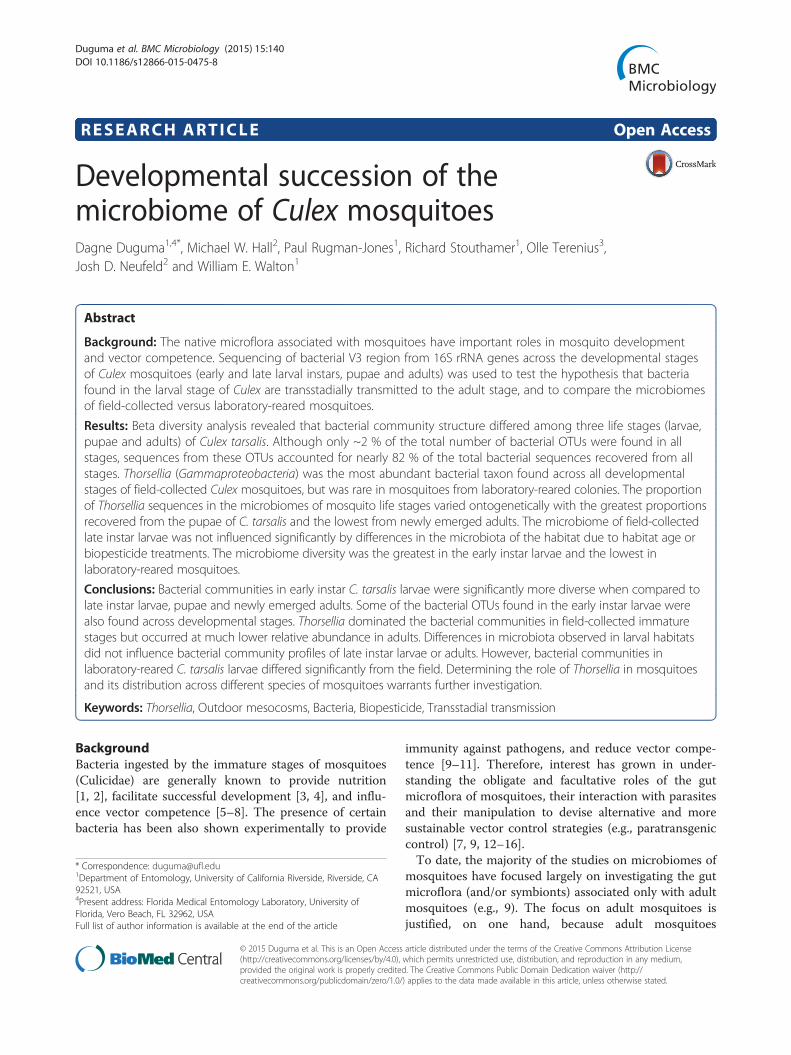
ResultsBacterial taxa in developmental stages of C. tarsalisA total of 14,634 OTUs (4,609,186 bacterial sequences)were generated from 41 Culex mosquito samples (39field-collected and two laboratory-reared). Proteobac-teria (56 %), Bacteroidetes (15 %), Cyanobacteria(14 %), Firmicutes (7 %), Actinobacteria (2 %), and Spi-rochaetes (2 %) were the most abundant bacterial phylafound in developmental stages of Culex mosquitoes(Additional file 1: Table S1). Unclassified sequencesaccounted for ~0.6 % of sequences, whereas, bacterialsequences unclassified to phyla accounted for 0.8 %of sequences. Another 30 phyla accounted for theremaining 1 % of the bacterial communities. Althoughthe relative abundance of bacterial taxa changed acrossthe mosquito developmental stages, Proteobacteriawere the dominant bacteria found in the guts of C. tar-salis (Fig. 1).A total of 235 bacterial OTUs, comprising 82 % of the
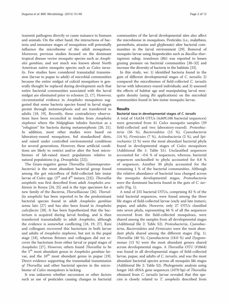
total bacterial sequences, were found among the threelife stages of field-collected larvae (early and late instars),pupae, and adults. However, only 27 OTUs classifiedinto seven phyla, representing 66 % of all the sequencesrecovered from the field-collected mosquitoes, wereshared among the samples from all developmental stages(Additional file 2: Table S2). Proteobacteria, Cyanobac-teria, Bacteroidetes and Firmicutes were the most abun-dant phyla shared among the different stages (Fig. 1).Thorsellia (40 %), Cyanobacteria (18.0 %) and Dysgono-monas (13 %) were the most abundant genera sharedacross developmental stages. A Thorsellia OTU (#2664)was found in all developmental stages of field-collectedlarvae, pupae, and adults of C. tarsalis, and was the mostabundant bacterial species across all mosquito life stages(Additional file 2: Table S2). Phylogenetic analysis usinglonger 16S rRNA gene sequences (1070 bp) of Thorselliaobtained from C. tarsalis larvae revealed that this spe-cies is closely related to T. anophelis described from
Duguma et al. BMC Microbiology (2015) 15:140 Page 2 of 13
Anopheles gambiae sensu stricto but may also contain aseparate strain (Fig. 2). The two Thorsellia sequences fromCulex mosquitoes were both 99 % similar to T. anophelis,but with individual differences. Out of the six clones se-quenced, five clones were identical to clone #6, whereasclone #7 was unique but had nucleotide differences that
are supported by the other Thorsellia species (data notshown).
Early instar larvae (not identified to species)A total of 5,888 OTUs (805,169 sequences from 7 sam-ples) in 34 bacterial phyla were recovered from early (1st
Fig. 1 Proportional sequence abundance of bacterial phyla across Culex developmental stages. The samples were taken 4, 14, 20 and 48 daysafter the onset of the experiment. Only phyla with an average abundance≥ 0.01 % were included. Numbers on the x-axis represent sampleidentification numbers. Stages of mosquitoes are denoted by Early instar, Late instar, Lab = lab reared late instar, Pupae, and Adults. Light barrefers to control; gray refers to low Bti and dark bar for high Bti treatments
Fig. 2 Molecular phylogenetic analysis of Thorsellia from Culex larvae by maximum likelihood method. The proportion of trees in which theassociated taxa clustered together is shown next to the branches (1000 bootstraps). The tree is drawn to scale, with branch lengths measured inthe number of substitutions per site
Duguma et al. BMC Microbiology (2015) 15:140 Page 3 of 13
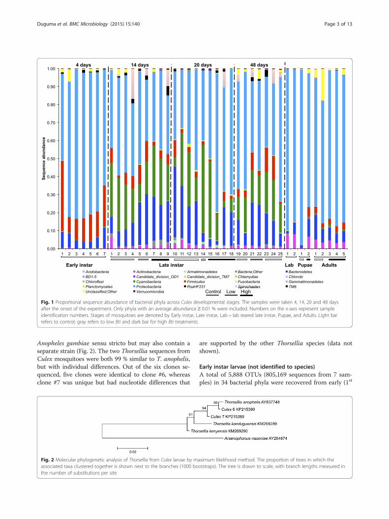
and 2nd) instars of Culex larvae. Proteobacteria (73 %),Firmicutes (18 %), and Bacteroidetes (7 %) dominatedthe early instar Culex larvae (Additional file 1: Table S1).Overall, bacterial taxa in Gammaproteobacteria (43 %),Betaprotobacteria (26 %) and Bacilli (13 %) were thethree most abundant classes found in the early stages ofthe mosquito life cycle (Additional file 1: Table S1).Thorselliaceae (27.2 %) and Comamonadaceae (21.2 %)were the two most abundant families found in early in-star larvae (Fig. 3). Thorsellia was the most abundant(19 %) genus found in the early stages followed by anunclassified taxon of Gammaproteobacteria (8.8 %) andAeromonas (7 %) (Additional file 3: Table S3).
Late instar C. tarsalis larvaeField-collected C. tarsalis larvaeOverall, 34 bacterial phyla (10,115 OTUs from 25 sam-ples) were found in field-collected late larval (3rd and4th) instars. Proteobacteria accounted for nearly half(49 %) of the sequences followed by Cyanobacteria(20 %), Bacteroidetes (18 %), and Firmicutes (6 %) (Fig. 1).Spirochaetes and Actinobacteria accounted for nearly
3 % and 2 %, respectively, of the sequences recoveredfrom late-instar larvae. Thorselliaceae (43.9 %) and Por-phyromonadaceae (24.9 %) were the two most abundantfamilies found in late-instar larvae (Fig. 2). Thorselliawas the most abundant genus in field-collected late in-star larvae (27 %) followed by Cyanobacteria (19 %) andDysgonomonas (Bacteroidetes; 11 %) (Additional file 4:Table S4).
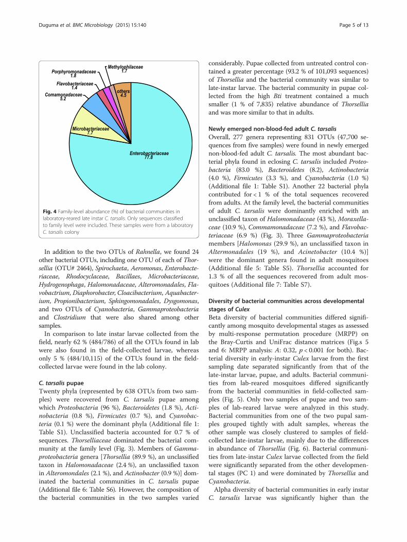
Laboratory-reared larvaeThe late-instar larvae from a laboratory colony were dom-inated by Proteobacteria (87 %) followed by Actinobacteria(8 %) and Bacteroidetes (4 %). Overall, 786 OTUs (319,786sequences from 2 samples) were obtained from thelaboratory-reared colonies. Enterobacteriaceae (77.8 %)was the most abundant family found in late instarlaboratory-reared mosquitoes (Fig. 4). Rahnella (Gamma-proteobacteria: Enterobacteriaceae) (64 %), unclassifiedEnterobacteriaceae (12 %), and unclassified Microbacteria-ceae (Actinobacteteria) (8 %) dominated the bacterialcommunities in the larvae from the laboratory-reared col-ony (Additional file 5: Table S5).
Fig. 3 Family-level abundance of bacterial communities. Family-level abundance (%) of bacterial communities in field-collected early (1st and 2nd)instar larvae, late (3rd and 4th) instar larvae, pupae and adults of C. tarsalis. Only sequences classified to family level were included. Because thetreatments effects on the gut bacterial community structure within each stage were not significantly different, mosquitoes from all treatmentswere included in this figure
Duguma et al. BMC Microbiology (2015) 15:140 Page 4 of 13
In addition to the two OTUs of Rahnella, we found 24other bacterial OTUs, including one OTU of each of Thor-sellia (OTU# 2464), Spirochaeta, Aeromonas, Enterobacte-riaceae, Rhodocyclaceae, Bacillaes, Microbacteriaceae,Hydrogenophaga, Halomonadaceae, Alteromonadales, Fla-vobactrium, Diaphorobacter, Cloacibacterium, Aquabacter-ium, Propionibacterium, Sphingomonadales, Dysgomonas,and two OTUs of Cyanobacteria, Gammaproteobacteriaand Clostridium that were also shared among othersamples.In comparison to late instar larvae collected from the
field, nearly 62 % (484/786) of all the OTUs found in labwere also found in the field-collected larvae, whereasonly 5 % (484/10,115) of the OTUs found in the field-collected larvae were found in the lab colony.
C. tarsalis pupaeTwenty phyla (represented by 638 OTUs from two sam-ples) were recovered from C. tarsalis pupae amongwhich Proteobacteria (96 %), Bacteroidetes (1.8 %), Acti-nobacteria (0.8 %), Firmicutes (0.7 %), and Cyanobac-teria (0.1 %) were the dominant phyla (Additional file 1:Table S1). Unclassified bacteria accounted for 0.7 % ofsequences. Thorselliaceae dominated the bacterial com-munity at the family level (Fig. 3). Members of Gamma-proteobacteria genera [Thorsellia (89.9 %), an unclassifiedtaxon in Halomonadaceae (2.4%), an unclassified taxonin Alteromondales (2.1 %), and Actinobacter (0.9 %)] dom-inated the bacterial communities in C. tarsalis pupae(Additional file 6: Table S6). However, the composition ofthe bacterial communities in the two samples varied
considerably. Pupae collected from untreated control con-tained a greater percentage (93.2 % of 101,093 sequences)of Thorsellia and the bacterial community was similar tolate-instar larvae. The bacterial community in pupae col-lected from the high Bti treatment contained a muchsmaller (1 % of 7,835) relative abundance of Thorselliaand was more similar to that in adults.
Newly emerged non-blood-fed adult C. tarsalisOverall, 277 genera representing 831 OTUs (47,700 se-quences from five samples) were found in newly emergednon-blood-fed adult C. tarsalis. The most abundant bac-terial phyla found in eclosing C. tarsalis included Proteo-bacteria (83.0 %), Bacteroidetes (8.2), Actinobacteria(4.0 %), Firmicutes (3.3 %), and Cyanobacteria (1.0 %)(Additional file 1: Table S1). Another 22 bacterial phylacontributed for < 1 % of the total sequences recoveredfrom adults. At the family level, the bacterial communitiesof adult C. tarsalis were dominantly enriched with anunclassified taxon of Halomonadaceae (43 %), Moraxella-ceae (10.9 %), Commamonadaceae (7.2 %), and Flavobac-teriaceae (6.9 %) (Fig. 3). Three Gammaproteobacteriamembers [Halomonas (29.9 %), an unclassified taxon inAltermonadales (19 %), and Acinetobacter (10.4 %)]were the dominant genera found in adult mosquitoes(Additional file 5: Table S5). Thorsellia accounted for1.3 % of all the sequences recovered from adult mos-quitoes (Additional file 7: Table S7).
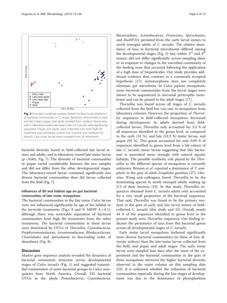
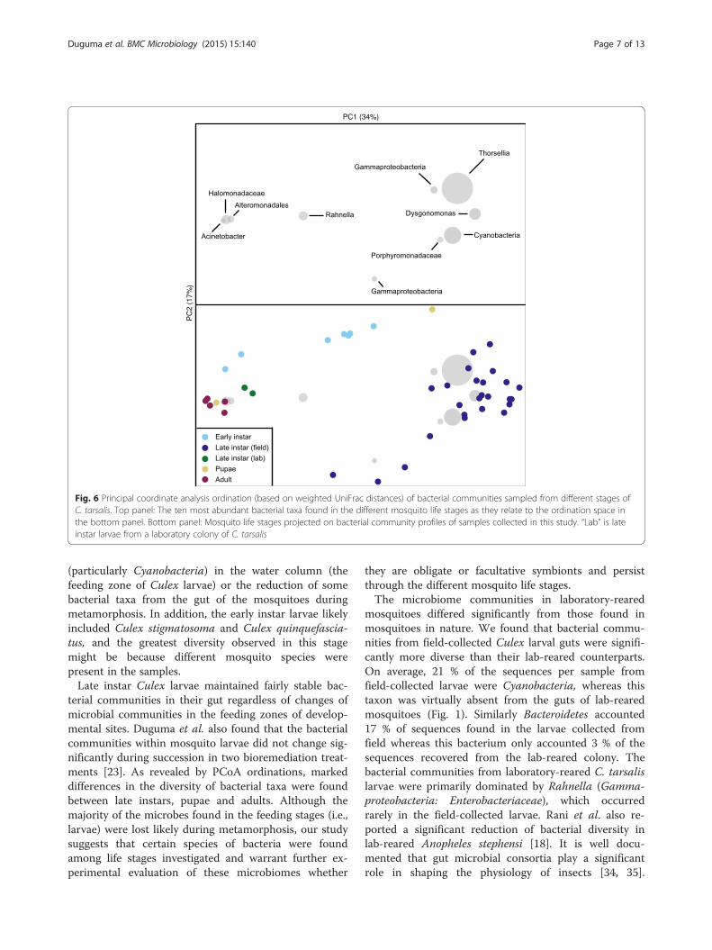
Diversity of bacterial communities across developmentalstages of CulexBeta diversity of bacterial communities differed signifi-cantly among mosquito developmental stages as assessedby multi-response permutation procedure (MRPP) onthe Bray-Curtis and UniFrac distance matrices (Fig.s 5and 6: MRPP analysis: A: 0.32, p < 0.001 for both). Bac-terial diversity in early-instar Culex larvae from the firstsampling date separated significantly from that of thelate-instar larvae, pupae, and adults. Bacterial communi-ties from lab-reared mosquitoes differed significantlyfrom the bacterial communities in field-collected sam-ples (Fig. 5). Only two samples of pupae and two sam-ples of lab-reared larvae were analyzed in this study.Bacterial communities from one of the two pupal sam-ples grouped tightly with adult samples, whereas theother sample was closely clustered to samples of field-collected late-instar larvae, mainly due to the differencesin abundance of Thorsellia (Fig. 6). Bacterial communi-ties from late-instar Culex larvae collected from the fieldwere significantly separated from the other developmen-tal stages (PC 1) and were dominated by Thorsellia andCyanobacteria.Alpha diversity of bacterial communities in early instar
C. tarsalis larvae was significantly higher than the
Fig. 4 Family-level abundance (%) of bacterial communities inlaboratory-reared late instar C. tarsalis. Only sequences classifiedto family level were included. These samples were from a laboratoryC. tarsalis colony
Duguma et al. BMC Microbiology (2015) 15:140 Page 5 of 13
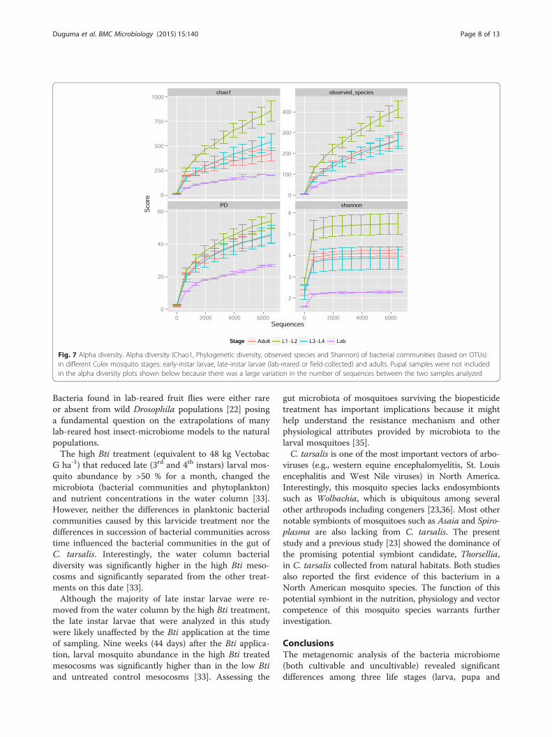
bacterial diversity found in field-collected late larval in-stars and adults, and in laboratory-reared late-instar larvae(p < 0.001, Fig. 7). The diversity of bacterial communitiesin pupae varied considerably between the two samplesand did not differ from the other developmental stages.The laboratory-reared larvae contained significantly lessdiverse bacterial communities than did larvae collectedfrom the field (Fig. 7).
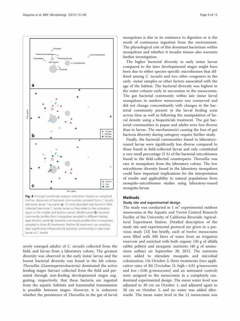
Influences of Bti and habitat age on gut bacterialcommunities of late instar mosquitoesThe bacterial communities in the late instar Culex larvaewere not influenced significantly by age of the habitat orthe larvicide treatments (Fig.s 8 and 9; MRPP A ≤ 0.1),although there was noticeable separation of bacterialcommunities from high Bti treatments from the othertreatments. The bacterial communities in these larvaewere dominated by OTUs of Thorsellia, Cyanobacteria,Porphyromonadaceae, Aeromonadaceae, Rhodocyclaceae,Clostridiales and Spirochaeta in descending order ofabundance (Fig. 8).
DiscussionMarker gene sequence analysis revealed the dynamics ofbacterial community structure across developmentalstages of Culex tarsalis (Fig. 1) and suggested transsta-dial transmission of some bacterial groups in Culex mos-quitoes from North America. Overall, 235 bacterialOTUs in the phyla Proteobacteria, Cyanobacteria,
Bacteroidetes, Actinobacteria, Firmicutes, Spirochaetes,and RsaHF231 persisted from the early larval instars tonewly emerged adults of C. tarsalis. The relative abun-dance of taxa in bacterial microbiome differed amongthe developmental stages (Fig. 5) but, within 3rd and 4th
instars, did not differ significantly across sampling datesor in response to changes in the microbial community ofthe feeding zone that occurred following the applicationof a high dose of biopesticides. Our study provides add-itional evidence that, contrary to a commonly acceptedhypothesis [17], metamorphosis does not completelyeliminate gut microbiota. In Culex pipiens mosquitoes,some bacterial communities from the larval stages wereshown to be sequestered in meconial peritrophic mem-brane and can be passed to the adult stages [17].Thorsellia was found across all stages of C. tarsalis
collected from the field but was rare in mosquitoes fromlaboratory colonies. However, the proportion of Thorsel-lia sequences in field-collected mosquitoes decreasedduring development. In adults derived from field-collected larvae, Thorsellia only accounted for 3.3 % ofall sequences identified to the genus level, as comparedto the early (31 %), and late (52.3 %) instar larvae, andpupae (93 %). This genus accounted for only 0.09 % ofsequences identified to genus level from a lab colony oflate C. tarsalis instar larvae suggesting that this bacter-ium is associated more strongly with natural aquatichabitats. The possible symbiotic role played by the Thor-sellia to the different species of mosquitoes is currentlyunknown. Briones et al. reported a dominance of T. ano-phelis in the guts of adult Anopheles gambiae [27]. Like-wise, Wang and colleagues found Thorsellia to be thedominating species in newly emerged adults making up2/3 of their bacteria [19]. In this study, Thorsellia se-quences obtained from C. tarsalis adults only accountedfor a very small proportion of the bacterial sequences.That said, Thorsellia was found to be the primary resi-dent in the guts of early and late larval instars of field-collected C. tarsalis (this study and 23). Overall, nearly44 % of the sequences identified to genus level in thepresent study were Thorsellia sequences. Our finding in-dicates the persistence of taxa from this bacterial genusacross all developmental stages of C. tarsalis.Early instar larval mosquitoes harbored significantly
more diverse bacterial communities (in three of four di-versity indices) than the late-instar larvae collected fromthe field, and pupae and adult stages. The early instarlarvae were sampled four days after the onset of the ex-periment and the bacterial communities in the guts ofthese mosquitoes mirrored the higher bacterial diversityobserved in the water column on this sampling date[33]. It is unknown whether the reduction of bacterialcommunities especially during the late stages of develop-ment was due to the dominance of phytoplankton
Fig. 5 Principal coordinate analysis (based on Bray-Curtis distances)of bacterial communities in C. tarsalis. Bacterial communities in earlyand late instars, pupae, and adults sampled from outdoor mesocosms,and in laboratory-reared late larval instars of C.tarsalis were significantlyseparated. Pupae and adults were collected only from high Btitreatment and untreated control (see material and methods fordetails). Late instar larvae were sampled from all treatments
Duguma et al. BMC Microbiology (2015) 15:140 Page 6 of 13
(particularly Cyanobacteria) in the water column (thefeeding zone of Culex larvae) or the reduction of somebacterial taxa from the gut of the mosquitoes duringmetamorphosis. In addition, the early instar larvae likelyincluded Culex stigmatosoma and Culex quinquefascia-tus, and the greatest diversity observed in this stagemight be because different mosquito species werepresent in the samples.Late instar Culex larvae maintained fairly stable bac-
terial communities in their gut regardless of changes ofmicrobial communities in the feeding zones of develop-mental sites. Duguma et al. also found that the bacterialcommunities within mosquito larvae did not change sig-nificantly during succession in two bioremediation treat-ments [23]. As revealed by PCoA ordinations, markeddifferences in the diversity of bacterial taxa were foundbetween late instars, pupae and adults. Although themajority of the microbes found in the feeding stages (i.e.,larvae) were lost likely during metamorphosis, our studysuggests that certain species of bacteria were foundamong life stages investigated and warrant further ex-perimental evaluation of these microbiomes whether
they are obligate or facultative symbionts and persistthrough the different mosquito life stages.The microbiome communities in laboratory-reared
mosquitoes differed significantly from those found inmosquitoes in nature. We found that bacterial commu-nities from field-collected Culex larval guts were signifi-cantly more diverse than their lab-reared counterparts.On average, 21 % of the sequences per sample fromfield-collected larvae were Cyanobacteria, whereas thistaxon was virtually absent from the guts of lab-rearedmosquitoes (Fig. 1). Similarly Bacteroidetes accounted17 % of sequences found in the larvae collected fromfield whereas this bacterium only accounted 3 % of thesequences recovered from the lab-reared colony. Thebacterial communities from laboratory-reared C. tarsalislarvae were primarily dominated by Rahnella (Gamma-proteobacteria: Enterobacteriaceae), which occurredrarely in the field-collected larvae. Rani et al. also re-ported a significant reduction of bacterial diversity inlab-reared Anopheles stephensi [18]. It is well docu-mented that gut microbial consortia play a significantrole in shaping the physiology of insects [34, 35].
Fig. 6 Principal coordinate analysis ordination (based on weighted UniFrac distances) of bacterial communities sampled from different stages ofC. tarsalis. Top panel: The ten most abundant bacterial taxa found in the different mosquito life stages as they relate to the ordination space inthe bottom panel. Bottom panel: Mosquito life stages projected on bacterial community profiles of samples collected in this study. “Lab” is lateinstar larvae from a laboratory colony of C. tarsalis
Duguma et al. BMC Microbiology (2015) 15:140 Page 7 of 13
Bacteria found in lab-reared fruit flies were either rareor absent from wild Drosophila populations [22] posinga fundamental question on the extrapolations of manylab-reared host insect-microbiome models to the naturalpopulations.The high Bti treatment (equivalent to 48 kg Vectobac
G ha-1) that reduced late (3rd and 4th instars) larval mos-quito abundance by >50 % for a month, changed themicrobiota (bacterial communities and phytoplankton)and nutrient concentrations in the water column [33].However, neither the differences in planktonic bacterialcommunities caused by this larvicide treatment nor thedifferences in succession of bacterial communities acrosstime influenced the bacterial communities in the gut ofC. tarsalis. Interestingly, the water column bacterialdiversity was significantly higher in the high Bti meso-cosms and significantly separated from the other treat-ments on this date [33].Although the majority of late instar larvae were re-
moved from the water column by the high Bti treatment,the late instar larvae that were analyzed in this studywere likely unaffected by the Bti application at the timeof sampling. Nine weeks (44 days) after the Bti applica-tion, larval mosquito abundance in the high Bti treatedmesocosms was significantly higher than in the low Btiand untreated control mesocosms [33]. Assessing the
gut microbiota of mosquitoes surviving the biopesticidetreatment has important implications because it mighthelp understand the resistance mechanism and otherphysiological attributes provided by microbiota to thelarval mosquitoes [35].C. tarsalis is one of the most important vectors of arbo-
viruses (e.g., western equine encephalomyelitis, St. Louisencephalitis and West Nile viruses) in North America.Interestingly, this mosquito species lacks endosymbiontssuch as Wolbachia, which is ubiquitous among severalother arthropods including congeners [23,36]. Most othernotable symbionts of mosquitoes such as Asaia and Spiro-plasma are also lacking from C. tarsalis. The presentstudy and a previous study [23] showed the dominance ofthe promising potential symbiont candidate, Thorsellia,in C. tarsalis collected from natural habitats. Both studiesalso reported the first evidence of this bacterium in aNorth American mosquito species. The function of thispotential symbiont in the nutrition, physiology and vectorcompetence of this mosquito species warrants furtherinvestigation.
ConclusionsThe metagenomic analysis of the bacteria microbiome(both cultivable and uncultivable) revealed significantdifferences among three life stages (larva, pupa and
Fig. 7 Alpha diversity. Alpha diversity (Chao1, Phylogenetic diversity, observed species and Shannon) of bacterial communities (based on OTUs)in different Culex mosquito stages: early-instar larvae, late-instar larvae (lab-reared or field-collected) and adults. Pupal samples were not includedin the alpha diversity plots shown below because there was a large variation in the number of sequences between the two samples analyzed
Duguma et al. BMC Microbiology (2015) 15:140 Page 8 of 13
newly emerged adults) of C. tarsalis collected from thefield, and larvae from a laboratory colony. The greatestdiversity was observed in the early instar larvae and thelowest bacterial diversity was found in the lab colony.Thorsellia (Gammaproteobacteria) dominated the activefeeding stages (larvae) collected from the field and per-sisted through non-feeding developmental stages sug-gesting, respectively, that these bacteria are ingestedfrom the aquatic habitats and transstadial transmissionis possible between stages. However, it is unknownwhether the persistence of Thorsellia in the gut of larval
mosquitoes is due to its resistance to digestion or is theresult of continuous ingestion from the environment.The physiological role of this dominant bacterium withinmosquitoes and whether it invades tissues also warrantsfurther investigation.The higher bacterial diversity in early instar larvae
compared to the later developmental stages might havebeen due to either species-specific microbiomes that dif-fered among C. tarsalis and two other congeners in theearly -instar samples or other factors associated with theage of the habitat. The bacterial diversity was highest inthe water column early in succession in the mesocosms.The gut bacterial community within late instar larvalmosquitoes in outdoor mesocosms was conserved anddid not change concomitantly with changes in the bac-terial community present in the larval feeding zoneacross time as well as following the manipulation of lar-val density using a biopesticide treatment. The gut bac-terial communities in pupae and adults were less diversethan in larvae. The mechanism(s) causing the loss of gutbacteria diversity during ontogeny require further study.Finally, the bacterial communities found in laboratory-
reared larvae were significantly less diverse compared tothose found in field-collected larvae and only constituteda very small percentage (5 %) of the bacterial microbiomesfound in the field-collected counterparts. Thorsellia wasrare in mosquitoes from the laboratory colony. The lowmicrobiome diversity found in the laboratory mosquitoescould have important implications for the interpretationof results and applicability to natural populations frommosquito-microbiomes studies using laboratory-rearedmosquito larvae.
MethodsStudy site and experimental designThe study was conducted in 1 m2 experimental outdoormesocosms at the Aquatic and Vector Control ResearchFacility of the University of California Riverside Agricul-tural Experiment Station. Detailed description of thestudy site and experimental protocol are given in a pre-vious study [33] but briefly, each of twelve mesocosmswere filled with 300 liters of water from an irrigationreservoir and enriched with both organic (50 g of alfalfarabbit pellets) and inorganic nutrients (40 g of ammo-nium sulfate) on September 28, 2012. The nutrientswere added to stimulate mosquito and microbialcolonization. On October 2, three treatments (two appli-cation rates of Bti [Vectobac G: high = 4.81 g/mesocosmand low = 0.06 g/mesocosm] and an untreated control)were assigned to the mesocosms in a completely ran-domized experimental design. The mean water level wasadjusted to 30 cm on October 1, and adjusted again to30 cm on October 5, and no water was added after-wards. The mean water level in the 12 mesocosms was
Fig. 8 Principal coordinate analysis ordination (based on weightedUniFrac distances) of bacterial communities sampled from C. tarsalislate-instar larvae. Top panel (a): 10 most abundant taxa found in field-collected late-instar C. tarsalis larvae as they relate to the ordinationspace in the middle and bottom panels. Middle panel (b): bacterialcommunity profiles from mosquitoes sampled in different habitatages. Bottom panel (c): bacterial community profiles from mosquitoessampled in three Bti treatments. Neither Bti treatment nor samplingdate significantly influenced the bacterial communities in late-instarlarvae of C. tarsalis
Duguma et al. BMC Microbiology (2015) 15:140 Page 9 of 13

28 (±0.32; mean ± SE) cm on October 12, and 25 (±0.28)cm on October 19 and 17 (±0.65) cm on November 16.
Mosquito sampling for bacterial DNA extractionFive early instar Culex larvae per mesocosm were col-lected on October 1 (4 days after starting the experi-ment), placed in 95 % ethanol in 15 mL sterilecentrifuge tubes and stored at -20 °C until DNA extrac-tion. The identification of these early instars was notpossible using morphology but late (3rd and 4th) instarCulex larvae collected the following day was comprisedof C. stigmatosoma (74 %), C. quinquefasciatus (23 %),and C. tarsalis (3 %). The proportion of C. tarsalis inthe mesocosms increased over time while C. stigmato-soma tended to be more abundant early in experiment.Five late (3rd and 4nd) instar larvae of C. tarsalis larvae
per mesocosm were sampled on three dates: October 12,October 18, and November 15 (i.e., 14, 20 and 48 days,respectively from the onset of the experiments) to deter-mine the influences of Bti treatments and habitat age onbacterial community structure associated with Culex lar-vae. These dates correspond to 10, 16 and 44 days afterBti treatments, respectively. Two groups of five late instarC. tarsalis larvae from a laboratory colony were alsosampled to compare bacterial communities of laboratory-reared mosquitoes with field-collected mosquitoes. C.tarsalis colonies have been maintained in the laboratoryfor > 5 years and the larvae were fed a mixture of yeastand ground rat chow [37].For pupae and adult mosquitoes, late instar larvae were
collected from the mesocosms, morphologically identifiedto C. tarsalis under a dissecting microscope, and thenreared to pupae or to adulthood. Water from the corre-sponding mesocosms was used to rear the larvae. Immedi-ately after pupation, two groups of five pupae from thehigh Bti treatment and the control were preserved in 95 %ethanol and placed in a -20 °C freezer until DNA extrac-tion. The remaining pupae were then transferred to rear-ing cages and, immediately after emergence, five groups of
five adult C. tarsalis (three from high Bti and two fromcontrol treatments) were placed in 95 % ethanol in 15 mLsterile tubes and kept in -20 °C freezer until DNAextraction.
DNA extraction, PCR and Illumina library preparationThe procedures of DNA extraction and amplification ofthe V3 hypervariable region of 16S rRNA genes usingpolymerase chain reaction (PCR) were similar to thoseused for late larval instars in previous study [23]. Inaddition to late instar larvae, early (1st and 2nd) instarlarvae, pupae and adults of C. tarsalis were also sampledin the present study. All mosquito samples within the15-mL tubes were sonicated for 3 min in iced sterilewater according to a previous study [23]. DNA was ex-tracted from only three (out of five) pooled, intact mos-quitoes in 1.5 mL microcentrifuge tubes per replicatemesocosm using a Qiagen kit as described in [23]. PCRand Illumina library preparation were also carried outaccording to a previous study [23]. After libraries wereprepared and quantified using an Agilent Bioanalyzer, allsamples were normalized to 10 nM using Tris-HCl(10 mM, pH 8.5) and combined to create two multi-plexed samples. The multiplexed samples were then sub-jected to a 2 × 150 base paired-end sequencing on aMiSeq Illumina platform at GENOSEQ (Sequencing andGenotyping Core) of the University of California LosAngeles, Los Angeles. Overall, a total of 41 mosquitosamples [two late-instar samples from laboratory col-onies, seven samples of field-collected early instars, 25samples of late instars (taken on three sampling dates),two samples of pupae and five samples of adult mosqui-toes] were submitted for sequencing.
Thorsellia cloning and phylogenyPCR in 25 μL reactions was performed with Illustra PuReTaq Ready-To-Go PCR Beads (GE Healthcare, Uppsala,Sweden), 0.4 μM each of forward and reverse Thorselliaprimers 207f (5’-GCACTAGGATGAACCCAGG-3’) and
Fig. 9 Principal coordinate analysis of bacterial communities in C. tarsalis late-instar larvae. Late-instar larvae collected from three Bti treatments(a), and three sampling dates (b)
Duguma et al. BMC Microbiology (2015) 15:140 Page 10 of 13
reverse primer 1277r (5’-CTTTATGAGTTCCGCTTACCC-3’), and 2 μL of DNA from larvae of C. tarsalis.These primers were designed for investigating Thorselliain Anopheles gambiae sensu latu in a previous study [38].The PCR program was 98 °C for 5 min followed by 30 cy-cles of [95 °C for 30 s, 55 °C for 30 s and 72 °C for 1 min]and a final step of 72 °C for 10 min. Amplification prod-ucts (1.1 kb) were cloned into TOPO 2.1 (Invitrogen) andsequenced at Macrogen (South Korea).Sequences were aligned with available GenBank acces-
sions for three described Thorsellia species (T. anophelis[AY837748], T. kanduguensis [KM269289], and T. kenyen-sis [KM269290]) and a single outgroup taxon (Arsenopho-nous nasoniae [AY264674]) using MEGA version 6 [39].Positions containing gaps or missing data were eliminated,resulting in a final dataset of 1070 positions. MEGA wasagain used to infer phylogentic relationships among thesequences using the Maximum Likelihood method basedon the Tamura-Nei model [40]. The initial tree for theheuristic search was obtained automatically by applyingNeighbor-Join and BioNJ algorithms to a matrix ofpairwise distances estimated using the Maximum Com-posite Likelihood (MCL) approach, and then selectingthe topology with superior log likelihood value. Adiscrete Gamma distribution was used to model evolu-tionary rate differences among sites [5 categories (+G,parameter = 0.05)].
Sequence analysis, alignment, taxonomy assignment andstatistical analysisAnalysis of the sequence reads was carried out usingQIIME [41] version 1.7.0 and AXIOME version 1.6.0[42] pipelines. Clustering of sequences to operationaltaxonomic units (OTUs) was carried out using cd-hit-est(multi-threaded version) with 97 % sequence identity[43]. Taxonomy assignment was conducted using theRDP classifier v2.2 with a confidence level of 0.6, andtrained against the SILVA v111 16S/18S database [44].All sequences that classified to Eukaryota were dis-carded. The statistical analyses of the sequences werecarried out using procedures described in [33]. Briefly,beta diversity analysis using principal coordinate analysis(PCoA) based on Bray-Curtis dissimilarity distancematrix was carried out to assess the significance of dif-ferences among samples from different mosquito devel-opmental stages, between Bti treatments and untreatedcontrols, and among sampling dates. Beta diversity ana-lyses were performed on OTU tables that were randomlysubsampled (without replacement) down to the samplewith the lowest number of sequences. Analyses includingall stages were subsampled down to 6,539 sequences persample, and analyses on late instar samples were subsam-pled down to 72,987 sequences per sample. The signifi-cance of the separation of sample groups in Bray-Curtis
and UniFrac ordinations was assessed by MRPP [45] viaQIIME. MRPP returns a within-group homogeneity valueof A and a p value, which represents the probability of theobserved differences between the groups occurring bychance. A values closer to 1 indicate increased samplesimilarity within-group, and an A value of 0 indicates thewithin-group similarity expected by chance. Alpha diver-sity measures based on phylogenetic distances were com-pared among samples within the mosquito developmentalstages.
Availability of supporting dataThe data set supporting the results of this article is avail-able in the ENA repository under project accessionnumber PRJEB6788. Two representative clone sequenceswere deposited in GenBank under accession numbersKP215389 and KP215390. The data set supporting theresults of this article are included within the article andits additional files.
Additional files
Additional file 1: Table S1. Bacteria phyla and percent (by number ofsequences) abundance in Culex life cycle stages.
Additional file 2: Table S2. Shared bacterial OTUs across developmentalstages of C. tarsalis collected from the field. Bacterial OTUs represented by atleast 1 sequence per sample were included.
Additional file 3: Table S3. The most abundant bacterial OTUs (≥1 %)found in field-collected early-instar Culex larvae.
Additional file 4: Table S4. The most abundant bacterial OTUs (≥1 %)found in field-collected late-instar C. tarsalis larvae.
Additional file 5: Table S5.The most abundant bacterial OTUs (≥1 %)found in laboratory-reared late-instar C. tarsalis larvae.
Additional file 6: Table S6. The most abundant bacterial OTUs (≥0.5 %)found in C. tarsalis pupae (reared from late-instar larvae collected fromthe field).
Additional file 7: Table S7.The most abundant bacterial OTUs (≥1 %)found in C. tarsalis adults (reared from late-instar larvae collected fromthe field).
Competing interestThe authors have declared no conflict of interests.
Authors’ contributionsDD and WEW conceived the experiment. DD and WEW designed andconducted the field experiment. DD and PRJ conducted the molecularlaboratory procedures. DD, MWH and JDN analyzed the data. RS contributedreagents. OT contributed the phylogenetic data and analysis. All authorscontributed to writing the manuscript and have approved its submission.
AcknowledgmentsDD acknowledges the Ian and Helen Moore fund for Marine and AquaticEntomology, UC President’s Dissertation Year Fellowship, and funding fromFlorida Department of Agriculture and Consumer Services Contract No.020180 to W. J. Tabachnick. JDN acknowledges funding from a DiscoveryGrant from the Natural Sciences and Engineering Research Council ofCanada (NSERC). WEW acknowledges funding from the AgriculturalExperiment Station at UC Riverside. We thank Drs. T. Paine and T. Miller forallowing us to use equipment in their laboratories. We thank LKJ Nilsson forlaboratory assistance. We also thank J. Newman for graphic assistance.
Duguma et al. BMC Microbiology (2015) 15:140 Page 11 of 13
Author details1Department of Entomology, University of California Riverside, Riverside, CA92521, USA. 2Department of Biology, University of Waterloo, Waterloo,Ontario N2L 3G1, Canada. 3Department of Ecology, Swedish University ofAgricultural Sciences (SLU), Uppsala, Sweden. 4Present address: FloridaMedical Entomology Laboratory, University of Florida, Vero Beach, FL 32962,USA.
Received: 21 January 2015 Accepted: 30 June 2015
References1. Merritt RW, Dadd RH, Walker ED. Feeding behavior, natural food, and
nutritional relationships of larval mosquitoes. Annu Rev Entomol.1992;37:349–74.
2. Clements AN. The biology of mosquitoes: Development, nutrition andreproduction. London: Chapman & Hall; 1992.
3. Minard G, Mavingui P, Moro CV. Diversity and function of bacterialmicrobiota in the mosquito holobiont. Parasit Vectors. 2013;6:1–12.
4. Coon KL, Vogel KJ, Brown MR, Strand MR. Mosquitoes rely on their gutmicrobiota for development. Mol Ecol. 2014;23:2727–39.
5. Pumpuni CB, Beier MS, Nataro JP, Guers LD, Davis JR. Plasmodiumfalciparum: inhibition of sporogonic development in Anopheles stephensi byGram-negative Bacteria. Exp Parasitol. 1993;7:195–9.
6. Okech B, Gouagna L, Yan G, Githure J, Beier J. Larval habitats of Anophelesgambiae s.s. (Diptera: Culicidae) influences vector competence toPlasmodium falciparum parasites. Malar J. 2007;6:50.
7. Cirimotich CM, Ramirez JL, Dimopoulos G. Native microbiota shapeinsect vector competence for human pathogens. Cell Host Microb.2011;10:307–10.
8. Takken W, Smallegange RC, Vigneau AJ, Johnston V, Brown M, Mordue-Luntz AJ, et al. Larval nutrition differentially affects adult fitness andPlasmodium development in the malaria vectors Anopheles gambiae andAnopheles stephensi. Parasit Vectors. 2013;6:345.
9. Dong Y, Manfredini F, Dimopoulos G. Implication of the mosquitomidgut microbiota in the defense against malaria parasites. PLoS Patho.2009;5, e1000423.
10. Ricci I, Valzano M, Ulissi U, Epis S, Cappelli A, Favia G. Symbiotic control ofmosquito borne disease. Patho Global Health. 2012;106:380–5.
11. Bando H, Okado K, Guelbeogo W, Badolo A, Aonuma H, et al. Intra-specificdiversity of Serratia marcescens in Anopheles mosquito midgut definesPlasmodium transmission capacity. Sci Rep. 2013;3.
12. Riehle MA, Jacobs-Lorena M. Using bacteria to express and displayanti-parasite molecules in mosquitoes: current and future strategies. InsectBiochem Mol Biol. 2005;35:699–707.
13. Boissière A, Tchioffo MT, Bachar D, Abate L, Marie A, et al. Midgutmicrobiota of the malaria mosquito vector Anopheles gambiae andinteractions with Plasmodium falciparum infection. PLoS Patho.2012;8, e1002742.
14. Osei-Poku J, Mbogo C, Palmer W, Jiggins F. Deep sequencing revealsextensive variation in the gut microbiota of wild mosquitoes from Kenya.Mol Ecol. 2012;21:5138–50.
15. Wang S, Ghosh AK, Bongio N, Stebbings KA, Lampe D, Jacobs-Lorena M.Fighting malaria with engineered symbiotic bacteria from vector mosquitoes.Proc Natl Acad Sci USA. 2012;109:12734–9.
16. Caljon G, De Vooght L, Van den Abbeele J. Options for the delivery ofanti-pathogen molecules in arthropod vectors. J Invert Pathol. 2013;112:S75–82.
17. Moll RM, Romoser WS, Modrakowski MC, Moncayo AC, Lerdthusnee K.Meconial peritrophic membranes and the fate of midgut bacteriaduring mosquito (Diptera: Culicidae) metamorphosis. J Med Entomol.2001;38:29–32.
18. Rani A, Sharma A, Rajagopal R, Adak T, Bhatnagar R. Bacterial diversityanalysis of larvae and adult midgut microflora using culture-dependent andculture-independent methods in lab-reared and field-collected Anophelesstephensi-an Asian malarial vector. BMC Microbiol. 2009;9:96.
19. Wang Y, Gilbreath T, Kukutla P, Yan G, Xu J. Dynamic gut microbiomeacross life history of the malaria mosquito Anopheles gambiae in Kenya.PLoS ONE. 2011;6, e24767.
20. Chavshin AR, Oshaghi MA, Vatandoost H, Yakhchali B, Raeisi A, Zarenejad F.Escherichia coli expressing a green fluorescent protein (GFP) in Anophelesstephensi: a preliminary model for paratransgenesis. Symbiosis. 2013;60:17–24.
21. Chavshin AR, Oshaghi MA, Vatandoost H, Yakhchali B, Raeisi A, Zarenejad F,et al. Malpighian tubules are important determinants of Pseudomonastransstadial transmission and longtime persistence in Anopheles stephensi.Parasit Vectors. 2015;8:36.
22. Chandler JA, Lang JM, Bhatnagar S, Eisen JA, Kop A. Bacterial communitiesof diverse Drosophila species: ecological context of a host–microbe modelsystem. PLoS Genet. 2011;7, e1002272.
23. Duguma D, Rugman-Jones P, Kaufman MG, Hall MW, Neufeld JD, StouthamerR, et al. Bacterial communities associated with Culex mosquito larvae andtwo emergent aquatic plants of bioremediation importance. PLoS One.2013;8, e72522.
24. Lindh JM, Terenius O, Faye I. 16S rRNA gene-based identification of midgutbacteria from field-caught Anopheles gambiae sensu lato and A. funestusmosquitoes reveals new species related to known insect symbionts. ApplEnviron Microbiol. 2005;71:7217–23.
25. Kämpfer P, Lindh J, Terenius O, Haghdoost S, Falsen E, Busse HJ, et al.Thorsellia anophelis gen nov, sp nov, a new member of theGammaproteobacteria. Int J Syst Evol Microbiol. 2006;56:335–8.
26. Kämpfer P, Glaeser SP, Nilsson LK, Eberhard T, Håkansson S, Guy L, et al.Proposal of Thorsellia kenyensis sp. nov. and Thorsellia kandunguensis sp.nov., isolated from the larvae of Anopheles arabiensis as members of thefamily Thorselliaceae fam. nov. Int J Syst Evol Microbiol. 2014;65:444–51.
27. Briones A, Shililu J, Githure J, Novak R, Raskin L. Thorsellia anophelis is thedominant bacterium in a Kenyan population of adult Anopheles gambiaemosquitoes. The ISME J. 2008;2:74–82.
28. Chavshin AR, Oshaghi MA, Vatandoost H, Pourmand MR, Raeisi A, TereniusO. Isolation and identification of culturable bacteria from wild Anophelesculicifacies, a first step in a paratransgenesis approach. Parasit Vector.2014;7:419.
29. Muturi EJ, Orindi BO, Kim CH. Effect of leaf type and pesticide exposureon abundance of bacterial taxa in mosquito larval habitats. PLoS One.2013;8, e71812.
30. Kaufman MG, Chen S, Walker ED. Leaf-associated bacterial and fungal taxashifts in response to larvae of the tree hole mosquito, Ochlerotatustriseriatus. Microb Ecol. 2008;55:673–84.
31. Xu Y, Chen S, Kaufman MG, Maknojia S, Bagdsarian M, Walker ED. Bacterialcommunity structure in treehole habitats of Ochlerotatus triseiatus:influences of larval feeding. J Am Mosq Control Assoc. 2008;24:219–27.
32. Walker ED, Kaufman MG, Merritt RW. An acute trophic cascade amongmicroorganisms in the tree hole ecosystem following removal ofomnivorous mosquito larvae. Community Ecol. 2010;11:171–8.
33. Duguma D, Hall MW, Rugman-Jones P, Neufeld JD, Stouthamer R, WaltonWE. Microbial communities and nutrient dynamics in experimentalmicrocosms are altered after application of a high dose of Bti. J ApplEcol. 2015;52:763–73.
34. Nkya TE, Akhouayri I, Kisinza W, David JP. Impact of environment onmosquito response to pyrethroid insecticides: facts, evidences andprospects. Insect Biochem Mol Biol. 2013;43:407–16.
35. Xia X, Zheng D, Zhong H, Qin B, Gurr GM, et al. DNA sequencing revealsthe midgut microbiota of diamondback moth, Plutella xylostella (L.)and a possible relationship with insecticide resistance. PLoS One.2013;8:e68852.
36. Rasgon J, Scott T. An initial survey for Wolbachia (Rickettsiales: Rickettsiaceae)infections in selected California mosquitoes (Diptera: Culicidae). J MedEntomol. 2004;41:255–7.
37. Peck GW, Walton WE. Effects of bacterial food quality and density ongrowth and whole body stoichiometry of Culex quinquefasciatus and Culextarsalis (Diptera: Culicidae). J Med Entomol. 2006;43:25–33.
38. Nilsson L: Isolation of Thorsellia from Kenyan Anopheles gambiae sensu latoand their breeding waters. MS thesis. Swedish University of AgriculturalSciences; 2012
39. Tamura K, Stecher G, Peterson D, Filipski A, Kumar S. MEGA6: MolecularEvolutionary Genetics Analysis version 6.0. Mol Biol Evol. 2013;30:2725–9.
40. Tamura K, Nei M. Estimation of the number of nucleotide substitutions inthe control region of mitochondrial DNA in humans and chimpanzees. MolBiol Evol. 1993;10:512–26.
41. Caporaso JG, Kuczynski J, Stombaugh J, Bittinger K, Bushman FD, CostelloEK, et al. QIIME allows analysis of high-throughput community sequencingdata. Nat Methods. 2010;7:335–6.
42. Lynch MD, Masella AP, Hall MW, Bartram AK, Neufeld JD. AXIOME:automated exploration of microbial diversity. GigaScience. 2013;2:3.
Duguma et al. BMC Microbiology (2015) 15:140 Page 12 of 13
43. Li W, Godzik A. Cd-hit: a fast program for clustering and comparinglarge sets of protein or nucleotide sequences. Bioinformatics.2006;22:1658–9.
44. Quast C, Pruesse E, Yilmaz P, Gerken J, Schweer T, Yarza P, et al. The SILVAribosomal RNA gene database project: improved data processing andweb-based tools. Nucl Acids Res. 2013;41:D590–6.
45. Mielke PW, Berry KJ, Johnson ES. Multi-response permutation procedures fora priori classifications. Commun Stat Theor Methods. 1976;5:1409–24.
Submit your next manuscript to BioMed Centraland take full advantage of:
• Convenient online submission
• Thorough peer review
• No space constraints or color figure charges
• Immediate publication on acceptance
• Inclusion in PubMed, CAS, Scopus and Google Scholar
• Research which is freely available for redistribution
Submit your manuscript at www.biomedcentral.com/submit
Duguma et al. BMC Microbiology (2015) 15:140 Page 13 of 13


![RESEARCH Open Access Wuchereria bancrofti · 2017. 9. 20. · Wuchereria bancrofti in Africa and transmitted mostly by Anopheles and Culex mosquitoes [1]. The adult worms parasitize](https://img.pdfslide.net/doc/110x75/610ab281b7ad480a9910fa27/research-open-access-wuchereria-bancrofti-2017-9-20-wuchereria-bancrofti-in.jpg)