Embed Size (px)
Citation preview
Development/Plasticity/Repair
The Wnt5/Planar Cell Polarity Pathway Regulates AxonalDevelopment of the Drosophila Mushroom Body Neuron
Kazumichi Shimizu,1,2 Makoto Sato,1,2 and Tetsuya Tabata1,2
1Institute of Molecular and Cellular Biosciences, University of Tokyo, Tokyo 113-0032, Japan, and 2Graduate Program in Biophysics and Biochemistry,Graduate School of Science, University of Tokyo, Tokyo 113-0033, Japan
Axonal development is a fundamental process for circuit formation in the nervous system and is dependent on many cellular events,including axon initiation, elongation, guidance, and branching. The molecular mechanisms underlying these events have been wellstudied, especially for axon guidance. In the presence of a guidance cue, the polarization of a growth cone precedes the turning response,which is one example of the diverse forms of cell polarity. Planar cell polarity (PCP) is another example of cell polarity. Although some PCPgenes are required for axonal tract formation in vertebrates, it remains elusive whether these genes participate in a common PCP pathwayconcertedly. Here, we show that essential PCP signaling components, encoded by frizzled ( fz), strabismus (stbm), flamingo ( fmi), anddishevelled (dsh), are cooperatively required for axonal targeting and branching of the Drosophila mushroom body (MB) neurons. Adetailed analysis of these mutants revealed that these components were required for the correct targeting and bifurcation of axons. Inaddition, we suggest that Wnt5 functions as a ligand in the PCP pathway in this process. Wnt5 mutants showed similar phenotypes to PCPmutants at the single-cell level and genetically interacted with PCP genes. Wnt5 was broadly expressed in the developing brain. Wepropose that Wnt5 and the PCP pathway concertedly regulate axonal development of the MB.
IntroductionBrain function is based on the precise wiring of neural circuits. Toconstruct such an accurate circuit, neurons must project theiraxons to correct targets during development. This process re-quires axon initiation, elongation, guidance, and branching.Growth cones are guided along specific pathways by attractive orrepulsive guidance cues, such as Ephrin/Eph, semaphorins, ne-trins, and Robo/Slit (Chilton, 2006). In addition to these fourfamilies of guidance molecules, factors characterized in other de-velopmental processes have been proven to guide the growthcones in recent years (Chilton, 2006). Components of the planarcell polarity (PCP) signaling pathway are examples of such factors(Wada and Okamoto, 2009; Tissir and Goffinet, 2010).
PCP refers to polarity within the plane of a cell sheet, and themolecular mechanisms underlying its generation have been stud-
ied in the epithelial polarization of Drosophila. Drosophila PCPgenes include genes such as frizzled ( fz), strabismus (stbm), fla-mingo ( fmi), dishevelled (dsh), prickle ( pk), and diego (dgo) (Si-mons and Mlodzik, 2008; Axelrod, 2009). The asymmetricdistribution of these factors defines the planar axis within a cell.Vertebrate homologues of PCP genes are also important for bothepithelial polarization and convergent extension (Simons andMlodzik, 2008). PCP components in Drosophila and vertebratesare also involved in axon guidance, target recognition, and den-dritic growth and maintenance (Takeichi, 2007; Wada and Oka-moto, 2009). However, the relationship among PCP componentsin these processes has not been addressed, and it is also unclearwhether these processes use the PCP signaling system per se.
In vertebrates, there is some evidence that Wnts function up-stream of PCP components (Heisenberg et al., 2000; Kilian et al.,2003; Gros et al., 2009). However, relationships between Wntsand PCP components are still mostly unclear. Especially in Dro-sophila, no Wnt ligand acting upstream of PCP components hasbeen identified.
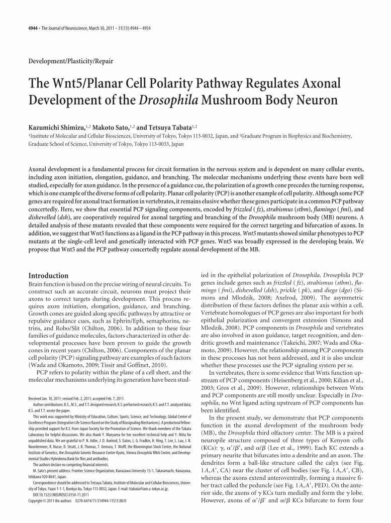
In the present study, we demonstrate that PCP componentsfunction in the axonal development of the mushroom body(MB), the Drosophila third olfactory center. The MB is a pairedneuropile structure composed of three types of Kenyon cells(KCs): �, ��/��, and �/� (Lee et al., 1999). Each KC extends aprimary neurite that bifurcates into a dendrite and an axon. Thedendrites form a ball-like structure called the calyx (see Fig.1A,A�, CA) near the cluster of cell bodies (see Fig. 1A,A�, CB),whereas the axons extend anteroventrally, forming a massive fi-ber tract called the peduncle (see Fig. 1A,A�, PED). On the ante-rior side, the axons of � KCs turn medially and form the � lobe.However, axons of ��/�� and �/� KCs bifurcate to form four
Received Jan. 10, 2011; revised Feb. 2, 2011; accepted Feb. 7, 2011.Author contributions: K.S., M.S., and T.T. designed research; K.S. performed research; K.S. and T.T. analyzed data;
K.S. and T.T. wrote the paper.This work was supported by Ministry of Education, Culture, Sports, Science, and Technology, Global Center of
Excellence Program (Integrative Life Science Based on the Study of Biosignaling Mechanisms). A predoctoral fellow-ship provided support for K.S. from Japan Society for the Promotion of Science. We thank members of the TabataLaboratory for helpful discussions. We also thank Y. Maeyama for her excellent technical help and Y. Nitta forunpublished data. We are grateful to P. N. Adler, J. D. Axelrod, S. Eaton, L. G. Fradkin, H. Hing, T. Lee, L. Luo, J. N.Noordermeer, R. Nusse, D. Strutt, J. B. Thomas, T. Uemura, T. Wolff, the Bloomington Stock Center, the NationalInstitute of Genetics, the Drosophila Genetic Resource Center Kyoto, Vienna Drosophila RNAi Center, and Develop-mental Studies Hybridoma Bank for flies and antibodies.
The authors declare no competing financial interests.M. Sato’s present address: Frontier Science Organization, Kanazawa University 13-1, Takaramachi, Kanazawa,
Ishikawa 920-8641, Japan.Correspondence should be addressed to Tetsuya Tabata, Institute of Molecular and Cellular Biosciences, Univer-
sity of Tokyo, Yayoi 1-1-1, Bunkyo-ku, Tokyo 113-0032, Japan. E-mail: [email protected]:10.1523/JNEUROSCI.0154-11.2011
Copyright © 2011 the authors 0270-6474/11/314944-11$15.00/0
4944 • The Journal of Neuroscience, March 30, 2011 • 31(13):4944 – 4954
discrete lobes: the ��, ��, �, and � lobes (see Fig. 1A,A�). Wefound that fz, stbm, and dsh functioned cooperatively in axontargeting and branching of KCs. In addition, we show geneticinteractions between PCP components and Wnt5, suggesting thatWnt5 acts as a candidate ligand for PCP components in the reg-ulation of the axonal development of the MB.
Materials and MethodsFly strains. Wnt5D7 (Yoshikawa et al., 2003), Wnt5400 (Fradkin et al.,2004), fzH51 (Jones et al., 1996), fzD21, fzK21, stbmA3 (Taylor et al., 1998),stbmstbm-6 (Wolff and Rubin, 1998), fmiE59 (Usui et al., 1999), and dgo380
(Feiguin et al., 2001) are null alleles or amorphic alleles. dsh1 is a hypo-morphic allele of dsh. pk1 and pksple-1 are hypomorphic alleles that lackthe expression of the sple and pk isoforms, respectively. pkpk-sple-13 is a nullallele that lacks both the sple and the pk isoforms (Gubb et al., 1999). Thefollowing Gal-4 lines were used to drive gene expression in KCs: OK107-Gal4, NP7175-Gal4, c739-Gal4, and 7B-Gal4. The following transgenicstrains were used in this study: UAS-GFP, UAS-mCD8::GFP (Lee et al.,1999), UAS-fz, UAS-stbm (Bastock et al., 2003), UAS-dsh (Rulifsonet al., 1996), UAS-dsh�DEP (Axelrod, 2001), and UAS-Wnt5 (Fradkin etal., 2004). UAS-dsRNA lines ( fz, 105493; stbm, 7376; fmi, 107993; Wnt5,101621) and UAS-dicer2 lines (60008, 60009) were obtained from theVienna Drosophila RNAi Center stock center.
Clonal analysis. To generate single-cell clones in the MB, we used themosaic analysis with a repressible cell marker (MARCM) technique (Leeand Luo, 1999). Pupae were heat-shocked at 37°C for 15 min for theFRT19A clones and for 5 min for the FRTG13 clones. The genotypes usedwere as follows: yw/Y; FRTG13 UAS-mCD8::GFP/ FRTG13 tubP-Gal80;hsFLP/�; OK107-Gal4/�, Wnt5D7/Y; FRTG13 UAS-mCD8::GFP/FRTG13 tubP-Gal80; hsFLP/�; OK107-Gal4/�, dsh1/Y; FRTG13UAS-mCD8::GFP/ FRTG13 tubP-Gal80; hsFLP/�; OK107-Gal4/�,FRT19A tubP-Gal80 hsFLP w/ yw FRT19A; UAS-mCD8::GFP/�; OK107-Gal4/�, FRT19A tubP-Gal80 hsFLP w/ yw FRT19A; UAS-mCD8::GFP/�; fzH51ri FRT2A/ fzD21; OK107-Gal4/�, FRT19A tubP-Gal80 hsFLP w/yw FRT19A; stbmstbm-6/ stbmA3; UAS-mCD8::GFP /�; OK107-Gal4/�.To generate the Wnt5 mutant neuroblast clones in the MB, we usedWnt5400. First- and second-instar larvae were heat-shocked five times for1 h at 37°C. The genotypes used were as follows: Wnt5400 FRT19A/FRT19A tubP-Gal80 hsFLP w; UAS-mCD8::GFP/�; OK107-Gal4/�.
Immunohistochemistry. Immunohistochemistry was performed as de-scribed previously (Huang and Kunes, 1996; Takei et al., 2004), exceptfor the staining of the brains for Wnt5. The following primary antibodieswere provided by the Developmental Studies Hybridoma Bank: mousemonoclonal anti-Fasciclin II (1D4 anti-Fasciclin II; 1:50), rat monoclo-nal anti-N-cadherin (DN-EX no. 8; 1:10), and mouse monoclonal anti-Flamingo (Flamingo no. 74; 1:10). Rabbit anti-Fz (Bastock and Strutt,2007) and rabbit anti-Stbm (Rawls and Wolff, 2003) were used at dilu-tions of 1:300, and 1:500, respectively. Rabbit anti-green fluorescent pro-tein (GFP) Alexa Fluor 488 conjugate was used at a dilution of 1:2000(Invitrogen). The following secondary antibodies were used at a dilutionof 1:200: anti-mouse Cy3 (Jackson ImmunoResearch), anti-mouse FITC(Jackson ImmunoResearch), anti-rat Cy3 (Jackson ImmunoResearch),and anti-rabbit Alexa Fluor 660 (Invitrogen). Specimens were mountedwith Vectashield mounting medium (Vector Laboratories) and viewedon a Zeiss LSM 710, a Zeiss LSM 510, a Zeiss LSM 5 EXCITER, or anOlympus FV1000 confocal microscope.
Generation of the Wnt5 antibody. A rabbit anti-Wnt5 antibody wasraised against a previously described region and affinity purified (Frad-kin et al., 1995), except that a His-tagged immunogen was used instead ofa trpE fusion protein (Takara Bio).
Wnt5 staining of larval and pupal brains. Wnt5 staining was performedas previously described with slight modifications (Yao et al., 2007). Dis-sected brains were incubated with anti-Wnt5 (1:500) in PBS overnight at4°C, washed twice for 5 min at room temperature, and fixed with PLP (para-formaldehyde/lysine/periodate) fixative for 1 h at room temperature.
Classification of mutant phenotypes. To classify the mutant phenotypes,we measured the width of the � and the � lobes at the middle of theirlengths in a three-dimensional image reconstructed by Imaris software
(Bitplane). We classified the MB as “normal” if the width of both the �lobe and the � lobe was above the cutoff values. If the width of either the� lobe or the � lobe was below the cutoff value, the phenotype of the MBwas classified as “lobe reduction.” When either or both of the � and �lobes were completely absent, we classified the MB as “lobe loss.” TheMBs in which all of the �/� neurons aggregated beside the calyx wereclassified as “posterior aggregation.” The cutoff values were set by sub-tracting 3 � SD from the mean width of wild type (mean, 23.91 pixels,and SD, 1.99 pixels, for the � lobes of the Canton S females; mean, 22.70pixels, and SD, 2.50 pixels, for the � lobes of the Canton S females; mean,25.93 pixels, and SD, 2.19 pixels, for the � lobes of the Canton S males;and mean, 23.72 pixels, and SD, 2.63 pixels, for the � lobes of the CantonS males).
Comparison of the signals of Wnt5 and N-cadherin in the Wnt5 mutantclone. To eliminate the noise outside of the calyx, background signalswere subtracted from the images of the single channels for both Wnt5and N-cadherin using ImageJ software (National Institutes of Health,Bethesda, MD). The subtracted image for Wnt5 was divided by that forN-cadherin and visualized by the height of the peaks in 2.5-dimensionalimages. We could obtain a similar result without subtraction of the back-ground signals.
ResultsDrosophila PCP components are expressed in the MBTo analyze the function of the PCP signal in neural development,we searched for neural tissues expressing Fz, one of the core PCPsignaling components. Fz was expressed strongly in the core andweakly in the periphery along the entire tract of the lobes and thepeduncle of the MB throughout its development (Fig. 1E–G�).Because the younger axons of KCs run within the core of the MB,Fz was expected to be expressed strongly in the axons of youngerKCs and weakly in the axons of older KCs.
We also examined the expression patterns of other PCP com-ponents and found that Stbm, Fmi were specifically expressed inthe core of the MB (Fig. 1H–J�) (data not shown). The expressionof these components in the MB was abolished in the correspond-ing mutants, confirming the specificity of the antibodies (datanot shown). Therefore, the major PCP signaling componentsexamined were all expressed in the axons of younger KCs. Theseobservations suggest the possible involvement of the PCP signalin axonal development of the MB.
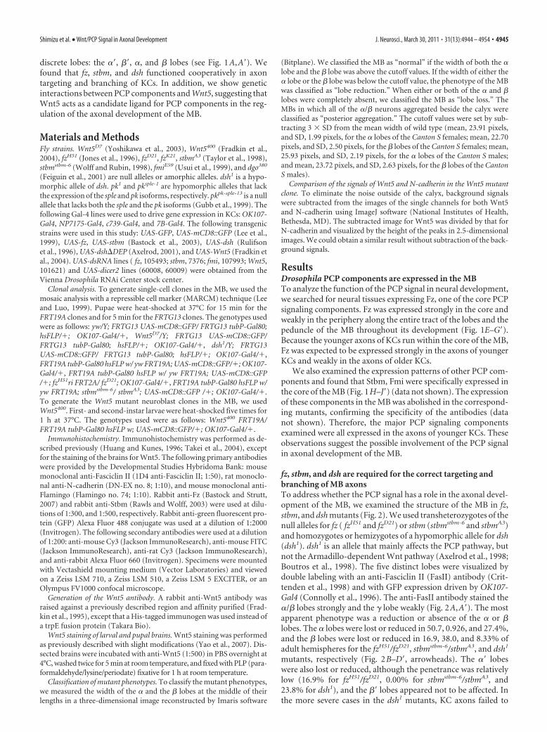
fz, stbm, and dsh are required for the correct targeting andbranching of MB axonsTo address whether the PCP signal has a role in the axonal devel-opment of the MB, we examined the structure of the MB in fz,stbm, and dsh mutants (Fig. 2). We used transheterozygotes of thenull alleles for fz ( fzH51 and fzD21) or stbm (stbmstbm-6 and stbmA3)and homozygotes or hemizygotes of a hypomorphic allele for dsh(dsh1). dsh1 is an allele that mainly affects the PCP pathway, butnot the Armadillo-dependent Wnt pathway (Axelrod et al., 1998;Boutros et al., 1998). The five distinct lobes were visualized bydouble labeling with an anti-Fasciclin II (FasII) antibody (Crit-tenden et al., 1998) and with GFP expression driven by OK107-Gal4 (Connolly et al., 1996). The anti-FasII antibody stained the�/� lobes strongly and the � lobe weakly (Fig. 2A,A�). The mostapparent phenotype was a reduction or absence of the � or �lobes. The � lobes were lost or reduced in 50.7, 0.926, and 27.4%,and the � lobes were lost or reduced in 16.9, 38.0, and 8.33% ofadult hemispheres for the fzH51/fzD21, stbmstbm-6/stbmA3, and dsh1
mutants, respectively (Fig. 2B–D�, arrowheads). The �� lobeswere also lost or reduced, although the penetrance was relativelylow (16.9% for fzH51/fzD21, 0.00% for stbmstbm-6/stbmA3, and23.8% for dsh1), and the �� lobes appeared not to be affected. Inthe more severe cases in the dsh1 mutants, KC axons failed to
Shimizu et al. • Wnt/PCP Signal in Axonal Development J. Neurosci., March 30, 2011 • 31(13):4944 – 4954 • 4945
project anteriorly and formed a ball-likeaggregate beside the calyx and cell body(1.19%) (Fig. 2E,E�,G,G�, arrow). Be-cause an absence of the �� lobes was al-ways associated with a loss of the � lobes(n � 23 for fzH51/fzD21 and n � 19 fordsh1), for the following analyses, the mu-tant phenotypes were scored by examin-ing the structure of the anti-FasII-stained�/� lobes as a measure. We assumed thatthe phenotype of an absence of the � lobesinvolved the loss of the �� lobes. Because anull allele of fmi ( fmiE59) is a lethal allele,fmi was instead knocked down in the MBusing RNA interference (RNAi) withOK107-Gal4; UAS-fmi dsRNA, and de-fects similar to the fzH51/fzD21, stbmstbm-6/stbmA3, and dsh1 mutants were observed(data not shown). Mutants of anothercore PCP gene, pk, showed the above phe-notypes with a relatively low penetrance(2.78%, n � 36 for pk1; 17.6%, n � 34 forpksple-1; 23.8%, n � 42 for pkpk-sple-13) (datanot shown). dgo mutants showed no phe-notype (data not shown).
Because individual �/� and ��/�� neu-rons bifurcate dorsally and medially toform the �/� and ��/�� lobes, the pheno-types of the �, �, and �� neurons de-scribed above can be explained by defectsin branch formation or the mistargetingof the bifurcated sister branches towardthe same direction. To distinguish be-tween these two possibilities, we usedthe MARCM technique in which clonesof cells are generated by mitotic recom-bination and marked by the expressionof mCD8::GFP (Lee and Luo, 1999). Weinduced a MARCM clone in the wild typeor mutant background using FRT19Afor fzH51/fzD21 and stbmstbm-6/stbmA3 orFRTG13 for dsh1 (Fig. 2 H–O�). In thewild type, an individual �/� neuron ex-tended a single axon within the peduncleand bifurcated at the anterior end of thepeduncle to form the vertical and medialbranches (Fig. 2H,H�,M,M�). An over-branching at the branch point of the lobeswas occasionally observed (12.0%, n � 25
Figure 1. The expression patterns of the PCP components during the development of the MB. A–D, Structure of the MB in theadult (A, A�), at the late third larval instar (B, B�), 24 h APF (C, C�), and 48 h APF (D, D�). Anterior perspectives of whole MBs (A–D)and oblique perspectives of right MBs (A�–D�) are indicated. E–J�, Expression patterns of Fz (E–G, magenta; E�–G�, white) and
4
Stbm (H–J, magenta; H�–J�, white) at the third larval instar(E, E�, H, H�), 24 h APF (F, F�, I, I�), and 48 h APF (G, G�, J, J�).All of the PCP components examined were expressed in thecore of the MB during the entire period of MB development.The structures of the MBs were visualized by the expression ofGFP (green) driven by OK107-Gal4. The abbreviations are asfollows: CB, Cell bodies; CX, calyx; PED, peduncle; VL, verticallobe; ML, medial lobe. Because the lobes at the late third larvalinstar and 24 h APF do not have clearly discrete lobes that arecomposed of distinct subtypes of KCs, the lobes are simply in-dicated as the vertical lobes (VL) and the medial lobes (ML) inB–C�, E–F�, and H–I�.
4946 • J. Neurosci., March 30, 2011 • 31(13):4944 – 4954 Shimizu et al. • Wnt/PCP Signal in Axonal Development
Figure 2. The phenotypes of the PCP gene mutants. A–G�, The structure of the MB in wild type (A, A�, F, F�), fzH51/fzD21 (B, B�), stbmstbm-6/stbmA3 (C, C�), and dsh1/Y (D, D�, E, E�, G, G�) mutants.The vertical or medial lobes were lost or reduced in the PCP mutants (B–D�, arrowheads). Anterior (A–E�) and dorsal views with the posterior side up and the anterior side down (F–G�) are indicated.In dsh1 mutants, a loss or reduction of the lobes (D, D�, arrowheads) or an aggregation of axons beside the calyx (E, E�, G, G�, arrow) was observed. In the case of axon aggregation, only a small portionof the � neurons projected toward the anterior side (E, E�, G, G�, yellow arrowhead). The MB was visualized by the expression of GFP driven by OK107-Gal4 (green) and the � and �/� lobes werevisualized by anti-FasII antibody (magenta). H–O�, The axonal projections of �/� KCs marked by GFP (H–O, O�, green; H�–O�, O�, white) in wild type (FRT 19A control clone in H, H�; FRT G13control clone in M, M�), fzH51/fzD21 mutants (I–J�), stbmstbm-6/stbmA3 mutants (K–L�), and dsh1 mutants (N–O�). The axon bifurcated into both the vertical and the medial lobes in the wild type (H, H�, M, M�). Theprojection of sister branches in the same direction (I, I�, arrowheads), the stalling of the branched axon (K, K�, arrowhead), and the reversed projection of the branched axon into (Figure legend continues.)
Shimizu et al. • Wnt/PCP Signal in Axonal Development J. Neurosci., March 30, 2011 • 31(13):4944 – 4954 • 4947
for the FRT19A control clone; 3.23%, n �31 for the FRTG13 control clone). How-ever, in the fzH51/fzD21, stbmstbm-6/stbmA3,and dsh1 mutants, guidance defects en-compassing the projection of sisterbranches toward the same direction andthe stalling of the bifurcated branch alongthe lobe or the peduncle were observed(guidance defects) (Fig. 2 I, I�, 37.2%, n �43 for fzH51/fzD21; K,K�, 20.5%, n � 39 forstbmstbm-6/stbmA3; N,N�, 15.5%, n � 58for dsh1). We also noticed defects inbranch formation, including an ectopicbranch at the peduncle, an overbranchingat the branch point of the lobes and nobranch at the branch point of the lobes(branching defects) (Fig. 2 J, J�, 25.6%,n � 43 for fzH51/fzD21; L,L�, 43.6%, n � 39for stbmstbm-6/stbmA3; O–O�, 32.8%, n �58 for dsh1). All of the clones extendedaxons beyond the branch point of thelobes regardless of the branching pheno-type. These results suggest that fz, stbm,and dsh are required for the correctbranching and targeting of the axons ofKCs, but not for the extension of axons.
Next, we examined genetic interac-tions among PCP genes to addresswhether these genes function in a cooper-ative manner. To assess the mutant phe-notypes, we measured the width of � and� lobes in three-dimensional recon-structed images and classified the MBsinto four classes: posterior aggregation,lobe loss, lobe reduction, and normal.Double heterozygotes of any two of fzH51,stbmA3, and dsh1 showed loss or reductionof the �/� lobes (Fig. 2P, stbm A3/�; fzH51/�, value of p is 6.4 �10�5; Q, dsh1/�; fzH51/�, value of p is 0.0067 using Fisher’s exacttest) with the phenotype of dsh1/�; stbmA3/� being much lessprominent (Fig. 2R, value of p is 0.38), although single heterozy-gotes of these genes showed no or little defects in the MB. Theseresults suggest that the PCP genes cooperatively function in theaxonal development of the MB.
fz, stbm, and dsh function in the KCs during axonaldevelopment of the MBBased on their expression patterns, the PCP components wereexpected to function in the KCs during axonal development ofthe MB. To confirm this, we attempted to rescue mutants of thePCP components using c739-Gal4, NP7175-Gal4, or 7B-Gal4, allof which are expressed in the developing MB. The lobe loss, lobereduction, or posterior aggregation of the �/� lobes in fzH51/fzD21
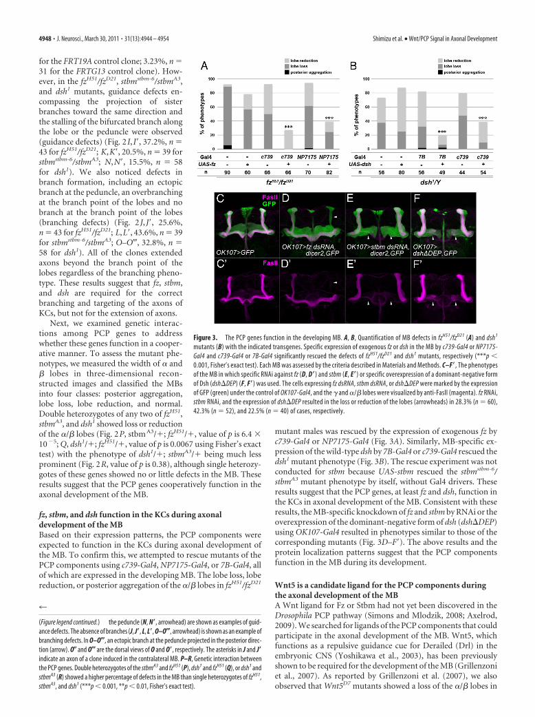
mutant males was rescued by the expression of exogenous fz byc739-Gal4 or NP7175-Gal4 (Fig. 3A). Similarly, MB-specific ex-pression of the wild-type dsh by 7B-Gal4 or c739-Gal4 rescued thedsh1 mutant phenotype (Fig. 3B). The rescue experiment was notconducted for stbm because UAS-stbm rescued the stbmstbm-6/stbmA3 mutant phenotype by itself, without Gal4 drivers. Theseresults suggest that the PCP genes, at least fz and dsh, function inthe KCs in axonal development of the MB. Consistent with theseresults, the MB-specific knockdown of fz and stbm by RNAi or theoverexpression of the dominant-negative form of dsh (dsh�DEP)using OK107-Gal4 resulted in phenotypes similar to those of thecorresponding mutants (Fig. 3D–F�). The above results and theprotein localization patterns suggest that the PCP componentsfunction in the MB during its development.
Wnt5 is a candidate ligand for the PCP components duringthe axonal development of the MBA Wnt ligand for Fz or Stbm had not yet been discovered in theDrosophila PCP pathway (Simons and Mlodzik, 2008; Axelrod,2009). We searched for ligands of the PCP components that couldparticipate in the axonal development of the MB. Wnt5, whichfunctions as a repulsive guidance cue for Derailed (Drl) in theembryonic CNS (Yoshikawa et al., 2003), has been previouslyshown to be required for the development of the MB (Grillenzoniet al., 2007). As reported by Grillenzoni et al. (2007), we alsoobserved that Wnt5D7 mutants showed a loss of the �/� lobes in
4
(Figure legend continued.) the peduncle (N, N�, arrowhead) are shown as examples of guid-ance defects. The absence of branches (J, J�, L, L�, O–O�, arrowhead) is shown as an example ofbranching defects. In O–O�, an ectopic branch at the peduncle projected in the posterior direc-tion (arrow). O� and O� are the dorsal views of O and O�, respectively. The asterisks in J and J�indicate an axon of a clone induced in the contralateral MB. P–R, Genetic interaction betweenthe PCP genes. Double heterozygotes of the stbmA3 and fzH51 (P), dsh1 and fzH51 (Q), or dsh1 andstbmA3 (R) showed a higher percentage of defects in the MB than single heterozygotes of fzH51,stbmA3, and dsh1 (***p � 0.001, **p � 0.01, Fisher’s exact test).
Figure 3. The PCP genes function in the developing MB. A, B, Quantification of MB defects in fzH51/fzD21 (A) and dsh1
mutants (B) with the indicated transgenes. Specific expression of exogenous fz or dsh in the MB by c739-Gal4 or NP7175-Gal4 and c739-Gal4 or 7B-Gal4 significantly rescued the defects of fzH51/fzD21 and dsh1 mutants, respectively (***p �0.001, Fisher’s exact test). Each MB was assessed by the criteria described in Materials and Methods. C–F�, The phenotypesof the MB in which specific RNAi against fz (D, D�) and stbm (E, E�) or specific overexpression of a dominant-negative formof Dsh (dsh�DEP) (F, F�) was used. The cells expressing fz dsRNA, stbm dsRNA, or dsh�DEP were marked by the expressionof GFP (green) under the control of OK107-Gal4, and the � and �/� lobes were visualized by anti-FasII (magenta). fz RNAi,stbm RNAi, and the expression of dsh�DEP resulted in the loss or reduction of the lobes (arrowheads) in 28.3% (n � 60),42.3% (n � 52), and 22.5% (n � 40) of cases, respectively.
4948 • J. Neurosci., March 30, 2011 • 31(13):4944 – 4954 Shimizu et al. • Wnt/PCP Signal in Axonal Development
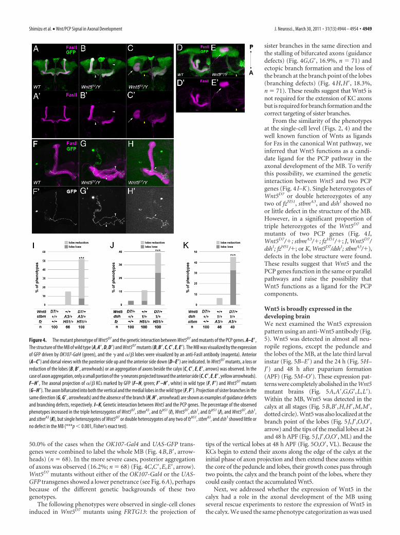
50.0% of the cases when the OK107-Gal4 and UAS-GFP trans-genes were combined to label the whole MB (Fig. 4B,B�, arrow-heads) (n � 68). In the more severe cases, posterior aggregationof axons was observed (16.2%; n � 68) (Fig. 4C,C�,E,E�, arrow).Wnt5D7 mutants without either of the OK107-Gal4 or the UAS-GFP transgenes showed a lower penetrance (see Fig. 6A), perhapsbecause of the different genetic backgrounds of these twogenotypes.
The following phenotypes were observed in single-cell clonesinduced in Wnt5D7 mutants using FRTG13: the projection of
sister branches in the same direction andthe stalling of bifurcated axons (guidancedefects) (Fig. 4G,G�, 16.9%, n � 71) andectopic branch formation and the loss ofthe branch at the branch point of the lobes(branching defects) (Fig. 4H,H�, 18.3%,n � 71). These results suggest that Wnt5 isnot required for the extension of KC axonsbut is required for branch formation and thecorrect targeting of sister branches.
From the similarity of the phenotypesat the single-cell level (Figs. 2, 4) and thewell known function of Wnts as ligandsfor Fzs in the canonical Wnt pathway, weinferred that Wnt5 functions as a candi-date ligand for the PCP pathway in theaxonal development of the MB. To verifythis possibility, we examined the geneticinteraction between Wnt5 and two PCPgenes (Fig. 4 I–K). Single heterozygotes ofWnt5D7 or double heterozygotes of anytwo of fzH51, stbmA3, and dsh1 showed noor little defect in the structure of the MB.However, in a significant proportion oftriple heterozygotes of the Wnt5D7 andmutants of two PCP genes (Fig. 4 I,Wnt5D7/�; stbmA3/�; fzH51/�; J, Wnt5D7/dsh1; fzH51/�; or K, Wnt5D7/dsh1; stbmA3/�),defects in the lobe structure were found.These results suggest that Wnt5 and thePCP genes function in the same or parallelpathways and raise the possibility thatWnt5 functions as a ligand for the PCPcomponents.
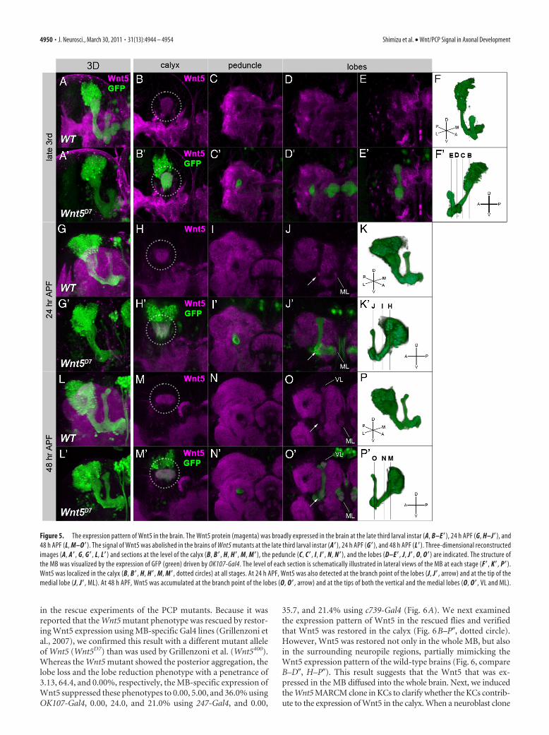
Wnt5 is broadly expressed in thedeveloping brainWe next examined the Wnt5 expressionpattern using an anti-Wnt5 antibody (Fig.5). Wnt5 was detected in almost all neu-ropile regions, except the peduncle andthe lobes of the MB, at the late third larvalinstar (Fig. 5B–E�) and the 24 h (Fig. 5H–J�) and 48 h after puparium formation(APF) (Fig. 5M–O�). These expression pat-terns were completely abolished in the Wnt5mutant brains (Fig. 5A,A�,G,G�,L,L�).Within the MB, Wnt5 was detected in thecalyx at all stages (Fig. 5B,B�,H,H�,M,M�,dotted circle). Wnt5 was also localized at thebranch point of the lobes (Fig. 5J,J�,O,O�,arrow) and the tips of the medial lobes at 24and 48 h APF (Fig. 5J,J�,O,O�, ML) and the
tips of the vertical lobes at 48 h APF (Fig. 5O,O�, VL). Because theKCs begin to extend their axons along the edge of the calyx at theinitial phase of axon projection and then extend these axons withinthe core of the peduncle and lobes, their growth cones pass throughtwo points, the calyx and the branch point of the lobes, where theycould easily contact the accumulated Wnt5.
Next, we addressed whether the expression of Wnt5 in thecalyx had a role in the axonal development of the MB usingseveral rescue experiments to restore the expression of Wnt5 inthe calyx. We used the same phenotype categorization as was used
Figure 4. The mutant phenotype of Wnt5D7 and the genetic interaction between Wnt5D7 and mutants of the PCP genes. A–E�,The structure of the MB of wild type (A, A�, D, D�) and Wnt5D7 mutants (B, B�, C, C�, E, E�). The MB was visualized by the expressionof GFP driven by OK107-Gal4 (green), and the � and �/� lobes were visualized by an anti-FasII antibody (magenta). Anterior(A–C�) and dorsal views with the posterior side up and the anterior side down (D–E�) are indicated. In Wnt5D7 mutants, a loss orreduction of the lobes (B, B�, arrowheads) or an aggregation of axons beside the calyx (C, C�, E, E�, arrows) was observed. In thecase of axon aggregation, only a small portion of the � neurons projected toward the anterior side (C, C�, E, E�, yellow arrowheads).F–H�, The axonal projection of �/� KCs marked by GFP (F–H, green; F�–H�, white) in wild type (F, F�) and Wnt5D7 mutants(G–H�). The axon bifurcated into both the vertical and the medial lobes in the wild type (F, F�). Projection of sister branches in thesame direction (G, G�, arrowheads) and the absence of the branch (H, H�, arrowhead) are shown as examples of guidance defectsand branching defects, respectively. I–K, Genetic interaction between Wnt5 and the PCP genes. The percentage of the observedphenotypes increased in the triple heterozygotes of Wnt5D7, stbmA3, and fzH51 (I), Wnt5D7, dsh1, and fzH51 (J), and Wnt5D7, dsh1,and stbmA3 (K), but single heterozygotes of Wnt5D7 or double heterozygotes of any two of fzH51, stbmA3, and dsh1 showed little orno defect in the MB (***p � 0.001, Fisher’s exact test).
Shimizu et al. • Wnt/PCP Signal in Axonal Development J. Neurosci., March 30, 2011 • 31(13):4944 – 4954 • 4949
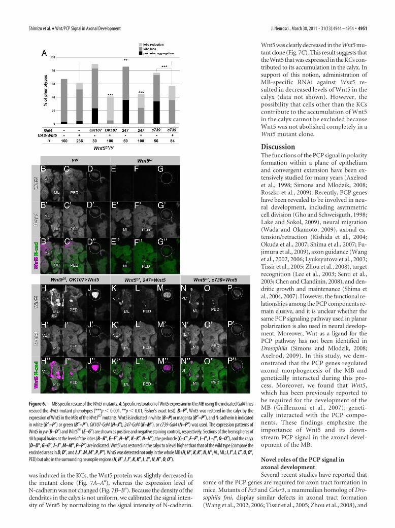
in the rescue experiments of the PCP mutants. Because it wasreported that the Wnt5 mutant phenotype was rescued by restor-ing Wnt5 expression using MB-specific Gal4 lines (Grillenzoni etal., 2007), we confirmed this result with a different mutant alleleof Wnt5 (Wnt5D7) than was used by Grillenzoni et al. (Wnt5400).Whereas the Wnt5 mutant showed the posterior aggregation, thelobe loss and the lobe reduction phenotype with a penetrance of3.13, 64.4, and 0.00%, respectively, the MB-specific expression ofWnt5 suppressed these phenotypes to 0.00, 5.00, and 36.0% usingOK107-Gal4, 0.00, 24.0, and 21.0% using 247-Gal4, and 0.00,
35.7, and 21.4% using c739-Gal4 (Fig. 6A). We next examinedthe expression pattern of Wnt5 in the rescued flies and verifiedthat Wnt5 was restored in the calyx (Fig. 6B–P, dotted circle).However, Wnt5 was restored not only in the whole MB, but alsoin the surrounding neuropile regions, partially mimicking theWnt5 expression pattern of the wild-type brains (Fig. 6, compareB–D, H–P). This result suggests that the Wnt5 that was ex-pressed in the MB diffused into the whole brain. Next, we inducedthe Wnt5 MARCM clone in KCs to clarify whether the KCs contrib-ute to the expression of Wnt5 in the calyx. When a neuroblast clone
Figure 5. The expression pattern of Wnt5 in the brain. The Wnt5 protein (magenta) was broadly expressed in the brain at the late third larval instar (A, B–E�), 24 h APF (G, H–J�), and48 h APF (L, M–O�). The signal of Wnt5 was abolished in the brains of Wnt5 mutants at the late third larval instar (A�), 24 h APF (G�), and 48 h APF (L�). Three-dimensional reconstructedimages (A, A�, G, G�, L, L�) and sections at the level of the calyx (B, B�, H, H�, M, M�), the peduncle (C, C�, I, I�, N, N�), and the lobes (D–E�, J, J�, O, O�) are indicated. The structure ofthe MB was visualized by the expression of GFP (green) driven by OK107-Gal4. The level of each section is schematically illustrated in lateral views of the MB at each stage (F�, K�, P�).Wnt5 was localized in the calyx (B, B�, H, H�, M, M�, dotted circles) at all stages. At 24 h APF, Wnt5 was also detected at the branch point of the lobes (J, J�, arrow) and at the tip of themedial lobe (J, J�, ML). At 48 h APF, Wnt5 was accumulated at the branch point of the lobes (O, O�, arrow) and at the tips of both the vertical and the medial lobes (O, O�, VL and ML).
4950 • J. Neurosci., March 30, 2011 • 31(13):4944 – 4954 Shimizu et al. • Wnt/PCP Signal in Axonal Development
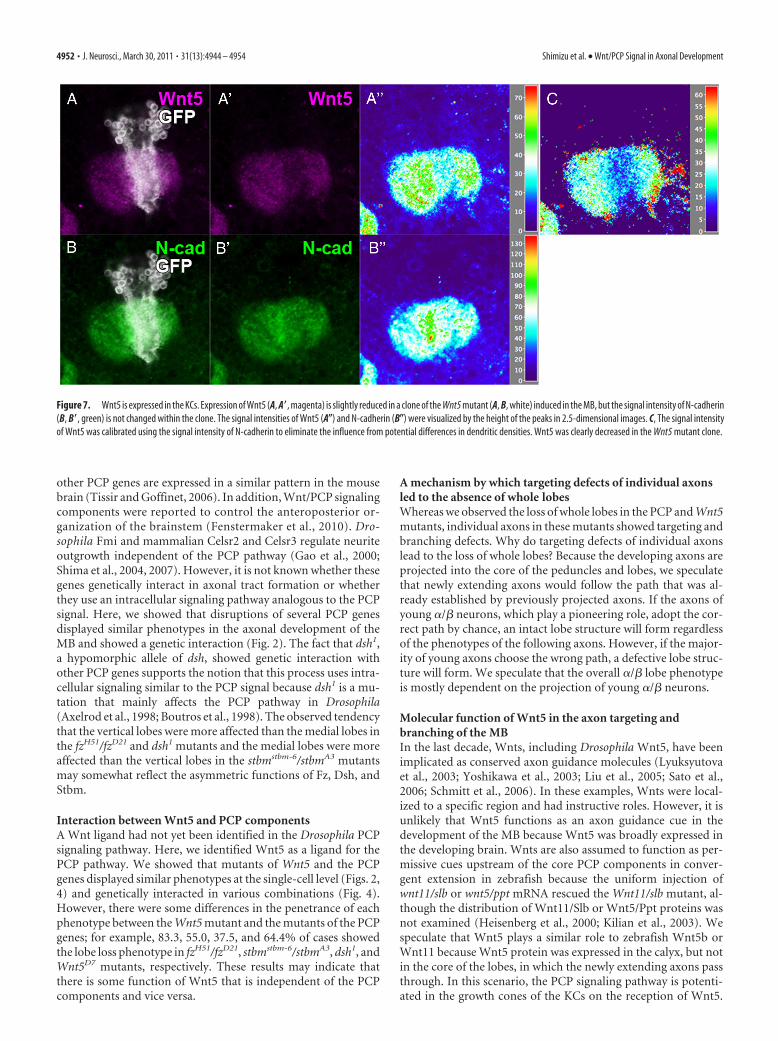
was induced in the KCs, the Wnt5 protein was slightly decreased inthe mutant clone (Fig. 7A–A), whereas the expression level ofN-cadherin was not changed (Fig. 7B–B). Because the density of thedendrites in the calyx is not uniform, we calibrated the signal inten-sity of Wnt5 by normalizing to the signal intensity of N-cadherin.
Wnt5 was clearly decreased in the Wnt5 mu-tant clone (Fig. 7C). This result suggests thatthe Wnt5 that was expressed in the KCs con-tributed to its accumulation in the calyx. Insupport of this notion, administration ofMB-specific RNAi against Wnt5 re-sulted in decreased levels of Wnt5 in thecalyx (data not shown). However, thepossibility that cells other than the KCscontribute to the accumulation of Wnt5in the calyx cannot be excluded becauseWnt5 was not abolished completely in aWnt5 mutant clone.
DiscussionThe functions of the PCP signal in polarityformation within a plane of epitheliumand convergent extension have been ex-tensively studied for many years (Axelrodet al., 1998; Simons and Mlodzik, 2008;Roszko et al., 2009). Recently, PCP geneshave been revealed to be involved in neu-ral development, including asymmetriccell division (Gho and Schweisguth, 1998;Lake and Sokol, 2009), neural migration(Wada and Okamoto, 2009), axonal ex-tension/retraction (Kishida et al., 2004;Okuda et al., 2007; Shima et al., 2007; Fu-jimura et al., 2009), axon guidance (Wanget al., 2002, 2006; Lyuksyutova et al., 2003;Tissir et al., 2005; Zhou et al., 2008), targetrecognition (Lee et al., 2003; Senti et al.,2003; Chen and Clandinin, 2008), and den-dritic growth and maintenance (Shima etal., 2004, 2007). However, the functional re-lationships among the PCP components re-main elusive, and it is unclear whether thesame PCP signaling pathway used in planarpolarization is also used in neural develop-ment. Moreover, Wnt as a ligand for thePCP pathway has not been identified inDrosophila (Simons and Mlodzik, 2008;Axelrod, 2009). In this study, we dem-onstrated that the PCP genes regulatedaxonal morphogenesis of the MB andgenetically interacted during this pro-cess. Moreover, we found that Wnt5,which has been previously reported tobe required for the development of theMB (Grillenzoni et al., 2007), geneti-cally interacted with the PCP compo-nents. These findings emphasize theimportance of Wnt5 and its down-stream PCP signal in the axonal devel-opment of the MB.
Novel roles of the PCP signal inaxonal developmentSeveral recent studies have reported that
some of the PCP genes are required for axon tract formation inmice. Mutants of Fz3 and Celsr3, a mammalian homolog of Dro-sophila fmi, display similar defects in axonal tract formation(Wang et al., 2002, 2006; Tissir et al., 2005; Zhou et al., 2008), and
Figure 6. MB specific rescue of the Wnt5 mutants. A, Specific restoration of Wnt5 expression in the MB using the indicated Gal4 linesrescued the Wnt5 mutant phenotypes (***p � 0.001, **p � 0.01, Fisher’s exact test). B–P�, Wnt5 was restored in the calyx by theexpression of Wnt5 in the MBs of the Wnt5D7 mutants. Wnt5 is indicated in white (B–P) or magenta (B�–P�), and N-cadherin is indicatedin white (B�–P�) or green (B�–P�). OK107-Gal4 (H–J�), 247-Gal4 (K–M�), or c739-Gal4 (N–P�) was used. The expression patterns ofWnt5 in yw (B–D�) and Wnt5D7 (E–G�) are shown as positive and negative staining controls, respectively. Sections of the hemispheres of48 h pupal brains at the level of the lobes (B–B�, E–E�, H–H�, K–K�, N–N�), the peduncle (C–C�, F–F�, I–I�, L–L�, O–O�), and the calyx(D–D�, G–G�, J–J�, M–M�, P–P�) are indicated. Wnt5 was restored in the calyx to a level higher than that of the wild type (compare theencircled areas in D, D�, and J, J�, M, M�, P, P�). Wnt5 was detected not only in the whole MB (H, H�, K, K�, N, N�, VL, ML; I, I�, L, L�, O, O�,PED) but also in the surrounding neuropile regions (H, H�, I, I�, K, K�, L, L�, N, N�, O, O�).
Shimizu et al. • Wnt/PCP Signal in Axonal Development J. Neurosci., March 30, 2011 • 31(13):4944 – 4954 • 4951
other PCP genes are expressed in a similar pattern in the mousebrain (Tissir and Goffinet, 2006). In addition, Wnt/PCP signalingcomponents were reported to control the anteroposterior or-ganization of the brainstem (Fenstermaker et al., 2010). Dro-sophila Fmi and mammalian Celsr2 and Celsr3 regulate neuriteoutgrowth independent of the PCP pathway (Gao et al., 2000;Shima et al., 2004, 2007). However, it is not known whether thesegenes genetically interact in axonal tract formation or whetherthey use an intracellular signaling pathway analogous to the PCPsignal. Here, we showed that disruptions of several PCP genesdisplayed similar phenotypes in the axonal development of theMB and showed a genetic interaction (Fig. 2). The fact that dsh1,a hypomorphic allele of dsh, showed genetic interaction withother PCP genes supports the notion that this process uses intra-cellular signaling similar to the PCP signal because dsh1 is a mu-tation that mainly affects the PCP pathway in Drosophila(Axelrod et al., 1998; Boutros et al., 1998). The observed tendencythat the vertical lobes were more affected than the medial lobes inthe fzH51/fzD21 and dsh1 mutants and the medial lobes were moreaffected than the vertical lobes in the stbmstbm-6/stbmA3 mutantsmay somewhat reflect the asymmetric functions of Fz, Dsh, andStbm.
Interaction between Wnt5 and PCP componentsA Wnt ligand had not yet been identified in the Drosophila PCPsignaling pathway. Here, we identified Wnt5 as a ligand for thePCP pathway. We showed that mutants of Wnt5 and the PCPgenes displayed similar phenotypes at the single-cell level (Figs. 2,4) and genetically interacted in various combinations (Fig. 4).However, there were some differences in the penetrance of eachphenotype between the Wnt5 mutant and the mutants of the PCPgenes; for example, 83.3, 55.0, 37.5, and 64.4% of cases showedthe lobe loss phenotype in fzH51/fzD21, stbmstbm-6/stbmA3, dsh1, andWnt5D7 mutants, respectively. These results may indicate thatthere is some function of Wnt5 that is independent of the PCPcomponents and vice versa.
A mechanism by which targeting defects of individual axonsled to the absence of whole lobesWhereas we observed the loss of whole lobes in the PCP and Wnt5mutants, individual axons in these mutants showed targeting andbranching defects. Why do targeting defects of individual axonslead to the loss of whole lobes? Because the developing axons areprojected into the core of the peduncles and lobes, we speculatethat newly extending axons would follow the path that was al-ready established by previously projected axons. If the axons ofyoung �/� neurons, which play a pioneering role, adopt the cor-rect path by chance, an intact lobe structure will form regardlessof the phenotypes of the following axons. However, if the major-ity of young axons choose the wrong path, a defective lobe struc-ture will form. We speculate that the overall �/� lobe phenotypeis mostly dependent on the projection of young �/� neurons.
Molecular function of Wnt5 in the axon targeting andbranching of the MBIn the last decade, Wnts, including Drosophila Wnt5, have beenimplicated as conserved axon guidance molecules (Lyuksyutovaet al., 2003; Yoshikawa et al., 2003; Liu et al., 2005; Sato et al.,2006; Schmitt et al., 2006). In these examples, Wnts were local-ized to a specific region and had instructive roles. However, it isunlikely that Wnt5 functions as an axon guidance cue in thedevelopment of the MB because Wnt5 was broadly expressed inthe developing brain. Wnts are also assumed to function as per-missive cues upstream of the core PCP components in conver-gent extension in zebrafish because the uniform injection ofwnt11/slb or wnt5/ppt mRNA rescued the Wnt11/slb mutant, al-though the distribution of Wnt11/Slb or Wnt5/Ppt proteins wasnot examined (Heisenberg et al., 2000; Kilian et al., 2003). Wespeculate that Wnt5 plays a similar role to zebrafish Wnt5b orWnt11 because Wnt5 protein was expressed in the calyx, but notin the core of the lobes, in which the newly extending axons passthrough. In this scenario, the PCP signaling pathway is potenti-ated in the growth cones of the KCs on the reception of Wnt5.
Figure 7. Wnt5 is expressed in the KCs. Expression of Wnt5 (A, A�, magenta) is slightly reduced in a clone of the Wnt5 mutant (A, B, white) induced in the MB, but the signal intensity of N-cadherin(B, B�, green) is not changed within the clone. The signal intensities of Wnt5 (A�) and N-cadherin (B�) were visualized by the height of the peaks in 2.5-dimensional images. C, The signal intensityof Wnt5 was calibrated using the signal intensity of N-cadherin to eliminate the influence from potential differences in dendritic densities. Wnt5 was clearly decreased in the Wnt5 mutant clone.
4952 • J. Neurosci., March 30, 2011 • 31(13):4944 – 4954 Shimizu et al. • Wnt/PCP Signal in Axonal Development
Wnt5 is expressed from the dendrites of the older KCs and regu-lates the axonal development of the younger KCs. During theinitial phase of projection, the growth cones of the KCs receiveWnt5 when they pass through the calyx, and the PCP pathway,potentiated by the Wnt5, regulates axonal projection andbranching. This may be the underlying cause of the aggregationof axons beside the calyx and the loss of the lobes.
Possible functions of the PCP signal in axonal targeting andbranching of KCsBased on the analysis of the PCP genes at the single-cell level,these genes are required for axonal targeting and branching. Dur-ing pathfinding, the growth cones at the tips of primary axonsmust detect small differences in the concentration gradients ofattractive and repulsive cues because the width of the KC growthcone is very small (�1 �m) (data not shown). Axonal branchingcan also be considered a polarized cellular process, and thus itencounters problems that are similar to that found in the axonalguiding process. To overcome this difficulty, signals from theexternal environment need to be amplified within the growthcone. The most prominent molecular feature of the PCP compo-nents, the ability to generate polarity within a cell that is gener-ated by reciprocal negative feedback between the Stbm and Fzcomplexes, may regulate this amplification process. In thismodel, ligand binding to its receptor activates the PCP signal.PCP components change their distribution from a symmetric toan asymmetric manner within a growth cone and form a receptorcomplex with the guidance receptor, which leads to the asymmet-ric distribution of the guidance receptor with a higher concentra-tion at the side closer to the source of the guidance cue, forexample. A small difference in the concentration of a guidancecue in the external environment is converted to a large differencein signal strength within a growth cone via the PCP machinery,although we could not detect any alteration in the distribution ofFz, Stbm, or Fmi in Wnt5 mutants at the immunohistochemicallevel. One candidate molecule that links axon guidance receptorsand the PCP components is PTK7, which forms a complex withPlexin-A1, genetically interacts with Vangl2, and recruits Dsh tothe plasma membrane (Lu et al., 2004; Toyofuku et al., 2004;Shnitsar and Borchers, 2008). Although off-track (otk), the Dro-sophila homolog of PTK7, is not reported to function in the PCPformation, OTK associates with Plexin-A to form a receptorcomplex downstream of Semaphorin 1a during axon guidance(Winberg et al., 2001). In addition, RNAi knockdown of otkshowed a loss or reduction of the �/� lobes (our unpublishedobservations).
Additional study is needed to reveal the relationship betweenWnt5 and the PCP signaling pathway in axonal development andthe molecular entity that links axon guidance and the PCP signal.
ReferencesAxelrod JD (2001) Unipolar membrane association of Dishevelled mediates
Frizzled planar cell polarity signaling. Genes Dev 15:1182–1187.Axelrod JD (2009) Progress and challenges in understanding planar cell po-
larity signaling. Semin Cell Dev Biol 20:964 –971.Axelrod JD, Miller JR, Shulman JM, Moon RT, Perrimon N (1998) Differ-
ential recruitment of Dishevelled provides signaling specificity in the pla-nar cell polarity and Wingless signaling pathways. Genes Dev12:2610 –2622.
Bastock R, Strutt D (2007) The planar polarity pathway promotes coordi-nated cell migration during Drosophila oogenesis. Development134:3055–3064.
Bastock R, Strutt H, Strutt D (2003) Strabismus is asymmetrically localisedand binds to Prickle and Dishevelled during Drosophila planar polaritypatterning. Development 130:3007–3014.
Boutros M, Paricio N, Strutt DI, Mlodzik M (1998) Dishevelled activatesJNK and discriminates between JNK pathways in planar polarity andwingless signaling. Cell 94:109 –118.
Chen PL, Clandinin TR (2008) The cadherin Flamingo mediates level-dependent interactions that guide photoreceptor target choice inDrosophila. Neuron 58:26 –33.
Chilton JK (2006) Molecular mechanisms of axon guidance. Dev Biol292:13–24.
Connolly JB, Roberts IJ, Armstrong JD, Kaiser K, Forte M, Tully T, O’Kane CJ(1996) Associative learning disrupted by impaired G(s) signaling inDrosophila mushroom bodies. Science 274:2104 –2107.
Crittenden JR, Skoulakis EM, Han KA, Kalderon D, Davis RL (1998) Tri-partite mushroom body architecture revealed by antigenic markers. LearnMem 5:38 –51.
Feiguin F, Hannus M, Mlodzik M, Eaton S (2001) The ankyrin repeat pro-tein Diego mediates Frizzled-dependent planar polarization. Dev Cell1:93–101.
Fenstermaker AG, Prasad AA, Bechara A, Adolfs Y, Tissir F, Goffinet A, ZouY, Pasterkamp RJ (2010) Wnt/planar cell polarity signaling controls theanterior–posterior organization of monoaminergic axons in the brains-tem. J Neurosci 30:16053–16064.
Fradkin LG, Noordermeer JN, Nusse R (1995) The Drosophila Wnt proteinDWnt-3 is a secreted glycoprotein localized on the axon tracts of theembryonic CNS. Dev Biol 168:202–213.
Fradkin LG, van Schie M, Wouda RR, de Jong A, Kamphorst JT, Radjkoemar-Bansraj M, Noordermeer JN (2004) The Drosophila Wnt5 protein me-diates selective axon fasciculation in the embryonic central nervoussystem. Dev Biol 272:362–375.
Fujimura L, Watanabe-Takano H, Sato Y, Tokuhisa T, Hatano M (2009)Prickle promotes neurite outgrowth via the Dishevelled dependent path-way in C1300 cells. Neurosci Lett 467:6 –10.
Gao FB, Kohwi M, Brenman JE, Jan LY, Jan YN (2000) Control of dendriticfield formation in Drosophila: the roles of flamingo and competition be-tween homologous neurons. Neuron 28:91–101.
Gho M, Schweisguth F (1998) Frizzled signalling controls orientation ofasymmetric sense organ precursor cell divisions in Drosophila. Nature393:178 –181.
Grillenzoni N, Flandre A, Lasbleiz C, Dura JM (2007) Respective roles of theDRL receptor and its ligand WNT5 in Drosophila mushroom body devel-opment. Development 134:3089 –3097.
Gros J, Serralbo O, Marcelle C (2009) WNT11 acts as a directional cue toorganize the elongation of early muscle fibres. Nature 457:589 –593.
Gubb D, Green C, Huen D, Coulson D, Johnson G, Tree D, Collier S, Roote J(1999) The balance between isoforms of the prickle LIM domain proteinis critical for planar polarity in Drosophila imaginal discs. Genes Dev13:2315–2327.
Heisenberg CP, Tada M, Rauch GJ, Saude L, Concha ML, Geisler R, Stemple DL,Smith JC, Wilson SW (2000) Silberblick/Wnt11 mediates convergent ex-tension movements during zebrafish gastrulation. Nature 405:76–81.
Huang Z, Kunes S (1996) Hedgehog, transmitted along retinal axons, trig-gers neurogenesis in the developing visual centers of the Drosophila brain.Cell 86:411– 422.
Jones KH, Liu J, Adler PN (1996) Molecular analysis of EMS-induced friz-zled mutations in Drosophila melanogaster. Genetics 142:205–215.
Kilian B, Mansukoski H, Barbosa FC, Ulrich F, Tada M, Heisenberg CP(2003) The role of Ppt/Wnt5 in regulating cell shape and movementduring zebrafish gastrulation. Mech Dev 120:467– 476.
Kishida S, Yamamoto H, Kikuchi A (2004) Wnt-3a and Dvl induce neuriteretraction by activating Rho-associated kinase. Mol Cell Biol24:4487– 4501.
Komiyama T, Sweeney LB, Schuldiner O, Garcia KC, Luo L (2007) Gradedexpression of semaphorin-1a cell-autonomously directs dendritic target-ing of olfactory projection neurons. Cell 128:399 – 410.
Lake BB, Sokol SY (2009) Strabismus regulates asymmetric cell divisionsand cell fate determination in the mouse brain. J Cell Biol 185:59 – 66.
Lee RC, Clandinin TR, Lee CH, Chen PL, Meinertzhagen IA, Zipursky SL(2003) The protocadherin Flamingo is required for axon target selectionin the Drosophila visual system. Nat Neurosci 6:557–563.
Lee T, Luo L (1999) Mosaic analysis with a repressible cell marker for studiesof gene function in neuronal morphogenesis. Neuron 22:451– 461.
Lee T, Lee A, Luo L (1999) Development of the Drosophila mushroom bod-
Shimizu et al. • Wnt/PCP Signal in Axonal Development J. Neurosci., March 30, 2011 • 31(13):4944 – 4954 • 4953
ies: sequential generation of three distinct types of neurons from a neu-roblast. Development 126:4065– 4076.
Liu Y, Shi J, Lu CC, Wang ZB, Lyuksyutova AI, Song X, Zou Y (2005) Ryk-mediated Wnt repulsion regulates posterior-directed growth of cortico-spinal tract. Nat Neurosci 8:1151–1159.
Lu X, Borchers AG, Jolicoeur C, Rayburn H, Baker JC, Tessier-Lavigne M(2004) PTK7/CCK-4 is a novel regulator of planar cell polarity in verte-brates. Nature 430:93–98.
Lyuksyutova AI, Lu CC, Milanesio N, King LA, Guo N, Wang Y, Nathans J,Tessier-Lavigne M, Zou Y (2003) Anterior-posterior guidance of com-missural axons by Wnt-frizzled signaling. Science 302:1984 –1988.
Okuda H, Miyata S, Mori Y, Tohyama M (2007) Mouse Prickle1 andPrickle2 are expressed in postmitotic neurons and promote neurite out-growth. FEBS Lett 581:4754 – 4760.
Rawls AS, Wolff T (2003) Strabismus requires Flamingo and Prickle func-tion to regulate tissue polarity in the Drosophila eye. Development130:1877–1887.
Roszko I, Sawada A, Solnica-Krezel L (2009) Regulation of convergence andextension movements during vertebrate gastrulation by the Wnt/PCPpathway. Semin Cell Dev Biol 20:986 –997.
Rulifson EJ, Micchelli CA, Axelrod JD, Perrimon N, Blair SS (1996) winglessrefines its own expression domain on the Drosophila wing margin. Nature384:72–74.
Sato M, Umetsu D, Murakami S, Yasugi T, Tabata T (2006) DWnt4 regu-lates the dorsoventral specificity of retinal projections in the Drosophilamelanogaster visual system. Nat Neurosci 9:67–75.
Schmitt AM, Shi J, Wolf AM, Lu CC, King LA, Zou Y (2006) Wnt-Ryk signal-ling mediates medial-lateral retinotectal topographic mapping. Nature439:31–37.
Senti KA, Usui T, Boucke K, Greber U, Uemura T, Dickson BJ (2003) Fla-mingo regulates R8 axon-axon and axon-target interactions in the Dro-sophila visual system. Curr Biol 13:828 – 832.
Shima Y, Kengaku M, Hirano T, Takeichi M, Uemura T (2004) Regulationof dendritic maintenance and growth by a mammalian 7-pass transmem-brane cadherin. Dev Cell 7:205–216.
Shima Y, Kawaguchi SY, Kosaka K, Nakayama M, Hoshino M, Nabeshima Y,Hirano T, Uemura T (2007) Opposing roles in neurite growth controlby two seven-pass transmembrane cadherins. Nat Neurosci 10:963–969.
Shnitsar I, Borchers A (2008) PTK7 recruits dsh to regulate neural crestmigration. Development 135:4015– 4024.
Simons M, Mlodzik M (2008) Planar cell polarity signaling: from fly devel-opment to human disease. Annu Rev Genet 42:517–540.
Takei Y, Ozawa Y, Sato M, Watanabe A, Tabata T (2004) Three DrosophilaEXT genes shape morphogen gradients through synthesis of heparan sul-fate proteoglycans. Development 131:73– 82.
Takeichi M (2007) The cadherin superfamily in neuronal connections andinteractions. Nat Rev Neurosci 8:11–20.
Taylor J, Abramova N, Charlton J, Adler PN (1998) Van Gogh: a new Dro-sophila tissue polarity gene. Genetics 150:199 –210.
Tissir F, Goffinet AM (2006) Expression of planar cell polarity genes duringdevelopment of the mouse CNS. Eur J Neurosci 23:597– 607.
Tissir F, Goffinet AM (2010) Planar cell polarity signaling in neural devel-opment. Curr Opin Neurobiol 20:572–577.
Tissir F, Bar I, Jossin Y, De Backer O, Goffinet AM (2005) ProtocadherinCelsr3 is crucial in axonal tract development. Nat Neurosci 8:451– 457.
Toyofuku T, Zhang H, Kumanogoh A, Takegahara N, Suto F, Kamei J, AokiK, Yabuki M, Hori M, Fujisawa H, Kikutani H (2004) Dual roles ofSema6D in cardiac morphogenesis through region-specific association ofits receptor, Plexin-A1, with off-track and vascular endothelial growthfactor receptor type 2. Genes Dev 18:435– 447.
Usui T, Shima Y, Shimada Y, Hirano S, Burgess RW, Schwarz TL, Takeichi M,Uemura T (1999) Flamingo, a seven-pass transmembrane cadherin, regu-lates planar cell polarity under the control of Frizzled. Cell 98:585–595.
Wada H, Okamoto H (2009) Roles of planar cell polarity pathway genes forneural migration and differentiation. Dev Growth Differ 51:233–240.
Wang Y, Thekdi N, Smallwood PM, Macke JP, Nathans J (2002) Frizzled-3is required for the development of major fiber tracts in the rostral CNS.J Neurosci 22:8563– 8573.
Wang Y, Zhang J, Mori S, Nathans J (2006) Axonal growth and guidancedefects in Frizzled3 knock-out mice: a comparison of diffusion tensormagnetic resonance imaging, neurofilament staining, and genetically di-rected cell labeling. J Neurosci 26:355–364.
Winberg ML, Tamagnone L, Bai J, Comoglio PM, Montell D, Goodman CS(2001) The transmembrane protein Off-track associates with Plexins andfunctions downstream of Semaphorin signaling during axon guidance.Neuron 32:53– 62.
Wolff T, Rubin GM (1998) Strabismus, a novel gene that regulates tissue polar-ity and cell fate decisions in Drosophila. Development 125:1149–1159.
Yao Y, Wu Y, Yin C, Ozawa R, Aigaki T, Wouda RR, Noordermeer JN,Fradkin LG, Hing H (2007) Antagonistic roles of Wnt5 and the Drlreceptor in patterning the Drosophila antennal lobe. Nat Neurosci10:1423–1432.
Yoshikawa S, McKinnon RD, Kokel M, Thomas JB (2003) Wnt-mediatedaxon guidance via the Drosophila Derailed receptor. Nature 422:583–588.
Zhou L, Bar I, Achouri Y, Campbell K, De Backer O, Hebert JM, Jones K,Kessaris N, de Rouvroit CL, O’Leary D, Richardson WD, Goffinet AM,Tissir F (2008) Early forebrain wiring: genetic dissection using condi-tional Celsr3 mutant mice. Science 320:946 –949.
4954 • J. Neurosci., March 30, 2011 • 31(13):4944 – 4954 Shimizu et al. • Wnt/PCP Signal in Axonal Development