Embed Size (px)
Citation preview
IHS Division of Diabetes Treatment & Prevention Foot Care Training Course Notes January 2010; Revised March 2011
Indian Health Service
Division of Diabetes Treatment and Prevention
Diabetes Foot Care Training
January 2010
Indian Health Service
Division of Diabetes Treatment and Prevention 5300 Homestead Road NE Albuquerque, NM 87110
www.diabetes.ihs.gov
IHS Division of Diabetes Treatment & Prevention Foot Care Training Course Notes January 2010; Revised March 2011
Table of Contents Introduction .......................................................................................................................... 3
Objectives ......................................................................................................................... 3
Screening for High Risk Patients: Overview ....................................................................... 4
Screening for High Risk Patients: Sensation ....................................................................... 5
Screening for High Risk Patients: Foot Deformities ........................................................... 6
Screening for High Risk Patients: Vascular ......................................................................... 7
Screening for High Risk Patients: Complete Foot Exam..................................................... 8
Interventions: Patient Education .......................................................................................... 9
Interventions: Foot Wear .................................................................................................... 10
Interventions: Podiatry Care .............................................................................................. 12
Interventions: Wound Care: Uncomplicated...................................................................... 13
Steps to debride an uncomplicated wound ..................................................................... 14
Interventions: Wound Care: Complicated.......................................................................... 16
Summary of Diabetic Foot Care ..................................................................................... 17
Implementation: Clinical .................................................................................................... 18
Implementation: Community ............................................................................................. 19
Resources and Summary .................................................................................................... 20
IHS Division of Diabetes Treatment & Prevention Page 3 Foot Care Training Course Notes January 2010; Revised March 2011
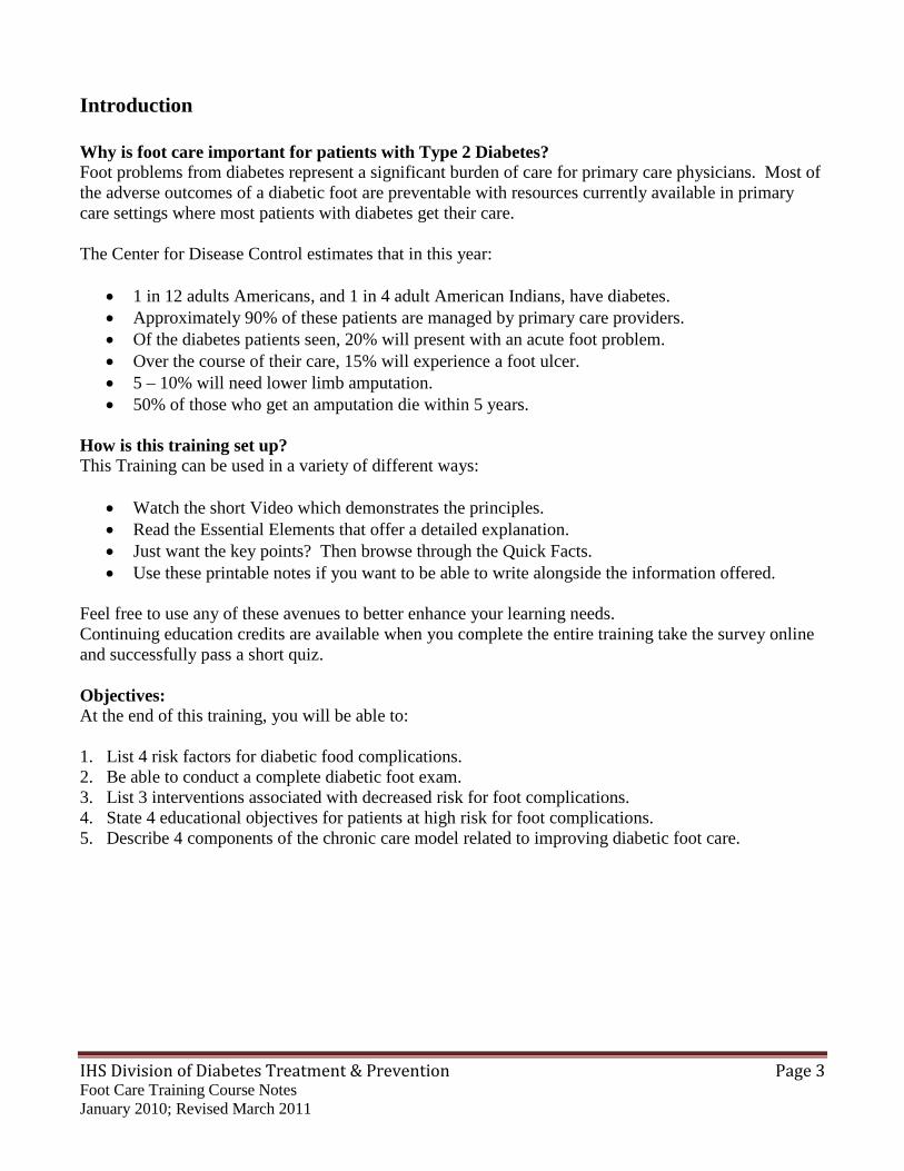
Introduction Why is foot care important for patients with Type 2 Diabetes? Foot problems from diabetes represent a significant burden of care for primary care physicians. Most of the adverse outcomes of a diabetic foot are preventable with resources currently available in primary care settings where most patients with diabetes get their care. The Center for Disease Control estimates that in this year:
• 1 in 12 adults Americans, and 1 in 4 adult American Indians, have diabetes. • Approximately 90% of these patients are managed by primary care providers. • Of the diabetes patients seen, 20% will present with an acute foot problem. • Over the course of their care, 15% will experience a foot ulcer. • 5 – 10% will need lower limb amputation. • 50% of those who get an amputation die within 5 years.
How is this training set up? This Training can be used in a variety of different ways:
• Watch the short Video which demonstrates the principles. • Read the Essential Elements that offer a detailed explanation. • Just want the key points? Then browse through the Quick Facts. • Use these printable notes if you want to be able to write alongside the information offered.
Feel free to use any of these avenues to better enhance your learning needs. Continuing education credits are available when you complete the entire training take the survey online and successfully pass a short quiz. Objectives: At the end of this training, you will be able to: 1. List 4 risk factors for diabetic food complications. 2. Be able to conduct a complete diabetic foot exam. 3. List 3 interventions associated with decreased risk for foot complications. 4. State 4 educational objectives for patients at high risk for foot complications. 5. Describe 4 components of the chronic care model related to improving diabetic foot care.
IHS Division of Diabetes Treatment & Prevention Page 4 Foot Care Training Course Notes January 2010; Revised March 2011
Screening for High Risk Patients: Overview Foot ulceration and amputation are preventable with resources currently available in primary care settings where most patients with diabetes get their care. There are several principle risk factors for ulceration and lower extremity amputation (LEA) among patients with diabetes:
• Neuropathy • Deformity • Limited joint mobility • Prior ulcer/ LEA • PVD • Onychomycosis
It is also important to assess non-foot related risk factors; some of them are potentially modifiable:
• Male sex • Duration of diabetes • Age • Hyperglycemia • Hypertension • Dyslipidemia • Smoking • Poor vision • Other complications such as renal disease
Patients at high risk for foot ulcer and amputation can be identified with simple criteria that involve several testing and inspection measures. Patients with all normal criteria are at low-risk, while those with insensitivity, deformity, absent pulses, or prior foot ulcers or amputations are at high-risk. These simple criteria have been validated in Indian Health Services (IHS) and adopted by most professional and public health organizations including the American Diabetes Association (ADA) and World Health Organization (WHO).
• Sensory testing with a 10 gram monofilament • Confirmation testing with a 128Hz tuning fork if sensate to monofilament • Foot inspection for deformity • Reports of prior ulcer or amputation • Checking for pedal pulses or taking measurements for an ABI pressure
IHS Division of Diabetes Treatment & Prevention Page 5 Foot Care Training Course Notes January 2010; Revised March 2011
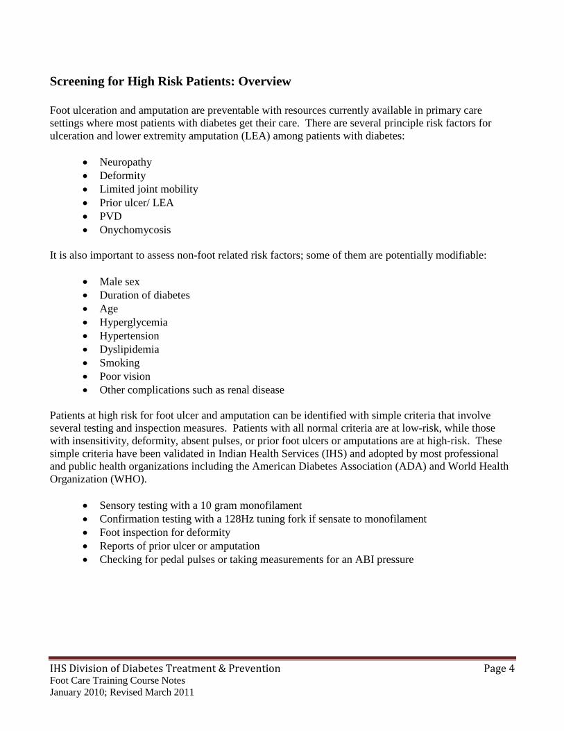
Screening for High Risk Patients: Sensation Monofilament and Vibration exams are two of the tests used to help identify high risk feet in people with diabetes. The monofilament exam involves using a 10 gram monofilament to test sensation on the great toe and 1st, 3rd and 5th metatarsal heads of each foot. The technique for monofilament testing is as follows:
• Press the 10 gram Semmes-Weinstein Monofilament (SWM) perpendicular to the skin to the point of bending. Hold for one second and then release.
• It is best to demonstrate the technique on your hand and then the patient’s hand first to decrease patient apprehension and to provide a reference point for the patient on their feet.
• When testing the feet, have the patient close their eyes and acknowledge sensation of pressure with a “yes” response.
• Test sites include the great toe and 1st, 3rd and 5th metatarsal heads of each foot. • An abnormal test results when the patient cannot feel any sensation on any of the monofilament
test sites. If results from the monofilament exam are normal, confirmation testing of vibratory sensation testing is recommended with a 128 Hz tuning fork. Approximately 10 – 15% of people who can feel the monofilament are still at high risk for developing foot problems. Accordingly, some experts recommend performing a sensory examination other than the monofilament. If a patient has a normal monofilament exam, the Indian Health Service recommends using a tuning fork.
• Using a 128Hz (C-note) tuning fork, tap the open end of the fork with the ball of your palm. • Holding the tip like a pencil, apply the tip of the fork to the great toe on the tip or first joint. • Have the patient notify you when they feel the vibration stops. An abnormal response occurs
when the patient loses vibratory sensation and the examiner still perceives it while holding the fork on the tip of either toe.
128 Hz tuning fork and monofilament
IHS Division of Diabetes Treatment & Prevention Page 6 Foot Care Training Course Notes January 2010; Revised March 2011
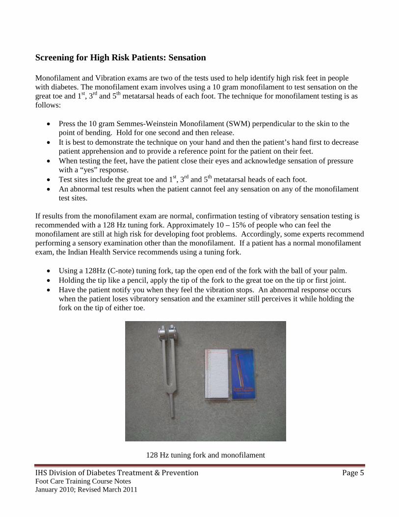
Screening for High Risk Patients: Foot Deformities To understand foot deformities, it is important to recognize that foot structure and function is incredibly complex and can easily go awry. Normal foot architecture is maintained through a balance of forces applied by muscles and tendons on bones. Atrophy of a muscle group through nerve damage can lead to deformity. It is important to watch areas that can be open to friction and repetitive micro-trauma as those sites can lead to callus and ulceration. Types of foot deformities include:
• The bunion, or hallux valgus, increases the risk for ulceration through a mechanism of increased pressures and friction and repetitive micro-trauma that out paces the healing capacity at the bunion site.
• Hammer and claw toe deformities develop from atrophy of the small muscles between the toes. Both the dorsal and plantar aspects of the involved toes are at risk for friction and pressure related trauma with subsequent ulceration.
• Charcot foot is one of the most severe diabetic foot deformity in which the entire mid-section of the foot collapses and forms a classic “rocker bottom” sole. It is caused by a combination of sensory and autonomic nerve dysfunction in which microscopic fractures to the tarsal bones trigger an inflammatory response and subsequent dissolution of the tarsal and metatarsal bones of the foot. The arch becomes inverted, which causes high plantar pressures, and is at extreme risk for ulceration.
IHS Division of Diabetes Treatment & Prevention Page 7 Foot Care Training Course Notes January 2010; Revised March 2011
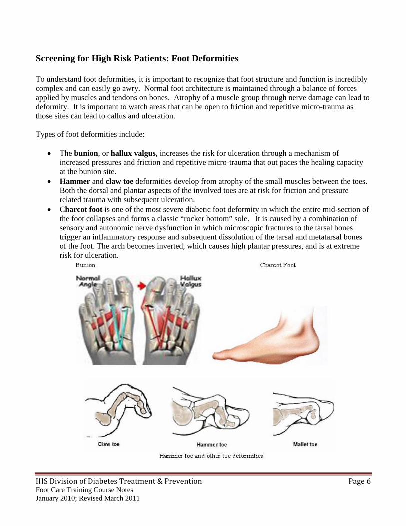
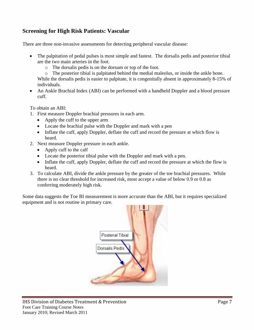
Screening for High Risk Patients: Vascular There are three non-invasive assessments for detecting peripheral vascular disease:
• The palpitation of pedal pulses is most simple and fastest. The dorsalis pedis and posterior tibial are the two main arteries in the foot.
o The dorsalis pedis is on the dorsum or top of the foot. o The posterior tibial is palpitated behind the medial maleolus, or inside the ankle bone.
While the dorsalis pedis is easier to palpitate, it is congenitally absent in approximately 8-15% of individuals.
• An Ankle Brachial Index (ABI) can be performed with a handheld Doppler and a blood pressure cuff.
To obtain an ABI: 1. First measure Doppler brachial pressures in each arm.
• Apply the cuff to the upper arm • Locate the brachial pulse with the Doppler and mark with a pen • Inflate the cuff, apply Doppler, deflate the cuff and record the pressure at which flow is
heard. 2. Next measure Doppler pressure in each ankle.
• Apply cuff to the calf • Locate the posterior tibial pulse with the Doppler and mark with a pen. • Inflate the cuff, apply Doppler, deflate the cuff and record the pressure at which the flow is
heard. 3. To calculate ABI, divide the ankle pressure by the greater of the toe brachial pressures. While
there is no clear threshold for increased risk, most accept a value of below 0.9 or 0.8 as conferring moderately high risk.
Some data suggests the Toe BI measurement is more accurate than the ABI, but it requires specialized equipment and is not routine in primary care.
IHS Division of Diabetes Treatment & Prevention Page 8 Foot Care Training Course Notes January 2010; Revised March 2011
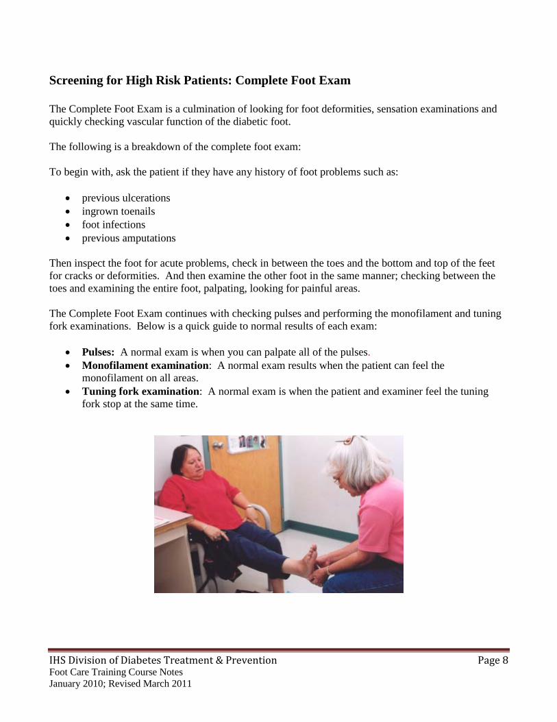
Screening for High Risk Patients: Complete Foot Exam The Complete Foot Exam is a culmination of looking for foot deformities, sensation examinations and quickly checking vascular function of the diabetic foot. The following is a breakdown of the complete foot exam: To begin with, ask the patient if they have any history of foot problems such as:
• previous ulcerations • ingrown toenails • foot infections • previous amputations
Then inspect the foot for acute problems, check in between the toes and the bottom and top of the feet for cracks or deformities. And then examine the other foot in the same manner; checking between the toes and examining the entire foot, palpating, looking for painful areas. The Complete Foot Exam continues with checking pulses and performing the monofilament and tuning fork examinations. Below is a quick guide to normal results of each exam:
• Pulses: A normal exam is when you can palpate all of the pulses. • Monofilament examination: A normal exam results when the patient can feel the
monofilament on all areas. • Tuning fork examination: A normal exam is when the patient and examiner feel the tuning
fork stop at the same time.
IHS Division of Diabetes Treatment & Prevention Page 9 Foot Care Training Course Notes January 2010; Revised March 2011
Interventions: Patient Education In the diabetic foot, ulcer and amputation result from a combination of risk factors coming together over time resulting in sequences called “causal pathways.” The most common pathway to ulceration is: Deformity or neuropathy minor trauma Poor circulation and infection gangrene and amputation Hence, patient education that supports the patient to take an active role in their foot care has been associated with improved outcomes in a wide variety of settings. Protecting the high-risk foot from injury is a primary objective in the high-risk patient. High-risk patients need education in:
• foot inspection, • nail care, • footwear selection and utilization, • environmental modifications to prevent minor foot trauma, • knowing when and whom to call for problems.
For patients at low risk, the emphasis of patient education should be on preventing neuropathy and peripheral vascular disease though controlling glucose, lipids and blood pressure, as well as smoking cessation. Education materials are more likely to be effective if they have been pre-tested for comprehension of learner objectives in the community they are to be used, such as ones developed by IHS. These materials can be ordered from IHS diabetes program website (www.diabetes.ihs.gov).
IHS Division of Diabetes Treatment & Prevention Page 10 Foot Care Training Course Notes January 2010; Revised March 2011
Interventions: Foot Wear After education, appropriate footwear is the second leg of preventative therapy. It is effective in preventing amputations in high-risk individuals through:
• cushioning the foot, • reducing callus formation, and • preventing ulceration.
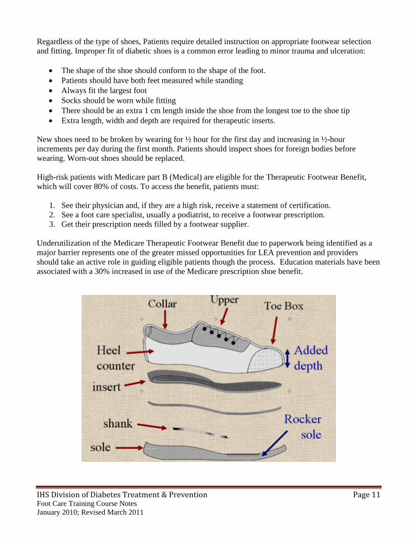
As a provider to patients with diabetes, it is important to know and understand the basic anatomy of a shoe:
• The collar provides ankle support • The heel counter is the rear and back side wall and provides lateral support • The upper is the top part of the shoe and may have laces or straps which affect the shoe fit. It
can also be known as a vamp. • The toe box is the protective covering for the toes. For a diabetic foot, the toe box can have ½-
1cm added depth to accommodate minor deformities. • A padded insert or liner is inside the shoe and can reduce slippage and friction. • The sole is the bottom of the shoe and provides traction. A rocker sole is beveled in the heel and
toe and can reduce pressure at the forefoot when walking. • Finally, a shank can be added under the arch of the foot to provide stability.
With this information, providers should instruct patients on selection and use of appropriate diabetic footwear.
• Patients with normal, low risk feet can wear standard shoes. • Patients with insensate feet should be encouraged to select the following:
o shoe with good padding and stability o adjustable uppers for a snug fit which reduce friction and slippage o firm heel counter to provide lateral stability o padded insert for cushioning o non-slip liner o broad sole with nominal lift. o rocker sole
Many of these features are common to commercial walking shoes. • Patients with minor deformities can usually be accommodated with a walking shoe (~$40 – 80)
or a therapeutic shoe with extra depth in the toe box region (~$100 – 200). • Patients with major deformities require custom made shoes built from the molding of the foot
(~$600 – 1200).
IHS Division of Diabetes Treatment & Prevention Page 11 Foot Care Training Course Notes January 2010; Revised March 2011
Regardless of the type of shoes, Patients require detailed instruction on appropriate footwear selection and fitting. Improper fit of diabetic shoes is a common error leading to minor trauma and ulceration:
• The shape of the shoe should conform to the shape of the foot. • Patients should have both feet measured while standing • Always fit the largest foot • Socks should be worn while fitting • There should be an extra 1 cm length inside the shoe from the longest toe to the shoe tip • Extra length, width and depth are required for therapeutic inserts.
New shoes need to be broken by wearing for ½ hour for the first day and increasing in ½-hour increments per day during the first month. Patients should inspect shoes for foreign bodies before wearing. Worn-out shoes should be replaced. High-risk patients with Medicare part B (Medical) are eligible for the Therapeutic Footwear Benefit, which will cover 80% of costs. To access the benefit, patients must:
1. See their physician and, if they are a high risk, receive a statement of certification. 2. See a foot care specialist, usually a podiatrist, to receive a footwear prescription. 3. Get their prescription needs filled by a footwear supplier.
Underutilization of the Medicare Therapeutic Footwear Benefit due to paperwork being identified as a major barrier represents one of the greater missed opportunities for LEA prevention and providers should take an active role in guiding eligible patients though the process. Education materials have been associated with a 30% increased in use of the Medicare prescription shoe benefit.
IHS Division of Diabetes Treatment & Prevention Page 12 Foot Care Training Course Notes January 2010; Revised March 2011
Interventions: Podiatry Care Routine podiatry care is associated with improved foot care outcomes including increased self-care knowledge, (30%) reduction in calluses, (54%) reduction in foot ulcers, and reduction in amputations (75%). There are three basic principle of routine podiatric care for patients with diabetes:
• Lubricate Skin • Trim nails • Reduce Callus
Patient with diabetes frequently have dry skin on their feet which can lead to increased friction and callus, cracking and infection. This is in part due to autonomic nerve dysfunction that controls sweat glands. Skin lubricants reduce friction, cracking, and risk for ulceration. Patients with dry skin should be instructed to apply a moisturizing lotion daily. Family members or other care givers can assist those for whom it is difficult to reach their feet. Both water and oil based lubricants are effective and is a matter of patient preference. There are several types of toenails to consider when trimming:
• Normal nails may only need straight or curved nail nippers and an emery board • For patients with curved or arched nails, you will need a straight nail nipper and an emery
board • Thick mycotic nails increase plantar pressures and can injure adjacent toes. Trimming can be
challenging because they can be both brittle and soft. A nail nipper and/or dremmel can be used to trim off sharp edges and debulk the nail, It is a best practice to refer such patients to a podiatrist or health professional skilled in nail care.
Calluses are formed by friction. They are common over foot deformities and exacerbated by poorly fitting shoes. Regular debridement of calluses has shown to reduce dynamic plantar pressures which in turn reduce risk for ulceration.

IHS Division of Diabetes Treatment & Prevention Page 13 Foot Care Training Course Notes January 2010; Revised March 2011
Interventions: Wound Care: Uncomplicated Despite the best prevention efforts, some patients progress to an ulcer. Basic principles of assessing and classifying wounds into those that can be safely managed by primary care and those that warrant referral to wound healing specialty services will be the focus. Assessment of the wound includes:
• Measuring the wound dimensions, length, diameter, depth in centimeters, and describe the shape. • Observe the wound margins for signs of epithealization and healing. • Measure the surrounding margin of errythema and cellulites. • Observe and probe for penetration to deep structures (fascia, tendon, bone, FB). • Look for lymphadentitis. • Look for the quality of blood flow.
Questions to answer include:
• Is the foot pink and warm? Or cool and dusky? • Are there obvious signs of gangrene? • Are pulses palpable or absent? • Is the capillary refill brisk or prolonged (> 2 seconds, or ABI < 0.4)? • Are there signs of a systemic infection? • Is the temperature > 100.5 F (39 C) or WBC > 12,000?
For the purpose of this training, a simplified classification will be used to determine if the wound is at high or low risk for healing with conventional outpatient management or whether referral to a specialist is indicated. Uncomplicated ulcers will be defined as:
• small in size (less than 2 cm in diameter). • superficial and lack deep space involvement. • having < 2cm margins of errythema and cellulites. • having adequate circulation. • having no systemic signs of infection. • likely to heal with conventional outpatient therapy.
Wounds with any of these findings greater than an uncomplicated wound would be characterized as complicated and warrant clinical consultation with a wound specialist and probable initial inpatient management. Wound healing is a delicate process of weaving a matrix of fibrin and elastin into which the epithelial cells at the margins can expand and take root. This process is promoted by a clean moist environment.
IHS Division of Diabetes Treatment & Prevention Page 14 Foot Care Training Course Notes January 2010; Revised March 2011
Steps to debride an uncomplicated wound
1. Wash foot with soap and water. Do not use Betadine as it is toxic to healing tissue. Anesthesia is usually not needed for this procedure.
2. The goal is to get the base of the ulcer clean to the point of bleeding and seepage by removing
necrotic tissue and fibrin exudates. This can be achieved by gently pulling the dry and necrotic tissue with a toothed forceps. Harder, more recalcitrant debris may need to be removed sharply with a #15 scalpel. Try to leave the new epithelial tissue at the margins of the ulcer undisturbed.
3. Next, the wound should be dressed to provide a clean moist environment to promote
epithelialization. Wound dressing is a delicate process and can be disturbed by too frequent of dressing changes and debridement. The balance is to provide judicious observation for infection and not disturb the healing process. There are some basic principles of dressing changes:
a. Wet to dry dressing is the mainstay. Cover the ulcer and foot with wet to dry saline dress
and wrap with Kerlix. b. Wounds with a lot of drainage or in which there is concern for infection may need to be
redressed daily. c. Occlusive dressings with less frequent changes are appropriate for wounds that have been
observed to be non-infected and progressing in a direction of healing. Monitor these wounds every 3 – 5 days.
d. Adsorbent compounds may be useful in “soupy” wounds. e. Hydrocolloid gels have a role in dry wounds. f. Enzymatic debridement can be useful in softening thick eschar.
4. Off loading the wound is crucial to the healing process. Off loading can be facilitated by a range
of adaptive footwear:
a. Half shoes offload the forefoot and heel b. Removable cast walkers with custom molded liners reduce pressure, but also carry a
higher price tag. c. Total contact casts, which could not be removed, are associated with the highest healing
rates. Comparable healing rates also occur if a removable cast is applied and then wrapped with a strand of fiberglass casting material to secure it to the foot.
d. At times, hospitalization or long-term care may be needed. Regardless of the method chosen, the wound should be assessed every 1 – 2 weeks. Ulcers can take up to several months to heal.
5. Monitor wound size and refer the patient to a wound specialist if the wound size is not reduced in size by 25% in 2 weeks or if it becomes complicated by extensive cellulitis, deep space infection, gangrene, or systemic infection. Wounds with evidence of infection should be cultured for aerobes and anaerobic bacteria using deep tissue fluids and tissue. Surface swab cultures are inaccurate and typically have highly resistant staph contaminants that can lead antibiotic therapy astray. Initial empiric therapy pending cultures should cover gram positives and gram negatives. Subsequent therapy should be directed by cultures.
IHS Division of Diabetes Treatment & Prevention Page 15 Foot Care Training Course Notes January 2010; Revised March 2011
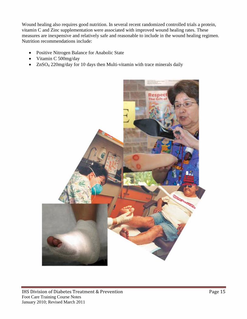
Wound healing also requires good nutrition. In several recent randomized controlled trials a protein, vitamin C and Zinc supplementation were associated with improved wound healing rates. These measures are inexpensive and relatively safe and reasonable to include in the wound healing regimen. Nutrition recommendations include:
• Positive Nitrogen Balance for Anabolic State • Vitamin C 500mg/day • ZnSO4 220mg/day for 10 days then Multi-vitamin with trace minerals daily
IHS Division of Diabetes Treatment & Prevention Page 16 Foot Care Training Course Notes January 2010; Revised March 2011
Interventions: Wound Care: Complicated Management of complicated wounds involves all of the basic principles covered on uncomplicated wounds plus getting consultation from a surgeon, vascular assessment and hospitalization for parental antibiotic therapy if infection is present. One of the most important principles in wound healing is patience. In one study fewer than 25% of wounds had healed at 12 weeks. There is a growing list of adjunctive wound healing agents that have been shown to accelerate wound healing rates by up to 50%. The common denominator is that these are selected patients with adequate circulation and no significant infections. In general, these therapies are costly. Adjunctive Wound Healing Therapies and approximate accelerated wound healing rates include:
• Growth factors (~15 – 25%) • Skin Graphs (~50%) • Hyperbaric Oxygen (~20%) • Electro-stimulation (~30%) • Maggot Therapy (~50%)
A rational approach to considering these adjunctive therapies is to start with conventional wet to dry dressing changes and first ensure that you have the basics of a clean wound, adequate circulation, blood glucose control and off loading. If the wound is healing at a rate of > 50% reduction in size in 4 weeks, then continue with conventional therapy. If not consider the use of these agents, recognizing that the resource could have greater impact if directed towards other interventions such as a case manager. In terms of vascular evaluation, the American Diabetes Association (ADA) recommends referral for surgical consultation if there is evidence of the following:
• Ulcer with clinical signs of ischemia • Non-healing ulcer • Rest pain • Nocturnal pain • Lifestyle limiting claudication

IHS Division of Diabetes Treatment & Prevention Page 17 Foot Care Training Course Notes January 2010; Revised March 2011
Summary of Diabetic Foot Care To summarize the evaluations and interventions of diabetic foot care:
1. Start by inspecting the feet for acute deformities on each visit. Even if there is no presence of an ulcer, it is important to ensure that the patient has had their annual diabetic foot exam of testing with the monofilament and assessing circulation, as 1 in 5 diabetic patients may present with a problem.
2. Gather patient history of ulceration and amputation. If no problems are detected in the history and complete foot exam, the patient is classified as low risk. Focus on self management education including blood glucose control, blood pressure control and smoking cessation. Follow up on this patient annually.
3. If the patient has an abnormal exam and/or history of ulceration and amputation, they are at high risk and emphasis should be on self management education, protective footwear, podiatry care, plus all measures used towards the low risk patient. High risk patients should be followed up every 2 to 3 months.
4. If an ulcer is detected at the visit, then conduct an assessment to determine if it is an uncomplicated or complicated ulcer.
5. Uncomplicated ulcers with small, superficial wounds not involving deep tissues, with limited infection and intact circulation can be managed as an outpatient with debridement, local wound care, off loading, oral antibiotics directed by culture if evidence of local infection, and weekly follow-up.
6. Patients with complex wounds should be hospitalized, have surgical consultation for debridement, vascular assessment, and initial parenteral antibiotics treatment directed by culture. Once on a healing trajectory, they can be managed as an uncomplicated ulcer.
IHS Division of Diabetes Treatment & Prevention Page 18 Foot Care Training Course Notes January 2010; Revised March 2011
Implementation: Clinical Formation of a foot care team, either as a separate team or as a sub-committee of the facility diabetes team or quality improvement team is an important first step to improving coordination of multi-disciplinary services. It is frequently difficult to get everyone in the room at one, so communication can be facilitated by telephone and email. Members include:
• clinic staff • consultants • community health • administrative leadership
Patient input can be valuable and can be obtained though clinic surveys and staff who are also patients. Teams can review the evidence relevant to their practice and develop a customized Clinical Proactive Guideline to their facility. Typical adaptations can include choice of footwear, dressing changes, mode of vascular assessment and patterns of referrals. The team also maps out implementing the guidelines into their practices including delineation of roles, documenting information, training needs, and measures for evaluation. Some clinics designate a foot care case manager. Below are two real life cases of foot managers in the Bemidji area:
• Charmaine Branchaud, RN was the foot care case management at Red Lake form 1995 – 2006. She received training in basic podiatry care by shadowing the local podiatrist for a year during his monthly outreach clinics to Red Lake. In addition to offering biweekly foot care clinic trimming nails and calluses, she coordinated a monthly wound clinic with a consultant vascular surgeon. Services were also coordinated with the local CHR’s who would bring patients into the wound care clinic and subsequently assist patients with dressing changes and follow-up care.
• In 2007, Emily Heinrich stepped in. Emily, an LPN, had shadowed Charmaine for several months to gain some skills and then took a 2 day course in Wisconsin. She works under Charmaine’s direct supervisions. RNs can become certified by the Wound, Ostomy and Continence Nurses Certification Board. The course is a two day program, one day of classroom learning and a second day of hands-on clinical training. This course does offer training for LPNs but only if the state board of nursing allows LPNs to function under the direct supervision of an RN. The link for more information on this training can be found under the Resources section on the webpage.”
• the foot care case manager position was taken over by Emily Heinrich, LPN. Emily received
her foot care training through a week long practicum in Eau Clair Wisconsin. Besides the Wound, Ostomy and Continence Nurses Certification Board exam (WOCNCB), training for health professionals is also available though the HRSA LEAP program and NHDC in Baton Rouge.
IHS Division of Diabetes Treatment & Prevention Page 19 Foot Care Training Course Notes January 2010; Revised March 2011
Administrative leadership support is often a requisite for carving out the time, clinic space and funding to support a local foot care program. Monitoring and evaluation that demonstrates improved patient care and outcome can provide compelling evidence to continue support for successful programs. Tracking patients with the DMS diabetes registry or iCare can facilitate identifying patients who are due for foot care services and scheduling them for follow up appointments.
Implementation: Community Outreach serves to provide access to pedorthic or foot wear services can improve access to foot wear and appropriate follow up. Outreach examples include:
• As a provider, look at the shoes that are in the stores and local community. Get an idea of what shoes meet the basic criteria for appropriate footwear and take note of the name brands, styles and prices so when you recommend shoes to your patient, you can give them specific examples, styles and price ranges.
• Meet with your local vendors that specialize in diabetic footwear and see if they are willing to come to your clinic on a certain day a week or a month to fit patients and dispense shoes.
Similarly, contact with regional wound healing consultants to provide on site out reach services which can be valuable to improve access as well as the consultant’s awareness of the patients' needs and support. The Chronic Care Model (CCM) is an organized, proactive, multi-component approach to healthcare delivery that utilizes community resources, policies and the organization of the health care system to create effective patient interactions and positive outcomes. Components of the Chronic Care Model include:
1. Self management support 2. Delivery system design 3. Decision support 4. Clinical Information systems 5. Leadership and Community Resources
The model recognizes that patients spend only a fraction of their time in the clinic and that it is critical to have links to community resources that support the patients in the settings where they live and work. With these components in place, studies have shown that the result is a prepared, proactive care team and an informed, activated patient, which translates into improved clinical outcomes for a wide-range of clinical conditions. The CCM can be adapted to diabetic foot care via the following approaches:
• Diabetes registry with foot risk status to track patients for needed care • A foot care team to screen patients for high risk foot conditions and provide risk appropriated
foot care education, podiatry care, and foot wear • Engaged and supportive clinic leadership • Links to community resources such as specialty outreach clinics
IHS Division of Diabetes Treatment & Prevention Page 20 Foot Care Training Course Notes January 2010; Revised March 2011
Resources and Summary In summary, topics covered in this training included:
1. How to screen and identify patients at risk for diabetic foot problems 2. Evidenced-based best practices associated with improved outcomes including self-management
education, the use of protective footwear, and routine podiatry care 3. Basic principles of wound management 4. How to integrate the principles into clinical practice
Most LEA can be prevented by the following methods:
• Screening high risk individuals and targeting them for patient education, podiatry care, and protective footwear.
• Creating diabetic foot screening forms into the charts that prompt referrals to appropriate providers.
• Develop a CPG, training a nurse in diabetic foot care, who can became the local Diabetes foot care case manager, and implement a patient tracking system, flow sheets, standing orders, and an evaluation system.
• Work with a vascular surgeon who can provide an outreach clinic in the community
Here are some Selected Internet Resources for Diabetic Foot Care • Indian Health Diabetes Best Practice Foot Care http://www.ihs.gov/medicalprograms/diabetes/index.cfm?module=toolsBPList A consensus-based approach, developed by Indian health system professionals, that anyone in clinical and community settings can use to implement or improve diabetes foot care. • Feet Can Last a Lifetime (Revised) www.niddk.nih.gov/health/diabetes/dateline/spri02/9.htm This updated kit is designed to provide health professionals with the tools and resources to prevent and treat diabetes foot problems among their patients. The revised kit includes foot care includes a quick-reference card for conducting a foot exam, a disposable monofilament for sensory testing, Medicare coverage information on therapeutic foot wear, patient education materials and reference and resource materials for health professionals. Kits available for $3 each. • Lower Extremity Amputation Prevention Program (LEAP) www.hrsa.gov/leap A comprehensive program that can dramatically reduce lower extremity amputations in individuals with diabetes mellitus.