Embed Size (px)
Citation preview

Thames Valley Diabetic Footcare Pathway, Version 1.63, November 2015 1
Thames Valley Strategic Clincal Network
Diabetes Footcare Pathway ‘from average to excellent’
Prepared by the TV SCN Diabetic Foot Reference Group
November 2015

Thames Valley Diabetic Footcare Pathway, Version 1.63, November 2015 2
Thames Valley Diabetes Footcare Pathway - Diabetic Foot Reference Group 2015 ‘Average to excellent….’ Purpose: Reduction of foot disease morbidity through promotion of foot education, screening and fast, effective triage of patients with active foot disease. This pathway provides a framework for the commissioning of Diabetic Foot services, in line with NICE guidance that can easily be customised for local use. The Diabetic Foot Reference Group will meet 6-monthly to review Diabetes foot care across Thames Valley, using parameters outlined here. Definitions used in the document: MDFT – Multidisciplinary Footcare Team FPS – Foot protection Service Resources:
1. Foot Reference Group meeting 9th September 2015, Reading 2. Foot Reference Group meeting, 3rd June 2015, High Wycombe 3. Foot Reference Group meeting, 4th March 2015, High Wycombe 4. NICE NG19 (published August 2015) replacing CG119, CG10, CG15 5. Putting Feet First ‘Fast track for a foot attack reducing amputations’, Diabetes UK campaign February 2013 6. Fixing footcare in Sheffield: Improving the pathway, Sheffield Teaching Hospitals NHS Foundation 7. Cheshire & Merseyside Strategic Clinical Networks Diabetes Footcare Service transformation work, 2014
Patient
Multidisciplinary Foot Care Team
GP
Podiatry Foot Protection Service
Practice Nurse
District Nurse Pharmacy
Community Tissue Viability Nurse
Employer
Family/Community

Thames Valley Diabetic Footcare Pathway, Version 1.63, November 2015 3
Contents
1. Pathways 4 2. Notes 16 Active Foot Disease 16 Antibiotic Guidelines 18 Patient Education 18 Training 19
Governance 19 Outcome Metrics and Quality Assurance 19
Notes for Commissioners 21 3. Appendices 22 Appendix 1 - Members of the Foot Reference Group 22
Appendix 2 - Resources for Podiatrists 23 Appendix 3 - Poster example 27

Thames Valley Diabetic Footcare Pathway, Version 1.63, November 2015 4
1. Pathways
Figure 1: Thames Valley Foot care pathway – initial diagnosis/first visit after diagnosis of diabetes in primary care.
Diagnosis of Diabetes
Manage Diabetes and Cardiovascular risk as per local policy and
initate care planning process
Refer for Formal Structured Education
according to local policy which will include footcare
advice
Specific Foot related education (A)
at time of diagnosis
Immediate Footcare examination (B) if a
problem is noted
Assign Risk Status, and follow appropriate
pathway. Give leaflet coresponding with
contact details
Book in to have formal foot examination (C)
at surgery
(within 12 weeks)

Thames Valley Diabetic Footcare Pathway, Version 1.63, November 2015 5
A – Immediate advice about footcare
1. Explain why foot at risk in diabetes. 2. Explain annual footcheck process and risk status. 3. Explain what to look out for (unexplained pain, redness, callus, corns, athletes foot, good fitting shoe wear, seek help urgently if there is
a problem with explanation of how as per figure 3). 4. Describe good foot care including use of moisturising creams (nb urea based creams e.g. 10% daily use particularly for dry skin, 25% use
sparingly on heels) 5. Formal foot review needed once a year – organise first appointment for this at the practice. There should be a trained healthcare
professional who will do this. B –Immediate footcare examination Examine both feet to diagnose any immediate risk i.e. ulcer, blister, gangrene, ischaemia, high pressure area, bacterial, fungal infection, dangerous footwear, etc. Allocate the patient into a risk category. This is important because a patient at increased risk is six times more likely to develop at foot ulcer than a low risk patient, and a high risk patient is eighty-three times more likely to develop a foot ulcer than a low risk patient. [Images in this section provided by Laurie King] Risk Category 1) Low risk (normal sensation, palpable pulses), 2) Increased risk (neuropathy or absent pulses or callus or deformity), 3) High risk (neuropathy or absent pulses plus deformity or skin changes/callus or previous ulcer or on renal replacement therapy) 4) Ulcerated foot Step 1: Identification of the presence of clinically significant sensory neuropathy (by the loss of the ability to feel a 10-g monofilament, vibration) and/or the abnormal build-up of callus.
Thames Valley Diabetic Footcare Pathway, Version 1.63, November 2015 6
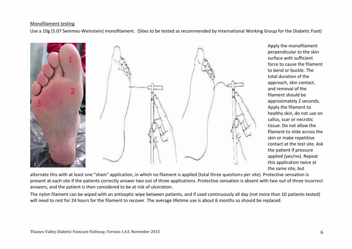
Monofilament testing
Use a 10g (5.07 Semmes-Weinstein) monofilament. (Sites to be tested as recommended by International Working Group for the Diabetic Foot)
Apply the monofilament perpendicular to the skin surface with sufficient force to cause the filament to bend or buckle. The total duration of the approach, skin contact, and removal of the filament should be approximately 2 seconds. Apply the filament to healthy skin, do not use on callus, scar or necrotic tissue. Do not allow the filament to slide across the skin or make repetitive contact at the test site. Ask the patient if pressure applied (yes/no). Repeat this application twice at the same site, but
alternate this with at least one "sham" application, in which no filament is applied (total three questions per site). Protective sensation is present at each site if the patients correctly answer two out of three applications. Protective sensation is absent with two out of three incorrect answers, and the patient is then considered to be at risk of ulceration.
The nylon filament can be wiped with an antiseptic wipe between patients, and if used continuously all day (not more than 10 patients tested) will need to rest for 24 hours for the filament to recover. The average lifetime use is about 6 months so should be replaced.

Thames Valley Diabetic Footcare Pathway, Version 1.63, November 2015 7
Vibration sense testing Use a tuning fork (128 Hz) or a graduated Rydel-Seiffer(64Hz) tuning fork.
First apply the tuning fork on the patient's wrists or elbow, so the patient knows what to expect. Ask the patient to close their eyes so as not to be able to see if and where the examiner applies the tuning fork. The tuning fork is applied on a bony part on the dorsal side (top) of the distal phalanx of the first toe. It should be applied perpendicularly with a constant pressure. Repeat this application twice, but alternate this with at least one "sham" application, in which the tuning fork is not vibrating. The test is positive if the patient correctly answered at least two out of three applications, and negative ("at risk for ulceration") with two out of three incorrect answers. If the patient is unable to sense the vibrations at the big toe, the test is repeated more proximally on the ankle, up to the knee.

Thames Valley Diabetic Footcare Pathway, Version 1.63, November 2015 8
Identify skin changes with pressure

Thames Valley Diabetic Footcare Pathway, Version 1.63, November 2015 9
Step 2: Identify if the arterial supply to the foot is reduced (by the absence of foot pulses, signs of tissue ischaemia or symptoms of intermittent claudication).
The vascular evaluation should include palpation of the pedal pulses. Do this for both pulses on both feet. The dorsalis pedis pulse can be found in the groove between the first and second metatarsal bones.
The posterior tibial pulse can be found behind the medial malleolus, one third of the distance from the medial malleolus to the bottom of the heel. Absence of either pulse means the foot is at risk and of significant concern if neither pulse is palpable. If the pulse is not palpable due to the presence of oedema in the foot, Doppler testing can be performed by a trained individual to assess blood supply.

Thames Valley Diabetic Footcare Pathway, Version 1.63, November 2015 10
Step 3: Identify deformities or problems of the foot (including bony deformities, dry skin or fungal infection) that may put it at risk. Identify Foot Deformity

Thames Valley Diabetic Footcare Pathway, Version 1.63, November 2015 11
Step 4: Identify identifying any issues with footwear

Thames Valley Diabetic Footcare Pathway, Version 1.63, November 2015 12
Step 5: Identify identifying other factors that may put the foot at risk (which may include reduced capacity for self care, impaired renal function, poor glycaemic control, cardiovascular and cerebrovascular disease, or previous amputation). Arrange assessment and management of these issues as required. Step 6: Assign Risk status. Give patient the appropriate leaflet responding to their level of Risk. http://www.oxfordhealth.nhs.uk/podiatry/leaflets/(This link should be changed to the HETV website link when up and running). Ensure this has local Podiatry contacts on it. If high risk, ensure the patient is advised to seek urgent medical attention if there is any concern with the foot. Excellent resources for all patients:https://www.diabetes.org.uk/Guide-to-diabetes/Monitoring/Feet If an ulcerated foot is present, then advise the patient to avoid weight bearing and ensure immediate referral to the local podiatry service or emergency department as per the pathways described below.
C – Formal Foot examination and verbal plus written advice as per local accredited policy or equivalent. All clinicians performing the testing need to have shown competence. Local examples: BHFT, Oxford, Bucks(Can provide links on HETV webpage when up and running) External Example:http://www.diabetesframe.org/

Thames Valley Diabetic Footcare Pathway, Version 1.63, November 2015 13
Figure 2: Footcare pathway for those with an established diagnosis of diabetes
Established diagnosis of Diabetes
Manage diabetes and cardiovascular risk as per local policy and
care planning process
Annual Review at the surgery
Footcare advice (A)
and Examination (C)
Assign Risk Status and give appropriate
leaflet with contact details of local
podiatry service
Thames Valley Diabetic Footcare Pathway, Version 1.63, November 2015 14
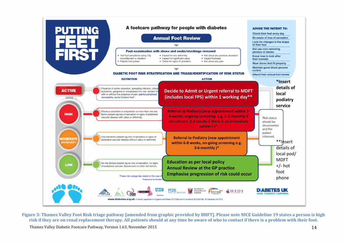
Figure 3: Thames Valley Foot Risk triage pathway [amended from graphic provided by BHFT]. Please note NICE Guideline 19 states a person is high risk if they are on renal replacement therapy. All patients should at any time be aware of who to contact if there is a problem with their foot.
Education as per local policy Annual Review at the GP practice Emphasise progression of risk could occur in the future
Referral to Podiatry (new appointment within 6-8 weeks, on-going screening e.g.
3-6 monthly )*
Referral to Podiatry (new appointment within 2-4 weeks, ongoing screening e.g. 1-2 monthly if no concern, 1-2 weeks if there is an immediate
concern )*
Decide to Admit or Urgent referral to MDFT (includes local FPS) within 1 working day**
*Insert details of local podiatry service **Insert details of local pod/ MDFT +/- hot foot phone

Thames Valley Diabetic Footcare Pathway, Version 1.63, November 2015 15
Figure 4: Thames Valley Triage of active foot disease. Please see Notes, p16 below for further details.
GP Surgery: Problem found at Annual Foot review or any review with the patient where foot problems are reported
Systemically unwell with Ulceration +/- Infection
or any of the following:
Rapidly progressing wound/cellulitis
Deep-seated soft-tissue or bone infection
Uncontrolled hyperglycaemia
Gangrene, ischaemic pain
Admit to Vascular Surgery
Secondary Care Pathway initated
No significant ischaemia
Admit to General Medicine
Secondary Care Pathway initiated
Systemically well with Ulceration +/- infection
Refer to MDFT/FPS within 1 working day
Patient to be triaged within 1 further day to decide how soon and where patient is seen
Infection: start antibiotics in line with local policy
GP to pescribe the same day
Iniate dressing changes with practice
nurse/district nurse
Callus, deformity, shoewear problem or
other
Refer to FPS within 1 working day
Patient to be triaged within 1 further day to
decide how soon and where patient
is seen
Suspected Charcot
Follow local Charcot guidelines
Refer direct to MDFT within 1 working day for triage within 1 further
working day
Offer non-weight bearing treatment until
MDFT review eg crutches

Thames Valley Diabetic Footcare Pathway, Version 1.63, November 2015 16
2. Notes Active foot disease Initial management of foot ulceration at the surgery:
1. Woundcare – local wound care. 2. Assess vascular status by checking pulses. Doppler can help qualify flow.
Vascular insufficiency needs to be highlighted when making referral to FPS/MDFT. 3. Optimise glycaemic control, using diabetes specialist nurse if necessary at intermediate or secondary care level. 4. Start antimicrobial therapy in line with local Antibiotic policy. 5. Advise patient to stay off their feet as much as possible, advise avoidance of tight fitting shoes, keep wound dry i.e. no showers! 6. Make referral to FPS/MDFT as per advice on local website/pathway. 7. Practice nurses /District nurses to be made aware of the patient for appropriate dressing changes (nb most diabetic foot wounds will
need cleaning and dressing changes at least 3x a week, with one of those usually being a podiatry appointment for debridement. Sometimes alternate/daily day dressings are needed if the wound is wet.
Suspected Charcot arthropathy at the surgery: Suspect Charcot arthropathy if there is redness, warmth, swelling or deformity (in particular where the skin is intact), especially in the presence of neuropathy or renal failure. A temperature difference between similar points on both feet is of particular concern. Charcot arthropathy should be considered even when deformity (which is a late sign) or pain are not present. Be aware that if a patient with diabetes fractures their foot ankle it may progress to Charcot arthropathy.
1. Immediate immobilisation is the key – advise patients not to weight bear, and provide non-weight bearing measures e.g. crutches. 2. Optimise glycaemic control. 3. Order a plain radiograph. 4. Make referral directly to MDFT/ consider admission over the weekend.

Thames Valley Diabetic Footcare Pathway, Version 1.63, November 2015 17
Foot Protection Service, formerly ‘Foot Protection Team’ role (see also additional resources for Podiatrists in Appendix): 1. Assess wound, vascular status, classify wound (use the Texas classification system or SINBAD scoring system). 2. Take swab of cleaned ulcer base, ideally tissue from ulcer base (send in dry pot to Microbiology). 3. Off-loading boot to be provided. 4. Decide if immediate referral to MDFT warranted, if lack of pulses to be seen in MDFT or suspected osteomyelitis for example as per
local policy. 5. Liaise with GPs regarding local antibiotic policy. 6. Maximum 4-week cut-off for referral to MDFT. 7. Referral directly to MSK podiatry/Orthotics as needed to achieve healing, prevent future breakdown. 8. On-going follow up until wound healed. 9. Advise patient to keep wound dry, and to consider ‘Limbo’ type device to keep area dry in shower. 10. Liaison with Practice nurses /District nurses re wound management. 11. Liaison with the MDFT e.g. rotating into MDFT/training sessions and regular communication. 12. Patient education and formation of agreed treatment plans, moving towards a care-planning approach. 13. Discharge planning to the appropriate level of Care e.g. Community clinics, FPS or High risk Podiatry clinics.
In-patient pathway and MDFT review: Each Trust should have a diabetic foot pathway that includes the following components:
1. Emergency Department staff education and referral pathways. 2. Foot examination on admission to hospital. 3. In-patient referral pathway to MDFT. 4. In-patient podiatry review. 5. Discharge pathway to ensure appropriate care given and follow up in MDFT clinic.
MDFT role:
1. As per FPS role. 2. MDT review to break through barriers to healing. 3. Consideration of long term mobility, limb salvage, preservation of health and independence should be the norm. In particular there
should be consideration as to whether re-vascularisation is appropriate. There should be no barrier to amputation if this is in the best interests of the patient. All options should be discussed fully with the patient to ensure informed consent occurs.
4. Consideration of occupational needs to help patients return to work

Thames Valley Diabetic Footcare Pathway, Version 1.63, November 2015 18
MDFT clinic minimum standard across region: 1. Podiatrist, Diabetologist, Vascular Surgeon, Orthotist in a combined clinic, at least monthly. 2. Aim for the clinic to run at least weekly with Diabetologist and Podiatrist. 5. Access to Vascular duplex, MRI, MRA, interventional radiology – ideally within 2-4 weeks if required. 6. Build up membership to full MDT as recommended in NICE guidance 7. Clear referral process to MDFT 8. Decide on how CV risk/Glycaemia should be aggressively targeted
Charcot Pathway:
1. Dedicated Orthopaedic input and Plaster room care 2. Ensure local policy available and advertised appropriately 3. Ensure follow up in High risk Podiatry clinic after resolution
Antibiotic guidelines Each area should have a local antibiotic policy that has been formulated bearing in mind local organisms/patterns. Each policy should cover primary and secondary care prescribing. Wound swabs should guide where there is lack of response to empirical treatment or to identify certain organisms for example MRSA needing certain organisms. Complicated wounds should be treated through the MDFT under the auspices of a dedicated Microbiologist. The principles of Antibiotic stewardship should be followed. Patient Education This should include structured education (diabetes structured education in line with local policies), advice at every annual review and podiatry review. As well as verbal information, written information should be given in the form of standardised leaflets (originating from NHS diabetes). These, as well as the Diabetes UK leaflets are available in other languages as well. Links: http://www.oxfordhealth.nhs.uk/podiatry/leaflets/ (To be replaced from link from HETV website, or each local provider can link to leaflets customised to their service). https://www.diabetes.org.uk/Guide-to-diabetes/Monitoring/Feet/

Thames Valley Diabetic Footcare Pathway, Version 1.63, November 2015 19
Training Several packages exist, and are accredited. Each area should specify what guidance it will use.
http://www.nwyhelearning.nhs.uk/elearning/northwest/shared/Diabetes_FootScreening/story.html http://www.diabetesframe.org/ https://www.diabetes.org.uk/Documents/Professionals/Competencies/The%20Podiatry%20Integrated%20Career%20and%20Competency%20Framework%20for%20Diabetes%20Foot%20Care%20-%20TRIEPodD-UK_May%202012.pdf
Governance Ensure local pathways are patient-centred in line with NHS Constitution, with focus on good patient experience. Dedicated governance procedures are in place in each Provider organisation Root cause analysis of all major lower limb amputations should be the norm. A minimum requirement of analysis of a random sample of 10 lower limb amputations/year has been agreed in Thames Valley. Learning points to be fed back at the Diabetic Foot Reference Group meeting. Outcome Metrics and Quality Assurance Data already collected per CCG:
1. Total episodes of inpatient care for diabetic foot disease 2. Annual episodes of care for diabetic foot disease per 1,000
adults with diabetes 3. Total nights in hospital due to diabetic foot disease 4. Annual nights in hospital for diabetic foot disease per 1,000
adults with diabetes 5. Episodes of care where an amputation is performed on
those with diabetes(Total, Major and Minor) 6. Annual amputations per 1,000 adults with diabetes (Total, Major and Minor)
(collected by YHPHO http://www.yhpho.org.uk/diabetesprofilesfoot/default.aspx)

Thames Valley Diabetic Footcare Pathway, Version 1.63, November 2015 20
Quality Assurance Measures could include: - Presence of a commissioned service in line with this guidance - Training uptake - Patient satisfaction questionnaires - Participation in the National Diabetic Foot Audit (NDFA) – on-going data collection and review of results - CQUINs such as number of admissions, length of stay, CV disease, time to healing, referral wait to be seen by podiatry/MD Outcome metrics, quality measures and results of Root Cause Analyses for lower limb amputations will be discussed at a regional Thames Valley level on a six monthly basis; at the diabetic foot reference group meetings.

Thames Valley Diabetic Footcare Pathway, Version 1.63, November 2015 21
Notes for Commissioners Points to consider:
1. This pathway provides robust and clear local pathways for integrated footcare of people with diabetes across all settings. This guidance should be used to provide a commissioned pathway for the care of the diabetic foot. The establishment of a foot protection service, integrated with a multidisciplinary foot care service has been deemed best practice via NICE guidance, and internationally in the management of the diabetic foot. This has been shown to be cost-effective through much work, presented nationally (see Marion Kerr’s work).
2. The details of the components of the foot protection service and multidisciplinary teams is outlines in NICE guidelines 19, published in August 2015 and specific notes to commissioners are included from page 37-41, https://www.nice.org.uk/guidance/ng19
3. Diabetic Foot disease is high priority for DUK and patient focus groups. 4. Commissioning activity along with diabetic foot outcomes is being looked at through the national diabetic foot audit (which will be
ongoing) 5. Nationally many areas have now achieved a commissioned, integrated pathway for the diabetic foot. So it is achievable, and we do not
want to be left behind. 6. Outcomes (YHPHO footcare profiles) in Thames Valley although on the whole not of concern compared with national averages, do show
that there could be significant improvements made when compared with the lowest rates of amputations achieved in some centers e.g. St Mary’s in London. Excellence is possible for our patients.
7. Savings that will come from the pathway in terms of reduced practice dressing, consultation and antibiotic costs, plus reduced admissions, length of stay etc.
8. Much of the service currently exists, and will simply need to be modified in line with this guidance. Any commissioning gaps should be addressed.
9. Outcome metrics, quality measures and results of Root Cause Analyses for lower limb amputations will be discussed at a regional Thames Valley level on a six monthly basis; at the diabetic foot reference group meetings.
10. Specifically to consider commissioning of the following: a. Adequate staffing to ensure the service continues despite annual leave. b. Access to off-loading boots and who can refer to Orthotics. c. Podiatry review over the weekends (a podiatrist to cover the Thames Valley area perhaps?). d. Hard to reach groups/vulnerable adults e.g. homeless, care homes, housebound, prison population. e. Hot Foot phone for advice. f. Transition care from Paeds to Adult services (Foot screening performed in Paeds clinic annually until age of 17-19).

Thames Valley Diabetic Footcare Pathway, Version 1.63, November 2015 22
3. Appendices Appendix 1: Members of the Thames Valley Diabetic Foot Reference Group Name Designation Area Contact
Dr Hema Heffernan Consultant in Diabetes, and Chair of Diabetes Foot Reference Group for TV SCN
Wexham Park Hospital, FHFT [email protected]
Dr Asif Ali Consultant in Diabetes MKUHFT [email protected] Mr T Boreham Patient representative Bucks [email protected] Ms Julia Coles SCN Domain 2 Lead NHS England [email protected] Dr Richard Croft Diabetes Lead, TV SCN NHS England [email protected] Dr Stephen Gardner Consultant in Diabetes Bucks NHS FT [email protected] Mr Keith Hilston Podiatry Lead BHFT [email protected] Dr Kathy Hoffman Bucks Diabetes lead Bucks CCGs [email protected] Mr Laurie King Podiatry Lead OUHFT [email protected]
Ms Susan Laybourn Diabetes foot lead MKUHFT [email protected]
Mr Tony Lloyd Patient representative RBFT [email protected]
Dr Aparna Pal Consultant in Diabetes RBHFT [email protected]
Mr Neil Sandys CVD Manager, TV SCN NHS England [email protected]
Ms Lesley Scott Diabetes Lead MKDC [email protected]
Mr Ed Sideso Consultant Surgeon OUHFT/RBHFT [email protected]
Ms Rebecca Tyrell Service improvement specialist NHS England [email protected] Key BHFT – Berkshire Healthcare Foundation Trust; FHFT –Frimley Health NHS Foundation Trust; FT – Foundation Trust; MKUHFT – Milton Keynes University Hospital Foundation Trust; MKDC Milton Keynes Diabetes Centre; OUHFT – Oxford University Hospitals Foundation Trust; RBFT Royal Berkshire Foundation Trust; TV SCN – Thames Valley Strategic Clinical Network

Thames Valley Diabetic Footcare Pathway, Version 1.63, November 2015 23
Appendix 2: Information for Podiatrists
Figure 1: An example of triage framework for Podiatrists (image provided by Laurie King)

Thames Valley Diabetic Footcare Pathway, Version 1.63, November 2015 24
Figure 2: TEXAS classification for use by Podiatry (image provided by Laurie King)

Thames Valley Diabetic Footcare Pathway, Version 1.63, November 2015 25
Figure 3: Management based on TEXAS classification for use by Podiatry (image provided by Laurie King)

Thames Valley Diabetic Footcare Pathway, Version 1.63, November 2015 26
Figure 4: TEXAS classification for use by Podiatry (image provided by Laurie King)

Thames Valley Diabetic Footcare Pathway, Version 1.63, November 2015 27
Appendix 3: Example Poster
Figure 1: ‘Hot Foot ‘ Phone Poster (image provided by Laurie King)