Embed Size (px)
Citation preview
DIABETIC FOOT SEPSIS
DR LYNNE TUDHOPE


2000 171 million
2030 366 million
International Diabetes Federation. IDF Diabetes Atlas, 5th edn. Brussels,
Belgium: International Diabetes Federation, 2011.
NUMBER OF PEOPLE WITH DIABETES GLOBALLY

2000 171 million
2010 285 million
2011 366 million
2030 366 million
2030 552 million
International Diabetes Federation. IDF Diabetes Atlas, 5th edn. Brussels,
Belgium: International Diabetes Federation, 2011.
NUMBER OF PEOPLE WITH DIABETES GLOBALLY

The evolution of mankind…
Inactivity

OBESITY


ADA DATA - MARCH 2013
• 41% increase in total cost of diabetes mellitus – $245 billion
– 1 in $5 spent on health care goes to diabetes
• 43% on hospital inpatient costs
33% of direct cost burden of diabetes is in the lower extremity
(only 0,17% of research funding in the USA is spent on the diabetic foot!)

SOUTH AFRICA AND THE STATE OF DIABETES CARE
• Estimated population of 52.98 million
• Exact prevalence of diabetes is unknown
– Estimated to be 5 – 7% of the population
• Indians 11 – 13%
• Coloureds 8 – 10%
• Blacks 5 – 8%
• Whites 4%
• ± 85% of these receive public (Government) sector medical care
– Overburdened and inefficient
• ± 15% receive medical care in the private sector – paid for either by
themselves or by medical insurance schemes

Botswana $816
South Africa $695 Namibia $468 Zimbabwe $56 Mozambique $37 Zambia $125 Tanzania $40 DRC $25 Malawi $31
MEAN DIABETES-RELATED EXPENDITURE PER PERSON WITH DIABETES (USD) 2011
Brazil $1038 USA $8468 UK $4267 Australia $4878 Canada $5106 Luxembourg $9341
International Diabetes Federation. IDF Diabetes Atlas, 5th edn. Brussels,
Belgium: International Diabetes Federation, 2011.
http://www.idf.org/diabetesatlas
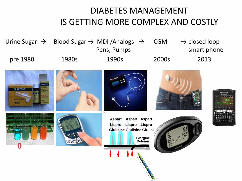
DIABETES MANAGEMENT IS GETTING MORE COMPLEX AND COSTLY
Urine Sugar → Blood Sugar → MDI /Analogs → CGM → closed loop Pens, Pumps smart phone
pre 1980 1980s 1990s 2000s 2013
0
Glargine Detemir
Lispro Lispro Lispro
Aspart Aspart Aspart
Glulisine Glulisine Glulisine

THE DIABETIC FOOT
• Nearly 80% of all non traumatic amputations occur in diabetics
• 85% of these begin with a foot ulcer
• 1 in 4 people with diabetes will have an ulcer in their lifetime
• 50% of these will become infected
• 50% of patients who have a foot ulcer die within 5 year
• Diabetic foot sepsis = amputation= loss of bipedalism

CAUSES OF PREVALENCE OF DIABETIC FOOT PROBLEMS IN SOUTH AFRICA
Health Care Related
Lack of Podiatrists (even in best hospitals)
Insufficient experience of those undertaking foot care
(surgeons, diabetologists, dermatologists)
Central Distribution of “ Good” health care services.
Shortage of finances
13

CAUSES OF PREVALENCE OF DIABETIC FOOT PROBLEMS IN SOUTH AFRICA
Health Care Related
Lack of health insurance of thousands of patients
Lack of health education (busy clinics, few national programs,
media, ….)
Insufficient national data about different health problems
Setting priorities (underestimation of foot problems)
14

CAUSES OF PREVALENCE OF DIABETIC FOOT PROBLEMS IN SOUTH AFRICA
Patients related Factors
High Prevalence of DM
Walking barefoot
Illiteracy
Associated comorbidities
Poor compliance of patients
15

THE AMPUTATION RATE IN SOUTH AFRICA?
• Published data shows a 60,2% rate of non traumatic lower limb amputation accountable to diabetes in public hospitals
• Unpublished data from two separate public hospitals showed an amputation rate of 78,5% however
• Limb salvage rate in a multidisciplinary clinic in a private hospital by contrast is 85% over a three year period
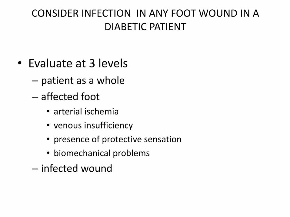
CONSIDER INFECTION IN ANY FOOT WOUND IN A DIABETIC PATIENT
• Evaluate at 3 levels
– patient as a whole
– affected foot
• arterial ischemia
• venous insufficiency
• presence of protective sensation
• biomechanical problems
– infected wound

EURODIALE
• PAD and peripheral neuropathy are both well known risk factors for diabetic foot ulceration and foot infection
• PAD and infection – PAD present in diabetes = X 5.5 increased risk for
diabetic foot infection
– 3x increase risk of amputation


20

SYSTEMIC EVALUATION
• ECG, stress test and even coronary angiography with intervention may be required
• Renal status and creatinine clearance
• Pulmonary function and chest preparation
• Control of diabetic status
• Identification and control of active infection
21

22
PHYSICAL EXAMINATION OF THE FOOT
Presence of peripheral pulses
Bruit
Skin temperature and colour
Hair loss, muscle and skin atrophy
Dependant hyperaemia
Skin ulceration or tissue loss

INFECTION?
• Erythema
• Swelling
• Induration
• Tenderness
• Malodor

Factors associated with increased risk of infection
• Positive probe to bone test
• Ulcer duration more than 30 days
• Traumatic wound
• Presence of PAD and PND
• Previous amputation
• Renal insufficiency
• History of walking barefoot

SIGNS OF POSSIBLE IMMINENT LIMB-THREATENING INFECTION
• Systemic inflammatory response
• Rapid progression of infection
• Extensive necrosis or gangrene
• Crepitus on examination or tissue gas on imaging
• Extensive ecchymoses or petechiae
• Bullae, especially hemorraghic
• Pain out of proportion to clinical findings
• Recent loss of neurologic function
• Critical limb ichemia
• Extensive soft tissue loss
• Extensive boney destruction, especiallymidfoot/hindfoot
• Failure of infection to improve with appropriate Rx

INFECTIONS
• Mild
• Moderate
• Severe

0 I II III
Lesion completely
epithelialised
0%
Superficial wound, not involving tendon capsule or bone
0%
Wound penetrating to
capsule or tendon
0%
Wound penetrating to joint or bone
Infection
12.5%
Infection
8.5%
Infection
28.5%
Infection
Ischemia
25%
Ischemia
25%
Ischemia
25%
Ischemia
Infection and ischemia
50%
Infection and ischemia
50%
Infection and ischemia
Infection and
ischemia
100%
A
B
C
D
TEXAS CLASSIFICATION
100%
100%
100%
92%
0%

IDSA IWGDF CLASSIFICATION OF DIABETIC FOOT INFECTION
Clinical manifestation of infection32mmHg PEDIS
grade
IDSA infection
severity
No symptoms or signs of infection 1 Uninfected
Infection present, as defined by the presence of at least 2 of the following
•Local swelling or induration
•Erythema
•Local tenderness or pain
•Local warmth
•Purulent discharge
Local infection involving skin and subcutaneous tissue without involvement of
deeper tissue and no systemic signs. Erythema >0,5 cm to ≤2cm around the ulcer
Exclude other causes of inflammatory response (acute Charcot, trauma, gout
fracture)
2 Mild
Local infection with erythema >2cm or involving structures deeper then skin and
subcutaneous tissues and no SIRS
3 Moderate
Local infection with signs of SIRS with ≥2 of following
•Temperature >38º or <36ºC
•Heart rate >90 beats/min
•Respiratory rate >20 breaths/min or PaCO2 <32 mmHg
•WBS >12 000 or < 4000 or ≥10% immature forms
4 Severe

INFECTION
• Polymicrobial – Aerobic gram+ cocci staphylococci, beta-
hemolytic streptococci
– Warm climates and exposure to water, gram- bacilli pseudomonas, E. coli
– + aerobic bacilli in chronic wounds
– Obligate anaerobes in ischemic / necrotic wounds
– MRSA in diabetic foot wounds ranges from 5 to 30% • usually if previously hospitalised

Antibiotic regimens
• Clinically uninfected? – No antimicrobial therapy
• Clinically infected? – Select antibiotic targeting likely pathogen – As narrow spectrum as possible – Empirical choice should cover
• Staphylococcus aureus • Gram + aerobic streptococci
• Only severe infections require IV Rx – Mild to moderate – 1 to 2 weeks Rx – Serious – 4 weeks of Rx

SUGGESTED ROUTE, SETTING AND DURATION OF Antibiotic Rx
Site of infection, by
severity or extent
Route of
administration
Setting Duration of
therapy
SOFT TISSUE ONLY
Mild Topical or oral Outpatient 1-2 weeks
Moderate Oral ( or initial IV) Outpatient/inpatient 1-3 weeks
Severe Initial IV, switch to
oral when possible
Inpatient, then
outpatient
2 – 4 weeks
BONE OR JOINT
No residual infected
tissue
IV or oral … 2-5 days
Residual infected soft
tissue (not bone)
IV or oral … 1-3 weeks
Residual infected(but
viable) bone
Initial IV, consider
oral switch
… 4-6 weeks
No surgery or residual
dead bone
Initial IV then oral
switch
≥3 months

SUGGESTED EMPIRIC
ANTIBIOTIC REGIMENS BASED
ON CLINICAL SEVERITY FOR
DIABETIC FOOT INFECTIONS

Non infected wound
• Should not be Rx with topical or systemic antibiotics
• After open bypass surgery – wait 4 to 8 days before definitive debridement
• After endovascular intervention – wait 3 to 4 weeks

OSTEOMYELITIS
• Present in up to 20% of mild to moderate infections • Present in 50% to 60% of severely infected wounds • Consider in ulcers that are deep, large chronic or over a
boney prominence • Charcot difficult to distinguish from osteomyelitis and can
co exist • Radiographic changes of osteomyelitis may lag clinical
disease by up to a month • Antibiotic Rx based on culture results of bone • Prolonged course of antibiotics (3-6 months) has a clinical
success rate of 65%-80% in diabetic foot osteomyelitis

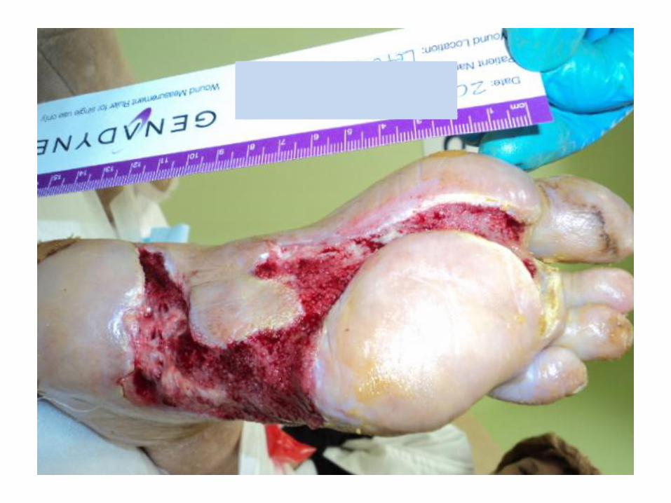
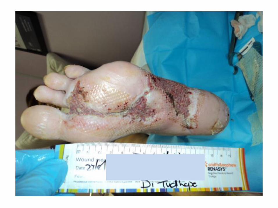
Wound • Surgical debridement of dead tissue • Appropriate antibiotic Rx • Removing pressure off the wound • Improve blood flow to the infected area
• Deep tissue specimen the best • Superficial wound swabs often contaminated • 50% volume decrease in 4 weeks or 10 to 15%
decrease each week • 40% of amputations are preventable with
appropriate wound care

Schematic diagram of cross-section of the foot.
Numbers 1 to 5 indicate metatarsal bones.
A,central plantar space; B,deep interosseous space;
C, lateral plantar space; D,medial plantar space

BASIC TOOLS OF DEBRIDEMENT
• Blades
• Forceps
• Scissors
• Curettes
• Rongeurs

OFFICE DEBRIDEMENT: CURETTE
remove biofilm on top of wound

OFFICE DEBRIDEMENT: SURGICAL BLADE OR NIPPERS
No 15 blade !

IDENTIFYING DEAD TISSUE
• Good
• Bad
EXCEPT VEIN
COLOUR CODING FOR IDIOTS

IDENTIFYING DEAD TISSUE: CLOTTED VEINS

DEBRIDEMENT OF NECROTIC MUSCLE

DEBRIDEMENT OF NECROTIC MUSCLE

DEBRIDEMENT OF NECROTIC MUSCLE

IDENTIFYING DEAD TISSUE: LIQUIFIED FASCIA

DEBRIDING FASCIA / TENDON: DEAD TENDON = INFECTION

DEBRIDING BONE: PUNCTATE BLEEDING
“paprika sign”


Debridement
stop when….
only normal tissue
remains &
odour gone

WHEN IS OBTAINING A BONE SPECIMEN FOR CULTURE AND HISTOLOGY JUSTIFIED?
• When there is uncertainty regarding the diagnosis of osteomyelitis despite clinical and imaging evaluations
• An absence (or confusing mix) of culture data from soft tissue specimens
• Failure of the patient to respond to empiric AB Rx

RECOMMENDATIONS FOR COLLECTION OF SPECIMENS FOR CULTURE FROM DIABETIC FOOT WOUNDS
• Do
– Obtain an appropriate specimen for culture from almost all infected wounds
– Cleans and debride the wound before obtaining specimen for culture
– Obtain a tissue specimen for culture by scraping with a sterile scalpel or dermal curette or biopsy from the base of a debrided ulcer
– Aspirate any purulent secretions using a sterile needle and syringe
– Promptly send specimens in a sterile container for aerobic and anaerobic culture
• Do not
– Culture a clinically uninfected lesion, unless for specific epidemiological purposes
– Obtain a specimen for culture without first cleansing or debriding the wound
– Obtain a specimen for culture by swabbing the wound or wound drainage

Entropy
• Common to all systems • Tend from a state of order to a state of chaos • Young patient….. held in check by the body’s intrinsic
system of repair and regeneration • Older and immune supressed patient…. This process is
abandoned by the body • It does not cause disease but it leaves an organism
vulnerable • Disease takes hold where the system is weakest • In humans??? • The cardiovascular system
52

53
Diabetic Vascular Disease
Retina, Kidney, Nerves
Myocardium
Microvascular
disease
Large Arteries
Increased Rate of
Atherosclerosis
Hyperglycemia Hyperglycemia
Hypertension
Hyperlipidemia
54
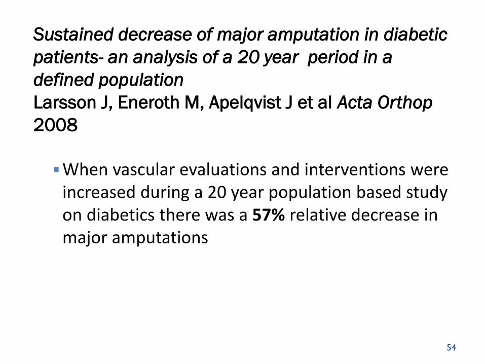
Sustained decrease of major amputation in diabetic
patients- an analysis of a 20 year period in a
defined population
Larsson J, Eneroth M, Apelqvist J et al Acta Orthop
2008
When vascular evaluations and interventions were increased during a 20 year population based study on diabetics there was a 57% relative decrease in major amputations

55
INDICATION FOR RX
Any diabetic in whom pulses are not easily palpable, has a critically
ischemic limb until proven otherwise.

56
Factors related to outcome of neuroischemic/ischemic foot
ulcer in diabetic patients
Apelqvist J, Elgzyri T, arsson J et al J Vasc Surg 2011
1151 diabetic patients with neuro ischemic ulceration < 50% considered ischemic prior to non-invasive
testing
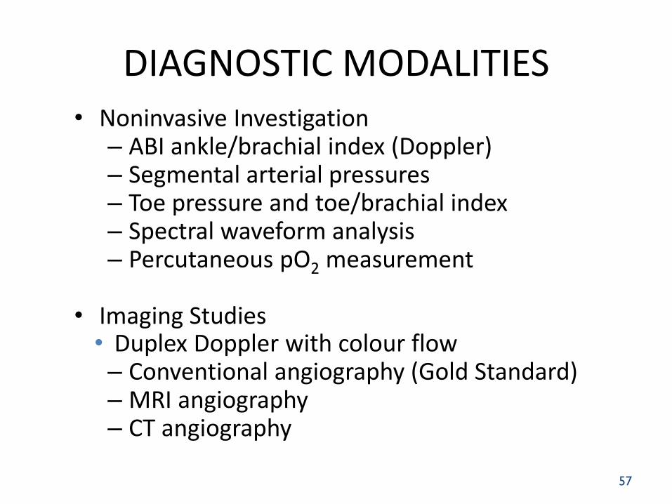
DIAGNOSTIC MODALITIES • Noninvasive Investigation
– ABI ankle/brachial index (Doppler) – Segmental arterial pressures – Toe pressure and toe/brachial index – Spectral waveform analysis – Percutaneous pO2 measurement
• Imaging Studies • Duplex Doppler with colour flow – Conventional angiography (Gold Standard) – MRI angiography – CT angiography
57
58
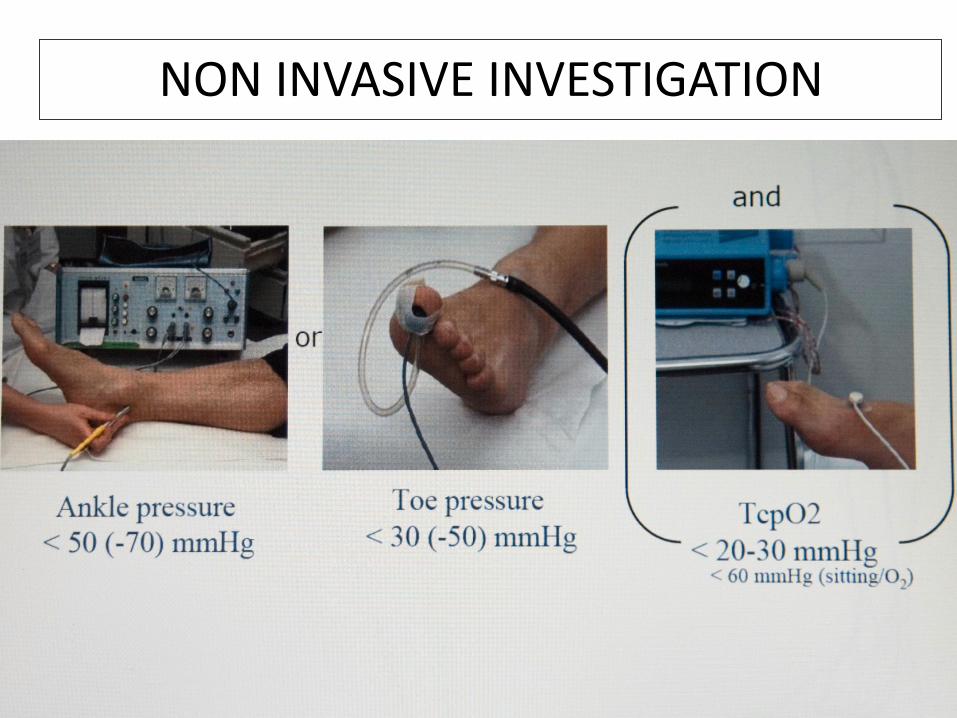
NON INVASIVE INVESTIGATION

ABPI – CONSENSUS STATEMENT
Diabetic > 50yrs
Screening ABPI in all diabetics
Repeat every 5 years if test is normal
Diabetic < 50 yrs
Screening ABPI if other PAD risk factors present
Smoking, hypertension, hyperlipidemia, duration of diabetes > 10yrs
Diagnostic ABPI in any diabetic with PAD symptoms; together with treadmill testing if ABPI and clinical symptoms do not correlate

IDEAL NON-INVASIVE TEST = TOE PRESSURES
Should be approximately 60% of brachial pressure
Toe pressures
> 45 mmHg = 85% primary healing
< 45 mmHg = 36% healing without amputation

< 34 mmHg (amputation 85%)
indicates the need for revascularization
> 34 < 40 mmHg (amputation 20%)
less pressing, although there remains a considerable probability of amputation
> 40 mmHg
revascularization is dependent on the severity of tissue loss and possible
morbidity caused by the procedure
Faglia E, Clerici G, Caminiti M et al. Predictive values of transcutaneous oxygen tension for above-the-ankle amputation in
diabetic patients with critical limb ischemia. Eur J Vasc Endovasc Surg. 2007 Jun;33(6):731-6
TcPO2 levels
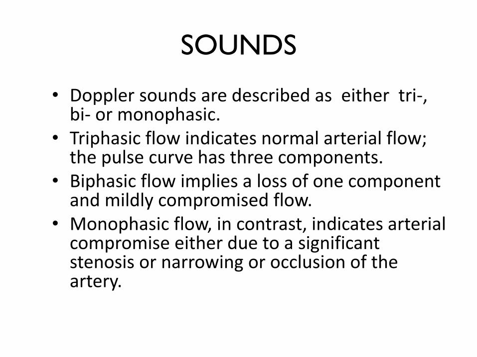
SOUNDS
• Doppler sounds are described as either tri-, bi- or monophasic.
• Triphasic flow indicates normal arterial flow; the pulse curve has three components.
• Biphasic flow implies a loss of one component and mildly compromised flow.
• Monophasic flow, in contrast, indicates arterial compromise either due to a significant stenosis or narrowing or occlusion of the artery.

Fig 7: Arterial arterial connections: By occluding arteries proximal or distal to the
arterial signal, it is possible to assess the direction of arterial flow

Direct vascular connections: arterial arterial connections
All the main arteries of the foot and ankle are directly connected to one another

Doppler: peroneal artery: anterior perforating branch Doppler anterior perforating branch of the peroneal
artery just medial to distal fibula

Doppler: Anterior tibial artery → dorsalis pedis
for
ante-
grade
flow

Doppler: antegrade flow posterior tibial artery
Locate
artery
for ante-
grade flow

68


THE IMPORTANCE OF ANGIOSOMES IN HEALING FOOT ULCERS
• Taylor describes at least 40 angiosomes in the body, of which 5 are found in the foot and ankle.
• These originate from the three main arteries in the lower extremity, the posterior tibial artery, the anterior tibial artery and the peroneal artery.
• The posterior tibial artery supplies the sole of the foot via the calcaneal branch, the medial plantar branch and the lateral plantar branch.
70

THE IMPORTANCE OF ANGIOSOMES IN HEALING FOOT ULCERS
• The anterior tibial artery supplies the anterior ankle and as the dorsali pedis artery, it also supplies the dorsum of the foot.
• The peroneal artery supplies the lateral anterior upper ankle via its anterior perforating branch and also supplies the plantar heel area via a calcaneal branch.
• Arterial to arterial connections are important because, despite the occlusion of one or more arteries to the foot, these connections provide an uninterrupted blood flow to the entire foot.
71

The importance of angiosomes in healing foot ulcers
• The treatment of ischemia in the diabetic foot should be aimed at the restoration of maximum blood flow to the foot with the restoration of pulsatile palpable foot pulses whenever possible
• This pulsatile flow increases the chance of wound healing and diminishes future skin breakdown and ulcer formation.
• In planning any surgical procedure on the foot, or when embarking on any course of wound care treatment, it is essential that optimum blood flow is obtained in the area of tissue breakdown.
• By understanding the principle of angiosomes and the vascular anatomy of the foot, wound healing and foot salvage will be easier to predict.

• It has been reported that up to 15% of bypasses to the foot fail to heal wounds on the foot, in spite of remaining patent, simply because these bypasses failed to revascularize the affected angiosome
• It is, therefore, crucial that bypass procedures are done to the right blood vessel, if existent ischemic ulcers are to be healed.
73
. 1

VASCULAR SUPPLY
• Regulation of vasculature
– Arterial supply maximization
– Periwound edema minimization


PERIPHERAL NEUROPATHY
• Affects sensory, motor and autonomic innervation
• Loss of sensation – wounds go unnoticed – Reduction of pain and tenderness…. No early warning
system
• Motor nerve damage – foot deformity, abnormal pressure points, callus, ulceration.
• Autonomic neuropathy – dry skin, heel cracks , tearing and infection


BARRIERS TO EFFECTIVE MANAGEMENT
• Importance of foot care not recognised
• Ignorance of improved patient outcomes with better foot care
• Non existent podiatry services
• Team approach lacking • Routine referral for amputation
• Limited training programs for healthcare providers

BARRIERS TO EFFECTIVE MANAGEMENT
• Services run by non specialist nurses not foot care specialists
• Barefoot walking common
• Faith healers, herbalists and home therapies
• Unaffordable footwear
• Poverty; limited access to care

THE DIABETIC FOOT
• Establish contacts in healthcare
• Raise funds
• Foot clinics beginning with a diabetes centre for excellence
• Multidisciplinary educational approach
• Establish attainable goals
• Recruit , train and retain
• Motivate healthcare professionals

SERVICES NEEDED
Foot Screening
Nail Care
Ulcer Care
Debridement
Offloading Devices
Education
97

In summary
• non infected wound? – no antibiotics
– specialised wound care
– off loading
• Infected wound? – stage wound
– chemical or mechanical debridement
– tissue and/or bone biopsy
– oral or IV antibiotics
– off loading

THE DIABETIC FOOT: Two decades of “progress”
1986: First Malvern Diabetic Foot Meeting
1987: Foot Council of ADA formed
1991: First International Diabetic Foot Meeting
1998: Diabetic Foot Study Group of EASD founded
1999: International Consensus group publishes “Guidelines on management”
2001: Formation of DFSIs
2002: First DFCon meeting, Universal City, Los Angeles
2004: Formation of GLEPED
2005: IDF designated year of the Diabetic Foot
2007: Formation of DFWG, South Africa
2007: Fifth International Diabetic Foot meeting
2009: Opening of first foot clinic in Colombia
2010: 13th biennial Malvern Diabetic Foot meeting
2010: First Pan-African Diabetic Foot meeting, Spier
2014: 2nd Pan-African Diabetic Foot meeting, Dar es Salaam

Laughter the best medicine?
• 5 year study – Leeds School of Healthcare
– “A hearty chuckle stimulates the diaphragm which in turn plays a vital role in moving blood around the body and speeds recovery from leg ulcers”
100