Embed Size (px)
Citation preview
2/9/2017
1
Diabetic Keratopthy(DK)
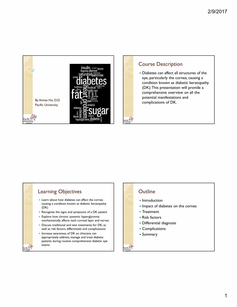
By Amiee Ho, O.D.
Pacific University
Course Description
� Diabetes can affect all structures of the eye, particularly the cornea, causing a condition known as diabetic keratopathy (DK). This presentation will provide a comprehensive overview on all the potential manifestations and complications of DK.
2
Learning Objectives
� Learn about how diabetes can affect the cornea causing a condition known as diabetic keratopathy (DK)
� Recognize the signs and symptoms of a DK patient
� Explore how chronic systemic hyperglycemia mechanistically affects each corneal layer and nerves
� Discuss traditional and new treatments for DK, as well as risk factors, differentials and complications
� Increase awareness of DK so clinicians can appropriately address, manage and treat diabetic patients during routine comprehensive diabetic eye exams
3
Outline
� Introduction
� Impact of diabetes on the cornea
� Treatment
� Risk factors
� Differential diagnosis
� Complications
� Summary
4
2/9/2017
2
Outline
� Introduction
� Impact of diabetes on the cornea
� Treatment
� Risk factors
� Differential diagnosis
� Complications
� Summary
5
Diabetes and The Eyes
6
https://s-media-cache-ak0.pinimg.com/originals/5b/4a/7f/5b4a7f7b0aedc5d43af2d1f247f9daac.jpghttps://www.pinterest.com/pin/117726977734572067/
We need to pay attention to cornea for DM patients because……
Diabetes can lead to corneal disease or Diabetic Keratopathy!!
7
https://socialanxietyinsti tute.org/sites/default/fi les/Focus.jpg 2000195019001850
1858
Francois
1967
Collier
1981
Richard Schultz•Cornea/anterior
segment abnormalities
•Delayed wound healing
•� risk of infection
•Published: comprehensive
review on DK
•Focus: pathopneumonic forms
of DK used for early diagnosis
•Published: “Diabetic Keratopathy”
History of DK
8

2/9/2017
3
Epidemiology of DK
� DK Prevalence estimations:◦ ~1/3 of patients with DM (Rao, Ioli)
◦ ~47-64% (Schultz, 1981)
◦ Epithelial lesions: ~2/3 (Rao)
� DK is believed to have high incidence:
◦ Rarely diagnosed (Wylegala)
◦ Underreported (Kaji)
◦ Overlooked
◦ Not considered serious or pathological (Kaji)
◦ Difficult confirming changes are only due to DM
9
Outline� Introduction
� Impact of diabetes on the cornea ◦ Pre-corneal tear film
◦ Epithelium (& basement membrane)
◦ Stroma
◦ Endothelium
◦ Corneal nerves
� Treatment
� Risk factors
� Differential diagnosis
� Complications
� Summary10
Basement M.
Review of Cornea
11
http://epomedicine.com/wp-content/uploads/2014/05/cornea-histology.png
Review of Cornea
12
Basement M.
http://epomedicine.com/wp-content/uploads/2014/05/cornea-histology.png
2/9/2017
4
THE PRE-CORNEAL TEAR FILM
13http://www.fruitycuties.com/archive/063-cartoon-onion-tear-gas-joke.htm
Tear Film Review
Keratoepitheliopathy
14
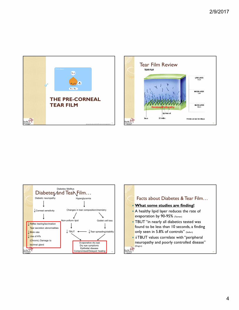
Diabetes Mellitus
Diabetic neuropathy Hyperglycemia
Changes in tear composition/chemistry
Non-uniform lipid Goblet cell loss
↓Tear spreading/stability↓ TBUT
Evaporative dry eye
Dry eye symptoms
Epithelial disease
Compromised/Delayed healing
Reflex tearing/lacrimation
Tear secretion abnormalities
Blink rate
Use of ATs
(Chronic) Damage to
lacrimal gland
↓
↓↓
Diabetes and Tear Film…
↓Corneal sensitivity
15
Facts about Diabetes & Tear Film…
�What some studies are finding!
� A healthy lipid layer reduces the rate of evaporation by 90-95% (Tasman)
� TBUT “in nearly all diabetics tested was found to be less than 10 seconds, a finding only seen in 5.8% of controls” (Seifart)
� �TBUT values correlate with “peripheral neuropathy and poorly controlled disease” (Dogru)
16
2/9/2017
5
Take home point
� Diabetes can reduce the effectiveness of tear film by altering structure and function causing….
17
Dry eyes
Damage corneal nerves
Decreased corneal
sensitivity
Basement M.
Review of Cornea
18
http://epomedicine.com/wp-content/uploads/2014/05/cornea-histology.png
CORNEAL EPITHELIUM&
BASEMENT MEMBRANE
19http://41.media.tumblr.com/ea3460fd6ebb5d56bec4376206e2bfa5/tumblr_n7diamoP9K1t7pikho1_1280.jpg
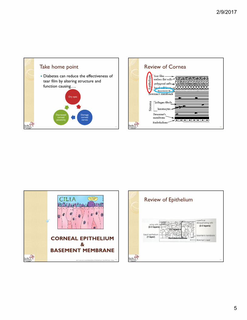
Review of Epithelium
5-7 layers
(2-3 layers) (2-3 layers)
(1 layer) hemidesmosomes
20

2/9/2017
6
Diabetes Mellitus
Hyperglycemia
Sugar
+
Amino acid
Maillard Reaction
(High heat)
Advanced Glycation End (AGE) Product
Deposits in epithelium
Changes in epithelial cells & Basement Membrane
Diabetic Keratoepitheliopathy
DM and Epithelium
21
Diabetic Keratoepitheliopathy
Signs/Symptoms:� Recurrent corneal erosion (Owen, Perry, Herse, Schultz
1981, 1984, Sato, Abdelkader)
� Slower wound repair (Hatchell, Herse, Sato)
� Delayed reepitheliazation (Kaji)
� SPK/Persistent epithelial defects (Herse, Owen, Schultz 1984)
� Increased epithelial fragility (Herse, Abdelkader)
� � risk of infection (i.e. fungal keratitis)� � defense properties and barrier
functions � edema (Gobbels, Yokoi, Gekka, Perry, Sato)
◦ 5.4x’s more permeable to water/ionic substances (Gobbels)
22
Diabetes and Epithelium
� Is AGE only in epithelium?
◦ Gradient of AGE: epithelium>stroma>endo
� Metabolism is mostly dependent on the aqueous humor (Zou)
� �expression of AGE productions, AGE receptors, and transcription factor nuclear factor kappa-B (NF-κB) in the lacrimal glands (Alves)
23http://www.hybridcornea.org/img/aboutcornea_clip_image004.jpg
Take home point
� Diabetes can produce excess AGE products that deposit in the epithelium altering structure and function causing Diabetic Keratoepitheliopathy
24

2/9/2017
7
Review of Cornea
25
Basement M.
http://epomedicine.com/wp-content/uploads/2014/05/cornea-histology.png
CORNEAL STROMA
Bypass Bowman’s Layer and onto…
26
Corneal Stroma
Signs/Symptoms:
� Wide spaced collagen fibril matrix � � transparency (Rehany)
� Transient stromal edema (Herse)
� Corneal lattice degeneration (Herse)
� Various forms of keratitis (Herse)
� Stromal ulceration/melting/perforation (Adbelkader, Lockwood)
� Stromal scarring (Adbelkader, Lockwood)
27
Review of Cornea
28
Basement M.
http://epomedicine.com/wp-content/uploads/2014/05/cornea-histology.png
2/9/2017
8
CORNEAL ENDOTHELIUM
29http://i2.wp.com/michaelduplessie.com/wp-content/uploads/2013/12/corneal-endothelium-normal.jpg
Review of Endothelium
30
• Single layer
• Metabolically active
• Hydration of cornea
https://www.reviewofoptometry.com/CMSImagesContent/2009/6/2_14368_1.gif
http://www.oculist.net/downaton502/prof/ebook/duanes/graphics/figures/v4/016a/004f.gifhttp://iovs.arvojournals.org/data/Journals/IOVS/932900/7g1022862001.jpeg
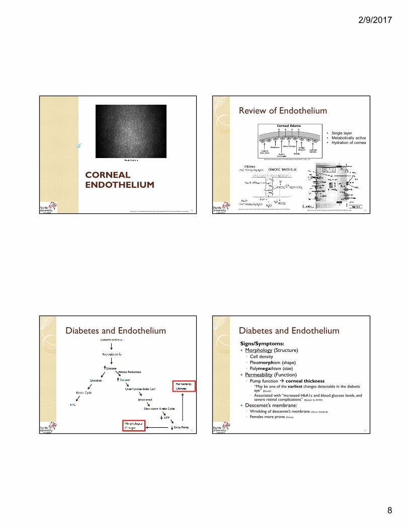
Diabetes and Endothelium
Aldose Reductase
31
Diabetes and Endothelium
Signs/Symptoms: � Morphology (Structure)◦ Cell density
◦ Pleomorphism (shape)
◦ Polymegathism (size)
� Permeability (Function)◦ Pump function � corneal thickness
� “May be one of the earliest changes detectable in the diabetic eye” (Busted)
� Associated with “increased HbA1c and blood glucose levels, and severe retinal complications” (Busted, Su DHW)
� Descemet’s membrane: ◦ Wrinkling of descemet’s membrane (Herse, Henkind)
◦ Females more prone (Herse)
32
2/9/2017
9
Comparing Endothelial Changes
33
Take home point
� Endothelium is the “powerhouse” of the cornea
� Diabetes can cause irreversible, detrimental changes to the structureand function of endo cells
� Corneal thickness
◦ May be earliest indicator of diabetes affecting eyes
◦ Associated with glucose fluctuations & severe retinal complications
34
CORNEAL NERVES AND SENSITIVITY
35
Review of Cornea
36
1/3 of ant stroma
Basement M.
http://epomedicine.com/wp-content/uploads/2014/05/cornea-histology.png
2/9/2017
10
Review of Corneal Nerves
37http://epomedicine.com/wp-content/uploads/2014/05/cornea-histology.png
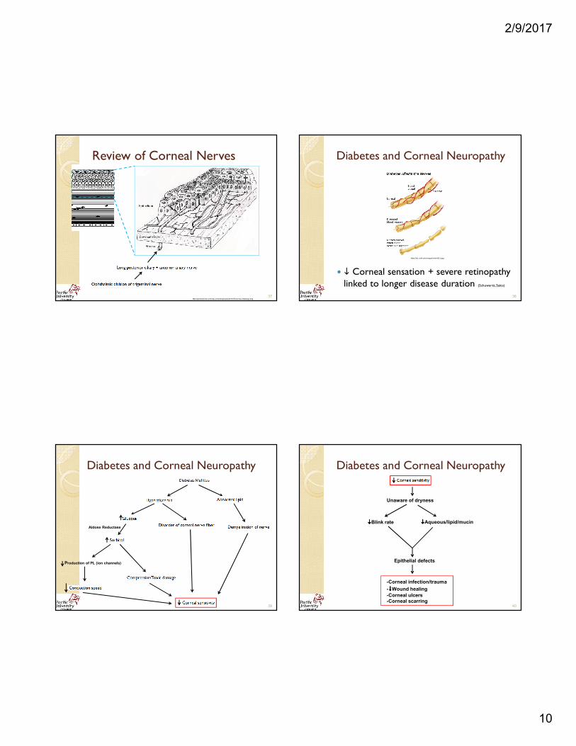
Diabetes and Corneal Neuropathy
� � Corneal sensation + severe retinopathy linked to longer disease duration (Schuwartz, Saito)
38
https://dtc.ucsf.edu/images/charts/5.d.jpg
Diabetes and Corneal Neuropathy
Production of PL (ion channels)
Aldose Reductase
39
Diabetes and Corneal Neuropathy
Unaware of dryness
����Aqueous/lipid/mucin����Blink rate
Epithelial defects
-Corneal infection/trauma
-����Wound healing
-Corneal ulcers
-Corneal scarring40

2/9/2017
11
Stages of Diabetic Corneal Neuropathy
Stage 1
• Superficial/epithelial
Stage 2
• Epithelial breakdown
Stage 3
• Stromal involvement• Ulcer/melting/perforation
(Adbelkader, Lockwood)41
Clinical advice: DK & Nerves
Course of nerve changes…
� Mild to moderate neuropathy
� OBJECTIVE change in long nerve fiber bundles
� Severe neuropathy
◦ SUBJECTIVE �Corneal sensitivity
� Instruments are more sensitive! (Rosenberg)
Clinical Pearl:
� Consider diabetic corneal neuropathy when pts develop unexplained corneal epithelial disease and ulcer (Lockwood)
42
Outline
� Introduction
� Impact of diabetes on the cornea
� Treatment
� Risk factors
� Differential diagnosis
� Complications
� Summary
43
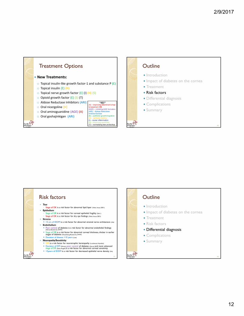
Treatment Options
� Standard Treatments:
o Preservative free topical lubricants
o Bandage contact lens
o Patching
o Tarsorrhaphy
o Induced ptosis
o Conjunctival flap
o Topical antiobiotic
o Topical steroid
44
2/9/2017
12
Treatment Options
� New Treatments:
o Topical insulin-like growth factor-1 and substance P (E)
o Topical insulin (E) (H)
o Topical nerve growth factor (E) (I) (H) (S)
o Opioid growth factor (E) (S) (T)
o Aldose Reductase Inhibitors (ARI)
o Oral nicergoline (H)
o Oral aminoguanidine (AGE) (A)
o Oral goshajinkigan (ARI)
**KEY**
(A) – improving attachment of epi
to basement MB
(AGE) – inhibiting AGE formation
(ARI) – Aldose Reductase
Inhibitor/Sorbitol
(E) – epithelial growth/migration
(H) – epithelial healing
(I) – ocular inflammation
(S) – improving sensitivity
(T) – normalizing tear production 45
Outline
� Introduction
� Impact of diabetes on the cornea
� Treatment
� Risk factors
� Differential diagnosis
� Complications
� Summary
46
Risk factors � Tear
◦ Stage of DK is a risk factor for abnormal lipid layer (Yokoi, Inoue 2001)
� Epithelium
◦ Stage of DR is a risk factor for corneal epithelial fragility (Saini )
◦ Stage of DK is a risk factor for dry eye findings (Yokoi, Inoue 2001)
� Stroma
� >5 yrs of IDDM is a risk factor for abnormal stromal nerve architecture (He)
� Endothelium
� Poor control of diabetes is a risk factor for abnormal endothelial findings (Herse, Busted, Su, DHW)
� Stage of DR is a risk factor for abnormal corneal thickness, thicker in earlier stages of diabetes (Rosenberg, Busted, Su, DHW)
� Duration of disease >10 years (Lee)
� Neuropathy/Sensitivity
� DM is a risk factor for neurotrophic keratopathy (Lockwood, Hyndiuk)
� Duration of DM (Herse), poor control of diabetes (Herse) and more advanced stage of DR (Saito, Rogell) is a risk factor for abnormal corneal sensitivity
� >5years of IDDM is a risk factor for decreased epithelial nerve density (He)
47
Outline
� Introduction
� Impact of diabetes on the cornea
� Treatment
� Risk factors
� Differential diagnosis
� Complications
� Summary
48
2/9/2017
13
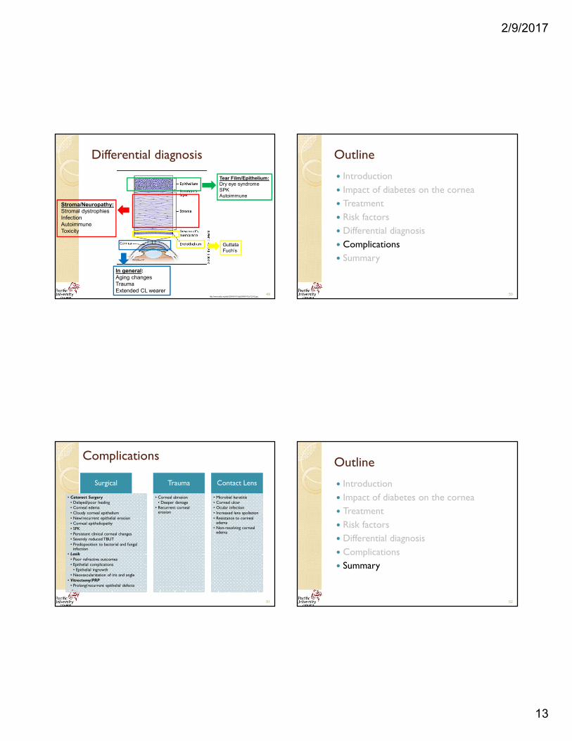
Differential diagnosis
Tear Film/Epithelium:
Dry eye syndrome
SPK
Autoimmune
Guttata
Fuch’s
Stroma/Neuropathy:
Stromal dystrophies
Infection
Autoimmune
Toxicity
In general:
Aging changes
Trauma
Extended CL wearer49
http://www.aafp.org/afp/2004/0701/afp20040701p123-f2.jpg
Outline
� Introduction
� Impact of diabetes on the cornea
� Treatment
� Risk factors
� Differential diagnosis
� Complications
� Summary
50
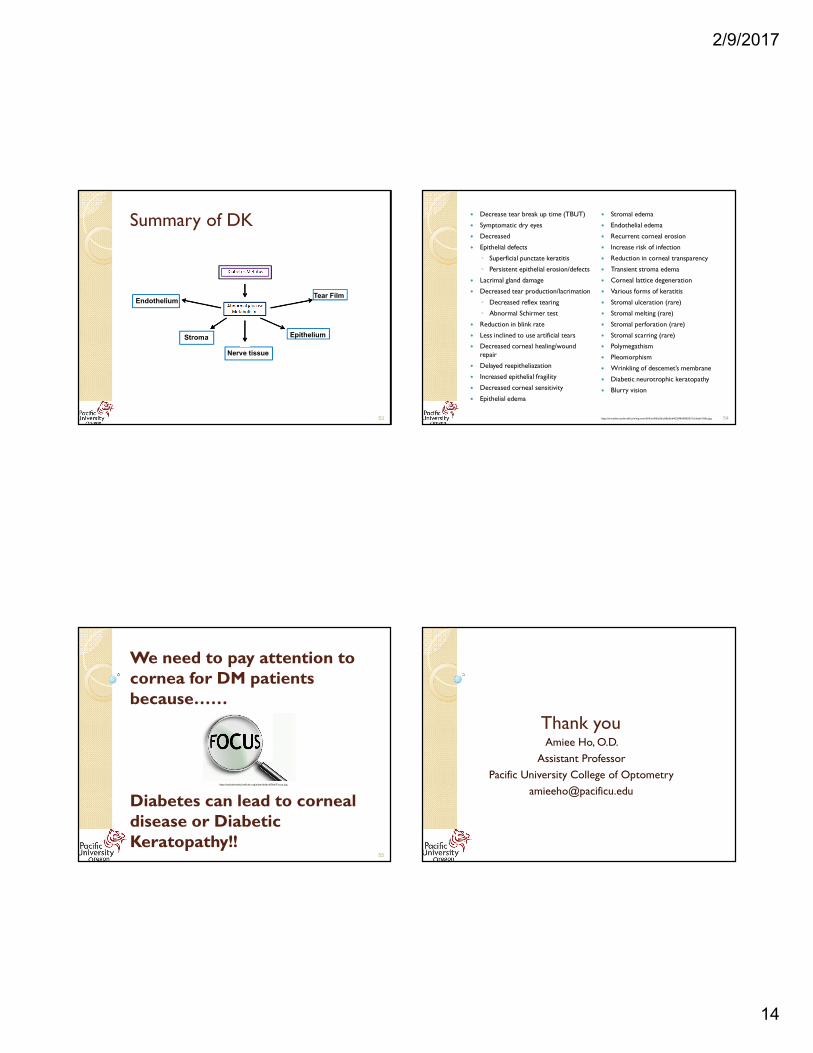
Complications
Surgical
• Cataract Surgery• Delayed/poor healing• Corneal edema
• Cloudy corneal epithelium
• New/recurrent epithelial erosion• Corneal epitheliopathy
• SPK
• Persistent clinical corneal changes• Severely reduced TBUT
• Predisposition to bacterial and fungal infection
• Lasik
• Poor refractive outcomes
• Epithelial complications • Epithelial ingrowth
• Neovascularization of iris and angle
• Vitrectomy/PRP• Prolong/recurrent epithelial defects
Trauma
• Corneal abrasion• Deeper damage
• Recurrent corneal erosion
Contact Lens
• Microbial keratitis• Corneal ulcer• Ocular infection
• Increased lens spoliation
• Resistance to corneal edema
• Non-resolving corneal edema
51
Outline
� Introduction
� Impact of diabetes on the cornea
� Treatment
� Risk factors
� Differential diagnosis
� Complications
� Summary
52
2/9/2017
14
Summary of DK
Nerve tissue
Endothelium
Stroma
Tear Film
Epithelium
53
� Decrease tear break up time (TBUT)
� Symptomatic dry eyes
� Decreased
� Epithelial defects
◦ Superficial punctate keratitis
◦ Persistent epithelial erosion/defects
� Lacrimal gland damage
� Decreased tear production/lacrimation
◦ Decreased reflex tearing
◦ Abnormal Schirmer test
� Reduction in blink rate
� Less inclined to use artificial tears
� Decreased corneal healing/wound repair
� Delayed reepitheliazation
� Increased epithelial fragility
� Decreased corneal sensitivity
� Epithelial edema
� Stromal edema
� Endothelial edema
� Recurrent corneal erosion
� Increase risk of infection
� Reduction in corneal transparency
� Transient stroma edema
� Corneal lattice degeneration
� Various forms of keratitis
� Stromal ulceration (rare)
� Stromal melting (rare)
� Stromal perforation (rare)
� Stromal scarring (rare)
� Polymegathism
� Pleomorphism
� Wrinkling of descemet’s membrane
� Diabetic neurotrophic keratopathy
� Blurry vision
54https://s-media-cache-ak0.pinimg.com/564x/c8/8a/5b/c88a5bd4039f466880937c5feafe769a.jpg
We need to pay attention to cornea for DM patients because……
Diabetes can lead to corneal disease or Diabetic Keratopathy!!
55
https://socialanxietyinsti tute.org/sites/default/fi les/Focus.jpg
Thank you Amiee Ho, O.D.
Assistant Professor
Pacific University College of Optometry
2/9/2017
15
References � Azar DT, Spurr-Michaud SJ, Tisdale AS, et al. Decreased penetration of anchoring fibrils into the diabetic stroma. A morphometric analysis. Arch Ophthalmol 1989; 107: 1520-3.
� Azar DT, Spurr-Michaud SJ, Tisdale AS, et al. Altered epithelial-basement membrane interactions in diabetic corneas. Arch Ophthalmol 1992; 110: 537-40.
� Bonini S. Lambiase A, Rama P, Capriogli G, Aloe L. Topical treatment with nerve growth factor for neurotrophic keratitis. Ophthalmology 2000; 107: 1347-52.
� Choo, MM, K. Prakash, T. Soong, N. Ramli, and AJ Kadir. "Corneal Changes in Type II Diabetes Mellitus in Malaysia." Int J Opthalmol 3.3 (2010): 234-36.
� Cisarik-Fredenburg P. Discoveries in research on diabetic keratopathy. Optometry 2001; 72: 691-704.
� Dogru M, Katakami C, Inoue M. Tear function and ocular surface changes in noninsulin-dependent diabetes mellitus. Ophthalmology 2001; 108: 586-592.
� Foulks, G.N., R.A. Thoft, H.D. Perry and F.I. Tolentino. Factors related to corneal epithelial complications after closed vitrectomy in diabetics. Arch. Ophthalmol. 97: 1076-1078, 1979.
� Friend J, Ishii Y, Thoft RA: Corneal epithelial changes in diabetic rats. Ophthalmic Res 14: 269-278, 1982.
� Fujishima H, Shimazaki J, Yagi Y, et al. Improvement of corneal sensation and tear dynamics in diabetic patients by oral aldose reductase inhibitor, ONO-2235: a preliminary study. Cornea1996; 15: 368-72.
� Gasset AR, Braverman LE, Fleming MC, et al: Tear glucose detection of hyperglycemia, Am J Ophthalmol 65: 414, 1968.
� Gekka M, Miyato K, Nagai Y, et al. Corneal epithelial barrier function in diabetic patients. Cornea 2004; 23:35-7.
� Gobbels M, Spitznas M, Oldendoerp J. Impairment of corneal epithelial barrier function in diabetics. Graefes Arch Clin Exp Ophthalmol 1989; 227:142-4.
� Goebbels M. Tear secretion and tear film function in insulin dependent patients with diabetes. Br J Ophthalmol 2000; 84: 19-21.
� Hatchell DL, Magolan JJ Jr, Besson MJ, Goldman AI, Pederson HJ, Schultz KJ. Damage to the epithelial basement membrane in the corneas of diabetic rabbits. Arch Ophthalmol 1983; 101: 469-471.
� He, Jiucheng, and Haydee EP Bazan. "Mapping the Nerve Architecture of Diabetic Human Corneas." Ophthalmology 119.5 (2012): 956-64.
� Henkind P, Wise GN: Descemet’s wrinkles in diabetes. Am J Ophthalmol 52: 371, 1961.
� Herse, Peter R. "A Review of Manifestations of Diabetes Mellitus in the Anterior Eye and Cornea." American Journal of Optometry & Physiological Optics 65.3 (1988): 224-30.
� "- Histology of the Cornea." - Histology of the Cornea. N.p., n.d. Web. 23 Sept. 2012. <http://simple-med.blogspot.com/2008/07/histology-of-cornea.html>.
� Hosotani H, Ohashi Y, Yamada M, et al. Reversal of abnormal corneal epithelial cell morphologic characteristics and reduced corneal sensitivity in diabetic patients by aldose reductase inhibitor, CT-112. Am J Ophthalmol 1995; 119: 288-94.
� Hyndiuk RA, Kazarian EL, Schultz RO, Seideman S. Neurotrophic corneal ulcers in diabetes mellitus. Arch Ophthalmol 1977; 95: 2193-2196
� Inoue, K., K. Okugawa, S. Amano, T. Oshika, E. Takamura, F. Egami, G. Umizu, K. Aikawa, and S. Kato. "Blinking and Superficial Punctate Keratopathy in Patients with Diabetes Mellitus." Eye 19.4 (2004): 418-21.
� Inoue K, Kato S, Inoue Y, Amano S, Oshika T. The corneal endothelium and thickness in type II diabetes mellitus. Jpn J Ophthalmol 2002; 46(1): 65-69.
57
References � Inoue, Kenji, MD, Satoshi Kato, MD, Chika Ohara, MD, Jiro Numaga, MD, Shiro Amano, MD, and Tetsuro Oshika, MD. "Ocular and Systemic Factors Relevant to Diabetic
Keratoepitheliopathy." Cornea 20.8 (2001): 798-801.
� Ioli-Spada G; ulterior contributo allo studio della cherato distrofia epiteliale punctata diabetic. Boll Ocul 43: 775, 1964.
� Kaji, Y. "Prevention of Diabetic Keratopathy." Br J Ophthalmol 89 (2005): 254-55.
� Kenyon K, Wafai Z, Michels R, et al. Corneal basement membrane abnormality in diabetes mellitus. Invest Ophthalmol Vis Sci 1978; 17(suppl): 245.
� Keoleian, GM, JM Pach, DO Hodge, SD Trocme, and WM Bourne. "Structural and Functional Studies of the Corneal Endothelium in Diabetes Mellitus." American Journal of Ophthalmology 113.1 (1992): 64-70.
� Lee JS, Oum BS, Choi HY, Lee JE, Cho BM. Differences in corneal thickness and corneal endothelium related to duration in diabetes. Eye 2006; 20(3):315-318.
� Lockwood, A., M. Hope-Ross, and P. Chell. "Neurotrophic Keratopathy and Diabetes Mellitus." Eye 20 (2006): 837-39.
� Matsuda M, Awata T, Ohashi Y, et al. The effects of aldose reductase inhibitor on the corneal endothelial morphology in diabetic rats. Curr Eye Res 1987; 6:391-7.
� Meyer LA, Ubels JL, Edelhauser HF. Corneal endothelial morphology in the rat. Effects of aging, diabetes, and topical aldose reductase inhibitor treatment. Invest Ophthalmol Vis Sci 1988; 29: 940-8.
� Morax V: Encycl fr Ophth IV, p 278, 1905.
� Nagaki, Yasunori, Seiji Hayasaka, Yoriko Hayasaka, Chiharu Kadoi, Nobuyasu Sekiya, and Katsutoshi Terasawa. "Effects of Goshajinkigan on Corneal Sensitivity, Superificial Punctate Keratopathy and Tear Secretion in Patients with Insulin-dependent Diabetes Mellitus." The American Journal of Chinese Medicine 31.1 (2003): 103-09.
� Nakamura M, Kawahara M, Morishige N, et al. Promotion of corneal epithelial wound healing in diabetic rats by the combination of a substance P-derived peptide (FGLM-NH2) and insulin-like growth factor-1. Diabetologia 2003: 46: 839-42.
� Ohashi Y, Matsuda M, Hosotani H, Tano Y, Ishimoto I, Fukuda M, Manabe R: Aldose Reductase Inhibitor (CT-112) eye drops for diabetic corneal epitheliopathy. Am J Ophthalmol 105:233-238, 1988.
� Ohguro N, Matsuda M, Ohashi Y, et al. Topical aldose reductase inhibitor for correcting corneal endothelial changes in diabetic patients. Br J Ophthalmol 1995; 79: 1074-7.
� Pierre EJ, Barrow RE, Hawkins HK. Effects of insulin on wound healing. J Trauma 1998; 44: 342-5.
� Rao, GN. "Dr. P. Siva Reddy Oration. Diabetic Keratopathy." Indian J Ophthalmol. 35.5-6 (1987): 16-36. Print.
� Rogell GD. Corneal hypesthesia and retinopathy in diabetic mellitus. Ophthalmology 1980;87:229-33.
� Rosenberg ME, Tervo TMT, Immonen IJ, et al. Corneal Structure and sensitivity in type 1 diabetes mellitus. Invest Ophthalmol Vis Sci. 2000; 41: 2915-21.
� Rosenthal SP. Acceleration of primary wound healing by insulin. Arch Surg 1968; 96: 53-5.
� Roszkowska AM, Tringali CG, Colosi P, Squeri CA, Ferreri G. Corneal endothelium evaluation in type I and type II diabetes mellitus. Ophthalmologica 1999;213(4):258-261.
� Saini JS, Khandalavla B. corneal epithelial fragility in diabetes mellitus. Can J Ophthalmol 1995; 30: 142-6.
� Saito J, Enoki M, Hara M. et al. Correlation of corneal sensation, but not of basal or reflex tear secretion, with the stage of diabetic retinopathy. Cornea 2003: 22: 15-18.
58
References � Sato, Euchi, MD, Fumihiko Mori, MD, PhD, Sho Igarashi, MD, Tohru Abiko, MD, PhD, Masumi Takeda, MD, PhD, Satoshi Ishiko, MD, PhD, and Akitoshi Yoshida, MD,
PhD. "Corneal Advanced Glycation End Product Increase in Patients With Proliferative Diabetic Retinopathy." Diabetes Care 24.3 (2001): 479-82.
� Schultz, Richard O., MD, Diane L. Van Horn, PhD, Mark A. Peters, MD, Kristine M. Klewin, MD, and William H. Schutten, MD. "Diabetic Keratopathy." Trans Am Ophthalmol Soc LXXIX (1981): 180-99.
� Schultz, Richard O., MD, Mark A. Peters, MD, Kathleen Sobocinski, MS, Kamal Nassif, MD, and Karen J. Schultz, BA. "Diabetic Corneal Neuropathy." Tr. Am. Ophth. 131 (1983): 107-24.
� Schultz, RO, M. Matsuda, RW Yee, HF Edelhauser, and KJ Schultz. "Corneal Endothelial Changes in Type I and Type II Diabetes Mellitus." American Journal of Ophthalmology 98.4 (1984): 401-10.
� Schwartz DE: Corneal sensitivity in diabetics. Am J Ophthalmol 91:174, 1974.
� Shenoy R, Khandekar R, Bialasiewicz A, Al Muniri A. Corneal endothelium in patients with diabetes mellitus: a historical cohort study. Eur J Ophthalmol 2009; 19(3): 369-375.
� Shoji, M., H. Sato, Y. Hirai, Y. Oguni, C.. Sugimoto, S. Morishita and C. Ito. Pharmacological effects of Gosha-jinki-gan-ryo extract: effects on enxperimental diabetes.
Folia. Pharmacol. Jpn. (Nippon Yakuri Zasshi) 99: 143-152, 1992.
� Siribunkum J, Kosrirukvongs P, Singalavanija A. Corneal abnormalities in diabetes. J Med Assoc Thai 2001;84(8):1075-1983.
� Stolwijk, T.R., J.A. van Best, J.P. Boot, H.H.P.J. Lemkes and J.A. Oosterhuis. Corneal epithelial barrier function after oxybuprocaine provocation in diabetes. Invest. Ophthalmol. Vis. Sci. 31: 436-439, 1990.
� Taylor HR, Kimsey RA. Corneal epithelial basement membrane changes in diabetes. Invest Ophthalmol Vis Sci. 1981; 20: 548-53.
� Tsubota K, Yamada M. The effect of aldose reductase inhibitor on the corneal epithelium. Cornea. 1993; 12: 161-2.
� Van Ort SR, Gerber RM. Topical application of insulin in the treatment of decubitus ulcers: a pilot study. Nurs Res 1976; 25: 9-12.
� Wylegala, E., L. Mocko, A. Woyna-Orlewicz, S. Teper, and B. Orzechowska-Wylegala. "Diabetic Complications within Ocular Surface." Polski Merkuriusz Lekarski 21.125 (2006): 495-97.
� Yokoi N, Nhya A, Komuro A, et al. Effects of aldose reductase inhibitor CT-112 on the corneal epithelial barrier of galactose-fed rats. Curr Eye Res 1997; 16: 595-9.
� Zagon IS, Klocek MS, Sassani JW, McLaughlin PJ. Dry eye reversal and corneal sensation restoration with topical naltrexone in diabetes mellitus. Arch Ophthalmol 2009; 127: 1468-73.
59
![The Guide - Diabetic Retinopathy - Vision Lossvisionloss.org.au/wp-content/uploads/2016/05/The... · the guide [diabetic retinopathy] What is Diabetic Retinopathy? Diabetic Retinopathy](https://img.pdfslide.net/doc/110x75/5e3ed00bf9c32e41ea6578a8/the-guide-diabetic-retinopathy-vision-the-guide-diabetic-retinopathy-what.jpg)