Embed Size (px)
Citation preview
VOLUME11DECEMBER20208
343
N. Meuleman, MD, PhD¹, M. Vercruyssen, MD¹, M. Cliquennois, MD², J. Caers, MD³, C. Doyen, MD, PhD4, G. Bries, MD5, C. Jacquy, MD6, M. Delforge, MD, PhD7, M-C. Vekemans, MD8
¹Department of Haematology, Institut Jules Bordet, Brussels, Belgium, ²Department of Haematology, Clinique St Pierre, Brussels, Belgium, ³Depart-
ment of Haematology, Centre Hospitalier Universitaire de Liège, Liège, Belgium, 4Department of Haematology, CHU UCL Namur Site Godinne,
Yvoir, Belgium, 5Department of Haematology, AZ Turnhout, Turnhout, Belgium, 6Department of Haematology, CHU Ambroise Paré, Mons, Belgium, 7Department of Haematology, UZ Leuven, Leuven, Belgium, 8Department of Haematology, Cliniques universitaires Saint-Luc, Brussels, Belgium.
Please send all correspondence to: N. Meuleman, MD, PhD, Department of Haematology, Institut Jules Bordet, Rue Heger Bordet 1,
1000 Brussels, Belgium, tel : +32 2 541 32 37, email: [email protected].
Conflict of interest: The authors have nothing to disclose and indicate no potential conflict of interest.
Keywords: AL amyloidosis, cardiac amyloidosis, consensus guidelines, nephrotic syndrome.
Acknowledgement: The authors contributed equally to the redaction of this manuscript.
INTRODUCTIONSystemic amyloidosis is a group of rare disorders caused by the deposition of misfolded proteins within virtually every organ, compromising their function. A large amount of different involved proteins has been described and iden-tification of these proteins is a first step in the diagnosis process as the treatments of the diverse entities are com-pletely different. AL amyloidosis is the most common type of systemic amyloidosis with a reported incidence of about 1:100.000.2,3 It is a life-threatening condition caused by the deposition, but also the local toxicity, of misfolded monoclonal light chains (FLC) produced by a small plasma cell clone or, rarely, by another B- lymphoproliferative clone. Its prog-nosis depends on the nature of the involved organs but also on the disease extension at diagnosis.4 Despite im-provements in the treatment of the disease, patients with advance organ damage still present a short survival. There-fore, early recognition of the symptoms and establish ment of the diagnosis are crucial.
CLINICAL PRESENTATIONClinical symptoms of AL amyloidosis are confounding and rarely specific, resulting in a significant delay in diag-nosis.5,6 Retrospective studies reported that the median time from first clinical signs to diagnostic is 1.2 years and that about a quarter of the patients had to see four or more specialists before the diagnosis.6 Fatigue, weight loss, weakness, lower limbs oedema and dyspnoea are the most frequently described symptoms while pathognomonic macro glossia and periorbital ecchymosis (racoon eyes) are only seen in 15% of the cases (Table 1).7 Every organ except the brain could be injured, but the heart and the kidneys are predominant sites of fibrils deposition. Indeed, dys-function of these organs is observed in 75% and 57% of the cases respectively.8 Involvement of the nerves, leading to autonomic dysfunction and/or symmetric sensory-motor neuropathy, is not uncommonly seen (22%) as is the liver (20%) and the gastrointestinal tract as well (15%). Soft tissue involvement, encountered in 15% of the patients, is highly specific of AL amyloidosis. Organs that may be
Diagnosis and treatment of AL amyloidosis: Belgian guidelines
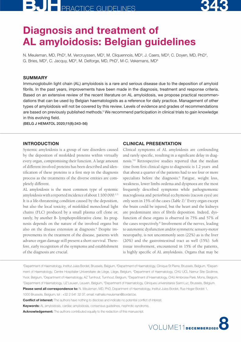
SUMMARY Immunoglobulin light chain (AL) amyloidosis is a rare and serious disease due to the deposition of amyloid fibrils. In the past years, improvements have been made in the diagnosis, treatment and response criteria. Based on an extensive review of the recent literature on AL amyloidosis, we propose practical recommendations that can be used by Belgian haematologists as a reference for daily practice. Management of other types of amyloidosis will not be covered by this review. Levels of evidence and grades of recommendations are based on previously published methods.¹ We recommend participation in clinical trials to gain knowledge in this evolving field.(BELG J HEMATOL 2020;11(8):343-56)
PRACTICE GUIDELINES
VOLUME11DECEMBER2020
344
affected include muscles, lungs, and joints. Amyloidosis can also be associated with bleeding disorders in relation to organ involvement, fragility of blood vessels and, in a small percentage of patients, a factor X deficiency.9
DIAGNOSISAs delayed diagnosis is associated with irreversible organ damage and as a direct consequence, a worse prognosis. Hence, all efforts must be made to an early diagnosis of AL amyloidosis. Therefore, an annual evaluation of albuminu-ria and dosage of NT pro BNP is recommended by experts for all patients presenting a monoclonal gammopathy of un-determined significance (MGUS) or a smoldering multiple myeloma (SMM) with abnormal LC ratio (LCR).5,8 A careful anamnesis and clinical examination should also be done, focusing on symptoms and clinical signs of AL amyloi-dosis (Table 1). It must be suspected in case of unex plained albuminuria, nephrotic syndrome, diastolic dysfunction associated with microvoltage electrocardiogram, autonomic dysfunction, unexplained symmetric sensory- motor neuro-pathy and, surely if characteristic soft tissue involvements are observed.Confirmation of the diagnosis requires several steps: 1) Confirming the presence of amyloid deposits; 2) Identi-fying the amyloid fibril subtype; 3) Evaluate the plasma cell clone; and 4) Evaluate the extent and severity of organ involvement (Table 2).8
DIAGNOSIS CONFIRMATION The diagnosis requires the demonstration of amyloid deposits in a tissue biopsy. Therefore, it is crucial to per-form a tissue biopsy when one suspects AL amyloidosis. Of course, performing a biopsy of an involved organ offers the best chance to demonstrate the presence of amyloid fibrils and clearly constitutes the most sensitive method. Because of the bleeding risk due to vessels fragility and potential coagulation perturbations, such a biopsy is only indicated when other approaches could not confirm the diagnosis. A biopsy of the abdominal sub-cutaneous fat (instructive video on www.amyloid.nl.) is non-invasive, much less expensive and allows to detect AL amyloid deposits in 84% of the cases and up to 90% if combined with bone marrow biopsy.10-12 If that method fails to show fibrils, a biopsy of the accessory salivary glands can show the amyloid fibrils in more than 50% of the remaining cases.13 Typically, the amyloid fibrils deposition is confirmed by the Congo red staining that allows to show apple-green birefringence of fibrils under polarized light.4 But once systemic amyloidosis is proved, identifying the type of fibrils is decisive. Indeed, an existing concomitant MGUS is clearly insufficient to make a diagnosis of AL amyloidosis, as 23% of the patients suffering of transthyretin amyloidosis (ATTR) present with a MGUS as well.14 To precise the fibrils type, immunohistochemistry is inexpensive and broadly available but should be performed in specialised centres.
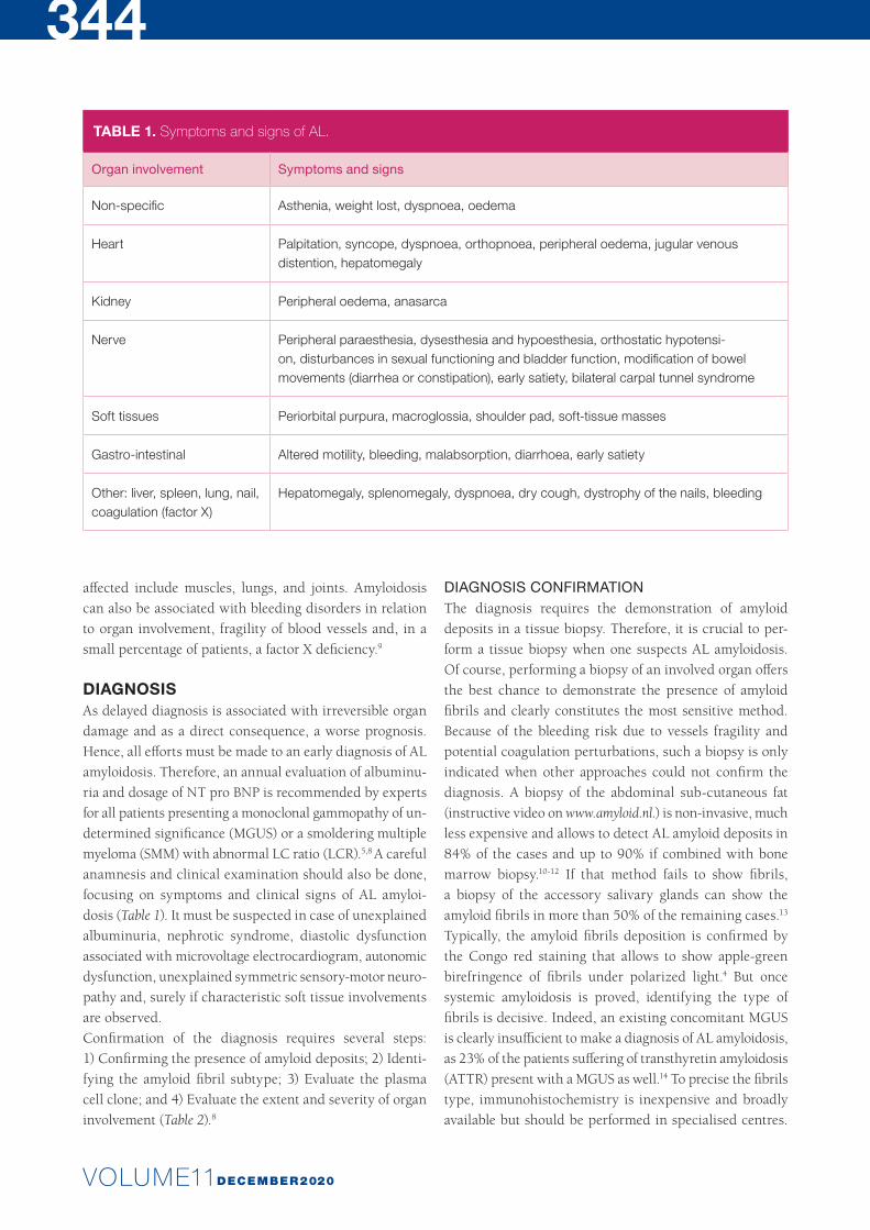
TABLE 1. Symptoms and signs of AL.
Organ involvement Symptoms and signs
Non-specific Asthenia, weight lost, dyspnoea, oedema
Heart Palpitation, syncope, dyspnoea, orthopnoea, peripheral oedema, jugular venous distention, hepatomegaly
Kidney Peripheral oedema, anasarca
Nerve Peripheral paraesthesia, dysesthesia and hypoesthesia, orthostatic hypotensi-on, disturbances in sexual functioning and bladder function, modification of bowel movements (diarrhea or constipation), early satiety, bilateral carpal tunnel syndrome
Soft tissues Periorbital purpura, macroglossia, shoulder pad, soft-tissue masses
Gastro-intestinal Altered motility, bleeding, malabsorption, diarrhoea, early satiety
Other: liver, spleen, lung, nail, coagulation (factor X)
Hepatomegaly, splenomegaly, dyspnoea, dry cough, dystrophy of the nails, bleeding
VOLUME11DECEMBER20208
345
Conversely, immunoelectron microscopy with gold-labelled antibodies has been reported to be a sensitive and very specific technique but is not available in the majority of the centres. Nowadays, mass spectrometry of amyloid deposits is considered to be the gold standard for amyloid typing.15,16 In some rare cases, gene sequencing may be required to confirm hereditary amyloidosis. In order to spare cardiac biopsy in elderly men suspected of senile amyloidosis (ATTRwt amyloidosis), it might be adequate to perform a cardiac scintigraphy using bone tracers (99Tc-pyrophosphate) to confirm the diagnosis.17 Actually, in the absence of any MGUS, if a grade 2 or 3 myocardial radiotracer uptake is observed, a specificity and a positive predictive value of 100% could be reached to identify an ATTR amyloidosis. Nevertheless, one must bear in mind that about 10% of AL amyloidosis have a positive bone scintigraphy.
PLASMA CELL CLONE EVALUATIONAfter confirmation of an AL Amyloidosis disease, an eva-luation of the plasma clone should be performed in order to define the underlying disease. This assessment requires serum and urine protein electrophoresis, serum and urine immunofixation, dosage of serum FLC, immunoglobulins quantification, bone marrow (BM) biopsy and aspiration with Fluorescent In Situ Hybridization (FISH) analysis. Typically, BM demonstrates a very low level of plasma cell infiltration (less than 5%). However, some cases can be diagnosed at a myeloma stage (>10%) and present a worse
prognosis.18 The cytogenetic evaluation is relevant as it has therapeutic implications.19 Indeed, the landscape of genetic abnormalities differs compared to multiple myeloma (MM). Hyperdiploidy and high-risk MM features are less frequent in AL amyloidosis while translocation t(11;14) is the most common feature, reported in up to 60% of the cases and, surprisingly, associated with a poor prognosis related to poor response to bortezomib and immunomodulatory therapy.19 Deletion of 1q21 has been reported as an inde-pendent adverse prognostic factor in patients treated with melphalan-dexamethasone.20 Even if cytogenetic assessment should be done at diagnosis regarding its clinical impli-cations, the typical low plasma cell infiltration leads fre-quently to technical issues and inconclusive results.
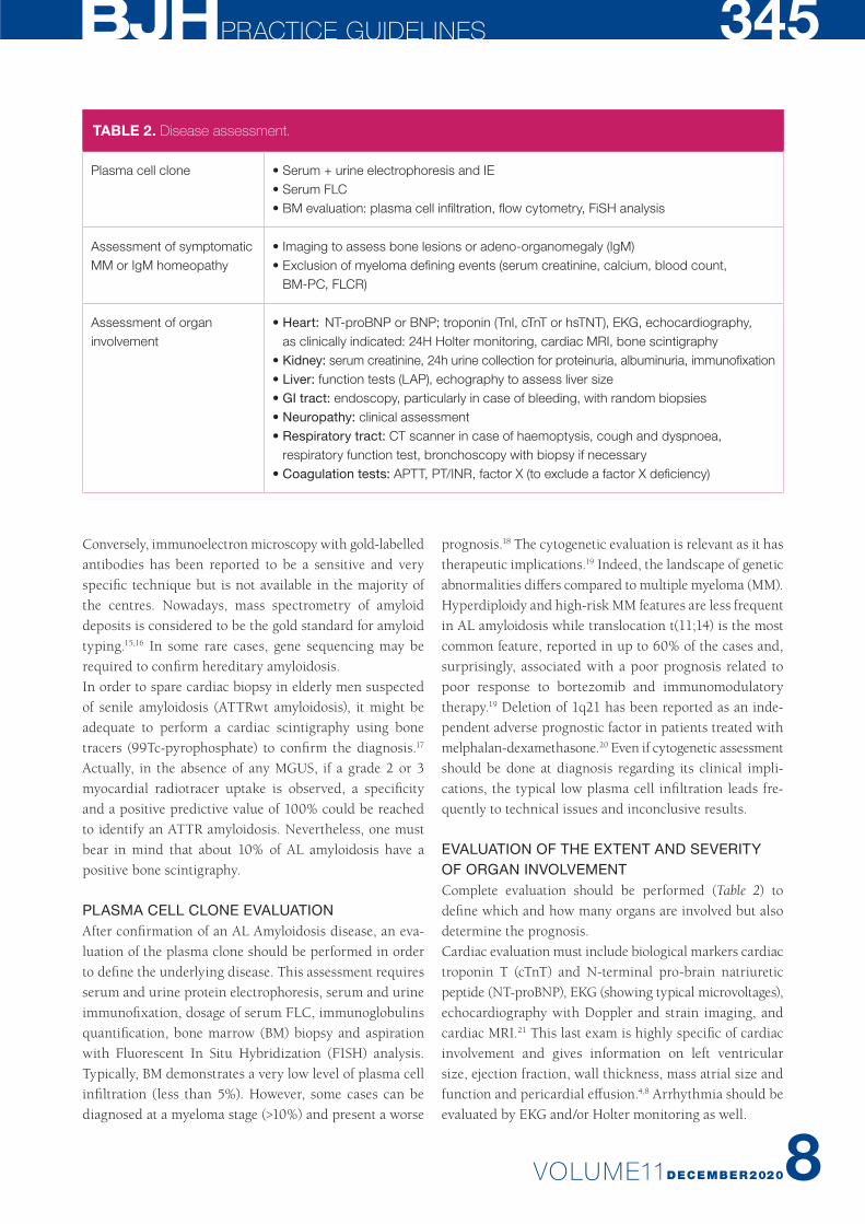
EVALUATION OF THE EXTENT AND SEVERITY OF ORGAN INVOLVEMENTComplete evaluation should be performed (Table 2) to define which and how many organs are involved but also determine the prognosis. Cardiac evaluation must include biological markers cardiac troponin T (cTnT) and N-terminal pro-brain natriuretic peptide (NT-proBNP), EKG (showing typical microvoltages), echocardiography with Doppler and strain imaging, and cardiac MRI.21 This last exam is highly specific of cardiac involvement and gives information on left ventricular size, ejection fraction, wall thickness, mass atrial size and function and pericardial effusion.4,8 Arrhythmia should be evaluated by EKG and/or Holter monitoring as well.
TABLE 2. Disease assessment.
Plasma cell clone • Serum + urine electrophoresis and IE• Serum FLC• BM evaluation: plasma cell infiltration, flow cytometry, FiSH analysis
Assessment of symptomatic MM or IgM homeopathy
• Imaging to assess bone lesions or adeno-organomegaly (IgM)• Exclusion of myeloma defining events (serum creatinine, calcium, blood count,
BM-PC, FLCR)
Assessment of organ involvement
• Heart: NT-proBNP or BNP; troponin (TnI, cTnT or hsTNT), EKG, echocardiography, as clinically indicated: 24H Holter monitoring, cardiac MRI, bone scintigraphy
• Kidney: serum creatinine, 24h urine collection for proteinuria, albuminuria, immunofixation• Liver: function tests (LAP), echography to assess liver size• GI tract: endoscopy, particularly in case of bleeding, with random biopsies• Neuropathy: clinical assessment • Respiratory tract: CT scanner in case of haemoptysis, cough and dyspnoea,
respiratory function test, bronchoscopy with biopsy if necessary• Coagulation tests: APTT, PT/INR, factor X (to exclude a factor X deficiency)
PRACTICE GUIDELINES
VOLUME11DECEMBER2020
346
Evaluation of kidney involvement must include 24-hour urine analysis with protein, albumin and ions level of excretion and creatinine clearance.22 Serum ions should be evaluated as well. Cholesterol level should be measured in case of nephrotic syndrome.Abdominal ultrasound could be useful to investigate the liver and spleen. Upper and lower endoscopy may be required in case of GI bleeding, modification of bowel movements habits or significant weight loss. Coagulation tests and dosage of factor X are part of the baseline disease evaluation.Finally, in order to rule out multiple myeloma (MM), low voltage total body CT scans, FDG Pet-CT or whole-body MRI should be performed.
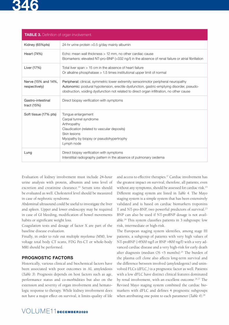
PROGNOSTIC FACTORSHistorically, various clinical and biochemical factors have been associated with poor outcomes in AL amyloidosis (Table 3). Prognosis depends on host factors such as age, performance status and co-morbidities but also on the extension and severity of organ involvement and hemato-logic response to therapy. While kidney involvement does not have a major effect on survival, it limits quality of life
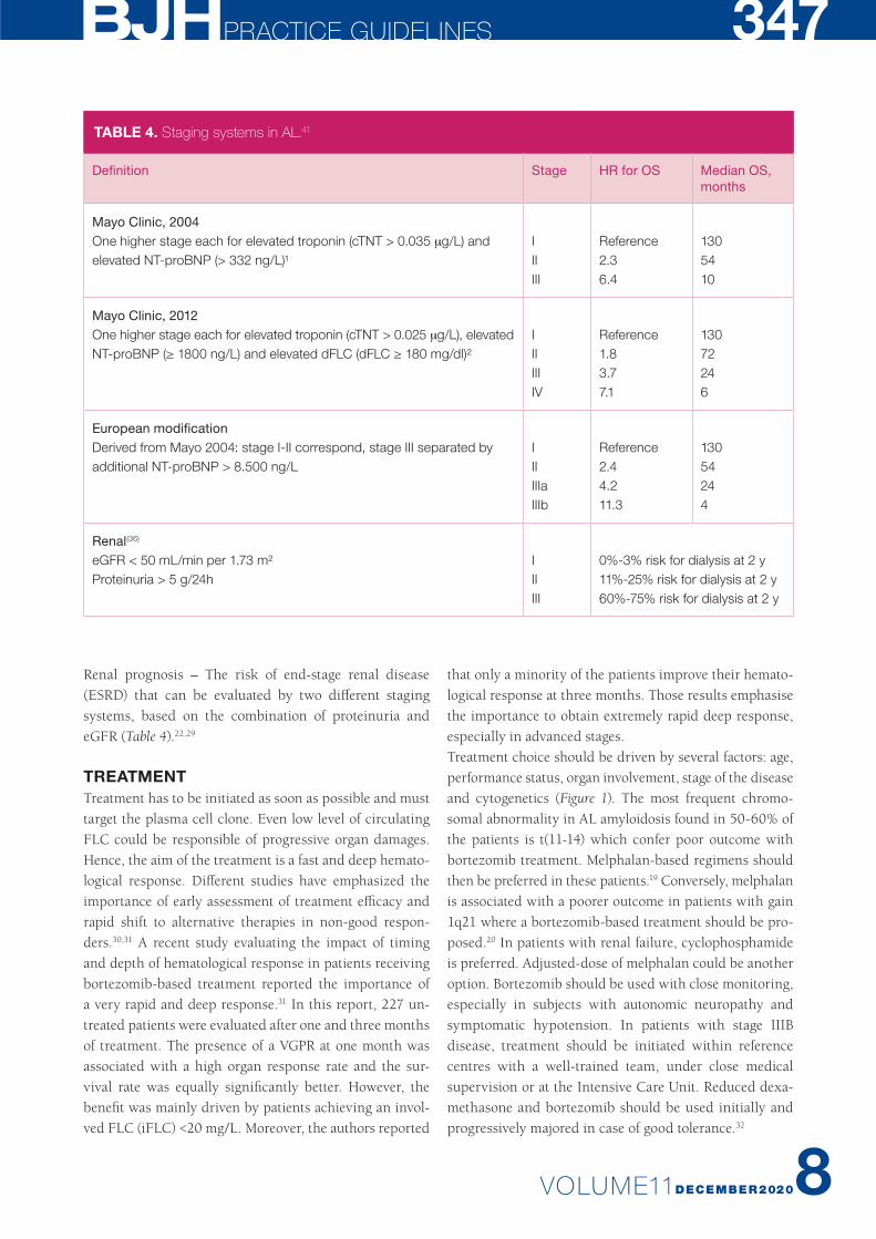
and access to effective therapies.17 Cardiac involvement has the greatest impact on survival, therefore, all patients, even without any symptoms, should be assessed for cardiac risk.23 Different staging system are listed in Table 4. The Mayo staging system is a simple system that has been extensively validated and is based on cardiac biomarkers troponins T and NT-pro-BNP, two powerful predictors of survival.23 BNP can also be used if NT-proBNP dosage is not avail-able.24 This system classifies patients in 3 subgroups: low risk, intermediate or high-risk.The European staging system identifies, among stage III patients, a subgroup of patients with very high values of NT-proBNP (>8500 ng/l or BNP >800 ng/l) with a very ad-vanced cardiac disease and a very high-risk for early death after diagnosis (median OS <5 months).25 The burden of the plasma cell clone also affects long-term survival and the difference between involved (amyloido genic) and unin-volved FLCs (dFLC,) is a prognostic factor as well. Patients with a low dFLC have distinct clinical features dominated by renal involvement, with an excellent outcome.26,27 The Revised Mayo staging system combined the cardiac bio-markers with dFLC and defines 4 prognostic subgroups when attributing one point to each parameter (Table 4).28
TABLE 3. Definition of organ involvement.
Kidney (65%pts) 24-hr urine protein >0.5 g/day mainly albumin
Heart (74%) Echo: mean wall thickness > 12 mm, no other cardiac causeBiomarkers: elevated NT-pro-BNP (>332 ng/l) in the absence of renal failure or atrial fibrillation
Liver (17%) Total liver span > 15 cm in the absence of heart failure Or alkaline phosphatase > 1.5 times institutional upper limit of normal
Nerve (15% and 14%, respectively)
Peripheral: clinical, symmetric lower extremity sensorimotor peripheral neuropathyAutonomic: postural hypotension, erectile dysfunction, gastric-emptying disorder, pseudo- obstruction, voiding dysfunction not related to direct organ infiltration, no other cause
Gastrointestinal tract (15%)
Direct biopsy verification with symptoms
Soft tissue (17% pts) Tongue enlargementCarpal tunnel syndrome ArthropathyClaudication (related to vascular deposits)Skin lesionsMyopathy by biopsy or pseudohypertrophyLymph node
Lung Direct biopsy verification with symptomsInterstitial radiography pattern in the absence of pulmonary oedema
VOLUME11DECEMBER20208
347
Renal prognosis – The risk of end-stage renal disease (ESRD) that can be evaluated by two different staging systems, based on the combination of proteinuria and eGFR (Table 4).22,29
TREATMENTTreatment has to be initiated as soon as possible and must target the plasma cell clone. Even low level of circulating FLC could be responsible of progressive organ damages. Hence, the aim of the treatment is a fast and deep hemato-logical response. Different studies have emphasized the importance of early assessment of treatment efficacy and rapid shift to alternative therapies in non-good respon-ders.30,31 A recent study evaluating the impact of timing and depth of hematological response in patients receiving bortezomib-based treatment reported the importance of a very rapid and deep response.31 In this report, 227 un-treated patients were evaluated after one and three months of treatment. The presence of a VGPR at one month was associated with a high organ response rate and the sur-vival rate was equally significantly better. However, the benefit was mainly driven by patients achieving an invol-ved FLC (iFLC) <20 mg/L. Moreover, the authors reported
that only a minority of the patients improve their hemato-logical response at three months. Those results emphasise the importance to obtain extremely rapid deep response, especially in advanced stages. Treatment choice should be driven by several factors: age, performance status, organ involvement, stage of the disease and cytogenetics (Figure 1). The most frequent chromo-somal abnormality in AL amyloidosis found in 50-60% of the patients is t(11-14) which confer poor outcome with bortezomib treatment. Melphalan-based regimens should then be preferred in these patients.19 Conversely, melphalan is associated with a poorer outcome in patients with gain 1q21 where a bortezomib-based treatment should be pro-posed.20 In patients with renal failure, cyclophosphamide is preferred. Adjusted-dose of melphalan could be another option. Bortezomib should be used with close monitoring, especially in subjects with autonomic neuropathy and symptomatic hypotension. In patients with stage IIIB disease, treatment should be initiated within reference centres with a well-trained team, under close medical supervision or at the Intensive Care Unit. Reduced dexa-methasone and bortezomib should be used initially and progressively majored in case of good tolerance.32
TABLE 4. Staging systems in AL.41
Definition Stage HR for OS Median OS, months
Mayo Clinic, 2004One higher stage each for elevated troponin (cTNT > 0.035 µg/L) and elevated NT-proBNP (> 332 ng/L)¹
IIIIII
Reference2.36.4
1305410
Mayo Clinic, 2012One higher stage each for elevated troponin (cTNT > 0.025 µg/L), elevated NT-proBNP (≥ 1800 ng/L) and elevated dFLC (dFLC ≥ 180 mg/dl)²
IIIIIIIV
Reference1.83.77.1
13072246
European modificationDerived from Mayo 2004: stage I-II correspond, stage III separated by additional NT-proBNP > 8.500 ng/L
IIIIIIaIIIb
Reference2.44.211.3
13054244
Renal(36)
eGFR < 50 mL/min per 1.73 m²Proteinuria > 5 g/24h
IIIIII
0%-3% risk for dialysis at 2 y11%-25% risk for dialysis at 2 y60%-75% risk for dialysis at 2 y
PRACTICE GUIDELINES

VOLUME11DECEMBER2020
348
FRONTLINE TREATMENTAUTOLOGOUS STEM CELL TRANSPLANTATIONMore than 50 non-randomised trials have confirmed the efficacy of high dose melphalan (HDM, 200 mg/m2) fol-lowed by ASCT, with organ response rates up to 65% in a proportion of eligible patients. However, this intensive therapy can only be offered to 15-25% of patients with AL amyloidosis. A phase III randomised trial published in 2007 comparing oral melphalan-dexamethasone (M-DEX) to HDM-ASCT reported a worse survival in the group of patients treated with high dose chemotherapy.33 Those results were linked to a very high transplanted related mortality (TRM) including 15% of deaths during stem cell mobilisation and collection. With improvement of suppor-tive care and better selection of subjects over time, more recent studies reported a decrease of TRM (2.4%) and a significant improvement of median overall survival over 10 years.34 This highlights the crucial role of patient selec-tion and the experience of the transplantation center Candidates to HDM-ASCT must be selected regarding
their performance status, organs function (cardiac, renal, pulmonary, hepatic), baseline systolic blood pressure and cardiac biomarkers. There may be some small differences in criteria for ASCT eligibility between centres, the BHS recommendations are listed in Table 2.4
Patients with a BM infiltration over 10% have an inferior complete remission (CR) rate, progression-free survival (PFS) and overall survival (OS).35 It has been reported that induction therapy before ASCT improves outcomes (CR and OS) among AL patients who have greater than 10% BMPC probably due to a rapid reduction of monoclonal light chains and a better selection of patients fit enough to receive high-dose melphalan (HDM).35 For patients with BMPC > 10% % or presenting at least one of the sympto-matic MM criteria, an induction with cyclophosphamide- bortezomib-dexamethasone (CyBorDex) should be given for two cycles or four in patients with MM criteria.A melphalan dose reduction has be proposed for frail patients, in order to reduce toxicity, but is associated with a reduced survival.36
FIGURE 1. Treatment of newly diagnosed AL amyloidosis.
ASCT: autologous stem cells transplantation, BM-PC: bone marrow plasma cells, CyBorDex: cyclophosphamide+borte-
zomib+dexamethasone, MDex: melphalan+dexamethasone, BMDex: bortezomib+melphalan+dexamethasone, HDM: high
dose melphalan.
ELIGIBLE FOR ASCT
YES
YES
CR
NO
NO
BM-PC > 10%
NO YES
Induction with 2-4Cycles of CyBorDex
HDM + ASCT
IConsolidation with 2Cycles of CyBorDex
Close surveillance of haematologicalor organ response / 3 months
EVALUATION: at least PR at 1 cycle and at least VRGP at 3 cycles
• If VGPR / haematological or organ progression (even in VGPR)
-> second line therapy
• If VGPR / CR => Close surveillance of haematological
or organ response / 3 months
Stage I to IIIa Stage IIIB• Low dose CyBorDex or BMDex* /MDEX if CI to bortezomib• Consider hospitalisation for monitoring
• CyBorDex or BMDex• BMDex in patients with t(11,14) or gain 1q21• MDEX if: • contraindication to Bortezomib: symptomatic neuropathy, fibrotic lung disease • Stage I and BM-PC< 10% and preserved renal function
HDM + ASCT
VOLUME11DECEMBER20208
349CONVENTIONAL CHEMOTHERAPYWhile M-DEX was until recently considered as one of the standard of treatment for patients with AL amyloidosis, the introduction of novel agents-based therapies has changed the game. Bortezomib is today a backbone in frontline therapy in AL amyloidosis. Two initial small studies, investigating the role of bortezomib, reported a hematological overall response rate (ORR) of 81-94% with about 50% of VGPR, results that were later confirmed on larger studies.37,38 In the European retrospective study, the hematological ORR was 65% with 43-49% of VGPR or better. However, patients with advanced cardiac stage (NT-pro-BNP >8500 ng/L) have still low response rates (42%, ≥ VGPR 23%) and poor survival (median, 7 months) mostly due to poor tolerability.39 In a large prospective observational study (n= 819), bortezomib- based treatment was associated with a prolonged OS of seven years and the best outcome were reached in patients with stringent dFLC responses, emphasising again the impor-tance of achieving a deep response.40 A recent phase III randomised trial comparing M-DEX to bortezomib-mel-phalan-dexamethasone (BM-DEX) reported a significant difference in terms of hematological response and OS.41 In that study, patients with severe cardiac disease (stage
IIIB), relevant ventricular arrhythmias, syncopes or pro-found hypotension were not eligible, and only 13% of the patients presented with a stage I disease. In the BM-DEX arm, the hematological ORR at 3 months was 79% versus 52% in the M-DEX arm (p= .002) and the number of patients reaching at least VGPR was also significantly higher in the BM-DEX arm (64% vs. 39%). More importantly, a 2- fold decrease of mortality was observed (HR: 0.50) in the BM-DEX arm with a median OS not reached compared to 34 months in the M-DEX arm. This is the first randomised trial in AL amyloidosis demonstrating a significant impact in terms of hematological response rate, PFS and OS. Based on these results, M-DEX is no more a standard option; expect for patients with contraindications to bortezomib or some selected patients with a stage I disease, a BM infil-tration less than 10% and preserved renal function.
MONOCLONAL ANTIBODY Daratumumab is an IgG1-kappa monoclonal antibody binding to CD38 antigen on the surface of plasma cells and is a major drug in the multiple myeloma treatment. After initial promising reports in heavily pre-treated AL amyloidosis patients, phase II trials and larger retrospective
RECOMMENDATIONS
If available, treatment of AL amyloidosis should be performed within the context of a clinical trial and in reference centres for advanced stages.
Treatment requires a multidisciplinary approach.
Considerations regarding initial therapy:• Bortezomib based regimens (BMDEX, CyBordDex) should be offered to all stage I-III patients if no
contraindications (level 1A, grade B), particularly in patients with renal dysfunction (level 2B, grade C).• In a selected population with stage I disease, BM-PC <10% and a normal kidney function, the M-DEX
regimen is still an option (level 2B, grade C).• Patients with t(11;14) should receive melphalan-based regimen (level 2A, grade B).• HDM followed by ASCT should only be performed in highly selected patients with minimal cardiac
involvement (cTnT < 0.06mcg/l, cTnI <0.1 mcg/l or NT-proBNP < 5000 ng/l) and adequate renal function (GRF >50 ml/min) (level 2A, grade B).
• For patients with a BM infiltration over 10% or presenting at least one of the symptomatic MM criteria, an induction with cyclophosphamide-bortezomib-dexamethasone (CyBorDex) should be given for 2 as the use of melphalan can significantly impact stem cell collection (level 2A, grade B).
• Dose-attenuated melphalan regimens are not recommended (level 2B, grade C).
Stem cell mobilisation should be performed with GCSF alone and in reference centres (level 3, grade C).
During stem cell reinfusion, cardiac monitoring is advised in patients with cardiac involvement (level 3, grade C).
Treatment of IgM AL amyloidosis should be driven by cytogenetic. Subjects with no t(11;14) require lymphoma/Waldenström treatments and other treatments targeting the plasma cell clone.
PRACTICE GUIDELINES
VOLUME11DECEMBER2020
350
studies reported outstanding results in relapsing refractory patients with ORR of 55-90% and up to 73% of patients reaching at least a VGPR.42-48 Moreover, all those reports showed a very rapid time to response of one month or less. In the light of these results, a phase III randomised trial (Andromeda study- Clinical Trials: NCT03201965) com-paring CyBorD to subcutaneous daratumumab-CyborD (dara-CyBorD) in newly diagnosed AL amyloidosis was launched.49,50 After a follow-up of eleven months, the initial report showed impressive results with a rate of VGPR or better of 79% in the dara-CyBorD arm versus 49% in the control arm, and a CR rate of 53% in the dara-CyBorD arm compared to 18% in the control arm (odds ratio, 5.1; 95% CI, 3.2-8.2; p< 0.0001). The organ response rate was also significantly better in patients receiving daratumumab with a 6-month cardiac response rate of 42% for dara-CyBorD versus 22% for CyBorD (p= 0.0029) and a 6-month renal response rate of 54% versus 27%, (p< 0.0001). There was no significant difference in terms of toxicity between both groups. It is the first time that such results have been achieved in untreated AL systemic amyloidosis and this regimen will probably become the new standard of care for newly diagnosed AL amyloidosis.Localised amyloidosis is a different entity, presenting deposits in bladder, larynx, the skin, eyelids, stomach, and colon or as solitary pulmonary nodules and can be treated by resection or with laser therapy. Unlike the systemic form, localised amyloidosis is associated with a very good prognosis, with a rare evolution to extensive disease.51
IgM related amyloidosis is a distinct subtype of amyloi-
dosis. Presentation is different from non-IgM amyloidosis with more peripheral nerve and soft tissue involvements and less cardiac injury (56% vs. 73%, p= 0.002).52 Two subtypes of IgM amyloidosis have been characterized, one consisting of pure plasma cell diseases (23%) associated with the same high prevalence of t(11;14) as in non- IgM amyloidosis, with no MYD88 mutations. Those subjects should be treated as non-IgM AL amyloidosis. Another subtype consisting of lymphoplasmacytic neoplasm asso-ciated with MYD88L265P and CXCR4 mutation who should be treated with lymphoma/Waldenström regimens.
RELAPSING-REFRACTORY PATIENTSWHEN TO RESTART A TREATMENT?When to retreated patients is still a matter of debate; how-ever, treatment should be anticipated before organ pro-gression. In a report from the Mayo Clinic on 235 patients initially treated with ASCT, subjects with a dFLC ≥50 mg/L at diagnosis and who achieved at least VGPR were able to tolerate a gradual rise in dFLC.53 This strategy should not be applied to patients with a dFLC <50 mg/L (23% of the patients) since about two-thirds of them, in fact, presented organ progression with a dFLC <50 mg/L. The Pavia group reported their experience in 259 patients who responded to upfront non-transplant therapy.54 About two-thirds of patients had a high-risk dFLC progression at the time of second line treatment which was defined as a dFLC >20 mg/L, a level >20% of baseline value, or a >50% increase from the value reached at best response. The presence of a high-risk dFLC progression preceded cardiac progression
RECOMMENDATIONS
At reappearance or progression of the biological markers, evaluation of biological relapse according to the dFLC at diagnosis and the best response observed on first line therapy should be performed as well as an evaluation of organ progression (level 2B, grade B).
Subjects with initial cardiac presentation should be treated as soon as there is a hematologic relapse as increase in cardiac biomarkers is associated with a worse OS (level 2B, grade A).
Patients with no advanced stage or severe organ damage and a dFLC >50 mg/l at diagnosis may be able to tolerate progressive increase of dFLC. A close monitoring is recommended anyway (level 2B, grade C).
For patients with organ progression without any other identified causes, an active search for the amyloidogenic clone should be performed (use MDR technique for patients still in CR) and a new line of treatment should be initiated as soon as possible (level 2B, grade BC).
Those recommendations should be balanced by the great fragility of some patients in whom a close monitoring could be an option.
VOLUME11DECEMBER20208
351by a median of 6 months in 85% of the subjects and, in this study, cardiac progression at the time of reinitiating a treat-ment was the only independent predictor of OS. Authors conclude that a high-risk dFLC progression should trigger the start of a rescue treatment. It has also been demonstrated that, in a small proportion of patients, organ progression or relapse could occur without hematological progression. When organ progression without modification of dFLC is suspected, it is essential to exclude another cause of organ deterioration before reinitiating therapy. Those results emphasise the possible toxicity of small amount of FLC.
TREATMENTTreatment decision should be driven by previous treatment, previous and expected toxicities, performance status and patient fragilities, organ dysfunctions and also by FISH cytogenetics.Retreatment with the same regimen has been investigated in a large (n= 1327) retrospective study of the Mayo Clinic and was associated with a significantly reduced TTNT (22 m vs. 32.3 m; p= .01). However, it was not associated with a significant impact on OS (30.8 m vs. 51.1 m; p= 0.5).55 This option could be recommended in patients with con-traindications to other drugs or who experienced a deep response and long interval before progression with the previous line of treatment.
MONOCLONAL ANTIBODIESIn four large reports and two phase II studies, daratumu-mab was associated with a very rapid response (<1 month), a high level of hematological response (ORR 55-90% and VGPR or better 47-73%), cardiac (22-55%) and renal res-ponse (31-67%).48-48 Daratumumab was administrated in monotherapy for a fixed period of time (six months) or for 24 months treatment and more.44,45,47,48 Two retrospective studies reported patients treated in combination with bortezomib or lenalidomide with a rate of at least VGPR of 59-66%.37 No difference in overall hematologic response rates was seen between patients treated with daratumumab single agent and those who received daratumumab combi-nations (after sixteen infusions, 81% vs. 88%, p= .470). In addition, CR/VGPR rate was similar in patients treated with single agent daratumumab or combination regimens (59% vs. 60%, p= .488). Despite the distinct characteristics of the studied populations, the heterogeneity of the applied regimens and duration of treatment, all reported a rapid and high level of deep responses, results that were never reported in relapsed/refractory Al amyloidosis before. Con-sidering those results, daratumumab should be offered to all patient not previously exposed to anti-CD38 therapy.
PROTEASOME INHIBITORSBortezomib should be offered to patients never exposed to the drug and who have no contraindications such as sig-nificant neuropathy or fibrotic lung disease. If the absence of other valid option, reduced bortezomib doses should be used with close monitoring, especially in subjects with autonomic neuropathy and symptomatic hypotension.Ixazomib is an interesting alternative as it is an oral pro-teasome inhibitor (PI) with less neurological side effects. After the initial promising results of a phase I-II study, the ixazomib–dexamethasone combination was compared to the physician choice in the Tourmaline–AL1 phase III trial.56,57 The primary end point, hematological ORR, was not met, but the ixazomib arm was associated with better results in terms of CR (26 vs. 17%), organ response rates (36 vs. 11%) and TTNT (27 vs. 13 months; p= 0.019).Preliminary results of treatment with Carfilzomib admini-strated at the dosage of 20/36 mg/m2 in a phase I-II study reported interesting RR with 63% ORR and 46%VGPR or better. In this trial, the median NT-pro-BNP was 542 pg/ml and grade 3/4 adverse events occurred in 71% of the patients.58 More safety data are still needed, especially in regard of the well-known cardiotoxicity of this PI.
IMMUNOMODULATORY DRUGSTreatments with lenalidomide or pomalidomide are effec-tive in patients exposed to borzetomib, alkylating agent and thalidomide. Before the era of anti-CD38 drugs, im-munomodulatory drugs (IMiDS) were frequently used as first option in relapsing patients. Prospective phase II study with lenalidomide showed an ORR of 38-61% with 41% a to 23% of organ responses.59-61 However, low level of deep hematological response is observed with 25-28% of VGPR or better and less than 10% of CR.62,63 This level might be increased with prolonged treatment. Physicians should be aware of the evaluation of cardiac responses in patients under IMiDs can be difficult as there could be associated with a possible increase of NT-proBNP in some cases without any worsening of cardiac function.64 The maximum dose tolerated of lenalidomide is lower than in MM and should not be higher than 15mg. Lenalidomide should be avoided in patients with a nephrotic syndrome as it could worsen the renal function.65 If no other option could be offered, a close monitoring of the renal function is then highly recommended.Pomalidomide-dexamethasone in the setting of relapse is an interesting option with a reported ORR of 42-96% and VGPR or better of 48-61%, better results compared to those reported with lenalidomide. A paroxysmal increase of NT-proBNP has also been reported in 89% of the patients,
PRACTICE GUIDELINES
VOLUME11DECEMBER2020
352
in the absence of any alteration of cardiac function. Hema-tological response to pomalidomide is rapidly achieved, in a median time of one to two months, in the absence of adverse toxicity profile.62,63
ANTI-FIBRIL THERAPIESAfter first encouraging results with NEOD001, a murine
antibody targeting misfolded light chains, the phase III study was closed due to futility.65 Other monoclonal antibodies targeting light chains and serum amyloid protein (SAP) are under investigations. It has been demonstrated that doxy-cycline has an anti-fibril activity. First interesting results have been published in association with anti-plasma cell therapy.66,67 A phase II trial on 25 patients reported a safe use
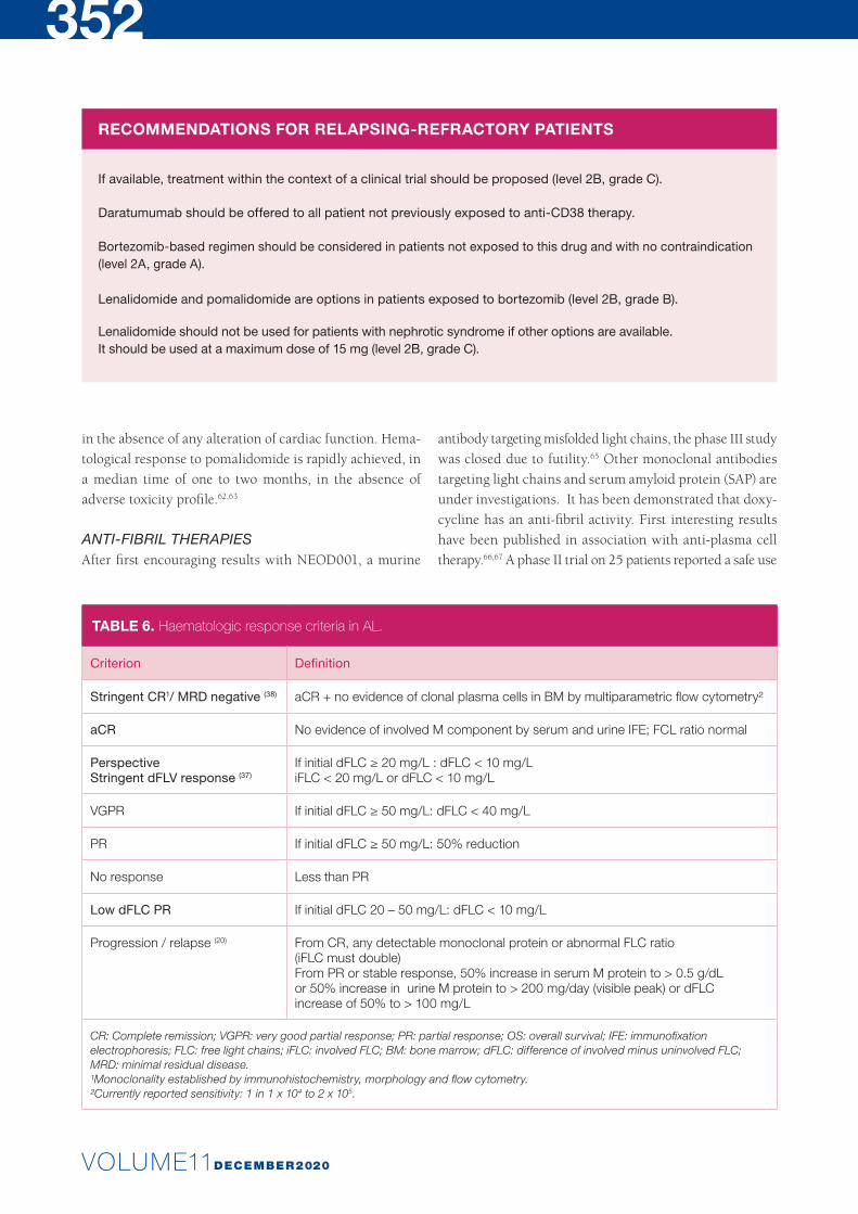
TABLE 6. Haematologic response criteria in AL.
Criterion Definition
Stringent CR1/ MRD negative (38) aCR + no evidence of clonal plasma cells in BM by multiparametric flow cytometry²
aCR No evidence of involved M component by serum and urine IFE; FCL ratio normal
PerspectiveStringent dFLV response (37)
If initial dFLC ≥ 20 mg/L : dFLC < 10 mg/LiFLC < 20 mg/L or dFLC < 10 mg/L
VGPR If initial dFLC ≥ 50 mg/L: dFLC < 40 mg/L
PR If initial dFLC ≥ 50 mg/L: 50% reduction
No response Less than PR
Low dFLC PR If initial dFLC 20 – 50 mg/L: dFLC < 10 mg/L
Progression / relapse (20) From CR, any detectable monoclonal protein or abnormal FLC ratio (iFLC must double)From PR or stable response, 50% increase in serum M protein to > 0.5 g/dL or 50% increase in urine M protein to > 200 mg/day (visible peak) or dFLC increase of 50% to > 100 mg/L
CR: Complete remission; VGPR: very good partial response; PR: partial response; OS: overall survival; IFE: immunofixation electrophoresis; FLC: free light chains; iFLC: involved FLC; BM: bone marrow; dFLC: difference of involved minus uninvolved FLC; MRD: minimal residual disease.¹Monoclonality established by immunohistochemistry, morphology and flow cytometry.²Currently reported sensitivity: 1 in 1 x 10⁴ to 2 x 10⁵.
RECOMMENDATIONS FOR RELAPSING-REFRACTORY PATIENTS
If available, treatment within the context of a clinical trial should be proposed (level 2B, grade C).
Daratumumab should be offered to all patient not previously exposed to antiCD38 therapy.
Bortezomibbased regimen should be considered in patients not exposed to this drug and with no contraindication (level 2A, grade A).
Lenalidomide and pomalidomide are options in patients exposed to bortezomib (level 2B, grade B).
Lenalidomide should not be used for patients with nephrotic syndrome if other options are available. It should be used at a maximum dose of 15 mg (level 2B, grade C).
VOLUME11DECEMBER20208
353
and a lower 1-year mortality compared to historical data.67 A retrospective study also reported promising results with a higher one year OS in patients treated with bortezomib-
based regimens plus doxycycline comparing to patients treated without doxycycline. A phase II-III trial is ongoing in first line treatment.67
PRACTICE GUIDELINES
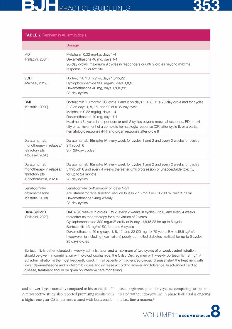
TABLE 7. Regimen in AL amyloidosis.
Dosage
MD(Palladini, 2004)
Melphalan 0.22 mg/kg, days 1-4Dexamethasone 40 mg, days 1-428-day cycles, maximum 9 cycles in responders or until 2 cycles beyond maximal response, PD or toxicity
VCD (Mikhael, 2012)
Bortezomib 1.3 mg/m², days 1,8,15,22Cyclophosphamide 300 mg/m², days 1,8,15Dexamethasone 40 mg, days 1,8,15,2228-day cycles
BMD (Kastritis, 2020)
Bortezomib 1.3 mg/m² SC: cycle 1 and 2 on days 1, 4, 8, 11 a 28-day cycle and for cycles 3–8 on days 1, 8, 15, and 22 of a 35-day cycle. Melphalan 0.22 mg/kg, days 1-4Dexamethasone 40 mg, days 1-4Maximum 8 cycles in responders or until 2 cycles beyond maximal response, PD or toxi-city or achievement of a complete hematologic response (CR) after cycle 6, or a partial hematologic response (PR) and organ response after cycle 6
Daratumumab mono therapy in relapse/ refractory pts(Roussel, 2020)
Daratumumab 16mg/kg IV, every week for cycles 1 and 2 and every 2 weeks for cycles 3 through 6Six 28-day cycles
Daratumumab monotherapy in relapse/ refractory pts(Sanchorawala, 2020)
Daratumumab 16mg/kg IV, every week for cycles 1 and 2 and every 2 weeks for cycles 3 through 6 and every 4 weeks thereafter until progression or unacceptable toxicity, for up to 24 months28-day cycles
Lenalidomide- dexamethasone(Kastritis, 2018)
Lenalidomide: 5–15mg/day on days 1–21Adjustment for renal function: reduce to less < 15 mg if eGFR <30 mL/min/1.73 m² Dexamethasone 24mg weekly 28-day cycles
DaraCyBorD(Palladini, 2020)
DARA SC weekly in cycles 1 to 2, every 2 weeks in cycles 3 to 6, and every 4 weeks thereafter as monotherapy for a maximum of 2 years Cyclophosphamide 300 mg/m2² orally or IV days 1,8,15,22 for up to 6 cyclesBortezomib 1.3 mg/m² SC for up to 6 cyclesDexamethasone 40 mg days 1, 8, 15, and 22 (20 mg if > 70 years, BMI ≤18.5 kg/m², hypervolemia including heart failure) poorly controlled diabetes mellitus) for up to 6 cycles28 days cycles
Bortezomib is better tolerated in weekly administration and a maximum of two cycles of bi-weekly administration should be given. In combination with cyclophosphamide, the CyBorDex regimen with weekly bortezomib 1.3 mg/m² SC administration is the most frequently used. In frail patients or if advanced cardiac disease, start the treatment with lower dexamethasone and bortezomib doses and increase according answer and tolerance. In advanced cardiac disease, treatment should be given on intensive care monitoring.
VOLUME11DECEMBER2020
354RESPONSE EVALUATIONBoth hematologic and organ criteria for response are summarised in Table 6.22,30 Hematological response should be assessed every cycle, by measuring FLC and organ res-ponse at least every two cycles. However, organ responses are usually delayed and only observed months after hema-tological response is obtained. Normalisation of the serum FLC ratio in response to therapy is a strong predictor of survival, regardless of the therapeutic strategy applied.69,70 To have a chance to achieve organ response in a small frac-tion of patients, a dFLC decrease below 40 mg/l should be the minimal goal to reach (VGPR).30 The prognostic value of dFLC response is superior compared to the M-protein response.71 Different studies reported the interest to obtain a stringent dFLC response defined as a dFLC <10 mg/L which translate in a significant improvement of OS.31,72 Patients with a low dFLC <50 mg/L have a better prognosis and more frequently renal kidney involvement. Similarly, a reduction of NT-proBNP >30% and >300 ng/l (or 50 ng/l for BNP) is associated with a significant better OS.73
SUPPORTIVE ORGAN CARE In the ASCT setting, particular attention should be given during all the process. Stem cell collection and ASCT have to be performed under intense surveillance and in specia-lised centres. In patients with cardiac involvement, arrhyth-mia prophylaxis with amiodarone should be discussed before stem collection and ASCT. During the aplasia period, physician should avoid G-CSF use if there is a cardiac and/or renal involvement as there is a high risk of fluid retention. Therefore, special attention should be taken with fluid administration. As there is an increased risk of bleeding, the cut-off for platelet transfusion should be increase at
20.000/mm3 and regularly blood test in the stool should be performed.Early mortality is related to organ involvement, particularly to advanced stage cardiac dysfunction. As organ responses are often delayed, supportive measures remain critical in the management of AL amyloidosis. For patients with cardiac involvement, management of fluid overload using loop diu retics and/or spironolactone is the key of treatment.4,74 Special attention should be paid in the dosage of these drugs in patients with autonomic neuropathy as they are exposed to an increased risk of hypotension or in patients with renal amyloidosis. Close monitoring of renal function and serum ions has to be done at the initiation of therapy. Cardiac arrhythmias are frequent in patients with advanced stages of the disease.75 Digoxin is contra-indicated for atrial fibrillation, as it has been described to bind to amyloid depo-sits and is associated increased cardiac arrhythmias. Ami-odarone is the drug of choice in this setting. In patients for whom the electrophysiological study reveals a high risk of arrhythmia, prophylactic amiodarone 200 mg should be considered. ACE inhibitors should be avoided and are usu-ally poorly tolerated, particularly in patients with hypo ten-sion, cardiac or autonomic involvement.76 Beta-blockers and calcium- channel blockers are contraindicated. In patients with recurrent cardiogenic syncope, or complex ventricular arrhythmias, pacemakers and defibrillators could be dis-cussed even if no benefice in survival has been reported.In patients with renal amyloidosis, diuretic, salt, and fluid restriction is the backbone of the treatment.77 However, a close monitoring of renal function and blood pressure is required after starting diuretics. In subjects with nephrotic syndrome, prophylactic anticoagulation should be consi-dered with caution and well balanced with the higher risk of
RECOMMENDATIONS
Symptomatic cardiac failure should be managed with salt depletion, loos diuretics and/or spironolactone (level 3, grade C).
In cases of cardiac involvement, ACE inhibitors, betablockers and calciumchannel blockers should be avoided, particularly in patients with dysautonomia, impaired renal function and hypotension (level 3, grade C).
Arrhythmia can be prevented by prophylactic amiodarone (level 3, grade C). Digoxin is contraindicated (level 3, grade C).
Nephrotic syndrome should be managed with diuretics, fluid and salt restriction (level 4, grade C).
ACE inhibitors should be restricted to patients with no significant cardiac involvement or dysautonomia (level 4, grade C).
VOLUME11DECEMBER20208
355bleeding. There is no data that showed an interest to pres-cribe ACE inhibitors to minimise proteinuria in AL amy-loidosis. If it is used, it should be started with caution and only if there is no cardiac or autonomic involvement. Hyper-cholesterolemia should be corrected. Even if there is no renal insufficiency at diagnosis, worsening of the clearance of creatinine could be observed during treatment, because of nephrotoxic drugs, iodine contrast exposure or depletion. Orthostatic hypotension is seen in about 15% of patients.77 It may be improved by wearing support stockings. Mido-drine, an alpha-adrenergic agonist, is the most effective drug for orthostatic hypotension in patients with amyloi-dosis but can cause supine hypertension and tachycardia. The initial dose is 2.5md t.i.d and could be increased to a maximum dosage of 10 mg t.i.d.
CONCLUSIONAL amyloidosis is a rare and probably underdiagnosed disease. Major advances have been made in diagnosis, supportive care and treatments. However, patients with advanced stage IIIB diseases have still a very limited OS and major efforts have still to been made to detect earlier that serious condition. Information about when to suspect amyloidosis should be given to cardiologists, nephrologists, neurologists, internists, general practitioners and screening should be done in patients with MGUS and SMM annually. Achievement of a rapid and deep response is crucial to improve the prognostic of these patients. After completion of treatment, patients should be closely monitored and therapy should be resumed as soon as any reappearance or evolution of the biological clone is observed, particularly in patients with cardiac involvement or severe organ disease at diagnosis. Patients should always be treated in the con-text of a clinical trial, if available, and in reference centres. Most of the effective drugs are not registered in this in-dication and should be prescribed in the context of MM therapy, when BM-PC are ≥10%, otherwise treatment should be request as samples or in a medical need program. Anti- CD38 therapy is highly effective and will change the prog-nosis of this disease. Daratumumab should be offered at relapse to all patients not exposed to anti-CD38 antibodies. Based on the Andromeda study, it will move to front-line therapy as soon as available.
REFERENCES1. Smith A, Wisloff F, Samson D. Br J Haematol. 2006;138(4):410-51.
2. Kyle RA, Larson DR, Kurtin PJ, et al. Mayo Clin Proc. 2019 ;94(3):465-71.
3. Wechalekar AD, Gillmore JD, Hawkins PN. Lancet. 2016;387(10038):
2641-54.
4. Merlini G. Hematology Am Soc Hematol Educ Program. 2017(1):1-12.
5. Di Girolamo M, Monno D, Pirro MR, et al. Amyloid. 2011;18(suppl 1):83-5.
6. Lousada I, Comenzo RL, Landau H, et al. Adv Ther. 2015;32(10):920-8.
7. Merlini G, Dispenzieri A, Sanchorawala V, et al. Nat Rev Dis Primers.
2018;4(1):38.
8. Gertz M, Dispenzieri A. JAMA. 2020;324(1):79-89.
9. Patel G, Hari P, Szabo A, et al. Hematol Oncol Stem Cell Ther. 2019;12(1):10-14.
10. Hazenberg B. Fat aspiration procedure for the detection of amyloid [Internet].
2011 [cited 17/02/2020]. Available from: http://www.amyloid.nl/Files/Fat%20
aspiration%20procedure02.pdf.
11. Quarta CC, Gonzalez-Lopez E, Gilbertson JA, et al. Eur Heart J. 2017;
38(24):1905-8.
12. Kimmich C, Schönland S, Kräker S, et al. Amyloid. 2017;24(1):52-9.
13. Foli A, Palladini G, Caporali R, et al. Amyloid. 2011;18 Suppl 1:80-2.
Erratum in: Amyloid. 2011;18 Suppl 1:82.
14. Geller HI, Singh A, Mirto TM, et al. Mayo Clin Proc. 2017;92(12):1800-5.
15. Muchtar E, Gertz MA, Kyle RA, et al. Mayo Clin Proc. 2019;94(3):472-83.
16. Vrana JA, Gamez JD, Madden BJ, et al. Blood. 2009;114(24):4957-9.
17. Gillmore J, Maurer M, Falk R, et al. Circulation. 2016;133:2404-12.
18. Tovar N, Rodríguez-Lobato LG, Cibeira MT, et al. sAmyloid. 2018;25(2):79-85.
19. Muchtar E, Dispenzieri A, Kumar S K, et al. Leukemia. 2017;31(7):1562-9.
20. Bochtler T, Hegenbart U, Kunz C, et al. Amyloid. 2014;21(1):9-17.
21. Grogan M, Dispenzieri A, Gertz MA. Heart. 2017;103(14):1065-72.
22. Palladini G, Hegenbart U, Milani P, et al. Blood. 2014;124(15):2325-32.
23. Dispenzieri A, Gertz MA, Kyle RA, et al. J Clin Oncol. 2004;22(18):3751-7.
24. Lilleness B, Ruberg F, Mussinelli R, et al. Blood. 2019;133(3):215-23.
25. Wechalekar A, Schonland S, Kastritis E, et al. Blood. 2013;121(17):3420-7.
26. Dittrich T, Benner A, Kimmich C, et al. Haematologica. 2019;104(7):1451-7.
27. Milani P, Basset M, Russo F, et al. Blood. 2017;130(5):625-31.
28. Kumar S, Dispenzieri A, Lacy MQ, et al. J Clin Oncol. 2012;30(9):989-95.
29. Kastritis E, Gavriatopoulou M, Roussou M, et al. Am J Hematol. 2017;
92(7):632-9.
30. Palladini G, Dispenzieri A, Gertz MA, et al. J Clin Oncol. 2012;30(36):4541-9.
31. Kastritis E, Fotiou D, Theodorakakou F, et al. Amyloid. 2020;27:1-9.
32. Le Bras F, Molinier-Frenkel V, Guellich A, et al. Eur J Cancer. 2017;76:183-7.
33. Jaccard A, Moreau P, Leblond V, et al. N Engl Med. 2007;357(11):1083-93.
34. Sidiqi MH, Aljama MA, Buadi FK, et al. J Clin Oncol. 2018;36(13):1323-9.
35. Hwa YL, Kumar SK, Gertz MA, et al. Am J Hematol. 2016;91(10):984-8.
36. Sanchorawala V, Sun F, Quillen K, et al. Blood. 2015;126(20):2345-7.
37. Mikhael J, Schuster S, Jimenez-Zepeda V, et al. Blood. 2012;119(19):4391-4.
38. Venner C, Lane T, Foard D, et al. Blood. 2012;119(19):4387-90.
39. Palladini G, Sachchithanantham S, Milani P, et al. Blood. 2015;126(5):612-5.
40. Manwani R, Cohen O, Sharpley F, et al. Blood. 2019;134(25):2271-80.
41. Kastritis E, Leleu X, Arnulf B, et al. J Clin Oncol. 2020;38(28):3252-60.
42. Popat R, Dowling E, Achhala S, et al. Br J Haematol. 2018;182(6):936-9.
43. Kaufman GP, Schrier SL, Lafayette RA, et al. Blood. 2017;130(7):900-2.
44. Milani P, Fazio F, Basset M, et al. Am J Hematol. 2020;95(8):900-5.
45. Chung A, Kaufman GP, Sidana S, et al. Blood Adv. 2020;4:458-66.
46. Kimmich CR, Terzer T, Benner A, et al. Blood. 2020;135:1517-30.
47. Roussel M, Merlini G, Chevret S, et al. Blood. 2020;135:1531-40.
48. Sanchorawala V, Sarosiek S, Schulman A, et al. Blood. 2020;135:1541-7.
PRACTICE GUIDELINES
VOLUME11DECEMBER2020
35649. Palladini G, Kastritis E, Maurer MS, et al. Blood. 2020;136(1):71-80.
50. Kastritis E, Palladini G, Minnema MC, et al. EHA Library. Kastritis E. 06/14/20;
303396; LB2604.
51. Kourelis TV, Kyle RA, Dingli D, et al. Mayo Clin Proc. 2017;92(6):908-17.
52. Sidana S, Larson P, Greipp P, et al. Leukemia. 2020;34(5):1373-82.
53. Hwa Y, Warsame R, Gertz M, et al. Blood. 2017;130(13):1578-84.
54. Palladini G, Milani P, Foli A. Blood. 2018;131(5):525-32.
55. Tandon N, Sidana S, Gertz M, et al. Am J Hematol. 2017;92(6):549-54.
56. Sanchorawala V, Palladini G, Kukreti V, et al. Blood. 2017;130:597-605.
57. Kastritis E. Am Society of Clin Oncol. 2020.
58. Cohen AD, Landau H, Scott EC, et al. Blood. 2016;128:645.
59. Palladini G, Russo P, Milani P, et al. Haematologica. 2013;98(3):433-6.
60. Mahmood S, Venner CP, Sachchithanantham S, et al. Br J Haematol. 2014;
166(6):842-8.
61. Kastritis E, Gavriatopoulou M, Roussou M, et al. Amyloid. 2018;25(4):234-41.
62. Palladini G, Milani P, Foli A, et al. Blood. 2017;129:2120-3.
63. Dispenzieri A, Buadi F, Laumann K, et al. Blood. 2012;129:5397-404.
64. Dispenzieri A, Dingli D, Kumar SK, et al. Am J Hematol. 2010;85(10):757-9.
65. Specter R, Sanchorawala V, Seldin DC, et al. Nephrol Dial Transplant. 2011;
26(3):881-6.
66. Gertz M, Cohen AD, Comenzo RL, et al. Blood. 2019;134:3166.
67. Wechalekar AD, Whelan C. Blood Cancer J. 2017;7:e546.
68. D’Souza A, Szabo A, Flynn KE, et al. E Clinical Medicine. 2020;23:10361.
69. Dispenzieri A, Lacy M, Katzmann J, et al. Blood. 2006;107(8):3378-83.
70. Lachmann H, Gallimore R, Gillmore J, et al. Br J Haematol. 2003;122(1):78-84.
71. Kumar S, Dispenzieri A, Lacy M, et al. Am J Hematol. 2011;86(3):251-5.
72. Muchtar E, Gertz M, Lacy M, et al. Am J Hematol. 2020. Online ahead of print.
73. Palladini G, Foli A, Milani P, et al. Amyloid. 2010;17(s1):85.
74. Falk RH, Alexander KM, Liao R, et al. J Am Coll Cardiol. 2016;68(12):1323-41.
75. Giancaterino S, Urey MA, Darden D, et al. JACC Clin Electrophysiol. 2020;
6(4):351-61.
76. Kapoor P, Thenappan T, Singh E, et al. Am J Med. 2011;124(11):1006-15.
77. Cibeira MT, Ortiz-Pérez JT, Quintana LF, et al. Acta Haematol. 2020;
143(4):335-42.