Embed Size (px)
Citation preview
DIAGNOSIS OF PROSTATE DISEASES
• HISTORY• DIGITAL EXAMINATION• BIOPSY OR FINE NEEDLE
ASPIRATION.
• HISTORY• Cancer of the prostate usually presents with
clinical symptoms of prostatism.• Advanced prostatic adenocarcinomas
typically cause urinary obstruction. • It may also present in advanced cases with
signs and symptoms of metastasis (e.g. back pain due to vertebral metastasis).
• [ They may also present as incidental findings during microscopic examination of the tissue surgically removed for non-malignant disease, particularly BPH…………..OR sometimes ………….
• Tumors of the prostate are detected during autopsy on patients that showed no clinical evidence of prostatic cancer.]
• DIGITAL EXAMINATION• …. a hard painless mass in the gland is
found during rectal examination.• These tumors are easier to feel than to
see; they are firmer than hyperplastic nodules and poorly circumscribed.

DIRECT RECTAL EXAMINATION
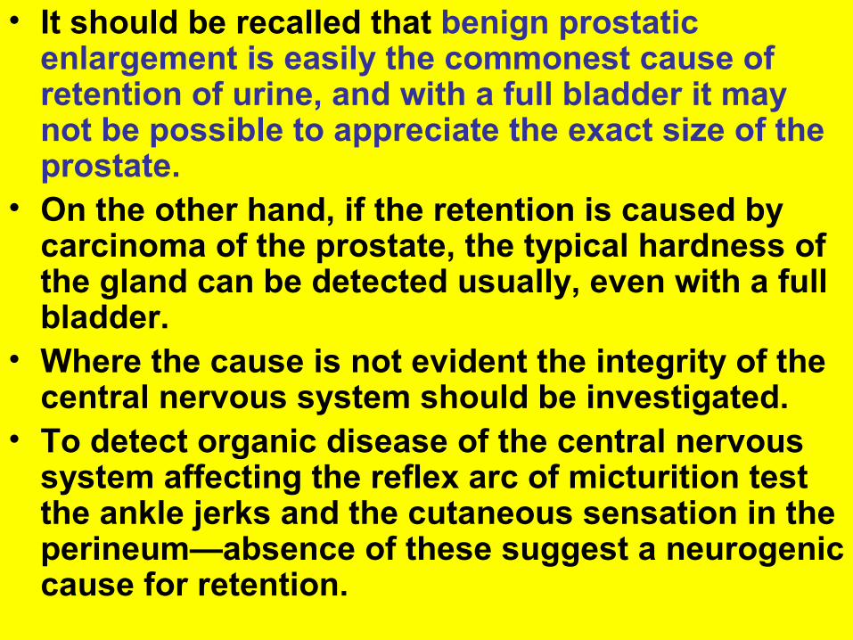
• The Prostate and its Adnexa• As a prelude to palpation of the prostate, the patient is
asked to empty his bladder. When possible the act of micturition should be watched, for loss of projectile power is significant.
• It is inadvisable to examine the prostate before a general examination of the abdomen has been conducted at which special attention should be paid to the bladder, for if by palpation and percussion it is found to be distended in spite of urination, not only is that discovery of cardinal importance, but it also foretells that the rectal findings on the state of the prostate are likely to be unreliable.
• It should be recalled that benign prostatic enlargement is easily the commonest cause of retention of urine, and with a full bladder it may not be possible to appreciate the exact size of the prostate.
• On the other hand, if the retention is caused by carcinoma of the prostate, the typical hardness of the gland can be detected usually, even with a full bladder.
• Where the cause is not evident the integrity of the central nervous system should be investigated.
• To detect organic disease of the central nervous system affecting the reflex arc of micturition test the ankle jerks and the cutaneous sensation in the perineum—absence of these suggest a neurogenic cause for retention.
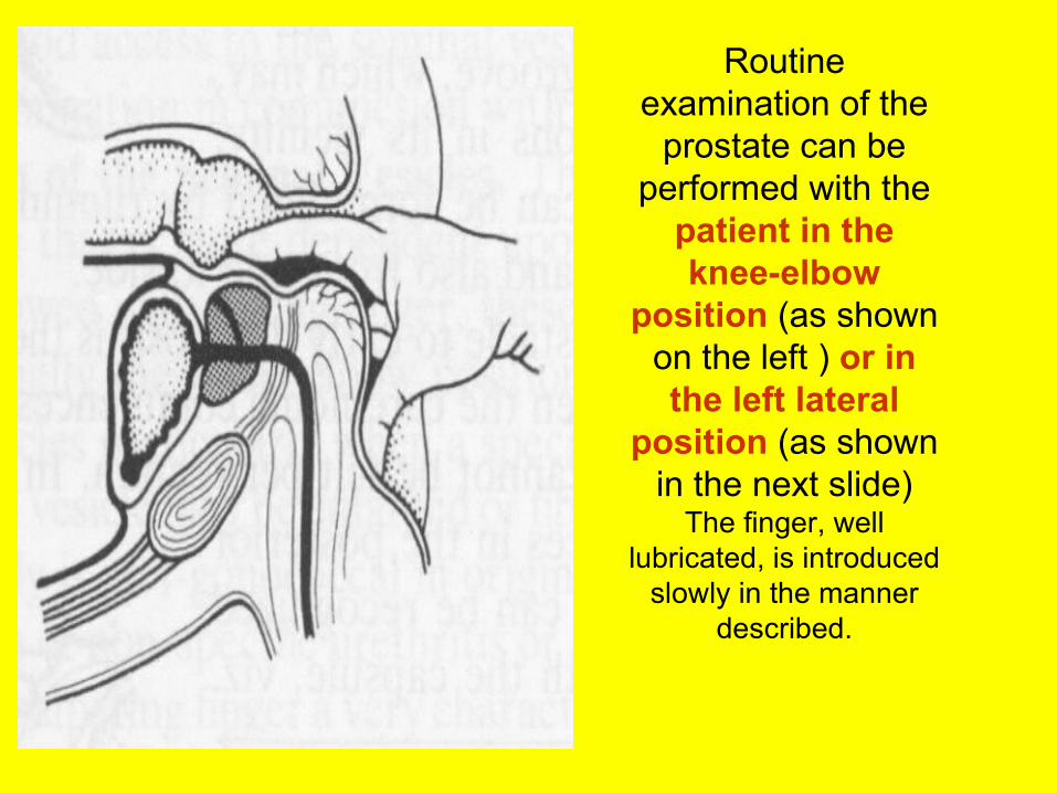
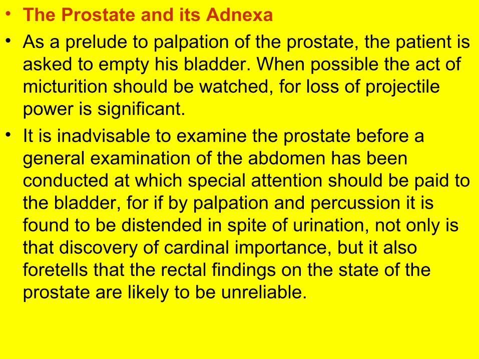
Routine examination of the
prostate can be performed with the
patient in the knee-elbow
position (as shown on the left ) or in the left lateral
position (as shown in the next slide)
The finger, well lubricated, is introduced
slowly in the manner described.
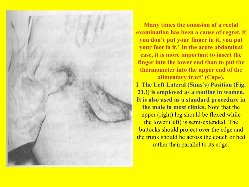
Many times the omission of a rectal examination has been a cause of regret. if
you don’t put your finger in it, you put your foot in it.’ In the acute abdominal case, it is more important to insert the
finger into the lower end than to put the thermometer into the upper end of the
alimentary tract’ (Cope).I. The Left Lateral (Sims’s) Position (Fig. 21.!) is employed as a routine in women. It is also used as a standard procedure in
the male in most clinics. Note that the upper (right) leg should be flexed whilethe lower (left) is semi-extended. The
buttocks should project over the edge and the trunk should be across the couch or bed
rather than parallel to its edge.
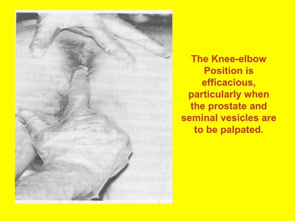
The Knee-elbow Position is efficacious,
particularly when the prostate and
seminal vesicles are to be palpated.
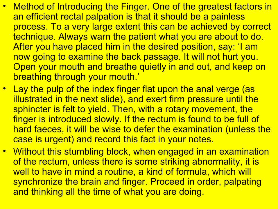
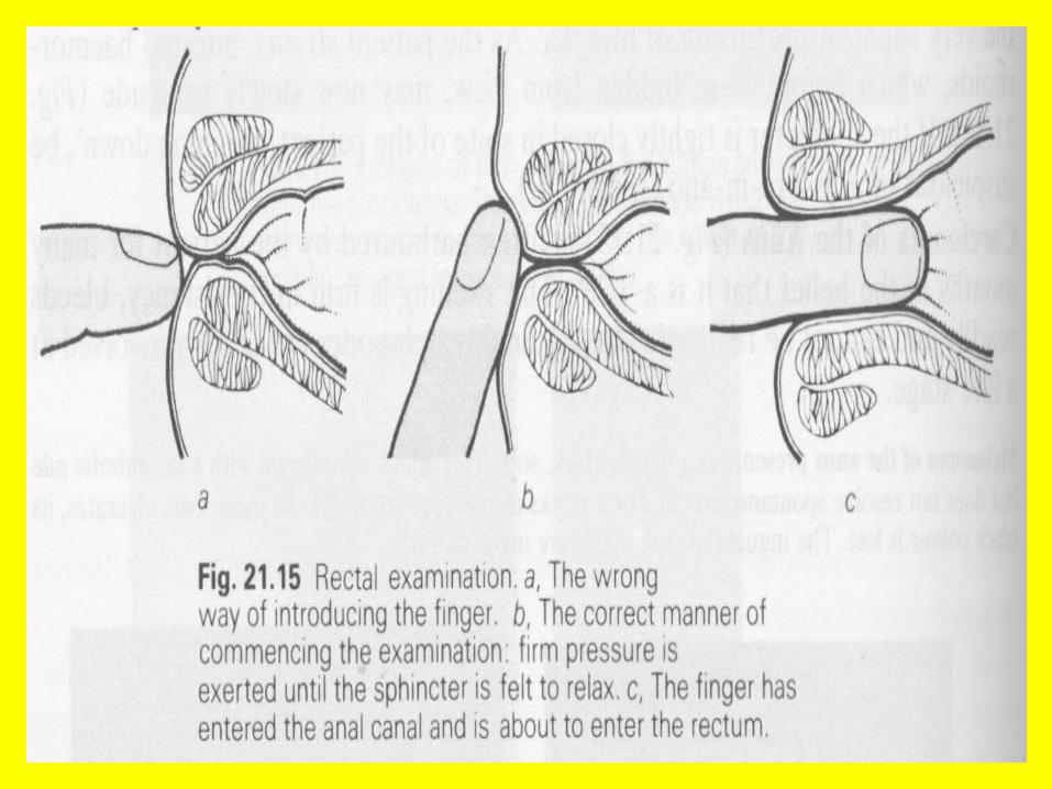
• Method of Introducing the Finger. One of the greatest factors in an efficient rectal palpation is that it should be a painless process. To a very large extent this can be achieved by correct technique. Always warn the patient what you are about to do. After you have placed him in the desired position, say: ‘I am now going to examine the back passage. It will not hurt you. Open your mouth and breathe quietly in and out, and keep on breathing through your mouth.’
• Lay the pulp of the index finger flat upon the anal verge (as illustrated in the next slide), and exert firm pressure until the sphincter is felt to yield. Then, with a rotary movement, the finger is introduced slowly. If the rectum is found to be full of hard faeces, it will be wise to defer the examination (unless the case is urgent) and record this fact in your notes.
• Without this stumbling block, when engaged in an examination of the rectum, unless there is some striking abnormality, it is well to have in mind a routine, a kind of formula, which will synchronize the brain and finger. Proceed in order, palpating and thinking all the time of what you are doing.
• It is not unusual for the advancing finger to push a lesion of the rectum before it. Therefore, in its upward course, after the anorectal ring has been passed, endeavour to steer the finger tip clear of the rectal walls.
• Having reached the highest limit, flex the finger and withdraw it partially. Repeat the process at other cardinal points of the compass, for a soft lesion of the rectal wall is more likely to be felt on the downward stroke of the finger than on its upward course.
• Having completed the routine palpation, look at your finger for blood, mucus, or pus: better still, wipe it on a gauze swab, which will show up the colour of the discharge.
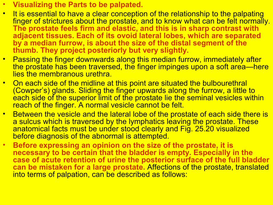
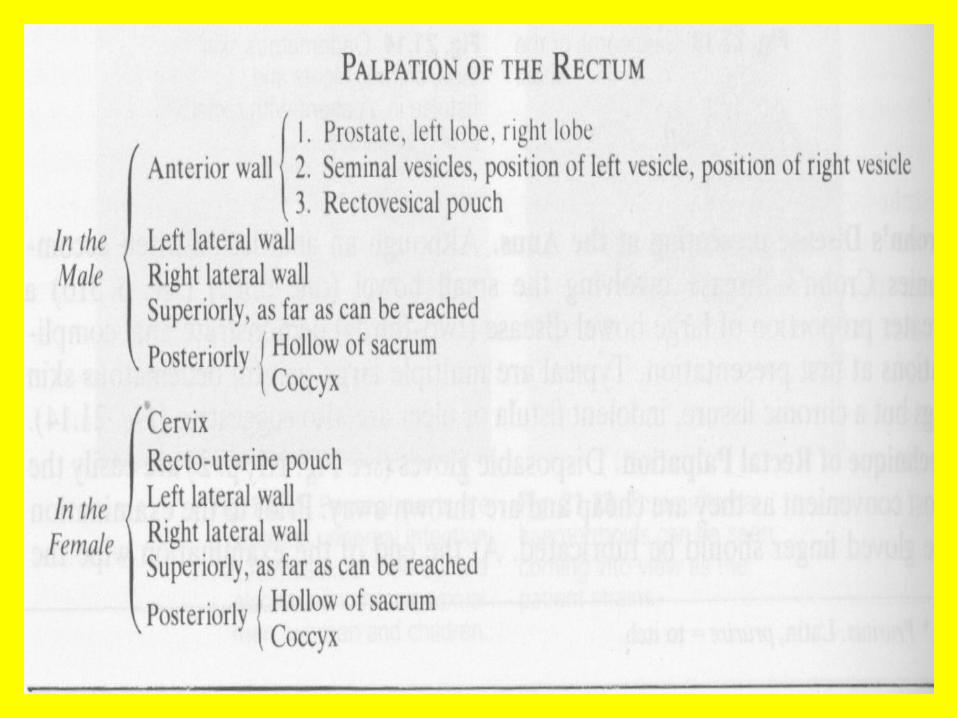
• Visualizing the Parts to be palpated. • It is essential to have a clear conception of the relationship to the palpating
finger of strictures about the prostate, and to know what can be felt normally. The prostate feels firm and elastic, and this is in sharp contrast with adjacent tissues. Each of its ovoid lateral lobes, which are separated by a median furrow, is about the size of the distal segment of the thumb. They project posteriorly but very slightly.
• Passing the finger downwards along this median furrow, immediately after the prostate has been traversed, the finger impinges upon a soft area—here lies the membranous urethra.
• On each side of the midline at this point are situated the bulbourethral (Cowper’s) glands. Sliding the finger upwards along the furrow, a little to each side of the superior limit of the prostate lie the seminal vesicles within reach of the finger. A normal vesicle cannot be felt.
• Between the vesicle and the lateral lobe of the prostate of each side there is a sulcus which is traversed by the lymphatics leaving the prostate. These anatomical facts must be under stood clearly and Fig. 25.20 visualized before diagnosis of the abnormal is attempted.
• Before expressing an opinion on the size of the prostate, it is necessary to be certain that the bladder is empty. Especially in the case of acute retention of urine the posterior surface of the full bladder can be mistaken for a large prostate. Affections of the prostate, translated into terms of palpation, can be described as follows:
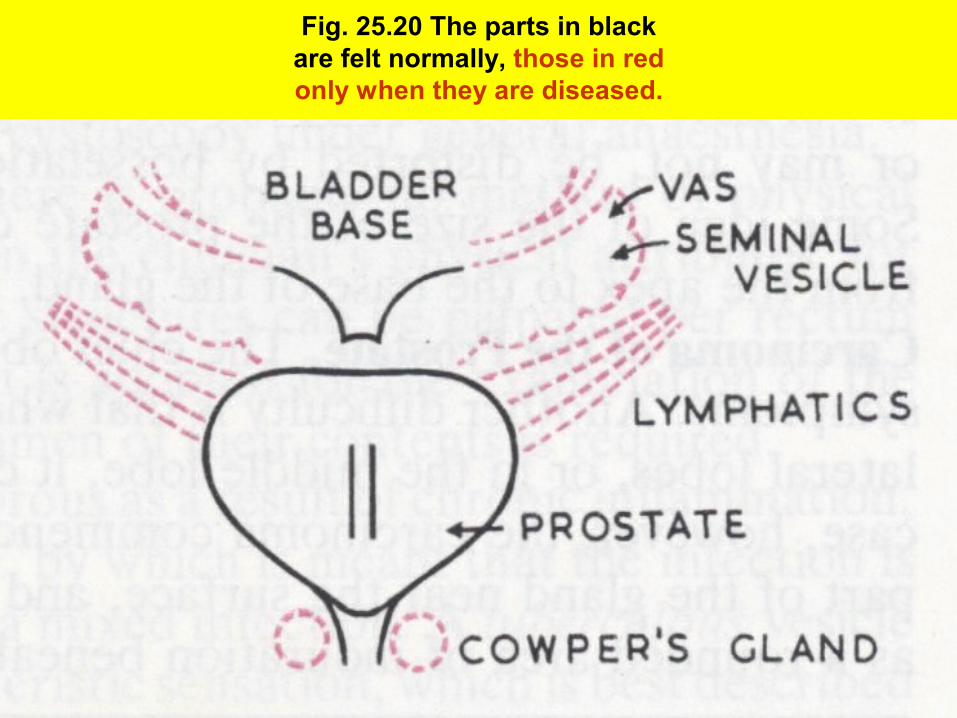
Fig. 25.20 The parts in blackare felt normally, those in redonly when they are diseased.
• Acute Prostatitis. Palpation must be very gentle. An enlarged, swollen, tense but slightly oedematous, tender, hot prostate is diagnostic of acute prostatitis. Sometimes acute prostatitis is associated with acute seminal vesiculitis. If an abscess or abscesses have formed they will be detected as areas of softening.
• Chronic prostatitis is not common. It can follow acute prostatitis, or be chronic from the commencement. Diagnosis is often uncertain and it is not always easy to decide if the symptoms are due to the prostatic infection. The gland may be slightly enlarged or normal in size. Similarly, tenderness may be slight or absent. Frequently the prostate is somewhat nodular, with occasional boggy spots. Clinical differentiation from carcinoma may prove difficult. Microscopical examination of the fluid expressed by prostatic massage (described above) for pus cells is the only reliable method of demonstrating the infection.
• Tuberculous Prostatitis. Almost always one, and sometimes both, seminal vesicles are implicated. Both these structures are hard, irregular and can be best described as ‘craggy’. The vas deferens is nearly always involved.
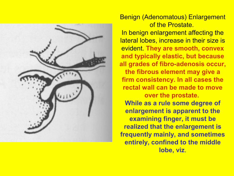
Benign (Adenomatous) Enlargement of the Prostate.
In benign enlargement affecting the lateral lobes, increase in their size is evident. They are smooth, convex and typically elastic, but because all grades of fibro-adenosis occur,
the fibrous element may give a firm consistency. In all cases the rectal wall can be made to move
over the prostate. While as a rule some degree of enlargement is apparent to the
examining finger, it must be realized that the enlargement is
frequently mainly, and sometimes entirely, confined to the middle
lobe, viz.

• The palpating finger should pass over the entire gland. It will note the presence of the median groove, which may, or may not, be distorted by bosselations in its vicinity.
• Some idea of the size of the prostate can be formulated by running the finger from the apex to the base of the gland, and also from side to side.
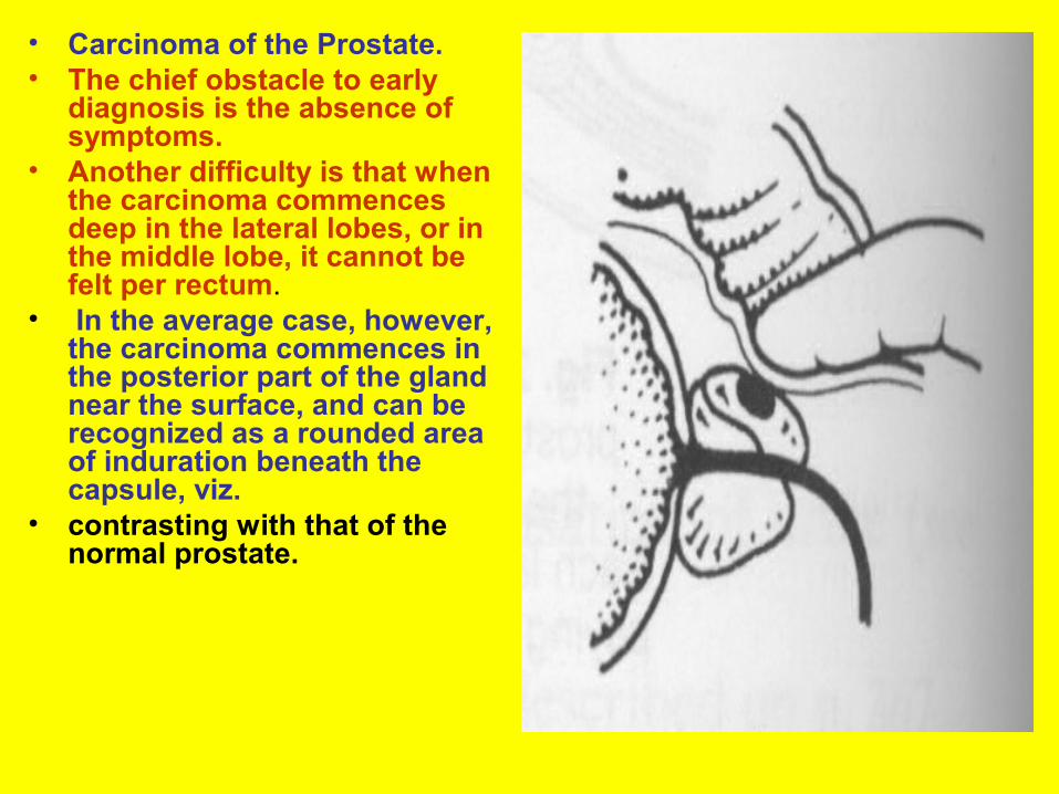
• Carcinoma of the Prostate. • The chief obstacle to early
diagnosis is the absence of symptoms.
• Another difficulty is that when the carcinoma commences deep in the lateral lobes, or in the middle lobe, it cannot be felt per rectum.
• In the average case, however, the carcinoma commences in the posterior part of the gland near the surface, and can be recognized as a rounded area of induration beneath the capsule, viz.
• contrasting with that of the normal prostate.
• As it increases in size, the nodule acquires stony hardness. • Somewhat later the vertical median groove between the
lateral lobes (see Fig. 25.20) becomes obliterated.• Further spread takes place, particularly in an upward and
outward direction around the seminal vesicles, giving rise to extensions shaped like the horns of a bull, which are quite characteristic. These are due to involvement of lymphatics (see Fig. 25.20).
• The normal mobility of the prostate gland becomes reduced with the extracapsular extention of the neoplasm.
• Later extension takes place in a backward direction, producing a stony-hard irregular mass obliterating the normal contour and the sulcus of the gland.
• Finally, although most cases of carcinoma of the prostate commence per primam, a few arise in a prostate that is already the seat of adenomatous enlargement; therefore always regard with particular suspicion an area of discrete induration in benign enlargement of the gland.
• Contracture of the Bladder Neck (Fibrous Prostate). The prostate is either normal in size or, more usually, smaller. The gland is distinctly harder than usual, but its shape is preserved and its contour smooth; the latter serves to differentiate the condition from carcinoma, but when the gland is stony hard, laboratory tests and biopsy are required to eliminate a scirrhous carcinoma.
• Prostatic Calculi. When these small stones are near enough the periphery to be detected by palpation, they are so embedded in the fibrous stroma as to simulate the irregular hardness of a carcinoma. Very occasionally the stones are comparatively free, and impart to the finger an impression of ‘beads in a bag’.
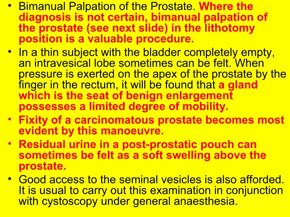
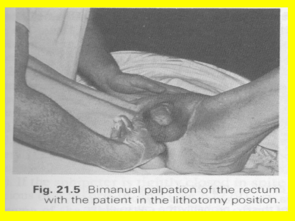
• Bimanual Palpation of the Prostate. Where the diagnosis is not certain, bimanual palpation of the prostate (see next slide) in the lithotomy position is a valuable procedure.
• In a thin subject with the bladder completely empty, an intravesical lobe sometimes can be felt. When pressure is exerted on the apex of the prostate by the finger in the rectum, it will be found that a gland which is the seat of benign enlargement possesses a limited degree of mobility.
• Fixity of a carcinomatous prostate becomes most evident by this manoeuvre.
• Residual urine in a post-prostatic pouch can sometimes be felt as a soft swelling above the prostate.
• Good access to the seminal vesicles is also afforded. It is usual to carry out this examination in conjunction with cystoscopy under general anaesthesia.
• BIOPSY OR FINE NEEDLE ASPIRATION. • Transrectal needle biopsy, often guided by
ultrasound, is useful to confirm the diagnosis, although incidental carcinomas can be found in transurethral resections for nodular hyperplasia.
• If you're going to operate for obstruction anyway, there's no reason to biopsy first.
• Ed opines………• * Future pathologists: With all these guys getting
needle biopsies nowadays, you need to try to find tiny cancers. You must section several levels of the core biopsy (Am. J. Clin. Path. 107: 26, 1997; Arch. Path. Lab. Med. 122: 833, 1998).
• Transrectal needle biopsy, often guided by ultrasound, is useful to confirm the diagnosis, although incidental carcinomas can be found in transurethral resections for nodular hyperplasia.
• Or it may turn up in a routine prostatectomy specimen. (If you're going to operate for obstruction anyway, there's no reason to biopsy first.)
• * Future pathologists: With all these guys getting needle biopsies nowadays, you need to try to find tiny cancers. You must section several levels of the core biopsy (Am. J. Clin. Path. 107: 26, 1997; Arch. Path. Lab. Med. 122: 833, 1998).
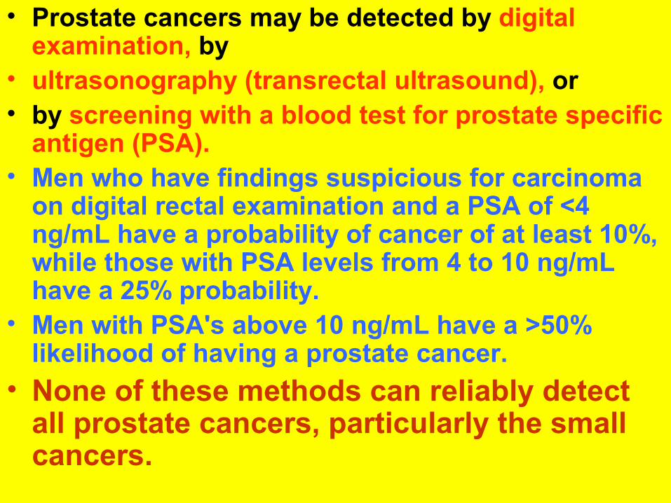
• Prostate cancers may be detected by digital examination, by
• ultrasonography (transrectal ultrasound), or • by screening with a blood test for prostate specific
antigen (PSA). • Men who have findings suspicious for carcinoma
on digital rectal examination and a PSA of <4 ng/mL have a probability of cancer of at least 10%, while those with PSA levels from 4 to 10 ng/mL have a 25% probability.
• Men with PSA's above 10 ng/mL have a >50% likelihood of having a prostate cancer.
• None of these methods can reliably detect all prostate cancers, particularly the small cancers.