Embed Size (px)
Citation preview
J. clin. Path. (1952), 5, 183.
THE DIAGNOSIS OF RETICULOSARCOMAOF THE THYROID GLAND-
BY
G. B. D. SCOTTFrom the Department ofPathology, University ofAberdeen
(RECEIVED FOR PUBLICATION OCTOBER 16, 1951)
The difficulty of making a histological distinction between reticulosarcomataand certain anaplastic carcinomata is well known and this applies particularly tothe thyroid gland. From a study of the literature it is not easy to obtain an accurateidea of the frequency of reticulosarcomata of the thyroid, using the term reticulo-sarcoma to include lymphoblastic and lymphocytic (lymphosarcoma) tumours aswell as the reticulum cell and other varieties (Robb-Smith, 1938).
Rice (1932) described four cases of lymphoblastic reticulosarcoma of the thyroidand one of a more primitive- type, possibly a reticulum cell tumour, these casesoccurring between the years 1922 and 1929. Vaux (1937) described a case ofdictyocytic reticulosarcoma of the thyroid occurring amongst 25 malignant thyroidtumours. Portmann (1940), in a review of 184 malignant thyroid tumours, recordedthe presence of 11 cases of lymphosarcoma, but did not subdivide them into theirpredominant cell types. He also recorded 14 cases as " unclassifiable," but did notstate why this was so or give any description of these tumours. Joll (1941) reviewed127 malignant thyroid tumours and recorded seven cases of sarcoma. He statedthat the reticulum cell variety was the commonest, but gave no figures. TheMassachusetts General Hospital recorded, with necropsy findings, four examplesof reticulosarcoma of the kidneys (1939, 1941, 1948), one of which (Case No. 34032,1948) showed coexistent reticulosarcoma of the thyroid. Kellett and Sutherland (1949)recorded five cases of thyroid reticulosarcoma. From the description given of them,one appeared to be of polymorphic type, two reticulum cell, and two lymphoblastic.However, they did not state over what period of time these tumours occurred norwhat percentage they formed of the total malignant thyroid tumours encounteredin that period. Of the 43 primary malignant thyroid tumours examined histologi-cally in this department between January 1, 1938, and January 1, 1951, 13 werepredominantly anaplastic but with small foci of obvious carcinoma, while ninewere completely anaplastic, and, of these, only one (Case 3 below) was proved bysubsequent events to be a lymphoblastic reticulosarcoma.
There can be no doubt that the frequency with which a diagnosis of reticulo-sarcoma of the thyroid is made varies greatly according to the criteria acceptableto different observers, and it is with the intention of evaluating these diagnosticcriteria that the following case of anaplastic carcinoma and two cases of authenticatedreticulosarcoma of the thyroid gland are presented.
on April 1, 2022 by guest. P
rotected by copyright.http://jcp.bm
j.com/
J Clin P
athol: first published as 10.1136/jcp.5.2.183 on 1 May 1952. D
ownloaded from
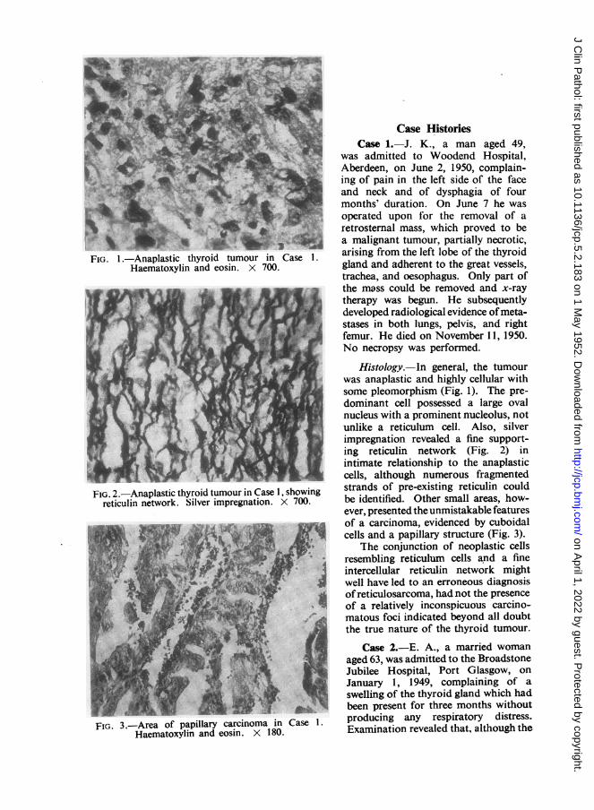
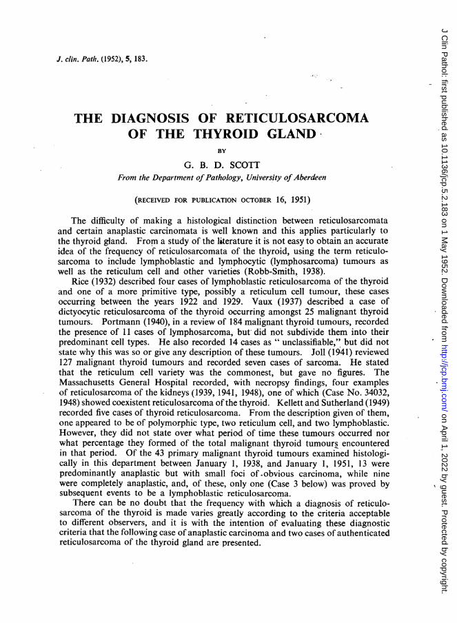
FIG. 1 -Anaplastic thyroid tumour in CaseHaematoxylin and eosin. X 700.
IT % IfI w -
FIG. 2.-Anaplastic thyroid tumour in Case 1, showingreticulin network. Silver impregnation. X 700.
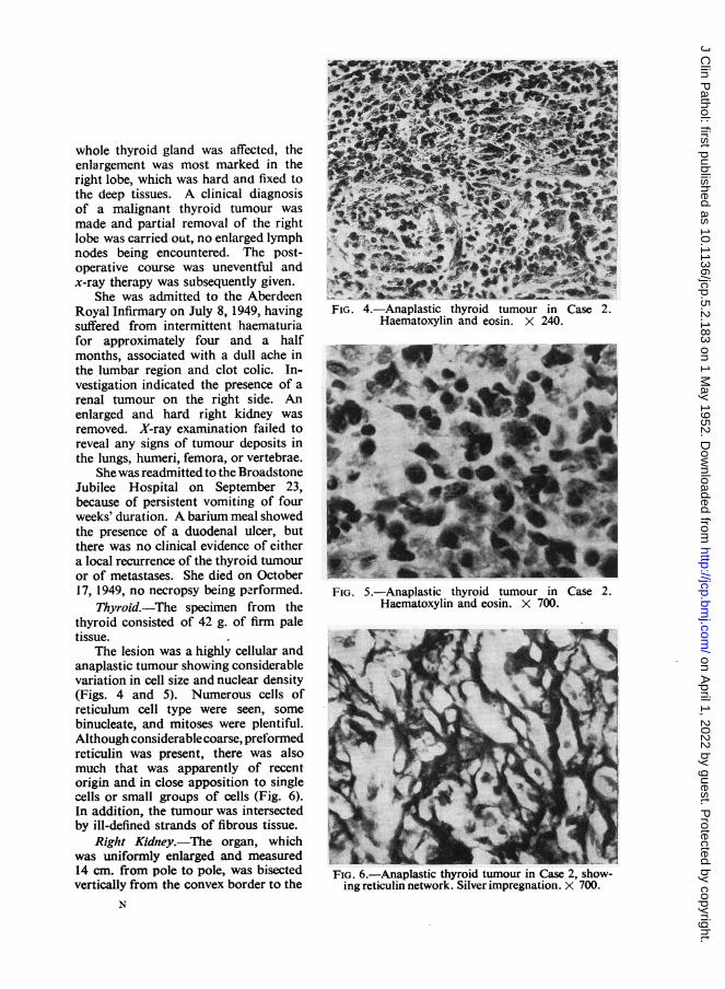
FIG. 3.-Area of papillary carcinoma inHaematoxylin and eosin. X 180.
Case HistoriesCase 1.-J. K., a man aged 49,
was admitted to Woodend Hospital,Aberdeen, on June 2, 1950, complain-ing of pain in the left side of the faceand neck and of dysphagia of fourmonths' duration. On June 7 he wasoperated upon for the removal of aretrosternal mass, which proved to bea malignant tumour, partially necrotic,arising from the left lobe of the thyroidgland and adherent to the great vessels,trachea, and oesophagus. Only part ofthe mass could be removed and x-raytherapy was begun. He subsequentlydeveloped radiological evidence ofmeta-stases in both lungs, pelvis, and rightfemur. He died on November 11, 1950.No necropsy was performed.
Histology.-ln general, the tumourwas anaplastic and highly cellular withsome pleomorphism (Fig. 1). The pre-dominant cell possessed a large ovalnucleus with a prominent nucleolus, notunlike a reticulum cell. Also, silverimpregnation revealed a fine support-ing reticulin network (Fig. 2) inintimate relationship to the anaplasticcells, although numerous fragmentedstrands of pre-existing reticulin couldbe identified. Other small areas, how-ever, presented the unmistakable featuresof a carcinoma, evidenced by cuboidalcells and a papillary structure (Fig. 3).
The conjunction of neoplastic cellsresembling reticulum cells and a fineintercellular reticulin network mightwell have led to an erroneous diagnosisofreticulosarcoma, had not the presenceof a relatively inconspicuous carcino-matous foci indicated beyond all doubtthe true nature of the thyroid tumour.
Case 2.-E. A., a married womanaged 63, was admitted to the BroadstoneJubilee Hospital, Port Glasgow, onJanuary 1, 1949, complaining of aswelling of the thyroid gland which hadbeen present for three months withoutproducing any respiratory distress.Examination revealed that, although the
on April 1, 2022 by guest. P
rotected by copyright.http://jcp.bm
j.com/
J Clin P
athol: first published as 10.1136/jcp.5.2.183 on 1 May 1952. D
ownloaded from
whole thyroid gland was affected, theenlargement was most marked in theright lobe, which was hard and fixed tothe deep tissues. A clinical diagnosisof a malignant thyroid tumour wasmade and partial removal of the rightlobe was carried out, no enlarged lymphnodes being encountered. The post-operative course was uneventful andx-ray therapy was subsequently given.
She was admitted to the AberdeenRoyal Infirmary on July 8, 1949, havingsuffered from intermittent haematuriafor approximately four and a halfmonths, associated with a dull ache inthe lumbar region and clot colic. In-vestigation indicated the presence of arenal tumour on the right side. Anenlarged and hard right kidney wasremoved. X-ray examination failed toreveal any signs of tumour deposits inthe lungs, humeri, femora, or vertebrae.
Shewas readmitted to the BroadstoneJubilee Hospital on September 23,because of persistent vomiting of fourweeks' duration. A barium meal showedthe presence of a duodenal ulcer, butthere was no clinical evidence of eithera local recurrence of the thyroid tumouror of metastases. She died on October17, 1949, no necropsy being performed.
Thyroid.-The specimen from thethyroid consisted of 42 g. of firm paletissue.
The lesion was a highly cellular andanaplastic tumour showing considerablevariation in cell size and nuclear density(Figs. 4 and 5). Numerous cells ofreticulum cell type were seen, somebinucleate, and mitoses were plentiful.Although considerable coarse, preformedreticulin was present, there was alsomuch that was apparently of recentorigin and in close apposition to singlecells or small groups of cells (Fig. 6).In addition, the tumour was intersectedby ill-defined strands of fibrous tissue.
Right Kidney.-The organ, whichwas uniformly enlarged and measured14 cm. from pole to pole, was bisectedvertically from the convex border to the
N
FIG. 4.-Anaplastic thyroid tumour in Case 2.Haematoxylin and eosin. X 240.
Pk..... i.. a-Xi%ss
FIG. 5.-Anaplastic thyroid tumour in Case 2.Haematoxylin and eosin. X 700.
FIG. 6.-Anaplastic thyroid tumour in Case 2, show-ing reticulin network. Silver impregnation. X 700.
on April 1, 2022 by guest. P
rotected by copyright.http://jcp.bm
j.com/
J Clin P
athol: first published as 10.1136/jcp.5.2.183 on 1 May 1952. D
ownloaded from
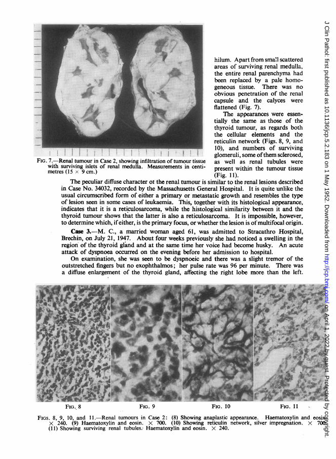
U;wr i i hilum. Apart from sma'l scatteredareas of surviving renal medulla,the entire renal parenchyma hadbeen replaced by a pale homo-geneous tissue. There was noobvious penetration of the renalcapsule and the calyces wereflattened (Fig. 7).
The appearances were essen-tially the same as those of thethyroid tumour, as regards boththe cellular elements and thereticulin network (Figs. 8, 9, and10), and numbers of surviving
*tf glomeruli, some ofthem sclerosed,FIG. 7.-Renal tumour in Case 2, showing infiltration of tumour tissue as well as renal tubules were
with surviving islets of renal medulla. Measurements in centi- present within the tumour tissuemetres (15 x 9 cm.) (Fig. 11).
The peculiar diffuse character ot the renal tumour is similar to the renal lesions describedin Case No. 34032, recorded by the Massachusetts General Hospital. It is quite unlike theusual circumscribed form of either a primary or metastatic growth and resembles the typeof lesion seen in some cases of leukaemia. This, together with its histological appearance,indicates that it is a reticulosarcoma, while the histological similarity between it and thethyroid tumour shows that the latter is also a reticulosarcoma. It is impossible, however,to determine which, ifeither, is the primary focus, or whether the lesion is ofmultifocal origin.
Case 3.-M. C., a married woman aged 61, was admitted to Stracathro Hospital,Brechin, on July 21, 1947. About four weeks previously she had noticed a swelling in theregion of the thyroid gland and at the same time her voice had become husky. An acuteattack of dyspnoea occurred on the evening before her admission to hospital.
On examination, she was seen to be dyspnoeic and there was a slight tremor of theoutstretched fingers but no exophthalmos; her pulse rate was 96 per minute. There wasa diffuse enlargement of the thyroid gland, affecting the right lobe more than the left.
* *
FIG. 8 FIG. 9 FIG. 10 FIG. 11
FIGS. 8, 9, 10, and 11.-Renal tumours in Case 2: (8) Showing anaplastic appearance. Haematoxylin and eosin.X 240. (9) Haematoxylin and eosin. X 700. (10) Showing reticulin network, silver impregnation. X 700.(11) Showing surviving renal tubules. Haematoxylin and eosin. X 240.
on April 1, 2022 by guest. P
rotected by copyright.http://jcp.bm
j.com/
J Clin P
athol: first published as 10.1136/jcp.5.2.183 on 1 May 1952. D
ownloaded from
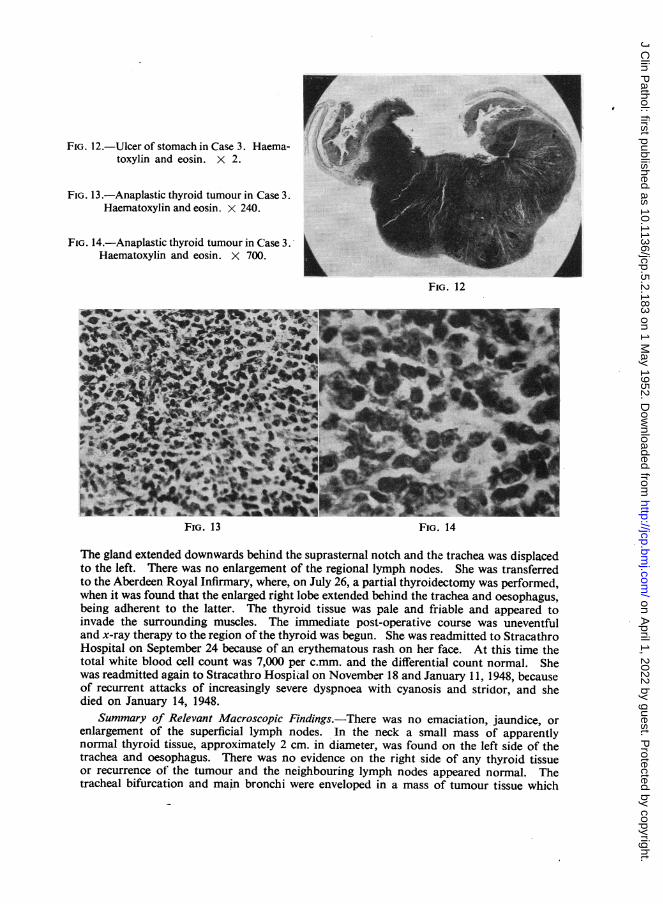
FIG. 12.-Ulcer of stomach in Case 3. Haema-toxylin and eosin. X 2.
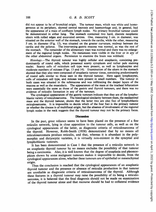
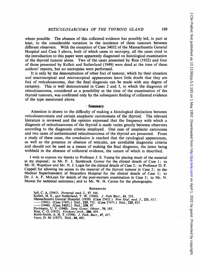
FIG. 13.-Anaplastic thyroid tumour in Case 3.Haematoxylin and eosin. X 240.
FIG. 14.-Anaplastic thyroid tumour in Case 3.Haematoxylin and eosin. X 700.
rI::/
FIG. 12
- -. --; a&i6 t-o 4
a
IP."W 9'... IOF 4' 11- A.
WO p gAt 4lk.. I%- :.914 10,t
L '4* .'9w - ft-j
FiG. 13 FIG. 14
The gland extended downwards behind the suprasternal notch and the trachea was displacedto the left. There was no enlargement of the regional lymph nodes. She was transferredto the Aberdeen Royal Infirmary, where, on July 26, a partial thyroidectomy was performed,when it was found that the enlarged right lobe extended behind the trachea and oesophagus,being adherent to the latter. The thyroid tissue was pale and friable and appeared toinvade the surrounding muscles. The immediate post-operative course was uneventfuland x-ray therapy to the region of the thyroid was begun. She was readmitted to StracathroHospital on September 24 because of an erythematous rash on her face. At this time thetotal white blood cell count was 7,000 per c.mm. and the differential count normal. Shewas readmitted again to Stracathro Hospiial on November 18 and January 11, 1948, becauseof recurrent attacks of increasingly severe dyspnoea with cyanosis and stridor, and shedied on January 14, 1948.
Summary of Relevant Macroscopic Findings.-There was no emaciation, jaundice, orenlargement of the superficial lymph nodes. In the neck a small mass of apparentlynormal thyroid tissue, approximately 2 cm. in diameter, was found on the left side of thetrachea and oesophagus. There was no evidence on the right side of any thyroid tissueor recurrence of the tumour and the neighbouring lymph nodes appeared normal. Thetracheal bifurcation and main bronchi were enveloped in a mass of tumour tissue which
-.i-0,I...
F.'"-4.6as, :-
on April 1, 2022 by guest. P
rotected by copyright.http://jcp.bm
j.com/
J Clin P
athol: first published as 10.1136/jcp.5.2.183 on 1 May 1952. D
ownloaded from
G. B. D. SCOTT
did not appear to be of bronchial origin. The tumour mass, which was white and homo-geneous at its periphery, showed central necrosis and haemorrhage and, in general, hadthe appearance of a mass of confluent lympb nodes. No primary bronchial tumour couldbe demonstrated in either lung. The stomach contained two hard, discrete neoplasticulcers with rolled edges and excavated centres. One, measuring 3 cm. in diameter, wassituated on the anterior wall of the stomach, towards the cardia, while the other, measuring5 cm. in diameter (Fig. 12), was situated on the greater curvature, midway between thecardia and the pylorus. The intervening gastric mucosa was normal, as was the rest ofthe stomach. The remainder ofthe alimentary tract was normal and there was no enlarge-ment of the regional lymph nodes. No metastases were visible in the liver or in any ofthe other abdominal organs. Permission to examine the brain was withheld.
Histology.-The thyroid tumour was highly cellular and anaplastic, consisting pre-dominantly of round cells, which possessed scanty cytoplasm and rather pale stainingnuclei. Scanty cells of reticulum cell type were present, as well as lymphocytes andmoderate numbers of mitoses (Figs. 13 and 14). Examination of the ulcers in the stomachshowed that they also were composed of anaplastic tumour tissue, consisting predominantlyof round cells similar to those seen in the thyroid tumour. Here again lymphocytes,cells of reticulum cell type, and mitoses were present in small numbers. The tumour inboth cases was situated in the submucosa and was infiltrating the deeper layers of themucosa as well as the muscularis. The appearances of the mass at the tracheal bifurcationwere essentially the same as those of the gastric and thyroid tumours, and there was noevidence of reticulin formation in any of the tumours.
The cytological appearances of the gastric tumours indicate that they are of the lympho-blastic variety of reticulosarcoma. The histological similarity between them, the mediastinalmass and the thyroid tumour, shows that the latter two are also foci of lymphoblasticreticulosarcoma. It is impossible to decide which of the four foci is the primary tumouror whether the disease is of multifocal origin, but the absence of involvement of the regionallymph nodes in the neck suggests that the thyroid tumour may not be the primary focus.
DiscussionIn the past, great reliance seems to have been placed on the presence of a fine
reticulin network, lying in close apposition to the tumour cells, as well as on thecytological appearances of the latter, as diagnostic criteria of reticulosarcoma ofthe thyroid. However, Robb-Smith (1938) demonstrated that by no means allreticulosarcomata produce reticulin, and that, whereas it is abundant in the poly-morphic and dictyocytic varieties, it is virtually non-existent in the syncytial andlymphoblastic tumours.
It has been demonstrated in Case 1 that the presence of a reticulin network inan anaplastic thyroid tumour by no means excludes the possibility of that tumourbeing a carcinoma. Also, it is well known that the degree of anaplasia and pleomor-phism shown by some malignant tumours makes it impossible to decide, from thecytological appearances alone, whether these tumours are of epithelial or mesenchymalorigin.
Thus the conclusion is reached that the cytological appearances of an anaplasticthyroid tumour and the presence or absence of reticulin production in that tumourare unreliable as diagnostic criteria of reticulosarcoma of the thyroid. Althoughthese features in a thyroid tumour may raise the possibility of its being a reticulo-sarcoma, it is believed that the final diagnosis should not be made on examinationof the thyroid tumour alone and that recourse should be had to collateral evidence
188
on April 1, 2022 by guest. P
rotected by copyright.http://jcp.bm
j.com/
J Clin P
athol: first published as 10.1136/jcp.5.2.183 on 1 May 1952. D
ownloaded from
RETICULOSARCOMA OF THE THYROID GLAND
where possible. The absence of this collateral evidence has possibly led, in part atleast, to the considerable variation in the incidence of these tumours betweendifferent observers. With the exception of Case 34032 of the Massachusetts GeneralHospital and Case 3 above, both of which came to necropsy, all the cases cited inthe introduction to this paper were apparently diagnosed on histological examinationof the thyroid tumour alone. Two of the cases presented by Rice (1932) and fourof those presented by Kellett and Sutherland (1949) were dead at the time of theseauthors' reports, but no necropsies were performed.
It is only by the demonstration of other foci of tumour, which by their situationand macroscopical and microscopical appearances leave little doubt that they arefoci of reticulosarcoma, that the final diagnosis can be made with any degree ofcertainty. This is well demonstrated in Cases 2 and 3, in which the diagnosis ofreticulosarcoma, considered as a possibility at the time of the examination of thethyroid tumours, was confirmed only by the subsequent finding of collateral evidenceof the type mentioned above.
SummaryAttention is drawn to the difficulty of making a histological distinction between
reticulosarcomata and certain anaplastic carcinomata of the thyroid. The relevantliterature is reviewed and the opinion expressed that the frequency with which adiagnosis of reticulosarcoma of the thyroid is made varies greatly between observersaccording to the diagnostic criteria employed. One case of anaplastic carcinomaand two cases of authenticated reticulosarcoma of the thyroid are presented. Froma study of these cases, the conclusion is reached that the cytological appearances,as well as the presence or absence of reticulin, are unreliable diagnostic criteriaand should not be used as a means of making the final diagnosis, the latter beingwithheld in the absence of collateral evidence, the nature of which is described.
I wish to express my thanks to Professor J. S. Young for placing much of the materialat my disposal; to Mr. F. J. Sambrook Gowar for the clinical details of Case 1; toMr. H. Wapshaw and Mr. N. J. Logie for the clinical details of Case 2; to Professor D. F.Cappell for allowing me access to the material of the thyroid tumour in Case 2; to theMedical Superintendent of Stracathro Hospital for the clinical details of Case 3; toDr. J. A. F. McLean for details of the post-mortem examination in Case 3; to Mr. N.Mowat for technical assistance; and to Mr. W. H. Carnie for the photographs.
REFEREN'CESJoIll, C A. (1941). Po0tgrad. med. J., 17, 166.Kellett, H. S., and Sutherland, T. W. (1949). J. Path Bact., 61, 233.Massachusetts General Hospital. (1939) (Case 25421.) New Engi. med. J., 221, 617.
(1941). (Case 27451.) Ibid., 225, 752. (Case 27471.) Ibid., 225, 833.(1948). (Case 34032.) Ibid., 238, 97.
Portmann, U. V. (1940). Surg. Gynec. Obstet., 70, 185.Rice, C. 0. (1932). Virchows Arch., 286, 459.Robb-Smith, A. H. T. (1938). J. Path. Bact., 47, 457.Vaux, D. M. (1937). Ibid., 44, 463.
189
on April 1, 2022 by guest. P
rotected by copyright.http://jcp.bm
j.com/
J Clin P
athol: first published as 10.1136/jcp.5.2.183 on 1 May 1952. D
ownloaded from