Embed Size (px)
Citation preview

Diagnostic Value of Knee Arthrometryin the Prediction of Anterior CruciateLigament Strain During Landing
Ata M. Kiapour,*y PhD, Samuel C. Wordeman,z§ BS, Mark V. Paterno,k{ PT, PhD, SCS, ATC,Carmen E. Quatman,z# MD, PhD, Jason W. Levine,y MD, Vijay K. Goel,y PhD,Constantine K. Demetropoulos,** PhD, and Timothy E. Hewett,z§{#yyzz PhDInvestigation performed at the Engineering Center for Orthopaedic Research Excellence(ECORE), The University of Toledo, Toledo, Ohio
Background: Previous studies have indicated that higher knee joint laxity may be indicative of an increased risk of anterior cru-ciate ligament (ACL) injuries. Despite the frequent clinical use of knee arthrometry in the evaluation of knee laxity, little data exist tocorrelate instrumented laxity measures and ACL strain during dynamic high-risk activities.
Purpose/Hypotheses: The purpose of this study was to evaluate the relationships between ACL strain and anterior knee laxitymeasurements using arthrometry during both a drawer test and simulated bipedal landing (as an identified high-risk injurioustask). We hypothesized that a high correlation exists between dynamic ACL strain and passive arthrometry displacement. Thesecondary hypothesis was that anterior knee laxity quantified by knee arthrometry is a valid predictor of injury risk such that speci-mens with greater anterior knee laxity would demonstrate increased levels of peak ACL strain during landing.
Study Design: Controlled laboratory study.
Methods: Twenty cadaveric lower limbs (mean age, 46 6 6 years; 10 female and 10 male) were tested using a CompuKT kneearthrometer to measure knee joint laxity. Each specimen was tested under 4 continuous cycles of anterior-posterior shear force(6134 N) applied to the tibial tubercle. To quantify ACL strain, a differential variable reluctance transducer (DVRT) was arthro-scopically placed on the ACL (anteromedial bundle), and specimens were retested. Subsequently, bipedal landing from 30 cmwas simulated in a subset of 14 specimens (mean age, 45 6 6 years; 6 female and 8 male) using a novel custom-designeddrop stand. Changes in joint laxity and ACL strain under applied anterior shear force were statistically analyzed using paired sam-ple t tests and analysis of variance. Multiple linear regression analyses were conducted to determine the relationship betweenanterior shear force, anterior tibial translation, and ACL strain.
Results: During simulated drawer tests, 134 N of applied anterior shear load produced a mean peak anterior tibial translation of3.1 6 1.1 mm and a mean peak ACL strain of 4.9% 6 4.3%. Anterior shear load was a significant determinant of anterior tibialtranslation (P\ .0005) and peak ACL strain (P = .04). A significant correlation (r = 0.52, P\ .0005) was observed between anteriortibial translation and ACL strain. Cadaveric simulations of landing produced a mean axial impact load of 4070 6 732 N. Simulatedlanding significantly increased the mean peak anterior tibial translation to 10.4 6 3.5 mm and the mean peak ACL strain to 6.8% 6
2.8% (P\ .0005) compared with the prelanding condition. Significant correlations were observed between peak ACL strain duringsimulated landing and anterior tibial translation quantified by knee arthrometry.
Conclusion: Our first hypothesis is supported by a significant correlation between arthrometry displacement collected during lax-ity tests and concurrent ACL strain calculated from DVRT measurements. Experimental findings also support our second hypoth-esis that instrumented measures of anterior knee laxity predict peak ACL strain during landing, while specimens with greater kneelaxity demonstrated higher levels of peak ACL strain during landing.
Clinical Relevance: The current findings highlight the importance of instrumented anterior knee laxity assessments as a potentialindicator of the risk of ACL injuries in addition to its clinical utility in the evaluation of ACL integrity.
Keywords: ACL; knee; laxity; arthrometry; injury
The anterior cruciate ligament (ACL) is the primary pas-sive restraint to anterior translation of the tibia with
respect to the femur.1,4,11,19,20 A cadaveric study by Butleret al4 demonstrated that the ACL provides 85% to 87% ofthe passive anterior tibial restraint at knee flexion anglesof approximately 20� to 30�. Dynamic knee stability isaffected by both passive (ligamentous) and active (neuro-muscular) joint restraints.29,30 The increased physiologicallaxity of the knee joint may contribute to deficits in
The American Journal of Sports Medicine, Vol. XX, No. XDOI: 10.1177/0363546513509961� 2013 The Author(s)
1
AJSM PreView, published on November 25, 2013 as doi:10.1177/0363546513509961
dynamic joint stability and, as a result, an increased risk ofACL injuries during high-risk maneuvers.23,28,31-33
Instrumented measurements of anterior-posterior (A-P)knee laxity, or arthrometry, are most often assessed duringanterior drawer and Lachman tests. Knee arthrometersoffer objective, quantitative, and accurate information thatcannot be obtained from a Lachman examination. Thesemethods are well established as diagnostic clinical tools toassess ACL integrity as well as in research to provide objec-tive measures of passive joint stability.23,34 Previous pro-spective clinical studies have reported that higher kneejoint laxity may indicate an increased risk of ACL inju-ries.23,28,31-33 However, no studies have determined the cor-relation between knee joint laxity and ACL strain (asa measure of the risk of ACL injuries) during high-riskmaneuvers. Torry and colleagues31 have reported a strongcorrelation between passive anterior knee laxity, measuredby knee arthrometry, and peak anterior tibial translation,measured in vivo during dynamic drop landing, in 16healthy adults. They concluded that anterior laxity can beconsidered as an established parameter in evaluating therisk of noncontact ACL injuries. In a prospective 2-yearstudy of 44 high school and college athletes, Woodford-Rogers and colleagues33 reported substantially greater kneelaxity within the ACL-injured group (obtained from the con-tralateral uninjured knee) compared with the athletes withintact ACLs. They further demonstrated that knee laxity isamong the best predictors of athletes’ group classification(ACL-injured or ACL-intact). Finally, they reported thatgreater knee joint laxity may result in an elevated risk ofACL injuries. Uhorchak et al32 reported that female athleteswith a higher passive joint restraint, defined as generalizedjoint laxity greater than 1 standard deviation above themean, demonstrated an increased risk of ACL injuries. Sim-ilarly, Ramesh et al28 observed increased rates of ACL inju-ries in patients with increased overall joint laxity,specifically those with increased knee joint laxity. Further,a prospective study of 1558 young female athletes by Myeret al23 reported a significant correlation between side-to-side differences in passive A-P knee joint laxity and thepotential risk of ACL injuries. They have suggested thatincreased passive A-P laxity of the knee joint may be associ-ated with an increased risk of ACL injuries.
Despite the frequent clinical use of knee arthrometry,little data exist to correlate arthrometric measurements
and ACL strain. The purpose of this study was to evaluatethe relationships between anterior knee laxity measure-ments and ACL strain under both a passive drawer testand simulated bipedal landing (as an identified high-riskinjurious task) using a cadaveric model. We hypothesizedthat a high correlation exists between arthrometry dis-placement and ACL strain during the laxity test. The sec-ondary hypothesis was that specimens with higheranterior knee laxity would demonstrate higher levels ofpeak ACL strain during landing.
MATERIALS AND METHODS
Knee Arthrometry (Passive Drawer Test)
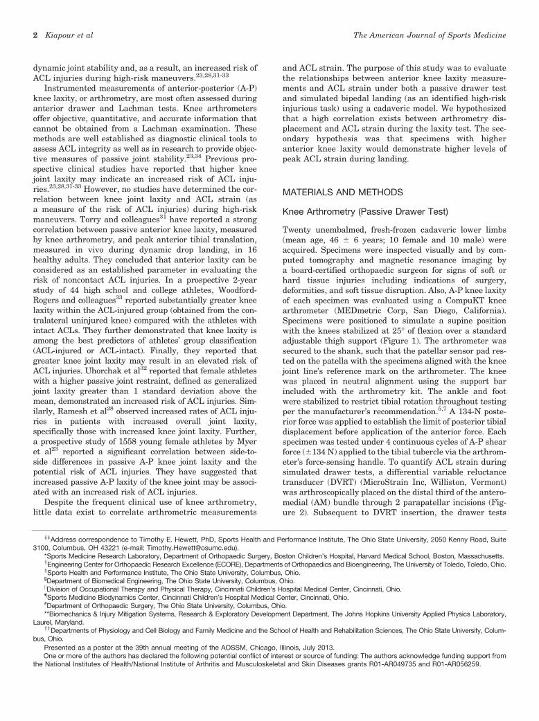
Twenty unembalmed, fresh-frozen cadaveric lower limbs(mean age, 46 6 6 years; 10 female and 10 male) wereacquired. Specimens were inspected visually and by com-puted tomography and magnetic resonance imaging bya board-certified orthopaedic surgeon for signs of soft orhard tissue injuries including indications of surgery,deformities, and soft tissue disruption. Also, A-P knee laxityof each specimen was evaluated using a CompuKT kneearthrometer (MEDmetric Corp, San Diego, California).Specimens were positioned to simulate a supine positionwith the knees stabilized at 25� of flexion over a standardadjustable thigh support (Figure 1). The arthrometer wassecured to the shank, such that the patellar sensor pad res-ted on the patella with the specimens aligned with the kneejoint line’s reference mark on the arthrometer. The kneewas placed in neutral alignment using the support barincluded with the arthrometry kit. The ankle and footwere stabilized to restrict tibial rotation throughout testingper the manufacturer’s recommendation.5,7 A 134-N poste-rior force was applied to establish the limit of posterior tibialdisplacement before application of the anterior force. Eachspecimen was tested under 4 continuous cycles of A-P shearforce (6134 N) applied to the tibial tubercle via the arthrom-eter’s force-sensing handle. To quantify ACL strain duringsimulated drawer tests, a differential variable reluctancetransducer (DVRT) (MicroStrain Inc, Williston, Vermont)was arthroscopically placed on the distal third of the antero-medial (AM) bundle through 2 parapatellar incisions (Fig-ure 2). Subsequent to DVRT insertion, the drawer tests
zzAddress correspondence to Timothy E. Hewett, PhD, Sports Health and Performance Institute, The Ohio State University, 2050 Kenny Road, Suite3100, Columbus, OH 43221 (e-mail: [email protected]).
*Sports Medicine Research Laboratory, Department of Orthopaedic Surgery, Boston Children’s Hospital, Harvard Medical School, Boston, Massachusetts.yEngineering Center for Orthopaedic Research Excellence (ECORE), Departments of Orthopaedics and Bioengineering, The University of Toledo, Toledo, Ohio.zSports Health and Performance Institute, The Ohio State University, Columbus, Ohio.§Department of Biomedical Engineering, The Ohio State University, Columbus, Ohio.kDivision of Occupational Therapy and Physical Therapy, Cincinnati Children’s Hospital Medical Center, Cincinnati, Ohio.{Sports Medicine Biodynamics Center, Cincinnati Children’s Hospital Medical Center, Cincinnati, Ohio.#Department of Orthopaedic Surgery, The Ohio State University, Columbus, Ohio.**Biomechanics & Injury Mitigation Systems, Research & Exploratory Development Department, The Johns Hopkins University Applied Physics Laboratory,
Laurel, Maryland.yyDepartments of Physiology and Cell Biology and Family Medicine and the School of Health and Rehabilitation Sciences, The Ohio State University, Colum-
bus, Ohio.
Presented as a poster at the 39th annual meeting of the AOSSM, Chicago, Illinois, July 2013.One or more of the authors has declared the following potential conflict of interest or source of funding: The authors acknowledge funding support from
the National Institutes of Health/National Institute of Arthritis and Musculoskeletal and Skin Diseases grants R01-AR049735 and R01-AR056259.
2 Kiapour et al The American Journal of Sports Medicine

were repeated. To calculate absolute strain values, the ACL’sreference length was calculated based on the methoddescribed by Howe et al14 and Fleming et al10 to identifythe DVRT’s length corresponding to the ligament-slack and-taut conditions. It was assumed that the mean strain acrossthe ACL’s AM bundle was equal to the change in length ofthe measured segment divided by the reference length(Kiapour AM, Quatman CE, Ditto RC, et al, ‘‘Influence ofAxial Rotation Moments on ACL Strain: A Cadaveric Studyof Single- and Multi-Axis Loading of the Knee.’’ Paper pre-sented at the proceedings of 37th ASB annual meeting,2012).16 All instrumented knee joint drawer tests were per-formed by a single licensed physical therapist (intrarater reli-ability [intraclass correlation coefficient] = 0.92).27 Analogdata (both arthrometer and DVRT) were collected at 100 Hz.
Dynamic Testing(Cadaveric Simulation of Bipedal Landing)
Subsequent to arthrometry, a subset of 14 specimens(mean age, 45 6 6 years; 6 female and 8 male) were testedunder dynamic conditions. It is noteworthy to mention that6 of 20 specimens used in the first part of the study wererandomly selected for multiple trials to better design andoptimize the dynamic test setup and loading protocol.These 6 trial specimens were eliminated from the secondset of the experiments (dynamic testing) because of tissuedamage and ACL failure during trials and the tune-up pro-cess. Specimens were stored at –20�C. Before testing,specimens were slowly thawed to room temperature. Speci-mens were sectioned at the midfemoral diaphysis (30 cmabove the joint line), and all soft tissues up to 15 cm prox-imal to the joint line were dissected. The remaining bonystructure of the proximal femur of each specimen was pot-ted for rigid attachment to the testing frame. The quadri-ceps (rectus femoris) and hamstring (semitendinosus,biceps femoris, and semimembranosus) tendons werethen isolated and clamped inside metal tendon grips toallow for the application of simulated muscle loads. Themusculature and skin directly around the knee joint were
maintained intact. The foot and ankle were also main-tained intact to provide a realistic load-transfer interface.Saline solution was used throughout specimen preparationand testing to maintain tissue hydration.
A novel custom-designed drop stand was used to simu-late dynamic landing.17,26 Specimens were mounted insuch a way to simulate lower extremity postures duringground strike, while landing from a jump with knees flexedat 25� (Figure 3). Each specimen was rigidly fixed at theproximal femur, and the tibia was orientated verticallywith the foot above. Transknee muscle forces were simu-lated utilizing multiple cable-pulley systems along withstatic weights to apply constant forces to the quadriceps(1200 N) and hamstring (800 N). Adjustable pulley systemswere used to maintain the physiological line of action ofeach muscle (Figure 3). An athletic shoe was placed onthe foot to provide realistic load transfer during initial con-tact. Subsequent to the application of muscle loads, thefloor platform was set upon the shoe to simulate a foot-planted position. Bipedal landing was simulated by therelease of half the body weight (350 N) from a height of30 cm utilizing a hemispherical impactor with an inte-grated weight stack. The drop weight exerted an impulsiveaxial compressive force that simulated the ground-reactionforce during landing from a jump. Moreover, ACL strainwas calculated based on the measurements of the DVRTsensor on the ACL’s AM bundle. The tibiofemoral jointkinematics were captured using an Optotrak 30203-dimensional motion capture system (Northern Digital,Waterloo, Ontario, Canada) along with 2 arrays of 3 infra-red light-emitting diode markers rigidly attached to thefemur and tibia. The obtained coordinates from eachmarker were then transferred to a joint local coordinatesystem to obtain anatomic joint kinematics.35 The initialposition of the tibia with respect to the femur in anunloaded position was considered as the reference framein the calculation of joint kinematics. The knee joint rota-tions were defined as Euler angles of the tibia’s referenceframe relative to the femur’s reference frame. Two 6-axis
Figure 1. The knee arthrometry procedure. Figure 2. Insertion of the differential variable reluctancetransducer on the anteromedial bundle of the anterior cruci-ate ligament.
Vol. XX, No. X, XXXX The Effect of Joint Laxity on Risk of ACL Injury 3

load cells (RA Denton Corp, Rochester Hills, Michigan)incorporated within the floor pad and femoral fixture wereused to capture all forces and moments delivered to theknee joint and reaction forces and moments during simu-lated landings. Data collection from all data acquisitionunits was synchronized utilizing a simultaneous triggeringsystem. Analog data (load cell and DVRT) were collectedat 4 kHz, while motion data were collected at 400 Hz.
Statistical Analysis
Paired sample t tests were used to investigate changes inboth anterior tibial translation and ACL strain under sim-ulated passive drawer tests (before and after DVRT inser-tion) and bipedal landing (before landing and peak). Ananalysis of variance (ANOVA) with a post hoc Bonferronicorrection for multiple comparisons was used to studythe relationships between applied shear force, joint laxity,and ACL strain. Multiple linear regression analyses wereconducted to determine the correlation between anteriordrawer load, anterior tibial translation, and ACL strain.
Correlations were classified as poor (\0.4), good (0.4-0.74), and strong (�0.75) based on the determined Pearsoncorrelation coefficient (r).25 Differences were considered tobe statistically significant for P \ .05.
RESULTS
A summary of the mean anterior tibial translation for bothbefore and after DVRT insertion along with the mean calcu-lated ACL strain during simulated drawer tests is presentedin Table 1. No significant difference in anterior tibial trans-lation was observed before and after DVRT insertion on theACL’s AM bundle under 67 N (P = .92), 89 N (P = .98), and134 N (P = .41) of anterior drawer force. During kneearthrometry (simulated passive drawer tests), an increasedanterior shear force resulted in significantly greater ante-rior tibial translation (anterior knee laxity) and meanACL strain (Figure 4). Further, linear regression analysiswith the assumption that 0-mm anterior tibial translationcorresponds with 0% ACL strain (y-intercept = 0) demon-strated a good statistically significant correlation (r = 0.52,P \ .0005) between arthrometer-measured anterior kneelaxity and resultant ACL strain (Figure 5).
A summary of peak axial impact force, anterior tibialtranslation, and ACL strain during simulated landings ispresented in Table 2. Before impact, muscle forces in thequadriceps and hamstring muscle groups produceda mean anterior tibial translation of 3.5 6 3.1 mm andACL strain of 2.0% 6 2.2%. Simulated landing resulted ina mean axial impact load of 4070 6 732 N over a period of70 milliseconds. Loads generated by axial impact increasedthe mean anterior tibial translation by 6.9 6 2.3 mm andincreased the mean ACL strain by 4.9% 6 2.6%. Simulatedlanding significantly increased peak anterior tibial transla-tion to 10.4 6 3.5 mm (P \ .0005) and peak ACL strain to6.8% 6 2.8% (P \ .0005) compared with the prelanding con-dition. No tissue failure was observed across anatomicstructures of the knee after testing.
Significant correlations were observed between resul-tant peak ACL strains during simulated landing and ante-rior tibial translation under anterior shear forces (duringsimulated drawer tests) of 67 N (r = 0.59, P = .02), 89 N(r = 0.57, P = .03), and 134 N (r = 0.62, P = .01). It was
Figure 3. Custom-designed drop-stand testing apparatus(top) and cable-pulley system used for the simulation ofknee muscle forces (bottom).
TABLE 1Summary of Knee Arthrometry Data (N = 20)a
Applied Anterior Shear Load
67 N (15 lb) 89 N (20 lb) 134 N (30 lb)
ATT before DVRTinsertion, mm
1.1 6 1.0 1.9 6 1.2 2.8 6 1.4
ATT after DVRTinsertion, mm
1.1 6 0.6 1.8 6 0.7 3.1 6 1.1
ACL strain, % 2.1 6 3.1 3.5 6 3.5 4.9 6 4.3
aValues are expressed as mean 6 standard deviation. ACL,anterior cruciate ligament; ATT, anterior tibial translation;DVRT, differential variable reluctance transducer.
4 Kiapour et al The American Journal of Sports Medicine

further shown that the specimens with higher magnitudesof anterior tibial translation during knee arthrometry test-ing demonstrate greater levels of peak ACL strain duringsimulated landings (Figure 6). To better understand thecorrelation between the risk of ACL injuries (measure bypeak ACL strain) and quantified anterior knee laxity,graphs presented in Figure 6 were divided into 4 quad-rants by 2 lines representing the mean values of anteriortibial translation (vertical line) and peak ACL strain (hor-izontal line) for the tested specimens. A substantial num-ber of the specimens (67%, 72%, and 83% under 67-N,89-N, and 134-N shear loads, respectively) with anteriorknee laxity greater than the mean were located at the firstquadrant (top right, high-risk zone), demonstratinggreater peak ACL strain than the mean during landing.However, the majority of the specimens (75%, 86%, and88% under 67-N, 89-N, and 134-N shear loads, respec-tively) with anterior knee laxity less than the mean werelocated in the third quadrant (bottom left, low-risk zone),demonstrating lower peak ACL strain than the mean dur-ing simulated landing.
DISCUSSION
Injuries of the ACL are exceedingly common and often dev-astating to adolescents and young adults.6,12 The associ-ated costs and resulting long-term disability havegenerated significant interest in the investigation of non-contact ACL injury mechanisms. Landing after a jumphas been identified as a high-risk task that may, under cer-tain conditions, lead to noncontact ACL injuries.3,15,24
Instrumented A-P knee laxity tests are well-establishedand reliable techniques that have been used extensivelyby clinicians to quantify knee joint laxity and to evaluateACL integrity. Previous prospective clinical investigationshave demonstrated that patients with higher knee laxityare at an increased risk of ACL injuries.23,28,31-33 Despitesubstantial research on the effects of knee joint laxityand the risk of ACL injuries, the biomechanical relation-ship between anterior knee laxity and ACL strain (asa measure of the risk of ACL injuries) during high-riskactivities has yet to be established. This study aimed toinvestigate the correlation between anterior knee laxityand ACL strain during a high-risk dynamic landing taskusing a cadaveric model.
Knee arthrometry was used to quantify the A-P kneelaxity of 20 relatively young, intact cadaveric lower limbswith and without DVRTs. The insertion of DVRTs acrossthe AM bundle of the ACL resulted in no significant differ-ence in instrumented laxity measures. Under simulateddrawer tests, applied anterior shear forces were shown tobe a significant factor in anterior tibial translation (ante-rior knee laxity) and resultant ACL strain levels. It wasdemonstrated that greater anterior shear forces can leadto increases in anterior tibial translation and ACL strain.This is in agreement with previous findings that demon-strate that the ACL provides up to 87% of the anterior tib-ial restraining force.1,4,11,19,20 A significant linearcorrelation was established between the magnitudes ofanterior tibial translation and ACL strain at 25� of knee
10
ACL Strainp=0.04
8
6
4
2
%)
Stra
in (%
ge A
CL S
Ave
rag
067 N (15 lb) 89 N (20 lb) 134 N (30 lb)
Anterior Shear Force
0
1
2
3
4
5
67 N (15 lb) 89 N (20 lb) 134 N (30 lb)
Ant
erio
r Ti
bial
Tra
nsla
�on
(mm
)
Anterior Shear Force
Anterior Knee Laxityp<0.0005
p=0.02
Figure 4. Change in anterior knee laxity (top) and meananterior cruciate ligament strain (bottom) under applied ante-rior shear force during the passive knee drawer test.
r=0.52p<0.0005
-3
0
3
6
9
12
15
18
0 1 2 3 4 5
ACL
Str
ain
(%)
Anterior Knee Laxity (mm)
67 N (15 lb)
89 N (20 lb)
134 N (30 lb)
Figure 5. Quantified anterior cruciate ligament strain versusanterior tibial translation during knee arthrometry.
Vol. XX, No. X, XXXX The Effect of Joint Laxity on Risk of ACL Injury 5

TABLE 2Summary of Change in ATT and ACL Strain During Simulated Landinga
Specimens ATT, mm ACL Strain, %
Identification No. Sex Age, y Side Peak Axial Impact, N Increased Knee Flexion, deg Impact Induced Peak Impact Induced Peak
1-C080044 F 52 L 3468 1.1 5.8 10.9 1.4 3.42-C090105 M 38 L 3188 0.1 3.1 8.0 1.0 6.93-C080033 M 51 L 3561 –0.1 8.6 14.6 5.6 8.54-S090574 F 49 R 4050 0.8 12.0 16.5 7.6 9.45-C090155 M 46 L 4400 0.4 6.6 5.2 5.7 4.46-C090105 M 38 R 4155 1.0 4.3 5.3 9.1 13.37-C090155 M 46 R 3394 0.7 5.4 10.8 1.7 6.28-C090361 M 34 R 2869 1.6 6.6 7.6 4.7 5.19-C090508 F 45 L 5076 –0.2 4.9 15.2 5.7 9.310-C090508 F 45 R 5036 0.7 8.4 8.8 8.9 8.511-C080044 F 52 R 3616 0.1 6.9 8.5 2.2 6.012-C090552 M 45 R 4751 0.6 8.9 13.8 6.4 7.413-C090552 M 45 L 4453 1.6 6.4 11.2 4.4 5.714-S090706 F 50 L 4962 3.1 9.4 9.9 3.7 2.2
aACL, anterior cruciate ligament; ATT, anterior tibial translation; F, female; impact induced, the change caused by application of the axialimpact load during simulated landings; L, left; M, male; R, right.
r=0.59p=0.02
0
2
4
6
8
10
12
14
0 0.5 1 1.5 2 2.5 3 3.5 4 4.5 5
Peak
ACL
Str
ain
(%)
Anterior Knee Laxity (mm)
67 N (15 lb) of Anterior Shear Force
High-Risk Zone
Low-Risk Zone
r=0.57p=0.03
0
2
4
6
8
10
12
14
0 0.5 1 1.5 2 2.5 3 3.5 4 4.5 5
Peak
ACL
Str
ain
(%)
Anterior Knee Laxity (mm)
89 N (20 lb) of Anterior Shear Force
High-Risk Zone
Low-Risk Zone
r=0.62p=0.01
0
2
4
6
8
10
12
14
0 0.5 1 1.5 2 2.5 3 3.5 4 4.5 5
Peak
ACL
Str
ain
(%)
Anterior Knee Laxity (mm)
134 N (30 lb) of Anterior Shear Force
High-Risk Zone
Low-Risk Zone
A B
C
Figure 6. Peak anterior cruciate ligament strain (during simulated bipedal landing) versus peak anterior tibial translation (quan-tified by the knee arthrometer) over a range of anterior shear loads.
6 Kiapour et al The American Journal of Sports Medicine

flexion. This direct correlation indicates that kneearthrometry is a good indicator of ACL strain and, there-fore, has intrinsic value in the evaluation of ACL loadingcharacteristics and the diagnosis of a functionally compro-mised ACL. In an in vivo study of 5 human patients, Flem-ing et al9 established a similar correlation (r = 0.59)between anterior tibial translation and ACL strain (mea-sured by a Hall effect transducer placed on the ACL’sAM bundle) during passive laxity testing.
Subsequent to the instrumented laxity evaluation, bipedallandings after a jump from a 30-cm height were simulated ona subset of 14 specimens. Results demonstrated the ability ofaxial impact loads (that simulate the vertical component ofthe ground-reaction force) to generate anterior translationof the tibia with respect to the femur. The impact-inducedanterior translation of the tibia relative to the femur can bebiomechanically described as the tibiofemoral joint reactionto generated shear forces due to the specific anatomy of thetibial plateau under shallow knee flexion angles, as demon-strated by other investigators.8,21,22 As expected, theimpact-induced anterior tibial translation resulted in ele-vated ACL strain levels after peak axial impact.
Significant direct linear relationships between theassessed anterior knee laxity after arthrometry andpeak ACL strain during simulated landings wereobserved. Specimens with greater anterior knee laxitydemonstrated an increased risk of ACL injuries, as themajority of these specimens were located within thehigh-risk zone. In contrast, specimens with lower anteriorknee laxity had a lower risk of ACL injuries, as the major-ity of these were located within the low-risk zone (Figure6). Although no studies have been able to directly quantifyACL strain during landing from a jump in vivo, theresults from this study indicate that patients withincreased anterior knee laxity may experience higherACL strains during landing, which may place them atan increased risk of ACL injuries.
This study is the first to establish a significant correla-tion between anterior knee joint laxity measured byarthrometry and peak ACL strain during simulateddynamic landing conditions. The instrumented evaluationof A-P knee laxity has been identified as one of the mostcommon, accurate, and valid approaches to evaluate ACLintegrity. The CompuKT arthrometer has been used exten-sively to quantify knee joint laxity and demonstrates good-to-excellent intrarater reliability.34 Considering ACLstrain as an established quantifiable measure for the riskof ACL injuries, the current findings highlight the impor-tance of instrumented knee A-P laxity assessment asa potential indicator of the risk of ACL injuries in additionto its clinical utility in the evaluation of ACL integrity.
Study Limitations
Despite the strong findings reported, there are inherent lim-itations, as with any experimental study. As noted by Grosset al,13 a number of factors, including arthrometer place-ment, flexion angle, and speed and angle of force applicationmay influence variables quantified during instrumented lax-ity testing. We have attempted to minimize these problems
and associated measurement errors by having a single,highly reliable licensed physical therapist, with over 15 yearsof experience with the CompuKT knee arthrometer, collectall arthrometry measurements. Further, the change in A-Pknee translation due to muscle co-contraction during kneearthrometry testing in vivo has been identified as one ofthe main limitations of this technique. However, the use ofCompuKT arthrometry in the current cadaveric work iswell justified because of the lack of any active neuromuscularcontrols. Moreover, the use of the CompuKT knee arthrome-ter has provided the operator with the advantage of livevisual feedback regarding the curve shape and test consis-tency compared with traditional knee arthrometers (ie,KT1000 arthrometer). This would increase the reliability ofthe results by allowing the operator to adjust the place-ment/pull angle based on the observed live feedback. Finally,the lack of significant differences between data before andafter DVRT insertion demonstrates consistent measure-ments using this approach in addition to demonstratingthat the minimally invasive placement of DVRTs did notaffect knee laxity measures. Another limitation was thepotential differences in tissue properties associated withcadaveric specimens compared with in vivo tissue propertiesof young athletes, which can affect the accuracy of absolutereported values. We have tried to minimize this artifact bytesting relatively young specimens (mean age, 46 6 6 years).Further, ACL strain was represented only by local strainmeasurements across the AM bundle. The attachment ofa second DVRT to the posterolateral bundle of the ACL wouldhave been associated with compromise of the posterior jointcapsule and potential measurement artifacts.2 The choice toplace a single DVRT on the ACL’s AM bundle was basedon previous work that found the AM bundle’s strain to bea good representation of overall ACL strain.18 Finally, theutilized cadaveric model of landing was associated witha number of assumptions and simplifications including, butnot limited to, static muscle loading, orientation of theground-reaction force relative to the tibia, exclusion oftransknee muscles other than the quadriceps and hamstring,and limited range of knee flexion. Care was taken to under-stand these limitations during the interpretation of ourfindings.
CONCLUSION
The current experimental findings support our first hypoth-esis, demonstrating a significant direct correlation betweenarthrometry displacement during laxity tests and concur-rent ACL strain calculated from DVRT measurements.Results further support our second hypothesis that instru-mented measures of anterior knee laxity predict peak ACLstrain during simulated landings. It also indicates thatthe specimens with higher anterior knee laxity exhibitincreased peak ACL strain during simulated landing.
ACKNOWLEDGMENT
The authors thank Nathaniel Bates for his assistance.
Vol. XX, No. X, XXXX The Effect of Joint Laxity on Risk of ACL Injury 7

REFERENCES
1. Arms S, Boyle J, Johnson R, Pope M. Strain measurement in the
medial collateral ligament of the human knee: an autopsy study.
J Biomech. 1983;16(7):491-496.
2. Bach JM, Hull ML. Strain inhomogeneity in the anterior cruciate liga-
ment under application of external and muscular loads. J Biomech
Eng. 1998;120(4):497-503.
3. Boden BP, Dean GS, Feagin JA, Garrett WE. Mechanisms of anterior
cruciate ligament injury. Orthopedics. 2000;23(6):573-578.
4. Butler DL, Noyes FR, Grood ES. Ligamentous restraints to anterior-
posterior drawer in the human knee: a biomechanical study.
J Bone Joint Surg Am. 1980;62(2):259-270.
5. Daniel DM, Malcom LL, Losse G, Stone ML, Sachs R, Burks R.
Instrumented measurement of anterior laxity of the knee. J Bone
Joint Surg Am. 1985;67(5):720-726.
6. Daniel DM, Stone ML, Dobson BE, Fithian DC, Rossman DJ, Kauf-
man KR. Fate of the ACL-injured patient: a prospective outcome
study. Am J Sports Med. 1994;22(5):632-644.
7. Daniel DM, Stone ML, Sachs R, Malcom L. Instrumented measure-
ment of anterior knee laxity in patients with acute anterior cruciate lig-
ament disruption. Am J Sports Med. 1985;13(6):401-407.
8. Dejour H, Bonnin M. Tibial translation after anterior cruciate ligament
rupture: two radiological tests compared. J Bone Joint Surg Br.
1994;76(5):745-749.
9. Fleming BC, Beynnon BD, Nichols CE, Johnson RJ, Pope MH. An in
vivo comparison of anterior tibial translation and strain in the antero-
medial band of the anterior cruciate ligament. J Biomech.
1993;26(1):51-58.
10. Fleming BC, Beynnon BD, Tohyama H, et al. Determination of a zero
strain reference for the anteromedial band of the anterior cruciate lig-
ament. J Orthop Res. 1994;12(6):789-795.
11. Fukubayashi T, Torzilli PA, Sherman MF, Warren RF. An in vitro bio-
mechanical evaluation of anterior-posterior motion of the knee: tibial
displacement, rotation, and torque. J Bone Joint Surg Am.
1982;64(2):258-264.
12. Griffin LY, Agel J, Albohm MJ, et al. Noncontact anterior cruciate lig-
ament injuries: risk factors and prevention strategies. J Am Acad
Orthop Surg. 2000;8(3):141-150.
13. Gross SM, Carcia CR, Gansneder BM, Shultz SJ. Rate of force appli-
cation during knee arthrometer testing affects stiffness but not dis-
placement measurements. J Orthop Sports Phys Ther. 2004;34(3):
132-139.
14. Howe JG, Wertheimer C, Johnson RJ, Nichols CE, Pope MH, Beyn-
non B. Arthroscopic strain gauge measurement of the normal anterior
cruciate ligament. Arthroscopy. 1990;6(3):198-204.
15. Kiapour AM. Non-Contact ACL Injuries During Landing: Risk Factors
and Mechanisms [dissertation]. Toledo, Ohio: Department of Bioengi-
neering, The University of Toledo; 2013.
16. Kiapour AM, Quatman CE, Ditto RC, et al. Global quasi-static
mechanical characterization of the human knee under single- and
multi-axis unconstrained loading conditions. In: Proceedings of
2012 ASME Summer Bioengineering Conference. Fajardo, Puerto
Rico: American Society of Mechanical Engineers (ASME);
2012:44809:1119-1120.
17. Levine JW, Kiapour AM, Quatman CE, et al. Clinically relevant injury
patterns after an anterior cruciate ligament injury provide insight into
injury mechanisms. Am J Sports Med. 2013;41(2):385-395.
18. Markolf KL, Gorek JF, Kabo JM, Shapiro MS. Direct measurement of
resultant forces in the anterior cruciate ligament: an in vitro study per-
formed with a new experimental technique. J Bone Joint Surg Am.
1990;72(4):557-567.
19. Markolf KL, Graff-Radford A, Amstutz HC. In vivo knee stability:
a quantitative assessment using an instrumented clinical testing
apparatus. J Bone Joint Surg Am. 1978;60(5):664-674.
20. Markolf KL, Mensch JS, Amstutz HC. Stiffness and laxity of the knee:
the contributions of the supporting structures. A quantitative in vitro
study. J Bone Joint Surg Am. 1976;58(5):583-594.
21. McLean SG, Oh YK, Palmer ML, et al. The relationship between ante-
rior tibial acceleration, tibial slope, and ACL strain during a simulated
jump landing task. J Bone Joint Surg Am. 2011;93(14):1310-1317.
22. Meyer EG, Haut RC. Excessive compression of the human tibio-
femoral joint causes ACL rupture. J Biomech. 2005;38(11):2311-2316.
23. Myer GD, Ford KR, Paterno MV, Nick TG, Hewett TE. The effects of
generalized joint laxity on risk of anterior cruciate ligament injury in
young female athletes. Am J Sports Med. 2008;36(6):1073-1080.
24. Olsen OE, Myklebust G, Engebretsen L, Bahr R. Injury mechanisms
for anterior cruciate ligament injuries in team handball: a systematic
video analysis. Am J Sports Med. 2004;32(4):1002-1012.
25. Portney LG, Watkins MP. Foundations of Clinical Research: Applications
to Practice. 2nd ed. Upper Saddle River, New Jersey: Prentice Hall; 1999.
26. Quatman CE, Kiapour AM, Demetropoulos CK, et al. Preferential
loading of the ACL compared with the MCL during landing: a novel
in sim approach yields the multiplanar mechanism of dynamic valgus
during ACL injuries [published online October 11, 2013]. Am J Sports
Med. doi:10.1177/0363546513506558
27. Quatman CE, Paterno MV, Wordeman SC, Kaeding CC. Longitudinal
anterior knee laxity related to substantial tibial tunnel enlargement
after anterior cruciate ligament revision. Arthroscopy. 2011;27(8):
1160-1163.
28. Ramesh R, Von Arx O, Azzopardi T, Schranz PJ. The risk of anterior
cruciate ligament rupture with generalised joint laxity. J Bone Joint
Surg Br. 2005;87(6):800-803.
29. Rozzi SL, Lephart SM, Gear WS, Fu FH. Knee joint laxity and neuro-
muscular characteristics of male and female soccer and basketball
players. Am J Sports Med. 1999;27(3):312-319.
30. Shultz SJ, Carcia CR, Perrin DH. Knee joint laxity affects muscle acti-
vation patterns in the healthy knee. J Electromyogr Kinesiol. 2004;
14(4):475-483.
31. Torry MR, Myers C, Pennington WW, et al. Relationship of anterior
knee laxity to knee translations during drop landings: a bi-plane fluo-
roscopy study. Knee Surg Sports Traumatol Arthrosc. 2011;19(4):
653-662.
32. Uhorchak JM, Scoville CR, Williams GN, Arciero RA, St Pierre P, Tay-
lor DC. Risk factors associated with noncontact injury of the anterior
cruciate ligament: a prospective four-year evaluation of 859 West
Point cadets. Am J Sports Med. 2003;31(6):831-842.
33. Woodford-Rogers B, Cyphert L, Denegar CR. Risk factors for anterior
cruciate ligament injury in high school and college athletes. J Athl
Train. 1994;29(4):343-346.
34. Wordeman SC, Paterno MV, Quatman CE, Bates NA, Hewett TE.
Arthrometric curve-shape variables to assess anterior cruciate liga-
ment deficiency. Clin Biomech (Bristol, Avon). 2012;27(8):830-836.
35. Yu B, Lin CF, Garrett WE. Lower extremity biomechanics during the
landing of a stop-jump task. Clin Biomech (Bristol, Avon). 2006;
21(3):297-305.
For reprints and permission queries, please visit SAGE’s Web site at http://www.sagepub.com/journalsPermissions.nav
8 Kiapour et al The American Journal of Sports Medicine