Embed Size (px)
Citation preview

Hindawi Publishing CorporationISRN EndoscopyVolume 2013, Article ID 769519, 3 pageshttp://dx.doi.org/10.5402/2013/769519
Clinical StudyDiagnostic Yield of Routine Duodenal Biopsies in IronDeficiency Anemia for Celiac Disease Diagnosis
Houria Chellat, Mouna Salihoun, Nawal Kabbaj, Leila Amrani, Ilham Serraj, Zakia Chaoui,M’hamed Nya, Mohammed Acherki, and Naïma Amrani
EFD-Hepatogastroenterology Unit, University of Mohammed 5 Souissi, Rabat 10000, Morocco
Correspondence should be addressed to Mouna Salihoun; [email protected]
Received 16 December 2012; Accepted 20 January 2013
Academic Editors: P. Born, B. Braden, and M. Sewitch
Copyright © 2013 Houria Chellat et al. This is an open access article distributed under the Creative Commons Attribution License,which permits unrestricted use, distribution, and reproduction in any medium, provided the original work is properly cited.
Background. Iron deficiency anemia (IDA) is a recognised feature of celiac disease (CD) in adults and can be its only presentation.Aim. To define the prevalence of CD in Moroccan adult patients with IDA of obscure origin and to determine the yield of smallbowel biopsy performed during routine endoscopy.Methods. 437 patients with IDA of obscure origin were included. 4 endoscopicmucosal biopsies were taken from the second part of duodenum and 2 biopsies from antrum and fundus, respectively. Endoscopicaspect and severity of anemia were correlated with histological diagnoses using coefficient Kappa. Results. 29 out of 437 patients(6.63%) had CD. Endoscopic aspect was normal in 66%, a mosaic pattern of mucosa in 17%, and scalloping of the small bowelfolds in 17%. 12 patients had Marsh III, 8 had Marsh II, 6 had Marsh I, and 3 had Marsh IV lesions. There was no correlationbetween degree of anemia, endoscopic aspect, and severity of duodenal lesions (Kappa = −0.167). Conclusion. Routine duodenalbiopsy gives an additional 6.63% diagnostic benefit of CD and should be indicated in all patients with IDA. The finding of normalendoscopic appearance of mucosa should not preclude duodenal biopsies.
1. Background
Occult, chronic blood loss from the gastrointestinal tract isthe most common cause of iron deficiency anemia. The dif-ferent lesions responsible for chronic blood loss include bothupper and lower gastrointestinal tract sourceswith causes andincidences varying widely among different studies [1].
First described in 1888 by Samuel Gee, adult celiac diseasehas now been well recognized as a disease characterized bydamage to the small bowel mucosa induced by gluten. Adultceliac disease can be a cause of malabsorption of severalnutrients in addition to having amalignant potential. Anemiacan be a presenting and/or significant feature of this diseaseand can occur in any age, sex, or ethnic group [1].
This autoimmune enteropathy is common not only inEurope but also in African populations, specifically in theMaghreb area. The primary aim of this study is to define theprevalence of CD in Moroccan adult patients with iron defi-ciency anemia of obscure origin and to determine the yield of
small bowel biopsy performed during routine endoscopy.Thesecondary one is to determine the correlation between degreeof anemia, endoscopic aspect, and severity of duodenallesions.
2. Methods
We retrospectively studied 437 patients with IDA of obscureorigin and nonspecific gastrointestinal symptoms are col-lected between 2005 and 2011. All patients had a diagnosis ofIDA with either hemoglobin < 12 g/dL or ferritin < 15 𝜇g/L.
4 endoscopic mucosal biopsies were taken from thesecond part of the duodenum in these patients and 2 biopsiesfrom antrum and fundus, respectively. Biopsy specimenswere fixed in buffered formalin and immediately submit-ted for histopathology study. Histopathologic examinationresults of patients were stratified according to Marsh clas-sification system: normal mucosa was defined as Marsh 0;increased number of intraepithelial lymphocytes as Marsh I;
2 ISRN Endoscopy
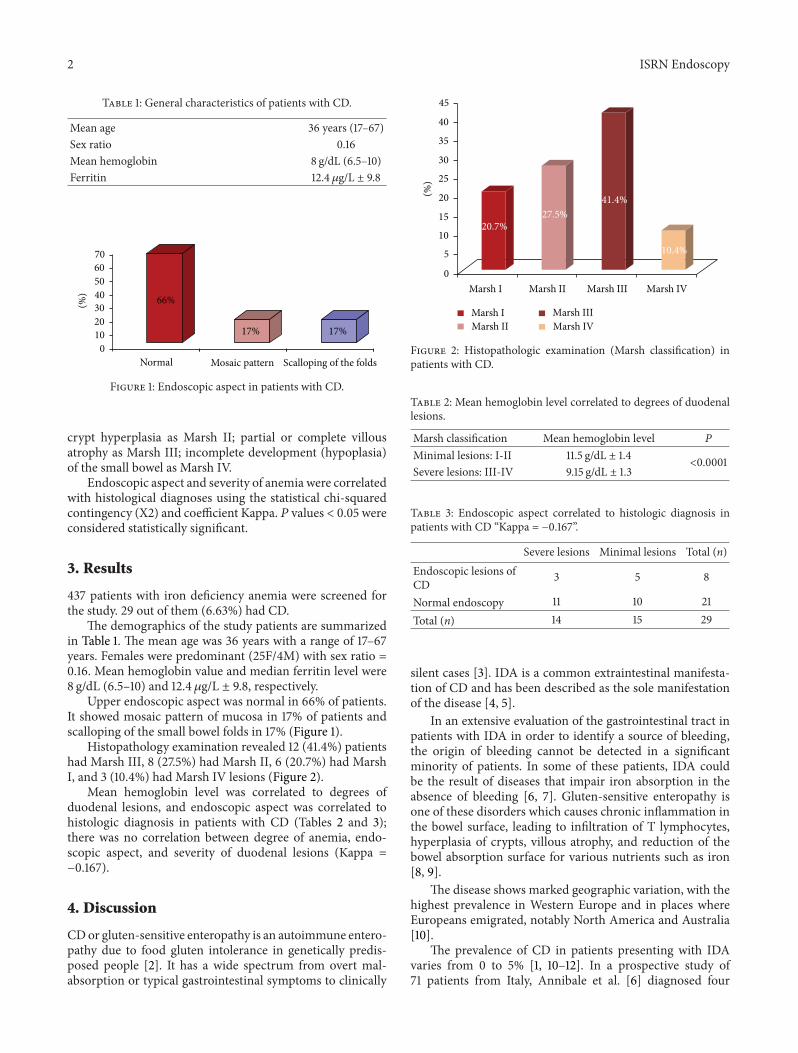
Table 1: General characteristics of patients with CD.
Mean age 36 years (17–67)Sex ratio 0.16Mean hemoglobin 8 g/dL (6.5–10)Ferritin 12.4 𝜇g/L ± 9.8
Normal Mosaic pattern Scalloping of the folds
70605040302010
0
(%)
17% 17%
66%
Figure 1: Endoscopic aspect in patients with CD.
crypt hyperplasia as Marsh II; partial or complete villousatrophy as Marsh III; incomplete development (hypoplasia)of the small bowel as Marsh IV.
Endoscopic aspect and severity of anemia were correlatedwith histological diagnoses using the statistical chi-squaredcontingency (X2) and coefficient Kappa. 𝑃 values < 0.05 wereconsidered statistically significant.
3. Results
437 patients with iron deficiency anemia were screened forthe study. 29 out of them (6.63%) had CD.
The demographics of the study patients are summarizedin Table 1. The mean age was 36 years with a range of 17–67years. Females were predominant (25F/4M) with sex ratio =0.16. Mean hemoglobin value and median ferritin level were8 g/dL (6.5–10) and 12.4 𝜇g/L ± 9.8, respectively.
Upper endoscopic aspect was normal in 66% of patients.It showed mosaic pattern of mucosa in 17% of patients andscalloping of the small bowel folds in 17% (Figure 1).
Histopathology examination revealed 12 (41.4%) patientshad Marsh III, 8 (27.5%) had Marsh II, 6 (20.7%) had MarshI, and 3 (10.4%) had Marsh IV lesions (Figure 2).
Mean hemoglobin level was correlated to degrees ofduodenal lesions, and endoscopic aspect was correlated tohistologic diagnosis in patients with CD (Tables 2 and 3);there was no correlation between degree of anemia, endo-scopic aspect, and severity of duodenal lesions (Kappa =−0.167).
4. Discussion
CDor gluten-sensitive enteropathy is an autoimmune entero-pathy due to food gluten intolerance in genetically predis-posed people [2]. It has a wide spectrum from overt mal-absorption or typical gastrointestinal symptoms to clinically
45
40
35
30
25
20
15
10
5
0
(%)
Marsh IIMarsh IIIMarsh IV
Marsh I
Marsh I Marsh II Marsh III Marsh IV
20.7%27.5%
41.4%
10.4%
Figure 2: Histopathologic examination (Marsh classification) inpatients with CD.
Table 2: Mean hemoglobin level correlated to degrees of duodenallesions.
Marsh classification Mean hemoglobin level PMinimal lesions: I-II 11.5 g/dL ± 1.4
<0.0001Severe lesions: III-IV 9.15 g/dL ± 1.3
Table 3: Endoscopic aspect correlated to histologic diagnosis inpatients with CD “Kappa = −0.167”.
Severe lesions Minimal lesions Total (n)Endoscopic lesions ofCD 3 5 8
Normal endoscopy 11 10 21Total (n) 14 15 29
silent cases [3]. IDA is a common extraintestinal manifesta-tion of CD and has been described as the sole manifestationof the disease [4, 5].
In an extensive evaluation of the gastrointestinal tract inpatients with IDA in order to identify a source of bleeding,the origin of bleeding cannot be detected in a significantminority of patients. In some of these patients, IDA couldbe the result of diseases that impair iron absorption in theabsence of bleeding [6, 7]. Gluten-sensitive enteropathy isone of these disorders which causes chronic inflammation inthe bowel surface, leading to infiltration of T lymphocytes,hyperplasia of crypts, villous atrophy, and reduction of thebowel absorption surface for various nutrients such as iron[8, 9].
The disease shows marked geographic variation, with thehighest prevalence in Western Europe and in places whereEuropeans emigrated, notably North America and Australia[10].
The prevalence of CD in patients presenting with IDAvaries from 0 to 5% [1, 10–12]. In a prospective study of71 patients from Italy, Annibale et al. [6] diagnosed four

ISRN Endoscopy 3
patients (5.7%) with CD. In a study of 114 patients with IDA,conducted in the United Kingdom, 2.6% of these patients hadCD [13]. In another study, CD was present in 5.7% of patientswith IDA [14]. In a study from USA, Grisolano et al. [15]identified nine cases (8.7%) in 103 patients with IDA. In ourstudy, 6.63% of patients with IDA had CD. This disparity ofprevalence could possibly be related to differences in localprevalence of CD as well as patient selection criteria.
Endoscopic diagnoses, symptoms and the prevalence ofanemia were correlated with the histological diagnoses ina prospective study of 1000 patients [16]. There was nocorrelation between clinical symptoms, the prevalence ofanemia, and the diagnosis of coeliac disease or giardiasis inthis cohort. Our result is concording with this study.
These studies emphasize the importance of the prevalenceof disease in different patient groups and its effect on thepredictive value of the diagnostic test.Thus, they demonstratethe important yield of upper endoscopy in the evaluation ofpatients with IDA.
5. Conclusion
Routine duodenal sampling during the upper endoscopicexamination gives an additional 6.63% diagnostic benefit inour study (near to the most reports in the literature), andthis practice should be included in the diagnostic workup ofpatients with IDA. Moreover, performing duodenal biopsy isstill necessary even though the endoscopic appearance of themucosa is normal.
Conflict of Interests
The authors declare no conflict of interests.
References
[1] U. S. Karnam, L. R. Felder, and J. B. Raskin, “Prevalence ofoccult celiac disease in patients with iron-deficiency anemia: aprospective study,” Southern Medical Journal, vol. 97, no. 1, pp.30–34, 2004.
[2] V. M. De Lima, L. Gandolfi, J. A. D. A. Pires, and R. Pratesi,“Prevalence of celiac disease in dyspeptic patients,” Arquivos deGastroenterologia, vol. 42, no. 3, pp. 153–156, 2005.
[3] C.Gonen,N.Yilmaz,M.Yalcin, I. Simsek, andO.Gonen, “Diag-nostic yield of routine duodenal biopsies in iron deficiencyanaemia: a study from Western Anatolia,” European Journal ofGastroenterology and Hepatology, vol. 19, no. 1, pp. 37–41, 2007.
[4] G. R. Corazza, R. A.Valentini,M. L. Andreani et al., “Subclinicalcoeliac disease is a frequent cause of iron-deficiency anaemia,”Scandinavian Journal of Gastroenterology, vol. 30, no. 2, pp. 153–156, 1995.
[5] U. Schmitz, Y. Ko, S. Seewald, R. Dusing, and H. Vetter, “Iron-deficiency anemia as the sole manifestation of celiac disease,”Clinical Investigator, vol. 72, no. 7, pp. 519–521, 1994.
[6] B. Annibale, G. Capurso, A. Chistolini et al., “Gastrointestinalcauses of refractory iron deficiency anemia in patients withoutgastrointestinal symptoms,” American Journal of Medicine, vol.111, no. 6, pp. 439–445, 2001.
[7] D. C. Rockey and J. P. Cello, “Evaluation of the gastrointestinaltract in patients with iron-deficiency anemia,”TheNew EnglandJournal of Medicine, vol. 329, no. 23, pp. 1691–1695, 1993.
[8] M. Silano, U. Volta, A. Mecchia et al., “Delayed diagnosis ofcoeliac disease increases cancer risk,” BMC Gastroenterology,vol. 7, article 8, 2007.
[9] F. Zamani, M. Mohamadnejad, R. Shakeri et al., “Glutensensitive enteropathy in patients with iron deficiency anemia ofunknown origin,”World Journal of Gastroenterology, vol. 14, no.48, pp. 7381–7385, 2008.
[10] R. J. Farrell and C. P. Kelly, “Celiac sprue,” The New EnglandJournal of Medicine, vol. 346, pp. 180–188, 2002.
[11] S. Gee, “On the coeliac affection,” St Bartholomew’s HospitalReport, vol. 24, pp. 17–24, 1888.
[12] G. C. Power and R. E. Smith, “Prevalence of occult celiac diseasein adults with iron deficiency anemia,”The American Journal ofGastroenterology, vol. 86, p. 231, 1991.
[13] A. S.McIntyre and R. G. Long, “Prospective survey of investiga-tions in outpatients referred with iron deficiency anaemia,”Gut,vol. 34, no. 8, pp. 1102–1107, 1993.
[14] M. T. Kepczyk and C. S. C. Kadakia, “Prospective evaluation ofgastrointestinal tract in patients with iron-deficiency anemia,”Digestive Diseases and Sciences, vol. 40, no. 6, pp. 1283–1289,1995.
[15] S. W. Grisolano, A. S. Oxentenko, J. A. Murray, L. J. Burgart, R.A. Dierkhising, and J. A. Alexander, “The usefulness of routinesmall bowel biopsies in evaluation of iron deficiency anemia,”Journal of Clinical Gastroenterology, vol. 38, no. 9, pp. 756–760,2004.
[16] J. J. W. Tischendor, K. Wopp, K. L. Streetz et al., “The value ofduodenal biopsy within routine upper endoscopy: a prospectivestudy in 1000 patients,” Zeitschrift fur Gastroenterologie, vol. 46,no. 8, pp. 771–775, 2008.

Submit your manuscripts athttp://www.hindawi.com
Stem CellsInternational
Hindawi Publishing Corporationhttp://www.hindawi.com Volume 2014
Hindawi Publishing Corporationhttp://www.hindawi.com Volume 2014
MEDIATORSINFLAMMATION
of
Hindawi Publishing Corporationhttp://www.hindawi.com Volume 2014
Behavioural Neurology
International Journal of
EndocrinologyHindawi Publishing Corporationhttp://www.hindawi.com
Volume 2014
Hindawi Publishing Corporationhttp://www.hindawi.com Volume 2014
Disease Markers
BioMed Research International
Hindawi Publishing Corporationhttp://www.hindawi.com Volume 2014
OncologyJournal of
Hindawi Publishing Corporationhttp://www.hindawi.com Volume 2014
Hindawi Publishing Corporationhttp://www.hindawi.com Volume 2014
Oxidative Medicine and Cellular Longevity
PPARRe sea rch
Hindawi Publishing Corporationhttp://www.hindawi.com Volume 2014
The Scientific World JournalHindawi Publishing Corporation http://www.hindawi.com Volume 2014
Immunology ResearchHindawi Publishing Corporationhttp://www.hindawi.com Volume 2014
Journal of
ObesityJournal of
Hindawi Publishing Corporationhttp://www.hindawi.com Volume 2014
Hindawi Publishing Corporationhttp://www.hindawi.com Volume 2014
Computational and Mathematical Methods in Medicine
OphthalmologyJournal of
Hindawi Publishing Corporationhttp://www.hindawi.com Volume 2014
Diabetes ResearchJournal of
Hindawi Publishing Corporationhttp://www.hindawi.com Volume 2014
Hindawi Publishing Corporationhttp://www.hindawi.com Volume 2014
Research and TreatmentAIDS
Hindawi Publishing Corporationhttp://www.hindawi.com Volume 2014
Gastroenterology Research and Practice
Parkinson’s DiseaseHindawi Publishing Corporationhttp://www.hindawi.com Volume 2014
Evidence-Based Complementary and Alternative Medicine
Volume 2014Hindawi Publishing Corporationhttp://www.hindawi.com
![Crypt of Cthulhu #38 (1987.Cryptic)[CosmicJukebox] of Cthulhu/Misc/Crypt of Cthulhu/Crypt of... · Eastertide1986/5 foundinLovecraft'scellargallery: Alockedportfolio,boundintanned](https://img.pdfslide.net/doc/110x75/5b975f8609d3f27e758c8cfe/crypt-of-cthulhu-38-1987crypticcosmicjukebox-of-cthulhumisccrypt-of-cthulhucrypt.jpg)