Embed Size (px)
Citation preview

Diagnostics and genetic variation of an invasive microsporidium (Nosema ceranae) in honey bees
(Apis mellifera)
Dr. M. M. Hamiduzzaman
School of Environmental Sciences University of Guelph, Canada

Importance of honey bees
Honey bees are social, beneficial, economic insects
Honey bee
Pollination
Health/medicine
Economy
Agriculture
Foods
Industry
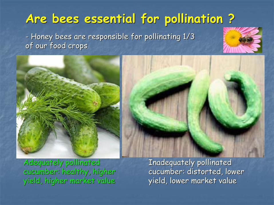
Are bees essential for pollination ?
Inadequately pollinated cucumber: distorted, lower yield, lower market value
Adequately pollinated cucumber: healthy, higher yield, higher market value
- Honey bees are responsible for pollinating 1/3 of our food crops

Bee colony losses
World
USA
Honey bee colony mortality in Canada as well other countries has been increasing
Winter bee mortality in Canada
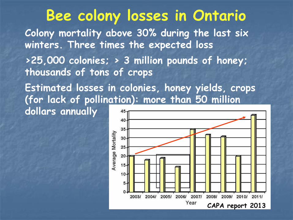
Bee colony losses in Ontario Colony mortality above 30% during the last six winters. Three times the expected loss
>25,000 colonies; > 3 million pounds of honey; thousands of tons of crops
Estimated losses in colonies, honey yields, crops (for lack of pollination): more than 50 million dollars annually
CAPA report 2013
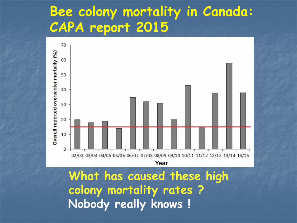
What has caused these high colony mortality rates ? Nobody really knows !
Bee colony mortality in Canada: CAPA report 2015
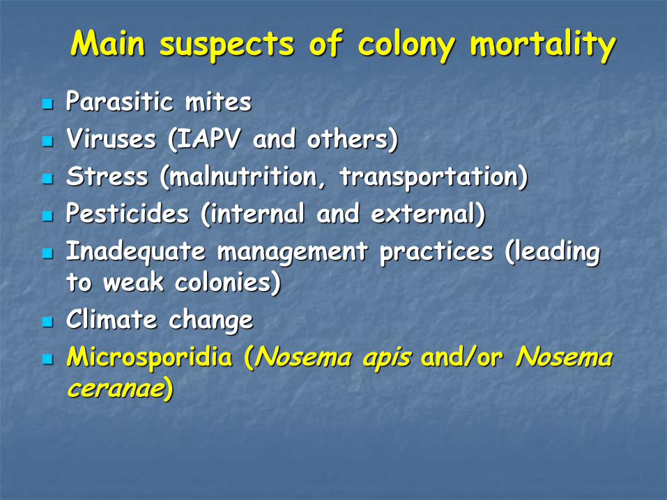
Parasitic mites
Viruses (IAPV and others)
Stress (malnutrition, transportation)
Pesticides (internal and external)
Inadequate management practices (leading to weak colonies)
Climate change
Microsporidia (Nosema apis and/or Nosema ceranae)
Main suspects of colony mortality
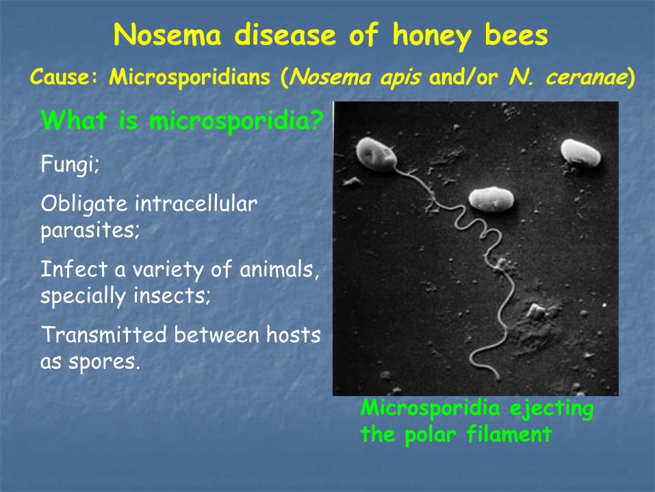
Nosema disease of honey bees
What is microsporidia?
Fungi;
Obligate intracellular parasites;
Infect a variety of animals, specially insects;
Transmitted between hosts as spores.
Microsporidia ejecting the polar filament
Cause: Microsporidians (Nosema apis and/or N. ceranae)
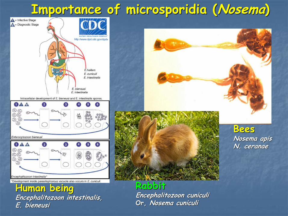
Importance of microsporidia (Nosema)
Bees Nosema apis N. ceranae
Rabbit Encephalitozoon cuniculi Or, Nosema cuniculi
Human being Encephalitozoon intestinalis, E. bieneusi

Nosema (Dysentery symptom)
Fecal staining on the outside and entrance of the hive
Nosema disease (Nosemosis) of honey bees
Cause: Microsporidians (N. ceranae and/or N. apis)
Nosema Dysentery symptom

History of Nosema disease in bees
The first report of N. apis was in Europe (Germany) in the Western honey bees, Apis mellifera (Zander, 1909). Almost a century after that N. ceranae was reported in East Asia (China) in the Asian honey bees, Apis cerana (Fries et al., 1996). Originally it was thought that N. ceranae was restricted to Asian honey bees (Apis cerana). N. ceranae was reported in European bees (Apis mellifera) since 2006 by many research groups in all over the world.

Life cycle of Nosema apis. The spore injects its contents into a gut epithelial cell, multiplies, and eventually causes the cell to burst and release the new spores back into the gut. Nosema can also reproduce “vegetatively” cell to cell.
Life cycle of Nosema disease
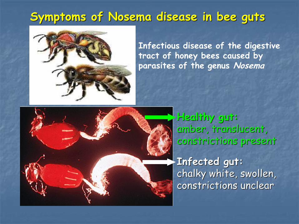
Symptoms of Nosema disease in bee guts
Healthy gut: amber, translucent, constrictions present
Infected gut: chalky white, swollen, constrictions unclear
Infectious disease of the digestive tract of honey bees caused by parasites of the genus Nosema
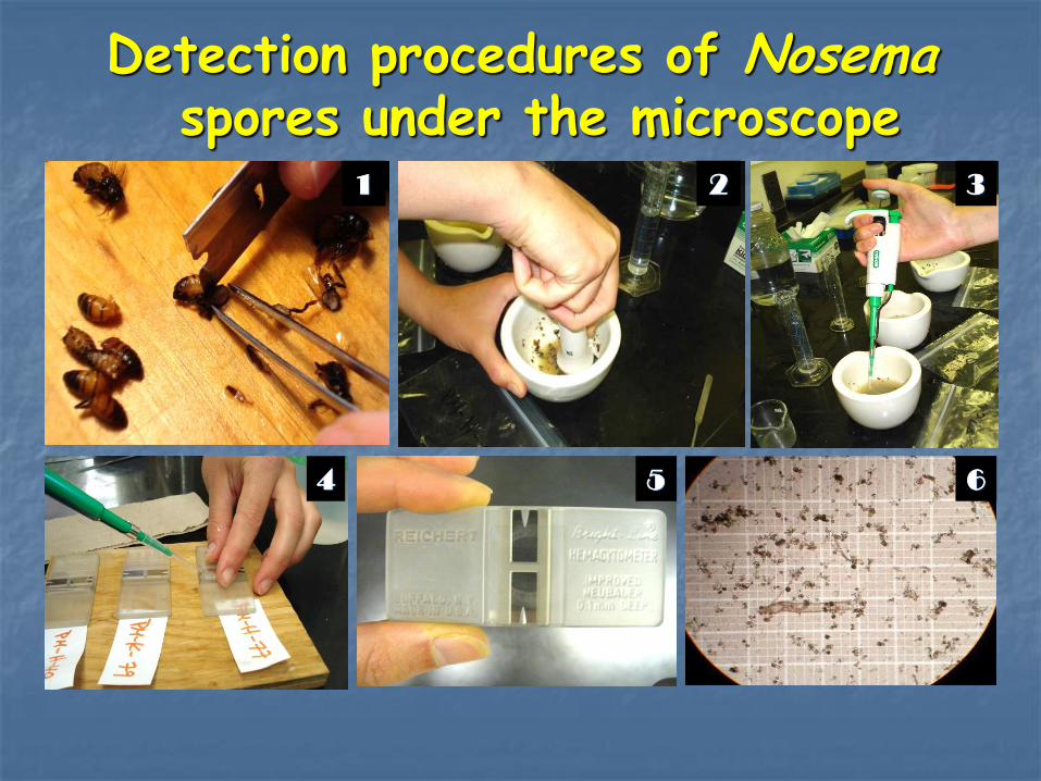
Detection procedures of Nosema spores under the microscope
1 2 3
6 4 5
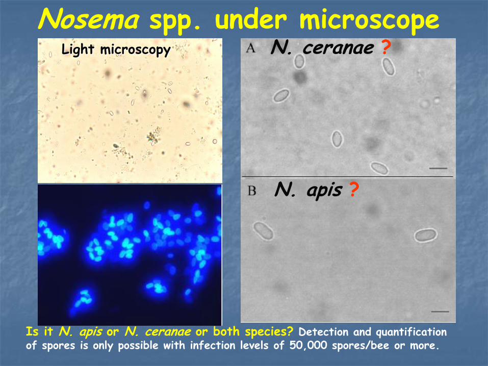
Fluorescent microscopy
N. ceranae ?
N. apis ?
Light microscopy
Nosema spp. under microscope
Is it N. apis or N. ceranae or both species? Detection and quantification of spores is only possible with infection levels of 50,000 spores/bee or more.

Challenges ! - Recently N. ceranae was found in Canada and it is replacing N. apis
- Good/reliable lab. methods to diagnose and quantify N. ceranae and/or N. apis infections are needed
- Analyze the genetic diversity of N. ceranae

Objectives
- Develop a simple and reliable multiplex PCR (polymerase chain reaction) method to diagnose and quantify Nosema disease (N. ceranae and/or N. apis) in honey bees - Determine genetic variation in N. ceranae samples collected from different regions of the world using genetic markers and sequencing techniques

Honey Bee Research Centre (HBRC) method
Commercial Kit for DNA extraction
10-20 bees per sample
Extraction of spores & filtration needed
Germination buffer needed
Incubation period needed
DNA extracted with CTAB buffer developed
Single bee per sample
Total abdomen used (no spore extraction needed)
No germination buffer needed
No incubation period needed
Kit method (Roche Diagnosis)
DNA extraction method
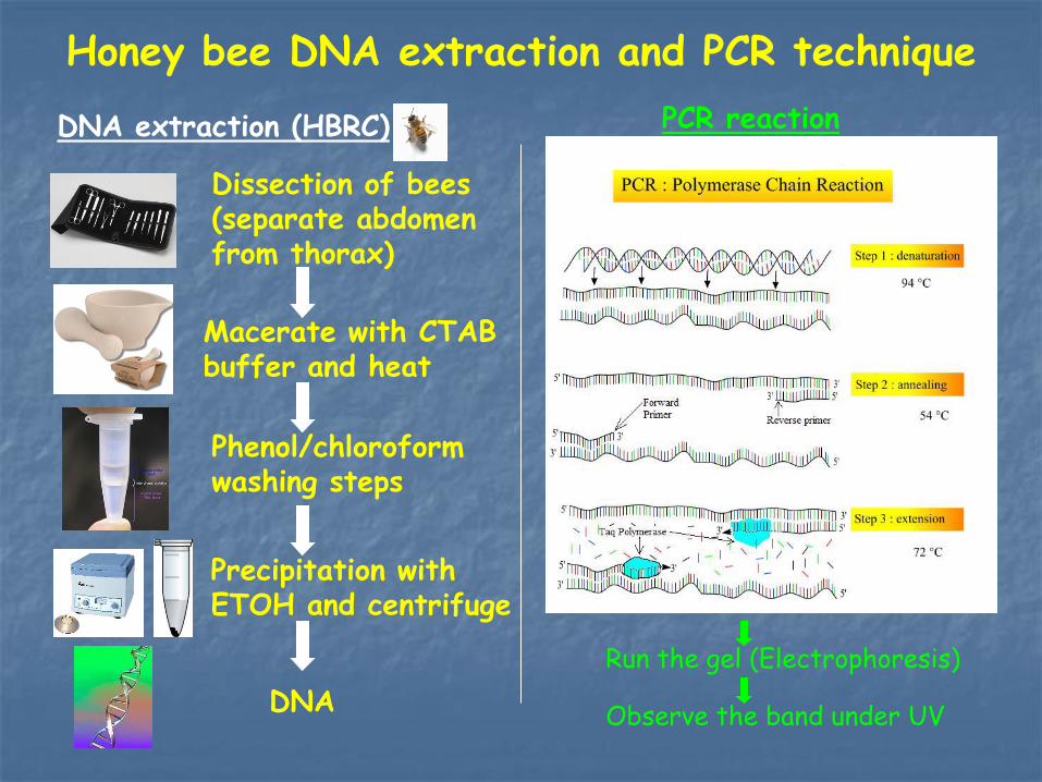
Honey bee DNA extraction and PCR technique
DNA extraction (HBRC)
Dissection of bees (separate abdomen from thorax)
DNA
Phenol/chloroform washing steps
Precipitation with ETOH and centrifuge
Macerate with CTAB buffer and heat
PCR reaction
Run the gel (Electrophoresis)
Observe the band under UV
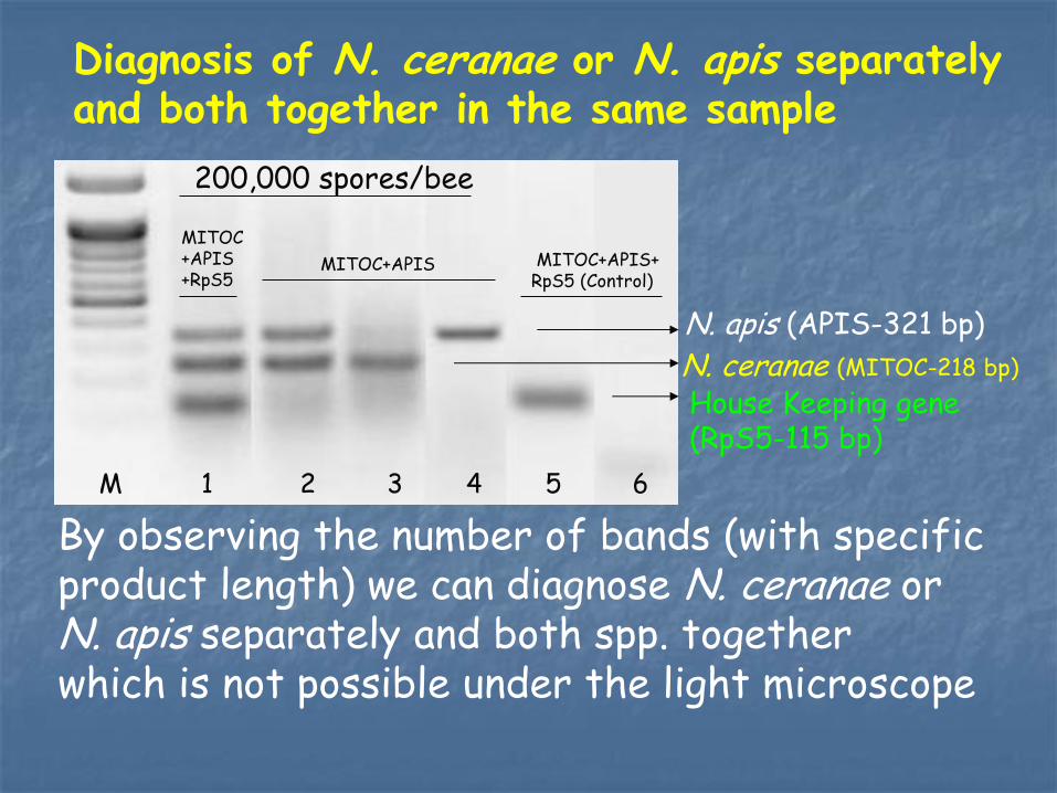
Diagnosis of N. ceranae or N. apis separately and both together in the same sample
N. apis (APIS-321 bp)
N. ceranae (MITOC-218 bp)
House Keeping gene (RpS5-115 bp)
By observing the number of bands (with specific product length) we can diagnose N. ceranae or N. apis separately and both spp. together which is not possible under the light microscope
200,000 spores/bee
MITOC +APIS +RpS5
MITOC+APIS MITOC+APIS+ RpS5 (Control)
M 1 2 3 4 5 6

Sensitivity of the HBRC method
What is the lowest number of spores in a sample that can be detected with the HBRC method ?
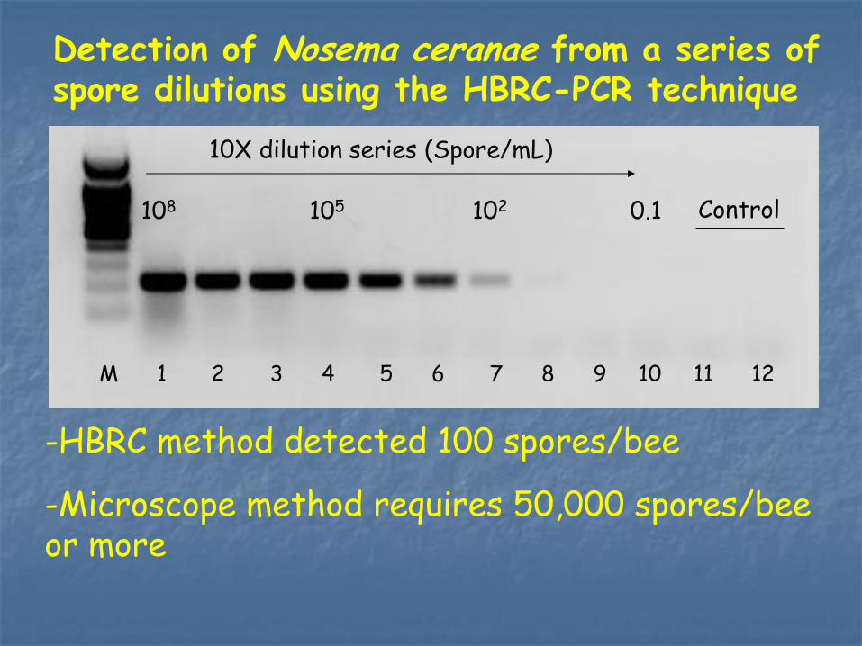
Detection of Nosema ceranae from a series of spore dilutions using the HBRC-PCR technique
M 1 2 3 4 5 6 7 8 9 10 11 12
108 102
10X dilution series (Spore/mL)
105 0.1 Control
-HBRC method detected 100 spores/bee
-Microscope method requires 50,000 spores/bee or more

Ratio of the expression level of target gene over HK gene was positively correlated with infection levels
M 1 2 3 4 5 6 7
H K gene
N. ceranae
Lane 1-5: Different infection levels (spores/bee)
Healthy bee
Clean spore
Lane 1-7: MITOC + RpS5
Quantification of Nosema infection levels using the HBRC method
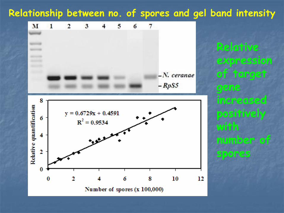
Relationship between no. of spores and gel band intensity
Relative expression of target gene increased positively with number of spores
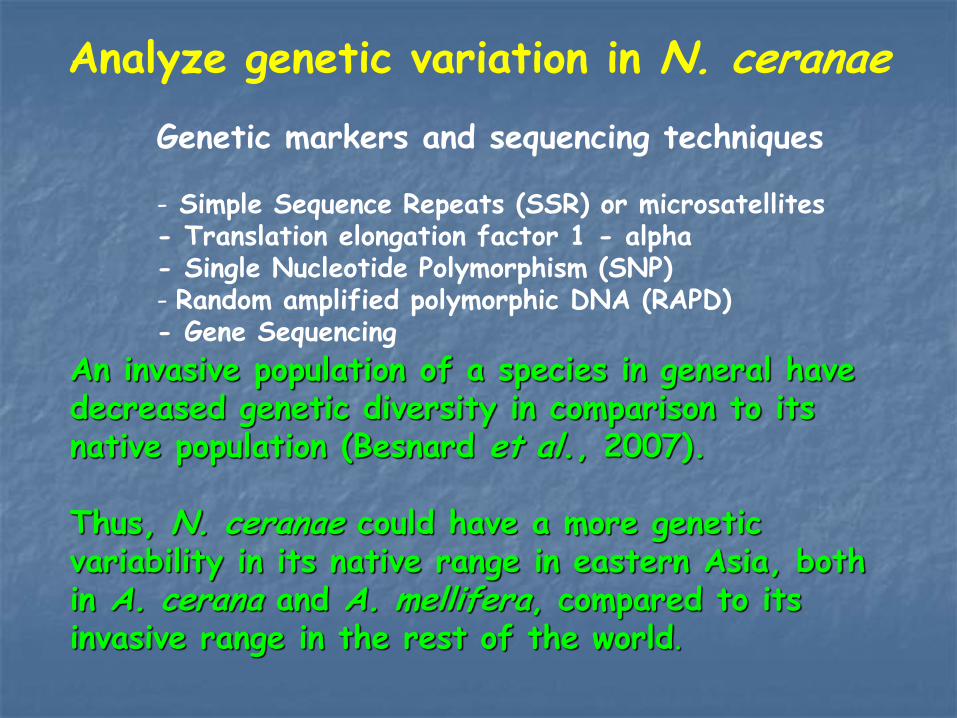
Analyze genetic variation in N. ceranae
An invasive population of a species in general have decreased genetic diversity in comparison to its native population (Besnard et al., 2007). Thus, N. ceranae could have a more genetic variability in its native range in eastern Asia, both in A. cerana and A. mellifera, compared to its invasive range in the rest of the world.
Genetic markers and sequencing techniques - Simple Sequence Repeats (SSR) or microsatellites - Translation elongation factor 1 - alpha - Single Nucleotide Polymorphism (SNP) - Random amplified polymorphic DNA (RAPD) - Gene Sequencing
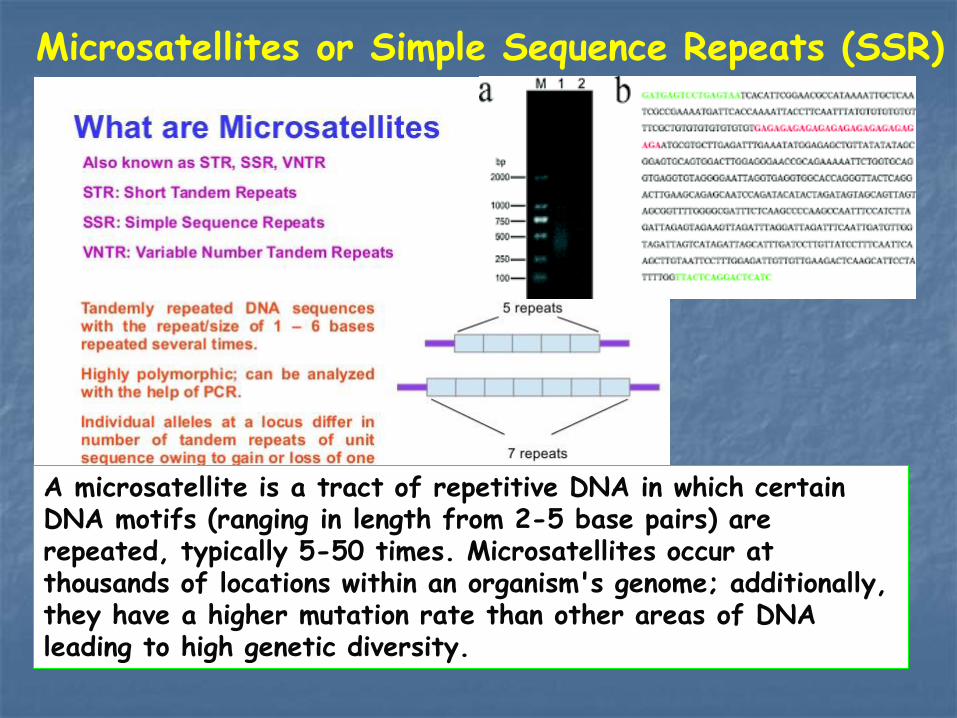
A microsatellite is a tract of repetitive DNA in which certain DNA motifs (ranging in length from 2-5 base pairs) are repeated, typically 5-50 times. Microsatellites occur at thousands of locations within an organism's genome; additionally, they have a higher mutation rate than other areas of DNA leading to high genetic diversity.
Microsatellites or Simple Sequence Repeats (SSR)
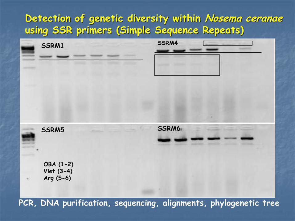
Detection of genetic diversity within Nosema ceranae using SSR primers (Simple Sequence Repeats)
PCR, DNA purification, sequencing, alignments, phylogenetic tree
SSRM1 SSRM4
SSRM5 SSRM6
OBA (1-2) Viet (3-4) Arg (5-6)

Single Nucleotide Polymorphism (SNP)
A single-nucleotide polymorphism (SNP, pronounced snip) is a DNA sequence variation occurring when a single nucleotide adenine (A), thymine (T), cytosine (C), or guanine (G) in the genome (or other shared sequence) differs between members of a species or paired chromosomes in an individual.

VI8 ATGTTAAGAAATATTGCTGTAACTAGATCTATAGAAAAATACAGAGAATC 50
VI10 ATGTTAAGAAATATTGTTGATACTAGAACTATAGAAAAATACAGAGAATC 50
GrCom ATGTTAAGAAATATTGTTGATACTAGAACTATAGAAAAATACAGAGAATC 50
VI7 ATGTTAAGAAATATTGTTGATACTAGAACTATAGAAAAATACAGAGAATC 50
VI11 ATGTTAAGAAATATTGTTGATACTAGAACTATAGAAAAATACAGAGAATC 50
VI9 ATGTTAAGAAATATTGTTGATACTAGAACTATAGAAAAATACAGAGAATC 50
* * * ** * * * * * * * ** * * * * * * ** * * * * * * ** * * * * * * ** * * * * * * * *
Nucleotide sequence alignment of the translation elongation factor 1- alpha of Nosema ceranae infected honey bees

1.The HBRC method was simple, more reliable, and cheaper than current methods
2. It is possible to detect N. ceranae or N. apis separately or together with the HBRC method which is not possible under the microscope
3. 100 spores/bee was detectable with the HBRL method when 50,000 spores/bee are needed under the microscope
4. First time it is shown that the quantification of Nosema infection levels is possible with a simple PCR technique
5. SSR primers and SNPs found as good molecular markers to determine genetic diversity within Nosema ceranae populations
Conclusions

Application of the HBRC method - This method could be useful in epidemiological evaluations for regulatory purposes or disease control
- This method could be handy for researchers to study in depth N. apis and/or N. ceranae because it diagnoses single-bee samples
- SSR primers and translation elongation factor 1 – alpha could be useful to detect and track N. ceranae genotypes for parasite control, determine the sources of infestation and regulatory purpose when outbreaks occur
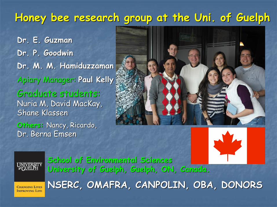
Honey bee research group at the Uni. of Guelph
Dr. E. Guzman
Dr. P. Goodwin
Dr. M. M. Hamiduzzaman
Apiary Manager: Paul Kelly
Graduate students: Nuria M, David MacKay, Shane Klassen
Others: Nancy, Ricardo, Dr. Berna Emsen
School of Environmental Sciences University of Guelph, Guelph, ON, Canada.
NSERC, OMAFRA, CANPOLIN, OBA, DONORS
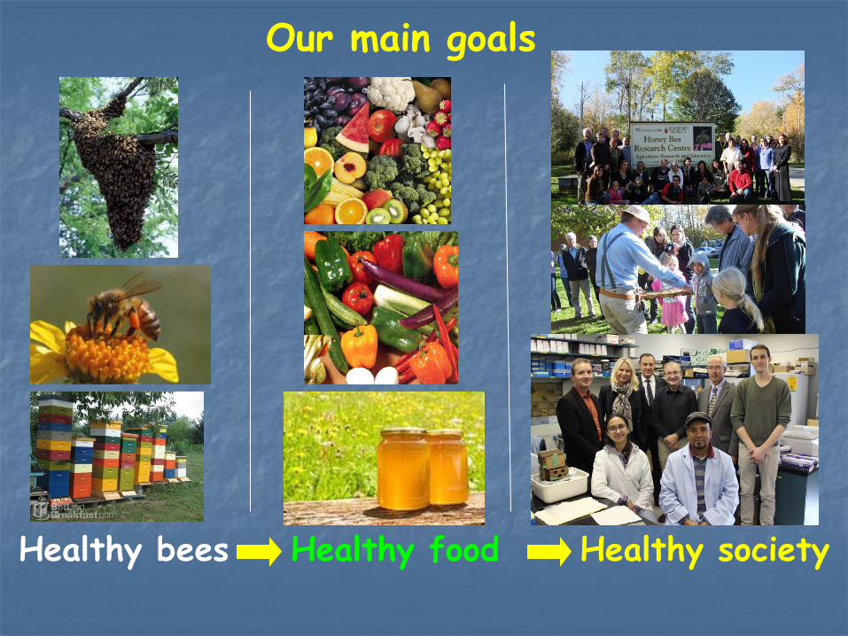
Our main goals
Healthy bees Healthy food Healthy society