Embed Size (px)
Citation preview

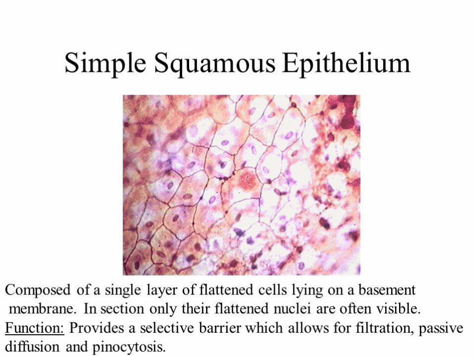
Diagram of simple squamous epithelium. The epithelial cells are separated from the subjacent connective tissue by a basement membrane.
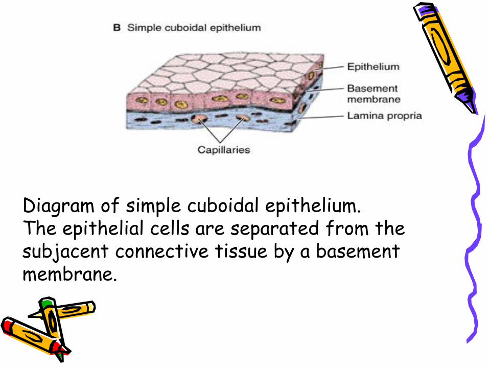
Diagram of simple cuboidal epithelium. The epithelial cells are separated from the subjacent connective tissue by a basement membrane.
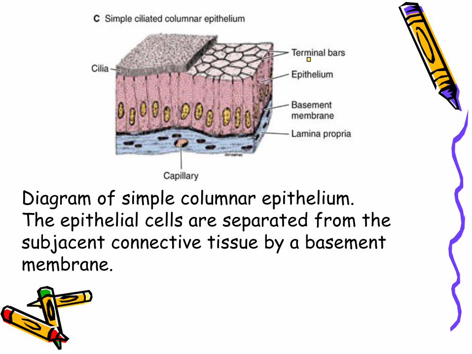
Diagram of simple columnar epithelium. The epithelial cells are separated from the subjacent connective tissue by a basement membrane.
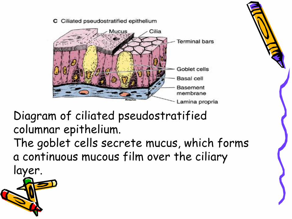
Diagram of ciliated pseudostratified columnar epithelium. The goblet cells secrete mucus, which forms a continuous mucous film over the ciliary layer.
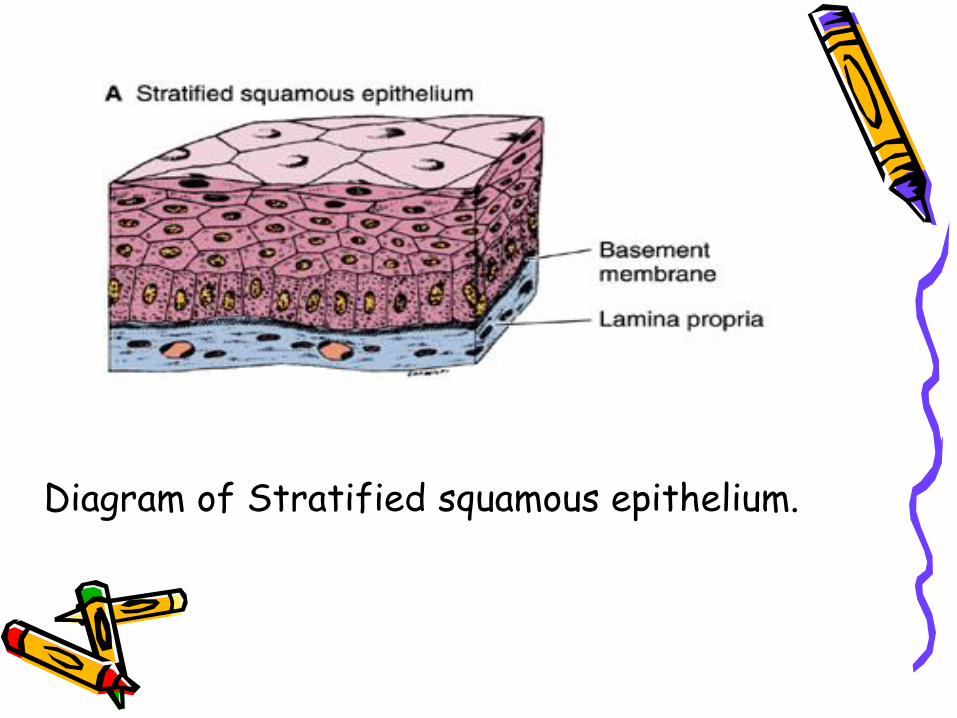
Diagram of Stratified squamous epithelium.
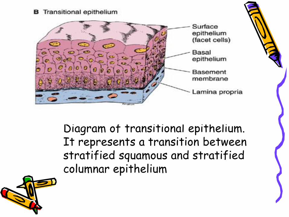
Diagram of transitional epithelium. It represents a transition between stratified squamous and stratified columnar epithelium
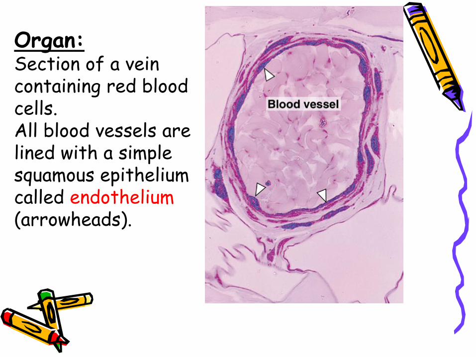
Organ:Section of a vein containing red blood cells. All blood vessels are lined with a simple squamous epithelium called endothelium (arrowheads).
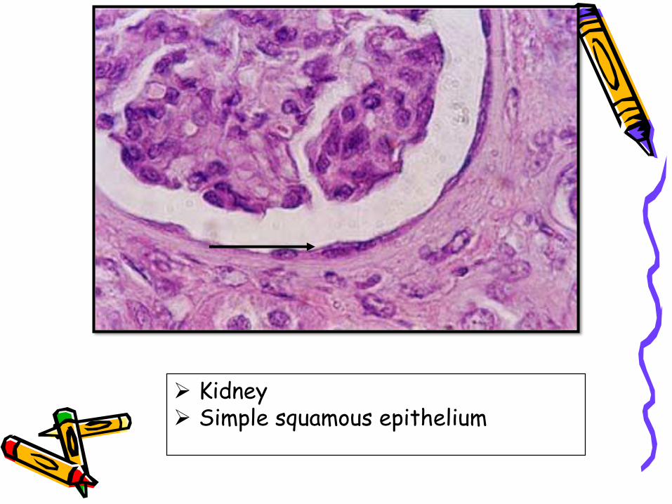
Kidney Simple squamous epithelium
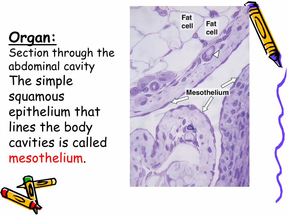
Organ:Section through the abdominal cavityThe simple squamous epithelium that lines the body cavities is called mesothelium.
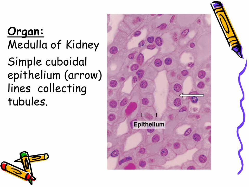
Organ:Medulla of Kidney
Simple cuboidal epithelium (arrow) lines collecting tubules.
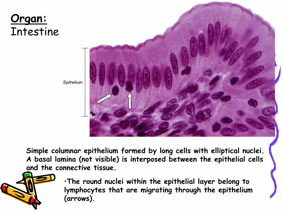
Simple columnar epithelium formed by long cells with elliptical nuclei. A basal lamina (not visible) is interposed between the epithelial cells and the connective tissue.
Organ:Intestine
•The round nuclei within the epithelial layer belong to lymphocytes that are migrating through the epithelium (arrows).
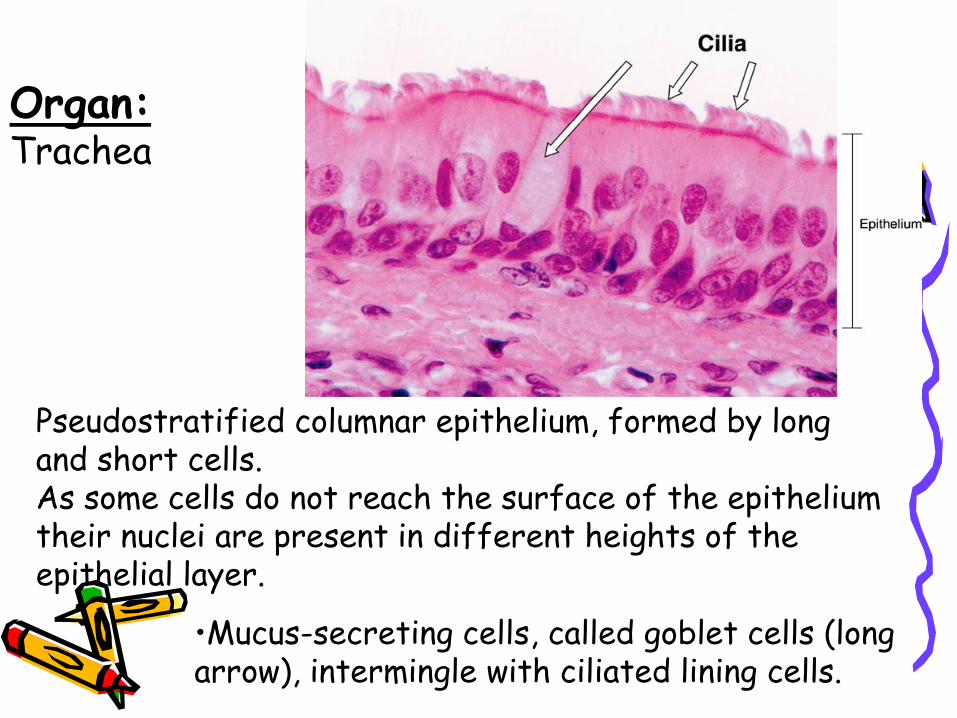
Pseudostratified columnar epithelium, formed by long and short cells. As some cells do not reach the surface of the epithelium their nuclei are present in different heights of the epithelial layer.
Organ:Trachea
•Mucus-secreting cells, called goblet cells (long arrow), intermingle with ciliated lining cells.
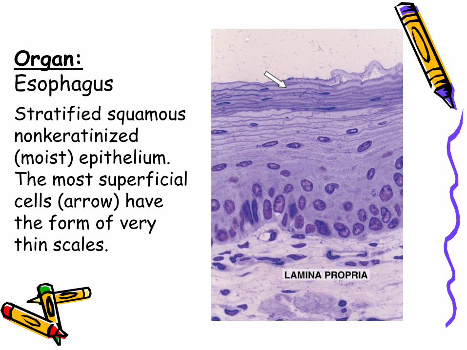
Organ:Esophagus
Stratified squamous nonkeratinized (moist) epithelium.The most superficial cells (arrow) have the form of very thin scales.
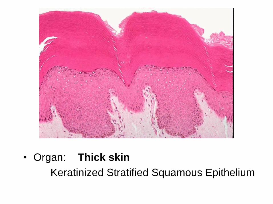
• Organ: Thick skin
Keratinized Stratified Squamous Epithelium
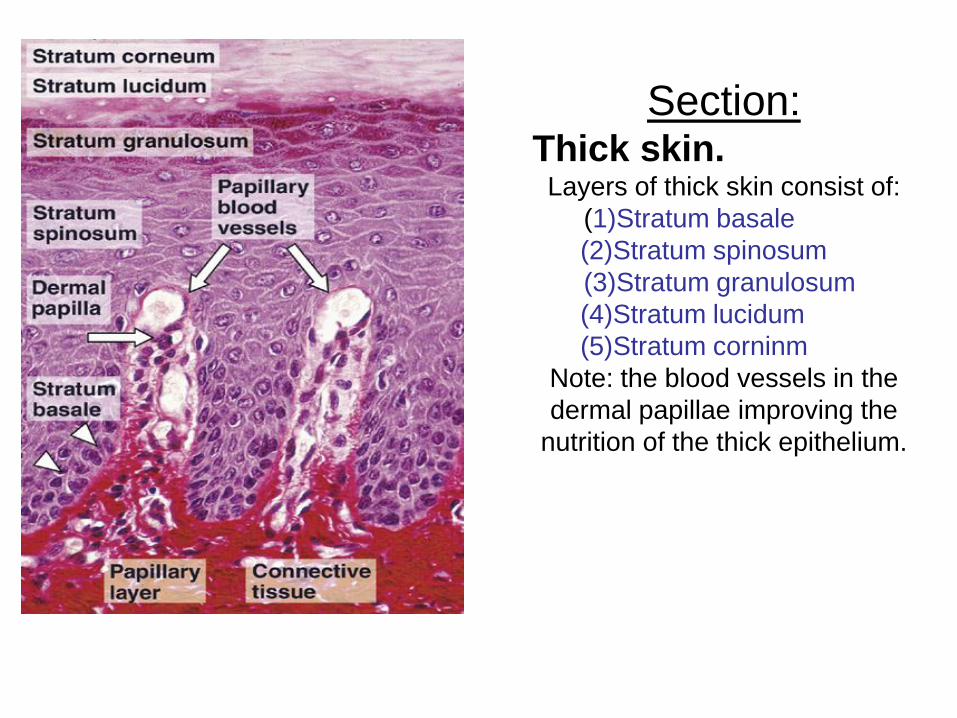
Section:Thick skin.
Layers of thick skin consist of:
(1)Stratum basale
(2)Stratum spinosum
(3)Stratum granulosum
(4)Stratum lucidum
(5)Stratum corninm
Note: the blood vessels in the
dermal papillae improving the
nutrition of the thick epithelium.
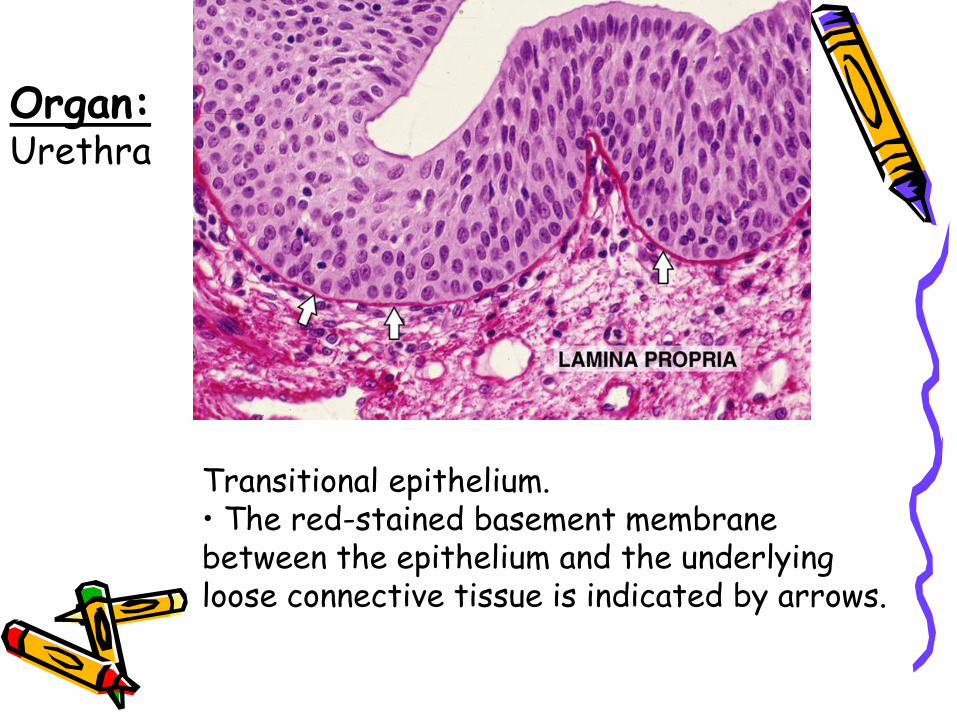
Transitional epithelium.• The red-stained basement membrane between the epithelium and the underlying loose connective tissue is indicated by arrows.
Organ:Urethra
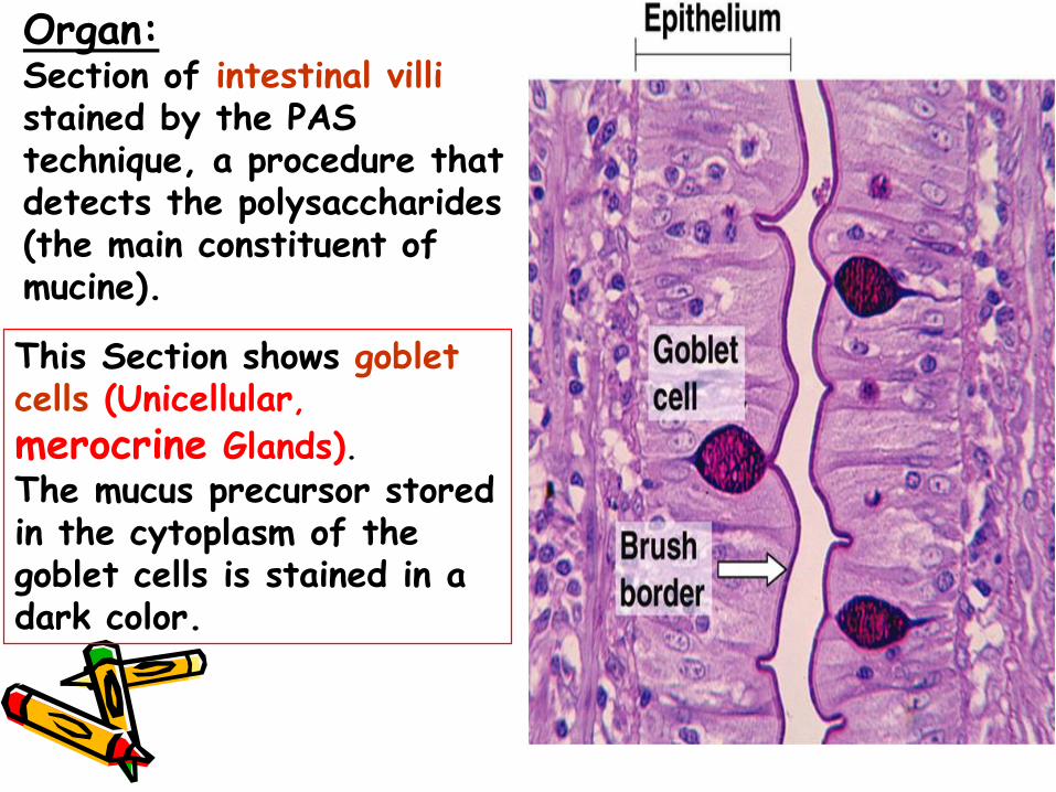
This Section shows goblet cells (Unicellular,
merocrine Glands).The mucus precursor stored in the cytoplasm of the goblet cells is stained in a dark color.
Organ:Section of intestinal villistained by the PAS technique, a procedure that detects the polysaccharides (the main constituent of mucine).
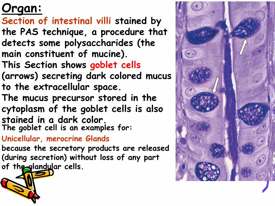
Organ:Section of intestinal villi stained by the PAS technique, a procedure that detects some polysaccharides (the main constituent of mucine).This Section shows goblet cells(arrows) secreting dark colored mucus to the extracellular space. The mucus precursor stored in the cytoplasm of the goblet cells is also stained in a dark color. The goblet cell is an examples for:
Unicellular, merocrine Glandsbecause the secretory products are released (during secretion) without loss of any part of the glandular cells.
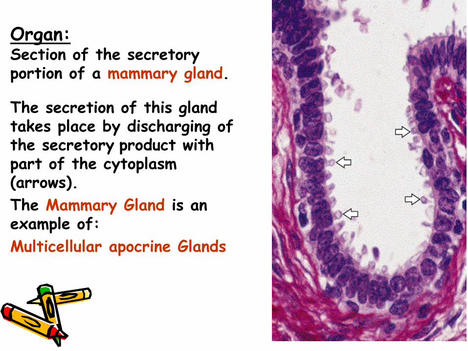
Organ:Section of the secretory portion of a mammary gland.
The secretion of this gland takes place by discharging of the secretory product with part of the cytoplasm (arrows).
The Mammary Gland is an example of:
Multicellular apocrine Glands
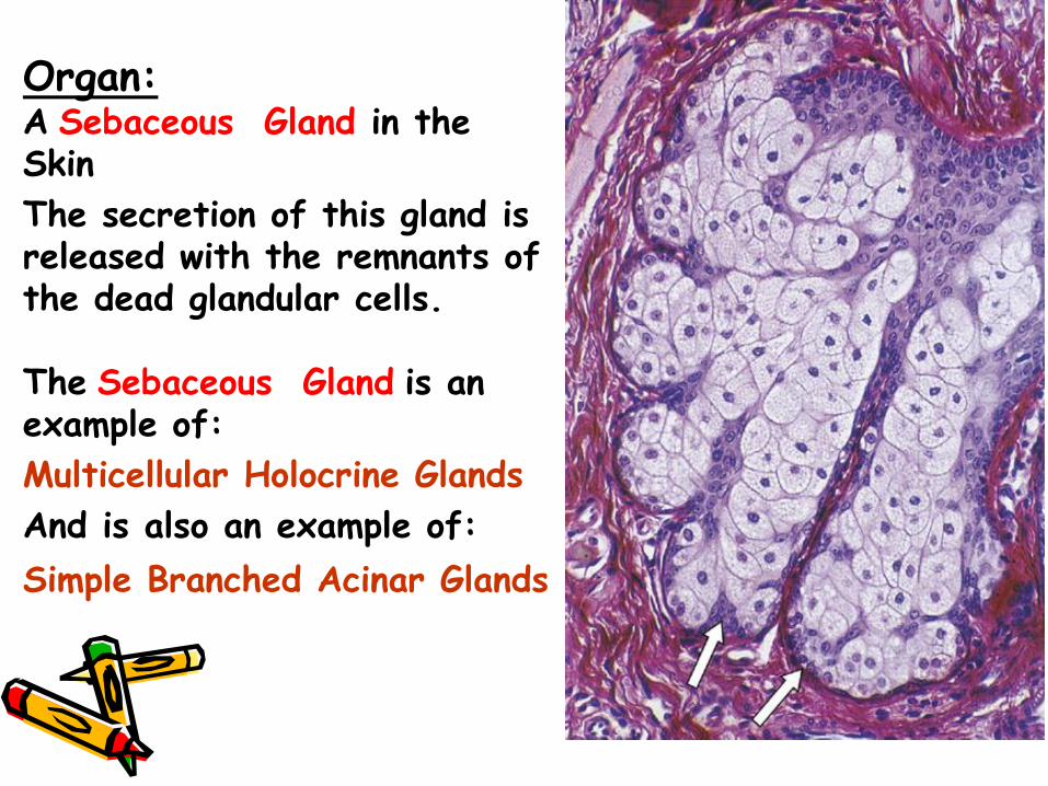
Organ:A Sebaceous Gland in the Skin
The secretion of this gland is released with the remnants of the dead glandular cells.
The Sebaceous Gland is an example of:
Multicellular Holocrine Glands
And is also an example of:
Simple Branched Acinar Glands
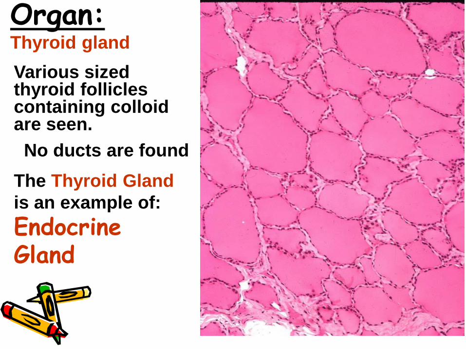
Various sized thyroid follicles containing colloid are seen.
No ducts are found
The Thyroid Gland
is an example of:
Endocrine Gland
Organ:Thyroid gland
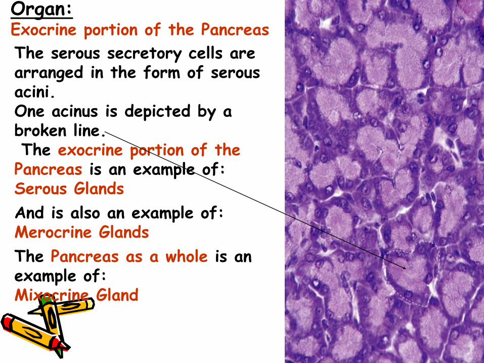
The serous secretory cells are arranged in the form of serous acini. One acinus is depicted by a broken line.The exocrine portion of thePancreas is an example of:Serous Glands
And is also an example of:Merocrine Glands
The Pancreas as a whole is an example of:Mixocrine Gland
Organ:Exocrine portion of the Pancreas
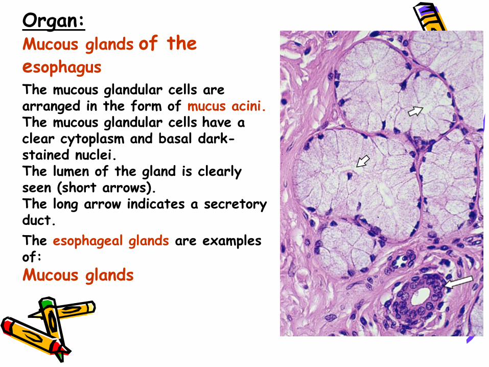
Organ:Mucous glands of the esophagusThe mucous glandular cells are arranged in the form of mucus acini.The mucous glandular cells have a clear cytoplasm and basal dark-stained nuclei. The lumen of the gland is clearly seen (short arrows). The long arrow indicates a secretory duct.
The esophageal glands are examples of:
Mucous glands
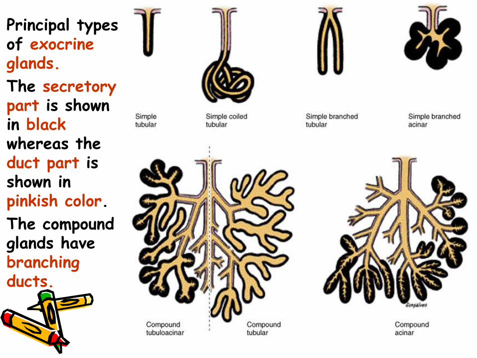
Principal types of exocrine glands.
The secretory part is shown in blackwhereas the duct part is shown in pinkish color.
The compound glands have branching ducts.
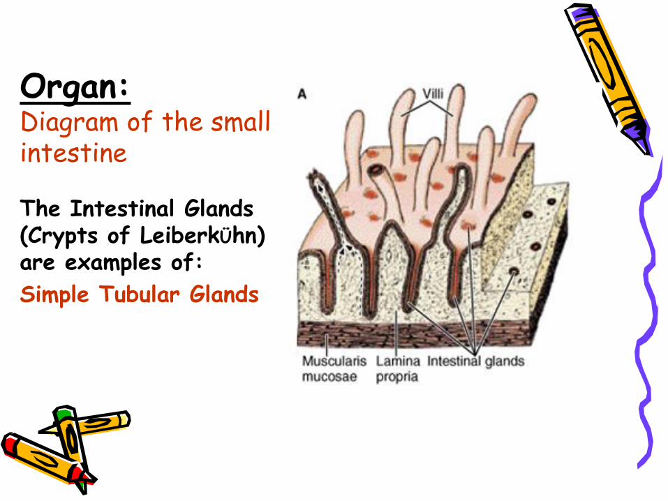
Organ:Diagram of the small intestine
The Intestinal Glands (Crypts of LeiberkÜhn) are examples of:
Simple Tubular Glands
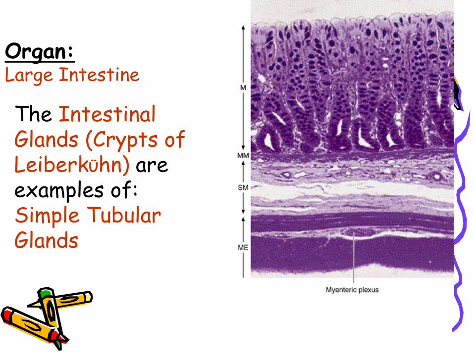
The Intestinal Glands (Crypts of LeiberkÜhn) are examples of:Simple Tubular Glands
Organ:Large Intestine
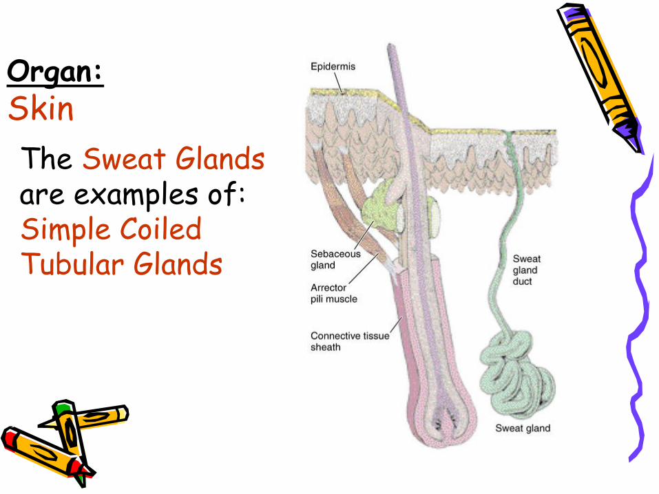
The Sweat Glandsare examples of:Simple Coiled Tubular Glands
Organ:Skin
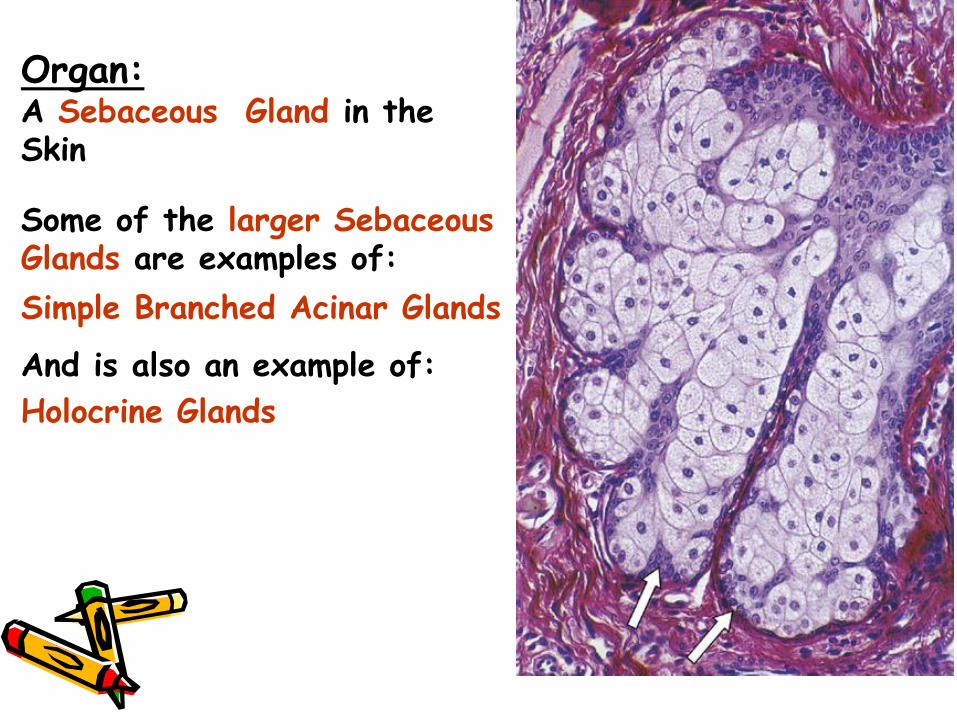
Organ:A Sebaceous Gland in the Skin
Some of the larger Sebaceous Glands are examples of:
Simple Branched Acinar Glands
And is also an example of:
Holocrine Glands
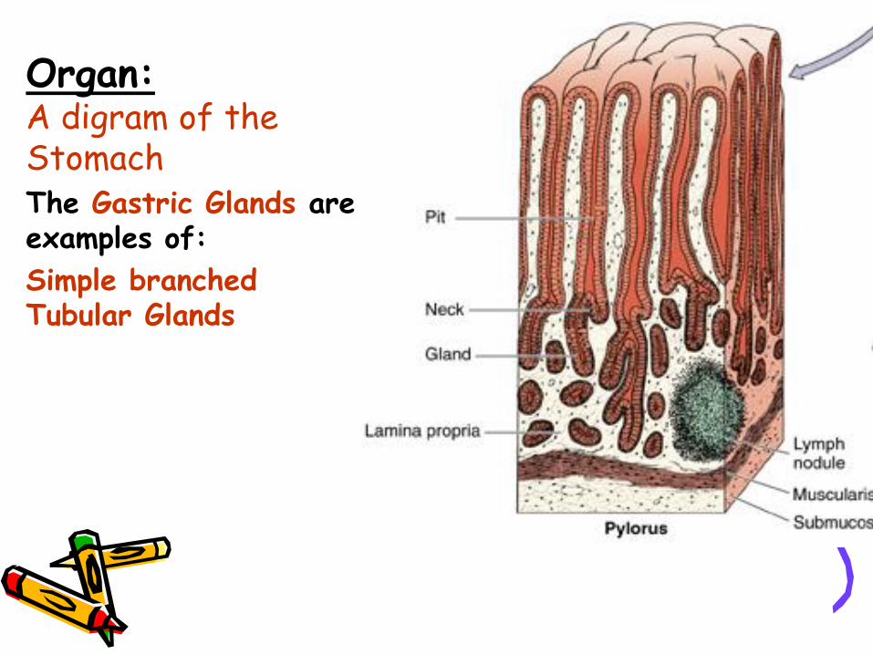
Organ:A digram of the StomachThe Gastric Glands are examples of:
Simple branched Tubular Glands
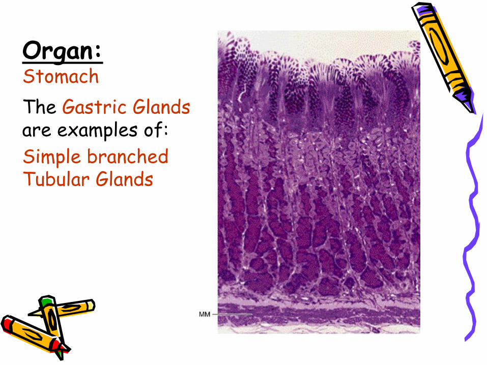
Organ:Stomach
The Gastric Glandsare examples of:
Simple branched Tubular Glands
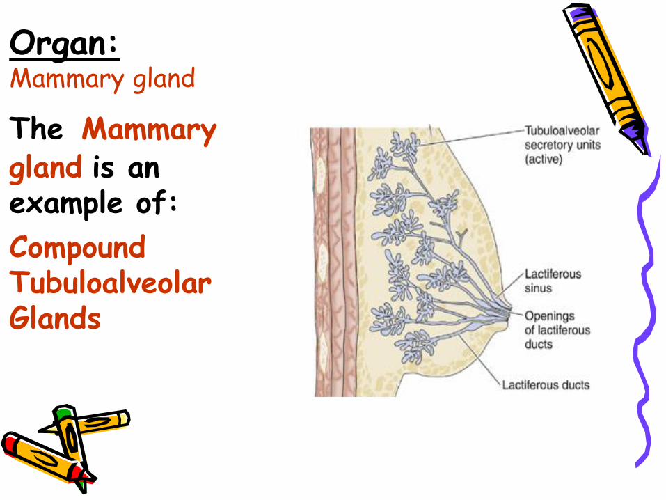
Organ:Mammary gland
The Mammary gland is an example of:
Compound Tubuloalveolar Glands
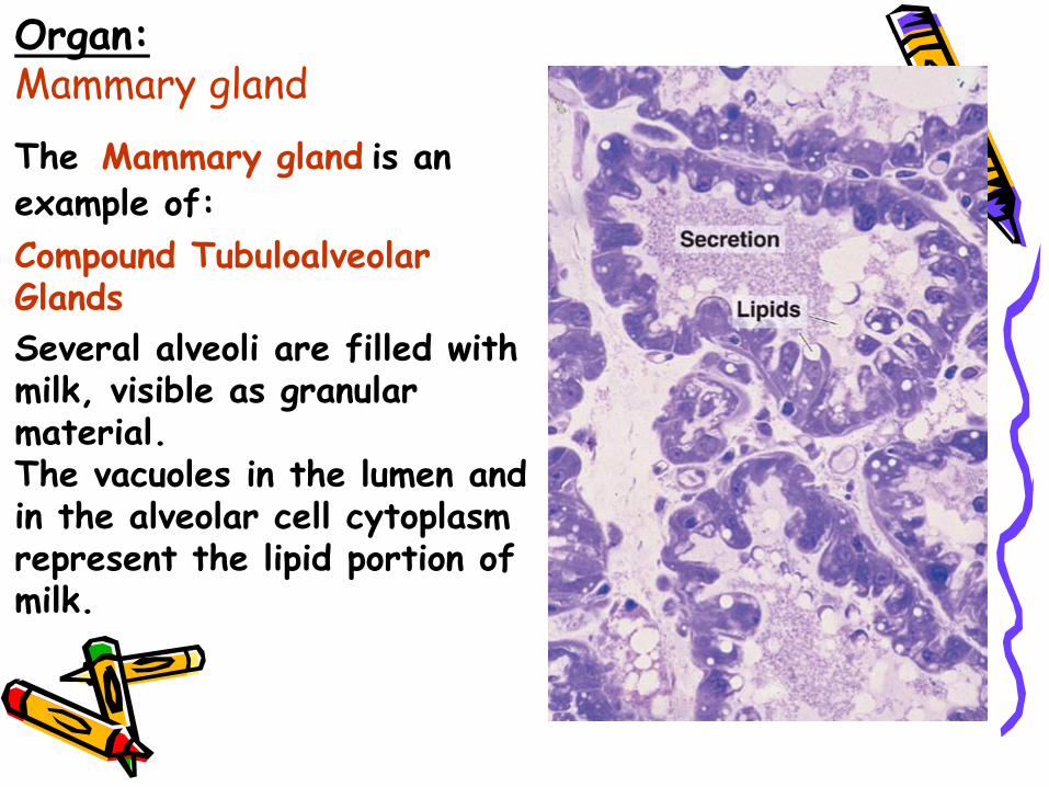
Organ:Mammary gland
The Mammary gland is an example of:
Compound Tubuloalveolar Glands
Several alveoli are filled with milk, visible as granular material. The vacuoles in the lumen and in the alveolar cell cytoplasm represent the lipid portion of milk.
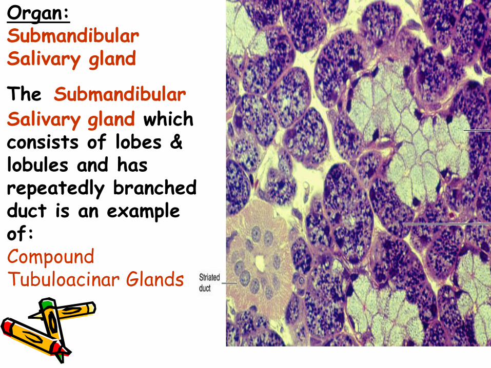
Organ:Submandibular Salivary gland
The Submandibular Salivary gland which consists of lobes & lobules and has repeatedly branched duct is an example of:Compound Tubuloacinar Glands