Embed Size (px)
Citation preview
DIASTOLIC LV FUNCTION AND DIASTOLIC HEART FAILURE
Dr.Deepak Raju
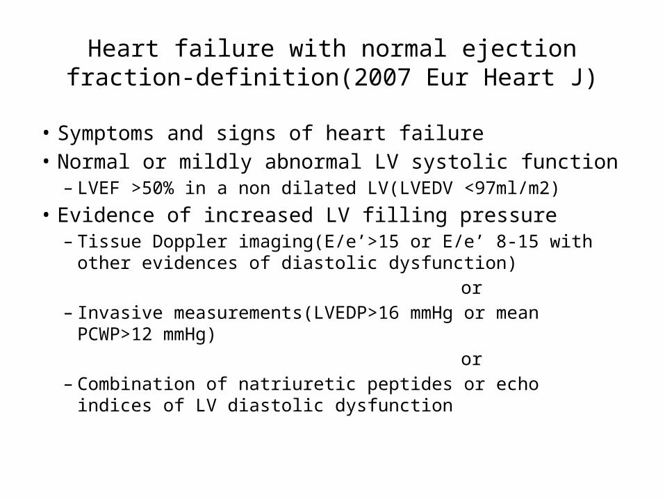
Heart failure with normal ejection fraction-definition(2007 Eur Heart J)
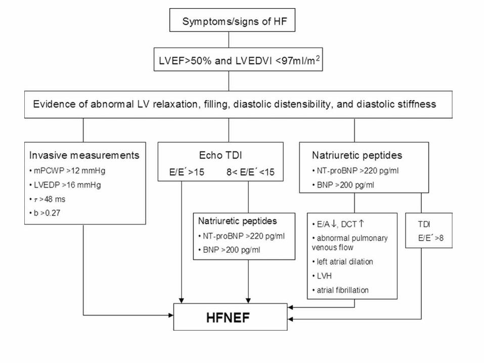
• Symptoms and signs of heart failure• Normal or mildly abnormal LV systolic function
– LVEF >50% in a non dilated LV(LVEDV <97ml/m2) • Evidence of increased LV filling pressure
– Tissue Doppler imaging(E/e’>15 or E/e’ 8-15 with other evidences of diastolic dysfunction)
or– Invasive measurements(LVEDP>16 mmHg or mean PCWP>12
mmHg) or– Combination of natriuretic peptides or echo indices of LV diastolic
dysfunction
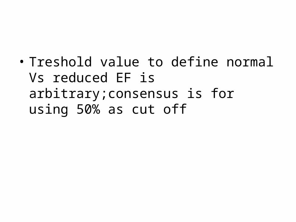
• Treshold value to define normal Vs reduced EF is arbitrary;consensus is for using 50% as cut off
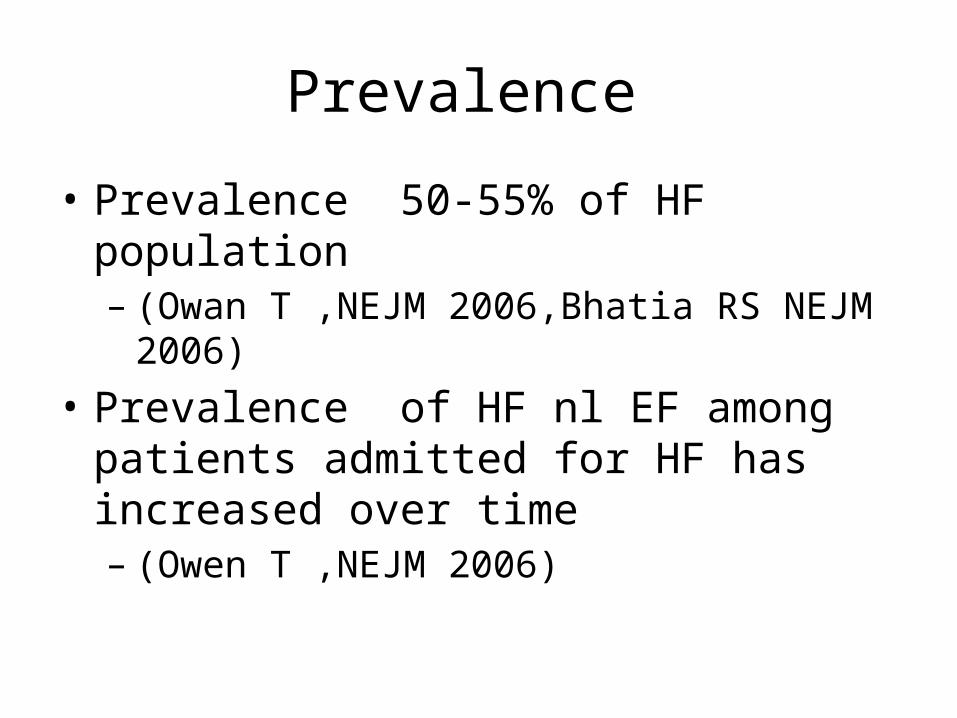
Prevalence
• Prevalence 50-55% of HF population– (Owan T ,NEJM 2006,Bhatia RS NEJM 2006)
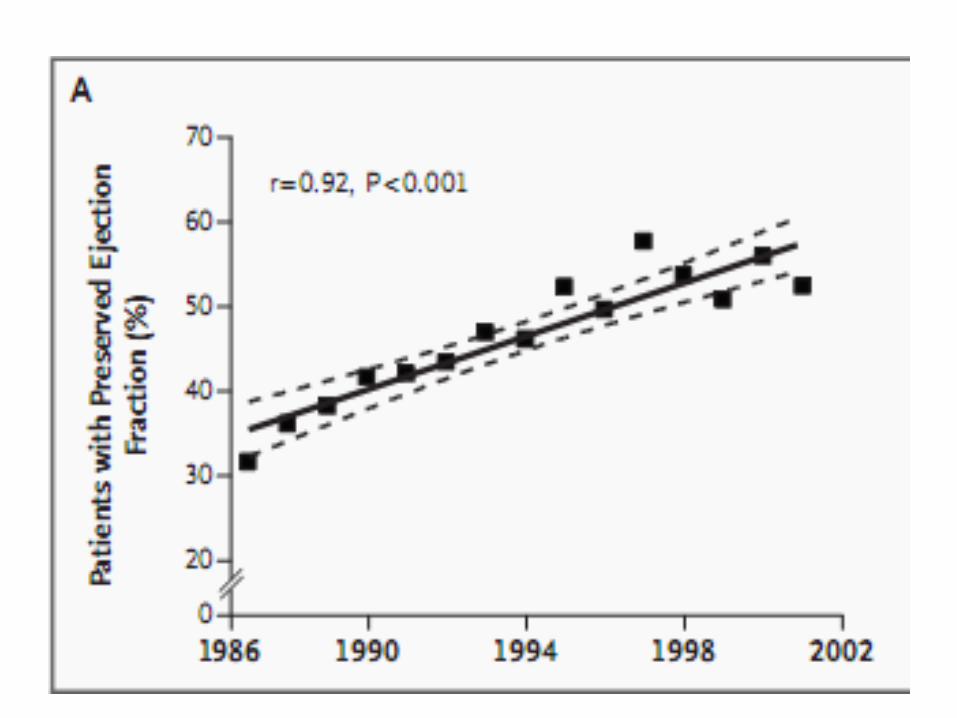
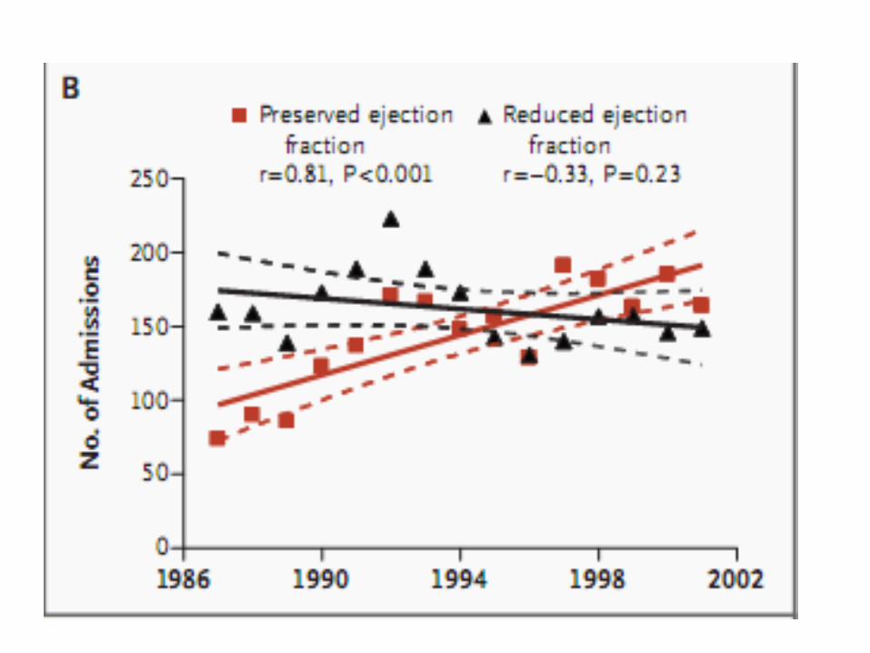
• Prevalence of HF nl EF among patients admitted for HF has increased over time– (Owen T ,NEJM 2006)
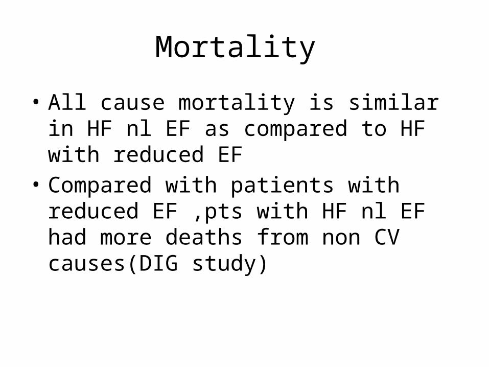
Mortality
• All cause mortality is similar in HF nl EF as compared to HF with reduced EF
• Compared with patients with reduced EF ,pts with HF nl EF had more deaths from non CV causes(DIG study)
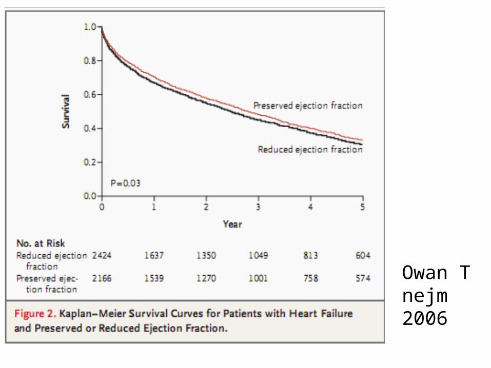
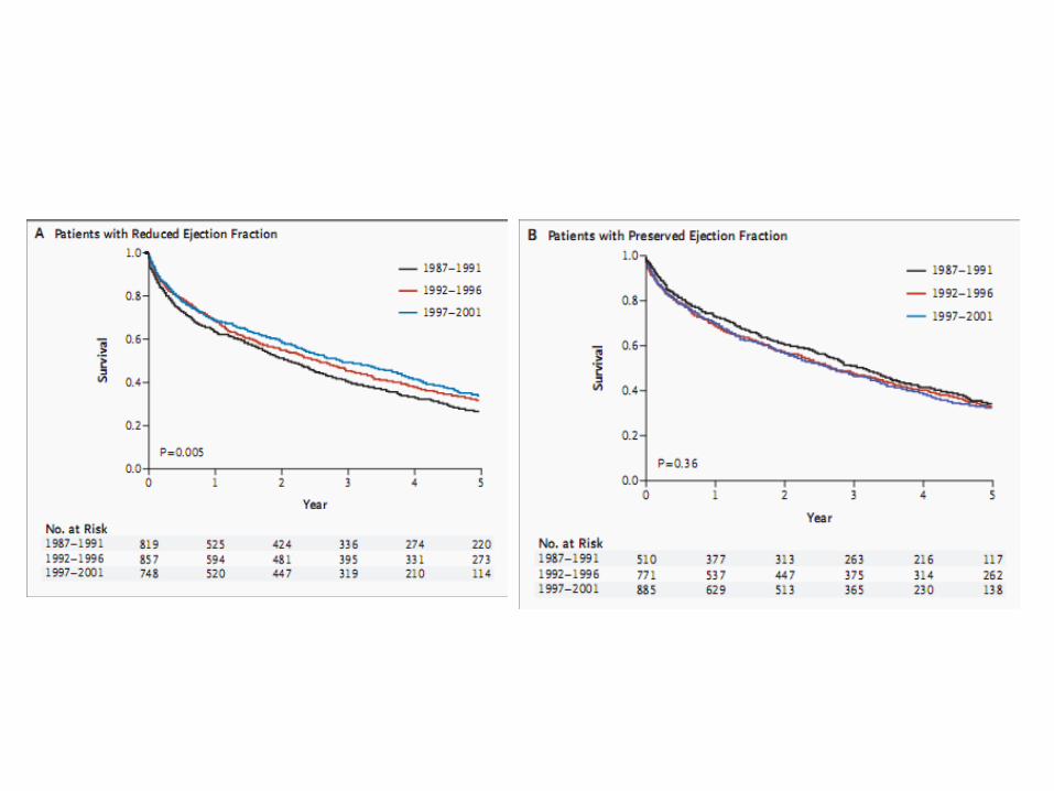
Owan Tnejm 2006
• Survival for HF with reduced EF have improved over time ,but not for HF nl EF
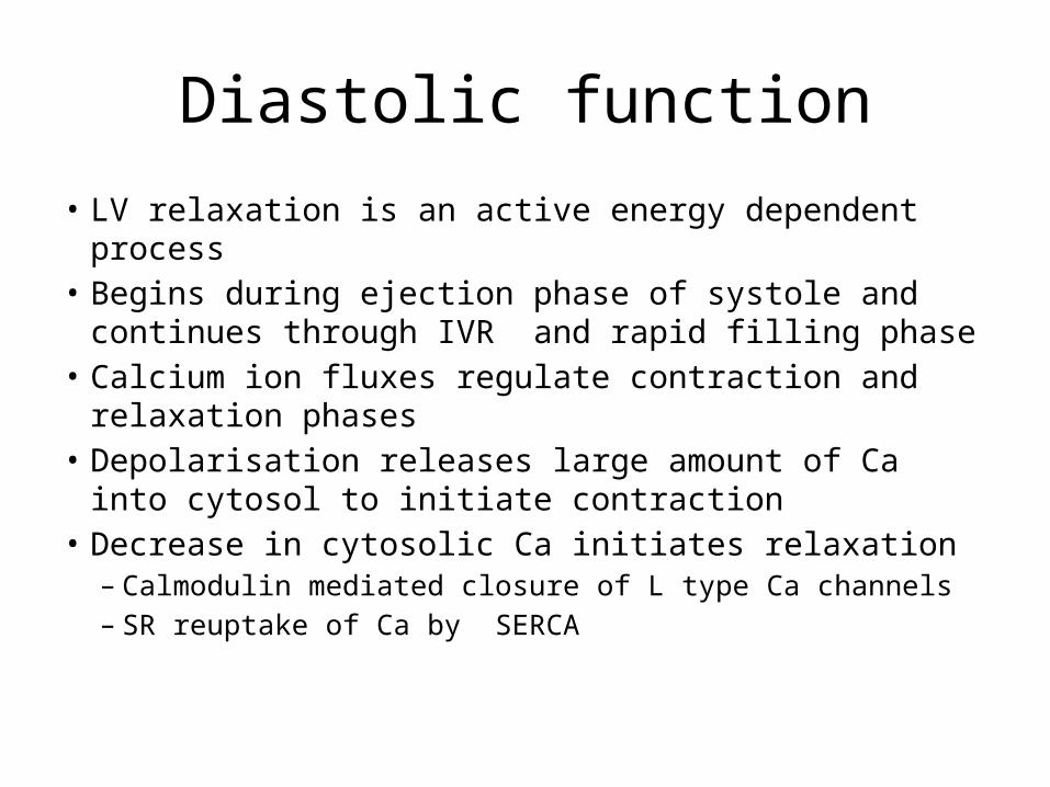
Diastolic function
• LV relaxation is an active energy dependent process• Begins during ejection phase of systole and continues
through IVR and rapid filling phase • Calcium ion fluxes regulate contraction and relaxation
phases• Depolarisation releases large amount of Ca into
cytosol to initiate contraction• Decrease in cytosolic Ca initiates relaxation
– Calmodulin mediated closure of L type Ca channels– SR reuptake of Ca by SERCA
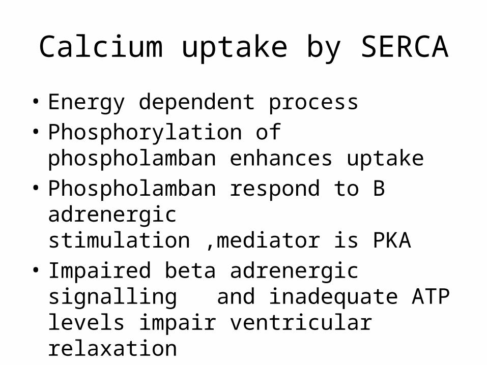
Calcium uptake by SERCA
• Energy dependent process• Phosphorylation of phospholamban enhances
uptake• Phospholamban respond to B adrenergic
stimulation ,mediator is PKA• Impaired beta adrenergic signalling and
inadequate ATP levels impair ventricular relaxation
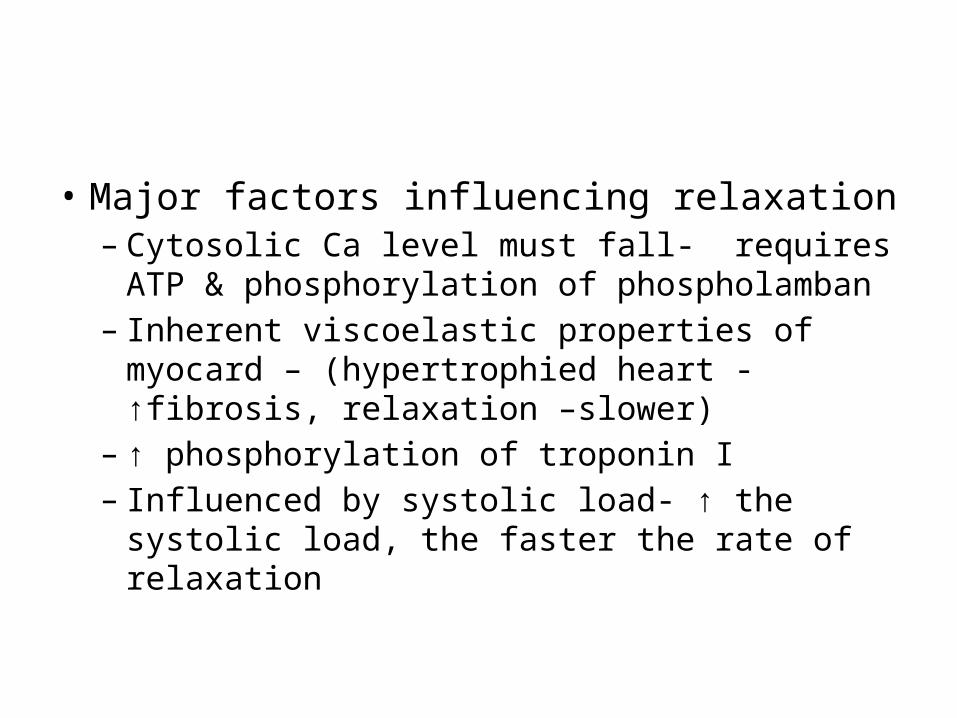
• Major factors influencing relaxation– Cytosolic Ca level must fall- requires ATP &
phosphorylation of phospholamban– Inherent viscoelastic properties of myocard –
(hypertrophied heart -↑fibrosis, relaxation –slower)
– ↑ phosphorylation of troponin I – Influenced by systolic load- ↑ the systolic load,
the faster the rate of relaxation
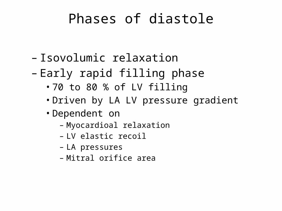
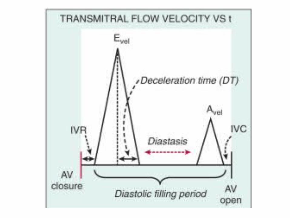
Phases of diastole
– Isovolumic relaxation– Early rapid filling phase• 70 to 80 % of LV filling• Driven by LA LV pressure gradient• Dependent on
– Myocardioal relaxation– LV elastic recoil– LA pressures– Mitral orifice area
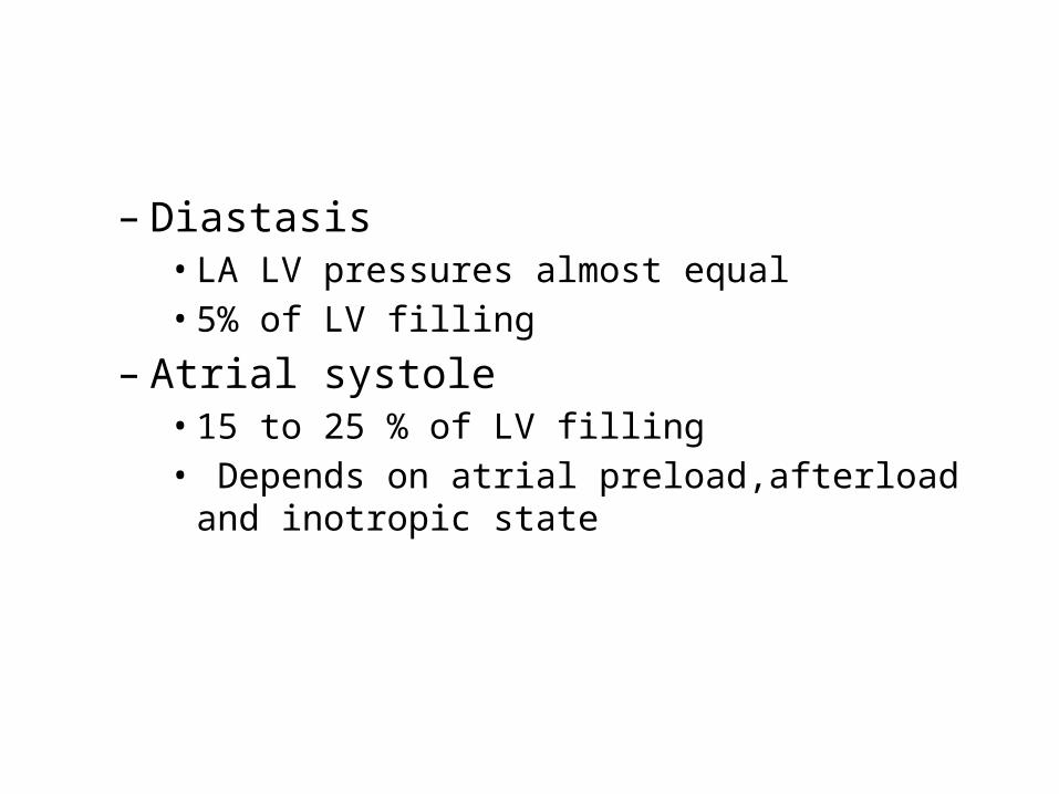
– Diastasis• LA LV pressures almost equal• 5% of LV filling
– Atrial systole• 15 to 25 % of LV filling• Depends on atrial preload,afterload and inotropic state
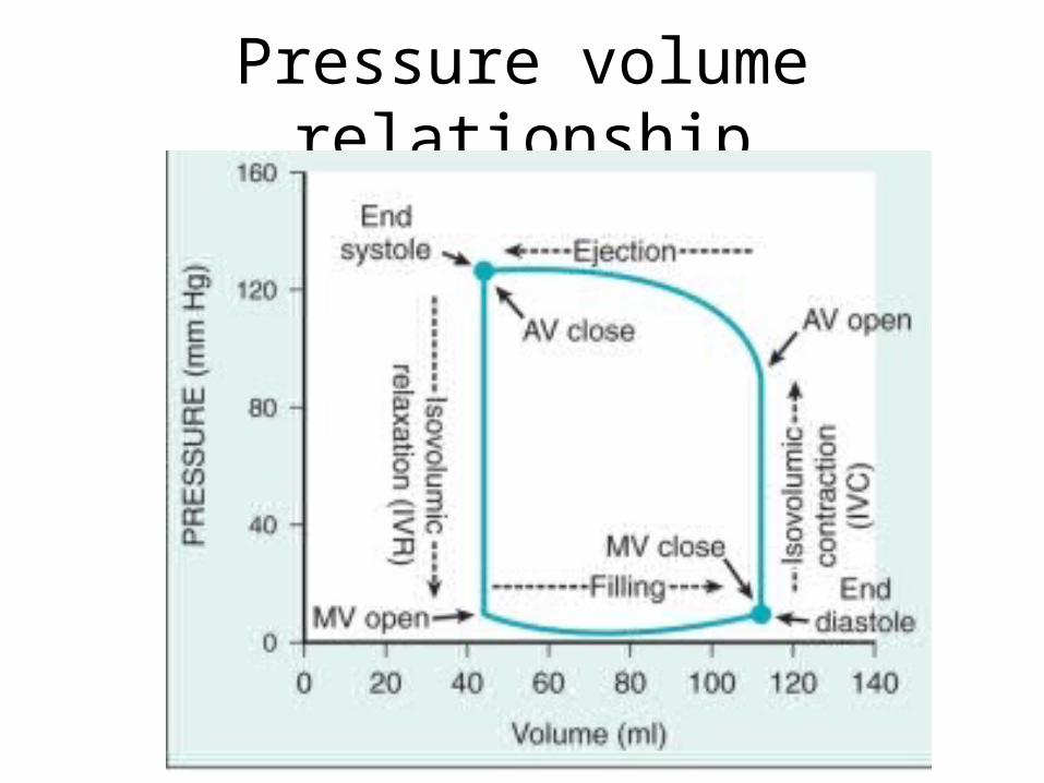
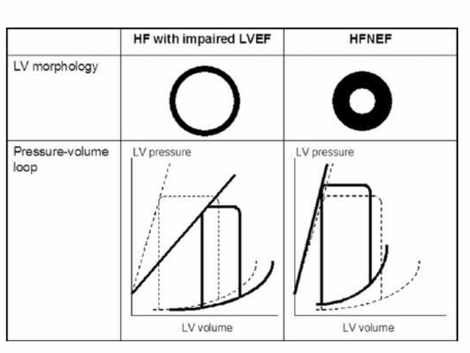
Pressure volume relationship
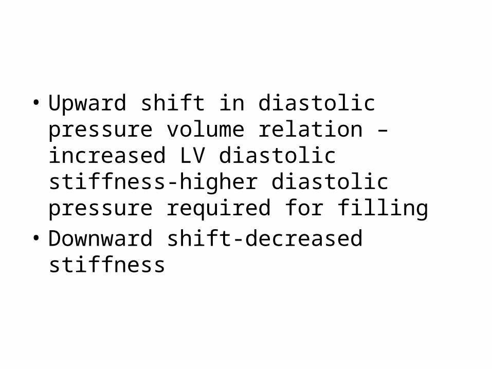
• Upward shift in diastolic pressure volume relation –increased LV diastolic stiffness-higher diastolic pressure required for filling
• Downward shift-decreased stiffness
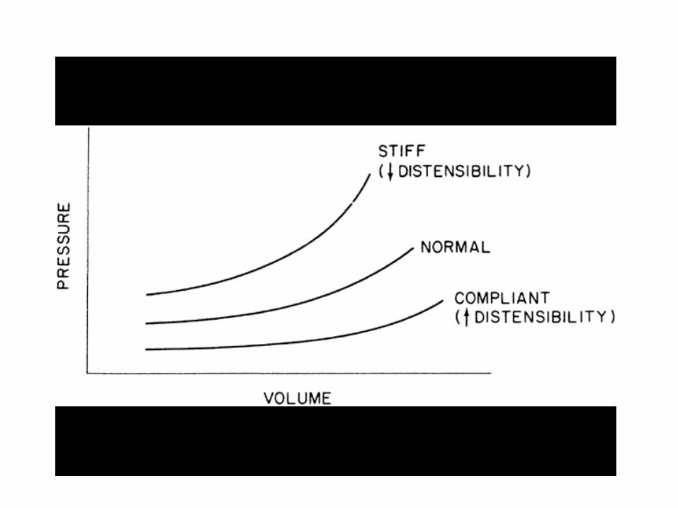
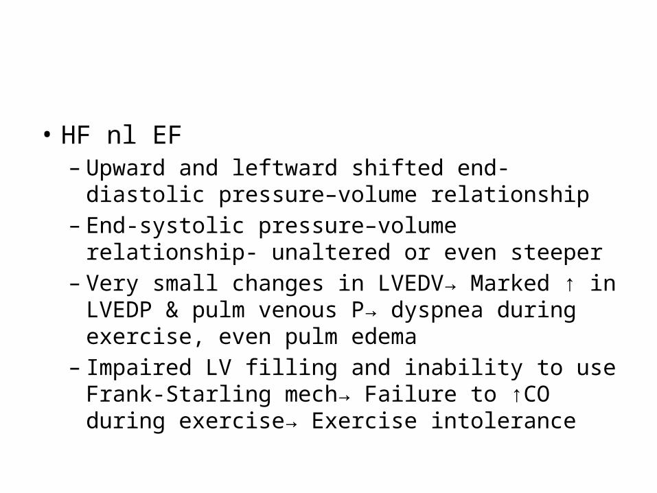
• HF nl EF – Upward and leftward shifted end-diastolic pressure–
volume relationship– End-systolic pressure–volume relationship- unaltered or
even steeper– Very small changes in LVEDV→ Marked ↑ in LVEDP &
pulm venous P→ dyspnea during exercise, even pulm edema
– Impaired LV filling and inability to use Frank-Starling mech→ Failure to ↑CO during exercise→ Exercise intolerance
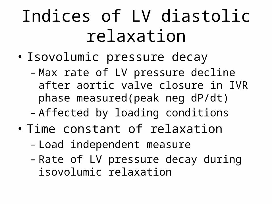
Indices of LV diastolic relaxation
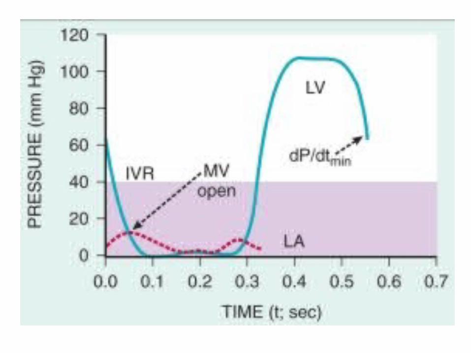
• Isovolumic pressure decay– Max rate of LV pressure decline after aortic valve
closure in IVR phase measured(peak neg dP/dt)– Affected by loading conditions
• Time constant of relaxation– Load independent measure– Rate of LV pressure decay during isovolumic
relaxation
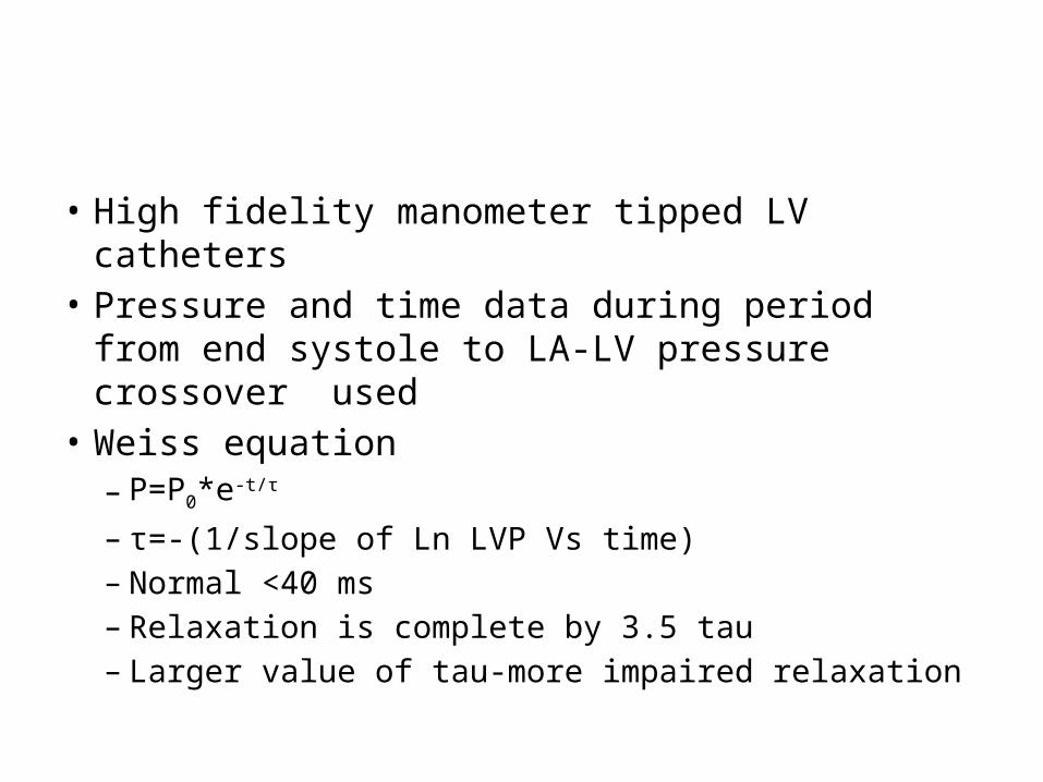
• High fidelity manometer tipped LV catheters• Pressure and time data during period from end
systole to LA-LV pressure crossover used• Weiss equation– P=P0*e-t/τ
– τ=-(1/slope of Ln LVP Vs time)– Normal <40 ms– Relaxation is complete by 3.5 tau– Larger value of tau-more impaired relaxation
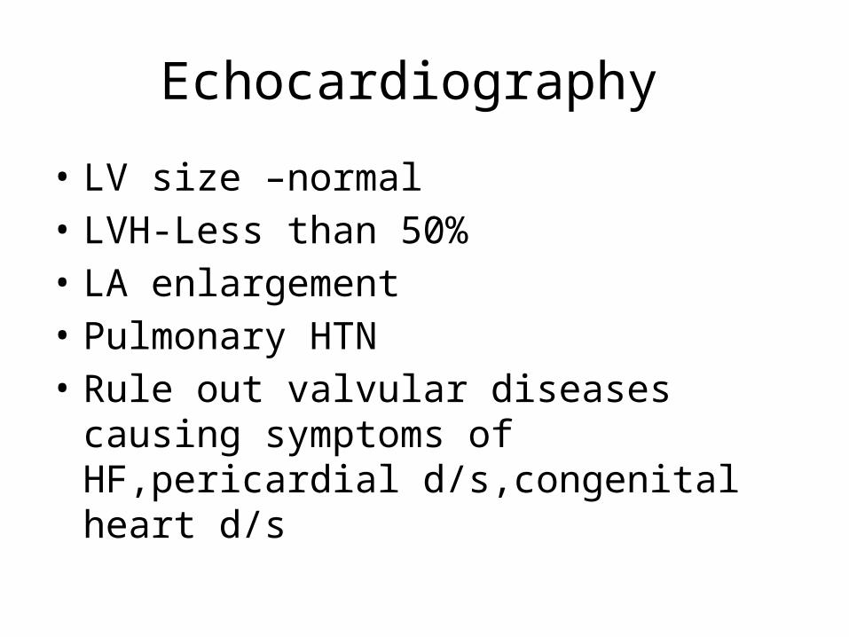
Echocardiography
• LV size –normal • LVH-Less than 50%• LA enlargement• Pulmonary HTN• Rule out valvular diseases causing symptoms
of HF,pericardial d/s,congenital heart d/s
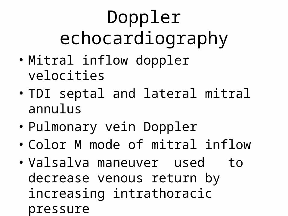
Doppler echocardiography
• Mitral inflow doppler velocities• TDI septal and lateral mitral annulus• Pulmonary vein Doppler• Color M mode of mitral inflow • Valsalva maneuver used to decrease venous
return by increasing intrathoracic pressure
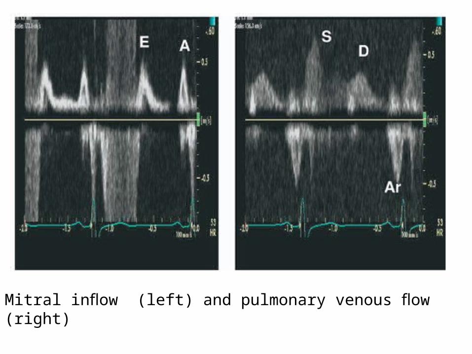
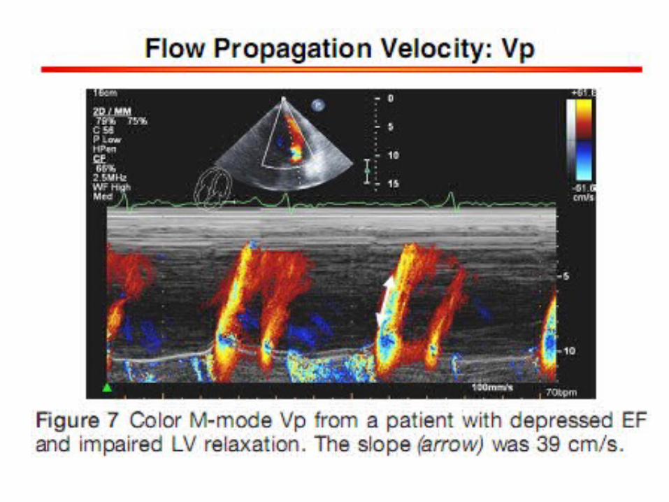
Mitral inflow (left) and pulmonary venous flow (right)

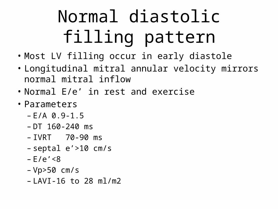
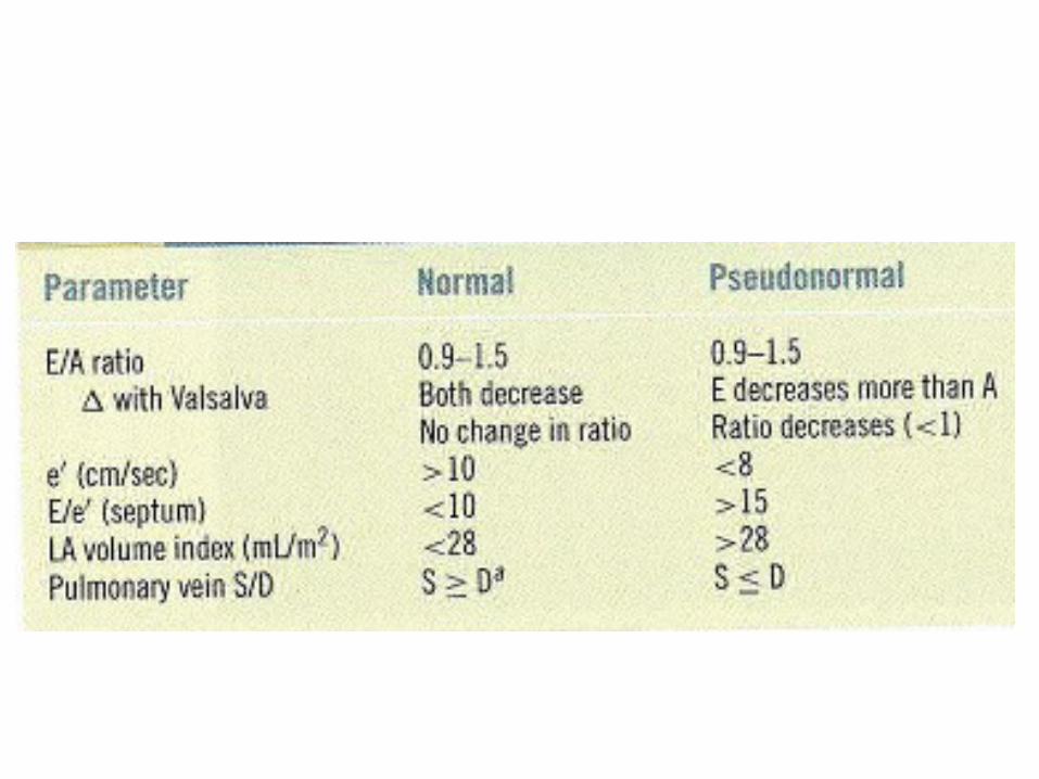
Normal diastolic filling pattern
• Most LV filling occur in early diastole • Longitudinal mitral annular velocity mirrors normal mitral
inflow• Normal E/e’ in rest and exercise• Parameters
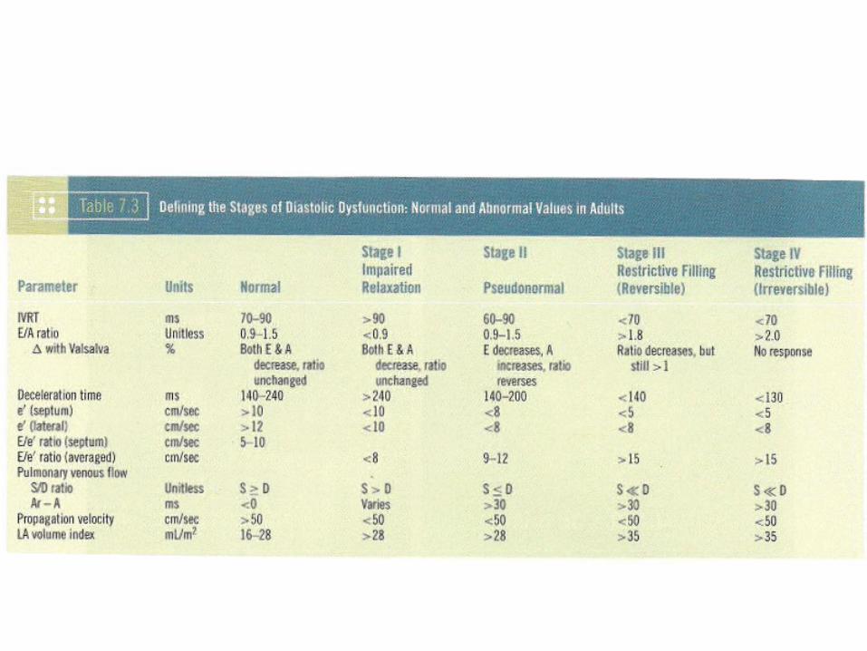
– E/A 0.9-1.5– DT 160-240 ms– IVRT 70-90 ms– septal e’>10 cm/s– E/e’<8– Vp>50 cm/s– LAVI-16 to 28 ml/m2
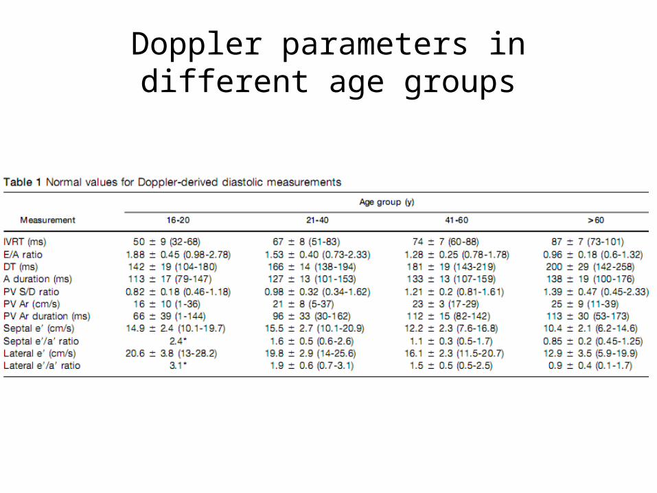
Doppler parameters in different age groups
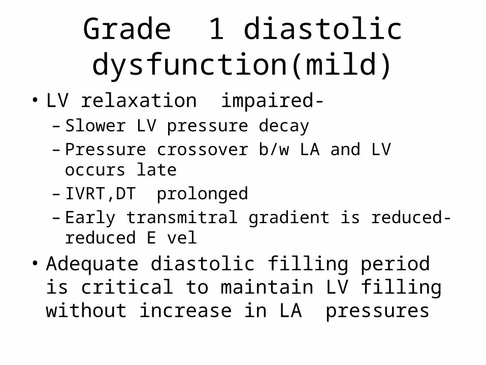
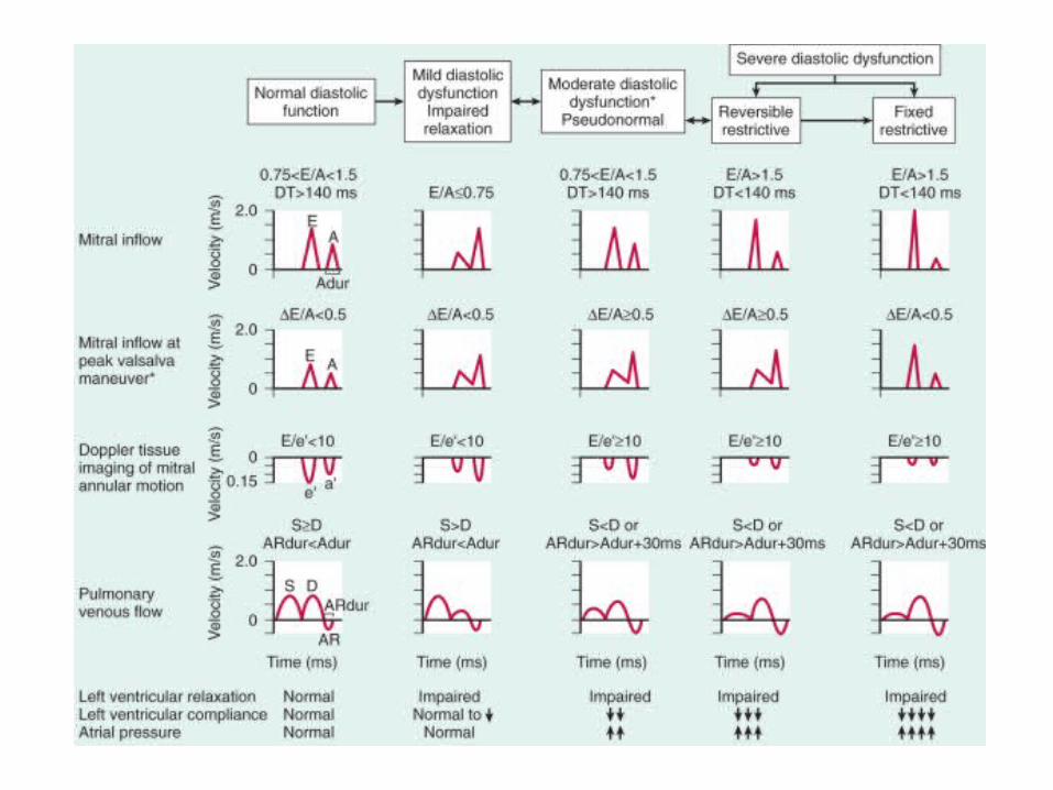
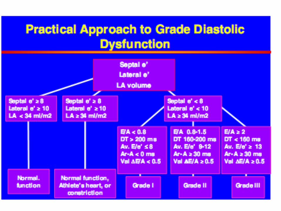
Grade 1 diastolic dysfunction(mild)
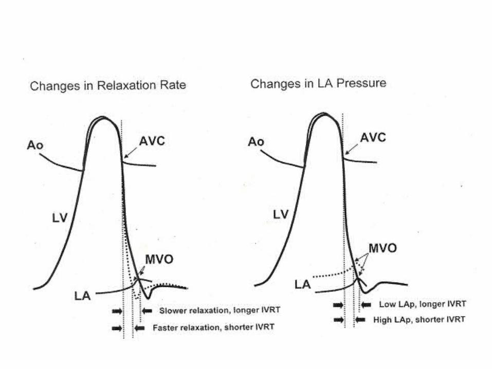
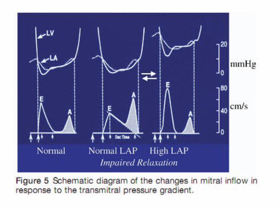
• LV relaxation impaired-– Slower LV pressure decay– Pressure crossover b/w LA and LV occurs late– IVRT,DT prolonged– Early transmitral gradient is reduced-reduced E vel
• Adequate diastolic filling period is critical to maintain LV filling without increase in LA pressures
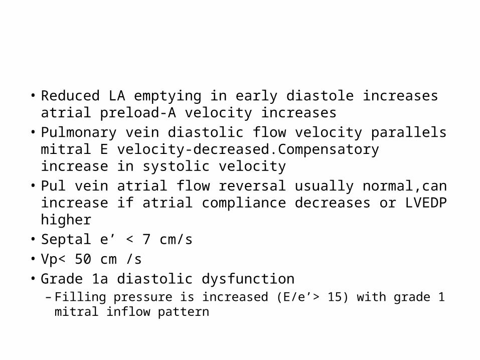
• Reduced LA emptying in early diastole increases atrial preload-A velocity increases
• Pulmonary vein diastolic flow velocity parallels mitral E velocity-decreased.Compensatory increase in systolic velocity
• Pul vein atrial flow reversal usually normal,can increase if atrial compliance decreases or LVEDP higher
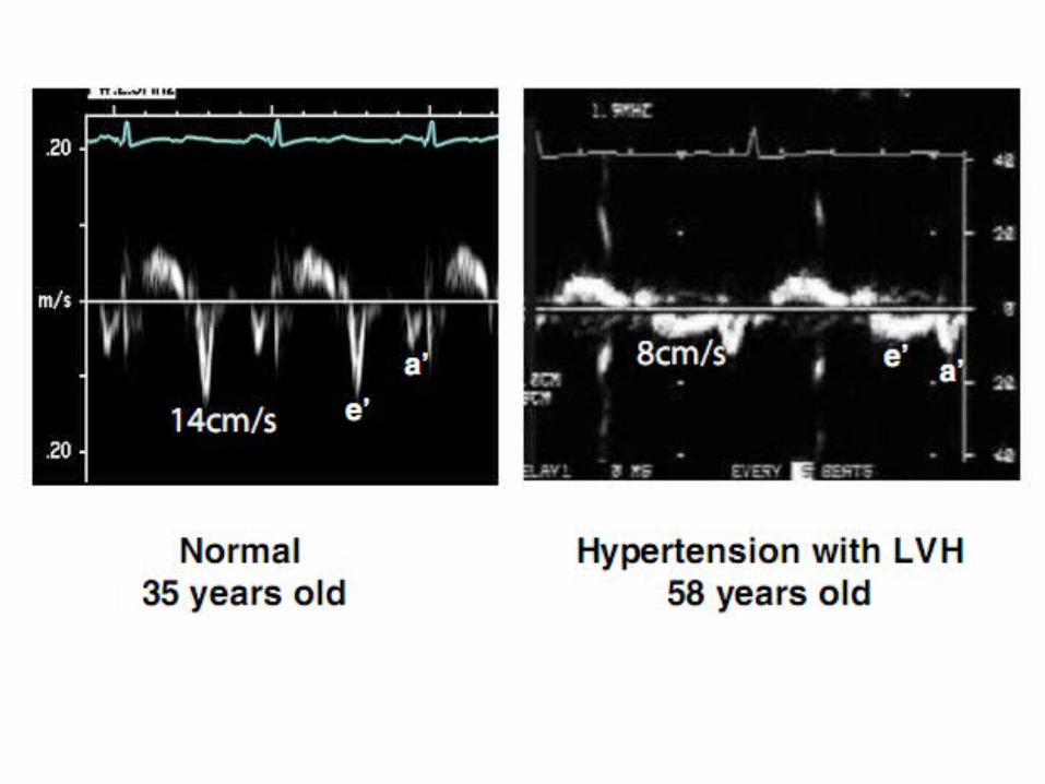
• Septal e’ < 7 cm/s• Vp< 50 cm /s• Grade 1a diastolic dysfunction
– Filling pressure is increased (E/e’> 15) with grade 1 mitral inflow pattern
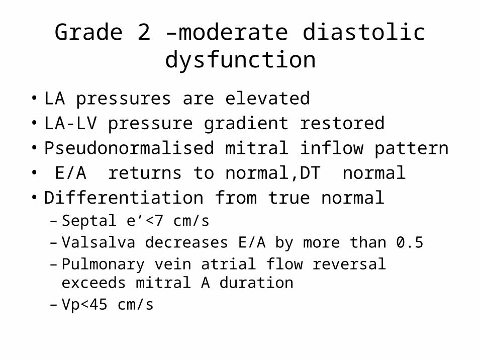
Grade 2 –moderate diastolic dysfunction
• LA pressures are elevated• LA-LV pressure gradient restored• Pseudonormalised mitral inflow pattern• E/A returns to normal,DT normal• Differentiation from true normal– Septal e’<7 cm/s– Valsalva decreases E/A by more than 0.5– Pulmonary vein atrial flow reversal exceeds mitral A
duration– Vp<45 cm/s
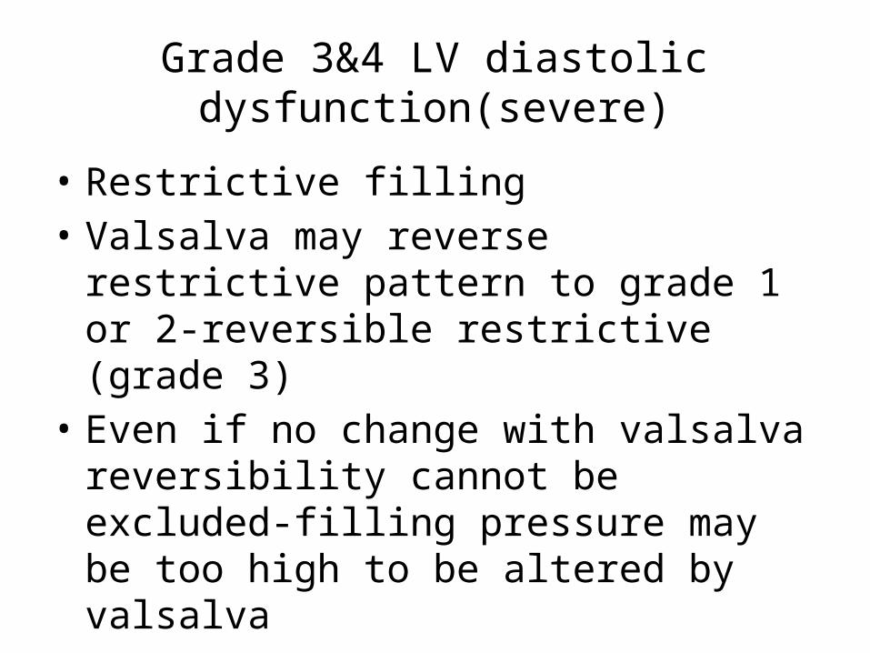
Grade 3&4 LV diastolic dysfunction(severe)
• Restrictive filling • Valsalva may reverse restrictive pattern to
grade 1 or 2-reversible restrictive (grade 3)• Even if no change with valsalva reversibility
cannot be excluded-filling pressure may be too high to be altered by valsalva
• Grade 4 dysfunction not used in ASE rec.
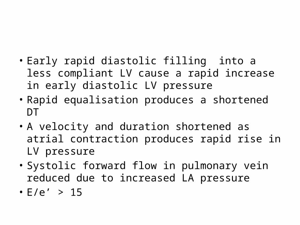
• Early rapid diastolic filling into a less compliant LV cause a rapid increase in early diastolic LV pressure
• Rapid equalisation produces a shortened DT• A velocity and duration shortened as atrial
contraction produces rapid rise in LV pressure• Systolic forward flow in pulmonary vein
reduced due to increased LA pressure• E/e’ > 15
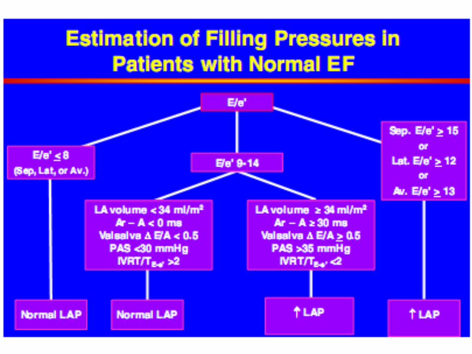
E/e’ ratio in rest and exercise
• E/e’ ratio > 15 correspond to PCWP> 20 mmHg at rest and exercise
• Normal-increase in E and e’ velocity with exercise to maintain ratio
• In a subset of patients with diasolic dysfunction –increase in PCWP with exercise occur–increase in E not accompanied by increase in e’ to elevate the ratio
• PCWP normal if E/e’< 8
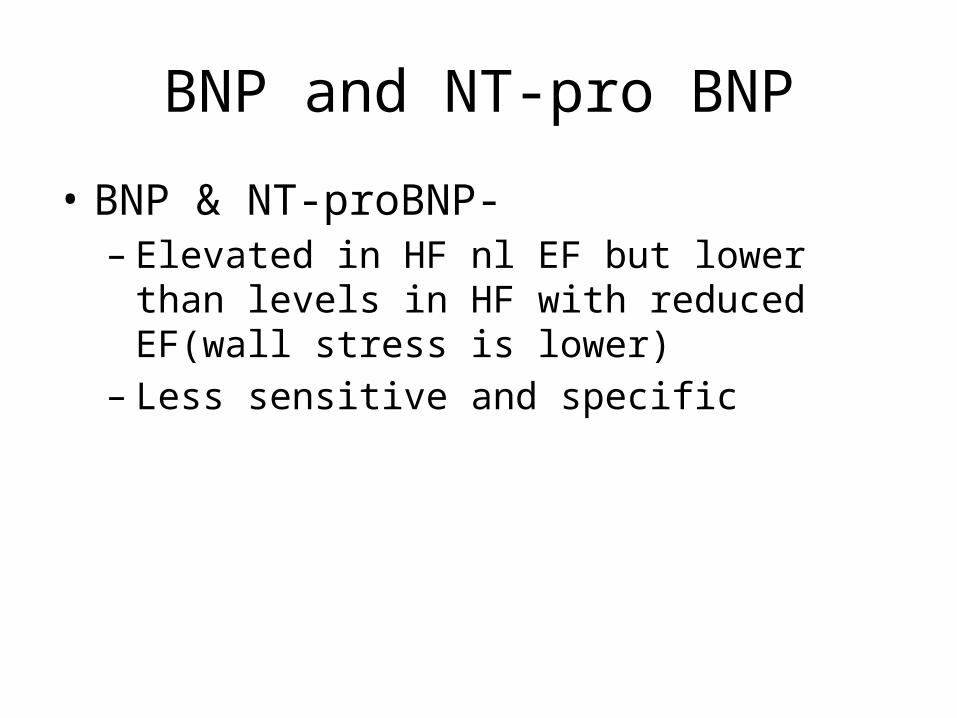
BNP and NT-pro BNP
• BNP & NT-proBNP- – Elevated in HF nl EF but lower than levels in HF
with reduced EF(wall stress is lower)– Less sensitive and specific
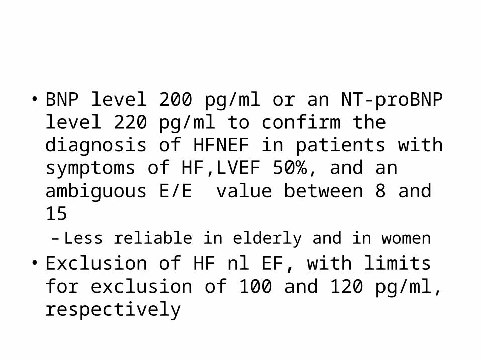
• BNP level 200 pg/ml or an NT-proBNP level 220 pg/ml to confirm the diagnosis of HFNEF in patients with symptoms of HF,LVEF 50%, and an ambiguous E/E value between 8 and 15 – Less reliable in elderly and in women
• Exclusion of HF nl EF, with limits for exclusion of 100 and 120 pg/ml, respectively
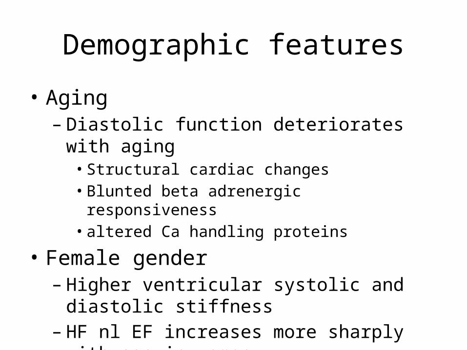
Demographic features
• Aging– Diastolic function deteriorates with aging• Structural cardiac changes• Blunted beta adrenergic responsiveness• altered Ca handling proteins
• Female gender– Higher ventricular systolic and diastolic stiffness– HF nl EF increases more sharply with age in
women
• HTN– LVH which increases diastolic stiffness– Ischemia produces exaggerated increase in filling pressure
• CAD– a/c ischemia causes diastolic dysfunction– Role of CAD in c/c diastolic dysfunction uncertain– Guidelines recommend revascularisation in pts in whom
ischemia is felt to contribute to LV diastolic dysfunction– Reduced coronary microvascular density-impaired
coronary flow reserve-diastolic dysfunction in stress
• AF– AF in 20-40 % of pts with diastolic dysfn– AF may cause decompensation in pts with HF nl EF
• Obesity– 30-50% of pts– Risk for HTN,DM,CAD,AF-contribute to diastolic
dysfn
• Diabetes– Prevalence similar in both forms of HF– Structural changes
• Myocyte hypertrophy• Increased extracellular matrix• Intramyocardial microangiopathy
• Renal dysfn– No diff in both forms of HF– b/l RAS with rapid onset pul edema is a cause of HF
nl EF
• Rare causes– Hypertrophic cardiomyupathy– Infiltrative cardiomyopathies– Ideopathic restrictive cardiomyopathy– Radiation heart disease
• Pulmonary HTN– Prevalence is 86%– a/w decreased survival– Cause is resting or exertional pul venous HTN
Factors causing a/c decompensation
• Uncontrolled HTN• Arrhythmias• ischemia• Infection,anemia• Dietary changes
Treatment
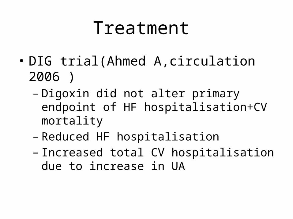
• DIG trial(Ahmed A,circulation 2006 )– Digoxin did not alter primary endpoint of HF
hospitalisation+CV mortality– Reduced HF hospitalisation– Increased total CV hospitalisation due to increase
in UA
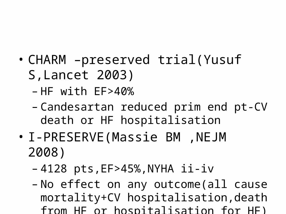
• CHARM –preserved trial(Yusuf S,Lancet 2003)– HF with EF>40%– Candesartan reduced prim end pt-CV death or HF
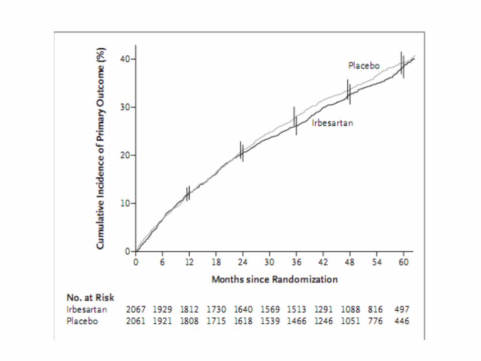
hospitalisation• I-PRESERVE(Massie BM ,NEJM 2008)– 4128 pts,EF>45%,NYHA ii-iv– No effect on any outcome(all cause mortality+CV
hospitalisation,death from HF or hospitalisation for HF)
• PEP-CHF(eur heart journal 2006)– Perindopril Vs placebo,age >70,HF nl EF– All cause mortality+HF hospitalisation– No benefit inprimary end pt– Trend towards reduced HF hospitalisation
• Hong Kong diastolic heart failure study– Diuretics,diuretic +irbesartan,diuretic +ramipril– Hospitalisations,6 min walk ,QOL similar
• TOPCAT-spiranolactone• RELAX-sildenafil• RESET-atrial pacing
Guidelines
• Class I– Control HTN– Control ventrcular rate in AF– Diuretics for pulmonary congestion
• Class II a– Revascularisation in whom ischemia is judged to
have an adverse effect on cardiac function

• Thank you


![1210 DIASTOLIC Hypertension[2]](https://img.pdfslide.net/doc/110x75/577cdd4a1a28ab9e78acb724/1210-diastolic-hypertension2.jpg)