Embed Size (px)
Citation preview

Sponsored by
AAGLAdvancing Minimally Invasive Gynecology Worldwide
Didactic: The Ins and Outs of Midurethral Slings:
Safe Implantation and Surgical Management
of Complications
PROGRAM CHAIR
Eric R. Sokol, MD
PROGRAM CO-CHAIR
Charles R. Rardin, MD
Dobie L. Giles, MD, MSMichael D. Moen, MD
Cheryl B. Iglesia, MDBeri M. Ridgeway, MD
GLOBAL CONGRESSON MINIMALLY INVASIVE GYNECOLOGYNOV. 17-21, 2014 | Vancouver, British Columbia
43rd AAGL

Professional Education Information Target Audience This educational activity is developed to meet the needs of residents, fellows and new minimally invasive specialists in the field of gynecology. Accreditation AAGL is accredited by the Accreditation Council for Continuing Medical Education to provide continuing medical education for physicians. The AAGL designates this live activity for a maximum of 3.75 AMA PRA Category 1 Credit(s)™. Physicians should claim only the credit commensurate with the extent of their participation in the activity. DISCLOSURE OF RELEVANT FINANCIAL RELATIONSHIPS As a provider accredited by the Accreditation Council for Continuing Medical Education, AAGL must ensure balance, independence, and objectivity in all CME activities to promote improvements in health care and not proprietary interests of a commercial interest. The provider controls all decisions related to identification of CME needs, determination of educational objectives, selection and presentation of content, selection of all persons and organizations that will be in a position to control the content, selection of educational methods, and evaluation of the activity. Course chairs, planning committee members, presenters, authors, moderators, panel members, and others in a position to control the content of this activity are required to disclose relevant financial relationships with commercial interests related to the subject matter of this educational activity. Learners are able to assess the potential for commercial bias in information when complete disclosure, resolution of conflicts of interest, and acknowledgment of commercial support are provided prior to the activity. Informed learners are the final safeguards in assuring that a CME activity is independent from commercial support. We believe this mechanism contributes to the transparency and accountability of CME.

Table of Contents
Course Description ........................................................................................................................................ 1 Disclosure ...................................................................................................................................................... 2 Office Evaluation of Urinary Incontinence C.B. Iglesia ..................................................................................................................................................... 3 Urodynamic Assessment of Urinary Incontinence D.L. Giles ..................................................................................................................................................... 14 Behavioral and Functional Treatment of Urinary Incontinence M.D. Moen .................................................................................................................................................. 22 Refractory OAB: Botox, PTNS and Neuromodulation C.B. Iglesia ................................................................................................................................................... 28 Surgery for Stress Urinary Incontinence M.D. Moen ....................................................................................................................................... 36 Sling Selection: Retropubic, Transobturator or Minisling E.R. Sokol ..................................................................................................................................................... 46 Management of Sling Complications C.R. Rardin ................................................................................................................................................... 53 Salvage Procedures for Refractory SUI B.M. Ridgeway ............................................................................................................................................ 58 Cultural and Linguistics Competency ......................................................................................................... 67

URO-‐607 Didactic: The Ins and Outs of Midurethral Slings:
Safe Implantation and Surgical Management of Complications Presented in affiliation with the American Urogynecologic Society (AUGS)
Eric R. Sokol, Chair Charles R. Rardin, Co-‐Chair
Faculty: Dobie L. Giles, Cheryl B. Iglesia, Michael D. Moen, Beri M. Ridgeway
This course provides a broad overview of the proper evaluation and management of female urinary incontinence. We will review conservative and surgical management strategies, including guideline-‐driven recommendations for the treatment of refractory urinary incontinence. Management of surgical complications, including mesh sling exposures and erosions, will also be discussed. Learning Objectives: At the conclusion of this course, the clinician will be able to: 1) Discuss standard algorithms for treatment of stress, urge, and mixed urinary incontinence; 2) compare different surgical treatment modalities for stress urinary incontinence; and 3) describe complications associated with mid-‐urethral slings and how to manage them.
Course Outline 7:00 Welcome, Introductions and Course Overview E.R. Sokol
7:05 Office Evaluation of Urinary Incontinence C.B. Iglesia
7:30 Urodynamic Assessment of Urinary Incontinence D.L. Giles
7:55 Behavioral and Functional Treatment of Urinary Incontinence M.D. Moen
8:20 Refractory OAB: Botox, PTNS and Neuromodulation C.B. Iglesia
8:45 Questions & Answers All Faculty
8:55 Break
9:10 Surgery for Stress Urinary Incontinence M.D. Moen
9:35 Sling Selection: Retropubic, Transobturator or Minisling E.R. Sokol
10:00 Management of Sling Complications C.R. Rardin
10:25 Salvage Procedures for Refractory SUI B.M. Ridgeway
10:50 Questions & Answers All Faculty
11:00 Adjourn
Page 1

PLANNER DISCLOSURE The following members of AAGL have been involved in the educational planning of this workshop and have no conflict of interest to disclose (in alphabetical order by last name). Art Arellano, Professional Education Manager, AAGL* Viviane F. Connor* Kimberly A. Kho* Frank D. Loffer, Medical Director, AAGL* Linda Michels, Executive Director, AAGL* M. Jonathon Solnik* Johnny Yi* SCIENTIFIC PROGRAM COMMITTEE Arnold P. Advincula Consultant: Blue Endo, Intuitive Surgical, SurgiQuest Other: Royalties: CooperSurgical William M. Burke* Rosanne M. Kho* Ted T.M. Lee Consultant: Ethicon Endo-‐Surgery Javier F. Magrina* Ceana H. Nezhat Consultant: Karl Storz Other: Medical Advisor: Plasma Surgical Other: Scientific Advisory Board: SurgiQuest Kevin J.E. Stepp Consultant: CONMED Corporation, Teleflex Other: Stock Ownership: Titan Medical Robert K. Zurawin Consultant: Bayer Healthcare Corp., CONMED Corporation, Ethicon Endo-‐Surgery, Hologic, Intuitive Surgical FACULTY DISCLOSURE The following have agreed to provide verbal disclosure of their relationships prior to their presentations. They have also agreed to support their presentations and clinical recommendations with the “best available evidence” from medical literature (in alphabetical order by last name). Dobie L. Giles* Cheryl B. Iglesia* Michael D. Moen* Charles R. Rardin* Beri M. Ridgeway* Eric R. Sokol Grants/Research: El.En Other: National Principle Investigator: American Medical Systems Other: Stock Ownership: Pelvalon Asterisk (*) denotes no financial relationships to disclose.
Page 2

Office Evaluation of Urinary Incontinence
Cheryl Iglesia, M.D.Section Director, FPMRS
MedStar Washington Hospital CenterProfessor, Ob/Gyn and Urology
Georgetown University School of Medicine
Disclosures
No financial conflicts of interest
Chair, AUGS Guidelines Committee
Chair, PFDN Advisory Board NICHD
Vice Chair, ACOG Pt Ed Board
FDA ObGyn Devices Panel Member
Objectives
• Define subtypes of UI
• Draw conclusions for occult SUI risk
• List 6 steps for the minimal pre-surgical evaluation of uncomplicated SUI
Urinary Incontinence (UI):Prevalence
13 million Americans
Gender
Female: 10%‐55%
Male: 2%‐5%
Prevalence and severity increase with age
Seen in over 50% of nursing home patients
ICS/IUGA Subtypes of Urinary Incontinence
Stress (activity-related) Incontinence SUI
Complaint of involuntary loss of urine on effort or physical exertion
Page 3

Urgency Urinary Incontinence UUI
Complaint of involuntary loss of urine associated with urgency
Other IUGA/ICS Definitions
Postural (urinary) incontinence: Complaint of involuntary loss of urine associated with change of position, e.g. rising from a seated position
Nocturnal enuresis: Complaint of involuntary loss of urine which occurs during sleep
Mixed (urinary) incontinence: Complaint of involuntary UUI and SUI.
Other UI Continuous (urinary) incontinence:
Complaint of continuous involuntary loss of urine.
Insensible (urinary) incontinence: Complaint of urinary incontinence where the woman has been unaware of how it occurred.
Coital incontinence: Complaint of involuntary loss of urine with coitus--further divided into that occurring with penetration and that occurring at orgasm.
Prevalence of Any UIBy Age and Severity
Minassian VA, et al. Obstet Gynecol. 2008;111(2 Pt 1):324‐331.
Defining Overactive Bladder
• Urinary urgency, with or without urge incontinence, usually with urinary frequency and nocturia, in the absence of pathologic or metabolic factors that would explain these symptoms
The International Continence Society defines OAB Syndrome as:
Assessment History
Physical exam
Voiding diary
Post void residual
Urine culture
Cough stress test
Urethral mobility Q-tip test
Single channel CMG
Multichannel CMG
Cystoscopy
Dynamic MRI and other imaging
Page 4

Evaluation Match symptoms to signs
Treat symptomsPay close attention to what bothers patient most
Validated Questionnaires PFDI/ PFIQ: pelvic floor disorders
inventory and pelvic floor impact questionnaire relates to bowel, bladder function (Barber MD et al AJOG 2005; 193: 103-13) I
Incorporates Urogenital Distress Inventory UDI and Urinary Impact Questionnaire UIQ
MESA screen for Mixed UI
Functional Incontinence: Causes(AKA Transient or Reversible Incontinence)
Patient‐related
Environmental‐related
Disease‐related
Medication‐related
Delirium
Infection
Atrophy
Pharmacologic
Psychologic
Endocrinologic
Restricted mobility
Stool impaction
“DIAPPERS”Mnemonic
Voiding Diary
TimeAmount Voided
Any leakage (yes/no)
ActivityIntake
Amount/ type
0700 500 ccs Yes On way to bathroom
0715 8 oz coffee and 4 oz OJ
0900 Yes Coughing
0915 300 ccs No
Bladder App voicesfor pfd.org
AUGS Bladder TrakHer for Iphone/Ipad
Lower Urinary Tract Function
• Bladder and urethral functions– Storage
– Micturition
• These functions are controlled by the central nervous system (CNS) through reflexes that coordinate the activity of:– Bladder (smooth muscle)
– Urethra (smooth and striated muscles)
– Pelvic floor muscles
Page 5

From Female Pelvic Medicine and Reconstructive SurgeryEds. Rogers, Sung, Thakar and Iglesia McGraw-Hill 2014
McGraw Hill 2014 Female Pelvic Medicine Reconstructive Surgery ed: Rogers, Iglesia
Lower Urinary Tract Innervation
+M3
Pelvic Nerve(Parasympathetic) ACh
+N
Pudendal Nerve(Somatic) ACh
‐3
+1Hypogastric Nerve
(Sympathetic) NE
Acetylcholine (ACh)
Evaluation of Incontinence and Prolapse
Physical detailed pelvic
including neurologic
Urine dip and PVR
Kegel PFM
POP-Q
Initial Assessment
Medical history
Symptoms
Other Pertinent History Gynecologic
Urologic
Neurologic
General Medical
Occupational hazards
Medications
Page 6

Drugs That Cause OAB‐Like Symptoms
Ouslander JG. New Engl J Med. 2004;350(8):786‐799.
• Alpha-adrenergic receptor agonists• Tricyclic antidepressants• Psychotropics (sedatives, hypnotics)• Cholinesterase inhibitors• Narcotic analgesics, opioids • Calcium channel blockers• Diuretics• Methylxanthines • NSAIDS
Evaluate All Sites
anterior wall prolapse: urethra, bladder(cystocele, urethrocele, cystourethrocele or enterocele)
apical prolapse: cervix or cuff/cul-de-sac/ small bowel(uterine prolapse, vaginal vault prolapse, enterocele)
posterior wall: rectum, sigmoid, small bowel (rectocele, sigmoidocele, enterocele)
perineal descent
Fixed reference point: hymen
Two points of measurement each
Anterior wall (Aa, Ba)
Posterior wall (Ap, Bp)
Apex (C, D)
Genital hiatus (gh), perineal body (pb),
and total vaginal length (tvl)
The POPQ System
Aa Ba C
GH PB TVL
Ap Bp D
Anterior wall
Posterior wall
Cervix or cuff
Posterior fornix
Genital hiatus Perineal body Total vaginal length
POPQ Software bardurological.com/po
p-q Boston Scientific
Pelvic Floor Institute
www.puritanmedproducts.com
Page 7
Gh and Pb Genital hiatus: from mid-urethra to
posterior fourchette, in centimeters.
Perineal body: from posterior fourchette to mid-anus, in centimeters.
Both with maximal valsalva
Genital Hiatus
Perineal Body
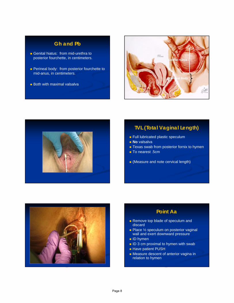
TVL (Total Vaginal Length) Full lubricated plastic speculum No valsalva Texas swab from posterior fornix to hymen To nearest .5cm
(Measure and note cervical length)
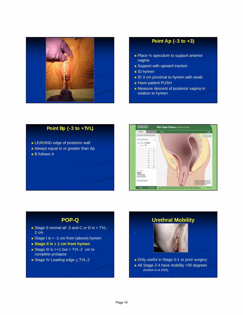
Point Aa Remove top blade of speculum and
discard Place ½ speculum on posterior vaginal
wall and exert downward pressure ID hymen ID 3 cm proximal to hymen with swab Have patient PUSH Measure descent of anterior vagina in
relation to hymen
Page 8

LEADING edge of anterior wall
Always equal to or greater than Aa
B follows A
Point Ba (-3 to +TVL)
Location of posterior fornix
Most distal edge of cervix or leading edge of vaginal cuff
Measure length of cervix with swab
Direct patient to PUSH
Allow speculum to be pushed out
Measure distance from cervix, posterior fornix to hymen
+/- TVL
Points C and D (with speculum)
2 digit exam
Middle finger in posterior fornix
1st finger on cervix
Have patient PUSH
Note cm of descent at introitus (hymen)
+/- TVL
Points C and D (Digital)
Page 9
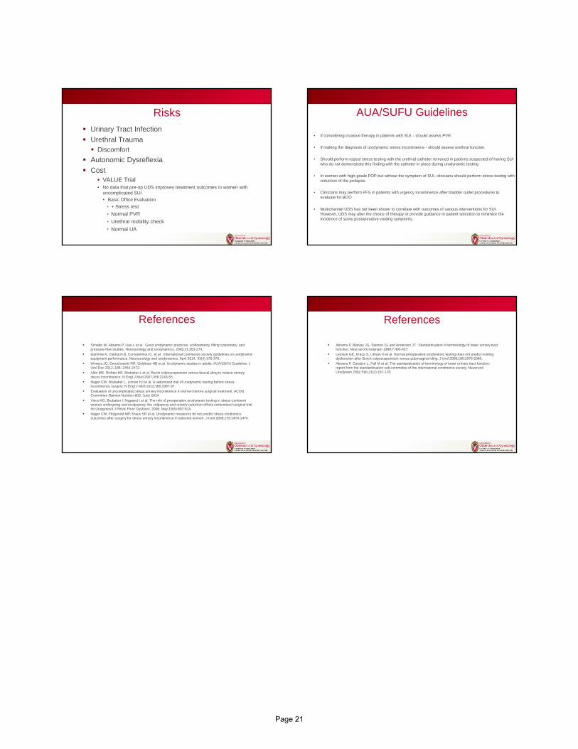
Place ½ speculum to support anterior vagina
Support with upward traction
ID hymen
ID 3 cm proximal to hymen with swab
Have patient PUSH
Measure descent of posterior vagina in relation to hymen
Point Ap (-3 to +3)
LEADING edge of posterior wall
Always equal to or greater than Ap
B follows A
Point Bp (-3 to +TVL)
POP-Q Stage 0 normal all -3 and C or D is < TVL-
2 cm
Stage I is < -1 cm from (above) hymen
Stage II is + 1 cm from hymen
Stage III is >+1 but < TVL-2 cm to complete prolapse
Stage IV Leading edge > TVL-2
Urethral Mobility
Only useful in Stage 0-1 or prior surgery
All Stage 2-4 have mobility >30 degrees (Noblett et al 2005)
�
Page 10

Weber AM and Walters M AJOG 2000
Are Urodynamics Really Necessary?
No!
Basic Office evaluation vs. Urodynamics Women with prolapse and stress incontinence
Basic office evaluation and urodynamic testing had same cure rate of incontinence (96%)
If surgery is the preferred treatment then urodynamic testing is not cost-effective.
(Weber and Walters MD)
Reduction Stress Testing Occult incontinence estimated 36-
80% of advanced prolapse (Richardson DA 1983,
Bump RC 1988, Bergman A 1988, Rosenzweig BA 1992, Chaikin D. J Urol 2000)
Decision to place a prophylactic Sling controversial (OPUS trial) Wei JT, Nygaard I, Richter HE, et al.A midurethral sling to reduce incontinence after vaginal prolapse repair. N Engl J Med 2012;366:2358-2367.
Reduction stress test negative for SUI: still up to 42% risk of de novo SUI postop
PFDN De Novo SUI Risk Calculator
Jelovsek JE, Chagin K, Brubaker L, et al. A model for predicting the risk of de novo stress urinary incontinence in women undergoing pelvic organ prolapse surgery. Obstet Gynecol 2014;123(2): 279-287
http://www.r-calc.com/ExistingFormulas.aspx?filter=CCQHS
Simple Cystometrogram CMG 51725
NEED: 14 FR red rubber catheter, 60 cc catheter tip syringe, 1 Liter saline or H2O
Insert urethral catheter and retrograde fill bladder
Provocative stress test
The observation of transurethral loss of liquid (urine) simultaneous with a cough or valsalva
Sensitivity best if standing with a full bladder.
Page 11

Consider Further Consultation
Hematuria Recurrent UTIs Neuro abnormality Voiding difficulty Decreased bladder capacity Elevated residual volume Fistula Urethral diverticulum Failure to improve on initial management Unclear history and physical exam
Should UDS be performed in all women before SUI surgery?
The VALUE Trial (UITN)
Trial Schema
SUI Treatment*
Patient Referral
Pre-Op Assessment and Diagnosis (basic office evaluation - BOE)
Predominant SUI
BOE
Eligible Refused
Eligible Enrolled
Ineligible
Random Assignment
UDS NO UDS
Predominant UUI
Follow-Up Assessments
*Any approved clinical care method Investigators use in their practice. *Any approved clinical care method Investigators use in their practice. *Any approved clinical care method Investigators use in their practice. *Any approved clinical care method Investigators use in their practice. *Any approved clinical care method Investigators use in their practice. *Any approved clinical care method Investigators use in their practice.
* Any approved clinical care method Investigators use in
their practice.
Outcomes.
Nager CW et al. N Engl J Med 2012;366:1987-1997
Randomized Trial of Office Evaluation with or Without Urodynamic Testing Before
Surgery for Stress Urinary Incontinence
NO DIFFERENCE compared with Basic Office Assessment
-In the absence of prolapse beyond the hymen,
-In the presence of documented leakage with cough, normal PVR
VALUE New England Journal of Medicine 2012
Nager C et al, Contemporary Clinical Trials 30 (2009) 531–539
Who needs urodynamics
Uncertain diagnosis and treatment plan difficulty
Uncertain about the relative contribution of urge and stress incontinence in mixed incontinence
Suspected poor urethral function (ISD?)- Previous surgery, severe incontinence, radiation, radical surgery and data could change treatment
Failure to respond to treatment, Failed surgery
Combined incontinence and emptying disorders
Symptoms of difficult bladder emptying
Elevated PVR- Voiding dysfunction
Page 12

ACOG/AUGS EvaluationUncomplicated SUI
6 steps Basic Evaluation
Summary
Evaluation of UI requires thorough review of urinary, sexual and anorectal complaints and treatment should match complaints
Examination should be done with a split speculum or standing
In cases of advanced prolapse, assess for occult urinary incontinence with prolapse reduced
SUI risk calculator may be useful for counseling re: prophylactic sling surgery
Page 13

Urodynamic Assessment of Urinary Incontinence
Dobie Giles, MD, MS, FACOGAssistant Professor, UW School of Medicine & Public Health
Division Director of Gynecology and Gynecologic Subspecialties
Chief, Female Pelvic Medicine and Reconstructive Surgery
Departments of Obstetrics & Gynecology and Urology
November 17, 2014
Disclosures
I have no financial relationships to disclose.
Objectives
Review the different components of Urodynamics
Discuss AUA/SUFU guidelines
Discuss ACOG/AUGS Committee Opinion
Goal of Urodynamics
To identify factors contributing to LUT dysfunction
To predict the consequences of LUT dysfunction on the upper tracts
To predict the consequences and outcomes of therapeutic intervention
To confirm and/or understand the effects of interventional techniques
To investigate the reasons for treatment failure
Urodynamic Indications
• Not every patient with Stress Urinary Incontinence or Urge Urinary Incontinence needs UDS
• Indications for complex UDS• Marked UUI symptoms, refractory to treatment
• - Inconclusive single channel• - S/s of neurologic disease• - History of prior anti-incontinence surgery or pelvic xrt• - Voiding difficulties/abnormalities
Page 14

Committee Opinion 603 (ACOG/AUGS)
Pre-op multichannel UDS is not necessary before planning primary anti-incontinence surgery in women with uncomplicated SUI Observed leakage by provocative stress measures
Normal UA
No POP beyond hymen
Normal PVR
RCT demonstrated outcomes 1 year after MUS were the same for those who had BOA compared with those who had pre-op UDS
Assessment of lower urinary tract
Obtain functional information about
Bladder filling
Storage
Emptying
A Problem with Storage
A Problem with Emptying
UDS Components
• Voiding studies• Uroflow
• Pressure flow
• Cystometry• Simple vs. Complex
• Single vs. Multichannel
• Electromyography
• Urethral Pressure Profilometry
• Video
UDS Components
• Voiding studies• Uroflow
• Pressure flow
• Cystometry• Simple vs. Complex
• Single vs. Multichannel
• Electromyography
• Urethral Pressure Profilometry
• Video
Uroflowmetry
Flowmeter – volume vs. time
Assess emptying phase
Simple, noninvasive first line screening test
Normal values less well established for females
Assess PVR at completion
Uroflowmetry: Interpretation
• Evaluate curve pattern– Continuous Smooth
– Continuous Fluctuating
– Intermittent
• Normal: Qmax in 1/3 of the total voiding time
Page 15

Uroflowmetry: Interpretation
Volume voided
Maximum Flow Rate (Qmax)
Should not exceed 1/3 of flow time
Average Flow Rate (Qave)
Voided volume/flow time
Flow time
Excluding interruptions
Voiding time
Including interruptions
Time to maximum flow
Abnormalities in flow
Continuous Smooth
Abnormalities in flow
Continuous Fluctuating
Abnormalities in flow
Intermittent
Uroflowmetry: Interpretation
Flow rate is a composite of the function of the bladder and the bladder outlet
Which one is responsible for the dysfunction?
Max flow rate in males
<40 generally over 25 ml/s
>60 generally over 15 ml/s
Women typically have a rate 5-10 ml/s faster
Volumes less than 150 ml should be repeated
UDS Components
• Voiding studies• Uroflow
• Pressure flow
• Cystometrogram (CMG)• Simple vs. Complex
• Single vs. Multichannel
• Electromyography
• Urethral Pressure Profile
• Video
Page 16

Cystometry
Test that measures pressure/volume relationship of the bladder
Assess detrusor activity, sensation, capacity, & compliance
Leak point pressures
Simple Cystometry
Saline, Catheter, Syringe
Watch meniscus
Sudden rise possible detrusor contraction or increase in abdominal pressure
Can determine sensations and capacity
Filling Cystometry Filling Cystometry
A catheter in the bladder
Two sensors/balloons
Pves
Pura
A catheter in the rectum (or vagina)
Pabd
8 Fr or less
Room temperature fluid
Fill rate 50 ml/min
Filling Cystometry
First Sensation
Notice fluid in the bladder
First Desire to Void
Pass urine at next convenient moment but can delay if necessary (watch tv and next commercial)
Strong Desire to Void
Persistent desire to void without the fear of leakage (can’t wait for next commercial)
Maximum Cystometric Capacity
Can no longer delay
Filling CystometryPves (pressure frombladder catheter)
Pabd (pressure in abdomen
Pdet (Pves-Pabd)
Page 17

Leak Point Pressures
Method of assessing urethral resistance and function
To be accurate, perform in standard fashion Catheter size, bladder volume, type of provocation,
reduction of prolapse
If large anterior wall prolapse Perform with and without reduction
Leak Point Pressures
Abdominal leak point pressure (ALPP)
Measures the outlet
Due to increased abdominal pressure in the absence of a detrusor contraction
VLPP
CLPP
Detrusor leak point pressure (DLPP)
Measures compliance
The lowest detrusor pressure at which urine leakage occurs in the absence of either a detrusor contraction or increased abdominal pressure
High DLPP (>40 cm H2O) concern for upper tract damage
Valsalva Leak Point Pressure / Abdominal Leak Point Pressure
Fill to 200 cc (some studies at 300 cc) Repeat every 100 cc
Begin coughing Not as accurate Unnecessary if leakage w/ Valsalva
Perform progressive Valsalva until leakage occurs
If still no leakage Continue filling or Remove catheters and Valsalva and cough again
to demonstrate leakage
Valsalva Leak Point Pressure
Pves
Pabd
Pdet
UDS Components
• Voiding studies• Uroflow
• Pressure flow
• Cystometry• Simple vs. Complex
• Single vs. Multichannel
• Electromyography
• Urethral Pressure Profilometry
• Video
Electromyography
Useful w/ neurologic history or occult neurogenic voiding dysfunction is suspected
Abnormalities in hx, PE, uroflow, or pvr should prompt
Study of the electrical potential generated by depolarization of muscle
During normal voiding there should be no activity
Increased activity during voiding may be characteristic of Detrusor Ext Sphincter Dyssynergia(DESD)
Page 18

Patch Electrodes Electromyography
UDS Components
• Voiding studies• Uroflow
• Pressure flow
• Cystometry• Simple vs. Complex
• Single vs. Multichannel
• Electromyography
• Urethral Pressure Profilometry
• Video
Pressure Flow Study
Performed after CMG
Ideally, voiding w/ abdominal, intravesical, intraurethral, and EMG measurements
Identifies 3 fundamental voiding states: Low detrusor pressure/high flow rate –
unobstructed
High detrusor pressure/low flow rate – obstructed
Low detrusor pressure/low flow rate – poor contractility
Pressure Flow Study
Committee Opinion 603 (ACOG/AUGS)
Elevated PVR in the absence of Pelvic Organ Prolapse is uncommon and should trigger an evaluation of the bladder-emptying mechanism, usually with a pressure-flow urodynamic study
Pressure Flow Study
Page 19

Abrams-Griffiths Pressure Flow
• Simplest Nomogram: Maximal flow rate vs. detrusor pressure
• Draw back: equivocal zone
UDS Components
• Voiding studies• Uroflow
• Pressure flow
• Cystometry• Simple vs. Complex
• Single vs. Multichannel
• Electromyography
• Urethral Pressure Profilometry
• Video
Urethral Pressure Profilometry Urethral Pressure Profilometry*Maximal urethral pressure (Pura)-highest pressure during withdrawal
*Maximal urethral closure pressure (MUCP)- Difference of Pves-Pura
*Functional urethral length – urethral length where intraurethral pressure > bladder pressure
*Total profile length – length of urethra measured by withdrawal technique
Video Urodynamics Video Urodynamics
Page 20
Risks Urinary Tract Infection
Urethral Trauma Discomfort
Autonomic Dysreflexia
Cost• VALUE Trial• No data that pre-op UDS improves treatment outcomes in women with
uncomplicated SUI
• Basic Office Evaluation
• + Stress test
• Normal PVR
• Urethral mobility check
• Normal UA
AUA/SUFU Guidelines
• If considering invasive therapy in patients with SUI – should assess PVR
• If making the diagnosis of urodynamic stress incontinence - should assess urethral function.
• Should perform repeat stress testing with the urethral catheter removed in patients suspected of having SUI who do not demonstrate this finding with the catheter in place during urodynamic testing
• In women with high-grade POP but without the symptom of SUI, clinicians should perform stress testing with reduction of the prolapse.
• Clinicians may perform PFS in patients with urgency incontinence after bladder outlet procedures to evaluate for BOO
• Multichannel UDS has not been shown to correlate with outcomes of various interventions for SUI. However, UDS may alter the choice of therapy or provide guidance in patient selection to minimize the incidence of some postoperative voiding symptoms.
References
Schafer W, Abrams P, Liao L et al. Good urodynamic practices: uroflowmetry, filling cystometry, and pressure-flow studies. Neurourology and urodynamics. 2002;21:261-274.
Gammie A, Clarkson B, Constantinou C. et al. International continence society guidelines on urodynamic equipment performance. Neurourology and urodynamics. April 2014; 33(4) 370-379.
Winters JC, Dmochowski RR, Goldman HB et al. Urodynamic studies in adults: AUA/SUFU Guideline. J Urol Dec 2012.;188: 2464-2472.
Albo ME, Richter HE, Brubaker L et al. Burch colposuspension versus fascial sling to reduce urinary stress incontinence. N Engl J Med 2007;356:2143-55.
Nager CW. Brubaker L, Litman HJ et al. A radomized trial of urodynamic testing before stress-incontinence surgery. N Engl J Med 2012;366:1987-97.
Evaluation of uncomplicated stress urinary incontinence in women before surgical treatment. ACOG Committee Opinion Number 603. June 2014.
Visco AG, Brubaker l, Nygaard I et al. The role of preoperative urodynamic testing in stress-continent women undergoing sacrocolpopexy: the colpopexy and urianry reduction efforts randomized surgical trial. Int Urogynecol J Pelvic Floor Dysfunct. 2008. May;19(5):607-614.
Nager CW, Fitzgerald MP, Kraus SR et al. Urodynamic measures do not predict stress continence outcomes after surgery for stress urinary incontinence in selected women. J Urol 2008;179:1470-1474.
References
Abrams P, Blaivas JG, Stanton SL and Andersen JT. Standardisation of terminology of lower urinary tract function. Neurourol Urodynam 1988:7:403-427.
Lemack GE, Kraus S, Litman H et al. Normal preoperative urodynamic testing does not predict voiding dysfunction after Burch colposuspension versus pubovaginal sling. J Urol 2008;180:2076-2080.
Abrams P, Cardozo L, Fall M et al. The standardisation of terminology of lower urinary tract function: report from the standardisation sub-committee of the international continence society. NeuorurolUrodynam 2002 Feb;21(2):167-178.
Page 21

Behavioral and Functional Treatment of Incontinence
Michael Moen, MD, FACOG, FACS
Professor of Obstetrics and GynecologyChicago Medical School/Rosalind Franklin University
Medical DirectorIllinois Urogynecology, Ltd.
Disclosures
I have no financial relationships to disclose.
Learning Objectives
Identify factors associated with screening, evaluation and treatment of women with urinary incontinence
Review literature supporting behavioral and functional treatments for urinary incontinence
Develop practical approach for initiation of behavioral and functional treatments for urinary incontinence
Page 22

Consequences of Incontinence A public episode of incontinence is seen as most
embarrassing symptom for the majority of patients with urinary incontinence
Leaking leads to lifestyle changes and coping behavior– Bathroom mapping, Avoiding social situations, Avoiding travel – Wearing dark clothing, Wearing pads/protection
Can impact intimate relationships (fear of leakage during/after intercourse)
Higher rate of depression in patients with incontinence
Screening for Incontinence
Perform focused history and physical exam
Rule out urinary tract infection
“Do you have bladder problems that are troublesome, or do you ever leak urine?”
YES
Office Evaluation-History Review Quality of life issues:
– Wear protection?
– Limit travel or social activities?
– Avoid exercise?
– Avoid sexual activity?
– Feel depressed?
Neuromuscular evaluation
Perineal sensation
Pelvic muscle strength
Office Evaluation-Exam Importance of Pelvic Floor Assessment
Assessment of pelvic muscle strength critical in identifying patients who might benefit from pelvic floor physical therapy
– 20-30% of asymptomatic women unable to adequately contract pelvic floor muscles
– 7-10% of women actually perform Valsalvamaneuver when attempting to contract pelvic floor muscles
Moen et al. J Pelvic Med 2007;13:113
Page 23

Office Evaluation-Exam Importance of Pelvic Floor Assessment
Majority of women familiar with “Kegels”
42% had been instructed to perform PMEs with 62.5% having received only verbal instruction and 38% were told to stop and start their stream when voiding
Only 23% able to adequately contract PFMs
12% performed Valsalva maneuver when attempting to contract pelvic floor muscles
Moen et al. IUJ 2009; 20:843–846
Functional Factors related to UI
Cognitive issues/Dementia
Mobility issues/Physical limitations (arthritis)
Constipation/Fecal impaction
Medications
– Diuretics; Sedatives; Hypnotics
Functional Treatments Cognitive issues/Dementia
– Prompted voiding; Absorbent products
Mobility issues/Physical limitations (arthritis)
– Bedside commode; clothing changes
Constipation/Fecal impaction
– Optimize bowel function
Medications
– Timing of diuretics
Functional/Behavioral Therapy Discuss normal fluid intake
– Avoid dehydration– Avoid excessive intake– Evening restrictions on fluid intake
Avoid exacerbating factors– Control of allergies, bronchitis, coughing– Heavy lifting
Page 24

Behavioral Therapy Advantages
– No reported side effects– Can be used in conjunction with other therapies
Disadvantages– Require patient education, motivation and
continued practice– Time consuming for health care provider
Goal– improve pelvic floor muscle function and
bladder control
Bladder Training Three basic components
– 1) education (bladder function, fluid intake)– 2) scheduled voiding with systematic delay– 3) positive reinforcement (follow-up)
Cure rates 47-100 % (10 studies) Outcomes may improve with addition of
– Pelvic Floor Muscle Exercises– Estrogen therapy in patients with atrophy
“it has been established without a doubt that loss of function of the pubococcygeus muscle is present in all women with true urinary stress incontinence”
“this condition is amenable to correction through reeducation of muscular function and resistive exercises that can be instituted as a simple office procedure”
Pelvic Floor Muscle Exercises
Benefits seen with as few as 30 contractions performed 3 times per week
Overall success for improving SUI 30-74%
Optimal results may be seen with 40-80 contractions performed per day
Wells J Am Ger Soc 1991
Pelvic Floor Muscle Rehabilitation
Pelvic Muscle Exercises (Kegel’s)– effective for stress and urge incontinence– may prevent incidence of incontinence
Pelvic Floor Physical Therapy–Biofeedback–Electrical Stimulation–Electromagnetic therapy
Page 25

Biofeedback Biofeedback Most useful in patients with weak or
uncoordinated muscle activity
Provide feedback (auditory/visual) to patient to improve exercise technique
Initial treatment with Physiotherapist 1-2x/wk
Home unit can be used daily
Symptom improvement: 48-83% (13 studies)
Electrical Stimulation Pelvic Floor Electrical Stimulation
Most useful in patients with inability to contract
Produces contraction of levator ani, external urethral sphincter and anal sphincter
Pelvic floor contraction is accompanied by reflex inhibition of detrusor contraction (via reflex arc through sacral micturition center)
Physiotherapist/Office and Home therapies
Improvement in 30-80%
Page 26

Incontinence Pessaries for SUI
Conclusions Bladder Training and Pelvic Muscle Exercises are
the primary behavioral treatments for incontinence
Many women and health care providers are unaware of the safety and effectiveness of Behavioral and Functional treatments for urinary incontinence
The majority of women with pelvic floor disorders have suboptimal pelvic muscle function highlighting the importance of pelvic floor exam and availablity of Pelvic Floor Physical Therapy
References Taylor et al. The self-reported prevalence and knowledge of urinary incontinence and barriers to
health care seeking in a community sample of Canadian women. Am J Med Sci 2013;3:97-102
Morrill et al. Seeking healthcare for pelvic floor disorders: a population-based study. Am J ObstetGynecol 2007;197:86.e1-e6
Rogers RG. Urinary stress incontinence in women. NEJM 2008;358:1029-36
Moen et al. Knowledge and performance of pelvic muscle exercises in women. J Pelvic Med Surg2007;13:113–117
Moen Pelvic floor muscle function in women presenting with pelvic floor disorders. Int UrogynecolJ 2009;20:843–846
Kegel AH. Progressive resistance exercise in the functional restoration of the perineal muscle. Am J Obstet Gynecol. 1948;56:238–249
Wells et al. Pelvic muscle exercise for stress urinary incontinence in elderly women. J Am Ger Soc1991;39:785-91
Olah et al. The conservative management of patients with symptoms of stress incontinence: a randomized, prospective study comparing weighted vaginal cones and interferential therapy. Am J Obstet Gynecol 1990;162:87-92
Washington et al. Barriers to pelvic floor physical therapy utilization for treatment of female urinary incontinence. Am J Obstet Gynecol 2011;205:152.e1-9
Page 27

Refractory OAB: Neuromodulation, PTNS and Botox
Cheryl B. Iglesia, MDSection Director, FPMRS
MedStar Washington Hospital CenterProfessor, Department of Ob/Gyn and
UrologyGeorgetown University School of Medicine
Washington, DC
Disclosures
• I have no financial disclosures
• Serve as chair of the Pelvic Floor Disorders Network Advisory Board, NICHD
• FDA ObGyn Devices Panel Member
• Chair, AUGS Guidelines committee
Objectives
• List indications for neuromodulation and detrusor botulinum toxin injections
• Cite evidence‐based complications from both modalities Yes
Refractory OAB
• Urgency urinary incontinence (UUI)
– Highly prevalent
– Significantly impacts QoL
• Most common therapy: Anticholinergics
• No one drug is superior
• Marginal efficacy
• Side effects: dry mouth, constipation, CNS
Hartmann et al. Treatment of Overactive Bladder in Women. Evidence Report No. 187, 2009.
2014 AUA/SUFU OAB Guidelines
Page 28

Sacral Nerve Stimulation
• 1981 Tanagho and Schmidt
• 1997 FDA approves for Urge Incontinence
• 1999 FDA approves for Urgency/Frequency and Non‐obstructive Urinary Retention
• 2002 FDA approves for OAB
• 2002 Wide use of staged technique/fluoro
• 2006 Small neurostimulator
• NOW over 100,000 implants worldwide
Neuromodulation SNS
• Implantable stimulation system
includes lead and pacemaker (IPG)
• Targets sacral nerves (S3)
• Modulates reflexes between bladder, sphincter, and pelvic floor
Mechanism of Action
• Incompletely understood
• Bladder is not the specific target
• Central afferent neuromodulation– Targets reflex centers in cord and pons
• Affects OAB and idiopathic retention– Blocks ascending sensory pathway inputs
– Suppresses guarding reflex pathways
SNS Indications
• Refractory OAB– Failed drugs and behavioral therapy
• Urinary Retention– Idiopathic non‐obstructive
• Non‐neurogenic etiology• Bowel dysfunction
– European indications– Fecal incontinence April 2011 FDA approval
How is it done?
• Test stimulation (outpatient)
• Trial period (1‐2 weeks)
– Percutaneous
– Implantable lead
• Implantation of IPG
Page 29

Test Stimulation
• Needle placed at S3
• Motor Response– Bellows/toe motion
• Sensation– Genital/anal sensation, comfort
Trial Period
• Review diary
• Success equals > 50% improvement
– Number of leaks/day
– Number of voids/day
– Volume voided/void
– Degree of urgency
Device Implantation
• Based on success of trial
• Completely reversible if declined/discontinued
• Change stimulation parameters externally
• Permanent devices last up to 10 years
Clinical Efficacy
• Urge incontinence N=38
– 45% completely dry
– 34% experienced >50% reduction in leaks
• Urgency Frequency N=33
– 31% normal voids 4‐7 per day
– 33% experienced >50% reduction in voids
• Retention N=38
– 61% stopped catheter use
– 16% experienced >50% decrease in retained volume
SNS Study Group J Urol 1999; 162; J Urol 2000; 163; J Urol 2001; 165
Systematic Review: SNS for Urge Incontinence
• 1,824 implants from 34 clinical trials
• From RCTs 80% of patients achieve >50% improvement of symptoms
• From Case Series: 67% improvement
Brazell M et al. Efficacy & Safety of SNS; J Urol 2006; 175: 835-841
Page 30

5 year results SNS• 17 centers, 163 patients, mean age 44.7, 87% female (Van Kerrebroeck J Urol 2007)
• Success rates– 68% UI
– 56% UF
– 71% Retention
• If success at one year, rate of success at 5 years– 89% UI
– 71% UF
– 76% Retention
Complications SNS
• Reoperation Rate 33%– Loss of efficacy
– Pain at lead or IPG site
– Infection
• Improvement– Use of fluoroscopy
– Staged approach and tined lead
– Recent study show reoperation <20%
Starkman NUU2007; van Voskuilen BJU 2007
Factors Predicting Success Rates
• For urge incontinence
– Age less than 55
– Non‐neurogenic
– Few comorbidities
• Staged approach using tined lead
– 80% test to implant rate
Amundsen C, J Urol 2007; Spinelli J Urol 2003
Review of SNS
• Ideal candidate
– 65% chance of at least 50% improvement
• Long‐term benefit is probable
• Safe, low morbidity and reversible
• Potential help for GI symptoms
• High likelihood of long‐term satisfaction
PTNS Posterior Tibial Nerve Stimulation
• Aka Percutaneous tibial nerve stimulation
• Urgent PC, Uroplasty, Minnetonka, MN
• Dr. Marshall Stoller, UCSF 2007 SANS unit
• FDA cleared for UUI 2000 and OAB 2010
PTNS: weekly 30 min sessions x 12 weeks
Page 31

Evidence for PTNS
• ORBIT trial: (Peters J Urol 2009)– 100 patients RCT PTNS vs Tolterodine 4mg ER– Global response assessment 79.5% reporting cure or improvement compared to 54.8% of subjects on tolterodine (p = 0.01).
– Similar improvement in frequency, UUI, urgency and nocturia
• SUMIT trial: (Peters J Urol 2010)– PTNS vs Sham– 55% vs 21% sham response (p<.001)
COCHRANE REVIEW PTNS
Botulinum Toxin in Urology
• Dykstra et al J Urol 1988; 139; 919
• Schurch et J Urol 1996; 155: 1023
• Smith and Chancellor J Urol 2004; 171:2128
• Schurch et al J Urol 2005; 174: 196
Why Use It
• Refractory OAB
– Marginal compliance with oral therapies
– Refractory to Interstim SNS
Botox mechanism of action
Page 32

Detrusor Botox
• FDA approved for UUI urge urinary incontinence
• Effective in:– Neurogenic DO
• Spinal cord injury, MS
– Post‐obstructive DO
• Expensive $490/100u
• Effect in 7‐10 days
• Lasts 3‐9 months
Botulinum Toxin Injection Technique
• 100 units of Botox in 20 cc saline volume
• 1 cc per injection site
– 5 units per site
• 20 sites, spare trigone
• Needle
– 5F sheath
– 23 gauge needle Williams (Cook), Corson (Bard), BoNee (Coloplast)
Botulinum Toxin A for Detrusor Overactivity (DO)
• Intradetrusor Botulinum toxin A vs placebo for neurogenic DO (n=59)1
– Reduction in mean incontinent episode frequency
• 32‐54% (200U)
• 42‐58% (300U)
• Case series of 231 patients with neurogenic DO (Reitz, 2004)
– 73% continent and remainder improved
1Schurch B. Neuro Urodyn 2004;23:609.
RUBI BOTOX RCT
• 200 u detrusor Botox in 28 pts vs. placebo injection in 15 pts
• Improvement in UUI symptoms in 60% Botox patients, lasting 6 months, median resp time 372 days Botox vs 62 days for placebo
• Retention requiring catheterization in 15% of Botox patients (PVR > 200 43%)
• Study halted
Anticholinergic vs Botox Comparison(ABC) Randomized Trial
Anthony G. Visco et al
Pelvic Floor Disorders Network
Supported by grants from The Eunice Kennedy Shriver National Institute of Child Health and Human Development
Objective
Safety Efficacy
6 MonthAnticholinergic Regimen
Single 100UBotox Injection
Page 33

RandomizationRandomization
Anticholinergic Meds+
Placebo Injection
Anticholinergic Meds+
Placebo Injection
Botox 100U+
Placebo Pills
Botox 100U+
Placebo Pills
Bladder Diaries(Months 1‐6)Bladder Diaries(Months 1‐6)
Bladder Diaries(Months 1‐6)Bladder Diaries(Months 1‐6)
Women with ≥5 UUI Episodes/3‐Day DiaryWomen with ≥5 UUI Episodes/3‐Day Diary
All Pills Stopped at 6 MonthsAll Pills Stopped at 6 Months
Methods
Inclusion Criteria: Anticholinergic hx (failed 2 previous attempts)
PVR < 150mL
Able to perform self-catheterization (CISC)
Primary Outcome:
∆ from baseline in the mean number of UUI episodes
over the 6-month period
Primary Outcome: Reduction in Mean UUI Episodes/Day Over 6 Months
‐4
‐3.5
‐3
‐2.5
‐2
‐1.5
‐1
‐0.5
0
0 1 2 3 4 5 6
Change
in UUIE From Baseline
Study Month
Anticholinergic Onabotulinum Toxin A
P=0.81
Anticholinergics: 3.36 UUI Epidodes/DayBotox: 3.29 UUI Epidodes/Day
AnticholinergicsN=127
BotoxN=120
P value
Urgency Incontinence
Complete Resolution (Cure) 13% 27% 0.003
Quality of Life:
OABq‐SF Severity Scale ‐44.6 ‐44.1 0.87
OABq‐SF QOL Scale 37.1 37.1 0.98
PFDI‐SF ‐43.7 ‐48.2 0.47
PFIQ‐SF ‐32.8 ‐33.9 0.88
Secondary Efficacy Outcomes
AnticholinergicsN=127
BotoxN=120
P value
Dry mouth 58 (46%) 37 (31%) P=0.02
Dry eyes 21 (17%) 29 (24%) P=0.12
Constipation 36 (28%) 25 (21%) P=0.06
CISC1 month2 months4 months6 months
0000
3 (3%)6 (5%)3 (3%)1 (1%)
0.110.010.110.49
UTI 16 (13%) 40 (33%) <0.0001
Secondary Outcomes: Side Effects Botox Duration of Effect
Botox
Page 34

Conclusions:
• Anticholinergic therapy and Botox 100 units:– Both significantly improve:
• Urgency urinary incontinence
• Quality of Life
– No significant difference between treatment groups
• Botox compared to anticholinergics:– Two-fold higher likelihood of complete resolution of UUI
– Higher transient urinary retention and UTI
– Less dry mouth
Future Research
• ROSETTA Trial: Refractor Overactive Bladder: Sacral neuromodulation vs. Botox Assessment Amundsen et al PFDN 2012‐2015
• Botox A 200 units versus Interstim; 380 pts
• Primary outcome effectiveness at 6 months
SUMMARY
• Make correct diagnosis first!• Start with behavioral modifications• Add anticholinergic with dosing flexibility• Choose whichever anticholinergic is covered by insurance
• Urodynamics if suboptimal response• Change to different anticholinergic if not tolerated• Consider Interstim SNS or PTNS• Consider Botox injection
Page 35

Surgery for Stress Urinary Incontinence
Michael Moen, MD, FACOG, FACS
Professor of Obstetrics and GynecologyChicago Medical School/Rosalind Franklin University
Medical DirectorIllinois Urogynecology, Ltd.
Disclosures
I have no financial relationships to disclose.
Learning Objectives
Understand the historical development of surgical procedures for stress incontinence
Analyze key comparative studies and literature concerning surgery for stress incontinence
Describe current practical approach to surgical treatment of stress incontinence
There is a type of urinary incontinence in women which most frequently comes on following childbirth. The onset of this affection manifests itself first by an occasional escape of a few drops of urine following some unusual exertion. Later, gushes of urine follow coughing, sneezng, laughing stooping, or walking; which may ultimately lead to an absolute loss of control, compelling the patient to wear some kind of protection…few infirmities are productive of so much inconvenience and mental depression or interfere so gravely with the present comfort and future prospects of its victims.
Dr. Howard Kelly, 1914
The methods of treatment for urinary incontinence have been legion, and we find some of the earlier procedures very crude, such as …ligation of the prepuce, the use of pressure bandages, painting of the external meatus with collodion. Cold-water foot-bath, cold hypogastric douches, lumbar infusions, aromatic baths, counter-irritation by means of blisters, injection of sacral nerves, lumbar puncture, subarachnoid mercurial injections, epidural injections of sterile water or salt solution, cauterization, pessaries, massage, and the use of electricity have played an important part in the treatment of incontinence.
Many operations have been devised in the surgical treatment of urinary incontinence and may be classified as A) those which serve to create an artificial channel which can be placed under voluntary control or B) operations which restore the urethra to the normal power of retentionExamples of group A include:Rutenberg - close urethra and establish vesico-abdominal fistula; control is made by means of a large pledget or ball-valve closing the sinusRose - rectovaginal fistula is made, following which vagina is completely closed; control of urine is effected by means of the sphincter ani
Page 36

Kelly procedure“ This affection is due to the loss of elasticity or
normal tone of urethal and vesical sphincter, so well shown by the cystoscopic picture, which in many cases presents a gaping internal sphincter orifice which closes sluggishly as the cystoscope is withdrawn. The point of vantage toward which the operative treatment should be directed is the internal orifice of the urehtra and sphincter of the bladder”
Technique
Key Publications
-16 of 20 patients cured(F/U 4 months to 13 years)
Key Publications Kennedy (1937) Beck (1982)
(Early) Sling procedures
Goebell (1910) - pyramidalis muscles
Frangenheim (1914) - pyramidalis muscles attached to strips of overlying fascia
Stoeckel (1917) - Goebell-Frangenheimprocedure combined with vaginal plastic operation at bladder neck (i.e. Kelly)
Martius (1929) - bulbocavernosus muscle and surrounding fatty tissue
Fascial Slings Aldridge (1942)
– “The new procedure that has been described was devised primarily with the hope of curing post-partum, urinary stress incontinence in women in whom vaginal plastic surgery seemed inadequate.”
– “The disadvantages of the procedure are that it requires a painstaking technique which should not be undertaken by a surgeon who has not acquired a modern conception of the anatomic structures in the anterior vaginal wall about the urethra and bladder.”
Page 37

Key Publications Ridley (1966) Parker (1979) McGuire (1987) Beck (1988) Breen (1997) In all of these articles, the Fascial Sling is
described as a salvage procedure for patients with recurrent stress incontinence
Marshall-Marchetti-Krantz (MMK)
Krantz technique
Burch procedure
Burch 1961
Key Publications
Burch 1961: 53 cases; 100% success
Page 38

Technique
From: Hertogs and Stanton, 1985
Comparative Studies
Needle Suspension Procedures Pereyra (1959); Stamey (1973); Raz (1981);
Gittes (1987); other variations “cure of urinary incontinence depends
exclusively on raising the internal vesical neck of the bladder upward and forward behind the symphysis pubis, the cystoscope offers the most accurate way of placing the suspending sutures exactly at the bladder neck”
Stamey 1980
Technique
Page 39

Technique
Paravaginal Defect Repair
Pathophysiology - “(lateral) defect in pubocervicalsegment of endopelvic fascia resulting in loss of urethrovesical angle and stress incontinence”
Technique
Key Publications Richardson (1981)
– 233 pts; 88% cured, 7% satisfactory, 5% failure
Shull (1989)– 149 patients; 97% success
Key Publications
Increases in urethral closure pressure during a cough probably arise because the urethra is compressed against a hammock-like supportive layer, rather than the urethra being truly "intraabdominal."
Page 40

Comparative Studies Comparative Studies
Modified Slings
Sling materials
– Synthetic
– Biologic
– Patch
Fixation techniques
– Bone anchors
Modified Slings
Synthetic Midurethral Slings Pathophysiology - loss of function of pubourethral
ligaments to maintain high-pressure zone at mid-urethra
Key Publications
Page 41

Key Publications Key Publications
Comparative Studies Comparative Studies
Comparative Studies
Page 42

SUI Surgery Trends
MUS variations
Retropubic
– Bottom-up
– Top-down
Transobturator
– Outside-in
– Inside-out
Single incision (Mini)
Retropubic vs. Transobturator Is orientation of support and issue?
Comparative Studies
Page 43

Comparative Studies Ogah 2009 62 trials; 7101 pts
MUS as effective as traditional slings, open RPU and LscRPU, with fewer complications
Retropubic route better than obturator
Schimpf 2014 MUS = Burch (o)
MUS > PVS (s)
Retropubic MUS > transobturator (o,s)
MUS > Mini (o,s)
Conclusions Concepts concerning stress incontinence
pathophysiology and surgical treatments for stress incontinence have varied considerably over time
Current options for surgical management of stress incontinence include retropubic urethropexy, pubovaginal sling and synthetic midurethral slings
Midurethral slings are the preferred treatment for most patients with stress incontinence
References Kelly HA, Dumm WM. Urinary incontinence in women without manifest injury to the
bladder. Surg Gynecol Obstet 1914;18:444-450
Kennedy WT. Incontinence of urine in the female, the urethral sphincter mechanism, damage of function, and restoration of control. Am J Obstet Gynecol 1937;34:576-89
Beck RP, McCormick S. Treatment of urinary stress incontinence with anterior colporrhaphy. Obstet Gynecol 1982;59:269-274
Aldridge AH. Transplantaation of fascia for relief of urinary stress incontinence. Am J Obstet Gynecol 1942;44:398-411
Ridley JH. Appraisal of the Goebell-Frangenheim-Stoeckel sling procedure. AmJ ObstetGynecol 1966;95:714-21
Parker RT, et al. Fascia lata urethrovesical suspension for recurrent stress urinary incontinence. Am J Obstet Gynecol 1979;135:843-852
McGuire EJ, Bennett CJ, Konnak JA, Sonda P, Savastano JA. Experience with pubovaginalslings for urinary incontinence at the University of Michigan. J Urol 1987;138:525-26
Beck RP, McCormick S, Nordstrom L. The fascia lata sling procedure for treating recurrent genuine stress incontinence of urine. Obstet Gynecot 1988;72:699-703
Page 44

References Breen JM, Geer BE, May ge. The fascia lata suburethral sling for treating recurrent urinary
stress incontinence. Am J Obstet Gynecol 1997;177:1363-66
Marshall V F, Marchetti A A, Krantz, K E. The correction of stress incontinence by simple vesicourethral suspension. Surg Gynecol Obstet 1949;88:599-618
Burch ]C. Urethrovaginal fixation to Cooper's ligament for correction of stress incontinence, cystocele, and prolapse. Am J Obstet Gynecol 1961;81:281-290
Albo ME, Richter HE, Brubaker L, et al. Burch colposuspension versus fascial sling to reduce urinary stress incontinence. N Engl J Med 2007;356:2143-55
Hertogs K, Stanton SL. Mechanism of urinary continence after colposuspension: barrier studies. Br J Obstet Gynaecol 1985;92: 1184-8.
Kammerer-Doak DN, Dorin MH, Rogers RG, Cousin MO. A randomized trial of Burch urethropexy and anterior colporrhaphy for stress urinary incontinence. ObstetGynecol. 1999;93:75–78
Pereyra AJ. A simplified surgical procedure for the correction of stress incontinence in women. West ] Surg Obstet Gynecol 1959;67:223-6
Stamey TA. Endoscopic suspension of vesical neck for urinary incontinence. Surg GynecolObstet 1973;136:547-554
References Raz S. Modified bladder neck suspension for female stress incontinence. Urology 1981
;17:82-84
Gittes RF. Non-incision pubovaginal suspension for stress incontinence.] Urol1987;138:568-70
Richardson AC, Edmonds PB, Williams NL. Treatment of stress urinary incontinence due to paravaginal fascial defect. Obstet Cynecol 1981;57:357-362
Shull BL, Baden WF. A six year experience with paravaginal defect repair for stress urinary incontinence. Am J Obstet Gynecol 1989;160:1432-40
DeLanceyJOL. Structural support of the urethra as it relates to stress urinary incontinence: the hammock hypothesis. Am J Obstet Gynecol 1994;170:1713-20
Bergman A, Elia G. Three surgical procedures for genuine stress incontinence: five-year follow-up of a prospective randomized study. Am J Obstet Gynecol. 1995;173:66–71
Colombo M, Milani R, Vitobello D, Maggioni A. A randomized comparison of Burch colposuspension and abdominal paravaginal defect repair for female stress urinary incontinence. Am J Obset Gynecol. 1996;175:78–84
Iosif S, Ulmsten U. Comparative urodynamic studies of continent and stress incontinent women in pregnancy and in the puerperium. Am J Obstet Gynecol 1981;140:645-650
References Ulmsten U, Henriksson L, Johnson P, Varhos G. An ambulatory surgical procedure under
local anesthesia for treatment of female urinary incontinence. Int Urogynecol J Pelvic Floor Dysfunct 1996;7:81-6
Ulmsten U, Falconer C, Johnson P, Jomaa M, Lanner L, Nilsson CG, et al. A multicentre study of tension-free vaginal tape (TVT) for surgical treatment of stress urinary incontinence. Int Urogynaecol J 1998;9:210-3
Nilsson CG, Kuuva N, Falconer C, Rezapour M, Ulmsten U. Long-term results of the TVT procedure for surgical treatment of female stress incontinence. Int Urogynecol J 2001;12(suppl 2):S5-8
Nilsson CG, Palva K, Rezapour M, Falconer C. Eleven years prospective follow-up of the tension-free vaginal tape procedure for treatment of stress urinary incontinence. IntUrogynecol J 2008;19:1043–1047
Nilsonn CG, Palva K, Aarnio R, Morcos E, Falconer C. Seventeen years’ follow-up of the tension-free vaginal tape procedure for female stress urinary incontinence. Int Urogynecol J 2013;24:1265–1269
Rezapour M, Ulmsten U. Tension-Free Vaginal Tape (TVT) in Women with Recurrent Stress Urinary Incontinence – A Long-term Follow up. Int Urogynecol J 2001 Suppl 2:S9–S11
References Palva K, Nilsson CG (2009) Effectiveness of the TVT procedure as a repeat mid-urethra
operation for treatment of stress incontinence. Int Urogynecol J Pelvic Floor Dysfunct20(7):769–774
Ward KL, Hilton P; UK and Ireland TVT Trial Group. A prospective multicenter randomized trial of tension-free vaginal tape and colposuspension for primary urodynamic stress incontinence: two-year follow-up. Am J Obstet Gynecol 2004;190:324-31
Ward KL, Hilton PUK and Ireland TVT Trial Group. Tension-free vaginal tape versus colposuspension for primary urodynamic stress incontinence: 5-year follow up. BJOG 2008;115:226-33
Paraiso MFR, Walters MD, Karram MM, Barber MD. Laparoscopic Burch colposuspensionversus tension-free vaginal tape: a randomized trial. Obstet Gynecol 2004;104:1249-58
Rogers RG. Urinary stress incontinence in women. NEJM 2008;358:1029-36
Wu JM, Visco AG, Weidner AC, et al. Is Burch colposuspension ever cost-effective compared with tension-free vaginal tape for stress incontinence? Am J Obstet Gynecol 2007;197:62.e1-62.e5
Jonsson Funk M, Levin PJ, Wu JM. Trends in the surgical management of stress urinary incontinence. Obstet Gynecol 2012;119:845-51
References Palma P. A requiem to the Burch. Int Urogynecol J 2007;18:589–590
Barber MD, Kleeman S, Karram MM, et al. Transobturator tape compared with tension-free vaginal tape for the treatment of stress urinary incontinence: a randomized controlled trial. Obstet Gynecol 2008;111:611-21
Schierlitz L, Dwyer PL, Rosamilia A, et al. Effectiveness of tension-free vaginal tape compared with transobturator tape in women with stress urinary incontinence and intrinsic sphincter deficiency: a randomized controlled trial. Obstet Gynecol 2008;112:1253-61
Richter HE, Albo ME, Zyczynski HM, et al. Retropubic versus transobturator midurethralslings for stress incontinence. N Engl J Med 2010;362:2066-76
Maslow K, Gupta C, Klippenstein P, Girouard L. Randomized clinical trial comparing TVT Secur system and trans vaginal obturator tape for the surgical management of stress urinary incontinence Int Urogynecol J 2014:25:909–914
Basu M, Duckett J. Three-year results from a randomised trial of a retropubic mid-urethral sling versus the Miniarc single incision sling for stress urinary incontinence. Int UrogynecolJ 2013;24:2059–2064
References Djehdian LM, Araujo MP, Takano CC, Del-Roy CA, Sartori MGF, Girão MJBC, Castro
RA. Transobturator Sling Compared With Single-Incision Mini-Sling for the Treatment of Stress Urinary Incontinence: A Randomized Controlled Trial. Obstet Gynecol2014;123:553-61
Ogah J, Cody JD, Rogerson L. Minimally invasive synthetic suburethral sling operations for stress urinary incontinence in women. Cochrane Database of Systematic Reviews 2009, Issue 4. Art. No.: CD006375. DOI: 10.1002/14651858.CD006375.pub2
Schimpf MO, Rahn DD, Wheeler TL, et al. Sling surgery for stress urinary incontinence in women: a systematic review and metaanalysis. Am J Obstet Gynecol 2014;211:71.e1-27
Page 45

Sling Selection: Retropubic, Transobturator or Minisling
Eric R. Sokol, MD
Associate Professor of Obstetrics and Gynecology
Associate Professor of Urology, by Courtesy
Co-Chief, Urogynecology and Pelvic Reconstructive Surgery
Stanford University School of Medicine
Disclosure
• Grants/Research: El.En
• Other: National Principle Investigator: American Medical Systems
• Other: Stock Ownership:Pelvalon
Objectives
1. Review the different types of synthetic midurethral slings
2. Understand how to choose which sling to use
3. Critically appraise the literature comparing outcomes of
different types of slings
Slings are first-line treatment for SUI• MUS more successful for SUI than PFPT (subjective improvement 91% vs
64%, subjective cure 85% vs 53%, objective cure 77% vs 59%) *
• Same cure rate for PFPT before sling vs sling alone
• Greater improvement in OAB sx after MUS vs PT
• MUS also as effective as bladder neck slings and colposuspension with less morbidity
* Patients with only mild POP, no prior UI surgery, 12 month outcomes reported
* Labrie J et al. Surgery vs physiotherapy for SUI. NEJM 2013
Advantages of mid-urethral slings
• Short OR time
• Outpatient
• Local anesthesia possible
• Simple and reproducible
• Highly effective
• Low rate of serious adverse events
• Less voiding dysfunction than pubovaginal slings
Treatment - Slings
Retropubic
Infrapubic
Obturator
Page 46

Mid-Urethral Slings
LYNX
SOLYX
TVT
MINIARC
ADJUST
OBTRYX
Slings Available
Aris/ Supris/ Altis
T-Sling DesaraSparc/ Monarc/ Miniarc
Align/ AjustObtryx/ LynxAdvantage/ Solyx
TVT/ Abbrevo/ Exact
Company Coloplast Coloplast Caldera AMS Bard Boston Sci Gynecare
Material PP PP PP PP PP PP PP
Mono or Multifilament
Mono Mono Mono Mono Mono Mono Mono
Knitted Yes Yes Yes Yes Yes Yes Yes
Mass Density 70 g/m² 98g/m² 113g/m² 112g/m² 114g/m² 108g/m² 94g/m²
Thickness 0.30mm 0.54mm 0.54mm 0.64mm 0.70mm 0.65mm 0.65mm
Length 600mm 450mm 450mm 540mm 500mm 445mm 510mm
Width 11mm 11mm 11mm 11mm 12mm 11mm 10mm
Fiber diameter 80µm 180µm 178µm 150µm 150µm 150µm 150µm
Pores sizes 330-550µm 698µm <1000µm 920µm <1000µm 1344µm 1437µm
Elasticity 7.5% 17% 25% 30.1% ~30% ~30% 51.9%
Sheath No Yes Yes Yes Yes Yes Yes
Types of midurethral slings
• Retropubic slings: bottom-up or top-down
• Transobturator slings: in-to-out or out-to-in
• Single incision slings: fixed or adjustable
Retropubic slings (eg, TVT and SPARC)
Tension-Free Vaginal Tape
• Introduced by Ulmsten in 1995
• Millions done worldwide
• Indicated for UH or ISD
• Safe, effective, and economical
• Good long-term outcomes
Downward-pass slings
Possible advantages More control over needle
introducer near rectus fascia
Lower risk of bowel and vascular injury
Possible disadvantages More risk to urethra
Possible inferior outcomes vsTVT 1,2
1Lord HE. BJU Int 2006;98:367-76
2Gandhi S. J Pelvic Floor Dysfunct 2006;17:125-30
Page 47

Retropubic slings: bottom-up or top-down?
• TVT SUPERIOR to SPARC in meta-analysis of 5 RCTs
• More effective
• Higher subjective cure rates
• Higher objective cure rates
• Lower complication rates
• Less bladder perforation
• Less mesh erosion
• Less voiding dysfunction
Ogah J, Cody JD, Rogerson L. Minimally invasive synthetic sling operations for stress urinary incontinence in women. Cochrane Database Syst Rev. 2009(4):CD006375
Transobturator slings: inside-out or outside-in?
• The two types of transobturator procedures are equally
effective with similar complication rates
• No difference in subjective SUI cure rates
• No difference in objective cure rates
• No difference in voiding difficulties
• No difference in de novo urgency symptoms
Latthe PM et al. Two routes of transobturator tape procedures in stress urinary incontinence: a meta-analysis with direct and indirect comparison of randomized trials. BJU Int. 2010;106(1):68
Should I choose a retropubic or a transobturatorsling?
Systematic review: TVT vs TOT
• Sung VW et al AJOG 2007;197:3-11
• 6 RCTs and 11 cohort studies• No difference in subjective failure
• TOT associated with:• Decreased risk of complications (OR 0.40; CI 0.19-0.83)
• Possible decreased de novo irritative voiding symptoms (OR 0.44; CI 0.24-0.88)
• Insufficient data to assess superiority of one sling
• Excludes 2 more recent RCTs
TVT vs TVT-O RCT
• 136 TVT; 131 TVT-O• 2 month follow-up
• No difference in efficacy*
• TVT-O associated with:• more postop groin pain (p<.001)
• slower return to voiding (3 hours; p=.03)
Laurikainen E, et al. Retropubic compared with transobturator tape placement in treatment of urinary incontinence. Obstet Gynecol 2007;109:1,4-10.
*Objective cure - defined as (-) CST
*Subjective cure - evaluated by QoL questionnaires
A multi-center RCT comparing TOT with TVT for surgical treatment of SUI
• Barber et al, Obstet Gynecol 2008; 111:611• Non-inferiority design• 180 women with USI +/- POP • 3 centers• 92% follow-up at 15.5 +/- 5 months
Outcomes TOT TVT
Abnormal bladder function 42% 46% p= 0.64
Negative CST 90% 91% p= 0.88
Retention 3% 6%
Bladder perforation 0% 7%* p= 0.02
Page 48
Pivotal trials: retropubic vs transobturator
• 597 women randomized to transobturator vs retropubic1
• No difference in subjective success (56% vs 62%)1
• No difference in objective success (81% vs 78%)1
• At 3 years2: • More in TOT group had repeat surgery (20% vs 1.4%)• More in TOT group had neurologic symptoms (9.8% vs 4%)
• More voiding dysfunction requiring surgery in TVT group (2.7% vs 0%)2
• No differences found2:• Postop urge incontinence• Satisfaction with procedure• QoL
1Richter HE et al. N Engl J Med 2010; 362:20662Schierlitz L et al. Obstet Gynecol 2012; 119:321
Cochrane review – Midurethral slings
AUTHORS' CONCLUSIONS: The current evidence base suggests that minimally invasive synthetic suburethral sling operations are as effective as traditional suburethral slings, open retropubic colposuspension and laparoscopic colposuspension in the short term but with less postoperative complications. Women were less likely to be continent after operations performed via the obturator (rather than retropubic) route, but they had fewer complications. Most of the trials had short term follow up and the quality of the evidence was variable.
Ogah J, Cody JD, Rogerson L. Minimally invasive synthetic sling operations for stress urinary incontinence in women. Cochrane Database Syst Rev. 2009(4):CD006375
Cochrane review: AUGS/IUGA 2014 UPDATE!
AUTHORS' CONCLUSIONS: There is evidence that transobturatortapes are as effective as retropubic tapes with lower rates of bladder perforation and postoperative voiding dysfunction but higher rates of groin pain (which in most cases is of short duration). “Inside-out” transobturator tapes are as efficacious as “outside-in” transobturatortapes. Higher rates of vaginal perforation with “outside-in” tapes did not translate into more tape extrusions. Available evidence regarding QoLsuggests an improvement from baseline in both groups, but not between groups.
Ford A et al. Oral paper PP05 - Cochrane review: minimally invasive synthetic suburethral sling operations for stress urinary incontinence in women. AUGS/IUGA 2014 Scientific Meeting
What about ISD?
• Some data suggest that retropubic approach more
effective for ISD
• TVT more effective both with and without concurrent POP repair
• At 6 months, 21% TVT vs 45% TOT had urodynamic SUI
• Risk ratio of repeat surgery was 2.6 times higher in the TOT group
Schierlitz J et al. Effectiveness of tension-free vaginal tape compared to transobturator tape in women with stress urinary incontinence and intrinsic sphincter deficiency: a randomized controlled trial. Obstet Gynecol 2008; 112:1253
Why consider single-incision slings?
• No abdominal or thigh incisions
• Reduced risk of surgical injury to adductor muscles
• Improved peri/post-operative patient comfort
• Potential for decrease in vascular and nerve injury
• Use of local anesthesia• Improved patient experience/tolerability• Change the site of care for patients • More convenience and lower overall healthcare delivery cost
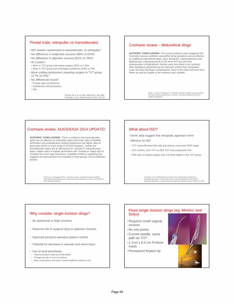
Fixed single incision slings (eg, MiniArc and Solyx)
• Requires small vaginal incision
• No exit points• Curved needle, same path as TOT
• 1.1cm x 8.5 cm Prolenemesh
• Permanent fixation tip
Page 49

Adjustable slings (eg, Ajust and Altis)In which patients might one consider using a single incision sling?
• Less severe SUI (no ISD)
• Very active, athletic patient
• Obese patient
• High anesthesia risk
• History of prior retropubic or pelvic surgery
• Office based procedure
• Physician comfort and familiarity
• Part of a research trial
Cochrane Review: Single-Incision Slings
AUTHORS' CONCLUSIONS: TVT-Secur is inferior to standard mid-urethral slings for the treatment of women with stress incontinence and has already been withdrawn from clinical use. Not enough evidence has been found on other single-incision slings compared with retropubic or transobturator slings to allow reliable comparisons. Additional adequately powered and high-quality trials with longer-term follow-up are required. Trials should clearly describe the fixation mechanism of these single-incisions slings: It is apparent that, although clubbed together as a single group, a significant difference in fixation mechanisms may influence outcomes.
Nambier A, Cody JD, Jeffrey ST. Single-incision sling operations for urinary incontinence in women. Cochrane Database Syst Rev. 2014;6:CD008708
Emerging data on single-incision slings
• 234 women randomized to MiniArc vs Monarc TOT
• 3 women in each arm had repeat surgery for SUI in f/u
• No significant difference in subjective outcomes (absence of SUI)
• No significant difference in objective outcomes (absence of USI or CST)
• Similar results on QoL questionnaires:
• ICIQ-UI
• ICIQ-OAB
• PISQ-12
• IIQ-7
• PGI-I
• No difference in pad weights at 6 months
• CONCLUSION: Two year results suggest comparable cure rates between MiniArc and
Monarc, with longer follow up planned
Lee J et al. Oral paper PP03 - MiniArc versus Monarc suburethral sling in women with stress urinary incontinence: an RCT with 24 month follow up. AUGS/IUGA 2014 Scientific Meeting
Emerging data on single-incision slings
• 100 women randomized to TVT-O vs Ajust single-incision sling
• No difference in subjective outcomes at 1 year
• No significant difference objective outcomes at 1 year (89.8% Ajust vs 87.2%
TVT-O with negative CST)
• Similar results on QoL questionnaires:
• ICIQ
• IQOL
• VAS scales
• Likert scales
• CONCLUSION: 1 year results suggest comparable subjective and objective outcomes
between TVT-O and Ajust slings
Masata J et al. Oral paper PP08 - Randomized trial of a comparison of the efficacy of TVT-O and single incision tape Ajust in the treatment of stress urinary incontinence in women – 1 year follow up. AUGS/IUGA 2014 Scientific Meeting
Adjustable Single-Incision Slings (eg, Altis and Ajust)
Page 50

Components: Anchors
• PP anchors with semi-flexible tines designed for secure retention in tissue
• Anchors are individually designed to maximize holding force and minimize tissue damage
• Allows for obturator membrane placement
Static
Dynamic
Components: Tensioning
• Dynamic anchor designed for two-way adjustability intra-operatively
• Dynamic anchor holding force designed to prevent sling movement from pelvic floor events during tissue in-growth period
Placement of an adjustable SIS
• Using inside-out technique and aim tip of introducer through dissected track towards “X” landmark
• Upon entry, ensure introducer is flush skin and handle is parallel with descending pubic ramus
• “Cephalad drift”
Placement of an adjustable SIS
• Advance introducer into internus fascia with a push until a slight “pop” is felt, avoiding a twisting motion
• Once the “pop” is felt, turn introducer approximately ¼ turn, advancing anchor through obturator membrane
• Do not over rotate
Placement of an adjustable SIS
• Remove introducer by rotating in opposite direction
• NOTE: Once anchor is placed into the tissue, anchor is not designed to be retracted or advanced further
Tensioning an adjustable SIS sling
• Tension sling by pulling suture loop across patient’s midline until desired support is achieved
Page 51

Tensioning and Positioning
• Sling placement under urethra without tension
• Metz may be inserted between sling and urethra to confirm no tension to urethra
Final Result of Tensioning
References• Listed on individual slides
Page 52

Management of Sling Complications
Charles R. Rardin, MDAssistant ProfessorBrown Medical School/Women and Infants’ HospitalOf Rhode IslandDivision of UrogynecologyProvidence, RI
Disclosures
I have no financial relationships to disclose.
Objectives
To review the anatomy of minimally invasive, midurethral slings
To describe the anatomic basis of complications of these slings
To review the prevention, diagnosis and management of these complications
Bladder Perforation
Bladder Perforation
Commonest complication for TVT
Reported incidence from 0.8 to 34%
Most commonly < 10%
Not effect the efficacy of the treatment BUT it is a major issue if it is missed
Treatment
Most surgeons drain pt for 24 to 48 hours with Foley catheter but nothing in literature to prove this
If grossly bloody must drain until hematuria clears – 3/95 (3%) had hematuria for 3-5 days (McLennan)
? No long term sequelae as long as injury not missed
Page 53

Intravesical Erosion? Delayed recognition
Does delayed erosion occur? Is it failed recognition initially or can it work its way
through with time? – unknown How close is too close - concern when mesh or
needle can be seen through the mucosa
Presenting symptoms Recurrent UTI Pain with voiding Urgency UI Microscopic hematuria of new onset
Treatment
Must do cystoscopy with persistent symptoms
Cystoscopic resection Can be difficult as need to apply traction to
one end and cut as close to mucosa as can and hope material retracts Baracat 2005 – 6 pts resected and one required 2
procedures
Resection of penetrating mesh
Combined endoscopic and cystoscopic Place laparoscopic trocar (or grasping needle)
through the dome of the bladder to grasp and apply tension to mesh – cut from below
Open laparotomy Open the bladder at the dome and directly
resect
Transurethral Erosion/Placement
Urinary Retention Voiding Dysfunction
immediately post-op r/o hematoma d/c with Foley 24-72 hr
If patient still unable to void at 3-10 days: consider reopening site under
local, place right-angle under mesh, and pull down slightly 5-10 mm
Page 54

Urinary Retention If beyond ~ 10 days
consider cutting mesh under local in midline after 4 weeks of catheterization
All cases of impaired emptying were completely resolved
Irritative symptoms resolved (30%) or improved (70%)
61% patients remained continent 26% were improved over
baseline 13% had recurrence of stress
incontinence CR Rardin, Obstet Gynecol 2002
Management of Retention
Try to determine whether the problem is over-correction Negative Qtip angle “speed bump” on catheterization or scope Elevated pDet on pressure-flow studies
If any of these are present, consider releasing sling
If none are present – trial of sacral neuromodulation?
Will they be continent?
Sling release for refractory voiding dysfunction Laurikainen. Int Urogynecol J 2006; 17:119
50/9040 (0.6%) – 82% relief; 24/50 (49%) continent
Rardin. Obstet Gynecol 2002;100:898 23/1175 (1.9%) – 100% relief; 14/23 (61%) continent
Karram. Obstet Gynecol 2003;101;929 6/350 (1.7%) – 100% relief; 1/6 (33%) continent
Klutke. Urology 2002;58:697 17/600 (2.8%) – 100 relief; 16/17 (94%) continent
Bowel Perforation Healthy 56-year old woman
with SUI for 3 years
History includes RSO, followed by TAH/LSO
Uneventful TVT with cystoscopy
3 hours postop: severe abdominal pain; laparoscopy performed
Meschia M, Int Urogyn J 2002
Bowel Perforation “We think that the
occurrence of this complication could be related to incorrect insertion of the needle which was brought up to the abdominal wall instead of in close contact with the pubic bone.”
Meschia M, Int Urogyn J 2002
Bowel Perforation
A
Page 55

The Relationship of TVT to the Vascular Anatomy of the Retropubic Space
Study performed on 10 fresh cadavers
Measured distance from the needle to vessel
Conclusion: “If the TVT needle is laterally directed or externally rotated in the course of insertion, major vascular injury may result”
T.W. Muir et al. Paper presentation, 22nd
Annual Meeting, AUGS, Oct. 2001
Vascular Injury
Vaginal Mesh Erosions
Occurs with TVT and TOT but incidence much higher in literature with TOT Material
Dissection
Location
Mesh issues Extrusion
Chronic infection
Risk Factors & Clinical Presentation
Risk factors No correlation to age, BMI, previous surgery,
concomitant surgery Type of mesh
Presentation 1 – 45 weeks, even longer Early ones may be incorrect placement – i.e. though
the lateral vaginal wall Vaginal bleeding Persistent discharge Dyspareunia Asymptomatic – noted on exam
Treatment
Prevention Make sure you don’t perforate laterally especially
TOT TOT – penetrate at the appropriate place ? Well estrogenized
Treatment ? Local estrogen Local removal in office – cut piece protruding
Depends on material – multifilaments are chronically infecteed
OR Removal of piece exposed and suture closed Removal of whole sling – especially important with solid
mesh and mesh with small pore size – usually vaginal Occasionally extensive exploration, debridement and removal
Page 56

Pain syndrome – obturator neuralgia
Postoperative pain
Incidence is unclear – often not addressed in the literature
Long-term followup of TOT procedures: up to 12% of patients with pain issues (Giberti, J Urol 2007)
Mesh kits – more mesh in these compartments
Adductor longus
Adductor brevis
Adductor magnus
Obturator internis and externis
Gracilis
Anatomy of the Obturator space
Obturator Sling – Pain issues
Anatomy review
Hallmarks of Obturator Neuralgia
Shooting or sharp pain of the affected groin; radiations in a variety of directions
Often delayed onset
Exacerbated by activity; relieved with rest
May have vaginal and/or groin tenderness
Treatment of Obturator Pain
Observation
Physical Therapy
Gabapentin
Kenalog/marcaine injections
Revision or excision
Surgical Treatment of Obturator Pain
Page 57

Salvage Procedures for Refractory SUI
Salvage Procedures for Refractory SUI
Beri Ridgeway, MDAssistant Professor of Surgery
Cleveland Clinic Lerner College of Medicine
DisclosuresDisclosures
I have no financial relationships to disclose.
ObjectivesObjectives
• Discuss considerations for patient with refractory SUI
• Describe salvage procedures for refractory SUI
- Urethra bulking agents
- Repeat sling
- Burch urethropexy
- Autologous fascial sling
- Obstructing sling
- Diversion
• Discuss pros and cons of salvage procedures
Patient with Refractory SUIPatient with Refractory SUI
• Verify diagnosis
- Thorough history
- Physical examination
- Multichannel urodynamics
- Consider cystoscopy
• Consider alternative diagnoses
- Urge incontinence
- Fistula
- Ectopic ureter
Urethral BulkingUrethral Bulking
Rationale for Urethral Bulking:Why it Works
Rationale for Urethral Bulking:Why it Works
• Increases coaptation of the urethra by augmenting submucosal layer
• Increases compressive force inward toward the urethral lumen
• Theoretically should only work in cases where there is at least some component of ISD
- Most SUI has some component of urethral incompetence
Page 58

Urethral Bulking AgentsSubmucosal Placement
Urethral Bulking AgentsSubmucosal Placement
Increased urethral resistance against rises in abdominal pressure
Treating SUI with Bulking AgentsTreating SUI with Bulking Agents
• Registration studies for most agents are in the “ISD” population defined by leak point pressures +urethral mobility
• Results for all approved agents are similar
- 25-35% cure, 25-50% improvement, 25-40% failure
• Gluteraldhyde cross-linked bovine collagen (Contigen®) was the first popular agent
- No longer manufactured
• Carbon coated zirconium beads Durasphere®
• Calcium hydroxylapatite Coaptite®
• Silicone Macroparticles Macroplastique®
Bulking Agents as an Option:When to Use
Bulking Agents as an Option:When to Use
• Patient decisions on treatment are related to expectations and level of risk
• Although improvement more likely than cure, safety and lack of convalescence remains the main advantage
• No perfect agent, nothing yet proven superior
Urethral Bulking: HowInjection Technique
Urethral Bulking: HowInjection Technique
• Can be transurethral or periurethral
• Most often done with local anesthesia in an office setting
• Injection quantity vary by product
- Err on the side of “too little” vs. “too much”
- Can re-inject at 2-4 weeks
• Modest or transient improvement after first injection is not a bad sign – just need a bit more
Transurethral TechniqueTransurethral Technique
Page 59

Complications of Bulking Agents
Complications of Bulking Agents
• Hematuria, pain• UTI• Transient voiding dysfunction or
retention• Extrusion of material• Granulomatous reaction• Prolapse of urethral mucosa• Urethrovaginal fistula- rare in normal
tissue
• Retrospective study
• Patients retreated after failed midurethral sling
- N=165
• Repeat sling or urethral bulking
- 46% of subjects noted significant improvement after bulking
- Significantly less that repeat sling
Repeat SlingRepeat Sling• Repeat sling does work
- 54% of subjects
• Doesn’t work as well as primary sling cohort
• Significant QOL improvement even if deemed “failure” by criteria
• No clear advantage over sling type for repeat procedure
• Underpowered
Repeat slingRepeat sling
• No need to excise previous sling- However, I will excise if there is any history of
voiding dysfunction or urge component
• Sling choice- I usually will use a retropubic sling in these
situations
Page 60

Burch UrethropexyBurch Urethropexy
Burch UrethropexyBurch Urethropexy
• 1st described in 1961
• Modification of MMK
• Endopelvic fascia to Cooper’s ligament
Brubaker et al. NEJM 354:1557, 2006. Burch. AJOG 81: 281, 1961.
Brubaker
A
C D
B
Hurt
A: Dissect open the space of
Retzius
B: Use permanent sutures:
- 1st stitch: 2 cm lateral to
proximal third of urethra
- 2nd stitch: 1 cm lateral & 1 cm
cephalad to 1st stitch
- Cystoscopy
C: Place sutures through
Cooper’s ligament
D: Tie with suture bridge to
stabilize a hypermobile urethra
Page 61

• Cochrane Review: Open retropubic colposuspension
(Burch and MMK) is an effective long-term treatment
- 1 year cure: 85-90%
- 5 year cure: 70%
• No good studies evaluating outcomes in recurrent SUI
- Expert Opinion: works best if urethral hypermobility present
Procedure 1 year 5 year
Burch urethropexy 89% 82%
Pereyra needle suspension 65% 43%
Kelly plication 63% 37%
Bergman & Elia. AJOG 173: 66, 1995. Lapitan et al. Cochrane Review, 2012.
ComplicationsComplications
• Bladder perforation1 2%
• Detrusor overactivity1 5 - 27%
• Unique complications2
- Osteitis pubis: painful inflammatory, noninfectious disease, which is self-limited
• Treat with rest, NSAIDs, and physical therapy
- Osteomyelitis: infectious disease requiring extended course of antibiotics
1. ACOG Practice Bulletin, June 2005. 2. Frederick et al. Urology 64:669, 2004.
Autologous Fascial SlingAutologous Fascial Sling
Pubovaginal SlingPubovaginal Sling
• Sling placed at the level of bladder neck
• Sling extends into the retropubic space on both sides
• Can be done with Autologous fascia, allograft, xenograft or synthetic material
Page 62

Autologous Fascia SlingsAutologous Fascia Slings
• Morgan et al reported an 85% cure rate in patients with at least 5 years of follow-up
• Chaikin et al reported a 95% cure rate in 20 patients with at least 10 years of follow-up
Autologous Fascia Sling Outcomes
Autologous Fascia Sling Outcomes
Author Year Op Pts F/U(months)
# Eval Continence
Morgan 2000 autoRF 247 51 235 92
Chaikin 1998 autoRF 251 37 251 92
Hassoun Ghoniem
1998 autoRF 112 41 78 78
Zaragoza 1996 autoRF 60 25 60 95
Haab 1997 autoRF/FL 40 48 37 46
Kaplan 1996 autoRF/VWS
79 23 79 96
Govier 1997 autoFL 32 14 32 87
If Pubovaginal Slings Work So Well, Why Use Anything Else?
If Pubovaginal Slings Work So Well, Why Use Anything Else?
• Morbidity- Fascial harvest and infection site risk- Dissection- Bleeding risk- ?Denervation- Pain and Hernia Risk
• Time• Durability with activity:….. “I felt a
pop..and then leaked”
Proportion of Patients with Treatment Failure at 2 Years, According to Overall Composite Criteria, Composite Criteria Specific to Stress Incontinence, and Other Criteria
Burch vs Autologous Fascia Sling
Albo ME et al. N Engl J Med 2007;356:2143-2155
Albo ME et al. N Engl J Med 2007;356:2143-2155
Page 63
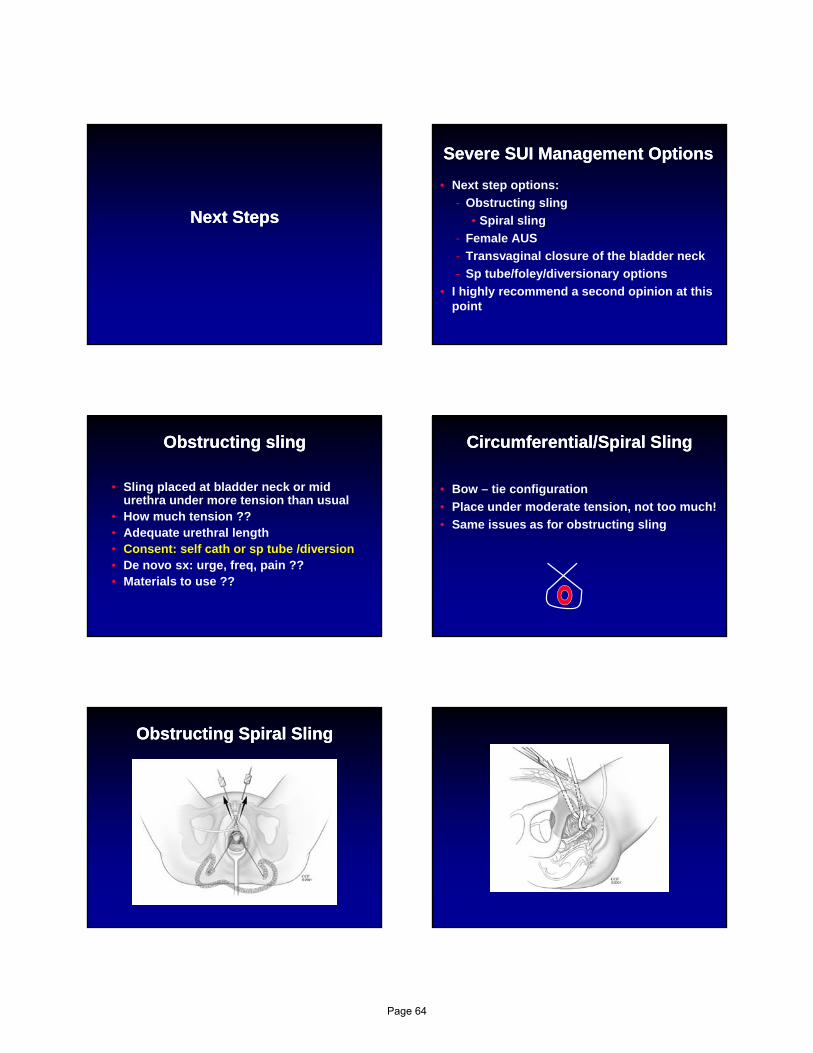
Next StepsNext Steps
Severe SUI Management OptionsSevere SUI Management Options
• Next step options:
- Obstructing sling
• Spiral sling
- Female AUS
- Transvaginal closure of the bladder neck
- Sp tube/foley/diversionary options
• I highly recommend a second opinion at this point
Obstructing slingObstructing sling
• Sling placed at bladder neck or mid urethra under more tension than usual
• How much tension ??• Adequate urethral length• Consent: self cath or sp tube /diversion• De novo sx: urge, freq, pain ??• Materials to use ??
Circumferential/Spiral SlingCircumferential/Spiral Sling
• Bow – tie configuration
• Place under moderate tension, not too much!
• Same issues as for obstructing sling
Obstructing Spiral SlingObstructing Spiral Sling
Page 64

Female AUSFemale AUS
• Not approved for use in USA currently
• Severe SUI• Issues with AUS in
females:- Erosion- Mechanical failure- Infection- Device use
• Indications ???
Transvaginal Closure of BNIndications
Transvaginal Closure of BNIndications
• Severe incontinence (failed surgeries)
• Destroyed urethra from indwelling Foley
• Trauma (surgical or non surgical)
• Congenital
• Neurogenic bladder (para,quad, MMC) with inability to do urethral ISC
• Urinary retention with difficulties to do urethral ISC
Page 65

Urinary DrainageUrinary Drainage
• Permanent suprapubic catheter
• Incontinent vesicostomy
• Continent vesicostomy
• Incontinent stoma (small bowel)
• Continent augmentation
• Upper tract diversion (Bricker or continent diversion)
Diversion of UrineDiversion of Urine
- Suprapubic catheter
- Continent or incontinent vesicostomy
- Continent or incontinent augmentation
- Continent or incontinent urinary diversion
ConclusionsConclusions
• Verify diagnosis – history, physical examination, urodynamics and ?cystoscopy
- UI severity and type
- Urethral hypermobility
• Bulking
• Repeat sling
• Burch
• Fascial sling
• Obstructive sling, closure of bladder neck, diversion
ReferencesReferences
References, cont.References, cont.
Page 66
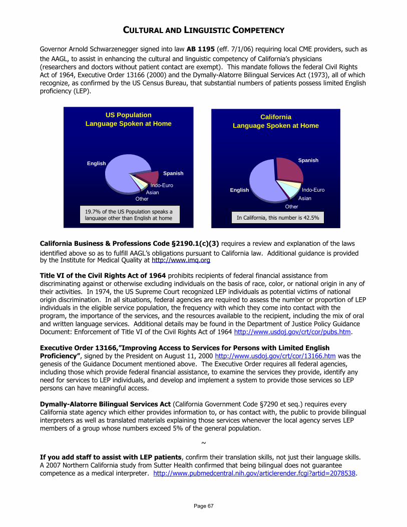
CULTURAL AND LINGUISTIC COMPETENCY Governor Arnold Schwarzenegger signed into law AB 1195 (eff. 7/1/06) requiring local CME providers, such as
the AAGL, to assist in enhancing the cultural and linguistic competency of California’s physicians
(researchers and doctors without patient contact are exempt). This mandate follows the federal Civil Rights Act of 1964, Executive Order 13166 (2000) and the Dymally-Alatorre Bilingual Services Act (1973), all of which
recognize, as confirmed by the US Census Bureau, that substantial numbers of patients possess limited English proficiency (LEP).
California Business & Professions Code §2190.1(c)(3) requires a review and explanation of the laws
identified above so as to fulfill AAGL’s obligations pursuant to California law. Additional guidance is provided by the Institute for Medical Quality at http://www.imq.org
Title VI of the Civil Rights Act of 1964 prohibits recipients of federal financial assistance from
discriminating against or otherwise excluding individuals on the basis of race, color, or national origin in any of their activities. In 1974, the US Supreme Court recognized LEP individuals as potential victims of national
origin discrimination. In all situations, federal agencies are required to assess the number or proportion of LEP individuals in the eligible service population, the frequency with which they come into contact with the
program, the importance of the services, and the resources available to the recipient, including the mix of oral
and written language services. Additional details may be found in the Department of Justice Policy Guidance Document: Enforcement of Title VI of the Civil Rights Act of 1964 http://www.usdoj.gov/crt/cor/pubs.htm.
Executive Order 13166,”Improving Access to Services for Persons with Limited English
Proficiency”, signed by the President on August 11, 2000 http://www.usdoj.gov/crt/cor/13166.htm was the genesis of the Guidance Document mentioned above. The Executive Order requires all federal agencies,
including those which provide federal financial assistance, to examine the services they provide, identify any
need for services to LEP individuals, and develop and implement a system to provide those services so LEP persons can have meaningful access.
Dymally-Alatorre Bilingual Services Act (California Government Code §7290 et seq.) requires every
California state agency which either provides information to, or has contact with, the public to provide bilingual
interpreters as well as translated materials explaining those services whenever the local agency serves LEP members of a group whose numbers exceed 5% of the general population.
~
If you add staff to assist with LEP patients, confirm their translation skills, not just their language skills.
A 2007 Northern California study from Sutter Health confirmed that being bilingual does not guarantee competence as a medical interpreter. http://www.pubmedcentral.nih.gov/articlerender.fcgi?artid=2078538.
US Population
Language Spoken at Home
English
Spanish
AsianOther
Indo-Euro
California
Language Spoken at Home
Spanish
English
OtherAsian
Indo-Euro
19.7% of the US Population speaks a language other than English at home In California, this number is 42.5%
Page 67