Embed Size (px)
Citation preview
Aus dem Bereich der Experimentellen Neurologie
Theoretische Medizin und Biowissenschaften der Medizinischen Fakultät
der Universität des Saarlandes Homburg/Saar
Die Bedeutung von Phospholipiden und oxidierten Lipiden für die Prozessierung des Amyloid-Vorläufer Proteins
(APP) und die Alzheimer Krankheit
Dissertation zur Erlangung des Grades einer Doktorin der Naturwissenschaften
der Medizinischen Fakultät
der UNIVERSITÄT DES SAARLANDES
2016
vorgelegt von
Viola J. Haupenthal
geboren am 24. August 1982 in Saarbrücken
Inhaltsverzeichnis Seite
(A) Auflistung des eigenen Anteils und des Anteils von Mitautoren an den Publikationen 1
(B) Abkürzungen ................................................................................................................... 5
(1) Zusammenfassung ........................................................................................................... 7
(1.1) Die Bedeutung von Phospholipiden und oxidierten Lipiden für die Prozessierung
des Amyloid-Vorläufer Proteins (APP) und die Alzheimer Krankheit ..................... 7
(1.2) The importance of phospholipids and oxidized lipids in Amyloid Precursor Protein
processing (APP) and Alzheimer´s disease .............................................................. 9
(2) Einleitung ............................................................................................................................ 11
(2.1) Die Alzheimer Krankheit – Grundlagen und Epidemiologie ........................................ 11
(2.2) Neurobiologische Grundlagen der Alzheimer Krankheit ............................................. 13
(2.3) Fragestellung der Arbeit .............................................................................................. 17
(3) Methoden ........................................................................................................................... 18
(3.1) Methoden der Zellkultur .............................................................................................. 18
(3.1.1) Kultivierung eukaryotischer Zellen ....................................................................... 18
(3.1.2) Präparation von primären kortikalen Mausneuronen ......................................... 18
(3.1.3) Inkubation mit Phospholipiden ............................................................................ 19
(3.1.4) Inkubation mit PL .................................................................................................. 19
(3.1.5) Inkubation mit oxidierten Lipiden, Lipidperoxidationsprodukten oder DHA ....... 19
(3.1.6) Bestimmung der Zytotoxizität .............................................................................. 20
(3.2) Proteinanalytik ............................................................................................................. 21
(3.2.1) Bestimmung der Proteinkonzentration ................................................................ 21
(3.2.2) Herstellung von Zell-/ Gewebe- Homogenaten .................................................... 21
(3.2.3) Herstellung von Zelllysaten ................................................................................... 22
(3.2.4) Aufarbeitung humaner post-mortem Gehirnproben ............................................ 23
(3.2.5) Post-nukleäre Fraktionierung und Isolation von Membranen ............................. 23
(3.2.6) In vitro Inkubationen............................................................................................. 24
(3.2.7) Bestimmung der β- und γ-Sekretase Aktivität nach in vitro Inkubation .............. 24
(3.2.8) Bestimmung der Sekretase Aktivität auf lebenden Zellen ................................... 25
(3.2.9) Bestimmung der α-Sekretase- und ADAM-10 Aktivität nach in vitro Inkubation 25
(3.2.10) Bestimmung des Aβ, sAPPβ und sAPPα Gehalts ................................................ 26
(3.2.11) Bestimmung des ADAM-17, BACE-1 und PS-1 Gehalts ...................................... 27
(3.3.) Lipidanalytik ................................................................................................................ 28
(3.3.1) Extraktion von Lipiden aus humanen post-mortem Gehirnen ............................. 28
(3.3.2) Messung der DHA-Stabilität.................................................................................. 28
(3.3.3) Bestimmung des Lipidgehalts durch Massenspektrometrie ................................ 28
(3.3.4) Bestimmung des Phospholipidgehalts mittels Dünnschichtchromatographie .... 29
(3.3.5) Bestimmung des HNE-Gehalts .............................................................................. 30
(3.3.6) Bestimmung der Lipidperoxidation ...................................................................... 30
(3.4) RNS-Analytik ................................................................................................................. 31
(3.4.1) Isolation von RNS .................................................................................................. 31
(3.4.1) cDNS-Sythese und Echtzeitpolymerasekettenreaktion (RT-PCR) ......................... 31
(3.5) Statistische Auswertung .............................................................................................. 32
(4) Publikationen ...................................................................................................................... 33
(4.1) Zusammenfassung von Publikation 1 und Beschreibung des Eigenanteils ................. 33
(4.2) Zusammenfassung von Publikation 2 und Beschreibung des Eigenanteils ................. 69
(4.3) Zusammenfassung von Publikation 3 und Beschreibung des Eigenanteils ................. 87
(5) Erweiterte Zusammenfassung .......................................................................................... 102
(5.1) Lipide in der AD .......................................................................................................... 102
(5.2) Im Kontext: Phospholipide, DHA und oxidierte Lipide in der APP Prozessierung und
ihre Rolle für die AD ............................................................................................ 104
(6) Zusammenfassung und Ausblick ....................................................................................... 117
(7) Beteiligung an weiteren Publikationen ............................................................................. 119
(8) Literaturverzeichnis .......................................................................................................... 121
(C) Danksagung ....................................................................................................................... 133
Kooperationen
1
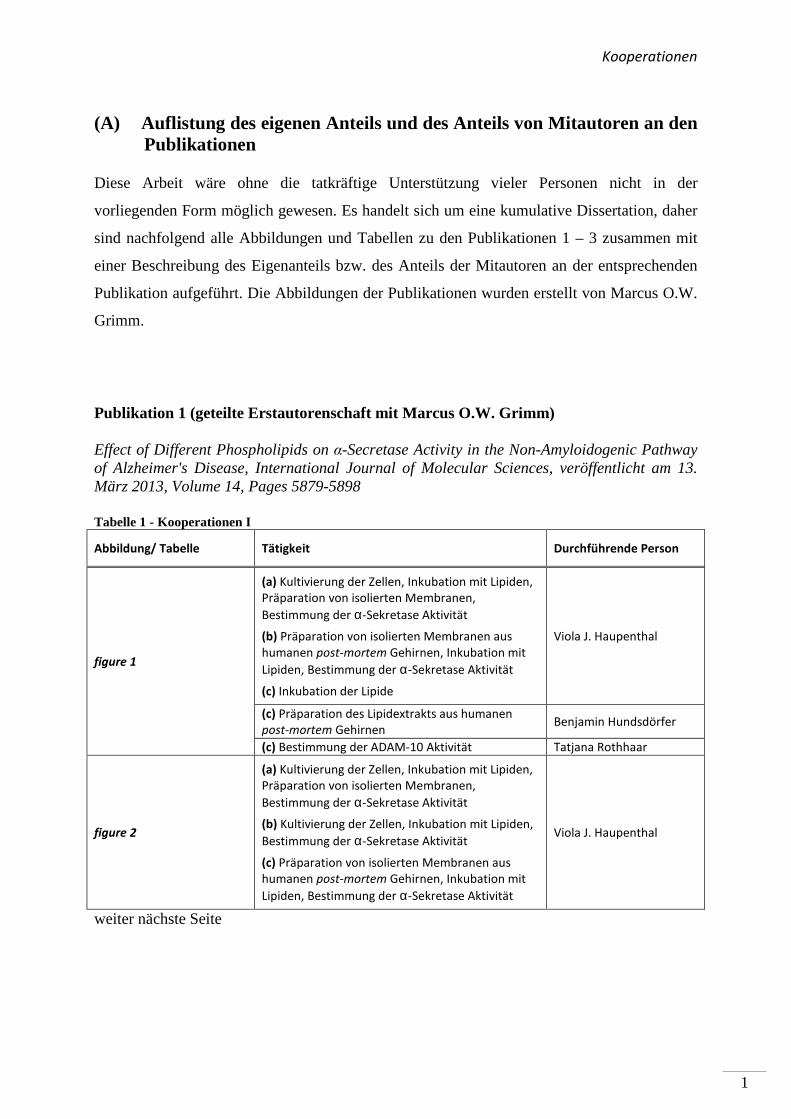
(A) Auflistung des eigenen Anteils und des Anteils von Mitautoren an den Publikationen
Diese Arbeit wäre ohne die tatkräftige Unterstützung vieler Personen nicht in der
vorliegenden Form möglich gewesen. Es handelt sich um eine kumulative Dissertation, daher
sind nachfolgend alle Abbildungen und Tabellen zu den Publikationen 1 – 3 zusammen mit
einer Beschreibung des Eigenanteils bzw. des Anteils der Mitautoren an der entsprechenden
Publikation aufgeführt. Die Abbildungen der Publikationen wurden erstellt von Marcus O.W.
Grimm.
Publikation 1 (geteilte Erstautorenschaft mit Marcus O.W. Grimm)
Effect of Different Phospholipids on α-Secretase Activity in the Non-Amyloidogenic Pathway of Alzheimer's Disease, International Journal of Molecular Sciences, veröffentlicht am 13. März 2013, Volume 14, Pages 5879-5898
Tabelle 1 - Kooperationen I
Abbildung/ Tabelle Tätigkeit Durchführende Person
figure 1
(a) Kultivierung der Zellen, Inkubation mit Lipiden,
Präparation von isolierten Membranen,
Bestimmung der α-Sekretase Aktivität
(b) Präparation von isolierten Membranen aus
humanen post-mortem Gehirnen, Inkubation mit
Lipiden, Bestimmung der α-Sekretase Aktivität
(c) Inkubation der Lipide
Viola J. Haupenthal
(c) Präparation des Lipidextrakts aus humanen
post-mortem Gehirnen Benjamin Hundsdörfer
(c) Bestimmung der ADAM-10 Aktivität Tatjana Rothhaar
figure 2
(a) Kultivierung der Zellen, Inkubation mit Lipiden,
Präparation von isolierten Membranen,
Bestimmung der α-Sekretase Aktivität
(b) Kultivierung der Zellen, Inkubation mit Lipiden,
Bestimmung der α-Sekretase Aktivität
(c) Präparation von isolierten Membranen aus
humanen post-mortem Gehirnen, Inkubation mit
Lipiden, Bestimmung der α-Sekretase Aktivität
Viola J. Haupenthal
weiter nächste Seite
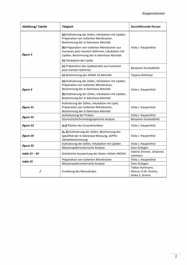
Kooperationen
2
Abbildung/ Tabelle Tätigkeit Durchführende Person
figure 3
(a) Kultivierung der Zellen, Inkubation mit Lipiden,
Präparation von isolierten Membranen,
Bestimmung der α-Sekretase Aktivität
(b) Präparation von isolierten Membranen aus
humanen post-mortem Gehirnen, Inkubation mit
Lipiden, Bestimmung der α-Sekretase Aktivität
(c) Inkubation der Lipide
Viola J. Haupenthal
(c) Präparation des Lipidextrakts aus humanen
post-mortem Gehirnen Benjamin Hundsdörfer
(c) Bestimmung der ADAM-10 Aktivität Tatjana Rothhaar
figure 4
(a) Kultivierung der Zellen, Inkubation mit Lipiden,
Präparation von isolierten Membranen,
Bestimmung der α-Sekretase Aktivität
(b) Kultivierung der Zellen, Inkubation mit Lipiden,
Bestimmung der α-Sekretase Aktivität
Viola J. Haupenthal
figure S1
Kultivierung der Zellen, Inkubation mit Lipid,
Präparation von isolierten Membranen,
Bestimmung der α-Sekretase Aktivität
Viola J. Haupenthal
figure S2 Aufarbeitung der Proben Viola J. Haupenthal
Dünnschichtchromatographische Analyse Benjamin Hundsdörfer
figure S3 (a-j) Plotten der Enzymkinetiken Viola J. Haupenthal
figure S4
(a, b) Kultivierung der Zellen, Bestimmung der
Spezifität der α-Sekretase Messung, sAPPα-
Gehaltsbestimmung
Viola J. Haupenthal
figure S5 Kultivierung der Zellen, Inkubation mit Lipiden Viola J. Haupenthal
Massenspektrometrische Analyse Sven Grösgen
table S1 – S4 Statistische Auswertung der Daten mittels ANOVA Valerie Zimmer, Johannes
Lehmann
table S5 Präparation von isolierten Membranen Viola J. Haupenthal
Massenspektrometrische Analyse Sven Grösgen
/ Erstellung des Manuskripts
Tobias Hartmann,
Marcus O.W. Grimm,
Heike S. Grimm
Kooperationen
3
Publikation 2 (Mitautorenschaft)
Plasmalogens Inhibit APP Processing by Directly Affecting γ-secretase Activity in Alzheimer's Disease, The Scientific World Journal, veröffentlicht am 01. April 2012, Volume 2012, Article ID 141240, 15 Pages
Tabelle 2 - Kooperationen II
Abbildung/ Tabelle Tätigkeit Durchführende Person
table 2
Aufarbeitung der humanen post-mortem
Gehirnproben Tatjana L. Rothhaar
Massenspektrometrische Analyse Sven Grösgen
table 3 Kultivierung der Zellen, Inkubation der Lipide Tatjana L. Rothhaar
Real Time PCR Analyse Sven Grösgen
figure 2
Kultivierung der Zellen, Inkubation mit Lipiden,
Aufarbeitung der Proben Tatjana L. Rothhaar
Bestimmung des ADAM-17, BACE-1, PS1-Gehalts Verena K. Burg
figure 3, 4, 6
Inkubation mit Lipiden, Präparation von isolierten
Membranen, Messung der β- bzw. γ-Sekretase
Aktivität
Tatjana L. Rothhaar
figure 5
Herstellung PNFs Tatjana L. Rothhaar
Inkubation mit Lipiden, Präparation von isolierten
Membranen, Bestimmung der α-Sekretase
Aktivität
Viola J. Haupenthal
figure S1 Kultivierung der Zellen, Aufarbeitung der Proben,
Bestimmung der Zytotoxizität Tatjana L. Rothhaar
figure S2
Herstellung PNFs Tatjana L. Rothhaar
Präparation von isolierten Membranen, Inkubation
mit Inhibitoren, Bestimmung der α-
Sekretaseaktivität
Viola J. Haupenthal
figure S3 & S4
Präparation von isolierten Membranen, Inkubation
mit Inhibitoren, Bestimmung der β- & γ-
Sekretaseaktivität
Tatjana L. Rothhaar
/ Erstellung des Manuskripts
Tobias Hartmann,
Marcus O.W. Grimm,
Heike S. Grimm,
Mattias Riemenschneider,
Benjamin Hundsdörfer,
Janine Mett
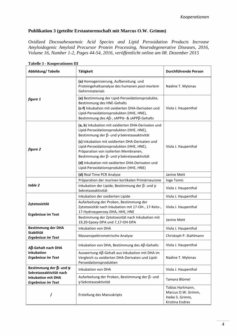
Kooperationen
4
Publikation 3 (geteilte Erstautorenschaft mit Marcus O.W. Grimm)
Oxidized Docosahexaenoic Acid Species and Lipid Peroxidation Products Increase Amyloidogenic Amyloid Precursor Protein Processing, Neurodegenerative Diseases, 2016, Volume 16, Number 1-2, Pages 44-54, 2016, veröffentlicht online am 08. Dezember 2015
Tabelle 3 - Kooperationen III
Abbildung/ Tabelle Tätigkeit Durchführende Person
figure 1
(a) Homogenisierung, Aufbereitung und
Proteingehaltsanalyse des humanen post-mortem
Gehirnmaterials
Nadine T. Mylonas
(a) Bestimmung der Lipid-Peroxidationsprodukte,
Bestimmung des HNE-Gehalts
(c-f) Inkubation mit oxidierten DHA-Derivaten und
Lipid-Peroxidationsprodukten (HHE, HNE),
Bestimmung des Aβ-, sAPPα- & sAPPβ-Gehalts
Viola J. Haupenthal
figure 2
(a, b) Inkubation mit oxidierten DHA-Derivaten und
Lipid-Peroxidationsprodukten (HHE, HNE),
Bestimmung der β- und γ-Sekretaseaktivität
(c) Inkubation mit oxidierten DHA-Derivaten und
Lipid-Peroxidationsprodukten (HHE, HNE),
Präparation von isolierten Membranen,
Bestimmung der β- und γ-Sekretaseaktivität
(d) Inkubation mit oxidierten DHA-Derivaten und
Lipid-Peroxidationsprodukten (HHE, HNE)
Viola J. Haupenthal
(d) Real Time PCR Analyse Janine Mett
table 2
Präparation der murinen kortikalen Primärneurone Inge Tomic
Inkubation der Lipide, Bestimmung der β- und γ-
Sekretaseaktivität Viola J. Haupenthal
Zytotoxizität
Ergebnisse im Text
Inkubation der oxidierten Lipide Viola J. Haupenthal
Aufarbeitung der Proben, Bestimmung der
Zytotoxizität nach Inkubation mit 17-OH-, 17-Keto-,
17-Hydroxyperoxy-DHA, HHE, HNE
Viola J. Haupenthal
Bestimmung der Zytotoxizität nach Inkubation mit
19,20-Epoxy-DPA und 7,17-OH-DPA Janine Mett
Bestimmung der DHA
Stabilität
Ergebnisse im Text
Inkubation von DHA Viola J. Haupenthal
Massenspektrometrische Analyse Christoph P. Stahlmann
Aββββ-Gehalt nach DHA
Inkubation
Ergebnisse im Text
Inkubation von DHA, Bestimmung des Aβ-Gehalts Viola J. Haupenthal
Auswertung Aβ-Gehalt aus Inkubation mit DHA im
Vergleich zu oxidierten DHA-Derivaten und Lipid-
Peroxidationsprodukten
Nadine T. Mylonas
Bestimmung der ββββ- und γγγγ-
Sekretaseaktivität nach
Inkubation mit DHA
Ergebnisse im Text
Inkubation von DHA Viola J. Haupenthal
Aufarbeitung der Proben, Bestimmung der β- und
γ-Sekretaseaktivität Tamara Blümel
/ Erstellung des Manuskripts
Tobias Hartmann,
Marcus O.W. Grimm,
Heike S. Grimm,
Kristina Endres
Abkürzungen
5
(B) Abkürzungen
°C Grad Celsius
µM micro Molar
AA Arachidonsäure
Aβ Amyloid β
AD Alzheimer Krankheit
ADAM α-Sekretase A-Disintegrin-and-metalloprotease
AICD intrazelluläre APP-Domäne
Aph engl. Anterior pharynx-defective
APO E Apolipoprotein E
APP Amyloid-Vorläufer Protein
BACE β-Sekretase
BSA Bovine serum albumin
COX Cyclooxigenase
CTF C-terminale Domäne von APP
DHA Docosahexaensäure
DMEM Dublecco´s modified eagle medium
DNS Desoxyribonukleinsäure
ECE Endothelin konvertierendes Enzym
ECL Verstärkte Chemolumineszenz
EDTA Ethylendiamintetraessigsäure
EGTA Ethylenglycoltetraessigsäure
EOAD Frühe Form der Alzheimer Krankheit
EPA Eicosapentaensäure
FAD Familiäre Form der Alzheimer Krankheit
FCS Fetales Kälberserum
g/g Gramm / Erdbeschleunigung
GSK3β Glykogensynthase-Kinase 3
h Stunden
HBSS Salzhaltige Pufferlösung für Primärneurone
HDL Lipoprotein mit hoher Dichte
HHE 4-Hydroxyhexenal
HMGCR 3-Hydroxy-3-Methylglutaryl-Coenzym-A-Reduktase
Abkürzungen
6
HNE 4-Hydroxynonenal
HPLC Hochleistungsflüssigkeitschromatographie
IDE Insulin degradierendes Enzym
IP Immunopräzipitation
LDH Lactatdehydrogenase-Messung
LDL Lipoprotein mit niedriger Dichte
LOAD Späte Form der Alzheimer Krankheit
LOX Lipoxigenase
LRP1 Lipoproteinrezeptor 1
Min Minute
Me Methanol
mM millimolar
NP-40 Nonidet-P-40 Lösung
NPD-1 Neuroprotektin-D1
NTF Neurofibrilläre Bündel
OD Optische Dichte
P p-Wert, Signifikanzniveau
PBS Phosphatgepufferte Salzlösung
PC Phosphatidylcholin
PE Phosphatidylethanolamin
PL Plasmalogen
PS Phosphatidylserin
PSEN engl. Presenilin enhancer
RIP Regulierte Intramembran Proteolyse
RNS Ribonukleinsäure
RT Raumtemperatur
RT-PCR Echtzeit-Polymerasekettenreaktion
SAD Sporadische Form der Alzheimer Krankheit
sAPP lösliches APP
s Sekunde
SDS Natrium-Dodecylsulfat
PAGE Polyakrylamid-Gelelektrophorese
SPT Serinpalmitoyl-Coenzym A-Synthase
TBA Thiobarbitursäure
wt Wildtyp
Zusammenfassung
7
(1) Zusammenfassung
(1.1) Die Bedeutung von Phospholipiden und oxidierten Lipiden für die Prozessierung des Amyloid-Vorläufer Proteins (APP) und die Alzheimer Krankheit
Die molekularen Grundlagen der Alzheimer Krankheit (AD) werden intensiv erforscht. Eine
besondere Bedeutung kommt dabei der proteolytischen Prozessierung des Amyloid-Vorläufer
Proteins (APP) zu. Die amyloidogene Prozessierung des APP durch die β- und γ-Sekretase
resultiert in der Bildung des Amyloid-β (Aβ)-Peptids. Aβ stellt ein Hauptbestandteil der Aβ-
Plaques dar, welche neben den Neurofibrillenbündeln, ein Kennzeichen der Alzheimer
Krankheit sind. Die nicht amyloidogene APP Prozessierung durch die α- und γ-Sekretase
verhindert die Aβ-Bildung. Da die Lipidzusammensetzung der Membran die Sekretasen und
APP beeinflussen kann, wurden in dieser Arbeit Phospholipide und oxidierte Lipide
hinsichtlich ihrer Wirkung auf die APP Prozessierung analysiert.
In Publikation 1 wurde der Einfluss von Phospholipiden auf die Aktivität der α-Sekretase im
nicht amyloidogenen Weg der APP Prozessierung betrachtet. Es wurden Phospholipide
verwendet, welche sich durch unterschiedliche Fettsäuren, hinsichtlich Kettenlänge,
Sättigungsgrad und Position der Doppelbindung unterschieden. Zusätzlich wurden
Phospholipide mit unterschiedlichen Kopfgruppen betrachtet. In Experimenten mit entweder
lebenden SH-SY5Y Zellen oder isolierten Membranen dieser Zellen aktivierten kurzkettige,
gesättigte Phospholipide die α-Sekretase, während langkettige, gesättigte Phospholipide
keinen Effekt auf die Aktivität hatten. Langkettige, ungesättigte Phospholipide hingegen
steigerten die Aktivität der α-Sekretase. Die aktivierende Wirkung von gesättigten,
kurzkettigen und ungesättigten, langkettigen Phospholipiden auf die α-Sekretase konnte in
einem ex vivo Ansatz zusätzlich in Membranen aus humanen post-mortem Gehirnen
nachgewiesen werden. Der Effekt der verwendeten Phospholipide wurde auf die α-Sekretase
ADAM-10 zurückgeführt. Die Variation der Kopfgruppe und die Position der Doppelbindung
innerhalb der Fettsäuren der Phospholipide hatten schwache oder keine Effekte auf die
Aktivität der α-Sekretase.
In Publikation 2 dieser Arbeit wurde untersucht, welchen Einfluss Plasmalogene (PL), eine
Untergruppe der Phospholipide, auf die proteolytische Prozessierung des APP haben. In
Versuchen mit isolierten Membranen von SH-SY5Y Zellen oder Mausgehirnen und lebenden
Zusammenfassung
8
SH-SY5Y Zellen verursachten PL eine Reduktion der γ-Sekretase Aktivität. Die Aktivität der
α- und β- Sekretase war weitestgehend unbeeinflusst durch die verwendeten PL. Die
Reduktion der γ-Sekretase Aktivität wurde durch einen direkten Effekt der PL auf die
Enzymaktivität hervorgerufen. Auch in isolierten Membranen von Gehirnen von AD-
Patienten zeigte sich eine signifikante Reduktion der γ-Sekretase durch die verwendeten PL.
Zuletzt wurde in Publikation 3 die Auswirkung von oxidierten Docosahexaensäure (DHA)-
Derivaten und Lipid-Peroxidationsprodukten der ω-3 und ω-6 Fettsäuren auf die APP
Prozessierung analysiert. Es wurde nachgewiesen, dass die Lipidperoxidation in humanen
post-mortem Gehirnen von AD-Patienten erhöht war. Die oxidierten DHA Derivate und
Lipid-Peroxidationsprodukte steigerten die Produktion von Aβ und sAPPβ, während der
Gehalt an sAPPα weitestgehend unverändert blieb. Eine Mischung aus allen oxidierten
Lipiden und DHA resultierte in einer Steigerung der Aβ-Produktion und invertierte damit den
positiven Effekt von nicht oxidiertem DHA. Der Effekt der oxidierten DHA-Derivate und
Lipid-Peroxidationsprodukte auf die Aβ Produktion konnte auf einen direkten Einfluss auf die
β-Sekretase Aktivität und die Expression des β-site-cleaving-enzyme (BACE-1)
zurückgeführt werden. Die γ-Sekretase Aktivität wurde weitestgehend durch
Expressionseffekte der oxidierten Lipide auf die einzelnen Bestandteile des Enzyms
hervorgerufen.
Zusammenfassend konnte in dieser Arbeit gezeigt werden, dass die verwendeten
Phospholipide und oxidierten Lipide die Prozessierung des APP beeinflussen. Dabei wirkten
diese Lipide auf die Aktivität der Sekretasen und beeinflussten zum Teil auch die Expression
der Sekretasen. Diese Ergebnisse unterstreichen die Bedeutung von Lipiden für die
molekularen Grundlagen der Alzheimer Krankheit und geben Hinweise auf mögliche
Präventionsansätze zur Reduktion der Aβ-Bildung.
Zusammenfassung
9
(1.2) The importance of phospholipids and oxidized lipids in Amyloid Precursor Protein (APP) processing and Alzheimer´s disease
The molecular principles of Alzheimer’s disease are intensively investigated. With regard to
that, the proteolytic processing of the Amyloid Precursor Protein (APP) is of importance. The
amyloidogenic processing of APP via the β- and γ-secretase results in the production of
Amyloid-β (Aβ). Aβ is a major constituent of Aβ-plaques, which represent together with
neurofibrillary tangles a hallmark of Alzheimer’s disease. The non amyloidogenic processing
of APP via the α- and γ-secretase precludes Aβ-formation. The processing of APP depends on
the lipid environment of the membranes, therefore in this thesis phospholipids and oxidized
lipids are analyzed with regard to that.
In publication 1 the influence of phospholipids on the α-secretase activity in the non
amyloidogenic pathway of APP processing is analyzed. Phospholipids differing in fatty acid
chain length and saturation, as well as position of double bonds and the head groups were
used. In experiments based on living SH-SY5Y cells or isolated membranes of these cells
short chain, saturated fatty acids activated the α-secretase, while phospholipids with long
chain saturated fatty acids did not influence the activity of the α-secretase. Long chain,
unsaturated fatty acid phospholipids again increased the α-secretase activity. Using an ex vivo
approach the activation of phospholipids with short chain saturated fatty acids and long chain
unsaturated fatty acids was demonstrated in isolated membranes of human post-mortem brain
samples, too. The activating effect of the above mentioned lipids could be traced back to the
α-secretase ADAM-10. The variation of the position of double bonds within the
phospholipids or the head group mainly showed no effect on α-secretase activity.
In publication 2 of this thesis, the influence of plasmalogens (PL), a subgroup of
phospholipids, on APP processing was analyzed. PL decreased γ-secretase activity in
membranes of SH-SY5Y cells or mouse brains and living SH-SY5Y cells. The activity of α-
and β-secretase was mainly unchanged by the analyzed PL. The decreased γ-secretase activity
was caused by a direct effect of PL on the activity of the enzyme. In line with these results,
PL decreased γ-secretase activity in isolated membranes of human post-mortem brain samples
of Alzheimer’s disease patients.
Zusammenfassung
10
Finally, in publication 3 the effect of oxidized docosahexaenoic acid (DHA) derivatives and
the lipid peroxidation products of ω-3 and ω-6 fatty acids, were analyzed. It was
demonstrated, that lipid peroxidation is increased in post-mortem brains of Alzheimer´s
disease patients. In SH-SY5Y cells the oxidized DHA-derivatives and lipid peroxidation
products increased the production of Aβ and sAPPβ, whereas the sAPPα level during non
amyloidogentic cleavage of APP, was mainly unaffected. Furthermore, a mixture of all
oxidized lipids together with DHA increased the Aβ level, too. The effects of oxidized DHA
derivatives and lipid peroxidation products on Aβ production were traced back to a direct
effect of the oxidized lipids on β-secretase activity and the expression of β-site-cleaving
enzyme (BACE1). The oxidized lipids increased also the γ-secretase activity, resulting from
an elevated gene expression of the γ-secretase components.
To summarize, in this thesis the influence of the analyzed phospholipids and oxidized lipids
are able to influence APP processing. The lipids affected the activity of the secretases and
partly their expression. The results underline the importance of lipids for the molecular
mechanisms of Alzheimer’s disease and reveal possible prevention actions for the reduction
of Aβ.
Einleitung
11
(2) Einleitung
(2.1) Die Alzheimer Krankheit – Grundlagen und Epidemiologie
Laut Welt-Alzheimer-Bericht leiden weltweit zirka 46 Millionen Menschen an Demenz.
Zudem wird sich die Zahl bis zum Jahre 2050 voraussichtlich verdreifachen. Die Alzheimer
Krankheit, im Folgenden als AD (Alzheimer Demenz) bezeichnet, ist mit 50-75% aller
Erkrankungen die häufigste Form von Demenz (Prince et al. 2015).
Weltweit betrachtet ist sowohl die Prävalenz, als auch die Inzidenz der AD in westlichen
Ländern, wie Westeuropa, Nordamerika und Lateinamerika am höchsten. Gefolgt werden
diese Länder von China, wo weltweit die meisten über 60-jährigen AD-Patienten leben: 151
Millionen im Jahre 2001. Im Vergleich dazu lebten in Westeuropa zu diesem Zeitpunkt 90
Millionen AD-Fälle diesen Alters (Ferri et al. 2005).
Vor über 100 Jahren wurde die AD erstmals beschrieben und von dem Psychiater und
Neuropathologen Alois Alzheimer anhand des Falles von Auguste Deter charakterisiert. Die
Patientin litt an Vergesslichkeit, Desorientierung und Verwirrtheit. Seine Beobachtungen
beschrieb Alzheimer im Jahre 1906 während eines Kongresses in Tübingen (Ramirez-
Bermudez 2012).
Im Anfangsstadium äußert sich die AD bei den Betroffenen unter anderem durch eine
eingeschränkte Erinnerungsfähigkeit des Kurzzeitgedächtnisses und leichte Einschränkungen
bei alltäglichen Aufgaben. Der progressive und irreversible Verlauf der AD führt zur
kontinuierlichen Verschlechterung der Symptome und die Beeinträchtigungen im Alltag der
Betroffenen nehmen zu. Nach und nach treten zusätzlich Verwirrtheitszustände, Probleme mit
Zeit- und Ortsangaben, Rückzug aus dem sozialen Umfeld, Wortfindungsstörungen,
Schreibschwäche und letztlich gravierende Persönlichkeitsveränderungen, sowie Depression
und Bettlägerigkeit auf. Der Tod tritt meist durch Sekundärerkrankungen ein (Alzheimer´s
Association 2014).
Die AD kann in der sporadischen und der familiären Form auftreten. Die sporadische Form
(SAD, sporadische Alzheimer Demenz) ist die häufigste Ausprägung der Krankheit. Die SAD
tritt ungefähr in der siebten Dekade des Lebens, also im letzten Lebensabschnitt auf und wird
dann auch als LOAD (engl. Late Onset AD, späte AD) bezeichnet. Die Risikofaktoren, sowie
die Ursachen für die SAD sind mannigfaltig. Das Alter stellt dafür den größten Risikofaktor
Einleitung
12
dar (Finckh 2006). Als genetischer Risikofaktor gilt unter Anderem der Apolipoprotein E
(Apo E) Status. Es wird zwischen Apo E ɛ1-4 unterschieden. Ist eine Person Träger von nur
einem Allel des Apo E ɛ4 Gens, so steigert dies ihr Risiko an AD zu erkranken deutlich
(Hauser & Ryan 2013; Corder et al. 1993).
Die weitaus seltenere Form der AD wird familiäre Alzheimer Krankheit (FAD, Familiäre
Alzheimer Demenz) genannt. In diesem Fall tritt eine familiäre Anhäufung der Krankheit auf
und die Personen erkranken meist vor dem 60. Lebensjahr. Deshalb wird diese Form der AD
auch als EOAD (engl. Early Onset Alzheimer´s Disease, frühe Form der Alzheimer Demenz)
bezeichnet. Die FAD wird in den meisten Fällen autosomal-dominant vererbt, dies hängt von
den entsprechenden Genen, die die Mutation tragen, ab. Als Risikofaktoren gelten Mutationen
im Amyloid-Vorläufer Protein (APP, engl. Amyloid Precursor Protein), Presenilin 1 (PS1)
und Presenilin 2 (PS2); die Verteilung dieser Mutationen in Familien, welche an FAD
erkrankten, ist 15-20% (APP-Mutation), 75-80% (PS1-Mutation) und weniger als 5% (PS2-
Mutation) (Wu et al. 2012; Blennow et al. 2006).
Zusätzlich zu den genannten Ursachen der AD werden weitere Risikofaktoren, die zu der
Entstehung von AD beitragen, diskutiert. Darunter fallen persönliche und kulturelle
Lebensumstände, wie z.B. Ernährung, Bildung und andere Krankheiten. Im Zusammenhang
mit anderen Krankheiten gilt es zu erwähnen, dass Faktoren, die das Herz-Kreislaufsystem
betreffen, das Risiko an AD zu erkranken beeinflussen (Qiu et al. 2009). Vaskuläre
Risikofaktoren, z.B. Bluthochdruck, hoher Gesamtcholesterinspiegel im Blut sowie Diabetes
steigern Risiko für eine AD. Für die meisten dieser Risikofaktoren ist das Auftreten im
mittleren Lebensabschnitt entscheidend, zusammengefasst in (Launer et al. 2015; Kivipelto et
al. 2001; de la Monte 2008; Sims et al. 2015). Zusätzlich kommt der Ernährung eine wichtige
Rolle bei der Entstehung von AD zu. Übergewicht und Fettleibigkeit steigern das Risiko für
Herz-/Kreislauferkrankungen. Auch hier ist der mittlere Lebensabschnitt entscheidend
(Kivipelto et al. 2005; Koivisto et al. 2016). Zahlreiche Nahrungsbestandteile, wie z.B.
Antioxidantien, Vitamine und Lipide beeinflussen das AD-Risiko. Die Reduktion von
oxidativem Stress durch Antioxidantien wird als protektiv gegenüber AD angesehen (Vina et
al. 2011). Oxidativer Stress, der zusammen mit inflammatorischen Prozessen im Gehirn von
AD-Patienten auftreten kann, steigert hingegen das AD Risiko (Obeid & Herrmann 2006). So
wurden z.B. Cytokine, die bei der Neuroinflammation und der Aktivierung von Mikroglia im
Gehirn auftreten (Swardfager et al. 2015), bei AD-typischen Veränderungen im Gehirn
nachgewiesen. Allerdings haben Studien mit anti-inflammatorischer Medikation bislang
keinen eindeutigen Erfolg bei der Prävention von AD erzielen können (Galasko et al. 2012;
Einleitung
13
Aisen et al. 2003; ADAPT Research Group, Lyketsos et al. 2007). Als weiterer Aspekt eines
gesteigerten AD Risikos wird ein erhöhter Homocysteinspiegel im Blutplasma diskutiert
(Seshadri et al. 2002; Zylberstein et al. 2015). Dieser kann neurotoxische Effekte, wie
mitochondriale Dysfunktion, DNA Schäden und oxidativen Stress hervorrufen (Lin et al.
2014; Kim & Pae 1996; Obeid & Herrmann 2006).
Nicht zuletzt wird diskutiert, ob Rauchen und Alkoholkonsum das Risiko an AD zu erkranken
steigert (Ruitenberg et al. 2002; Huang et al. 2002; Anttila et al. 2004; Anstey et al. 2007).
Die Wirkung von Lipiden bei der Entstehung von AD wird in der Erweiterten
Zusammenfassung in Kapitel 5 ausführlich diskutiert.
(2.2) Neurobiologische Grundlagen der Alzheimer Krankheit
Charakteristisch für die AD sind intrazelluläre Ansammlungen des hyperphosphorylierten
Tau-Proteins und extrazelluläre Aggregate des Amyloid-β Peptids (Aβ), auch als Plaques
bezeichnet. Die Tau-Ansammlungen bilden Neurofibrillenbündel (engl. Neurofibrillary
tangles, NFT) innerhalb der Zellen aus. Diese schädigen die Zellen und führen unter anderem
zur Blockade des axonalen Transports in Neuronen (Alonso et al. 1996; LaPointe et al. 2009).
Aβ resultiert aus der Proteolyse des APP, welches im Körper ubiquitär exprimiert wird. Die
physiologische Funktion von APP ist bis heute nicht abschließend aufgeklärt. APP hat
wichtige Funktionen in dem Lipidhaushalt (Grimm, Rothhaar & Hartmann 2012), dies ist in
Kapitel 4 beschrieben. Eine Rolle von APP unter anderem bei der Zell-Zelladhäsion, der
neuronalen Plastizität und der Synapsenbildung wird diskutiert (Wolfe & Guénette 2007).
APP ist ein Typ-1-Transmembranprotein und kommt in Neuronen gehäuft entlang der Axone
vor, wo es in Zusammenhang mit dem schnellen anterograden Transport steht (Satpute-
Krishnan et al. 2006). APP wird sequenziell von mehreren Enzymen gespalten. Diese
Prozessierung kann amyloidogen oder nicht amyloidogen ablaufen. Während der
amyloidogenen Prozessierung wird APP durch die β- und γ-Sekretase gespalten, während es
in der nicht amyloidogenen Prozessierung durch die α- und γ- Sekretase prozessiert wird. Die
amyloidogene Prozessierung resultiert in der Bildung des Aβ-Peptids (Grimm & Hartmann
2013). Der Einfluss von Lipiden auf die APP Proteolyse wird u.a. durch die subzelluläre
Lokalisation der Prozessierungswege deutlich. Die nicht amyloidogene Prozessierung des
APP durch die α- und γ- Sekretase findet an der an der Zelloberfläche statt (Sisodia 1992;
Einleitung
14
Kojro et al. 2001). Im Gegensatz dazu ist die amyloidogene Prozessierung des APP durch die
β- und γ- Sekretase mit dem endosomalen Kompartiment assoziiert. Zusätzlich wird das
Modell der Lipid Rafts bei der amyloidogenen Prozessierung diskutiert (Ehehalt et al. 2003;
Merched et al. 2000; Choy et al. 2012). Die nicht amyloidogene Prozessierung ist unabhängig
von Lipid Rafts. Das Modell der Lipid rafts beschreibt dicht gepackte Membranabschnitte,
welche reich an Cholesterin und Sphingomyelin sind (Sonnino et al. 2013; Karnovsky et al.
1982; Simons & Van Meer 1988).
Während der amyloidogenen Prozessierung wird APP N-terminal durch die β-Sekretase
BACE-1 (engl. β-site APP cleaving enzyme 1) gespalten. BACE-1 ist eine Aspartatprotease
und ein Typ-1 Membranprotein, welches vorwiegend innerhalb des endolysosomalen
Komplexes vorliegt und dort, bedingt durch den sauren pH-Wert, die größte proteolytische
Aktivität aufweist (Vassar 1999). Der β-Sekretase Schnitt resultiert im extrazellulär oder
endolysosomal vorliegenden löslichen sAPPβ (soluble APPβ) und dem in der Membran
verbleibenden C-terminalen Fragment βCTF (β-cleaved C-terminal Fragment), welches im
Folgenden durch die γ-Sekretase gespalten werden kann (Esch et al. 1990; Haass, Koo, et al.
1992; De Strooper et al. 1998; Wolfe et al. 1999). Diese Art der Proteolyse wird regulierte
Intramembranproteolyse (RIP) genannt. Diese Spaltung findet innerhalb der
Lipiddoppelschicht statt und erfordert spezielle Bedingungen, was den Einfluss der Lipide bei
der Prozessierung des APP weiter verdeutlicht (Brown et al. 2000). Die γ-Sekretase ist eine
Aspartatprotease und wird als funktioneller Multienzymkomplex von mindestens den
folgenden vier Untereinheiten gebildet: PS 1 oder 2, Anterior-Pharynx-defective 1a oder b
(Aph1a oder Aph1b), Nicastrin und Presenilin Enhancer 2 (PSEN2) (Selkoe & Wolfe 2016).
Nach dem Schnitt durch die γ-Sekretase liegt zytosolisch die intrazelluläre Domäne des APP,
AICD (engl. APP intracellular domain) vor, extrazellulär resultiert dieser Schnitt in der
Bildung des Aβ Peptids (Haass, Schlossmacher, et al. 1992; Haass et al. 1993). Aβ kommt
auch intrazellulär am endoplasmatischen Retikulum und dem endosomalen Kompartiment vor
(Grimm, Rothhaar & Hartmann 2012; Choy et al. 2012; Koo & Squazzo 1994; Hartmann et
al. 1997). Die Spezies Aβ40 und besonders Aβ42 besitzen ein hohes Aggregationspotential,
wodurch sich Aβ-Dimere, -Trimere, - Oligomere und schließlich Fibrillen ausbilden. Dieser
Prozess resultiert letztlich in der Bildung der Aβ Plaques im Gehirn von AD-Patienten.
Aggregate von Aβ sind besonders in Form von Oligomeren neurotoxisch, was bei AD-
Patienten nachgewiesen wurde (Bharadwaj et al. 2009; Mclean CA et al. 1999; Lue et al.
1999; Renner et al. 2010). Die biologische Funktion von sAPPβ ist noch nicht geklärt,
Einleitung
15
Einflüsse auf die Differenzierung von Neuronen und das Neuritenwachstum werden diskutiert
(Freude et al. 2011). Das intrazelluläre Produkt des γ-Sekretase Schnitts, AICD, ist
transkriptionell aktiv. Dies gilt besonders für AICD, welches aus der amyloidogenen
Prozessierung des APP resultiert. Begründet wird dieses Modell durch die subzelluläre
Lokalisation der amyloidogenen Prozessierung im endolysosomalen Kompartiment. Durch
Bindung von Fe65, TIP60 und anderer Cofaktoren wird AICD stabilisiert und vor dem Abbau
geschützt. Dann kann es durch Translokation in den Zellkern gelangen (Belyaev et al. 2010;
Müller et al. 2013). AICD reguliert unter anderem die Transkription des Aβ-degradierenden
Enzyms Neprilysin, LRP-1 (Low-density lipoprotein receptor 1), Gsk3β, SPT (Serin-
Palmitoyl-Coenzym A Transferase) und auch des APP selbst (Müller & Wild 2013;
Słomnicki & Leśniak 2008; Grimm, Mett, et al. 2015; Grimm, Mett, et al. 2013; Grimm,
Grösgen, Rothhaar, Burg, et al. 2011).
Im Gegensatz zur amyloidogenen Prozessierung durch die β- und γ-Sekretase, wird in der
nicht amyloidogenen Prozessierung des APP die Bildung von Aβ durch den Schnitt der α-
Sekretase verhindert (Esch et al. 1990). Dieser Schritt stellt ein entscheidendes Ereignis zur
Verhinderung der neurotoxischen Wirkung des Aβ dar und wird in der Prävention von AD
daher umfassend diskutiert (Lichtenthaler 2011; Postina et al. 2004; Nitsch et al. 2000;
Caccamo et al. 2016; Endres & Fahrenholz 2010). Die α-Sekretase spaltet APP an der
Plasmamembran, was extrazellulär in der Bildung des löslichen sAPPα (engl. soluble APPα)
und des membranständigen αCTF resultiert (Lammich et al. 1999). sAPPα besitzt
neuroprotektive Eigenschaften, fördert das Neuritenwachstum und ein protektiver Einfluss
gegenüber oxidativem Stress durch Aβ wird diskutiert (Chasseigneaux & Allinquant 2012).
Als α-Sekretasen gelten die Disintegrin- und Metalloproteasen ADAM-10 (engl. A disintegrin
and metalloprotease -10) und ADAM-17, beides sind Typ-I Transmembranproteine
(Lichtenthaler 2011; Black & White 1998; Mezyk et al. 2003). In murinen kortikalen
Primärneuronen ist hauptsächlich ADAM-10 für die konstitutive Aktivität der α-Sekretase
(Kuhn et al. 2010) verantwortlich, während die regulierte Aktivität der α-Sekretase von z.B.
ADAM-10, -17 und -9 übernommen werden kann (Kojro et al. 2006; Buxbaum et al. 1998;
Shen et al. 2016). Diese wird durch eine Vielzahl von Molekülen, z.B.
Phorbolmyristateacetate (PMA), Pituitary-Adenylatcyclase aktivierendes Polypeptid
(PACAP) und nicht zuletzt durch verschiedene Lipide beeinflusst (Kojro et al. 2006;
Buxbaum et al. 1998).
Einleitung
16
Nach dem Schnitt der α-Sekretase wird das membranständige αCTF durch die γ-Sekretase
gespalten, wodurch zytosolisch AICD und extrazellulär das kleine Fragment p3 freigesetzt
wird. AICD aus dem nicht amyloidogenen Weg wird im Gegensatz zum amyloidogenen Weg,
innerhalb der Zelle unter anderem durch das Insulin degradierende Enzym (engl. Insulin
degrading enzyme, IDE) degradiert weshalb die transkriptionelle Relevanz dieses AICD
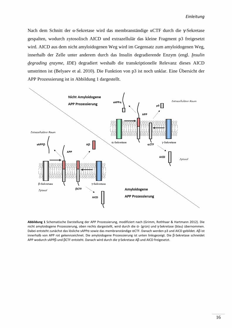
umstritten ist (Belyaev et al. 2010). Die Funktion von p3 ist noch unklar. Eine Übersicht der
APP Prozessierung ist in Abbildung 1 dargestellt.
Abbildung 1 Schematische Darstellung der APP Prozessierung, modifiziert nach (Grimm, Rothhaar & Hartmann 2012). Die
nicht amyloidogene Prozessierung, oben rechts dargestellt, wird durch die α- (grün) und γ-Sekretase (blau) übernommen.
Dabei entsteht zunächst das lösliche sAPPα sowie das membranständige αCTF. Danach werden p3 und AICD gebildet. Aβ ist
innerhalb von APP rot gekennzeichnet. Die amyloidogene Prozessierung ist unten linksgezeigt. Die β-Sekretase schneidet
APP wodurch sAPPβ und βCTF entsteht. Danach wird durch die γ-Sekretase Aβ und AICD freigesetzt.
Fragestellung
17
(2.3) Fragestellung der Arbeit
Trotz intensiver Forschung ist für Patienten mit AD bis zum heutigen Tag keine kausale
Therapie verfügbar. Die Behandlung beschränkt sich auf eine symptomatische Therapie, in
der Acetylcholinesterase-Hemmer (z.B. Donepezil) oder Glutamat-Antagonisten (z.B.
Memantin) eingesetzt werden (Anand et al. 2014). Aβ, welches aus der proteolytischen
Prozessierung des APP durch die Sekretasen resultiert, ist Hauptbestandteil der Plaques im
Gehirn von AD-Patienten. Es ist bekannt, dass einige Lipide die Aβ-Produktion reduzieren
können (Grimm, Kuchenbecker, Grösgen, et al. 2011; Burg et al. 2013). Die an der APP
Prozessierung beteiligten Sekretasen sind Transmembranproteine, die durch ihre
Lipidumgebung in ihrer Aktivität beeinflusst werden. Da die Prozessierung des APP direkt
von der α-, β- und γ-Sekretase abhängt, war es Ziel dieser Arbeit insbesondere den Einfluss
von Lipiden auf die Aktivität der an der APP Proteolyse beteiligten Sekretasen und die
Bildung des Aβ-Peptids zu untersuchen. Dazu wurden zum einen Phospholipide und
Plasmalogene (PL), die zu den häufigsten Lipidklassen des menschlichen Gehirns zählen, und
zum anderen oxidierte Derivate der Docosahexaensäure (DHA, C22:6) und Lipid-
Peroxidationsprodukte verwendet.
Der Gehalt an Phospholipiden und PL ist im Gehirn von AD-Patienten im Vergleich zum
gesunden Gehirn verändert. Zudem ist eine Wirkung von Phospholipiden auf die γ-Sekretase
und Aβ, welches aus der amyloidogenen Prozessierung des APP resultiert (Haass,
Schlossmacher, et al. 1992), gezeigt worden. Als Ergänzung wurde in dieser Arbeit der
Einfluss von Phospholipiden auf die nicht amyloidogenen Prozessierung untersucht, da dies
ein wichtiger Schritt zur Verhinderung der Aβ-Bildung darstellt. In einem weiteren Schritt
wurde der molekulare Wirkmechanismus von PL auf die APP Prozessierung analysiert.
Die Wirkung der ω-3 Fettsäure DHA in der Prävention oder Therapie der AD ist nicht
abschließend geklärt (Quinn et al. 2010; Freund-Levi et al. 2006; Chiu et al. 2008). Eine
mögliche Ursache für die heterogene Studienlage könnte die Instabilität der ω-3 Fettsäure
DHA darstellen, was zu einem erhöhten Gehalt an oxidierten Derivaten führen könnte. Daher
wurde in dieser Arbeit schließlich die Wirkung von verschiedenen oxidierten DHA-Derivaten
und den Lipid-Peroxidationsprodukten der ω-3 und ω-6 Fettsäuren auf die amyloidogene APP
Prozessierung untersucht.
Methoden
18
(3) Methoden
(3.1) Methoden der Zellkultur
(3.1.1) Kultivierung eukaryotischer Zellen In dieser Arbeit wurden die folgenden Zelllinien verwendet:
- Humane Neuroblastom Zelllinie (SH-SY5Y Wildtyp (wt)) (Biedler et al. 1973)
- Humane Neuroblastom Zelllinie stabil transfiziert mit der APP-Isoform mit 695
Aminosäuren (SH-SY5Y APP) von Heike Grimm (Grimm et al. 2003)
Die SH-SY5Y Zellen wurden direkt nach dem Auftauen in warmem DMEM-Medium
(Dublecco´s modified eagle`s medium, Sigma-Aldrich, Taufkirchen) mit 10% fetalem
Kälberserum FCS (Fetal Calf Serum, Pan Biotech, Aidenbach) und Aminosäurelösung
(MEM 100x, Sigma-Aldrich, Taufkirchen) kultiviert. Zusätzlich wurde dem Medium für die
SH-SY5Y-APP Zellen 300µg/ml Hygromyzin B (Pan Biotech, Aidenbach) zugesetzt.
Die Zellen wurden bis zur Konfluenz bei 5% CO2 in einem Inkubator (Heracell,
ThermoScientific, Waltham, Massachusetts, USA) kultiviert. Danach wurden die Zellen alle
3-4 Tage 1:5 (Wildtyp) und 1:3 (APP-überexprimierend) gesplittet. Dazu wurden die Zellen
mit warmem PBS gewaschen, nach Absaugen des PBS wurden die Zellen mit 1,5ml Trypsin
2min inkubiert. Durch wiederholtes auf- und abpipettieren wurden die Zellen vereinzelt und
dann auf neue Zellkulturplatten ausgesät. Vor Inkubationen wurden die Zellen entweder 1:3
auf 10cm-Platten/6-Loch Platten oder 1:1 auf 96-Loch-Platten ausgesät. Die Platten wurden
mindestens 24h vor Inkubationsbeginn unter Standardbedingungen inkubiert.
(3.1.2) Präparation von primären kortikalen Mausneuronen Durchgeführt von Inge Tomic.
Embryos von C57BI6/N wt-Mäusen (E14-18, Charles River, Sulzfeld) wurden entnommen
und 15min bei 37°C mit Trypsin behandelt (Sigma-Aldrich, Taufkirchen) und in warmem
HBSS 5x gewaschen. Danach wurden die Gehirne in 1ml HBSS durch Auf - und
Abpipettieren in einer Glaspipette homogenisiert. Die Neurone wurden auf einer 96-Loch
Methoden
19
Platte (Thermo Scientific, Karlsruhe), die mit Poly-L-Lysin (Sigma-Aldrich, Taufkirchen)
beschichtet wurde ausgesät und 7 Tage kultiviert.
(3.1.3) Inkubation mit Phospholipiden Die Phospholipide wurden mit, im Ultraschallbad für 15min, entgastem Ethanol (Sigma-
Aldrich, Taufkirchen) gelöst, in dunkle Glasflaschen aliquotiert und in flüssigem Stickstoff
gelagert. Die Konzentration dieser Lösungen war für Phosphatidylcholin (PC) 25mM, für
Phosphatidyserin (PS) 0,5µM und für Phosphatidylethanolamin (PE) 2,5mM.
Bei Konfluenz wurden die SH-SY5Y Zellen in DMEM mit 0,1% FCS für 4h inkubiert. Dann
wurden die Zellen für 8h und anschließend für 16h mit 10µM des jeweiligen Phospholipids
(Avanti Polar Lipids, Alabaster, Alabama, USA) in DMEM mit 0,1% FCS oder dem
Lösemittel als Kontrolle inkubiert.
(3.1.4) Inkubation mit PL Durchgeführt von Tatjana Rothhaar.
Die verwendeten PL (Avanti Polar Lipids, Alabaster, Alabama, USA) und
korrespondierenden Ester-Phospholipide wurden in entgastem Ethanol (Sigma-Aldrich,
Taufkirchen) in einer Konzentration von 50mM angesetzt, in dunkle Glasflaschen aliquotiert
und in flüssigem Stickstoff gelagert. Die konfluenten Zellen wurden unter reduziertem FCS-
Gehalt (0,1%) im Kulturmedium für 12h inkubiert. Dann wurden die Zellen für 2x 12h mit
100µM des jeweiligen PL oder dem korrespondierenden Ester-Phospholipid als Kontrolle
inkubiert.
(3.1.5) Inkubation mit oxidierten Lipiden, Lipid-Peroxidationsprodukten oder DHA
Die verwendeten oxidierten Lipide (Caymen Chemicals, Biomol, Hamburg) und Lipid-
Peroxidationsprodukte (Merck, Darmstadt) wurden in entgastem Ethanol (Sigma-Aldrich,
Taufkirchen) in einer Konzentration von 250µM gelöst. DHA (Sigma-Aldrich, Taufkirchen)
wurde in einer Konzentration von 50mM gelöst. Die Lipide wurden in dunklen Glasflaschen
(Wheaton, Millville, New Jearsey, USA) aliquotiert und mit Stickstoff überschichtet. Die
Lagerung erfolgte in flüssigem Stickstoff. Die Zellen wurden bei Konfluenz in DMEM mit
Methoden
20
0,1% FCS für 16h inkubiert. Dann wurden die Zellen für 24h unter Verwendung von 0,1%
w/v BSA (Bovine Serum Albumin, Sigma-Aldrich, Taufkirchen) mit 2µM oxidierten Lipiden,
Lipidperoxidationsprodukten, 20µM DHA (Sigma-Aldrich, Taufkirchen) oder dem
Lösemittel als Kontrolle inkubiert. Nach 8h erfolgte ein Mediumwechsel. Für die Inkubation
auf 6-Lochplatten wurden 2ml Medium pro Ansatz auf die Zellen gegeben, für die Inkubation
auf 96-Lochplatten 50µl Medium.
(3.1.6) Bestimmung der Zytotoxizität Für Publikation 2 wurde die Messung von Tatjana Rothhaar, für Publikation 3 in
Kooperation mit Janine Mett durchgeführt.
Die Zytotoxizität wurde mittels Lactatdehydrogenase-Assay (LDH-Assay, Roche, Grenzach-
Wyhlen) bestimmt. Dazu wurde unmittelbar nach Inkubationsende das Medium von den
Zellen abgenommen und 5min bei 1.000rpm zentrifugiert. Dann wurden jeweils 100µl pro
Probe auf eine 96-Loch Platte gegeben. Im Anschluss wurden die Catalyst und Dyesolution
im Verhältnis 1:45 vermischt und mit einer Multipipette (Multipette Plus, Eppendorf,
Hamburg) auf die Proben gegeben. Die Proben wurden dann 30min bei Raumtemperatur (RT)
unter leichtem Schütteln inkubiert und im Anschluss wurde die Reaktion durch Zugabe von
50µl Stopsolution beendet. Die OD wurde durch Messung in einem Photometer bei einer
Wellenlänge von 490nm bestimmt. Zusätzlich wurden die Zellen einer Platte mit 0,1% Triton-
X-100 lysiert um eine Positivkontrolle mit maximaler LDH-Konzentration zu erhalten. Diese
wurde linear verdünnt und als Standardreihe im Assay verwendet. Die Zytotoxizität der
Inkubation wurde dann über die Geradengleichung ermittelt.
Methoden
21
(3.2) Proteinanalytik
(3.2.1) Bestimmung der Proteinkonzentration Für Publikation 2 durchgeführt von Tatjana Rothhaar. Der Gesamtproteingehalt der Proben wurde mittels Bicinchoninsäure -Assay nach Smith et al.
(Smith et al. 1985) durchgeführt. Dabei wurde ein Protein-Standard (Bovines-Serum-
Albumin, Sigma-Aldrich, Taufkirchen) von 0-1,1mg/ml verwendet. Jede der Proben wurde
dreifach auf eine transparente 96-Loch Platte pipettiert und mit 200µl einer Arbeitslösung aus
Bicinchoninsäure (Sigma-Aldrich, Taufkirchen) und 4% w/v Kupfersulfat im Verhältnis 39:1
versetzt. Nach Inkubation der Platte bei 37°C für 15min wurde diese für weitere 15min bei
Raumtemperatur (RT) bei 300rpm auf einem Plattenschüttler inkubiert. Die optische Dichte
der Proben wurden dann bei 560nm in einem Photometer (Multiskan EX Thermo Scientific)
gemessen und die Standardreihe als Referenz zur Berechnung des Proteingehalts mittels der
Geradengleichung verwendet.
(3.2.2) Herstellung von Zell-/ Gewebe- Homogenaten Für Publikation 2 durchgeführt von Tatjana Rothhaar. Die SH-SY5Y Zellen wurden 3x mit kaltem PBS gewaschen und in Saccharosepuffer
(200mM Saccharose, 10mM Tris/HCL, pH 7,5) abgeschabt. Danach wurden die Zellen
mittels Minilyse durch Keramikkügelchen (Peqlab, Erlangen) bei höchster Intensität 30s lang
aufgeschlossen (Publikation 3) oder mit einem Potter (Publikation 1+2) bei maximaler
Drehzahl mit 25 Stößen homogenisiert. Die erhaltenen Homogenate wurden nach der
Proteinbestimmung mittels BCA-Tests auf 1mg/ml Gesamtproteingehalt eingestellt bevor sie
zur post-nukleären Fraktionierung verwendet wurden. Für Publikation 3 wurden die humanen
post-mortem Gehirne analog aufgeschlossen. Für Publikation 1 und 2 wurden die
Mausgehirne/Proben von humanen Gehirnen in 4°C kaltem Saccharosepuffer aufgenommen
und durch Verwendung eines Potters bei maximaler Drehzahl mit 25 Stößen (Publikation 1)
bei 1.500rpm Geschwindigkeit mit 50 Stößen (Publikation 2) aufgeschlossen. Die
Bestimmung des Gesamtproteingehalts erfolgte wie oben für die SH-SY5Y Zellen
beschrieben. Es wurden für Publikation 1 nur post-mortem Gehirne von Personen ohne
Alzheimer Pathologie, wie in nachfolgender Tabelle 4 aufgeführt, verwendet. Die Gehirne
wurden von Brain Bank (München) bezogen.
Methoden
22
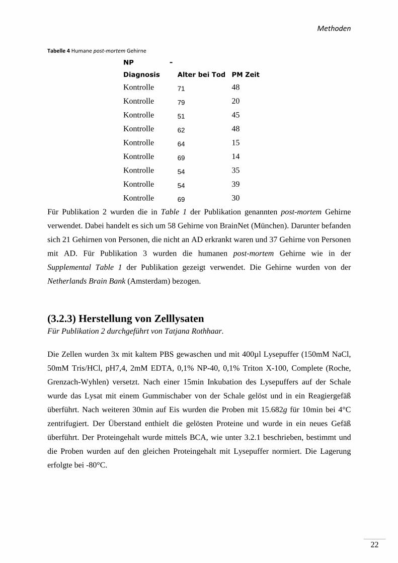
Tabelle 4 Humane post-mortem Gehirne
NP -
Diagnosis Alter bei Tod PM Zeit
Kontrolle 71 48
Kontrolle 79 20
Kontrolle 51 45
Kontrolle 62 48
Kontrolle 64 15
Kontrolle 69 14
Kontrolle 54 35
Kontrolle 54 39
Kontrolle 69 30
Für Publikation 2 wurden die in Table 1 der Publikation genannten post-mortem Gehirne
verwendet. Dabei handelt es sich um 58 Gehirne von BrainNet (München). Darunter befanden
sich 21 Gehirnen von Personen, die nicht an AD erkrankt waren und 37 Gehirne von Personen
mit AD. Für Publikation 3 wurden die humanen post-mortem Gehirne wie in der
Supplemental Table 1 der Publikation gezeigt verwendet. Die Gehirne wurden von der
Netherlands Brain Bank (Amsterdam) bezogen.
(3.2.3) Herstellung von Zelllysaten Für Publikation 2 durchgeführt von Tatjana Rothhaar.
Die Zellen wurden 3x mit kaltem PBS gewaschen und mit 400µl Lysepuffer (150mM NaCl,
50mM Tris/HCl, pH7,4, 2mM EDTA, 0,1% NP-40, 0,1% Triton X-100, Complete (Roche,
Grenzach-Wyhlen) versetzt. Nach einer 15min Inkubation des Lysepuffers auf der Schale
wurde das Lysat mit einem Gummischaber von der Schale gelöst und in ein Reagiergefäß
überführt. Nach weiteren 30min auf Eis wurden die Proben mit 15.682g für 10min bei 4°C
zentrifugiert. Der Überstand enthielt die gelösten Proteine und wurde in ein neues Gefäß
überführt. Der Proteingehalt wurde mittels BCA, wie unter 3.2.1 beschrieben, bestimmt und
die Proben wurden auf den gleichen Proteingehalt mit Lysepuffer normiert. Die Lagerung
erfolgte bei -80°C.
Methoden
23
(3.2.4) Aufarbeitung humaner post-mortem Gehirnproben Durchgeführt in Kooperation mit Nadine Mylonas.
Die Gehirnproben, die für Publikation 3 verwendet wurden, wurden von der Netherlands
brain bank in Amsterdam bezogen. Die humanen Gehirnproben wurden entsprechend ihres
Gewichts in vollentsalztem Wasser durch Minilyse für 30s bei maximaler Intensität mit
Keramik-Precellys homogenisiert. Der Proteingehalt wurde durch BCA-Test bestimmt und
die Homogenate auf einen Gesamtproteingehalt von 10mg/ml eingestellt. Die verwendeten
humanen Gehirne sind in Supplemental table 1 der Publikation 3 aufgelistet.
(3.2.5) Post-nukleäre Fraktionierung und Isolation von Membranen
Für Publikation 2 durchgeführt von Tatjana Rothhaar. Zur post-nukleären Fraktionierung wurden die Homogenate zunächst auf einen
Gesamtproteingehalt von 2mg/ml (β-, γ-Sekretasemessung) oder 1mg/ml (α-
Sekretasemessung) eingestellt. Dann wurden die Homogenate bei 900g und 4°C für 10min
zentrifugiert. Die resultierenden post-nukleären Fraktionen wurden weiterhin bei 135.520g
und 4°C für 75min in einer Ultrazentrifuge (Optima, Beckman-Coulter, Krefeld) zentrifugiert.
Das resultierende Pellet wurde für die Messung der β- und γ-Sekretaseaktivität in
Saccharosepuffer (200mM Saccharose, 10mM Tris/HCL, pH 7,5, 1mM EDTA)
aufgenommen. Zur Messung der α-Sekretaseaktivität wurde das Pellet mit Saccharosepuffer
(200mM Saccharose, 10mM Tris/HCL, pH 7,5; Publikation 1) oder HEPES-Puffer
(Publikation 2) unter Zugabe der folgenden Inhibitoren (Complete (Roche), 1µM β-Sekretase-
Inhibitor (Merck, Darmstadt), 1,46µM Pepstatin A (Sigma-Aldrich, Taufkirchen); 5µM
CaCl2, 5µM ZnCl2) aufgenommen. Die isolierten Membranen wurden unter Verwendung von
Kanülen mit kleiner werdendem Durchmesser (23,27G; BD, Franklin Lakes, New Jersey,
USA; U-100 Insulinspritze, Teruno, Sumerset, New Jersey, USA) solubilisiert (Publikation
1+2) oder durch Minilyse (Minilys Peqlab, Erlangen) mit mittlerer Intensität für 10s unter
Verwendung von Glaskügelchen (Peqlab, Erlangen; Publikation 3).
Methoden
24
(3.2.6) In vitro Inkubationen Für Publikation 2 wurden die Inkubationen in Kooperation mit Tatjana Rothhaar
durchgeführt.
Die post-nukleären Fraktionen von SH-SY5Y Zellen und Mausgehirnen wurden zunächst
10min bei 37°C im Wasserbad erwärmt mit 100µM Plasmalogen, 25µM Phospholipid oder
2µM oxidiertem Lipid inkubiert. Die isolierten Membranen aus humanen post-mortem
Gehirnen wurden nach dem Erwärmen mit 50µM Phospholipid bzw. 100µM Plasmalogen
inkubiert.
Die Inkubation wurde für 15 min bei 37°C unter leichtem Schütteln in 1ml Glasflaschen
(Wheaton, Millville, New Jearsey, USA) ausgeführt. Die PNFs wurden wie oben beschrieben
zentrifugiert und dann für die Bestimmung der α-, β- und γ-Sekretase Aktivität verwendet.
Zur Bestimmung der ADAM-10 Aktivität wurde 25µM Phospholipid oder Ethanol als
Kontrolle mit 100ng ADAM-10 (Sigma Aldrich, Taufkirchen, Germany) und Lipidextrakt aus
humanen Gehirnen (5µg pro 100µl) Saccharosepuffer für 1h bei 4°C unter leichtem Schütteln
inkubiert. Im Anschluss wurden die Proben 15min im Eiswasserbad sonifiziert.
(3.2.7) Bestimmung der ββββ- und γγγγ-Sekretase Aktivität nach in vitro Inkubation
Für Publikation 2 wurden die Messungen von Tatjana Rothhaar durchgeführt.
Nach der Inkubation wurden 100µl der isolierten Plasmamembranen als Dreifachbestimmung
auf eine schwarze 96-Loch Platte gegeben. Dies entsprach für die Messung aus SH-SY5Y
Zellen 125µg (β-Sekretase Messung) und 250µg (γ-Sekretase Messung) Gesamtprotein. Für
die Messung aus humanen und murinen Gehirnen wurden 250µg (β-Sekretase Messung) bzw.
500µg (γ-Sekretase Messung) an Gesamtprotein verwendet. Die Aktivität wurde nach Zugabe
von 20µM β-Sekretase Substrat oder 10µM γ-Sekretase Substrat gemessen. Die Spezifität der
Messung wurde durch Zugabe von spezifischen Inhibitoren für die β- und γ-Sekretase
bestimmt. Nach der Messung wurde das unspezifische Fluoreszenzsignal von den Werten
subtrahiert.
Die Fluoreszenz wurde 8000s lang bei einer Exzitation von 345 ± 5nm, sowie einer Emission
von 500 ± 2,5nm für die β-Sekretaseaktivität und 6000s lang bei einer Exzitation von 355 ±
10nm, sowie einer Emission von 440 ± 10nm für die γ-Sekretaseaktivität für 8000s bei 37 °C
bestimmt.
Methoden
25
(3.2.8) Bestimmung der Sekretase Aktivität auf lebenden Zellen Für Publikation 2 wurden die Messungen von Tatjana Rothhaar durchgeführt.
Nach der Inkubation mit Phospholipiden wurden die Zellen 1x mit Cell-Imaging-Solution
(140mM NaCl, 5mM KCl, 8mM CaCl2, 1mM MgCl2, 20mM HEPES, pH 7.4) gewaschen.
Anschließend wurde 50µl Cell-Imaging-Solution mit 10µM Phospholipid und 10µM α-
Sekretase Substrat auf die Zellen gegeben. Die α-Sekretase Aktivität wurde kontinuierlich für
10.000s in einem Fluorometer (Safire2 Fluorometer Tecan, Crailsheim, Germany) gemessen.
Die Fluoreszenz wurde bei einer Exzitation von 340 ± 10 nm, sowie einer Emission von 490 ±
10 nm mit einer Verstärkung von 150 bei 37 °C bestimmt.
Zur Bestimmung der β- und γ-Sekretaseaktivität wurde Cell-Imaging-Solution mit 30µM β-
und 24µM γ-Sekretase Substrat versetzt und pro Loch 50µl auf die 96-Loch Platte pipettiert.
Die Spezifität der Messung wurde durch Verwendung spezifischer Inhibitoren (Calbiochem,
Darmstadt) ermittelt und nach der Messung subtrahiert. Die Fluoreszenz wurde bei einer
Exzitation von 345 ± 5nm, sowie einer Emission von 500 ± 2,5nm für die β-Sekretaseaktivität
und einer Exzitation von 355 ± 10nm, sowie einer Emission von 440 ± 10nm für die γ-
Sekretaseaktivität bei 37 °C bestimmt.
(3.2.9) Bestimmung der αααα-Sekretase- und ADAM-10 Aktivität nach in vitro Inkubation
Die Messungen der ADAM-10 Aktivität wurde von Tatjana Rothhaar durchgeführt.
Nach der Inkubation wurden 100µl der isolierten Plasmamembranen der SH-SY5Y Zellen
und humanen post-mortem Gehirnen entsprechend 100µg an Gesamtprotein oder für
Mausgehirne entsprechend 50µg an Gesamtprotein als Dreifachbestimmung auf eine
schwarze 96-Loch Platte (Corning, New York, USA) gegeben. Die Aktivität wurde nach
Zugabe von 4µM α-Sekretase Substrat (Merck, Darmstadt) gemessen. Die Fluoreszenz wurde
wie oben beschrieben in einem Fluorometer (Safire2 Fluorometer Tecan, Crailsheim) für
10.000s nach Inkubation mit Phospholipiden auf lebenden Zellen und SH-SY5Y
Zellhomogenaten, 15.000s für Inkubation mit PLn auf Mausgehirnen gemessen. Zur
Bestimmung der Spezifität wurde ein EDTA-EGTA Gemisch, jeweils 5mM, eingesetzt.
Die Messung der ADAM-10 Aktivität wurde nach Zugabe von 4µM α-Sekretase Substrat
(Merck, Darmstadt) in einem Fluorometer (Infinite M1000 Pro Fluorometer, Tecan,
Methoden
26
Crailsheim) bestimmt, wobei die Fluoreszenz kontinuierlich über 15.000s bei einer
Verstärkung von 115 gemessen wurde.
(3.2.10) Bestimmung des Aβ, sAPPββββ und sAPPαααα Gehalts Die Bestimmung des Aβ-Gehalts wurde nach Ida et al. (Ida et al. 1996), die Bestimmung des
sAPPα Gehalts nach Grimm et al. (Grimm, Rothhaar, Grösgen, et al. 2012)durchgeführt. Das
konditionierte Medium wurde für 5min mit 13.000rpm bei 4°C zentrifugiert um tote Zellen
und Zellpartikel zu entfernen. Für die Bestimmung des Aβ-Gehalts wurden dem Medium
10µg W02-Antikörper und 20µl Protein-G-Sepharose (Sigma-Aldrich, Taufkirchen) zugesetzt
und die Ansätze bei 4°C für 16-18h auf einem Über-Kopf-Taumler (Reax2, Heidolph,
Schwabach) inkubiert. Die Proben wurden dann bei 13.000rpm in einer Tischzentrifuge
(5415D, Eppendorf, Hamburg) bei 4°C zentrifugiert und der Überstand abgesaugt. Das Pellet
wurde dann nacheinander 3x mit IP-Waschpuffer A, B und C (siehe Tabelle 6) gewaschen.
Während der Waschschritte wurden die Proben 10x invertiert und dann wie oben beschrieben
zentrifugiert.
Nach dem letzten Waschen wurde das Pellet mit 15µl 3x Protein-Proben-Puffer (187,5mM
Tris/HCl, 6% v/v SDS , 30% v/v Glycerol , 15% β-Mercaptoethanol , 0,03% w/v
Bromphenolblau) versetzt und 5min bei 98°C erhitzt.
Tabelle 5 IP-Waschpuffer
Puffer A Puffer B Puffer C
Tris/HCl 10mM 10mM 10mM
NaCl 250mM 500mM -
EDTA 2mM 2mM -
Triton X-100 0,1% (v/v) 0,1% -
NP-40 0,1% (v/v) 0,1% -
Anschließend wurde der Überstand auf ein 10-20% Grandientengel (Tris-Tricin Gel, Anamed,
Bad Ems) geladen und mittels SDS-PAGE aufgetrennt. Die Proben wurden danach auf eine
Nitrozellulosemembran (Whatman/GE Healthcare, Freiburg) für 1h (Aβ) und 3h (sAPPα/β)
bei 380mA in einer Kammer (Biorad, München) transferiert (Transferpuffer: 250 mM Trizma
Base; 1,57 M Glycin, 20% Methanol, 0,025% SDS).
Methoden
27
Die Membran wurde für die sAPPα- und Aβ-Messung 5min in der Mikrowelle (Micromat,
AEG, Frankfurt am Main) in 1xPBS gekocht. Die unspezifischen Antikörperbindestellen
wurden durch Inkubation mit 1xPBS mit 10% w/v Magermilchpulver für 1h geblockt. Die
Inkubation mit W02 Antikörper erfolgte für 1,5h in 1x PBS mit 2% w/v Magermilchpulver,
gefolgt von einer Inkubation mit dem HRP-gekoppelten Sekundärantikörper anti-Maus
(Dako, Hamburg) für 1h im gleichen Puffer. Die Inkubation mit sAPPβ-Antikörper (my
biosource, San Diego, Kalifornien, USA) wurde in einer Verdünnung von 1:400 in 1xPBS mit
5% Milch durchgeführt, gefolgt von einer 1h Inkubation mit dem Sekundärantikörper anti-
Kaninchen (Promega, Madison, Wisconsin).
Zwischen den Inkubationsschritten wurde die Membran jeweils 3x mit 1xPBS für 10min
gewaschen. Die Entwicklung erfolgte mit der Enhanced chemilumineszenz Methode (ECL,
Western Lightning PLUS ECL, Perkin Elmer, Waltham, Massachusetts, USA). Dabei wurde
die ECL-Lösung 1 im Verhältnis 1:1 mit ECL-Lösung 2 verdünnt und auf der Membran 1min
inkubiert. Dann wurde die Membran zwischen 2 Klarsichtfolien gelegt und die entstehende
Chemolumineszenz im Dunkeln durch einen Photofilm (Amersham Hyperfilm ECL, GE
Healthcare, Freiburg) detektiert.
(3.2.11) Bestimmung des ADAM-17, BACE-1 und PS-1 Gehalts Für Publikation 2 wurden die Messungen von Verena Burg durchgeführt.
Nach Inkubation mit PL wurden die Zellen wie oben beschrieben lysiert und die Proben
mittels SDS-PAGE aufgetrennt. Nach dem Western Blot wurde die Membran 1h mit 10% w/v
Magermilchpulver bei RT inkubiert und 1,5h mit spezifischen ADAM-17 (ab39162, abcam,
Cambridge, UK), BACE-1 (B0806, Sigma-Aldrich, Taufkirchen) oder PS1- (sc-7860, Santa
Cruz, Heidelberg) Antikörper in den Verdünnungen 1:5000, 1:1000 und 1:500 inkubiert.
Nach der Inkubation mit Sekundärantikörper anti-Kaninchen (W401B, Promega, Mannheim)
in einer Verdünnung von 1:10.000 wurde die Membran mittels der ECL Methode wie unter
3.2.10 beschrieben entwickelt.
Methoden
28
(3.3.) Lipidanalytik
(3.3.1) Extraktion von Lipiden aus humanen post-mortem Gehirnen
Durchgeführt von Sven Grösgen und Benjamin Hundsdörfer.
Die Lipidextraktion wurde mit leichten Modifikationen nach Bligh und Dyer durchgeführt
(Bligh & Dyer 1959). Die Gehirnproben wurden mit 3,75 mL CHCl3:MeOH:HCl (37%;
Sigma-Alrich Taufkirchen) (1:2:0.06; v/v/v) versetzt und 1 h bei RT durch einem Vortexer
(Bender-Hohbein, Bruchsal) geschüttelt. Dann wurden 1,25 mL CHCl3 hinzugegeben und 1 h
bei RT geschüttelt. Schließlich wurden 1,25 mL CHCl3 und 1,25 mL H2O hinzugefügt und die
Proben weitere 10min geschüttelt bevor sie mit 5.000rpm 10 min zentrifugiert wurden. Die
Phasentrennung ergibt eine obere, eine dünne mittlere und eine untere Phase. Die untere
Phase enthält Lipide und wurde unter kontinuierlichem Stickstofffluss evaporiert. Die Lipide
wurden in 1ml H2O resuspendiert und wieder bei 30°C evaporiert. Abschließend wurden die
Lipide in Ethanol gelöst. Zur Herstellung eines Gesamtlipidextraktes aus humanen post-
mortem Gehirnen wurden alle Proben vereinigt und auf eine Konzentration von 5 mM
eingestellt.
(3.3.2) Messung der DHA-Stabilität DMEM wurde mit 20µM PC-DHA/DHA versetzt und 16h unter
Standardzellkulturbedingungen inkubiert. Die Proben wurden zum Startzeitpunkt und nach
16h entnommen und in einem Gemisch aus Methanol und 5mM Ammoniumacetat (Sigma-
Aldirch, Taufkirchen) im Verhältnis 1:100 verdünnt. Im Anschluss wurden die Proben wie
unter 3.3.3 beschrieben mittels Massenspektrometrie vermessen.
(3.3.3) Bestimmung des Lipidgehalts durch Massenspektrometrie Für Publikation 1 und 2 wurde die Analytik von Sven Grösgen, für Publikation 3 von
Christoph Stahlmann durchgeführt.
Die Proben wurden auf einen Proteingehalt von 2mg/ml (Publikation 1) bzw. 6mg/ml
(Publikation 2) eingestellt.
Zur Bestimmung der Phospholipide wurde das Massenspektrometer 4000 QTRAP (ABSciex)
mit Turbo Spray Ion Source (AB SCIEX) verwendet. Der Phospholipidgehalt wurde mit dem
AbsoluteIDQ™ p150 (Biocrates Life Sciences AG, Innsbruck, Austria) bestimmt. Dazu
wurden 20 µl (Publikation 1) bzw. 10µl (Publikation 2) der Proben auf eine Membran
Methoden
29
(Whatman-GE Healthcare, Freiburg) in einer 96-Loch Sterilfilterplatte (0,45µM; Milipore,
Schwalbach) gegeben. Die Proben wurden unter Stickstofffluss (1-2 bar; 30 min; RT)
evaporiert. Dann wurden 20µl 5%iges Phenylisothiocyanat (v/v) (Verdünnung in
Ethanol/Wasser/Pyridin (1/1/1; v/v/v) jedem Loch zugegeben und die Platte für 20min bei RT
inkubiert und anschließend unter Stickstofffluss evaporiert (1-2 bar; 30 min; RT). Die Proben
wurden unter Zugabe von 300 µl 5 mM Ammoniumacetat in Methanol extrahiert und 30min
bei RT und 450 rpm geschüttelt. Anschließend wurden die Proben bei 500g für 2 min durch
die Sterilfilterplatte in eine 96-Loch Sammelplatte zentrifugiert. Die Proben wurden dann mit
600µl Laufpuffer A (161,5mM Ammoniumacetat in Wasser/Methanol, 3/297, v/v) verdünnt
und die Platte 2min bei RT und 450 rpm geschüttelt. Die Sammelplatte wurde mit einer
durchstechbaren Silikonmatte verschlossen und in den vorgekühlten Autosampler (10°C)
gestellt und die Messung gestartet. Die Proben wurden mit einem Gradienten aus Laufpuffer
A in das 4000 QTRAP Massenspektrometer injiziert (Phase1: 0min – 1,6min, 30µl/min
Laufpuffer A; Phase 2: 1,6min – 2,4min, 30µl/min Laufpuffer A; Phase 3: 2,4min – 2,8min,
200µl/min Laufpuffer A; Phase 4: 2,8min – 3min, 200µl/min Laufpuffer A). Die Detektion in
Triplicaten (20µl Injektionsvolumen) wurde mittels Analyst 1.4.2 software (AB SCIEX,
Darmstadt, Germany) durchgeführt. Folgende Parameter wurden für die
massenspektroskopische Untersuchung angewendet: Curtain gas (CUR) 20.0, collision gas
(CAD) medium, ion spray voltage (IS) 5500.0, Temperatur (TEM) 200.0, ion source gas 1
(GS1) 40, ion source gas 2 (GS2) 50, interface heater (ihe) on, Entrance potential (EP) 10,
Cell exit Potential (CXP) 15
(3.3.4) Bestimmung des Phospholipidgehalts mittels Dünnschichtchromatographie
Durchgeführt von Benjamin Hundsdörfer.
Eine Dünnschichtchromatographie-Kieselgelplatte (Merck, Darmstadt) wurde mit 1mM
EDTA in Wasser imprägniert und bei 100°C getrocknet. Die Platte wurde dann mit dem
Laufmittel A (Chloroform / Methanol / HPLC-Wasser im Verhältnis 60:40:10) gewaschen
und bei 100°C über Nacht getrocknet. Dann wurden 100µl der Lipidextrakte von SH-SY5Y
Zellen und humanen post-mortem Gehirnen langsam auf die Kieselgelplatte gegeben und
nacheinander mit Laufmitteln wie folgt aufgetrennt: Die Auftrennung mit Laufmittel B
(Chloroform / Methanol / HPLC-Wasser (65:40:5)) erfolgte bis 5cm; die Auftrennung mit
Laufmittel C (Ethyl-acetat / 2-Propanol / Ethanol / Chloroform / Methanol / 0,25 % KCl in
Methoden
30
HPLC-Wasser (35:5:20:22:15:9)) bis 10cm; mit Laufmittel D (Toluol / Diethylether /
Ethanol (60:40:3)) bis 12,5 cm und mit Laufmittel E (n-Heptan / Diethylether (94 : 8) bis
15cm. Zuletzt wurden die Proben mit Laufmittel F (n-Heptan) bis 0,5cm unter den oberen
Rand aufgetrennt. Zwischen den einzelnen Läufen wurde die Platte jeweils unter Heißluft
getrocknet. Die Lipide wurden dann durch Inkubation der Platte mit einer Kupfersulfatlösung
(10 % CuSO4 / 8% P2SO4) bei 200°C für 1-10min gefärbt.
(3.3.5) Bestimmung des HNE-Gehalts Der Gehalt an HNE wurde mittels enzymgekoppeltem Immunbindungsstest (Human 4-
Hydroxynonenal ELISA Kit Artikelnummer CSB-E16214h, Cusabio, Hubei Province, China)
entsprechend der Herstellerangaben gemessen. Die humanen Gehirnproben von Patienten mit
AD bzw. von den Kontrollpatienten wurden vereinigt und auf 790µg Gesamtprotein
eingestellt. Die Proben wurden bei 5.000g für 5 min zentrifugiert und die Überstände in ein
neues Reagiergefäß überführt. Ein Volumen von 100µl der Proben wurde für die Messung
verwendet, die gemäß dem Herstellerprotokoll durchgeführt wurde. Die Messung erfolgte bei
450nm, wobei eine Wellenlängenkorrektur durch zusätzliche Messung bei 560nm
vorgenommen wurde.
(3.3.6) Bestimmung der Lipidperoxidation Die humanen Gehirnproben von Patienten mit AD bzw. von Kontrollpatienten wurden
vereinigt und auf einen Gehalt von 2mg Gesamtprotein eingestellt. Die Bestimmung der
Lipidperoxidation wurde gemäß der Herstellerangaben (Lipid Peroxidation MDA Assay kit
Colorimetric, Fluorometric, Artikelnummer ab118970, Protokoll Version 7,
abcam,Cambridge,UK) kolorimetrisch durchgeführt. Demnach wurden die Homogenate im
Lysepuffer (Lysis Solution) für 20s bei maximaler Intensität lysiert. Nach anschließender
Zentrifugation der Proben wurde der Überstand mit TBA-Reagenz (engl. Thiobarbituric Acid,
Thiobartitursäure) versetzt und für 60min bei 95°C inkubiert. Die Proben wurden für 10min
auf Eis gestellt und anschließend die optische Dichte der Proben in einer 96-Loch Platte bei
532nm gemessen.
Methoden
31
(3.4) RNS-Analytik Für Publikation 2 wurde die Analytik von Sven Grösgen, für Publikation 3 von Janine Mett durchgeführt.
(3.4.1) Isolation von RNS Die Echtzeitpolymerasekettenreaktion (RT-PCR) wurde zur Bestimmung der mRNS Spiegel
in den Proben herangezogen. Die RNS aus Zellen wurde mittels 1ml Trizol pro 10cm Schale
(Thermo Fisher, Walham Massachusetts, USA) isoliert. Nach einer Inkubation von 5min bei
RT wurden die Proben mit 200µl Chlorophorm versetzt und 15s lang gevortext. Anschließend
wurden die Proben 3min lang inkubiert (RT) und danach bei 12.000rpm für 15min
zentrifugiert. Die obere Phase, welche die RNS enthielt, wurde abgenommen, mit 500µl
Isopropanol (Sigma-Aldrich, Taufkirchen) versetzt und für 10 min bei RT inkubiert, gefolgt
von einer Zentrifugation bei 12.000 rpm und 4°C.
Das resultierende RNS Pellet wurde mit 75% v/v Ethanol gewaschen und im Anschluss bei
7.500 rpm zunächst für 5min zentrifugiert dann bei RT getrocknet.
Die RNS wurde in 100µl RNase freiem Wasser aufgenommen und für 10min bei 55°C im
Wasserbad bei 55°C in Lösung gebracht.
Die RNS wurde auf eine Konzentration von 40µg/ml nach Messung der Proben bei 260nm
eingestellt.
(3.4.1) cDNS-Sythese und Echtzeitpolymerasekettenreaktion (RT-PCR)
Die Synthese der cDNS aus den Proben wurde mittels High Capacity cDNA RT Kit (Life
Technologies, Carlsbad, Kalifornien, USA) entsprechend der Herstellerangaben
durchgeführt. Zur Messung der mRNS Spiegel mittels RT-PCR wurde SYBR-Green (Thermo
Fisher, Walham Massachusetts, USA) in einem 7500 Fast Real Time PCR System Gerat (Life
Technologies, Carlsbad, Kalifornien, USA) für Publikation 2 oder einem PikoReal-Gerät
(Thermo Scientific, Waltham, Massachusetts, USA, PikoReal Software Version 2.1.158.545)
für Publikation 3 verwendet. 5µl der cDNS Proben wurden mit jeweils 0,5µl Primer forward
und 0,5µl Primer reverse, sowie 4µl Nuklease-freiem Wasser und 10µl SYBR Green Master
Mix in einer 96-Lochplatte (Life Technologies, Carlsbad, Kalifornien, USA) pipettiert. Die
Auswertung der Daten erfolgte mit der 2-��CT Methode (Livak & Schmittgen 2001) unter
Normierung auf die Expression des β-Actin. Die Sequenzen der Primer, welche in
Methoden
32
Publikation 2 verwendetet wurden, sind in Kapitel 4.2 in der entsprechenden Publikation
unter Abschnitt 2.10 aufgeführt.
Für Publikation 3 wurden die folgenden Primer verwendet:
APH1a: 5′-CAG CCA TTA TCC TGC TCC AT-3′ and 5′-GGA ATG TCA GTC CCG ATG TC-3′; APH1b: 5′-GTG TCA GCC CAG ACC TTC AT-3′ and 5′-CAG GCA GAG TTT CAG GCT TC-3′; BACE-1: 5′-AAT ACC TGC GGT GGA AGA TG-3′ and 5′-GCC CTC CAT GAT AAC AGC TC-3′; NCSTN: 5′-CTG TAC GGA ACC AGG TGG AG-3′ and 5′-GAG AGG CTG GGA CTG ATT TG-3′; PS1: 5′-CTC AAT TCT GAA TGC TGC CA-3′ and 5′-GGC ATG GAT GAC CTT ATA GCA-3′; PS2: 5′-GAT CAG CGT CAT CGT GGT TA-3′ and 5′-GGA ACA GCA GCA TCA GTG AA-3′; PEN2: 5′-CAT CTT CTG GTT CTT CCG AGA G-3′ and 5′-AGA AGA GGA AGC CCA CAG C-3′; β- Actin: 5′-CTT CCT GGG CAT GGA GTC-3′ and 5′-AGC ACT GTG TTG GCG TAC AG-3′.
Die Primer wurden von Eurofins MWG Operon (Ebersberg) bezogen.
(3.5) Statistische Auswertung Die Auswertung mittels ANOVA wurde für Publikation 1 in Kooperation mit Valerie Zimmer
und Johannes Lehmann , für Publikation 3 in Kooperation mit Nadine Mylonas durchgeführt.
Alle quantifizierten Daten basieren auf dem Mittelwert von wenigstens drei unabhängigen
Experimenten. Die Fehlerbalken zeigen die Standardabweichung vom Mittelwert. Die
statistische Signifikanz wurde durch Varianzanalyse mit post-hoc-Test oder zweiseitigen,
ungepaartem student-t-test bestimmt. Das Signifikanzniveau beträgt bei * p ≤ 0,05; ** p ≤
0,01 und *** p ≤ 0,001.
Alle beteiligten Personen waren zum Zeitpunkt der Versuchsdurchführung Angestellte oder
Studenten der Universität des Saarlandes.
Publikationen
33
(4) Publikationen
Die Daten der Publikationen 1-3 sind in Zusammenarbeit mit den Mitautoren entstanden. Der
Eigenanteil an den Publikationen ist unter den Abschnitten 4.1, 4.2 und 4.3 zusammengefasst
und kann auch den Kooperationstabellen 1-3 (siehe Seite 1-4) entnommen werden, in denen
für alle Abbildungen und Tabellen die durchführenden Personen genannt werden. Die
Publikationen 1 und 3 sind in geteilter Erstautorenschaft mit Marcus O.W. Grimm entstanden.
(4.1) Zusammenfassung von Publikation 1 und Beschreibung des Eigenanteils
Effect of Different Phospholipids on α-Secretase Activity in the Non-Amyloidogenic Pathway of Alzheimer’s Disease Haupenthal VJ*, Grimm MO*, Rothhaar TL, Zimmer VC, Grösgen S, Hundsdörfer B, Lehmann J, Grimm HS, Hartmann T * equally contributed
In Publikation 1 wurde der Einfluss von Phospholipiden auf die Aktivität der α-Sekretase im
nicht amyloidogenen Weg der APP-Prozessierung analysiert. Phospholipide können sich
einerseits in der Struktur der gebundenen Fettsäuren, andererseits in der Kopfgruppe
unterscheiden. Um den direkten Einfluss der Phospholipide auf die α-Sekretase Aktivität zu
untersuchen, wurden in dieser Arbeit Phosphatidylcholin (PC) - Spezies verwendet, die sich
in der Länge und dem Sättigungsgrad der gebundenen Fettsäuren, sowie der Position der
Doppelbindung in der Fettsäurekette unterschieden. Zusätzlich wurden Phospholipide mit
identischen Fettsäuren aber unterschiedlichen Kopfgruppen hinsichtlich ihrer Wirkung auf die
Aktivität der α-Sekretase analysiert. Da PC eine der Hauptspezies der Phospholipide im
humanen Gehirn darstellt (Söderberg et al. 1991), wurde in dieser Arbeit zunächst die
Wirkung von unterschiedlich langen Fettsäuren gebunden an PC auf die α-Sekretase Aktivität
analysiert. Dazu wurden Phospholipide verwendet, die sowohl an der sn1- als auch der sn2-
Position des Glycerin die gleiche Fettsäure gebunden hatten. PC-Spezies mit der Kettenlänge
von 10 C-Atomen (PC 10:0) bis hin zu 24 C-Atomen (PC 24:0) wurden mit isolierten
Membranen von SH-SY5Y Zellen und auf lebenden SH-SY5Y Zellen inkubiert. In Analogie
zu diesem Ansatz wurde PC gebunden an die ungesättigten Fettsäuren 18:1, 18:2, 18:3, 20:4,
20:5 und 22:6 betrachtet. Um die Wirkung der Position der Doppelbindung auf die α-
Sekretase Aktivität zu messen, wurde PC 18:1 mit einer Doppelbindung an Position 6 (D6) im
Vergleich zu PC18:1 mit der Doppelbindung an Position 9 (D9) der Fettsäuren verwendet.
Publikationen
34
Der Einfluss der Kopfgruppen auf die Aktivität der α-Sekretase wurde durch Verwendung
von Phosphatidylethanolamin (PE) und –serin (PS) gebunden an 12:0 und 14:0 im Vergleich
zu PC untersucht. Die Aufnahme der Lipide in die Zellen wurde exemplarisch für PC12:0 und
PC18:0 mittels Massenspektrometrie kontrolliert. Um eine bessere Vergleichbarkeit der
Phospholipide untereinander zu erzielen, wurde PC18:0, außer bei der Kopfgruppenanalyse,
für alle Versuche als Kontrolle verwendet, da es im Vergleich zur Lösemittelkontrolle keine
Veränderung der α-Sekretase Aktivität verursachte. Die Spezifität der α-Sekretase Messung
wurde durch Verwendung verschiedener Inhibitoren kontrolliert.
Die Versuche ergaben eine Steigerung der α-Sekretase Aktivität durch die kurzkettigen,
gesättigten Phospholipide PC10:0 & PC12:0. PC14:0 steigerte die Aktiviät der α-Sekretase
tendenziell, jedoch nicht signifikant. Hingegen bewirkten die langkettigen, gesättigten
Phospholipide PC16:0 – 24:0 keine Veränderung der α-Sekretase Aktivität.
Langkettige, ungesättigte Phospholipide verursachten ebenfalls einen Anstieg der α-Sekretase
Aktivität. Bemerkenswert ist, dass der signifikante Effekt erst durch Phospholipide, die
mindestens 4 Doppelbindungen (PC 20:4, PC 20:5, PC 22:6) enthielten, auftrat.
Phospholipide mit weniger Doppelbindungen steigerten die Aktivität der α-Sekretase nur
tendenziell.
In weiteren Analysen wurden keine Veränderungen der Aktivität der α-Sekretase durch die
Position der Doppelbindung innerhalb der Phospholipide oder durch Variation der
Kopfgruppe festgestellt. Lediglich PS12:0 und PS14:0 zeigten eine Veränderung der Aktivität
der α-Sekretase in lebenden SH-SY5Y Zellen bzw. deren isolierten Membranen.
Die steigernde Wirkung von kurzkettigen, gesättigten (PC12:0) und langkettigen,
ungesättigten Phospholipiden (PC22:6) auf die α-Sekretase Aktivität konnte exemplarisch
auch in isolierten Membranen von humanen post-mortem Gehirnen nachgewiesen und auf die
α-Sekretase ADAM-10 zurückgeführt werden.
Publikation 1 ist in geteilter Erstautorenschaft entstanden. Ich habe für diese Publikation die
Versuche koordiniert und in Zusammenarbeit mit Marcus O.W. Grimm geplant. Die
Inkubationen mit Lipiden, die Probenaufarbeitung und die nachfolgende Bestimmung der α-
Sekretase Aktivität wurde in allen Zellkultur und in vitro- Versuchen von mir durchgeführt
(figure 1 a+b, figure 2a-c, fgure 3 a+b, figure 4a+b, figure S1, figure S3, figure S4a). Auch
die Ergebnisse der Bestimmung der Spezifität der α-Sekretase Messung und des sAPPα-
Gehalts wurden von mir generiert (figure S4 a+b). Für die Messung der ADAM-10 Aktivität,
die von T. Rothhaar durchgeführt wurde (figure 1c+3c), sowie die Messung der Aufnahme
Publikationen
35
der Lipiden in die Zellen und isolierten Membranen, die von S. Grösgen durchgeführt wurde
(figure S5), habe ich die Inkubationen der Lipide durchgeführt.
Für die Bestimmung des PC-, PE- und PS-Gehalts mittels Dünnschichtchromatographie, die
von B. Hundsdörfer durchgeführt wurde (figure S2), und die Bestimmung des Lipidprofils,
durchgeführt von S. Grösgen (table S5), habe ich die Membranen aus SH-SY5Y Zellen und
humanen post-mortem Gehirnen isoliert.
Die statistische Auswertung der Daten mittels ANOVA habe ich in Zusammenarbeit mit V.
Zimmer und J. Lehmann durchgeführt (figure 1- 4, table S1-4).
Nachfolgend ist Publikation 1 zu finden. Das Original der Publikation ist auf der
Internetseite des Verlages MDPI AG (Basel, Schweiz), www.mdpi.com/journal/ijms,
unter der Nummer doi:10.3390/ijms14035879 zu finden.
Int. J. Mol. Sci. 2013, 14, 5879-5898; doi:10.3390/ijms14035879
International Journal of
Molecular Sciences ISSN 1422-0067
www.mdpi.com/journal/ijms
Article
Effect of Different Phospholipids on α-Secretase Activity in the Non-Amyloidogenic Pathway of Alzheimer’s Disease
Marcus O. W. Grimm 1,2,3,†,*, Viola J. Haupenthal 1,†, Tatjana L. Rothhaar 1,
Valerie C. Zimmer 1, Sven Grösgen 1, Benjamin Hundsdörfer 1, Johannes Lehmann 1,
Heike S. Grimm 1 and Tobias Hartmann 1,2,3
1 Experimental Neurology, Saarland University, Kirrberger Str. 1, 66421 Homburg/Saar, Germany;
E-Mails: [email protected] (V.J.H.); [email protected] (T.L.R.);
[email protected] (V.C.Z.); [email protected] (S.G.);
[email protected] (B.H.); [email protected] (J.L.);
[email protected] (H.S.G.); [email protected] (T.H.) 2 Neurodegeneration and Neurobiology, Saarland University, Kirrberger Str. 1,
66421 Homburg/Saar, Germany 3 Deutsches Institut für DemenzPrävention (DIDP), Saarland University, Kirrberger Str. 1,
66421 Homburg/Saar, Germany
† These authors contributed equally to this work.
* Author to whom correspondence should be addressed; E-Mail: [email protected];
Tel.: +49-6841-1647919; Fax: +49-6841-1647925.
Received: 13 December 2012; in revised form: 19 January 2013 / Accepted: 1 March 2013 /
Published: 13 March 2013
Abstract: Alzheimer’s disease (AD) is characterized by extracellular accumulation of
amyloid-β peptide (Aβ), generated by proteolytic processing of the amyloid precursor
protein (APP) by β- and γ-secretase. Aβ generation is inhibited when the initial ectodomain
shedding is caused by α-secretase, cleaving APP within the Aβ domain. Therefore, an
increase in α-secretase activity is an attractive therapeutic target for AD treatment. APP
and the APP-cleaving secretases are all transmembrane proteins, thus local membrane lipid
composition is proposed to influence APP processing. Although several studies have
focused on γ-secretase, the effect of the membrane lipid microenvironment on α-secretase
is poorly understood. In the present study, we systematically investigated the effect of fatty
acid (FA) acyl chain length (10:0, 12:0, 14:0, 16:0, 18:0, 20:0, 22:0, 24:0), membrane polar
lipid headgroup (phosphatidylcholine, phosphatidylethanolamine, phosphatidylserine),
OPEN ACCESS
Publika onen
36
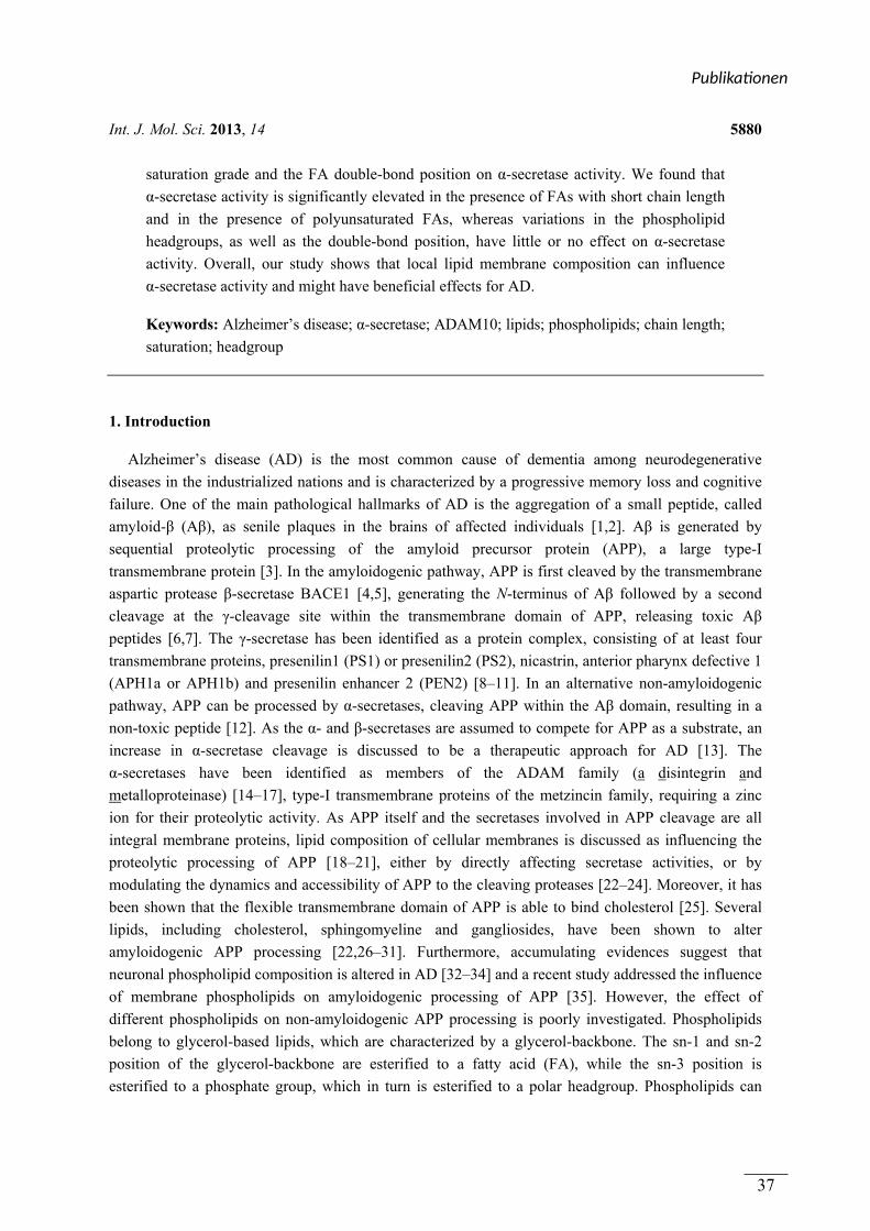
Int. J. Mol. Sci. 2013, 14 5880
saturation grade and the FA double-bond position on α-secretase activity. We found that
α-secretase activity is significantly elevated in the presence of FAs with short chain length
and in the presence of polyunsaturated FAs, whereas variations in the phospholipid
headgroups, as well as the double-bond position, have little or no effect on α-secretase
activity. Overall, our study shows that local lipid membrane composition can influence
α-secretase activity and might have beneficial effects for AD.
Keywords: Alzheimer’s disease; α-secretase; ADAM10; lipids; phospholipids; chain length;
saturation; headgroup
1. Introduction
Alzheimer’s disease (AD) is the most common cause of dementia among neurodegenerative
diseases in the industrialized nations and is characterized by a progressive memory loss and cognitive
failure. One of the main pathological hallmarks of AD is the aggregation of a small peptide, called
amyloid-β (Aβ), as senile plaques in the brains of affected individuals [1,2]. Aβ is generated by
sequential proteolytic processing of the amyloid precursor protein (APP), a large type-I
transmembrane protein [3]. In the amyloidogenic pathway, APP is first cleaved by the transmembrane
aspartic protease β-secretase BACE1 [4,5], generating the N-terminus of Aβ followed by a second
cleavage at the γ-cleavage site within the transmembrane domain of APP, releasing toxic Aβ
peptides [6,7]. The γ-secretase has been identified as a protein complex, consisting of at least four
transmembrane proteins, presenilin1 (PS1) or presenilin2 (PS2), nicastrin, anterior pharynx defective 1
(APH1a or APH1b) and presenilin enhancer 2 (PEN2) [8–11]. In an alternative non-amyloidogenic
pathway, APP can be processed by α-secretases, cleaving APP within the Aβ domain, resulting in a
non-toxic peptide [12]. As the α- and β-secretases are assumed to compete for APP as a substrate, an
increase in α-secretase cleavage is discussed to be a therapeutic approach for AD [13]. The
α-secretases have been identified as members of the ADAM family (a disintegrin and
metalloproteinase) [14–17], type-I transmembrane proteins of the metzincin family, requiring a zinc
ion for their proteolytic activity. As APP itself and the secretases involved in APP cleavage are all
integral membrane proteins, lipid composition of cellular membranes is discussed as influencing the
proteolytic processing of APP [18–21], either by directly affecting secretase activities, or by
modulating the dynamics and accessibility of APP to the cleaving proteases [22–24]. Moreover, it has
been shown that the flexible transmembrane domain of APP is able to bind cholesterol [25]. Several
lipids, including cholesterol, sphingomyeline and gangliosides, have been shown to alter
amyloidogenic APP processing [22,26–31]. Furthermore, accumulating evidences suggest that
neuronal phospholipid composition is altered in AD [32–34] and a recent study addressed the influence
of membrane phospholipids on amyloidogenic processing of APP [35]. However, the effect of
different phospholipids on non-amyloidogenic APP processing is poorly investigated. Phospholipids
belong to glycerol-based lipids, which are characterized by a glycerol-backbone. The sn-1 and sn-2
position of the glycerol-backbone are esterified to a fatty acid (FA), while the sn-3 position is
esterified to a phosphate group, which in turn is esterified to a polar headgroup. Phospholipids can
Publika onen
37
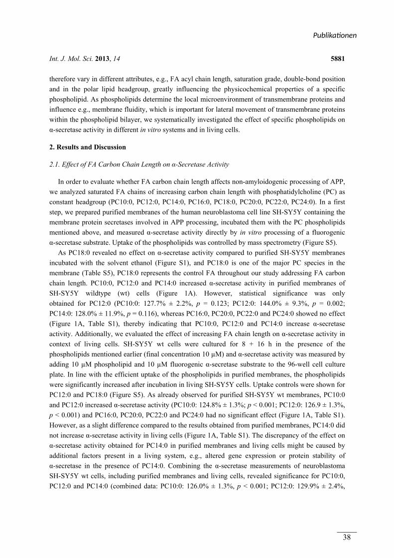
Int. J. Mol. Sci. 2013, 14 5881
therefore vary in different attributes, e.g., FA acyl chain length, saturation grade, double-bond position
and in the polar lipid headgroup, greatly influencing the physicochemical properties of a specific
phospholipid. As phospholipids determine the local microenvironment of transmembrane proteins and
influence e.g., membrane fluidity, which is important for lateral movement of transmembrane proteins
within the phospholipid bilayer, we systematically investigated the effect of specific phospholipids on
α-secretase activity in different in vitro systems and in living cells.
2. Results and Discussion
2.1. Effect of FA Carbon Chain Length on α-Secretase Activity
In order to evaluate whether FA carbon chain length affects non-amyloidogenic processing of APP,
we analyzed saturated FA chains of increasing carbon chain length with phosphatidylcholine (PC) as
constant headgroup (PC10:0, PC12:0, PC14:0, PC16:0, PC18:0, PC20:0, PC22:0, PC24:0). In a first
step, we prepared purified membranes of the human neuroblastoma cell line SH-SY5Y containing the
membrane protein secretases involved in APP processing, incubated them with the PC phospholipids
mentioned above, and measured α-secretase activity directly by in vitro processing of a fluorogenic
α-secretase substrate. Uptake of the phospholipids was controlled by mass spectrometry (Figure S5).
As PC18:0 revealed no effect on α-secretase activity compared to purified SH-SY5Y membranes
incubated with the solvent ethanol (Figure S1), and PC18:0 is one of the major PC species in the
membrane (Table S5), PC18:0 represents the control FA throughout our study addressing FA carbon
chain length. PC10:0, PC12:0 and PC14:0 increased α-secretase activity in purified membranes of
SH-SY5Y wildtype (wt) cells (Figure 1A). However, statistical significance was only
obtained for PC12:0 (PC10:0: 127.7% ± 2.2%, p = 0.123; PC12:0: 144.0% ± 9.3%, p = 0.002;
PC14:0: 128.0% ± 11.9%, p = 0.116), whereas PC16:0, PC20:0, PC22:0 and PC24:0 showed no effect
(Figure 1A, Table S1), thereby indicating that PC10:0, PC12:0 and PC14:0 increase α-secretase
activity. Additionally, we evaluated the effect of increasing FA chain length on α-secretase activity in
context of living cells. SH-SY5Y wt cells were cultured for 8 + 16 h in the presence of the
phospholipids mentioned earlier (final concentration 10 µM) and α-secretase activity was measured by
adding 10 µM phospholipid and 10 µM fluorogenic α-secretase substrate to the 96-well cell culture
plate. In line with the efficient uptake of the phospholipids in purified membranes, the phospholipids
were significantly increased after incubation in living SH-SY5Y cells. Uptake controls were shown for
PC12:0 and PC18:0 (Figure S5). As already observed for purified SH-SY5Y wt membranes, PC10:0
and PC12:0 increased α-secretase activity (PC10:0: 124.8% ± 1.3%; p < 0.001; PC12:0: 126.9 ± 1.3%,
p < 0.001) and PC16:0, PC20:0, PC22:0 and PC24:0 had no significant effect (Figure 1A, Table S1).
However, as a slight difference compared to the results obtained from purified membranes, PC14:0 did
not increase α-secretase activity in living cells (Figure 1A, Table S1). The discrepancy of the effect on
α-secretase activity obtained for PC14:0 in purified membranes and living cells might be caused by
additional factors present in a living system, e.g., altered gene expression or protein stability of
α-secretase in the presence of PC14:0. Combining the α-secretase measurements of neuroblastoma
SH-SY5Y wt cells, including purified membranes and living cells, revealed significance for PC10:0,
PC12:0 and PC14:0 (combined data: PC10:0: 126.0% ± 1.3%, p < 0.001; PC12:0: 129.9% ± 2.4%,
Publika onen
38
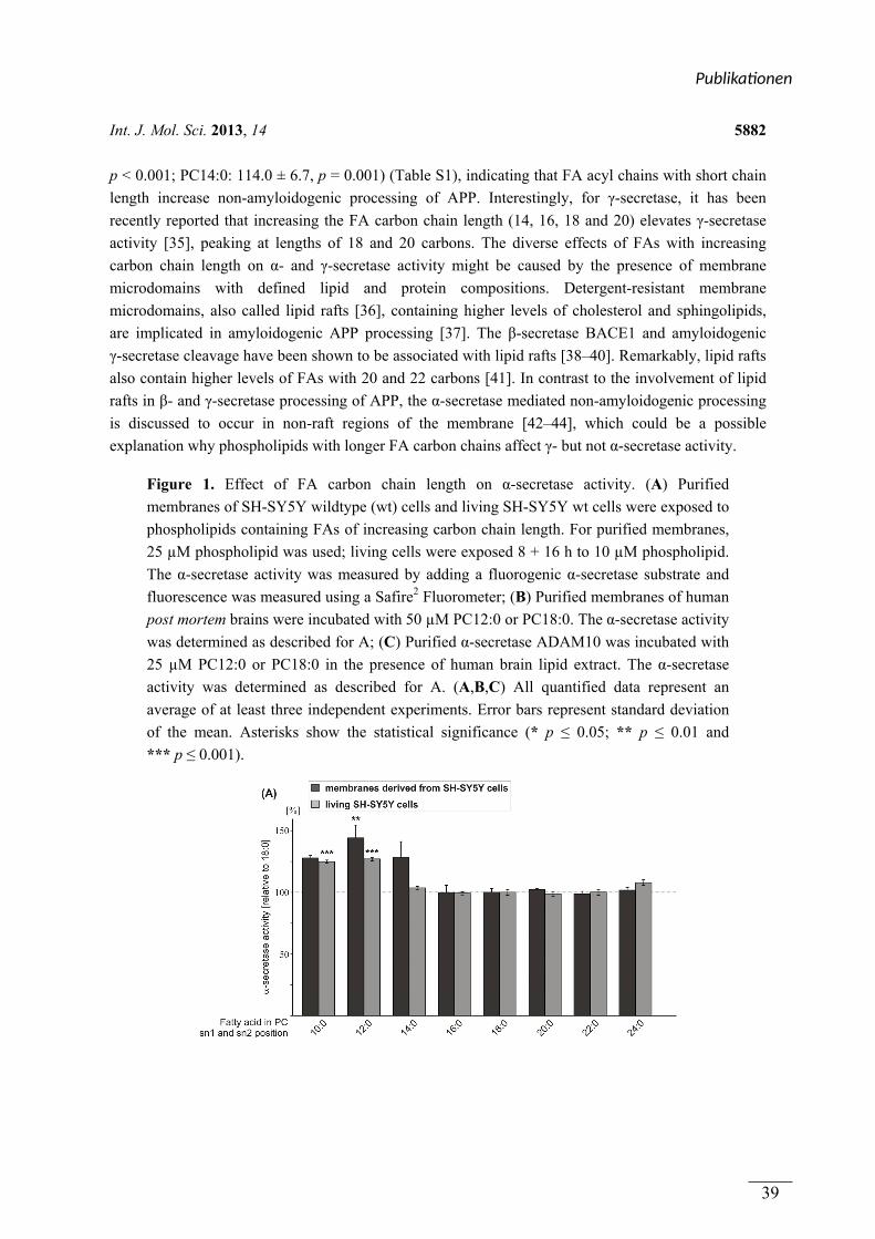
Int. J. Mol. Sci. 2013, 14 5882
p < 0.001; PC14:0: 114.0 ± 6.7, p = 0.001) (Table S1), indicating that FA acyl chains with short chain
length increase non-amyloidogenic processing of APP. Interestingly, for γ-secretase, it has been
recently reported that increasing the FA carbon chain length (14, 16, 18 and 20) elevates γ-secretase
activity [35], peaking at lengths of 18 and 20 carbons. The diverse effects of FAs with increasing
carbon chain length on α- and γ-secretase activity might be caused by the presence of membrane
microdomains with defined lipid and protein compositions. Detergent-resistant membrane
microdomains, also called lipid rafts [36], containing higher levels of cholesterol and sphingolipids,
are implicated in amyloidogenic APP processing [37]. The β-secretase BACE1 and amyloidogenic
γ-secretase cleavage have been shown to be associated with lipid rafts [38–40]. Remarkably, lipid rafts
also contain higher levels of FAs with 20 and 22 carbons [41]. In contrast to the involvement of lipid
rafts in β- and γ-secretase processing of APP, the α-secretase mediated non-amyloidogenic processing
is discussed to occur in non-raft regions of the membrane [42–44], which could be a possible
explanation why phospholipids with longer FA carbon chains affect γ- but not α-secretase activity.
Figure 1. Effect of FA carbon chain length on α-secretase activity. (A) Purified
membranes of SH-SY5Y wildtype (wt) cells and living SH-SY5Y wt cells were exposed to
phospholipids containing FAs of increasing carbon chain length. For purified membranes,
25 µM phospholipid was used; living cells were exposed 8 + 16 h to 10 µM phospholipid.
The α-secretase activity was measured by adding a fluorogenic α-secretase substrate and
fluorescence was measured using a Safire2 Fluorometer; (B) Purified membranes of human
post mortem brains were incubated with 50 µM PC12:0 or PC18:0. The α-secretase activity
was determined as described for A; (C) Purified α-secretase ADAM10 was incubated with
25 µM PC12:0 or PC18:0 in the presence of human brain lipid extract. The α-secretase
activity was determined as described for A. (A,B,C) All quantified data represent an
average of at least three independent experiments. Error bars represent standard deviation
of the mean. Asterisks show the statistical significance (* p ≤ 0.05; ** p ≤ 0.01 and
*** p ≤ 0.001).
Publika onen
39
Int. J. Mol. Sci. 2013, 14 5883
Figure 1. Cont.
As phospholipid PC12:0 showed the strongest effect on α-secretase activity in purified membranes
of SH-SY5Y wt cells and in living cells, we analyzed whether PC12:0 also increases non-amyloidogenic
APP processing in the lipid environment of human brain. Therefore, purified membranes of nine
healthy human post mortem brains were prepared and pooled, and α-secretase activity was measured in
the presence of PC12:0. Indeed, PC12:0 also significantly elevated α-secretase activity in purified
membranes of human post mortem brains compared to the control PC18:0 (PC12:0: 141.9% ± 7.6%,
p = 0.004) (Figure 1B; Table S1). The increase was nearly identical as observed for purified SH-SY5Y
wt membranes (PC12:0: 144.0% ± 9.3%, p = 0.002) (Figure 1A). Because these results suggest, that
PC12:0 directly increases α-secretase activity, we further analyzed the effect of PC12:0 on purified
enzyme. As the α-secretase ADAM10 has been recently shown to be the physiologically relevant
α-secretase in neurons [45], we selected ADAM10 to determine the effect of phospholipids on purified
α-secretase throughout this study. Purified ADAM10 was incubated with PC12:0 in the presence of
human post mortem brain lipid extract to reconstitute brain lipid environment. Also in this
experimental approach, ADAM10 activity was significantly increased in the presence of PC12:0
compared to PC18:0 (PC12:0: 132.0% ± 9.6%; p = 0.011) (Figure 1C, Table S1), validating that
PC12:0 directly influences α-secretase activity. However, although ADAM10 is discussed to be the
major α-secretase in neurons [45], further α-secretase candidates, e.g., ADAM17 and ADAM9 [14,16],
might be affected by PC12:0. This is a rather likely scenario, as we obtained for purified membranes of
SH-SY5Y wt cells and purified membranes of human post mortem brains, where total α-secretase and
not exclusively ADAM10 activity is determined, an even stronger increase in α-secretase activity.
However, our data suggest that ADAM10 is the α-secretase candidate mostly affected by shorter FA
acyl chain length.
2.2. Variations in the Phospholipid Headgroup on α-Secretase Activity
Beside FA carbon chain length the polar lipid headgroups of membrane phospholipids might affect
non-amyloidogenic APP processing. As we observed increased α-secretase activity in the presence of
Publika onen
40
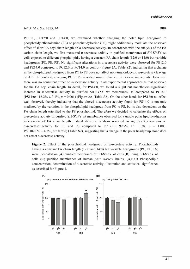
Int. J. Mol. Sci. 2013, 14 5884
PC10:0, PC12:0 and PC14:0, we examined whether changing the polar lipid headgroup to
phosphatidylethanolamine (PE) or phosphatidylserine (PS) might additionally modulate the observed
effect of short FA acyl chain length on α-secretase activity. In accordance with the analysis of the FA
carbon chain length, we first measured α-secretase activity in purified membranes of SH-SY5Y wt
cells exposed to different phospholipids, having a constant FA chain length (12:0 or 14:0) but variable
headgroups (PC, PE, PS). No significant alterations in α-secretase activity were observed for PE12:0
and PE14:0 compared to PC12:0 or PC14:0 as control (Figure 2A, Table S2), indicating that a change
in the phospholipid headgroup from PC to PE does not affect non-amyloidogenic α-secretase cleavage
of APP. In contrast, changing PC to PS revealed some influence on α-secretase activity. However,
there was no consistent effect on α-secretase activity in all experimental approaches as that observed
for the FA acyl chain length. In detail, for PS14:0, we found a slight but nonetheless significant,
increase in α-secretase activity in purified SH-SY5Y wt membranes, as compared to PC14:0
(PS14:0: 116.2% ± 3.1%, p = 0.001) (Figure 2A, Table S2). On the other hand, for PS12:0 no effect
was observed, thereby indicating that the altered α-secretase activity found for PS14:0 is not only
mediated by the variation in the phospholipid headgroup from PC to PS, but is also dependent on the
FA chain length esterified to the PS phospholipid. Therefore we decided to calculate the effects on
α-secretase activity in purified SH-SY5Y wt membranes observed for variable polar lipid headgroups
independent of FA chain length. Indeed statistical analysis revealed no significant alterations on
α-secretase activity for PE and PS compared to PC (PE: 99.7% +/− 1.0%, p = 1.000;
PS: 102.0% ± 4.5%, p = 0.936) (Table S2), suggesting that a change in the polar headgroup alone does
not affect α-secretase activity.
Figure 2. Effect of the phospholipid headgroup on α-secretase activity. Phospholipids
having a constant FA chain length (12:0 and 14:0) but variable headgroups (PC, PE, PS)
were incubated on (A) purified membranes of SH-SY5Y wt cells (B) living SH-SY5Y wt
cells (C) purified membranes of human post mortem brains. (A,B,C) Phospholipid
concentration, determination of α-secretase activity, illustration and statistical significance
as described for Figure 1.
Publika onen
41
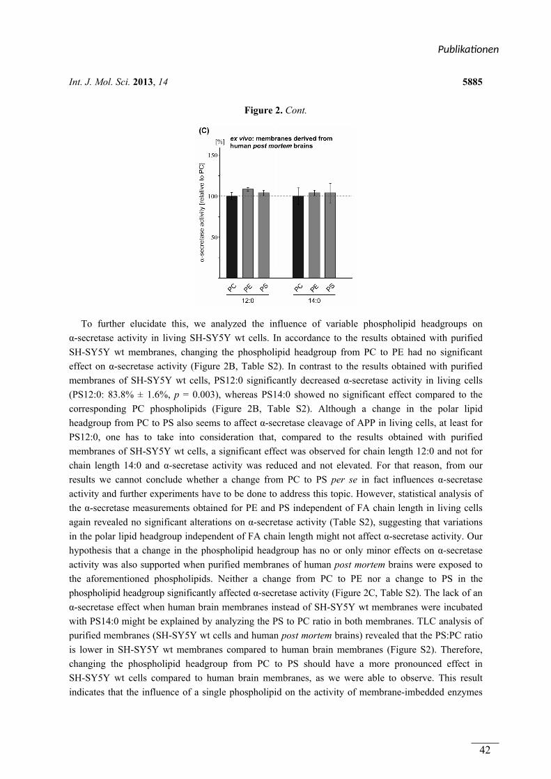
Int. J. Mol. Sci. 2013, 14 5885
Figure 2. Cont.
To further elucidate this, we analyzed the influence of variable phospholipid headgroups on
α-secretase activity in living SH-SY5Y wt cells. In accordance to the results obtained with purified
SH-SY5Y wt membranes, changing the phospholipid headgroup from PC to PE had no significant
effect on α-secretase activity (Figure 2B, Table S2). In contrast to the results obtained with purified
membranes of SH-SY5Y wt cells, PS12:0 significantly decreased α-secretase activity in living cells
(PS12:0: 83.8% ± 1.6%, p = 0.003), whereas PS14:0 showed no significant effect compared to the
corresponding PC phospholipids (Figure 2B, Table S2). Although a change in the polar lipid
headgroup from PC to PS also seems to affect α-secretase cleavage of APP in living cells, at least for
PS12:0, one has to take into consideration that, compared to the results obtained with purified
membranes of SH-SY5Y wt cells, a significant effect was observed for chain length 12:0 and not for
chain length 14:0 and α-secretase activity was reduced and not elevated. For that reason, from our
results we cannot conclude whether a change from PC to PS per se in fact influences α-secretase
activity and further experiments have to be done to address this topic. However, statistical analysis of
the α-secretase measurements obtained for PE and PS independent of FA chain length in living cells
again revealed no significant alterations on α-secretase activity (Table S2), suggesting that variations
in the polar lipid headgroup independent of FA chain length might not affect α-secretase activity. Our
hypothesis that a change in the phospholipid headgroup has no or only minor effects on α-secretase
activity was also supported when purified membranes of human post mortem brains were exposed to
the aforementioned phospholipids. Neither a change from PC to PE nor a change to PS in the
phospholipid headgroup significantly affected α-secretase activity (Figure 2C, Table S2). The lack of an
α-secretase effect when human brain membranes instead of SH-SY5Y wt membranes were incubated
with PS14:0 might be explained by analyzing the PS to PC ratio in both membranes. TLC analysis of
purified membranes (SH-SY5Y wt cells and human post mortem brains) revealed that the PS:PC ratio
is lower in SH-SY5Y wt membranes compared to human brain membranes (Figure S2). Therefore,
changing the phospholipid headgroup from PC to PS should have a more pronounced effect in
SH-SY5Y wt cells compared to human brain membranes, as we were able to observe. This result
indicates that the influence of a single phospholipid on the activity of membrane-imbedded enzymes
Publika onen
42
Int. J. Mol. Sci. 2013, 14 5886
strongly depends on the local lipid microenvironment. As Aβ deposition is a crucial event in AD and
α-seretase cleavage prevents the formation of Aβ by forming a non-toxic peptide [12], we conclude
from our results that changes in the phospholipid headgroup of phospholipid species with short FA
acyl chain length have no significant effects on α-secretase activity in the lipid environment of human
brain. Nevertheless, our results do not rule out that variations in the phospholipid headgroup might
play a role in AD by affecting other molecular pathways besides α-secretase activity. Several studies
addressed levels of the major membrane phospholipids in AD post mortem brains. PE and
phosphatidylinositol (PI) levels have been reported to be significantly decreased in AD
brains [33,34,46,47], whereas PS phospholipids are increased [33]. For PC Nitsch et al. reported a
significant decrease in AD brains [47], whereas PC was unaffected in the study by Wells et al. [33]. In
addition, mixtures of different phospholipid headgroup types, including PC, PE, PS, PI and
phosphatidic acid (PA) have been shown to alter γ-secretase cleavage in vitro [19,35]. Furthermore,
recently Nesic et al. reported, that α-secretase non-amyloidogenic APP processing is increased when
PE synthesis was inhibited in mammalian HEK293 cells whereas γ-secretase cleavage of APP is
reduced, resulting in decreased Aβ levels [48].
2.3. Effect of FA Saturation on α-Secretase Activity
Another important feature of membrane phospholipids is the saturation grade of the FA acyl chains.
In addition to FA chain length, the FA saturation grade strongly affects membrane fluidity, which is
important for membrane-dependent cellular functions, e.g., cell signaling, transmembrane protein
function, lateral diffusion of membrane proteins within the membrane, protein–lipid interactions and
vesicle formation [49]. To investigate whether FA acyl chain saturation of phospholipids influences
the proteolytic activity of α-secretase, we analyzed monounsaturated and polyunsaturated FA chains
esterified to a PC glycerol-backbone compared to saturated stearic acid (PC18:0, PC18:1, PC18:2,
PC18:3, PC20:4, PC20:5, PC22:6). Moreover, these FAs represent the most dominant species found in
brain (Table S5). Treatment of purified SH-SY5Y wt membranes with oleic acid (PC18:1 ∆9-cis),
linoleic acid (PC18:2), linolenic acid (PC18:3), arachidonic acid (AA, PC20:4) and docosahexaenoic
acid (DHA, PC22:6) showed no significant changes in α-secretase activity compared to the control
phospholipid PC18:0 (Figure 3A, Table S3). FA side chain eicosapentaenoic acid (EPA, PC20:5)
significantly elevated α-secretase activity (EPA/PC20:5: 123.1% ± 7.6%, p = 0.005) (Figure 3A,
Table S3). Exposing living SH-SY5Y wt cells to the aforementioned phospholipids (final
concentration 10 µM) revealed significantly increased α-secretase activity in the presence of
arachidonic acid, EPA and DHA, compared to control PC18:0 (AA/PC20:4: 124.7% ± 2.0%,
p = 0.001; EPA/PC20:5: 140.1% ± 14.2%, p < 0.001; DHA/PC22:6: 136.8% ± 2.2%, p < 0.001)
(Figure 3A, Table S3). Interestingly, in living cells, α-secretase activity was elevated with an
increasing number of double bonds within the acyl chains with the strongest increase for EPA and
DHA (Figure 3A), indicating that EPA and DHA are potent stimulators of non-amyloidogenic APP
processing in a living system. Calculating all α-secretase measurements for SH-SY5Y wt cells,
including purified membranes and living cells, showed similar results. Significance was obtained for
arachidonic acid, EPA and DHA (AA/PC20:4: 117.9% ± 3.5%, p = 0.002; EPA/PC20:5: 132.5% ± 5.9%,
p < 0.001; DHA/PC22:6: 123.5% ± 3.9%, p < 0.001) (Table S3).
Publika onen
43
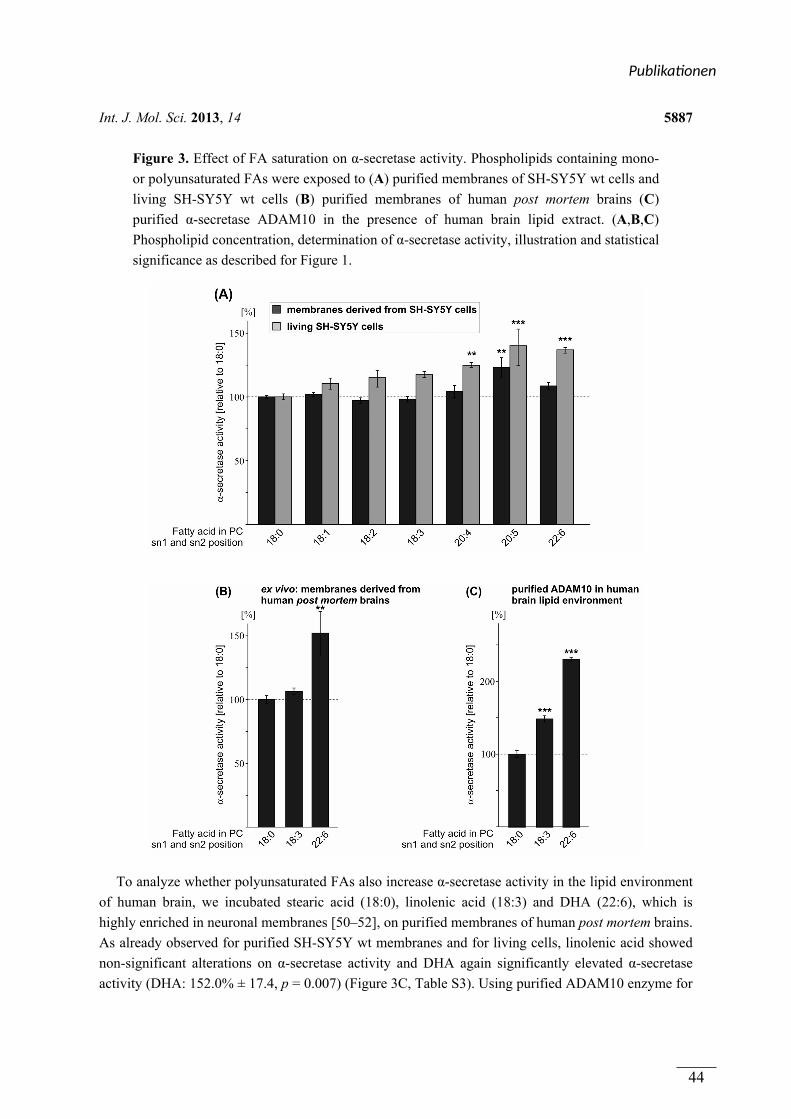
Int. J. Mol. Sci. 2013, 14 5887
Figure 3. Effect of FA saturation on α-secretase activity. Phospholipids containing mono-
or polyunsaturated FAs were exposed to (A) purified membranes of SH-SY5Y wt cells and
living SH-SY5Y wt cells (B) purified membranes of human post mortem brains (C)
purified α-secretase ADAM10 in the presence of human brain lipid extract. (A,B,C)
Phospholipid concentration, determination of α-secretase activity, illustration and statistical
significance as described for Figure 1.
To analyze whether polyunsaturated FAs also increase α-secretase activity in the lipid environment
of human brain, we incubated stearic acid (18:0), linolenic acid (18:3) and DHA (22:6), which is
highly enriched in neuronal membranes [50–52], on purified membranes of human post mortem brains.
As already observed for purified SH-SY5Y wt membranes and for living cells, linolenic acid showed
non-significant alterations on α-secretase activity and DHA again significantly elevated α-secretase
activity (DHA: 152.0% ± 17.4, p = 0.007) (Figure 3C, Table S3). Using purified ADAM10 enzyme for
Publika onen
44
Int. J. Mol. Sci. 2013, 14 5888
the incubation with linolenic acid (PC18:3) and DHA (PC22:6) in the presence of human post mortem
brain lipid extract, revealed that ADAM10 activity was significantly increased for both
polyunsaturated FAs (PC18:3: 148.2% ± 4.2%, p < 0.001; DHA/PC22:6: 229.7% ± 2.7%, p < 0.001)
(Figure 3C, Table S3). The extraordinary strong increase obtained with purified ADAM10 enzyme
might be explained by the lack of other membrane proteins potentially involved in α-secretase
regulation using brain lipid extract in contrast to purified membranes of human brains, which are not
devoid of integral membrane proteins. One might speculate that such a protein could be e.g.,
β-secretase BACE1, which competes with α-secretase for the initial cleavage of the substrate APP. In
purified membranes of human post mortem brains BACE1 is still present and can cleave the APP
substrate resulting in lower α-secretase cleavage compared to the α-secretase measurements using
purified α-secretase ADAM10 and brain lipid extract where BACE1 is lacking and ADAM10 has not
to compete with BACE1 for APP cleavage. In summary, all our results indicate that FA acyl chain
saturation of phospholipids affects α-secretase activity with a more pronounced effect with an
increasing number of double bonds within the hydrocarbon chains of the FAs. Arachidonic acid (four
double bonds), EPA (five double bonds) and DHA (six double bonds) significantly increased
α-secretase activity in living SH-SY5Y wt cells, whereas FAs with less than four double bonds
revealed minor or no significant effects. This finding is in line with the recent study by Yang et al.,
showing increased levels of α-secreted APP (sAPPα) in the culture medium of differentiated
SH-SY5Y cells exposed to arachidonic acid (20:4), EPA (20:5) and DHA (22:6) [53]. In the presence
of DHA sAPPα secretion has also been shown to be increased in HEK cells expressing the substrate
APP [54]. Additionally, stearic acid (18:0), oleic acid (18:1) and linoleic acid (18:2) showed no
changes in sAPPα secretion in the study by Yang et al. [53], validating our results obtained by
measuring α-secretase activity by the use of fluorogenic peptide. Furthermore, we validated our assay
by using α-secretase inhibitors. Utilizing these inhibitors, α-secretase activity was decreased and for
the non-cytotoxic α-secretase inhibitor GM6001, a reduction of sAPPα was also observed (Figure S4).
However, it cannot be ruled out that the natural substrate, the intact APP protein, has different physical
properties, e.g., affinity to the α-secretases, compared to the fluorogenic peptide and that these
differences might influence the effect strength of phospholipids on α-secretase activity.
Among the FAs analyzed, the main focus has to concentrate on DHA. DHA is highly enriched in
neuronal cell membranes [50–52] and DHA levels have been found to be decreased in the brain and
serum of AD patients and in AD post mortem brains [55–57]. Furthermore, several epidemiological
studies indicate an inverse correlation between DHA intake and AD incidence or cognitive
decline [58–61]. Importantly, DHA has also been shown to reduce Aβ generation in vitro and in
animal models of AD [35,62–68]. These findings, together with our finding that DHA strongly increases
non-amyloidogenic α-secretase activity in the lipid environment of human brain, indicate that DHA
might have protective effects on AD by decreasing amyloidogenic and increasing non-amyloidogenic
APP processing.
2.4. Effect of the Double-Bond Position on α-Secretase Activity
As we observed that FA chain length and the saturation grade of FAs influence α-secretase activity,
we asked whether shifting the position of the double-bond within the monounsaturated FA oleic acid
Publika onen
45
Int. J. Mol. Sci. 2013, 14 5889
PC18:1 might also affect α-secretase activity. Therefore, as a proof of principle, we analyzed PC18:1
∆6-cis and PC18:1 ∆9-cis. Exposure of purified SH-SY5Y wt membranes to the ∆6-cis isomer
revealed slightly—though significantly—increased α-secretase activity compared to PC18:1 ∆9-cis
(PC18:1 ∆6-cis: 106.1% ± 1.0%, p = 0.041) (Figure 4A, Table S4), whereas no effect on α-secretase
activity was obtained in living SH-SY5Y wt cells (Figure 4B, Table S4), thereby indicating that
changing the position of the double-bond within a monounsaturated FA chain has no or only minor
effects on non-amyloidogenic processing of APP. However, shifting the double-bond position further
from the center of the membrane bilayer has been recently shown to affect γ-secretase cleavage [35].
In this study, γ-secretase activity was reduced in PC18:1∆6-cis compared to PC18:1∆9-cis.
The observation that γ-secretase activity is affected more strongly by the position of the
double-bond-induced kink within the phospholipid bilayer as α-secretase activity might be explained
by the fact that γ-secretase cleaves the substrate APP within the middle of the phospholipid bilayer [69],
whereas α-secretase cleaves APP within the extracellular/luminal domain.
Figure 4. Effect of the double-bond position within the FA carbon chain on α-secretase
activity. Phospholipids containing either FA 18:1∆9-cis or FA 18:1∆6-cis were incubated
on (A) purified membranes of SH-SY5Y wt cells and (B) living SH-SY5Y wt cells;
(A,B) Phospholipid concentration, determination of α-secretase activity, illustration and
statistical significance as described for Figure 1.
3. Experimental Section
3.1. Chemicals and Reagents
All chemicals used here were purchased from Sigma Aldrich (Taufkirchen, Germany), if not stated
otherwise. Complete protease inhibitor cocktail was purchased from Roche (Grenzach-Wyhlen,
Germany), Aspartate protease inhibitor, β-secretase inhibitor, and fluorogenic substrate were
purchased from Calbiochem (Darmstadt, Germany). The following phospholipids were purchased
from Avanti Polar Lipids (Alabaster, AL, USA), except for PC20:5 (Sigma, Taufkirchen, Germany):
PC10:0—1,2-didecanoyl-sn-glycero-3-phosphocholine; PC12:0—1,2-dilauroyl-sn-glycero-3-
Publika onen
46
Int. J. Mol. Sci. 2013, 14 5890
phosphocholine; PC14:0—1,2-dimyristoyl-sn-glycero-3-phosphocholine; PC16:0—1,2-dipalmitoyl-
sn-glycero-3-phosphocholine; PC18:0—1,2-distearoyl-sn-glycero-3-phosphocholine; PC20:0—1,2-
diarachidoyl-sn-glycero-3-phosphocholine; PC22:0—1,2-dibehenoyl-sn-glycero-3-phosphocholine;
PC24:0—1,2-dilignoceroyl-sn-glycero-3-phosphocholine; PC18:1D9—1,2-dioleoyl-sn-glycero-3-
phosphocholine; PC18:1D6—1,2-dipetroselenoyl-sn-glycero-3-phosphocholine; PC18:2—1,2-
dilinoleoyl-sn-glycero-3-phosphocholine; PC18:3—1,2-dilinolenoyl-sn-glycero-3-phosphocholine;
PC20:4—1,2-diarachidonoyl-sn-glycero-3-phosphocholine; PC20:5—1,2-dieicosapentaenoyl-sn-
glycero-3-phosphocholine; PC22:6—1,2-didocosahexaenoyl-sn-glycero-3-phosphocholine;
PE12:01,2-dilauroyl-sn-glycero-3-phosphoethanolamine; PE14:0—1,2-dimyristoyl-sn-glycero-3-
phosphoethanolamine; PS12:0—1,2-dilauroyl-sn-glycero-3-phospho-L-serine; PS14:0—1,2-
dimyristoyl-sn-glycero-3-phospho-L-serine.
3.2. Cell Culture and Incubation with Phospholipids
SH-SY5Ywt cells were cultivated in Dulbecco’s Modified Eagle’s Medium supplemented with
10% FCS (PAN Biotech, Aidenbach, Germany) and non-essential amino acid solution. For incubation
with phospholipids, SH-SY5Ywt cells were plated on a 96-well plate (Thermo Scientific, Waltham,
MA, USA) and grown until confluency. Before incubation, cells were cultivated in medium containing
0.1% FCS for 4 h. Incubation was carried out for 8 h + 16 h with 10 µM phospholipid. Final
concentration of ethanol was less than 2‰, except PE- and PS-species (10‰).
3.3. Detection of α-Secretase Activity in Vivo
After incubation, SH-SY5Ywt cells were washed once with HEPES buffer, then 100 µL of cell
imaging solution (140 mM NaCl2, 5 mM KCl, 8 mM CaCl2, 1 mM MgCl2, 20 mM HEPES,
pH 7.4) containing 10 µM phospholipid and 10 µM fluorogenic α-secretase substrate (sequence:
Ac-RE-EDANS-VHHQK LVF-K-DABCYL-R-OH, Cat. No. 565767, Calbiochem, Darmstadt,
Germany) was added. Fluorescence was measured continuously for 10,000 s with an excitation
wavelength of 340 ± 10 nm and an emission wavelength of 490 ± 10 nm at 37 °C under light exclusion
using a Safire2 Fluorometer (Tecan, Crailsheim, Germany) [23]. Enzyme activity kinetics are shown in
the supplemental data section.
3.4. Human Post Mortem Brains
In total, nine human post mortem control brain samples were used. Brains were obtained from
BrainNet (Munich, Germany). No CERAD status was assigned to 4 out of 5 brain samples. The fifth
brain sample was assigned with CERAD status 0 (CERAD = the consortium to establish a registry for
AD, standardizing procedures for the evaluation and diagnosis if patients with AD. A, B, C, 0 as
described in http://cerad.mc.duke.edu/ [70]).
3.5. Preparation of Purified Membranes
SH-SY5Ywt cells and human post mortem brains were homogenized using a PotterS (Braun,
Melsungen, Germany) at maximum speed (25 strokes) on ice. Protein concentration was measured
Publika onen
47
Int. J. Mol. Sci. 2013, 14 5891
according to Smith et al. [71], as described in detail earlier [72]. Samples were adjusted to
1 mg/mL, centrifuged at 900 rcf for 10 min at 4 °C and the obtained post-nuclear fractions were
centrifuged at 55,000 rpm for 75 min at 4 °C. The pellet with purified membranes was resuspended
using cannulaes (BD, Franklin Lakes, NJ, USA) with decreasing diameter in sucrose buffer (200 mM
sucrose, 5 µM CaCl2, 5 µM ZnCl2) according to Grimm et al. [68].
3.6. Preparation of Human Brain Lipid Extract
Lipids were extracted according to Bligh and Dyer [73] with minor modifications [72,74]. In brief,
3.75 mL CHCl3:MeOH:HCl (37%) (1:2:0.06; v/v/v) was added to the sample and the mixture was
vortexed for 1 h at room temperature (RT). Then, 1.25 mL CHCl3 was added and vortexed again for
1 h at RT. Finally, 1.25 mL CHCl3 and 1.25 mL H2O were added and samples were vortexed for
another 10 min before centrifugation at 5000 rpm for 10 min. The lower phase containing lipids was
transferred to another glass tube and evaporated under nitrogen flow at 30 °C. Lipids were
resuspended in 1 mL H2O and the extraction cycle described earlier was repeated before lipids were
finally dissolved in ethanol.
3.7. Determination of α-Secretase Activity and in Vitro Incubation
Post-nuclear fractions of SH-SY5Ywt cells/human brain-purified membranes were incubated with
25 µM/50 µM phospholipids for 15 min at 37 °C while shaking (Multireax, Heidolph Instruments,
Schwabach, Germany). For determination of α-secretase activity, samples were adjusted to 100 µg of
protein and 4 µM of fluorogenic α-secretase substrate was added. Fluorescence was measured as
described above. Enzyme activity kinetics are shown in the supplemental data section.
3.8. Determination of ADAM10 Purified Enzyme Activity
Phospholipids (25 µM) were mixed with human brain lipid extract (5 µg lipid per 100 µL sucrose
buffer) and incubated with 100 ng of ADAM10 purified enzyme (Cat. no. A9975, Sigma Aldrich,
Taufkirchen, Germany) in sucrose buffer for 1 h at 4 °C while shaking, followed by sonification for
15 min on ice. ADAM10 purified enzyme activity was determined as described above using the
Infinite M1000 Pro Fluorometer. Enzyme activity kinetics are shown in the supplemental data section.
3.9. Detection of Phospholipid Species in SH-SY5Ywt Cells and Human Post Mortem Brains
Lipid extraction was performed as described above. One hundred microliter of each sample were
spotted on thin layer chromatography plate and lipids were analyzed as described before [75].
3.10. Mass Spectrometry Analysis
To determine phospholipid distribution in post-nuclear fractions and membranes derived from
human brains, phosphatidylcholine amounts were measured as described and validated in detail
earlier [76]. For analysis, we used 20 µL of post-nuclear fractions and human brain lipid extract
(for preparations see sections before). According to this method, additionally, the phospholipid uptake
into living cells and membranes of SH-SY5Y cells was determined.
Publika onen
48
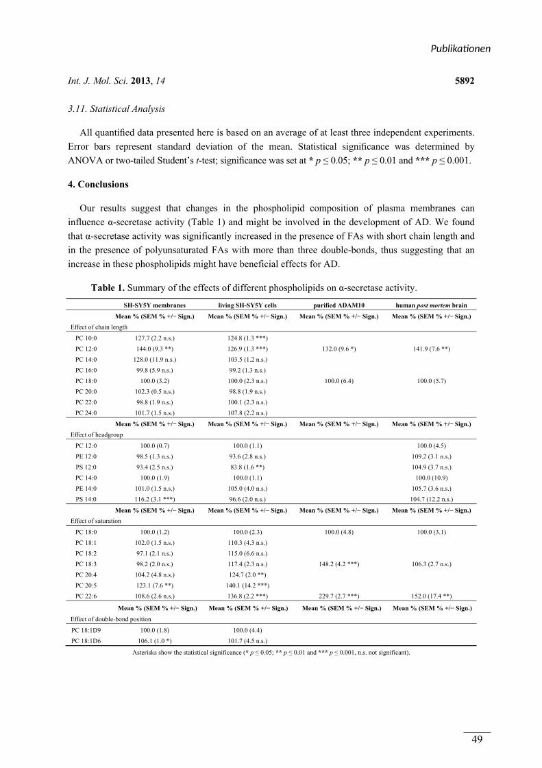
Int. J. Mol. Sci. 2013, 14 5892
3.11. Statistical Analysis
All quantified data presented here is based on an average of at least three independent experiments.
Error bars represent standard deviation of the mean. Statistical significance was determined by
ANOVA or two-tailed Student’s t-test; significance was set at * p ≤ 0.05; ** p ≤ 0.01 and *** p ≤ 0.001.
4. Conclusions
Our results suggest that changes in the phospholipid composition of plasma membranes can
influence α-secretase activity (Table 1) and might be involved in the development of AD. We found
that α-secretase activity was significantly increased in the presence of FAs with short chain length and
in the presence of polyunsaturated FAs with more than three double-bonds, thus suggesting that an
increase in these phospholipids might have beneficial effects for AD.
Table 1. Summary of the effects of different phospholipids on α-secretase activity.
SH-SY5Y membranes living SH-SY5Y cells purified ADAM10 human post mortem brain
Mean % (SEM % +/− Sign.) Mean % (SEM % +/− Sign.) Mean % (SEM % +/− Sign.) Mean % (SEM % +/− Sign.)
Effect of chain length
PC 10:0 127.7 (2.2 n.s.) 124.8 (1.3 ***)
PC 12:0 144.0 (9.3 **) 126.9 (1.3 ***) 132.0 (9.6 *) 141.9 (7.6 **)
PC 14:0 128.0 (11.9 n.s.) 103.5 (1.2 n.s.)
PC 16:0 99.8 (5.9 n.s.) 99.2 (1.3 n.s.)
PC 18:0 100.0 (3.2) 100.0 (2.3 n.s.) 100.0 (6.4) 100.0 (5.7)
PC 20:0 102.3 (0.5 n.s.) 98.8 (1.9 n.s.)
PC 22:0 98.8 (1.9 n.s.) 100.1 (2.3 n.s.)
PC 24:0 101.7 (1.5 n.s.) 107.8 (2.2 n.s.)
Mean % (SEM % +/− Sign.) Mean % (SEM % +/− Sign.) Mean % (SEM % +/− Sign.) Mean % (SEM % +/− Sign.)
Effect of headgroup
PC 12:0 100.0 (0.7) 100.0 (1.1) 100.0 (4.5)
PE 12:0 98.5 (1.3 n.s.) 93.6 (2.8 n.s.) 109.2 (3.1 n.s.)
PS 12:0 93.4 (2.5 n.s.) 83.8 (1.6 **) 104.9 (3.7 n.s.)
PC 14:0 100.0 (1.9) 100.0 (1.1) 100.0 (10.9)
PE 14:0 101.0 (1.5 n.s.) 105.0 (4.0 n.s.) 105.7 (3.6 n.s.)
PS 14:0 116.2 (3.1 ***) 96.6 (2.0 n.s.) 104.7 (12.2 n.s.)
Mean % (SEM % +/− Sign.) Mean % (SEM % +/− Sign.) Mean % (SEM % +/− Sign.) Mean % (SEM % +/− Sign.)
Effect of saturation
PC 18:0 100.0 (1.2) 100.0 (2.3) 100.0 (4.8) 100.0 (3.1)
PC 18:1 102.0 (1.5 n.s.) 110.3 (4.3 n.s.)
PC 18:2 97.1 (2.1 n.s.) 115.0 (6.6 n.s.)
PC 18:3 98.2 (2.0 n.s.) 117.4 (2.3 n.s.) 148.2 (4.2 ***) 106.3 (2.7 n.s.)
PC 20:4 104.2 (4.8 n.s.) 124.7 (2.0 **)
PC 20:5 123.1 (7.6 **) 140.1 (14.2 ***)
PC 22:6 108.6 (2.6 n.s.) 136.8 (2.2 ***) 229.7 (2.7 ***) 152.0 (17.4 **)
Mean % (SEM % +/− Sign.) Mean % (SEM % +/− Sign.) Mean % (SEM % +/− Sign.) Mean % (SEM % +/− Sign.)
Effect of double-bond position
PC 18:1D9 100.0 (1.8) 100.0 (4.4)
PC 18:1D6 106.1 (1.0 *) 101.7 (4.5 n.s.)
Asterisks show the statistical significance (* p ≤ 0.05; ** p ≤ 0.01 and *** p ≤ 0.001, n.s. not significant).
Publika onen
49
Int. J. Mol. Sci. 2013, 14 5893
Acknowledgments
We thank Inge Tomic for technical assistance. The research leading to these results has received
fundings from the EU FP7 project LipiDiDiet, Grant Agreement No 211696 (TH), the DFG (TH), the
Bundesministerium für Bildung, Forschung, Wissenschaft und Technologie via NGFNplus and KNDD
(TH), the HOMFOR 2009 (MG, TH) and the HOMFORexzellent 2011 (MG) (Saarland University
research grants). We thank Tecan Ltd. for their technical support.
Conflict of Interest
The authors declare no conflict of interest.
References
1. Masters, C.L.; Simms, G.; Weinman, N.A.; Multhaup, G.; McDonald, B.L.; Beyreuther, K.
Amyloid plaque core protein in Alzheimer disease and Down syndrome. Proc. Natl. Acad. Sci. USA
1985, 82, 4245–4249.
2. Selkoe, D.J. Alzheimer’s disease: Genes, proteins, and therapy. Physiol. Rev. 2001, 81, 741–766.
3. Dyrks, T.; Weidemann, A.; Multhaup, G.; Salbaum, J.M.; Lemaire, H.G.; Kang, J.; Muller-Hill, B.;
Masters, C.L.; Beyreuther, K. Identification, transmembrane orientation and biogenesis of the
amyloid A4 precursor of Alzheimer’s disease. EMBO J. 1988, 7, 949–957.
4. Vassar, R.; Bennett, B.D.; Babu-Khan, S.; Kahn, S.; Mendiaz, E.A.; Denis, P.; Teplow, D.B.;
Ross, S.; Amarante, P.; Loeloff, R.; et al. Beta-secretase cleavage of Alzheimer’s amyloid
precursor protein by the transmembrane aspartic protease BACE. Science 1999, 286, 735–741.
5. Sinha, S.; Anderson, J.P.; Barbour, R.; Basi, G.S.; Caccavello, R.; Davis, D.; Doan, M.;
Dovey, H.F.; Frigon, N.; Hong, J.; et al. Purification and cloning of amyloid precursor protein
beta-secretase from human brain. Nature 1999, 402, 537–540.
6. Haass, C.; Schlossmacher, M.G.; Hung, A.Y.; Vigo-Pelfrey, C.; Mellon, A.; Ostaszewski, B.L.;
Lieberburg, I.; Koo, E.H.; Schenk, D.; Teplow, D.B.; et al. Amyloid beta-peptide is produced by
cultured cells during normal metabolism. Nature 1992, 359, 322–325.
7. Shoji, M.; Golde, T.E.; Ghiso, J.; Cheung, T.T.; Estus, S.; Shaffer, L.M.; Cai, X.D.; McKay,
D.M.; Tintner, R.; Frangione, B.; et al. Production of the Alzheimer amyloid beta protein by
normal proteolytic processing. Science 1992, 258, 126–129.
8. Herreman, A.; Serneels, L.; Annaert, W.; Collen, D.; Schoonjans, L.; de Strooper, B. Total
inactivation of gamma-secretase activity in presenilin-deficient embryonic stem cells. Nat. Cell. Biol.
2000, 2, 461–462.
9. Takasugi, N.; Tomita, T.; Hayashi, I.; Tsuruoka, M.; Niimura, M.; Takahashi, Y.; Thinakaran, G.;
Iwatsubo, T. The role of presenilin cofactors in the gamma-secretase complex. Nature 2003, 422,
438–441.
10. Kimberly, W.T.; LaVoie, M.J.; Ostaszewski, B.L.; Ye, W.; Wolfe, M.S.; Selkoe, D.J.
Gamma-secretase is a membrane protein complex comprised of presenilin, nicastrin, Aph-1, and
Pen-2. Proc. Natl. Acad. Sci. USA 2003, 100, 6382–6387.
Publika onen
50
Int. J. Mol. Sci. 2013, 14 5894
11. Duering, M.; Grimm, M.O.; Grimm, H.S.; Schroder, J.; Hartmann, T. Mean age of onset in familial
Alzheimer’s disease is determined by amyloid beta 42. Neurobiol. Aging 2005, 26, 785–788.
12. Haass, C.; Hung, A.Y.; Schlossmacher, M.G.; Teplow, D.B.; Selkoe, D.J. Beta-Amyloid peptide
and a 3-kDa fragment are derived by distinct cellular mechanisms. J. Biol. Chem. 1993, 268,
3021–3024.
13. Lichtenthaler, S.F. Alpha-secretase in Alzheimer’s disease: Molecular identity, regulation and
therapeutic potential. J. Neurochem. 2011, 116, 10–21.
14. Buxbaum, J.D.; Liu, K.N.; Luo, Y.; Slack, J.L.; Stocking, K.L.; Peschon, J.J.; Johnson, R.S.;
Castner, B.J.; Cerretti, D.P.; Black, R.A. Evidence that tumor necrosis factor alpha converting
enzyme is involved in regulated alpha-secretase cleavage of the Alzheimer amyloid protein
precursor. J. Biol. Chem. 1998, 273, 27765–27767.
15. Lammich, S.; Kojro, E.; Postina, R.; Gilbert, S.; Pfeiffer, R.; Jasionowski, M.; Haass, C.;
Fahrenholz, F. Constitutive and regulated alpha-secretase cleavage of Alzheimer’s amyloid
precursor protein by a disintegrin metalloprotease. Proc. Natl. Acad. Sci. USA 1999, 96, 3922–3927.
16. Koike, H.; Tomioka, S.; Sorimachi, H.; Saido, T.C.; Maruyama, K.; Okuyama, A.;
Fujisawa-Sehara, A.; Ohno, S.; Suzuki, K.; Ishiura, S. Membrane-anchored metalloprotease
MDC9 has an alpha-secretase activity responsible for processing the amyloid precursor protein.
Biochem. J. 1999, 343, 371–375.
17. Allinson, T.M.; Parkin, E.T.; Turner, A.J.; Hooper, N.M. ADAMs family members as amyloid
precursor protein alpha-secretases. J. Neurosci. Res. 2003, 74, 342–352.
18. Hartmann, T.; Kuchenbecker, J.; Grimm, M.O. Alzheimer’s disease: The lipid connection.
J. Neurochem. 2007, 103, 159–170.
19. Osenkowski, P.; Ye, W.; Wang, R.; Wolfe, M.S.; Selkoe, D.J. Direct and potent regulation of
gamma-secretase by its lipid microenvironment. J. Biol. Chem. 2008, 283, 22529–22540.
20. Grimm, M.O.; Rothhaar, T.L.; Hartmann, T. The role of APP proteolytic processing in lipid
metabolism. Exp. Brain Res. 2012, 217, 365–375.
21. Lemkul, J.A.; Bevan, D.R. Lipid composition influences the release of Alzheimer’s amyloid
beta-peptide from membranes. Protein Sci. 2011, 20, 1530–1545.
22. Grimm, M.O.; Rothhaar, T.L.; Grösgen, S.; Burg, V.K.; Hundsdorfer, B.; Haupenthal, V.J.;
Friess, P.; Kins, S.; Grimm, H.S.; Hartmann, T. Trans fatty acids enhance amyloidogenic processing
of the Alzheimer amyloid precursor protein (APP). J. Nutr. Biochem. 2012, 23, 1214–1223.
23. Rothhaar, T.L.; Grosgen, S.; Haupenthal, V.J.; Burg, V.K.; Hundsdorfer, B.; Mett, J.;
Riemenschneider, M.; Grimm, H.S.; Hartmann, T.; Grimm, M.O. Plasmalogens inhibit APP
processing by directly affecting gamma-secretase activity in Alzheimer’s disease. Sci. World J.
2012, 2012, 141240.
24. Marenchino, M.; Williamson, P.T.; Murri, S.; Zandomeneghi, G.; Wunderli-Allenspach, H.;
Meier, B.H.; Kramer, S.D. Dynamics and Cleavability at the alpha-cleavage site of
APP(684–726) in different lipid environments. Biophys. J. 2008, 95, 1460–1473.
25. Barrett, P.J.; Song, Y.; van Horn, W.D.; Hustedt, E.J.; Schafer, J.M.; Hadziselimovic, A.;
Beel, A.J.; Sanders, C.R. The amyloid precursor protein has a flexible transmembrane domain and
binds cholesterol. Science 2012, 336, 1168–1171.
Publika onen
51
Int. J. Mol. Sci. 2013, 14 5895
26. Simons, M.; Keller, P.; de Strooper, B.; Beyreuther, K.; Dotti, C.G.; Simons, K. Cholesterol
depletion inhibits the generation of beta-amyloid in hippocampal neurons. Proc. Natl. Acad. Sci. USA
1998, 95, 6460–6464.
27. Grösgen, S.; Grimm, M.O.; Friess, P.; Hartmann, T. Role of amyloid beta in lipid homeostasis.
Biochim. Biophys. Acta 2010, 1801, 966–974.
28. Grimm, M.O.; Grimm, H.S.; Tomic, I.; Beyreuther, K.; Hartmann, T.; Bergmann, C. Independent
inhibition of Alzheimer disease beta- and gamma-secretase cleavage by lowered cholesterol
levels. J. Biol. Chem. 2008, 283, 11302–11311.
29. Grimm, M.O.; Grimm, H.S.; Patzold, A.J.; Zinser, E.G.; Halonen, R.; Duering, M.; Tschape, J.A.;
De Strooper, B.; Muller, U.; Shen, J.; et al. Regulation of cholesterol and sphingomyelin
metabolism by amyloid-beta and presenilin. Nat. Cell. Biol. 2005, 7, 1118–1123.
30. Zha, Q.; Ruan, Y.; Hartmann, T.; Beyreuther, K.; Zhang, D. GM1 ganglioside regulates the
proteolysis of amyloid precursor protein. Mol. Psychiatry 2004, 9, 946–952.
31. Grimm, M.O.; Zinser, E.G.; Grösgen, S.; Hundsdorfer, B.; Rothhaar, T.L.; Burg, V.K.;
Kaestner, L.; Bayer, T.A.; Lipp, P.; Muller, U.; et al. Amyloid precursor protein (APP) mediated
regulation of ganglioside homeostasis linking Alzheimer’s disease pathology with ganglioside
metabolism. PLoS One 2012, 7, e34095.
32. Svennerholm, L.; Gottfries, C.G. Membrane lipids, selectively diminished in Alzheimer brains,
suggest synapse loss as a primary event in early-onset form (type I) and demyelination in
late-onset form (type II). J. Neurochem. 1994, 62, 1039–1047.
33. Wells, K.; Farooqui, A.A.; Liss, L.; Horrocks, L.A. Neural membrane phospholipids in Alzheimer
disease. Neurochem. Res. 1995, 20, 1329–1333.
34. Prasad, M.R.; Lovell, M.A.; Yatin, M.; Dhillon, H.; Markesbery, W.R. Regional membrane
phospholipid alterations in Alzheimer’s disease. Neurochem. Res. 1998, 23, 81–88.
35. Holmes, O.; Paturi, S.; Ye, W.; Wolfe, M.S.; Selkoe, D.J. Effects of membrane lipids on the
activity and processivity of purified gamma-secretase. Biochemistry 2012, 51, 3565–3575.
36. Simons, K.; Ikonen, E. Functional rafts in cell membranes. Nature 1997, 387, 569–572.
37. Vetrivel, K.S.; Thinakaran, G. Membrane rafts in Alzheimer’s disease beta-amyloid production.
Biochim. Biophys. Acta 2010, 1801, 860–867.
38. Riddell, D.R.; Christie, G.; Hussain, I.; Dingwall, C. Compartmentalization of beta-secretase
(Asp2) into low-buoyant density, noncaveolar lipid rafts. Curr. Biol. 2001, 11, 1288–1293.
39. Vetrivel, K.S.; Cheng, H.; Lin, W.; Sakurai, T.; Li, T.; Nukina, N.; Wong, P.C.; Xu, H.;
Thinakaran, G. Association of gamma-secretase with lipid rafts in post-Golgi and endosome
membranes. J. Biol. Chem. 2004, 279, 44945–44954.
40. Vetrivel, K.S.; Cheng, H.; Kim, S.H.; Chen, Y.; Barnes, N.Y.; Parent, A.T.; Sisodia, S.S.;
Thinakaran, G. Spatial segregation of gamma-secretase and substrates in distinct membrane
domains. J. Biol. Chem. 2005, 280, 25892–25900.
41. Koumanov, K.S.; Tessier, C.; Momchilova, A.B.; Rainteau, D.; Wolf, C.; Quinn, P.J.
Comparative lipid analysis and structure of detergent-resistant membrane raft fractions isolated
from human and ruminant erythrocytes. Arch. Biochem. Biophys. 2005, 434, 150–158.
Publika onen
52
Int. J. Mol. Sci. 2013, 14 5896
42. Kojro, E.; Gimpl, G.; Lammich, S.; Marz, W.; Fahrenholz, F. Low cholesterol stimulates the
nonamyloidogenic pathway by its effect on the α-secretase ADAM 10. Proc. Natl. Acad. Sci. USA
2001, 98, 5815–5820.
43. Ehehalt, R.; Keller, P.; Haass, C.; Thiele, C.; Simons, K. Amyloidogenic processing of the
Alzheimer beta-amyloid precursor protein depends on lipid rafts. J. Cell Biol. 2003, 160, 113–123.
44. Parr-Sturgess, C.A.; Rushton, D.J.; Parkin, E.T. Ectodomain shedding of the Notch ligand
Jagged1 is mediated by ADAM17, but is not a lipid-raft-associated event. Biochem. J. 2010, 432,
283–294.
45. Kuhn, P.H.; Wang, H.; Dislich, B.; Colombo, A.; Zeitschel, U.; Ellwart, J.W.; Kremmer, E.;
Rossner, S.; Lichtenthaler, S.F. ADAM10 is the physiologically relevant, constitutive alpha-secretase
of the amyloid precursor protein in primary neurons. EMBO J. 2010, 29, 3020–3032.
46. Stokes, C.E.; Hawthorne, J.N. Reduced phosphoinositide concentrations in anterior temporal
cortex of Alzheimer-diseased brains. J. Neurochem. 1987, 48, 1018–1021.
47. Nitsch, R.M.; Blusztajn, J.K.; Pittas, A.G.; Slack, B.E.; Growdon, J.H.; Wurtman, R.J.
Evidence for a membrane defect in Alzheimer disease brain. Proc. Natl. Acad. Sci. USA 1992, 89,
1671–1675.
48. Nesic, I.; Guix, F.X.; Vennekens, K.; Michaki, V.; van Veldhoven, P.P.; Feiguin, F.;
de Strooper, B.; Dotti, C.G.; Wahle, T. Alterations in phosphatidylethanolamine levels affect the
generation of Abeta. Aging Cell 2012, 11, 63–72.
49. Escriba, P.V.; Gonzalez-Ros, J.M.; Goni, F.M.; Kinnunen, P.K.; Vigh, L.; Sanchez-Magraner, L.;
Fernandez, A.M.; Busquets, X.; Horvath, I.; Barcelo-Coblijn, G. Membranes: A meeting point for
lipids, proteins and therapies. J. Cell. Mol. Med. 2008, 12, 829–875.
50. Bazan, N.G.; Scott, B.L. Dietary omega-3 fatty acids and accumulation of docosahexaenoic acid
in rod photoreceptor cells of the retina and at synapses. Ups. J. Med. Sci. Suppl. 1990, 48, 97–107.
51. Ansari, K.A.; Shoeman, D.W. Arachidonic and docosahexanoic acid content of bovine brain
myelin: Implications for the pathogenesis of multiple sclerosis. Neurochem. Res. 1990, 15, 7–11.
52. Horrocks, L.A.; Farooqui, A.A. Docosahexaenoic acid in the diet: Its importance in maintenance
and restoration of neural membrane function. Prostaglandins Leukot Essent Fatty Acids 2004, 70,
361–372.
53. Yang, X.; Sheng, W.; Sun, G.Y.; Lee, J.C. Effects of fatty acid unsaturation numbers on
membrane fluidity and alpha-secretase-dependent amyloid precursor protein processing.
Neurochem. Int. 2011, 58, 321–329.
54. Eckert, G.P.; Chang, S.; Eckmann, J.; Copanaki, E.; Hagl, S.; Hener, U.; Muller, W.E.; Kogel, D.
Liposome-incorporated DHA increases neuronal survival by enhancing non-amyloidogenic APP
processing. Biochim. Biophys. Acta 2011, 1808, 236–243.
55. Soderberg, M.; Edlund, C.; Kristensson, K.; Dallner, G. Fatty acid composition of brain
phospholipids in aging and in Alzheimer’s disease. Lipids 1991, 26, 421–425.
56. Tully, A.M.; Roche, H.M.; Doyle, R.; Fallon, C.; Bruce, I.; Lawlor, B.; Coakley, D.; Gibney, M.J.
Low serum cholesteryl ester-docosahexaenoic acid levels in Alzheimer’s disease: A case-control
study. Br. J. Nutr. 2003, 89, 483–489.
Publika onen
53
Int. J. Mol. Sci. 2013, 14 5897
57. Cunnane, S.C.; Schneider, J.A.; Tangney, C.; Tremblay-Mercier, J.; Fortier, M.; Bennett, D.A.;
Morris, M.C. Plasma and brain fatty acid profiles in mild cognitive impairment and Alzheimer’s
disease. J. Alzheimers Dis. 2012, 29, 691–697.
58. Barberger-Gateau, P.; Letenneur, L.; Deschamps, V.; Peres, K.; Dartigues, J.F.; Renaud, S.
Fish, meat, and risk of dementia: Cohort study. Br. Med. J. 2002, 325, 932–933.
59. Morris, M.C.; Evans, D.A.; Bienias, J.L.; Tangney, C.C.; Bennett, D.A.; Wilson, R.S.;
Aggarwal, N.; Schneider, J. Consumption of fish and n-3 fatty acids and risk of incident
Alzheimer disease. Arch. Neurol. 2003, 60, 940–946.
60. Schaefer, E.J.; Bongard, V.; Beiser, A.S.; Lamon-Fava, S.; Robins, S.J.; Au, R.; Tucker, K.L.;
Kyle, D.J.; Wilson, P.W.; Wolf, P.A. Plasma phosphatidylcholine docosahexaenoic acid content
and risk of dementia and Alzheimer disease: The Framingham Heart Study. Arch. Neurol. 2006,
63, 1545–1550.
61. Van Gelder, B.M.; Tijhuis, M.; Kalmijn, S.; Kromhout, D. Fish consumption, n-3 fatty acids, and
subsequent 5-y cognitive decline in elderly men: The Zutphen Elderly Study. Am. J. Clin. Nutr.
2007, 85, 1142–1147.
62. Lukiw, W.J.; Cui, J.G.; Marcheselli, V.L.; Bodker, M.; Botkjaer, A.; Gotlinger, K.; Serhan, C.N.;
Bazan, N.G. A role for docosahexaenoic acid-derived neuroprotectin D1 in neural cell survival
and Alzheimer disease. J. Clin. Invest. 2005, 115, 2774–2783.
63. Perez, S.E.; Berg, B.M.; Moore, K.A.; He, B.; Counts, S.E.; Fritz, J.J.; Hu, Y.S.; Lazarov, O.;
Lah, J.J.; Mufson, E.J. DHA diet reduces AD pathology in young APPswe/PS1 Delta E9
transgenic mice: Possible gender effects. J. Neurosci. Res. 2010, 88, 1026–1040.
64. Oksman, M.; Iivonen, H.; Hogyes, E.; Amtul, Z.; Penke, B.; Leenders, I.; Broersen, L.;
Lutjohann, D.; Hartmann, T.; Tanila, H. Impact of different saturated fatty acid, polyunsaturated
fatty acid and cholesterol containing diets on beta-amyloid accumulation in APP/PS1 transgenic
mice. Neurobiol. Dis. 2006, 23, 563–572.
65. Calon, F.; Lim, G.P.; Yang, F.; Morihara, T.; Teter, B.; Ubeda, O.; Rostaing, P.; Triller, A.;
Salem, N., Jr.; Ashe, K.H.; et al. Docosahexaenoic acid protects from dendritic pathology in an
Alzheimer’s disease mouse model. Neuron 2004, 43, 633–645.
66. Hooijmans, C.R.; Rutters, F.; Dederen, P.J.; Gambarota, G.; Veltien, A.; van Groen, T.;
Broersen, L.M.; Lutjohann, D.; Heerschap, A.; Tanila, H.; et al. Changes in cerebral blood
volume and amyloid pathology in aged Alzheimer APP/PS1 mice on a docosahexaenoic acid
(DHA) diet or cholesterol enriched Typical Western Diet (TWD). Neurobiol. Dis. 2007, 28, 16–29.
67. Cole, G.M.; Ma, Q.L.; Frautschy, S.A. Omega-3 fatty acids and dementia. Prostaglandins Leukot
Essent Fatty Acids 2009, 81, 213–221.
68. Grimm, M.O.; Kuchenbecker, J.; Grösgen, S.; Burg, V.K.; Hundsdorfer, B.; Rothhaar, T.L.;
Friess, P.; de Wilde, M.C.; Broersen, L.M.; Penke, B.; et al. Docosahexaenoic acid reduces
amyloid beta production via multiple pleiotropic mechanisms. J. Biol. Chem. 2011, 286,
14028–14039.
69. Grziwa, B.; Grimm, M.O.; Masters, C.L.; Beyreuther, K.; Hartmann, T.; Lichtenthaler, S.F.
The transmembrane domain of the amyloid precursor protein in microsomal membranes is on
both sides shorter than predicted. J. Biol. Chem. 2003, 278, 6803–6808.
Publika onen
54
Int. J. Mol. Sci. 2013, 14 5898
70. Duyckaerts, C.; Hauw, J.J. Diagnosis and staging of Alzheimer disease. Neurobiol. Aging 1997,
18, S33–S42.
71. Smith, P.K.; Krohn, R.I.; Hermanson, G.T.; Mallia, A.K.; Gartner, F.H.; Provenzano, M.D.;
Fujimoto, E.K.; Goeke, N.M.; Olson, B.J.; Klenk, D.C. Measurement of protein using
bicinchoninic acid. Anal. Biochem. 1985, 150, 76–85.
72. Grimm, M.O.; Grösgen, S.; Rothhaar, T.L.; Burg, V.K.; Hundsdorfer, B.; Haupenthal, V.J.;
Friess, P.; Muller, U.; Fassbender, K.; Riemenschneider, M.; et al. Intracellular APP domain
regulates serine-palmitoyl-coa transferase expression and is affected in Alzheimer’s disease.
Int. J. Alzheimers Dis. 2011, 2011, 695413.
73. Bligh, E.G.; Dyer, W.J. A rapid method of total lipid extraction and purification. Can. J.
Biochem. Physiol. 1959, 37, 911–917.
74. Grimm, M.O.; Kuchenbecker, J.; Rothhaar, T.L.; Grösgen, S.; Hundsdorfer, B.; Burg, V.K.;
Friess, P.; Muller, U.; Grimm, H.S.; Riemenschneider, M.; et al. Plasmalogen synthesis is
regulated via alkyl-dihydroxyacetonephosphate-synthase by amyloid precursor protein processing
and is affected in Alzheimer’s disease. J. Neurochem. 2011, 116, 916–925.
75. Ruiz, J.I.; Ochoa, B. Quantification in the subnanomolar range of phospholipids and neutral lipids
by monodimensional thin-layer chromatography and image analysis. J. Lipid Res. 1997, 38,
1482–1489.
76. Grimm, M.O.; Grösgen, S.; Riemenschneider, M.; Tanila, H.; Grimm, H.S.; Hartmann, T.
From brain to food: Analysis of phosphatidylcholins, lyso-phosphatidylcholins and
phosphatidylcholin-plasmalogens derivates in Alzheimer’s disease human post mortem brains and
mice model via mass spectrometry. J. Chromatogr. A 2011, 1218, 7713–7722.
© 2013 by the authors; licensee MDPI, Basel, Switzerland. This article is an open access article
distributed under the terms and conditions of the Creative Commons Attribution license
(http://creativecommons.org/licenses/by/3.0/).
Publika onen
55
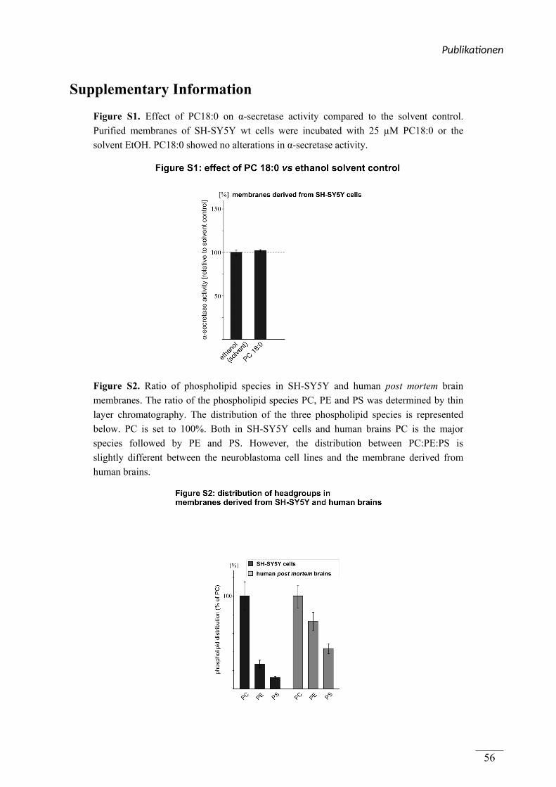
Supplementary Information
Figure S1. Effect of PC18:0 on α-secretase activity compared to the solvent control.
Purified membranes of SH-SY5Y wt cells were incubated with 25 µM PC18:0 or the
solvent EtOH. PC18:0 showed no alterations in α-secretase activity.
Figure S2. Ratio of phospholipid species in SH-SY5Y and human post mortem brain
membranes. The ratio of the phospholipid species PC, PE and PS was determined by thin
layer chromatography. The distribution of the three phospholipid species is represented
below. PC is set to 100%. Both in SH-SY5Y cells and human brains PC is the major
species followed by PE and PS. However, the distribution between PC:PE:PS is
slightly different between the neuroblastoma cell lines and the membrane derived from
human brains.
Publika onen
56
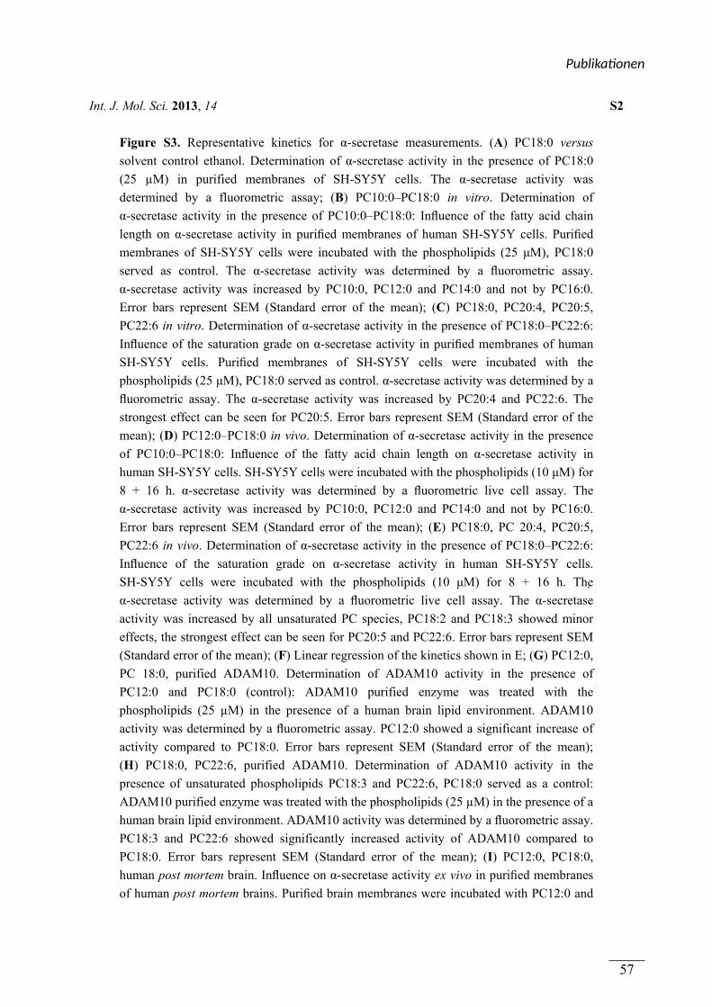
Int. J. Mol. Sci. 2013, 14 S2
Figure S3. Representative kinetics for α-secretase measurements. (A) PC18:0 versus
solvent control ethanol. Determination of α-secretase activity in the presence of PC18:0
(25 µM) in purified membranes of SH-SY5Y cells. The α-secretase activity was
determined by a fluorometric assay; (B) PC10:0–PC18:0 in vitro. Determination of
α-secretase activity in the presence of PC10:0–PC18:0: Influence of the fatty acid chain
length on α-secretase activity in purified membranes of human SH-SY5Y cells. Purified
membranes of SH-SY5Y cells were incubated with the phospholipids (25 μM), PC18:0
served as control. The α-secretase activity was determined by a fluorometric assay.
α-secretase activity was increased by PC10:0, PC12:0 and PC14:0 and not by PC16:0.
Error bars represent SEM (Standard error of the mean); (C) PC18:0, PC20:4, PC20:5,
PC22:6 in vitro. Determination of α-secretase activity in the presence of PC18:0–PC22:6:
Influence of the saturation grade on α-secretase activity in purified membranes of human
SH-SY5Y cells. Purified membranes of SH-SY5Y cells were incubated with the
phospholipids (25 μM), PC18:0 served as control. α-secretase activity was determined by a
fluorometric assay. The α-secretase activity was increased by PC20:4 and PC22:6. The
strongest effect can be seen for PC20:5. Error bars represent SEM (Standard error of the
mean); (D) PC12:0–PC18:0 in vivo. Determination of α-secretase activity in the presence
of PC10:0–PC18:0: Influence of the fatty acid chain length on α-secretase activity in
human SH-SY5Y cells. SH-SY5Y cells were incubated with the phospholipids (10 μM) for
8 + 16 h. α-secretase activity was determined by a fluorometric live cell assay. The
α-secretase activity was increased by PC10:0, PC12:0 and PC14:0 and not by PC16:0.
Error bars represent SEM (Standard error of the mean); (E) PC18:0, PC 20:4, PC20:5,
PC22:6 in vivo. Determination of α-secretase activity in the presence of PC18:0–PC22:6:
Influence of the saturation grade on α-secretase activity in human SH-SY5Y cells.
SH-SY5Y cells were incubated with the phospholipids (10 μM) for 8 + 16 h. The
α-secretase activity was determined by a fluorometric live cell assay. The α-secretase
activity was increased by all unsaturated PC species, PC18:2 and PC18:3 showed minor
effects, the strongest effect can be seen for PC20:5 and PC22:6. Error bars represent SEM
(Standard error of the mean); (F) Linear regression of the kinetics shown in E; (G) PC12:0,
PC 18:0, purified ADAM10. Determination of ADAM10 activity in the presence of
PC12:0 and PC18:0 (control): ADAM10 purified enzyme was treated with the
phospholipids (25 µM) in the presence of a human brain lipid environment. ADAM10
activity was determined by a fluorometric assay. PC12:0 showed a significant increase of
activity compared to PC18:0. Error bars represent SEM (Standard error of the mean);
(H) PC18:0, PC22:6, purified ADAM10. Determination of ADAM10 activity in the
presence of unsaturated phospholipids PC18:3 and PC22:6, PC18:0 served as a control:
ADAM10 purified enzyme was treated with the phospholipids (25 µM) in the presence of a
human brain lipid environment. ADAM10 activity was determined by a fluorometric assay.
PC18:3 and PC22:6 showed significantly increased activity of ADAM10 compared to
PC18:0. Error bars represent SEM (Standard error of the mean); (I) PC12:0, PC18:0,
human post mortem brain. Influence on α-secretase activity ex vivo in purified membranes
of human post mortem brains. Purified brain membranes were incubated with PC12:0 and
Publika onen
57
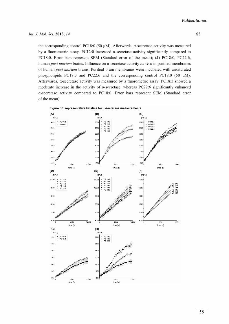
Int. J. Mol. Sci. 2013, 14 S3
the corresponding control PC18:0 (50 μM). Afterwards, α-secretase activity was measured
by a fluorometric assay. PC12:0 increased α-secretase activity significantly compared to
PC18:0. Error bars represent SEM (Standard error of the mean); (J) PC18:0, PC22:6,
human post mortem brains. Influence on α-secretase activity ex vivo in purified membranes
of human post mortem brains. Purified brain membranes were incubated with unsaturated
phospholipids PC18:3 and PC22:6 and the corresponding control PC18:0 (50 μM).
Afterwards, α-secretase activity was measured by a fluorometric assay. PC18:3 showed a
moderate increase in the activity of α-secretase, whereas PC22:6 significantly enhanced
α-secretase activity compared to PC18:0. Error bars represent SEM (Standard error
of the mean).
Publika onen
58
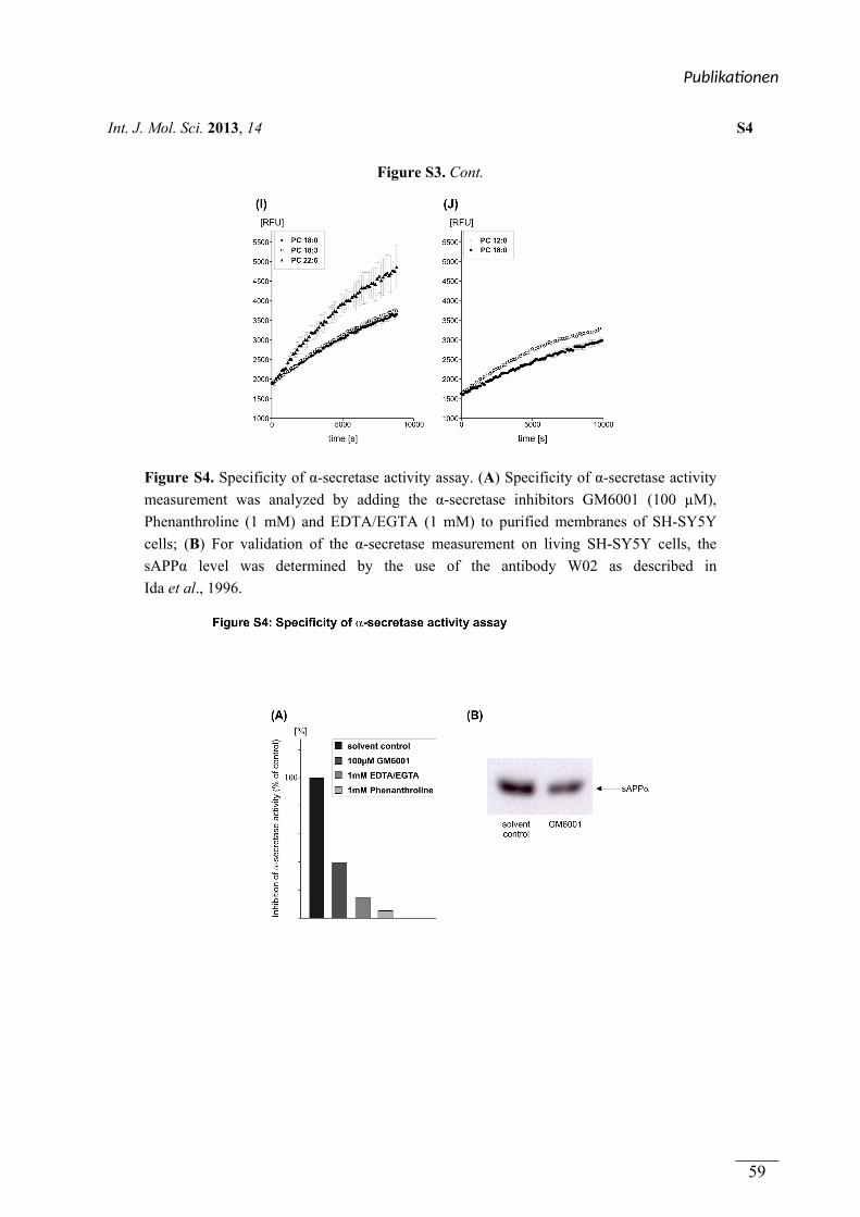
Int. J. Mol. Sci. 2013, 14 S4
Figure S3. Cont.
Figure S4. Specificity of α-secretase activity assay. (A) Specificity of α-secretase activity
measurement was analyzed by adding the α-secretase inhibitors GM6001 (100 µM),
Phenanthroline (1 mM) and EDTA/EGTA (1 mM) to purified membranes of SH-SY5Y
cells; (B) For validation of the α-secretase measurement on living SH-SY5Y cells, the
sAPPα level was determined by the use of the antibody W02 as described in
Ida et al., 1996.
Publika onen
59
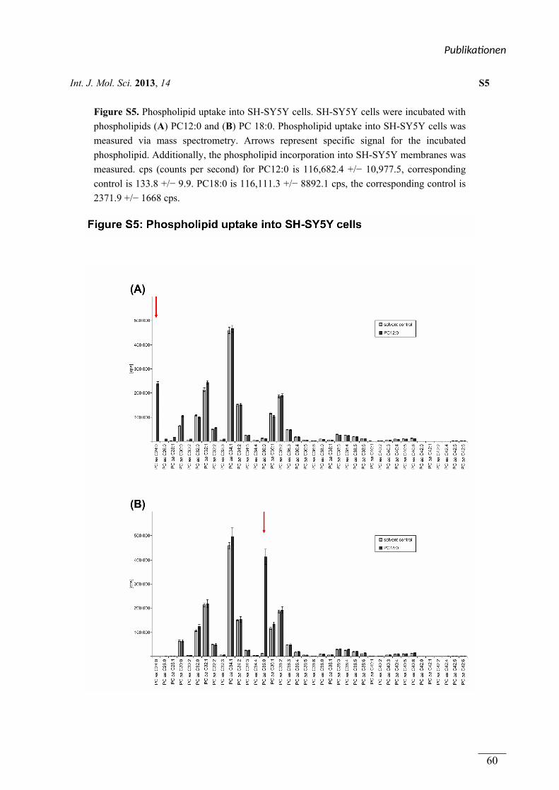
Int. J. Mol. Sci. 2013, 14 S5
Figure S5. Phospholipid uptake into SH-SY5Y cells. SH-SY5Y cells were incubated with
phospholipids (A) PC12:0 and (B) PC 18:0. Phospholipid uptake into SH-SY5Y cells was
measured via mass spectrometry. Arrows represent specific signal for the incubated
phospholipid. Additionally, the phospholipid incorporation into SH-SY5Y membranes was
measured. cps (counts per second) for PC12:0 is 116,682.4 +/− 10,977.5, corresponding
control is 133.8 +/− 9.9. PC18:0 is 116,111.3 +/− 8892.1 cps, the corresponding control is
2371.9 +/− 1668 cps.
Publika onen
60
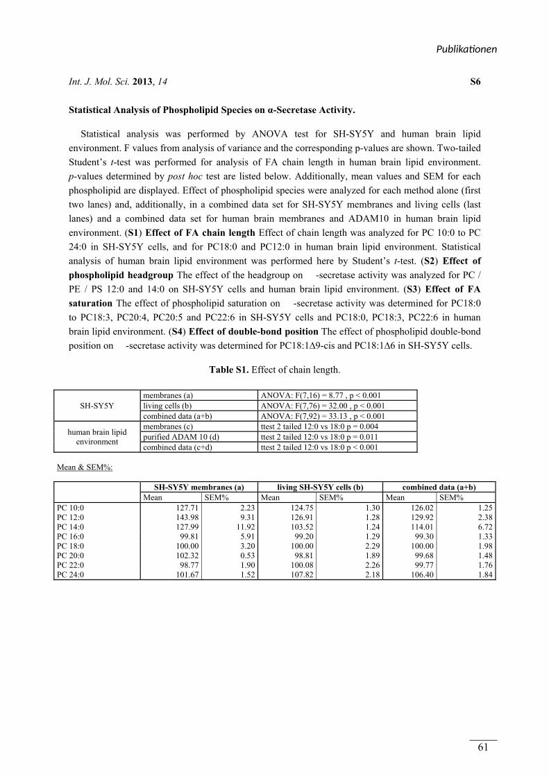
Int. J. Mol. Sci. 2013, 14 S6
Statistical Analysis of Phospholipid Species on α-Secretase Activity.
Statistical analysis was performed by ANOVA test for SH-SY5Y and human brain lipid
environment. F values from analysis of variance and the corresponding p-values are shown. Two-tailed
Student’s t-test was performed for analysis of FA chain length in human brain lipid environment.
p-values determined by post hoc test are listed below. Additionally, mean values and SEM for each
phospholipid are displayed. Effect of phospholipid species were analyzed for each method alone (first
two lanes) and, additionally, in a combined data set for SH-SY5Y membranes and living cells (last
lanes) and a combined data set for human brain membranes and ADAM10 in human brain lipid
environment. (S1) Effect of FA chain length Effect of chain length was analyzed for PC 10:0 to PC
24:0 in SH-SY5Y cells, and for PC18:0 and PC12:0 in human brain lipid environment. Statistical
analysis of human brain lipid environment was performed here by Student’s t-test. (S2) Effect of
phospholipid headgroup The effect of the headgroup on -secretase activity was analyzed for PC /
PE / PS 12:0 and 14:0 on SH-SY5Y cells and human brain lipid environment. (S3) Effect of FA
saturation The effect of phospholipid saturation on -secretase activity was determined for PC18:0
to PC18:3, PC20:4, PC20:5 and PC22:6 in SH-SY5Y cells and PC18:0, PC18:3, PC22:6 in human
brain lipid environment. (S4) Effect of double-bond position The effect of phospholipid double-bond
position on -secretase activity was determined for PC18:1∆9-cis and PC18:1∆6 in SH-SY5Y cells.
Table S1. Effect of chain length.
SH-SY5Y membranes (a) ANOVA: F(7,16) = 8.77 , p < 0.001 living cells (b) ANOVA: F(7,76) = 32.00 , p < 0.001 combined data (a+b) ANOVA: F(7,92) = 33.13 , p < 0.001
human brain lipid environment
membranes (c) ttest 2 tailed 12:0 vs 18:0 p = 0.004 purified ADAM 10 (d) ttest 2 tailed 12:0 vs 18:0 p = 0.011 combined data (c+d) ttest 2 tailed 12:0 vs 18:0 p < 0.001
Mean & SEM%:
SH-SY5Y membranes (a) living SH-SY5Y cells (b) combined data (a+b)
Mean SEM% Mean SEM% Mean SEM% PC 10:0 127.71 2.23 124.75 1.30 126.02 1.25PC 12:0 143.98 9.31 126.91 1.28 129.92 2.38PC 14:0 127.99 11.92 103.52 1.24 114.01 6.72PC 16:0 99.81 5.91 99.20 1.29 99.30 1.33PC 18:0 100.00 3.20 100.00 2.29 100.00 1.98PC 20:0 102.32 0.53 98.81 1.89 99.68 1.48PC 22:0 98.77 1.90 100.08 2.26 99.77 1.76PC 24:0 101.67 1.52 107.82 2.18 106.40 1.84
Publika onen
61
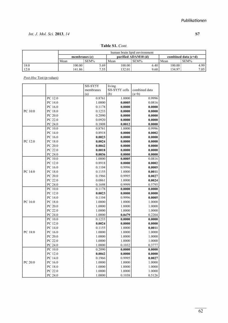
Int. J. Mol. Sci. 2013, 14 S7
Table S1. Cont.
human brain lipid environment
membranes (c) purified ADAM10 (d) combined data (c+d) Mean SEM% Mean SEM% Mean SEM%
18:0 100.00 5.69 100.00 6.40 100.00 4.9912:0 141.86 7.55 132.01 9.60 134.97 7.05
Post-Hoc Test (p-values)
SH-SY5Y membranes (a)
living SH-SY5Y cells (b)
combined data (a+b)
PC 10:0
PC 12.0 0.8761 1.0000 0.9996 PC 14.0 1.0000 0.0005 0.0816 PC 16.0 0.1178 0.0000 0.0000 PC 18.0 0.1233 0.0000 0.0000 PC 20.0 0.2090 0.0000 0.0000 PC 22.0 0.0920 0.0000 0.0000 PC 24.0 0.1808 0.0012 0.0000
PC 12:0
PC 10.0 0.8761 1.0000 0.9996 PC 14.0 0.8918 0.0000 0.0002 PC 16.0 0.0023 0.0000 0.0000 PC 18.0 0.0024 0.0000 0.0000 PC 20.0 0.0042 0.0000 0.0000 PC 22.0 0.0018 0.0000 0.0000 PC 24.0 0.0036 0.0000 0.0000
PC 14:0
PC 10.0 1.0000 0.0005 0.0816 PC 12.0 0.8918 0.0000 0.0002 PC 16.0 0.1104 0.9996 0.0005 PC 18.0 0.1155 1.0000 0.0011 PC 20.0 0.1966 0.9995 0.0027 PC 22.0 0.0861 1.0000 0.0024 PC 24.0 0.1698 0.9999 0.5795
PC 16:0
PC 10.0 0.1178 0.0000 0.0000 PC 12.0 0.0023 0.0000 0.0000 PC 14.0 0.1104 0.9996 0.0005 PC 18.0 1.0000 1.0000 1.0000 PC 20.0 1.0000 1.0000 1.0000 PC 22.0 1.0000 1.0000 1.0000 PC 24.0 1.0000 0.0479 0.2204
PC 18:0
PC 10.0 0.1233 0.0000 0.0000 PC 12.0 0.0024 0.0000 0.0000 PC 14.0 0.1155 1.0000 0.0011 PC 16.0 1.0000 1.0000 1.0000 PC 20.0 1.0000 1.0000 1.0000 PC 22.0 1.0000 1.0000 1.0000 PC 24.0 1.0000 0.1032 0.3777
PC 20:0
PC 10.0 0.2090 0.0000 0.0000 PC 12.0 0.0042 0.0000 0.0000 PC 14.0 0.1966 0.9995 0.0027 PC 16.0 1.0000 1.0000 1.0000 PC 18.0 1.0000 1.0000 1.0000 PC 22.0 1.0000 1.0000 1.0000 PC 24.0 1.0000 0.1038 0.5126
Publika onen
62
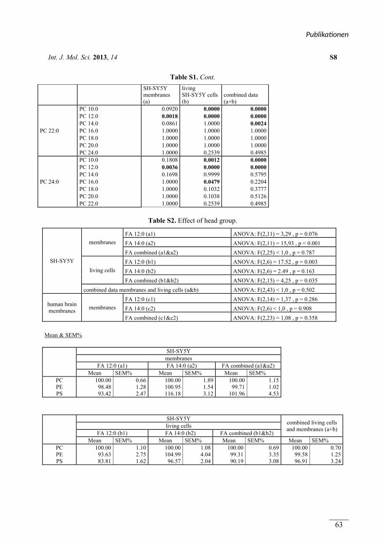
Int. J. Mol. Sci. 2013, 14 S8
Table S1. Cont.
SH-SY5Y membranes (a)
living SH-SY5Y cells (b)
combined data (a+b)
PC 22:0
PC 10.0 0.0920 0.0000 0.0000 PC 12.0 0.0018 0.0000 0.0000 PC 14.0 0.0861 1.0000 0.0024 PC 16.0 1.0000 1.0000 1.0000 PC 18.0 1.0000 1.0000 1.0000 PC 20.0 1.0000 1.0000 1.0000 PC 24.0 1.0000 0.2539 0.4985
PC 24:0
PC 10.0 0.1808 0.0012 0.0000 PC 12.0 0.0036 0.0000 0.0000 PC 14.0 0.1698 0.9999 0.5795 PC 16.0 1.0000 0.0479 0.2204 PC 18.0 1.0000 0.1032 0.3777 PC 20.0 1.0000 0.1038 0.5126 PC 22.0 1.0000 0.2539 0.4985
Table S2. Effect of head group.
SH-SY5Y
membranes
FA 12:0 (a1) ANOVA: F(2,11) = 3,29 , p = 0.076
FA 14:0 (a2) ANOVA: F(2,11) = 15,93 , p < 0.001
FA combined (a1&a2) ANOVA: F(2,25) < 1,0 , p = 0.787
living cells
FA 12:0 (b1) ANOVA: F(2,6) = 17.52 , p = 0.003
FA 14:0 (b2) ANOVA: F(2,6) = 2.49 , p = 0.163
FA combined (b1&b2) ANOVA: F(2,15) = 4,25 , p = 0.035
combined data membranes and living cells (a&b) ANOVA: F(2,43) < 1,0 , p = 0.502
human brain membranes
membranes
FA 12:0 (c1) ANOVA: F(2,14) = 1,37 , p = 0.286
FA 14:0 (c2) ANOVA: F(2,6) < 1,0 , p = 0.908
FA combined (c1&c2) ANOVA: F(2,23) = 1,08 , p = 0.358
Mean & SEM%
SH-SY5Y membranes
FA 12:0 (a1) FA 14:0 (a2) FA combined (a1&a2) Mean SEM% Mean SEM% Mean SEM%
PC 100.00 0.66 100.00 1.89 100.00 1.15 PE 98.48 1.28 100.95 1.54 99.71 1.02 PS 93.42 2.47 116.18 3.12 101.96 4.53
SH-SY5Y combined living cells and membranes (a+b)
living cells FA 12:0 (b1) FA 14:0 (b2) FA combined (b1&b2)
Mean SEM% Mean SEM% Mean SEM% Mean SEM% PC 100.00 1.10 100.00 1.08 100.00 0.69 100.00 0.70PE 93.63 2.75 104.99 4.04 99.31 3.35 99.58 1.25PS 83.81 1.62 96.57 2.04 90.19 3.08 96.91 3.24
Publika onen
63
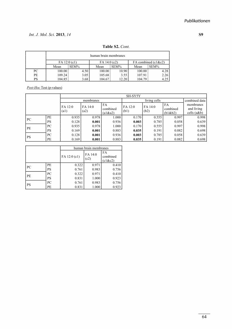
Int. J. Mol. Sci. 2013, 14 S9
Table S2. Cont.
human brain membranes
FA 12:0 (c1) FA 14:0 (c2) FA combined (c1&c2) Mean SEM% Mean SEM% Mean SEM%
PC 100.00 4.50 100.00 10.90 100.00 4.28 PE 109.24 3.05 105.68 3.55 107.91 2.26 PS 104.85 3.68 104.67 12.20 104.79 4.25
Post-Hoc Test (p-values)
SH-SY5Y membranes living cells combined data
membranes and living cells (a&b)
FA 12:0 (a1)
FA 14:0 (a2)
FA combined (a1&a2)
FA 12:0 (b1)
FA 14:0 (b2)
FA combined (b1&b2)
PC PE 0.935 0.978 1.000 0.170 0.555 0.997 0.998PS 0.128 0.001 0.936 0.003 0.785 0.058 0.639
PE PC 0.935 0.978 1.000 0.170 0.555 0.997 0.998PS 0.169 0.001 0.883 0.035 0.191 0.082 0.698
PS PC 0.128 0.001 0.936 0.003 0.785 0.058 0.639PE 0.169 0.001 0.883 0.035 0.191 0.082 0.698
human brain membranes
FA 12:0 (c1) FA 14:0 (c2)
FA combined (c1&c2)
PC PE 0.322 0.971 0.410 PS 0.761 0.983 0.756
PE PC 0.322 0.971 0.410 PS 0.831 1.000 0.923
PS PC 0.761 0.983 0.756 PE 0.831 1.000 0.923
Publika onen
64
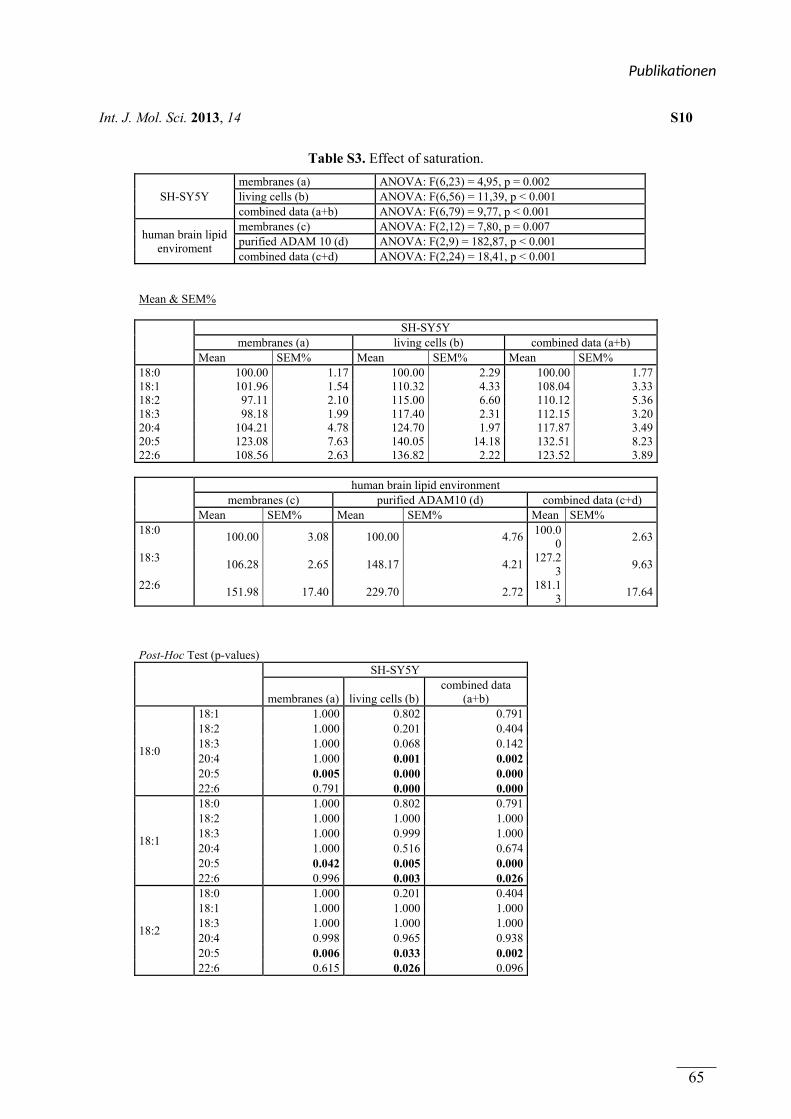
Int. J. Mol. Sci. 2013, 14 S10
Table S3. Effect of saturation.
SH-SY5Y membranes (a) ANOVA: F(6,23) = 4,95, p = 0.002 living cells (b) ANOVA: F(6,56) = 11,39, p < 0.001 combined data (a+b) ANOVA: F(6,79) = 9,77, p < 0.001
human brain lipid enviroment
membranes (c) ANOVA: F(2,12) = 7,80, p = 0.007 purified ADAM 10 (d) ANOVA: F(2,9) = 182,87, p < 0.001 combined data (c+d) ANOVA: F(2,24) = 18,41, p < 0.001
Mean & SEM%
SH-SY5Y
membranes (a) living cells (b) combined data (a+b) Mean SEM% Mean SEM% Mean SEM%
18:0 100.00 1.17 100.00 2.29 100.00 1.7718:1 101.96 1.54 110.32 4.33 108.04 3.3318:2 97.11 2.10 115.00 6.60 110.12 5.3618:3 98.18 1.99 117.40 2.31 112.15 3.2020:4 104.21 4.78 124.70 1.97 117.87 3.4920:5 123.08 7.63 140.05 14.18 132.51 8.2322:6 108.56 2.63 136.82 2.22 123.52 3.89
human brain lipid environment
membranes (c) purified ADAM10 (d) combined data (c+d) Mean SEM% Mean SEM% Mean SEM%
18:0 100.00 3.08 100.00 4.76
100.00
2.63
18:3 106.28 2.65 148.17 4.21
127.23
9.63
22:6 151.98 17.40 229.70 2.72
181.13
17.64
Post-Hoc Test (p-values)
SH-SY5Y
membranes (a) living cells (b)combined data
(a+b)
18:0
18:1 1.000 0.802 0.791 18:2 1.000 0.201 0.404 18:3 1.000 0.068 0.142 20:4 1.000 0.001 0.002 20:5 0.005 0.000 0.000 22:6 0.791 0.000 0.000
18:1
18:0 1.000 0.802 0.791 18:2 1.000 1.000 1.000 18:3 1.000 0.999 1.000 20:4 1.000 0.516 0.674 20:5 0.042 0.005 0.000 22:6 0.996 0.003 0.026
18:2
18:0 1.000 0.201 0.404 18:1 1.000 1.000 1.000 18:3 1.000 1.000 1.000 20:4 0.998 0.965 0.938 20:5 0.006 0.033 0.002 22:6 0.615 0.026 0.096
Publika onen
65
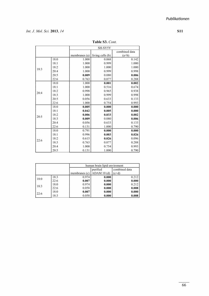
Int. J. Mol. Sci. 2013, 14 S11
Table S3. Cont.
SH-SY5Y
membranes (a) living cells (b)combined data
(a+b)
18:3
18:0 1.000 0.068 0.142 18:1 1.000 0.999 1.000 18:2 1.000 1.000 1.000 20:4 1.000 0.999 0.998 20:5 0.009 0.080 0.006 22:6 0.763 0.077 0.288
20:4
18:0 1.000 0.001 0.002 18:1 1.000 0.516 0.674 18:2 0.998 0.965 0.938 18:3 1.000 0.999 0.998 20:5 0.056 0.633 0.133 22:6 1.000 0.754 0.993
20:5
18:0 0.005 0.000 0.000 18:1 0.042 0.005 0.000 18:2 0.006 0.033 0.002 18:3 0.009 0.080 0.006 20:4 0.056 0.633 0.133 22:6 0.131 1.000 0.790
22:6
18:0 0.791 0.000 0.000 18:1 0.996 0.003 0.026 18:2 0.615 0.026 0.096 18:3 0.763 0.077 0.288 20:4 1.000 0.754 0.993 20:5 0.131 1.000 0.790
human brain lipid enviroment
membranes (c)purified ADAM 10 (d)
combined data (c+d)
18:0 18:3 0.974 0.000 0.212 22:6 0.007 0.000 0.000
18:3 18:0 0.974 0.000 0.212 22:6 0.058 0.000 0.008
22:6 18:0 0.007 0.000 0.000 18:3 0.058 0.000 0.008
Publika onen
66
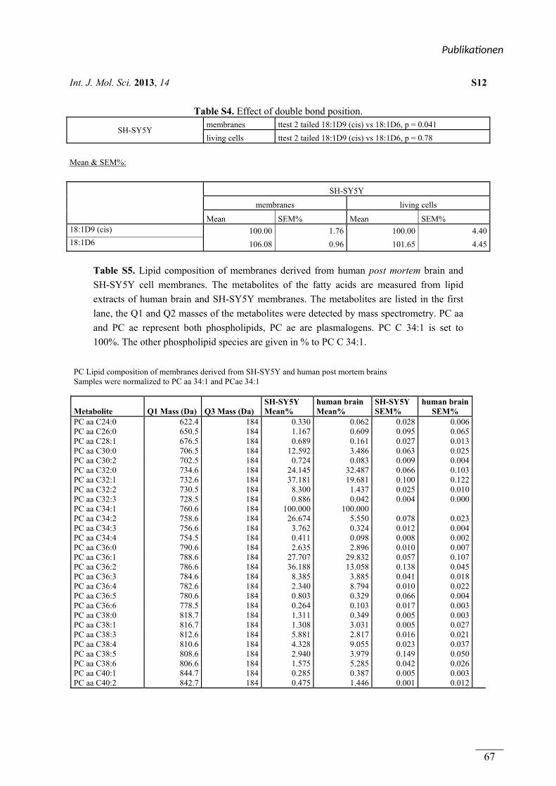
Int. J. Mol. Sci. 2013, 14 S12
Table S4. Effect of double bond position.
SH-SY5Y membranes ttest 2 tailed 18:1D9 (cis) vs 18:1D6, p = 0.041
living cells ttest 2 tailed 18:1D9 (cis) vs 18:1D6, p = 0.78
Mean & SEM%:
SH-SY5Y
membranes living cells
Mean SEM% Mean SEM% 18:1D9 (cis) 100.00 1.76 100.00 4.4018:1D6 106.08 0.96 101.65 4.45
Table S5. Lipid composition of membranes derived from human post mortem brain and
SH-SY5Y cell membranes. The metabolites of the fatty acids are measured from lipid
extracts of human brain and SH-SY5Y membranes. The metabolites are listed in the first
lane, the Q1 and Q2 masses of the metabolites were detected by mass spectrometry. PC aa
and PC ae represent both phospholipids, PC ae are plasmalogens. PC C 34:1 is set to
100%. The other phospholipid species are given in % to PC C 34:1.
PC Lipid composition of membranes derived from SH-SY5Y and human post mortem brains Samples were normalized to PC aa 34:1 and PCae 34:1
Metabolite Q1 Mass (Da) Q3 Mass (Da) SH-SY5Y Mean%
human brain Mean%
SH-SY5Y SEM%
human brain SEM%
PC aa C24:0 622.4 184 0.330 0.062 0.028 0.006PC aa C26:0 650.5 184 1.167 0.609 0.095 0.065PC aa C28:1 676.5 184 0.689 0.161 0.027 0.013PC aa C30:0 706.5 184 12.592 3.486 0.063 0.025PC aa C30:2 702.5 184 0.724 0.083 0.009 0.004PC aa C32:0 734.6 184 24.145 32.487 0.066 0.103PC aa C32:1 732.6 184 37.181 19.681 0.100 0.122PC aa C32:2 730.5 184 8.300 1.437 0.025 0.010PC aa C32:3 728.5 184 0.886 0.042 0.004 0.000PC aa C34:1 760.6 184 100.000 100.000 PC aa C34:2 758.6 184 26.674 5.550 0.078 0.023PC aa C34:3 756.6 184 3.762 0.324 0.012 0.004PC aa C34:4 754.5 184 0.411 0.098 0.008 0.002PC aa C36:0 790.6 184 2.635 2.896 0.010 0.007PC aa C36:1 788.6 184 27.707 29.832 0.057 0.107PC aa C36:2 786.6 184 36.188 13.058 0.138 0.045PC aa C36:3 784.6 184 8.385 3.885 0.041 0.018PC aa C36:4 782.6 184 2.340 8.794 0.010 0.022PC aa C36:5 780.6 184 0.803 0.329 0.066 0.004PC aa C36:6 778.5 184 0.264 0.103 0.017 0.003PC aa C38:0 818.7 184 1.311 0.349 0.005 0.003PC aa C38:1 816.7 184 1.308 3.031 0.005 0.027PC aa C38:3 812.6 184 5.881 2.817 0.016 0.021PC aa C38:4 810.6 184 4.328 9.055 0.023 0.037PC aa C38:5 808.6 184 2.940 3.979 0.149 0.050PC aa C38:6 806.6 184 1.575 5.285 0.042 0.026PC aa C40:1 844.7 184 0.285 0.387 0.005 0.003PC aa C40:2 842.7 184 0.475 1.446 0.001 0.012
Publika onen
67
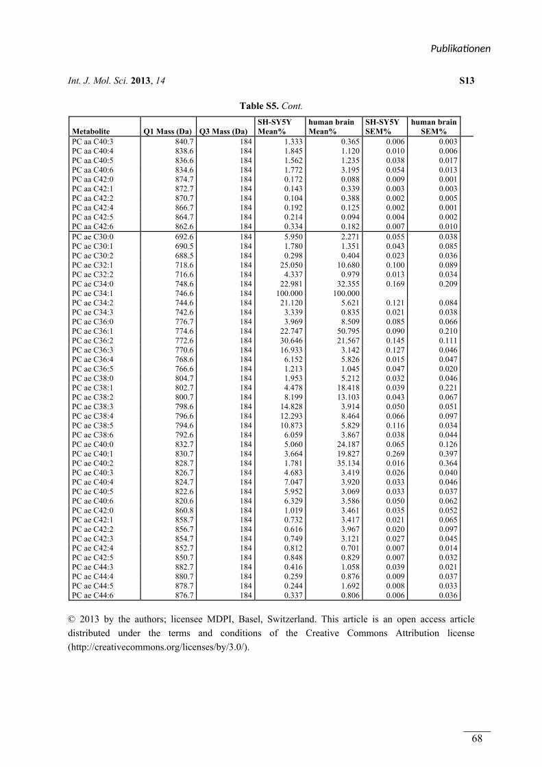
Int. J. Mol. Sci. 2013, 14 S13
Table S5. Cont.
Metabolite Q1 Mass (Da) Q3 Mass (Da) SH-SY5Y Mean%
human brain Mean%
SH-SY5Y SEM%
human brain SEM%
PC aa C40:3 840.7 184 1.333 0.365 0.006 0.003PC aa C40:4 838.6 184 1.845 1.120 0.010 0.006PC aa C40:5 836.6 184 1.562 1.235 0.038 0.017PC aa C40:6 834.6 184 1.772 3.195 0.054 0.013PC aa C42:0 874.7 184 0.172 0.088 0.009 0.001PC aa C42:1 872.7 184 0.143 0.339 0.003 0.003PC aa C42:2 870.7 184 0.104 0.388 0.002 0.005PC aa C42:4 866.7 184 0.192 0.125 0.002 0.001PC aa C42:5 864.7 184 0.214 0.094 0.004 0.002PC aa C42:6 862.6 184 0.334 0.182 0.007 0.010PC ae C30:0 692.6 184 5.950 2.271 0.055 0.038PC ae C30:1 690.5 184 1.780 1.351 0.043 0.085PC ae C30:2 688.5 184 0.298 0.404 0.023 0.036PC ae C32:1 718.6 184 25.050 10.680 0.100 0.089PC ae C32:2 716.6 184 4.337 0.979 0.013 0.034PC ae C34:0 748.6 184 22.981 32.355 0.169 0.209PC ae C34:1 746.6 184 100.000 100.000 PC ae C34:2 744.6 184 21.120 5.621 0.121 0.084PC ae C34:3 742.6 184 3.339 0.835 0.021 0.038PC ae C36:0 776.7 184 3.969 8.509 0.085 0.066PC ae C36:1 774.6 184 22.747 50.795 0.090 0.210PC ae C36:2 772.6 184 30.646 21.567 0.145 0.111PC ae C36:3 770.6 184 16.933 3.142 0.127 0.046PC ae C36:4 768.6 184 6.152 5.826 0.015 0.047PC ae C36:5 766.6 184 1.213 1.045 0.047 0.020PC ae C38:0 804.7 184 1.953 5.212 0.032 0.046PC ae C38:1 802.7 184 4.478 18.418 0.039 0.221PC ae C38:2 800.7 184 8.199 13.103 0.043 0.067PC ae C38:3 798.6 184 14.828 3.914 0.050 0.051PC ae C38:4 796.6 184 12.293 8.464 0.066 0.097PC ae C38:5 794.6 184 10.873 5.829 0.116 0.034PC ae C38:6 792.6 184 6.059 3.867 0.038 0.044PC ae C40:0 832.7 184 5.060 24.187 0.065 0.126PC ae C40:1 830.7 184 3.664 19.827 0.269 0.397PC ae C40:2 828.7 184 1.781 35.134 0.016 0.364PC ae C40:3 826.7 184 4.683 3.419 0.026 0.040PC ae C40:4 824.7 184 7.047 3.920 0.033 0.046PC ae C40:5 822.6 184 5.952 3.069 0.033 0.037PC ae C40:6 820.6 184 6.329 3.586 0.050 0.062PC ae C42:0 860.8 184 1.019 3.461 0.035 0.052PC ae C42:1 858.7 184 0.732 3.417 0.021 0.065PC ae C42:2 856.7 184 0.616 3.967 0.020 0.097PC ae C42:3 854.7 184 0.749 3.121 0.027 0.045PC ae C42:4 852.7 184 0.812 0.701 0.007 0.014PC ae C42:5 850.7 184 0.848 0.829 0.007 0.032PC ae C44:3 882.7 184 0.416 1.058 0.039 0.021PC ae C44:4 880.7 184 0.259 0.876 0.009 0.037PC ae C44:5 878.7 184 0.244 1.692 0.008 0.033PC ae C44:6 876.7 184 0.337 0.806 0.006 0.036
© 2013 by the authors; licensee MDPI, Basel, Switzerland. This article is an open access article
distributed under the terms and conditions of the Creative Commons Attribution license
(http://creativecommons.org/licenses/by/3.0/).
Publika onen
68
Publikationen
69
(4.2) Zusammenfassung von Publikation 2 und Beschreibung des
Eigenanteils
Plasmalogens Inhibit APP Processing by Directly Affecting γ-Secretase Activity in
Alzheimer’s Disease
Rothhaar TL, Grösgen S, Haupenthal VJ, Burg VK, Hundsdörfer B, Mett J, Riemenschneider
M, Grimm HS, Hartmann T, Grimm MO
In dieser Publikation wurde der Einfluss von Plasmalogenen (PL) auf die Aktivität der γ-
Sekretase untersucht. PL gehören zu den Glycerophospholipiden und sind durch eine
Vinyletherbindung an der sn1-Position des Glycerin zwischen der polaren Kopfgruppe und
den Fettsäureresten gekennzeichnet. An der sn2-Position des Glycerin liegt eine Esterbindung
der Fettsäure vor (Braverman & Moser 2012; Fahy et al. 2005). Die Struktur von PL ist in
figure 1 der Publikation 2 im Vergleich zum korrespondierenden Phospholipid dargestellt,
dieses trägt an sn1- und sn2-Position eine Esterbindung trägt und wird daher hier als
Esterphospholipid bezeichnet. Im humanen Gehirn stellen PE-PL, einen Anteil von bis zu
60% des Gehaltes an Glycerophospholipiden dar, zusammengefasst in (Farooqui & Horrocks
2001).
Um zu untersuchen welche PL im humanen Gehirn verändert sind, wurde in einem ersten
Schritt der Gehalt an PC-PL und PE-PL in humanen post-mortem Gehirnen von AD-Patienten
mittels Massenspektrometrie bestimmt. Als Kontrollgruppe wurden Gehirnproben von
Personen, die nicht an AD erkrankt waren, analysiert. Es konnte eine signifikante Reduktion
von fast allen PC-PL Spezies im AD-Gehirn gemessen werden. Am stärksten fiel der Effekt
bei PC-PL18:1 aus, daher wurde dieses PL für die folgenden Versuche ausgewählt. Zusätzlich
wurden PL mit mehrfach ungesättigten Fettsäuren verwendet, da diese in Neuronen des
Gehirns stark vertreten sind (Sun & Sun 1972). An der sn1-Position der verwendeten PL war
immer 18:0 gebunden. Als Kontrolle diente durchweg das korrespondierende Ester-
Phospholipid. Die verwendeten Lipide verursachten keine Zytotoxizität im Zellkulturmodell.
Die Spezifität der Sekretasemessungen wurde durch Einsatz von Inhibitoren kontrolliert.
Die analysierten PL bewirkten in SH-SY5Y Zellen keine Veränderung des Proteinspiegels
oder der Expression der α-, β- oder γ-Sekretase. Auch die Aktivität der β-Sekretase wurde
Publikationen
70
weitestgehend nicht durch PL verändert; einzig PC-PL 20:4 verursachte in lebenden SH-
SY5Y Zellen eine signifikante Reduktion der β-Sekretase Aktivität.
Im Gegensatz dazu reduzierten alle verwendeten PL signifikant die Aktivität der γ-Sekretase
und damit den letzten Schritt der Aβ-Synthese (Haass, Schlossmacher, et al. 1992). Die
Aktivität der α-Sekretase im nicht amyloidogenen Weg der APP Proteolyse wurde tendenziell
erhöht, für einige PL war dieser Effekt signifikant. Die Ergebnisse wurden in 6 humanen post-
mortem Gehirnproben von AD-Patienten verifiziert. In diesem ex vivo Experiment wurde
demonstriert, dass die Behandlung der Gehirnproben mit PL einer Reduktion der γ-Sekretase
Aktivität in 5 der 6 verwendeten Gehirnproben bewirkte.
Publikation 2 verdeutlicht, dass Veränderungen des PL-Spiegel nicht nur als Folge der AD-
Pathologie auftreten können, sondern auch direkt die amyloidogene APP-Prozessierung
beeinflussen.
Als Mitautor von Publikation 2 habe ich die Messung der α-Sekretase Aktivität in isolierten
Membranen von SH-SY5Y Zellen und Mausgehirnen durchgeführt. Dazu habe ich die
Inkubation von PNF´s aus SH-SY5Y Zellen bzw. Mausgehirnen mit PL, die Isolation von
Membranen, sowie die statistische Analyse der gewonnenen Daten durchgeführt (figure 5).
Weiterhin habe ich für diese Publikation die Spezifität der α-Sekretase Messung durch
Inkubation mit einem spezifischen α-Sekretase Inhibitor durchgeführt (figure S2).
Nachfolgend ist Publikation 2 zu finden. Das Original der Publikation ist auf der
Internetseite des Verlages Hindawi Publishing Cooperation (Kairo, Ägypten),
www.hindawi.com/journals/tswj/, unter der Nummer doi:10.1100/2012/141240 oder
unter der Internetseite http://dx.doi.org/10.1100/2012/141240 zu finden.
The Scientific World JournalVolume 2012, Article ID 141240, 15 pagesdoi:10.1100/2012/141240
The cientificWorldJOURNAL
Research Article
Plasmalogens Inhibit APP Processing by Directly Affectingγ-Secretase Activity in Alzheimer’s Disease
Tatjana L. Rothhaar,1 Sven Grosgen,1 Viola J. Haupenthal,1 Verena K. Burg,1
Benjamin Hundsdorfer,1 Janine Mett,1 Matthias Riemenschneider,2 Heike S. Grimm,1
Tobias Hartmann,1, 3, 4 and Marcus O. W. Grimm1, 3, 4
1 Experimental Neurology, Saarland University, Kirrbergerstraβe, 66421 Homburg/Saar, Germany2 Psychiatry, Saarland University, Kirrbergerstraβe, 66421 Homburg/Saar, Germany3 Deutsches Institut fur DemenzPravention (DIDP), Universitat des Saarlandes, Kirrbergerstraβe, 66421 Homburg/Saar, Germany4 Neurodegeneration and Neurobiology, Saarland University, Kirrbergerstraβe, 66421 Homburg/Saar, Germany
Correspondence should be addressed to Marcus O. W. Grimm, [email protected]
Received 10 October 2011; Accepted 30 November 2011
Academic Editor: Dietmar R. Thal
Copyright © 2012 Tatjana L. Rothhaar et al. This is an open access article distributed under the Creative Commons AttributionLicense, which permits unrestricted use, distribution, and reproduction in any medium, provided the original work is properlycited.
Lipids play an important role as risk or protective factors in Alzheimer’s disease (AD). Previously it has been shown thatplasmalogens, the major brain phospholipids, are altered in AD. However, it remained unclear whether plasmalogens themselvesare able to modulate amyloid precursor protein (APP) processing or if the reduced plasmalogen level is a consequence of AD.Here we identify the plasmalogens which are altered in human AD postmortem brains and investigate their impact on APPprocessing resulting in Aβ production. All tested plasmalogen species showed a reduction in γ-secretase activity whereas β- andα-secretase activity mainly remained unchanged. Plasmalogens directly affected γ-secretase activity, protein and RNA level ofthe secretases were unaffected, pointing towards a direct influence of plasmalogens on γ-secretase activity. Plasmalogens werealso able to decrease γ-secretase activity in human postmortem AD brains emphasizing the impact of plasmalogens in AD. Insummary our findings show that decreased plasmalogen levels are not only a consequence of AD but that plasmalogens alsodecrease APP processing by directly affecting γ-secretase activity, resulting in a vicious cycle: Aβ reduces plasmalogen levels andreduced plasmalogen levels directly increase γ-secretase activity leading to an even stronger production of Aβ peptides.
1. Introduction
Plasmalogens are glycerophospholipids and major constit-uents of neuronal membranes. Beside human brain, whereplasmalogens represent almost 20% of total glycerophospho-lipids, they can be found in all mammalian tissues, especiallyin the heart muscle [1–3]. Characteristic of plasmalogens isan enol ether double bond at the sn-1 position of the glycerolbackbone (Figure 1), which makes plasmalogens more sus-ceptible to oxidative stress than the corresponding ester-bonded glycerophospholipid, thus protecting cells from oxi-dative stress [4]. Beside their function as antioxidants, plas-malogens are involved in membrane fusion [5, 6], ion trans-port [7–9], and cholesterol efflux [10, 11]. Furthermore,plasmalogens can be hydrolyzed by plasmalogen-selective
phospholipase A2 [3, 12], generating fatty acids like arachi-donic acid, which is important for modulating ion channels,regulating different enzyme activities like protein kinase A,protein kinase C, NADPH oxidase, Na+K+-ATPase, and oth-ers [13]. Arachidonic acid released from plasmalogens canbe metabolized to eicosanoids, acting as second messengers[14]. Due to the fact that plasmalogens represent major con-stituents of neuronal membranes and are involved in dif-ferent cellular processes, it is not unexpected that neuronalfunction also depends on a delicate balance in lipid composi-tion of cellular membranes. Alterations of plasmalogen levelsoccur in several neurological disorders including Alzheimer’sdisease (AD) [15–17], spinal cord trauma [18], ischemia[19, 20], Niemann-Pick disease [21], and multiple sclerosis[22]. For AD, plasmalogen levels have been described to be
Publika onen
71
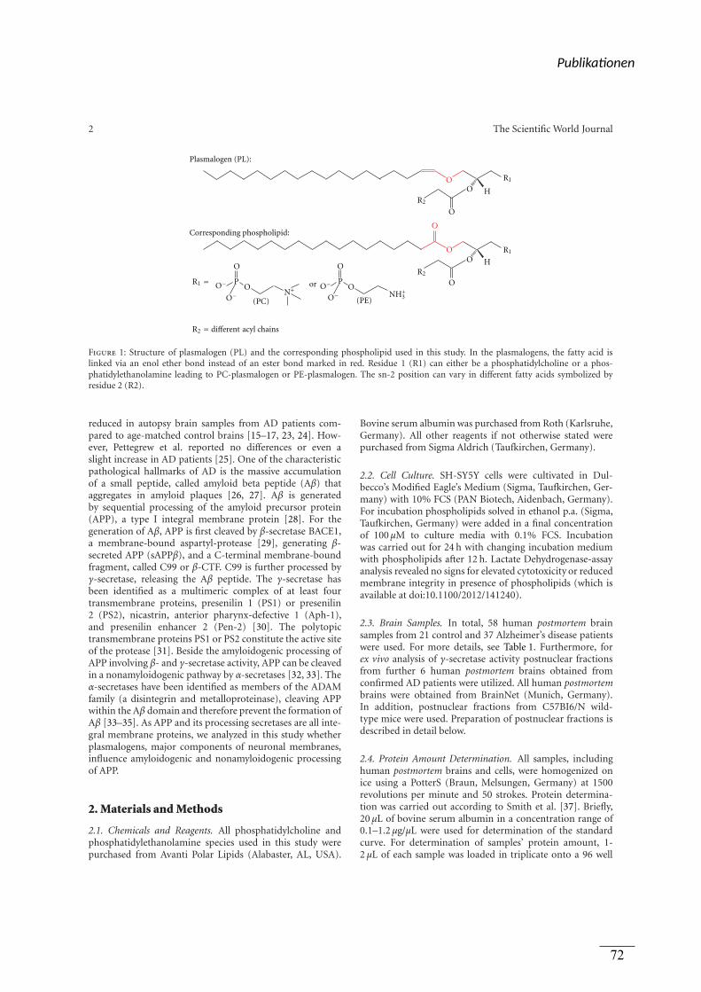
2 The Scientific World Journal
O
OP
(PC)
or
(PE)
O
OP
OO
H
O
O
O
O
O
H
N+O−
O−O−
O−
R1 =
R2 = different acyl chains
Plasmalogen (PL):
Corresponding phospholipid:
NH+3
R1
R1
R2
R2
Figure 1: Structure of plasmalogen (PL) and the corresponding phospholipid used in this study. In the plasmalogens, the fatty acid islinked via an enol ether bond instead of an ester bond marked in red. Residue 1 (R1) can either be a phosphatidylcholine or a phos-phatidylethanolamine leading to PC-plasmalogen or PE-plasmalogen. The sn-2 position can vary in different fatty acids symbolized byresidue 2 (R2).
reduced in autopsy brain samples from AD patients com-pared to age-matched control brains [15–17, 23, 24]. How-ever, Pettegrew et al. reported no differences or even aslight increase in AD patients [25]. One of the characteristicpathological hallmarks of AD is the massive accumulationof a small peptide, called amyloid beta peptide (Aβ) thataggregates in amyloid plaques [26, 27]. Aβ is generatedby sequential processing of the amyloid precursor protein(APP), a type I integral membrane protein [28]. For thegeneration of Aβ, APP is first cleaved by β-secretase BACE1,a membrane-bound aspartyl-protease [29], generating β-secreted APP (sAPPβ), and a C-terminal membrane-boundfragment, called C99 or β-CTF. C99 is further processed byγ-secretase, releasing the Aβ peptide. The γ-secretase hasbeen identified as a multimeric complex of at least fourtransmembrane proteins, presenilin 1 (PS1) or presenilin2 (PS2), nicastrin, anterior pharynx-defective 1 (Aph-1),and presenilin enhancer 2 (Pen-2) [30]. The polytopictransmembrane proteins PS1 or PS2 constitute the active siteof the protease [31]. Beside the amyloidogenic processing ofAPP involving β- and γ-secretase activity, APP can be cleavedin a nonamyloidogenic pathway by α-secretases [32, 33]. Theα-secretases have been identified as members of the ADAMfamily (a disintegrin and metalloproteinase), cleaving APPwithin the Aβ domain and therefore prevent the formation ofAβ [33–35]. As APP and its processing secretases are all inte-gral membrane proteins, we analyzed in this study whetherplasmalogens, major components of neuronal membranes,influence amyloidogenic and nonamyloidogenic processingof APP.
2. Materials and Methods
2.1. Chemicals and Reagents. All phosphatidylcholine andphosphatidylethanolamine species used in this study werepurchased from Avanti Polar Lipids (Alabaster, AL, USA).
Bovine serum albumin was purchased from Roth (Karlsruhe,Germany). All other reagents if not otherwise stated werepurchased from Sigma Aldrich (Taufkirchen, Germany).
2.2. Cell Culture. SH-SY5Y cells were cultivated in Dul-becco’s Modified Eagle’s Medium (Sigma, Taufkirchen, Ger-many) with 10% FCS (PAN Biotech, Aidenbach, Germany).For incubation phospholipids solved in ethanol p.a. (Sigma,Taufkirchen, Germany) were added in a final concentrationof 100 μM to culture media with 0.1% FCS. Incubationwas carried out for 24 h with changing incubation mediumwith phospholipids after 12 h. Lactate Dehydrogenase-assayanalysis revealed no signs for elevated cytotoxicity or reducedmembrane integrity in presence of phospholipids (which isavailable at doi:10.1100/2012/141240).
2.3. Brain Samples. In total, 58 human postmortem brainsamples from 21 control and 37 Alzheimer’s disease patientswere used. For more details, see Table 1. Furthermore, forex vivo analysis of γ-secretase activity postnuclear fractionsfrom further 6 human postmortem brains obtained fromconfirmed AD patients were utilized. All human postmortembrains were obtained from BrainNet (Munich, Germany).In addition, postnuclear fractions from C57BI6/N wild-type mice were used. Preparation of postnuclear fractions isdescribed in detail below.
2.4. Protein Amount Determination. All samples, includinghuman postmortem brains and cells, were homogenized onice using a PotterS (Braun, Melsungen, Germany) at 1500revolutions per minute and 50 strokes. Protein determina-tion was carried out according to Smith et al. [37]. Briefly,20 μL of bovine serum albumin in a concentration range of0.1–1.2 μg/μL were used for determination of the standardcurve. For determination of samples’ protein amount, 1-2 μL of each sample was loaded in triplicate onto a 96 well
Publika onen
72
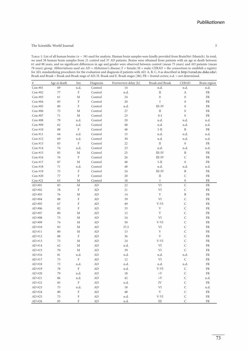
The Scientific World Journal 3
Table 1: List of all human brains (n = 58) used for analysis. Human brain samples were kindly provided from BrainNet (Munich). In total,we used 58 human brain samples from 21 control and 37 AD patients. Brains were obtained from patients with an age at death between61 and 88 years, and no significant differences in age and gender were observed between control (mean 75 years) and AD patients (mean78 years) group. Abbreviations used are AD = Alzheimer’s disease; F = female; M = male; CERAD = the consortium to establish a registryfor AD, standardizing procedures for the evaluation and diagnosis if patients with AD. A, B, C, 0 as described in http://cerad.mc.duke.edu/;Braak and Braak = Braak and Braak stage of AD; H. Braak and E. Braak stages [36]; FR = frontal cortex; n.d. = not determined.
# Age at death Sex Diagnosis Postmortem delay [h] Braak and Braak CERAD Brain region
Con #01 69 n.d. Control 14 n.d. n.d. n.d.
Con #02 77 F Control n.d. II A FR
Con #03 61 M Control 24 0 0 FR
Con #04 85 F Control 20 I 0 FR
Con #05 80 F Control n.d. III-IV 0 FR
Con #06 75 M Control 27 II 0 FR
Con #07 71 M Control 23 0-I 0 FR
Con #08 79 n.d. Control 20 n.d. n.d. n.d.
Con #09 62 n.d. Control 48 n.d. n.d. n.d.
Con #10 88 F Control 48 I-II B FR
Con #11 64 n.d. Control 15 n.d. n.d. n.d.
Con #12 69 n.d. Control 30 n.d. n.d. n.d.
Con #13 83 F Control 22 II 0 FR
Con #14 74 n.d. Control 23 n.d. n.d. n.d.
Con #15 85 M Control 25 III-IV B FR
Con #16 76 F Control 26 III-IV C FR
Con #17 87 M Control 48 I-II 0 FR
Con #18 71 n.d. Control 48 n.d. n.d. n.d.
Con #19 75 F Control 24 III-IV B FR
Con #20 77 F Control 20 II C FR
Con #21 63 M Control 18 I 0 FR
AD #01 83 M AD 22 VI C FR
AD #02 78 F AD 21 VI C FR
AD #03 76 M AD 14 V B FR
AD #04 88 F AD 39 VI C FR
AD #05 67 F AD 49 V-VI C FR
AD #06 82 F AD 33 V C FR
AD #07 80 M AD 12 V C FR
AD #08 75 M AD 24 VI C FR
AD #09 74 M AD 50 V-VI C FR
AD #10 83 M AD 37,5 VI C FR
AD #11 80 M AD 13 V C FR
AD #12 88 F AD 36 V C FR
AD #13 73 M AD 24 V-VI C FR
AD #14 62 M AD n.d. VI C FR
AD #15 70 M AD 39 VI C FR
AD #16 81 n.d. AD n.d. n.d. n.d. FR
AD #17 75 F AD 12 VI C FR
AD #18 73 n.d. AD n.d. n.d. n.d. FR
AD #19 78 F AD n.d. V-VI C FR
AD #20 79 n.d. AD 18 >V C FR
AD #21 86 n.d. AD 42 >V C n.d.
AD #22 85 F AD n.d. IV C FR
AD #23 75 n.d. AD 18 VI C n.d.
AD #24 80 F AD 48 V C FR
AD #25 73 F AD n.d. V-VI C FR
AD #26 85 F AD n.d. III C FR
Publika onen
73
4 The Scientific World Journal
Table 1: Continued.
# Age at death Sex Diagnosis Postmortem delay [h] Braak and Braak CERAD Brain region
AD #27 80 n.d. AD 5 >V C n.d.
AD #28 78 F AD n.d. VI C FR
AD #29 87 M AD 4 V C FR
AD #30 65 F AD 48 V-VI C FR
AD #31 82 F AD 14 V-VI C FR
AD #32 76 F AD 24 V-VI C FR
AD #33 79 F AD 20 V C n.d.
AD #34 87 M AD 26 V C FR
AD #35 68 F AD n.d. VI C FR
AD #36 85 n.d. AD n.d. n.d. n.d. FR
AD #37 83 M AD 48 V C FR
plate (Nunc, Langenselbold, Germany). 200 μL of reagentbuffer (CuSO4 : bicinchoninic acid; 1 : 39; Sigma Aldrich,Taufkirchen, Germany) was added to each well using a multi-channel pipette (Eppendorf, Hamburg, Germany). Incuba-tion took place for 15 minutes at 37◦C and afterwards forfurther 15 minutes at room temperature while shaking (IKA,Staufen, Germany) at 300 revolutions per minute. Absorb-ance was measured at a wavelength of 560 nm using a Multi-scanEX (Thermo Fisher Scientific, Schwerte, Germany).
2.5. Western Blot Analysis. For detection of ADAM17, PS1and BACE1 protein amount, proteins of cell lysate were sep-arated on 10%–20% Tricine gels (Anamed, Groß-Bieberau,Germany). Western Blot (WB) analysis was performed usingantibody ab39162 (1 : 5000; abcam, Cambridge, UK), sc-7860 (1 : 500; Santa Cruz, Heidelberg, Germany), and B0806(1 : 1000; Sigma, Taufkirchen, Germany) respectively. W401B(1 : 10000; Promega, Mannheim, Germany) was used as sec-ondary antibody, and detection was carried out using West-ern Lightning Plus-ECL solution (Perkin Elmer, Rodgau,Germany). Densiometric quantification was performed us-ing Image Gauge software.
2.6. Postnuclear Fractions. For preparing postnuclear frac-tions (PNFs) SH-SY5Y wild-type cells, mouse brains orhuman AD brains were washed with PBS and homogenizedin sucrose-buffer (pH 7.4) using a PotterS (Braun, Melsun-gen, Germany) at maximum speed. Protein amount wasadjusted to 2 mg/mL for β- and γ-secretase assay and to1 mg/mL for measuring α-secretase activity. After centrifuga-tion at 900 rcf for 10 min at 4◦C supernatants were collectedand stored at −80◦C.
2.7. In Vitro Incubation. PNFs were warmed up at 37◦C,and phospholipids solved in ethanol p.a. were added in afinal concentration of 100 μM. Samples were incubated whileshaking (Multireax, Heidolph Instruments, Schwabach, Ger-many) for 15 min at 37◦C before being centrifuged at55.000 rpm for 75 min at 4◦C for pelleting membranes.
2.8. Secretase Activities
α-Secretase Activity. Pelleted SH-SY5Y membranes were re-suspended in 1 mL, and purified mouse brain membraneswere resuspended in 2 mL Hepes-buffer pH 7.5. For solubili-sation, samples were passed through needles (BD, FranklinLakes, NJ, USA) with decreasing diameters. Samples weredispended in triplicate onto a black 96-well plate using100 μL per well corresponding to 100 μg for SH-SY5Y and50 μg for mouse brain membranes. After adding 3 μM α-secretase-substrate (Calbiochem, Darmstadt, Germany), flu-orescence was detected with excitation wavelength at 340 ±10 nm and emission wavelength at 490 ± 10 nm using Safire
2
Fluorometer (Tecan, Crailsheim, Germany). Kinetic wasplotted for 120 cycles with kinetic intervals of 120 s. For spec-ificity control, 10 mM EDTA/EGTA was used (supplementFigure S2).
β- and γ-Secretase Activities. Pelleted membranes were re-suspended in 800 μL sucrose-buffer for SH-SY5Y membranesand 400 μL for mouse brain and human AD brain mem-branes. Membranes were solubilized as described above.For analysing γ-secretase activity, samples were dispensedin triplicate on black 96-well plates (Corning, Lowell, MA,USA) using 100 μL per well corresponding to 250 μg forSH-SY5Y membranes and 500 μg for mouse and humanbrain membranes. After adding 10 μM γ-secretase sub-strate (Calbiochem, Darmstadt, Germany), fluorescence wasmeasured with excitation wavelength 355 ± 10 nm andfluorescence detection at 440 ± 10 nm in a Safire2 Fluo-rometer (Tecan, Crailsheim, Germany) at 37◦C under lightexclusion. Kinetics were plotted for 50 cycles with kineticintervals of 180 s. For determination of assay specificity, weused γ-secretase Inhibitor X (50 μM) (Calbiochem, Darm-stadt, Germany) (supplement Figure S3). For measuringβ-secretase activity, samples were dispensed in triplicateon black 96 well plates (50 μL per well corresponding to125 μg for SH-SY5Y membranes and 250 μg for mouse brainmembranes). β-secretase substrate (Calbiochem, Darmstadt,Germany) was added with a final concentration of 20 μM
Publika onen
74
The Scientific World Journal 5
and fluorescence was detected with an excitation wave-length at 345 ± 5 nm and emission wavelength at 500 ±2.5 nm under light exclusion at 37◦C. Kinetics were plottedfor 180 cycles with kinetic intervals of 60 s. Assay specificitywas validated using β-secretase Inhibitor II (1 μM) (Cal-biochem, Darmstadt, Germany) (supplement Figure S4). Forall secretase assays, the unspecificity was between 10% to30%. The secretase activities presented, were calculated bysubtracting the unspecific turnover determined by addingsecretase inhibitors.
2.9. Mass Spectrometry Analysis. For determination of phos-phatidylcholine and phosphatidylethanolamine levels inhuman control and AD brains, we used a 4000 quadrupolelinear-ion trap (QTrap) equipped with a Turbo-V ion source(AB SCIEX, Darmstadt, Germany) connected to a 1200 Agi-lent HPLC (Agilent, Boblingen, Germany). Briefly, sampleswere adjusted to 6 mg/mL protein amount and 10 μL of theadjusted samples was pipetted onto a membrane (Whatman,GE Healthcare, Freiburg, Germany) fixed in the wells of aMultiScreen, solvinert 96-well plate with a 0.45 μm sterilefilter at the bottom (Millipore, Schwalbach, Germany). This96 well plate was placed onto a 1 mL 96-well deep wellplate (Nunc, Langenselbold, Germany). Samples were driedunder a gentle flow of nitrogen for at least 30 min at roomtemperature. Meanwhile, phenylisothiocyanate was dilutedin ethanol : water : pyridine (1 : 1 : 1; v/v/v) to obtain a final5% phenylisothiocyanate solution. 20 μL of this solutionwas pipetted onto each membrane, and the plate wasincubated for 20 min at room temperature and afterwardsdried under a gentle flow of nitrogen for at least 30 min.Samples were extracted using 300 μL 5 mM ammoniumacetate buffer in methanol using a multichannel pipette(Eppendorf, Germany) and the plate was shaken at 300revolutions per minute using a plate shaker (IKA, Staufen,Germany) at room temperature for 30 min. Samples werecentrifuged (Thermo Scientific, Langenselbold, Germany)at 500×g for 2 min through 0.45 μm sterile filters intothe 96 deep well plate and further diluted with 600 μL of5 mM ammonium acetate dissolved in methanol : water(97 : 3, v/v) which also was used as the only running solvent.Finally plate was covered with a silicone mat and shook forfurther 2 min at 300 rpm at room temperature. Plate wasplaced into the cooled autosampler and detection was carriedout using Analyst 1.5 software (AB SCIEX, Darmstadt,Germany). Phosphatidylcholine species were detectedusing MRM transitions (PCae C34:1: 746,6 m/z − 184 m/z;PCae C36:4: 768,6 m/z − 184 m/z; PCae C36:2: 772,6 m/z− 184 m/z; PCae C36:1: 774,6 m/z − 184 m/z; PCae C38:6:792,6 m/z − 184 m/z; PCae C38:5: 794,6 m/z − 184 m/z;PCae C38:4: 796,6 m/z − 184 m/z; PCae C40:6: 820,6 m/z− 184 m/z), and phosphatidylethanolamine species weredetected using a neutral loss scan for 141 m/z (PEae 36:4:728,8 m/z; PEae 36:2: 732,8 m/z; PEae 38:6: 752,8 m/z;PEae 38:5: 754,8 m/z; PEae 38:4: 756,8 m/z; PEae 40:6:780,8 m/z). For both species detection, 20 μL sample wasinjected into sample loop with the following running solventgradient (0.0–2.4 min, 30 μL; 2.4–3.0 min, 200 μL; 3.0 min,30 μL).
2.10. Quantitative Real-Time Experiments. RNA was extract-ed in total from cells using TRIzol reagent (Invitrogen, Karl-sruhe, Germany), according to manufacturer’s protocols.High Capacity cDNA Reverse Transcription Kit (AppliedBiosystems, Darmstadt, Germany) was used to reverse-transcribe 2 μg total RNA. Quantitative real-time PCR anal-ysis was carried out using Fast SYBR Green Master Mix on7500 Fast Real-Time PCR System (7500 Fast System SDSSoftware 1.3.1.; Applied Biosystems, Darmstadt, Germany).All results were normalized to β-actin gene expression andchanges detected in gene expression were calculated using2−(ΔΔCt) method [38]. The following primer sequences wereused: ADAM17: 5′-CTG TGT GCC CTA TGT CGA TG-3′
and 5′-CAG CTG GTC AAT GAA ATC CC-3′; BACE1: 5′-GCA GGG CTA CTA CGT GGA GA-3′ and 5′-TAG TAGCGA TGC AGG AAG GG-3′; PSEN1: 5′-CTC AAT TCT GAATGC TGC CA-3′ and 5′-GGC ATG GAT GAC CTT ATAGCA-3′; PSEN2: 5′-GAT CAG CGT CAT CGT GGT TA-3′
and 5′-GGA ACA GCA GCA TCA GTG AA-3′; APH1a: 5′-CAG CCA TTA TCC TGC TCC AT-3′ and 5′-GGA ATG TCAGTC CCG ATG TC-3′; APH1b: 5′-GTG TCA GCC CAG ACCTTC AT-3′ and 5′-CAG GCA GAG TTT CAG GCT TC-3′;NCSTN: 5′-CTG TAC GGA ACC AGG TGG AG-3′ and 5′-GAG AGG CTG GGA CTG ATT TG-3′; PSENEN: 5′-CATCTT CTG GTT CTT CCG AGA G-3′ and 5′-AGA AGA GGAAGC CCA CAG C-3′; β-Actin: 5′-CTT CCT GGG CAT GGAGTC-3′ and 5′-AGC ACT GTG TTG GCG TAC AG-3′. Toverify the results obtained by quantitative real time experi-ments, samples were separated on 3% agarose gels in TBEbuffer (90 mM Tris, 90 mM boric acid, 2 mM EDTA, pH8.0).
2.11. Cytotoxicity Measurement. Cytotoxicity was measuredutilizing Lactate Dehydrogenase Cytotoxicity Assay Kit (Cay-man Chemical, Ann Arbor, USA) according to manufac-turer’s protocol.
2.12. Statistical Analysis. All quantified data presented hereis based on an average of at least three independent experi-ments. Error bars represent standard deviation of the mean.Statistical significance was determined by two-tailed Stu-dent’s t-test; significance was set at ∗P ≤ 0.05; ∗∗P ≤ 0.01and ∗∗∗P ≤ 0.001.
3. Results
3.1. Determination of PC-PL and PE-PL Levels in HumanAD Postmortem Brain. In our previous studies, we reporteda decrease in total phosphatidylethanolamine-plasmalogen(PE-PL) [39] and some phosphatidylcholine-plasmalogen(PC-PL) species [40] in postmortem AD brains. To elucidatethe question which plasmalogen species are mostly changedin AD, we determined in this study the major PE-PL and PC-PL species. In total, we analyzed 58 human brain samples, 21control brains with an average age of 75 years, and 37 brainsamples obtained from AD patients with an average age of 78years (Table 1). There were no significant differences in theage and sex of AD patients and controls. Mass spectrometryanalysis revealed that PC-PL levels were significantly reduced
Publika onen
75
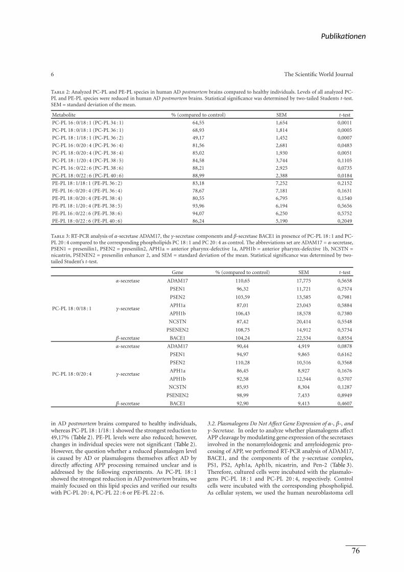
6 The Scientific World Journal
Table 2: Analyzed PC-PL and PE-PL species in human AD postmortem brains compared to healthy individuals. Levels of all analyzed PC-PL and PE-PL species were reduced in human AD postmortem brains. Statistical significance was determined by two-tailed Students t-test.SEM = standard deviation of the mean.
Metabolite % (compared to control) SEM t-test
PC-PL 16 : 0/18 : 1 (PC-PL 34 : 1) 64,55 1,654 0,0011
PC-PL 18 : 0/18 : 1 (PC-PL 36 : 1) 68,93 1,814 0,0005
PC-PL 18 : 1/18 : 1 (PC-PL 36 : 2) 49,17 1,452 0,0007
PC-PL 16 : 0/20 : 4 (PC-PL 36 : 4) 81,56 2,681 0,0483
PC-PL 18 : 0/20 : 4 (PC-PL 38 : 4) 85,02 1,930 0,0051
PC-PL 18 : 1/20 : 4 (PC-PL 38 : 5) 84,58 3,744 0,1105
PC-PL 16 : 0/22 : 6 (PC-PL 38 : 6) 88,21 2,925 0,0735
PC-PL 18 : 0/22 : 6 (PC-PL 40 : 6) 88,99 2,388 0,0184
PE-PL 18 : 1/18 : 1 (PE-PL 36 : 2) 83,18 7,252 0,2152
PE-PL 16 : 0/20 : 4 (PE-PL 36 : 4) 78,67 7,181 0,1631
PE-PL 18 : 0/20 : 4 (PE-PL 38 : 4) 80,55 6,795 0,1540
PE-PL 18 : 1/20 : 4 (PE-PL 38 : 5) 93,96 6,194 0,5656
PE-PL 16 : 0/22 : 6 (PE-PL 38 : 6) 94,07 6,250 0,5752
PE-PL 18 : 0/22 : 6 (PE-PL 40 : 6) 86,24 5,190 0,2049
Table 3: RT-PCR analysis of α-secretase ADAM17, the γ-secretase components and β-secretase BACE1 in presence of PC-PL 18 : 1 and PC-PL 20 : 4 compared to the corresponding phospholipids PC 18 : 1 and PC 20 : 4 as control. The abbreviations set are ADAM17 = α-secretase,PSEN1 = presenilin1, PSEN2 = presenilin2, APH1a = anterior pharynx-defective 1a, APH1b = anterior pharynx-defective 1b, NCSTN =nicastrin, PSENEN2 = presenilin enhancer 2, and SEM = standard deviation of the mean. Statistical significance was determined by two-tailed Student’s t-test.
Gene % (compared to control) SEM t-test
PC-PL 18 : 0/18 : 1
α-secretase ADAM17 110,65 17,775 0,5658
γ-secretase
PSEN1 96,32 11,721 0,7574
PSEN2 103,59 13,585 0,7981
APH1a 87,01 23,043 0,5884
APH1b 106,43 18,578 0,7380
NCSTN 87,42 20,414 0,5548
PSENEN2 108,75 14,912 0,5734
β-secretase BACE1 104,24 22,534 0,8554
PC-PL 18 : 0/20 : 4
α-secretase ADAM17 90,44 4,919 0,0878
γ-secretase
PSEN1 94,97 9,865 0,6162
PSEN2 110,28 10,516 0,3568
APH1a 86,45 8,927 0,1676
APH1b 92,58 12,544 0,5707
NCSTN 85,93 8,304 0,1287
PSENEN2 98,99 7,433 0,8949
β-secretase BACE1 92,90 9,413 0,4607
in AD postmortem brains compared to healthy individuals,whereas PC-PL 18 : 1/18 : 1 showed the strongest reduction to49,17% (Table 2). PE-PL levels were also reduced; however,changes in individual species were not significant (Table 2).However, the question whether a reduced plasmalogen levelis caused by AD or plasmalogens themselves affect AD bydirectly affecting APP processing remained unclear and isaddressed by the following experiments. As PC-PL 18 : 1showed the strongest reduction in AD postmortem brains, wemainly focused on this lipid species and verified our resultswith PC-PL 20 : 4, PC-PL 22 : 6 or PE-PL 22 : 6.
3.2. Plasmalogens Do Not Affect Gene Expression of α-, β-, andγ-Secretase. In order to analyze whether plasmalogens affectAPP cleavage by modulating gene expression of the secretasesinvolved in the nonamyloidogenic and amyloidogenic pro-cessing of APP, we performed RT-PCR analysis of ADAM17,BACE1, and the components of the γ-secretase complex,PS1, PS2, Aph1a, Aph1b, nicastrin, and Pen-2 (Table 3).Therefore, cultured cells were incubated with the plasmalo-gens PC-PL 18 : 1 and PC-PL 20 : 4, respectively. Controlcells were incubated with the corresponding phospholipid.As cellular system, we used the human neuroblastoma cell
Publika onen
76
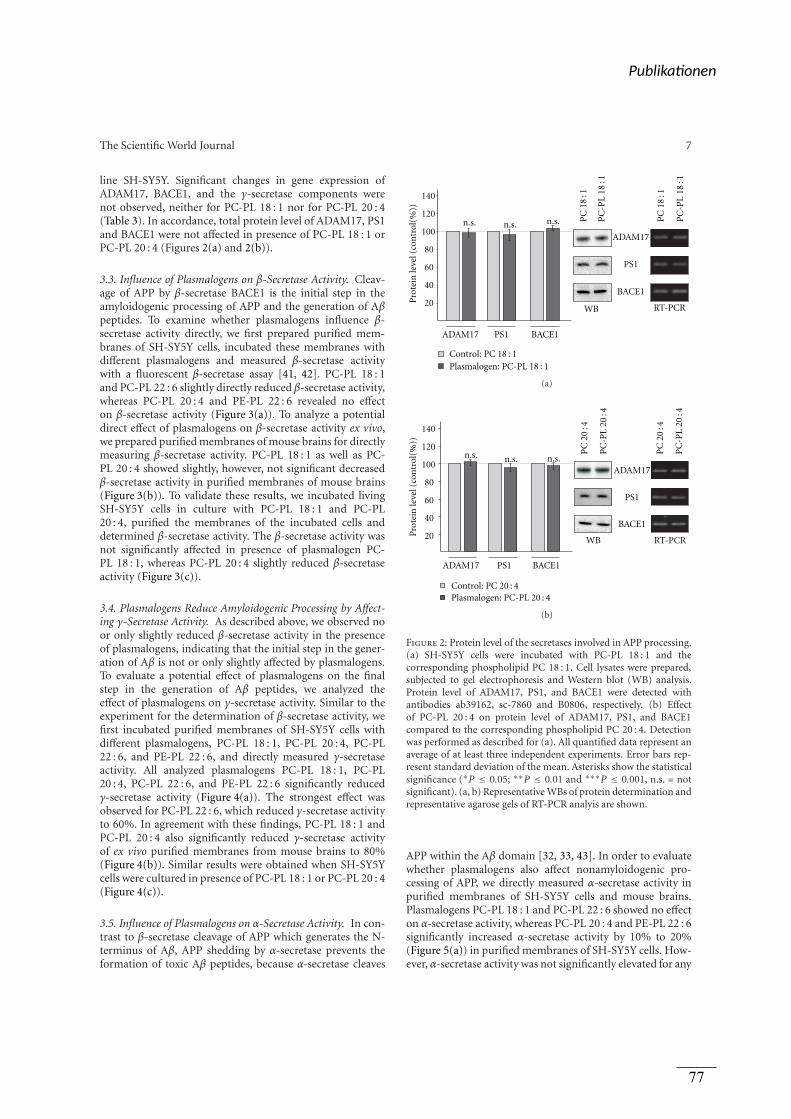
The Scientific World Journal 7
line SH-SY5Y. Significant changes in gene expression ofADAM17, BACE1, and the γ-secretase components werenot observed, neither for PC-PL 18 : 1 nor for PC-PL 20 : 4(Table 3). In accordance, total protein level of ADAM17, PS1and BACE1 were not affected in presence of PC-PL 18 : 1 orPC-PL 20 : 4 (Figures 2(a) and 2(b)).
3.3. Influence of Plasmalogens on β-Secretase Activity. Cleav-age of APP by β-secretase BACE1 is the initial step in theamyloidogenic processing of APP and the generation of Aβpeptides. To examine whether plasmalogens influence β-secretase activity directly, we first prepared purified mem-branes of SH-SY5Y cells, incubated these membranes withdifferent plasmalogens and measured β-secretase activitywith a fluorescent β-secretase assay [41, 42]. PC-PL 18 : 1and PC-PL 22 : 6 slightly directly reduced β-secretase activity,whereas PC-PL 20 : 4 and PE-PL 22 : 6 revealed no effecton β-secretase activity (Figure 3(a)). To analyze a potentialdirect effect of plasmalogens on β-secretase activity ex vivo,we prepared purified membranes of mouse brains for directlymeasuring β-secretase activity. PC-PL 18 : 1 as well as PC-PL 20 : 4 showed slightly, however, not significant decreasedβ-secretase activity in purified membranes of mouse brains(Figure 3(b)). To validate these results, we incubated livingSH-SY5Y cells in culture with PC-PL 18 : 1 and PC-PL20 : 4, purified the membranes of the incubated cells anddetermined β-secretase activity. The β-secretase activity wasnot significantly affected in presence of plasmalogen PC-PL 18 : 1, whereas PC-PL 20 : 4 slightly reduced β-secretaseactivity (Figure 3(c)).
3.4. Plasmalogens Reduce Amyloidogenic Processing by Affect-ing γ-Secretase Activity. As described above, we observed noor only slightly reduced β-secretase activity in the presenceof plasmalogens, indicating that the initial step in the gener-ation of Aβ is not or only slightly affected by plasmalogens.To evaluate a potential effect of plasmalogens on the finalstep in the generation of Aβ peptides, we analyzed theeffect of plasmalogens on γ-secretase activity. Similar to theexperiment for the determination of β-secretase activity, wefirst incubated purified membranes of SH-SY5Y cells withdifferent plasmalogens, PC-PL 18 : 1, PC-PL 20 : 4, PC-PL22 : 6, and PE-PL 22 : 6, and directly measured γ-secretaseactivity. All analyzed plasmalogens PC-PL 18 : 1, PC-PL20 : 4, PC-PL 22 : 6, and PE-PL 22 : 6 significantly reducedγ-secretase activity (Figure 4(a)). The strongest effect wasobserved for PC-PL 22 : 6, which reduced γ-secretase activityto 60%. In agreement with these findings, PC-PL 18 : 1 andPC-PL 20 : 4 also significantly reduced γ-secretase activityof ex vivo purified membranes from mouse brains to 80%(Figure 4(b)). Similar results were obtained when SH-SY5Ycells were cultured in presence of PC-PL 18 : 1 or PC-PL 20 : 4(Figure 4(c)).
3.5. Influence of Plasmalogens on α-Secretase Activity. In con-trast to β-secretase cleavage of APP which generates the N-terminus of Aβ, APP shedding by α-secretase prevents theformation of toxic Aβ peptides, because α-secretase cleaves
20
40
60
80
100
120
140
Pro
tein
leve
l (co
ntr
ol(%
))
ADAM17 PS1 BACE1
n.s.
ADAM17
PS1
BACE1
n.s. n.s.
WB RT-PCR
Plasmalogen: PC-PL 18 : 1Control: PC 18 : 1
PC
-PL
18:1
PC
18:1
PC
-PL
18:1
PC
18:1
(a)
20
40
60
80
100
120
140
Pro
tein
leve
l (co
ntr
ol(%
))
ADAM17
PS1
BACE1
ADAM17 PS1 BACE1
n.s. n.s. n.s.
WB RT-PCR
Plasmalogen: PC-PL 20 : 4Control: PC 20 : 4
PC
-PL
20:4
PC
20:4
PC
-PL
20:4
PC
20:4
(b)
Figure 2: Protein level of the secretases involved in APP processing.(a) SH-SY5Y cells were incubated with PC-PL 18 : 1 and thecorresponding phospholipid PC 18 : 1. Cell lysates were prepared,subjected to gel electrophoresis and Western blot (WB) analysis.Protein level of ADAM17, PS1, and BACE1 were detected withantibodies ab39162, sc-7860 and B0806, respectively. (b) Effectof PC-PL 20 : 4 on protein level of ADAM17, PS1, and BACE1compared to the corresponding phospholipid PC 20 : 4. Detectionwas performed as described for (a). All quantified data represent anaverage of at least three independent experiments. Error bars rep-resent standard deviation of the mean. Asterisks show the statisticalsignificance (∗P ≤ 0.05; ∗∗P ≤ 0.01 and ∗∗∗P ≤ 0.001, n.s. = notsignificant). (a, b) Representative WBs of protein determination andrepresentative agarose gels of RT-PCR analyis are shown.
APP within the Aβ domain [32, 33, 43]. In order to evaluatewhether plasmalogens also affect nonamyloidogenic pro-cessing of APP, we directly measured α-secretase activity inpurified membranes of SH-SY5Y cells and mouse brains.Plasmalogens PC-PL 18 : 1 and PC-PL 22 : 6 showed no effecton α-secretase activity, whereas PC-PL 20 : 4 and PE-PL 22 : 6significantly increased α-secretase activity by 10% to 20%(Figure 5(a)) in purified membranes of SH-SY5Y cells. How-ever, α-secretase activity was not significantly elevated for any
Publika onen
77
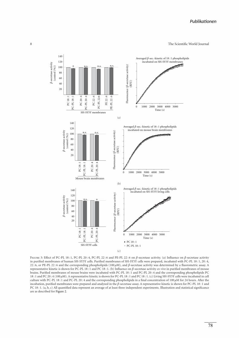
8 The Scientific World Journal
20
40
60
80
100
120
140
SH-SY5Y membranes
n.s. n.s. n.s.P
C18
:1
PC
-PL
18:1
PC
20:4
PC
-PL
20:4
PC
22:6
PC
-PL
22:6
PE
22:6
PE
-PL
22:6
∗
β-s
ecre
tase
acti
vity
(con
trol
(%))
0 1000 2000 3000 4000 5000Time (s)
Averaged β-sec.-kinetic of 18 : 1 phospholipidsincubated on SH-SY5Y membranes
Flu
ores
cen
ce(β
-sec
reta
seac
t ivi
ty)
(RFU
)
(a)
20
40
60
80
100
120
140
Mouse brain membranes
n.s. n.s.
PC
18:1
PC
-PL
18:1
PC
20:4
PC
-PL
20:4
β-s
ecre
tase
acti
vity
(con
trol
(%))
0 1000 2000 3000 4000 5000
Flu
ores
cen
ce(β
-sec
reta
seac
t ivi
ty)
(RFU
)
Averaged β-sec.-kinetic of 18 : 1 phospholipidsincubated on mouse brain membranes
Time (s)
(b)
PC
18:1
PC
-PL
18:1
PC
20:4
PC
-PL
20:4
20
40
60
80
100
120
140
SH-SY5Y cells
n.s.
β-s
ecre
tase
acti
vity
(con
trol
(%)) ∗
0 1000 2000 3000 4000 5000
Flu
ores
cen
ce(β
-sec
ret a
sea c
tivi
t y)
( RFU
)
PC 18 : 1
PC-PL 18 : 1
Averaged β-sec.-kinetic of 18 : 1 phospholipidsincubated on SH-SY5Y living cells
Time (s)
(c)
Figure 3: Effect of PC-PL 18 : 1, PC-PL 20 : 4, PC-PL 22 : 6 and PE-PL 22 : 6 on β-secretase activity. (a) Influence on β-secretase activityin purified membranes of human SH-SY5Y cells. Purified membranes of SH-SY5Y cells were prepared, incubated with PC-PL 18 : 1, 20 : 4,22 : 6, or PE-PL 22 : 6 and the corresponding phospholipids (100 μM), and β-secretase activity was determined by a fluorometric assay. Arepresentative kinetic is shown for PC-PL 18 : 1 and PC 18 : 1. (b) Influence on β-secretase activity ex vivo in purified membranes of mousebrains. Purified membranes of mouse brains were incubated with PC-PL 18 : 1 and PC-PL 20 : 4 and the corresponding phospholipids PC18 : 1 and PC 20 : 4 (100 μM). A representative kinetic is shown for PC-PL 18 : 1 and PC 18 : 1. (c) Living SH-SY5Y cells were incubated in cellculture with PC-PL 18 : 1 and PC-PL 20 : 4 and the corresponding phospholipids in a final concentration of 100 μM for 24 hours. After theincubation, purified membranes were prepared and analyzed in the β-secretase assay. A representative kinetic is shown for PC-PL 18 : 1 andPC 18 : 1. (a, b, c) All quantified data represent an average of at least three independent experiments. Illustration and statistical significanceare as described for Figure 2.
Publika onen
78
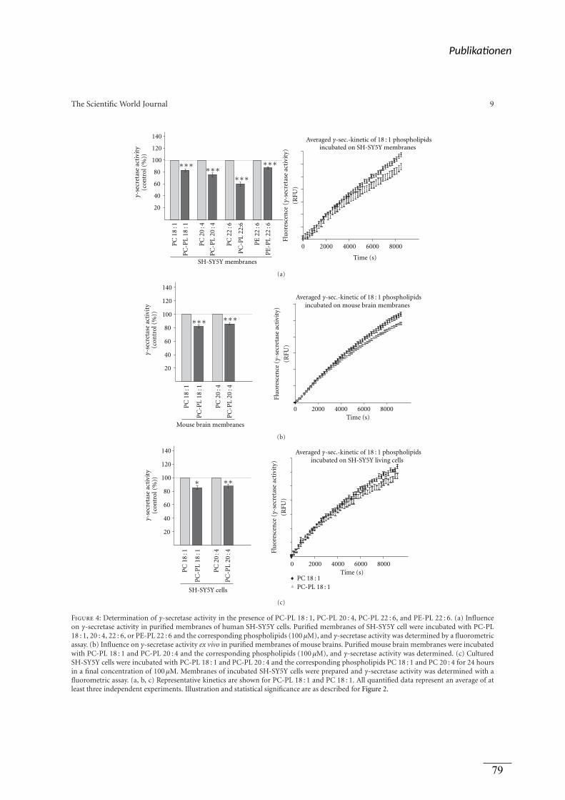
The Scientific World Journal 9
20
40
60
80
100
120
140
SH-SY5Y membranes
PC
18:1
PC
-PL
18:1
PC
20:4
PC
-PL
20:4
PC
22:6
PC
-PL
22:6
PE
22:6
PE
-PL
22:6
γ-se
cret
ase
acti
vity
(con
trol
(%))
∗∗∗ ∗∗∗∗∗∗
∗∗∗
0 2000 4000 6000 8000
Flu
ores
cen
ce(γ
-sec
reta
seac
tivi
ty)
(RFU
)
Averaged γ-sec.-kinetic of 18 : 1 phospholipidsincubated on SH-SY5Y membranes
Time (s)
(a)
20
40
60
80
100
120
140
Mouse brain membranes
PC
18:1
PC
-PL
18:1
PC
20:4
PC
-PL
20:4
γ-se
cret
ase
acti
vity
(con
trol
(%)) ∗∗∗∗∗∗
0 2000 4000 6000 8000
Averaged γ-sec.-kinetic of 18 : 1 phospholipidsincubated on mouse brain membranes
Flu
ores
cen
ce(γ
-sec
reta
seac
tivi
ty)
(RFU
)
Time (s)
(b)
0 2000 4000 6000 8000Time (s)
Averaged γ-sec.-kinetic of 18 : 1 phospholipidsincubated on SH-SY5Y living cells
Flu
ores
cen
ce(γ
-sec
reta
seac
tivi
ty)
(RFU
)
PC 18 : 1PC-PL 18 : 1
20
40
60
80
100
120
140
SH-SY5Y cells
PC
18:1
PC
-PL
18:1
PC
20:4
PC
-PL
20:4
γ-se
cret
ase
acti
vity
(con
trol
(%)) ∗∗∗
(c)
Figure 4: Determination of γ-secretase activity in the presence of PC-PL 18 : 1, PC-PL 20 : 4, PC-PL 22 : 6, and PE-PL 22 : 6. (a) Influenceon γ-secretase activity in purified membranes of human SH-SY5Y cells. Purified membranes of SH-SY5Y cell were incubated with PC-PL18 : 1, 20 : 4, 22 : 6, or PE-PL 22 : 6 and the corresponding phospholipids (100 μM), and γ-secretase activity was determined by a fluorometricassay. (b) Influence on γ-secretase activity ex vivo in purified membranes of mouse brains. Purified mouse brain membranes were incubatedwith PC-PL 18 : 1 and PC-PL 20 : 4 and the corresponding phospholipids (100 μM), and γ-secretase activity was determined. (c) CulturedSH-SY5Y cells were incubated with PC-PL 18 : 1 and PC-PL 20 : 4 and the corresponding phospholipids PC 18 : 1 and PC 20 : 4 for 24 hoursin a final concentration of 100 μM. Membranes of incubated SH-SY5Y cells were prepared and γ-secretase activity was determined with afluorometric assay. (a, b, c) Representative kinetics are shown for PC-PL 18 : 1 and PC 18 : 1. All quantified data represent an average of atleast three independent experiments. Illustration and statistical significance are as described for Figure 2.
Publika onen
79
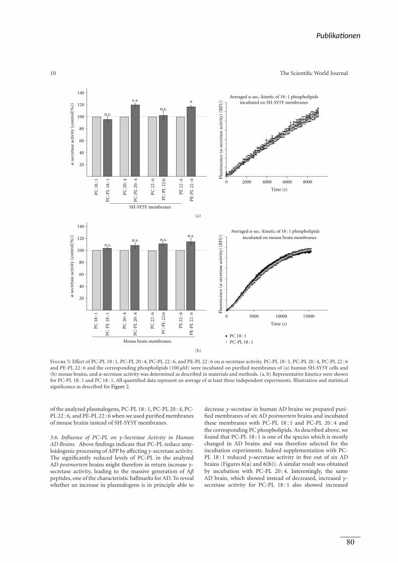
10 The Scientific World Journal
0 2000 4000 6000 8000
Flu
ores
cen
ce(α
-sec
reta
seac
tivi
ty)
(RFU
)
Averaged α-sec.-kinetic of 18 : 1 phospholipidsincubated on SH-SY5Y membranes
20
40
60
80
100
120
140
n.s.n.s.
SH-SY5Y membranes
PC
18:1
PC
-PL
18:1
PC
20:4
PC
-PL
20:4
PC
22:6
PC
-PL
22:6
PE
22:6
PE
-PL
22:6
∗∗ ∗
α-s
ecre
tase
acti
vity
(con
trol
(%))
Time (s)
(a)
Mouse brain membranes
20
40
60
80
100
120
140
n.s.n.s. n.s.
n.s.
PC
18:1
PC
-PL
18:1
PC
20:4
PC
-PL
20:4
PC
22:6
PC
-PL
22:6
PE
22:6
PE
-PL
22:6
α-s
ecre
tase
acti
vity
(con
trol
(%))
0 5000 10000 15000
Flu
ores
cen
ce(α
-sec
reta
seac
tivi
ty)
(RFU
)
Averaged α-sec.-kinetic of 18 : 1 phospholipidsincubated on mouse brain membranes
PC 18 : 1PC-PL 18 : 1
Time (s)
(b)
Figure 5: Effect of PC-PL 18 : 1, PC-PL 20 : 4, PC-PL 22 : 6, and PE-PL 22 : 6 on α-secretase activity. PC-PL 18 : 1, PC-PL 20 : 4, PC-PL 22 : 6and PE-PL 22 : 6 and the corresponding phospholipids (100 μM) were incubated on purified membranes of (a) human SH-SY5Y cells and(b) mouse brains, and α-secretase activity was determined as described in materials and methods. (a, b) Representative kinetics were shownfor PC-PL 18 : 1 and PC 18 : 1. All quantified data represent an average of at least three independent experiments. Illustration and statisticalsignificance as described for Figure 2.
of the analyzed plasmalogens, PC-PL 18 : 1, PC-PL 20 : 4, PC-PL 22 : 6, and PE-PL 22 : 6 when we used purified membranesof mouse brains instead of SH-SY5Y membranes.
3.6. Influence of PC-PL on γ-Secretase Activity in HumanAD Brains. Above findings indicate that PC-PL reduce amy-loidogenic processing of APP by affecting γ-secretase activity.The significantly reduced levels of PC-PL in the analyzedAD postmortem brains might therefore in return increase γ-secretase activity, leading to the massive generation of Aβpeptides, one of the characteristic hallmarks for AD. To revealwhether an increase in plasmalogens is in principle able to
decrease γ-secretase in human AD brains we prepared puri-fied membranes of six AD postmortem brains and incubatedthese membranes with PC-PL 18 : 1 and PC-PL 20 : 4 andthe corresponding PC phospholipids. As described above, wefound that PC-PL 18 : 1 is one of the species which is mostlychanged in AD brains and was therefore selected for theincubation experiments. Indeed supplementation with PC-PL 18 : 1 reduced γ-secretase activity in five out of six ADbrains (Figures 6(a) and 6(b)). A similar result was obtainedby incubation with PC-PL 20 : 4. Interestingly, the sameAD brain, which showed instead of decreased, increased γ-secretase activity for PC-PL 18 : 1 also showed increased
Publika onen
80
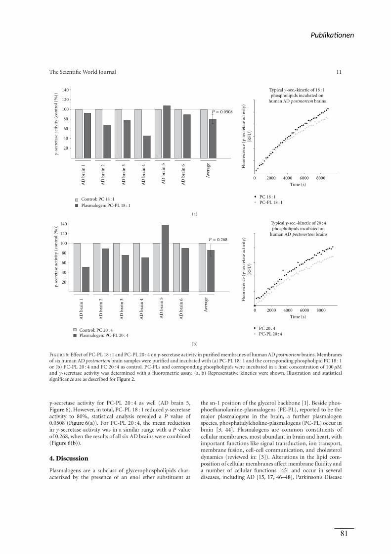
The Scientific World Journal 11
20
40
60
80
100
120
140
AD
bra
in 1
AD
bra
in 2
AD
bra
in 3
AD
bra
in 4
AD
bra
in 5
AD
bra
in 6
Ave
rage
Plasmalogen: PC-PL 18 : 1
Control: PC 18 : 1
γ-se
cret
ase
acti
vity
(con
trol
(%))
P = 0.0508
0 2000 4000 6000 8000
PC 18 : 1PC-PL 18 : 1
Flu
ores
cen
ce(γ
-sec
reta
seac
tivi
ty)
(RFU
)
Typical γ-sec.-kinetic of 18 : 1phospholipids incubated on
human AD postmortem brains
Time (s)
(a)
AD
bra
in 1
AD
bra
in 2
AD
bra
in 3
AD
bra
in 4
AD
bra
in 5
AD
bra
in 6
20
40
60
80
100
120
140
γ-se
cret
ase
acti
vity
(con
trol
(%))
Plasmalogen: PC-PL 20 : 4Control: PC 20 : 4
P = 0.268
0 2000 4000 6000 8000
PC 20 : 4PC-PL 20 : 4
Flu
ores
cen
ce(γ
-sec
reta
seac
tivi
ty)
(RFU
)
Typical γ-sec.-kinetic of 20 : 4phospholipids incubated on
human AD
Ave
rage
Time (s)
postmortem brains
(b)
Figure 6: Effect of PC-PL 18 : 1 and PC-PL 20 : 4 on γ-secretase activity in purified membranes of human AD postmortem brains. Membranesof six human AD postmortem brain samples were purified and incubated with (a) PC-PL 18 : 1 and the corresponding phospholipid PC 18 : 1or (b) PC-PL 20 : 4 and PC 20 : 4 as control. PC-PLs and corresponding phospholipids were incubated in a final concentration of 100 μMand γ-secretase activity was determined with a fluorometric assay. (a, b) Representative kinetics were shown. Illustration and statisticalsignificance are as described for Figure 2.
γ-secretase activity for PC-PL 20 : 4 as well (AD brain 5,Figure 6). However, in total, PC-PL 18 : 1 reduced γ-secretaseactivity to 80%, statistical analysis revealed a P value of0.0508 (Figure 6(a)). For PC-PL 20 : 4, the mean reductionin γ-secretase activity was in a similar range with a P valueof 0.268, when the results of all six AD brains were combined(Figure 6(b)).
4. Discussion
Plasmalogens are a subclass of glycerophospholipids char-acterized by the presence of an enol ether substituent at
the sn-1 position of the glycerol backbone [1]. Beside phos-phoethanolamine-plasmalogens (PE-PL), reported to be themajor plasmalogens in the brain, a further plasmalogenspecies, phosphatidylcholine-plasmalogens (PC-PL) occur inbrain [3, 44]. Plasmalogens are common constituents ofcellular membranes, most abundant in brain and heart, withimportant functions like signal transduction, ion transport,membrane fusion, cell-cell communication, and cholesteroldynamics (reviewed in: [3]). Alterations in the lipid com-position of cellular membranes affect membrane fluidity anda number of cellular functions [45] and occur in severaldiseases, including AD [15, 17, 46–48], Parkinson’s Disease
Publika onen
81
12 The Scientific World Journal
[49], Creutzfeldt-Jakob disease [50, 51], Gaucher disease[52], and Fabry disease [53, 54]. Recent studies have shownthat several lipids influence the proteolytic processing of APP.Cholesterol and GM1 are reported to increase the generationof Aβ [55–57], whereas docosahexaenoic acid and sphin-gomyelin decrease amyloidogenic processing of APP [42,58]. Beside their influence on the generation of Aβ, alteredlipid composition might also affect the recently identifiedphysiological function of APP. Beside the neuroprotectiveand memory enhancing effect of α-secreted APP [59–61], weand others could show that Aβ and the intracellular domainof APP (AICD), which is beside Aβ released by γ-secretasecleavage of β-CTF, regulate lipid homeostasis and gene tran-scription [39, 58, 62, 63]. AICD has been reported to affectgene transcription of several proteins, including APP, BACE,GSK3β [62], serine-palmitoyl-CoA transferase [63], andalkyl-dihydroxyacetonephosphate-synthase (AGPS) [39], arate limiting enzyme in plasmalogen synthesis. Althoughplasmalogen levels are altered in AD brain samples and aremajor constituents of neuronal membranes, so far, it is notknown whether plasmalogens affect the proteolytic process-ing of APP. Our studies on the proteolytic processing ofAPP revealed that plasmalogens decrease the amyloidogenicprocessing of APP. The detailed analysis of the secretasesinvolved in Aβ generation, β- and γ-secretase, revealedthat plasmalogens decrease amyloidogenic processing byreducing γ-secretase activity, whereas no or only a veryslight reduction in the enzymatic activity of β-secretase wasdetermined. All PC-PL and PE-PL species analyzed showeda highly significant decrease in γ-secretase activity to 60%–85% compared to the corresponding phospholipid lackingthe enol ether. All plasmalogens independent of the fatty acidshowed a decrease in γ-secretase activity further emphasizingthat the observed effect is due to the enol ether and not to thefatty acid. Analyzed protein levels of PS1 and BACE1 in pres-ence of PC-PL 18 : 1 and PC-PL 20 : 4 and RT-PCR analysisof PC-PL 18 : 1 or PC-PL 20 : 4 incubated cells showed thatplasmalogens do not affect gene expression of the secretasesinvolved in amyloidogenic processing of APP. This resultis in line with our finding that PC-PL 18 : 1 and PC-PL20 : 4 incubated cultured SH-SY5Y cells showed a similarreduction in γ-secretase activity like purified membranesof SH-SY5Y cells incubated with PC-PL 18 : 1 or PC-PL20 : 4, further strengthening our finding that plasmalogensdirectly reduce the enzymatic activity of γ-secretase and thatit is unlikely that indirect effects of plasmalogens mightinfluence γ-secretase activity. The α-secretase activity, whichprevents the formation of Aβ peptides, was in contrast toγ-secretase diversely affected by plasmalogens. Whereas PC-PL 18 : 1 and PC-PL 22 : 6 showed no significant differencesin α-secretase activity compared to PC 18 : 1 and PC 22 : 6,respectively, PC-PL 20 : 4 and PE-PL 22 : 6 significantlyincreased directly α-secretase activity compared to thecorresponding phospholipids suggesting that the fatty acidsor the phospholipid headgroups are at least able to modulatethe effect of the enol ether on α-secretase activity. However,this increase in α-secretase activity was only obtained whenpurified membranes of SH-SY5Y cells were used for the α-secretase assay, whereas on purified membranes of mouse
brains all plasmalogens showed no significant changes inα-secretase activity. Many matrix metalloproteases (MMPs)are known to contribute to the α-secretase activity initiatingthe nonamyloidogenic pathway [32–34, 64]. These MMPsare differentially expressed in different cell lines or tissues[65–67]. The different effect for PC-PL 20 : 4 and PE-PL22 : 6 might therefore be a result of different α-secretasecomposition in SH-SY5Y cells and mouse brains. Similarto the effect on the protein level and RNA level of BACE1and the γ-secretase components, PC-PL 18 : 1 and PC-PL20 : 4 revealed no changes in gene transcription of ADAM17.Because of the cell-type-specific effect of some plasmalogenspecies, we cannot exclude that plasmalogens influencesome other MMPs, which contribute to α-secretase activity.However these effects cannot be observed for all testedplasmalogens pointing out that in respect to α-secretase theenol ether has only a minor or modulating effect.
To test if our findings are relevant in AD, we analyzedwhether AD postmortem brains show altered levels of PC-PLand PE-PL. For PE-PL several studies have revealed that PE-PL level are reduced in AD brain [15, 17, 23, 68]. However,one study reported no differences or even a slight increasein PE-PL level in AD [25]. By analyzing 37 AD postmortembrain samples compared to 21 control brains not affectedby AD, we found PE-PL level to be reduced in AD brains;however, statistical analysis of the single PE-PL speciesrevealed no significance. These findings are in line with thereduced PE-PL level reported by Ginsberg et al. [15] and Hanet al. [24, 68] and our recent study that revealed also reducedPC-PL level in AD postmortem brain [40]. Interestingly,PC-PL level were significantly decreased in AD postmortembrain, indicating that PC-PL might play an important role inthe development of AD, although PC-PL are less abundantin neuronal membranes compared to PE-PL [44]. Theimportance of PC-PL is further substantiated by the resultsobtained for γ-secretase activities. As PC-PL level showedthe most prominent reduction in AD brains and AD brainsshow increased Aβ generation and accumulation, we testedwhether incubation of PC-PL 18 : 1 and PC-PL 20 : 4 on puri-fied membranes of human AD postmortem brains might alsoreduce γ-secretase activity. Indeed, supplementation withPC-PL 18 : 1 or PC-PL 20 : 4 on AD brain samples reducedγ-secretase activity in five out of six analyzed AD brains,further strengthening the importance of PC-PL in APPprocessing and probably the development of AD. Incubatingplasmalogens ex vivo on human postmortem material hasits clear limitations; further studies are required to clarifywhether an increase in plasmalogen levels are a suitabletarget, which might have a positive impact in AD.
However, the importance of plasmalogens on the patho-genesis of AD is further substantiated by our recent findingthat AGPS, a rate-limiting enzyme in plasmalogen synthesis,is regulated by APP processing [39]. Increased Aβ levels asobserved in AD lead to peroxisomal dysfunction and reducedAGPS protein stability, resulting in reduced AGPS proteinlevel and reduced plasmalogen de novo synthesis [39].Furthermore, Aβ peptides have been shown to increase theformation of reactive oxidative species [69–74], also reducingplasmalogen levels, because plasmalogens are susceptible
Publika onen
82
The Scientific World Journal 13
to oxidative stress and function as antioxidants. Reducedplasmalogen levels in AD might also be a result of increasedphospholipase A2 activity. Sanchez-Mejia et al. recentlyreported that Aβ stimulates phospholipase A2 [75, 76],responsible for the degradation of plasmalogens. Increasedlevels of Aβ peptides therefore, decrease plasmalogen levelsby reducing AGPS protein stability, increasing oxidativestress and activation of phospholipase A2. Therefore, in AD,a vicious cycle between APP processing and plasmalogenlevel occurs. Aβ peptides reduce the plasmalogen level andreduced plasmalogen level directly increase γ-secretase activ-ity leading to an even stronger production of Aβ peptides.In summary, our findings indicate that plasmalogens mightplay a crucial role in the development of AD and that adelicate balance in lipid composition of cellular membranesis important for neuronal function.
Acknowledgments
The authors gratefully thank BrainNet for the brain samples.The research leading to these results has received fundingsfrom the EU FP7 project LipiDiDiet, Grant Agreement no.211696 (TH), the DFG (TH), the Bundesministerium fur Bil-dung, Forschung, Wissenschaft und Technologie via NGFN-plus and KNDD (TH), the HOMFOR 2008 (MG), andHOMFOR 2009 (MG, TH) (Saarland University researchgrants).
References
[1] L. A. Horrocks and M. Sharma, “Plasmalogen and O-alkylglycerophospholipids,” in Phospholipids, J. N. Hawthorne andG. B. Answell, Eds., pp. 51–93, Elsevier, Amsterdam, TheNetherlands, 1982.
[2] R. W. Gross, “Identification of plasmalogen as the major phos-pholipid constituent of cardiac sarcoplasmic reticulum,” Bio-chemistry, vol. 24, no. 7, pp. 1662–1668, 1985.
[3] A. A. Farooqui and L. A. Horrocks, “Plasmalogens: workhorselipids of membranes in normal and injured neurons and glia,”Neuroscientist, vol. 7, no. 3, pp. 232–245, 2001.
[4] R. A. Zoeller, O. H. Morand, and C. R. Raetz, “A possible rolefor plasmalogens in protecting animal cells against photosen-sitized killing,” Journal of Biological Chemistry, vol. 263, no. 23,pp. 11590–11596, 1988.
[5] W. C. Breckenridge, I. G. Morgan, J. P. Zanetta, and G. Vincen-don, “Adult rat brain synaptic vesicles. II. Lipid composition,”Biochimica et Biophysica Acta, vol. 320, no. 3, pp. 681–686,1973.
[6] P. E. Glaser and R. W. Gross, “Plasmenylethanolamine facili-tates rapid membrane fusion: a stopped-flow kinetic investiga-tion correlating the propensity of a major plasma membraneconstituent to adopt an HII phase with its ability to promotemembrane fusion,” Biochemistry, vol. 33, no. 19, pp. 5805–5812, 1994.
[7] D. A. Ford and C. C. Hale, “Plasmalogen and anionic phos-pholipid dependence of the cardiac sarcolemmal sodium-calcium exchanger,” FEBS Letters, vol. 394, no. 1, pp. 99–102,1996.
[8] J. Duhm, B. Engelmann, U. M. Schonthier, and S. Streich,“Accelerated maximal velocity of the red blood cell Na+/K+pump in hyperlipidemia is related to increase in 1-palmitoyl,
2-arachidonoyl-plasmalogen phosphatidylethanolamine,” Bi-ochimica et Biophysica Acta, vol. 1149, no. 1, pp. 185–188,1993.
[9] C. Young, P. W. Gean, L. C. Chiou, and Y. Z. Shen, “Docosa-hexaenoic acid inhibits synaptic transmission and epilepti-form activity in the rat hippocampus,” Synapse, vol. 37, no.2, pp. 90–94, 2000.
[10] N. Nagan, A. K. Hajra, L. K. Larkins et al., “Isolation of aChinese hamster fibroblast variant defective in dihydroxy-acetonephosphate acyltransferase activity and plasmalogenbiosynthesis: use of a novel two-step selection protocol,”Biochemical Journal, vol. 332, part 1, pp. 273–279, 1998.
[11] H. Mandel, R. Sharf, M. Berant, R. J. Wanders, P. Vreken,and M. Aviram, “Plasmalogen phospholipids are involved inHDL-mediated cholesterol efflux: insights from investigationswith plasmalogen-deficient cells,” Biochemical and BiophysicalResearch Communications, vol. 250, no. 2, pp. 369–373, 1998.
[12] A. A. Farooqui and L. A. Horrocks, “Plasmalogens, phospho-lipase A2, and docosahexaenoic acid turnover in brain tissue,”Journal of Molecular Neuroscience, vol. 16, no. 2-3, pp. 263–272, 2001.
[13] A. A. Farooqui, T. A. Rosenberger, and L. A. Horrocks,“Arachidonic acid, neurotrauma, and neurodegenerative dis-eases,” in Handbook of Essential Fatty Acid Biology, S. Yehudaand D. I. Mostofsky, Eds., pp. 277–296, Humana Press,Totowa, NJ, USA, 1997.
[14] H. C. Yang, A. A. Farooqui, and L. A. Horrocks, “Plasmalogen-selective phospholipase A2 and its role in signal transduction,”Journal of Lipid Mediators and Cell Signalling, vol. 14, no. 1–3,pp. 9–13, 1996.
[15] L. Ginsberg, S. Rafique, J. H. Xuereb, S. I. Rapoport, and N. L.Gershfeld, “Disease and anatomic specificity of ethanolamineplasmalogen deficiency in Alzheimer’s disease brain,” BrainResearch, vol. 698, no. 1-2, pp. 223–226, 1995.
[16] K. Wells, A. A. Farooqui, L. Liss, and L. A. Horrocks, “Neuralmembrane phospholipids in Alzheimer disease,” Neurochemi-cal Research, vol. 20, no. 11, pp. 1329–1333, 1995.
[17] Z. Guan, Y. Wang, N. J. Cairns, P. L. Lantos, G. Dallner,and P. J. Sindelar, “Decrease and structural modificationsof phosphatidylethanolamine plasmalogen in the brain withAlzheimer disease,” Journal of Neuropathology and Experimen-tal Neurology, vol. 58, no. 7, pp. 740–747, 1999.
[18] P. Demediuk, R. D. Saunders, and D. K. Anderson, “Mem-brane lipid changes in laminectomized and traumatized catspinal cord,” Proceedings of the National Academy of Sciencesof the United States of America, vol. 82, no. 20, pp. 7071–7075,1985.
[19] P. Viani, I. Zini, G. Cervato, G. Biagini, L. F. Agnati, and B.Cestaro, “Effect of endothelin-1 induced ischemia on per-oxidative damage and membrane properties in rat striatumsynaptosomes,” Neurochemical Research, vol. 20, no. 6, pp.689–695, 1995.
[20] J. P. Zhang and G. Y. Sun, “Free fatty acids, neutral glycerides,and phosphoglycerides in transient focal cerebral ischemia,”Journal of Neurochemistry, vol. 64, no. 4, pp. 1688–1695, 1995.
[21] S. Schedin, P. J. Sindelar, P. Pentchev, U. Brunk, and G. Dallner,“Peroxisomal impairment in Niemann-Pick type C disease,”Journal of Biological Chemistry, vol. 272, no. 10, pp. 6245–6251, 1997.
[22] T. Yanagihara and J. N. Cumings, “Alterations of phospho-lipids, particularly plasmalogens, in the demyelination ofmultiple sclerosis as compared with that of cerebral oedema,”Brain, vol. 92, no. 1, pp. 59–70, 1969.
Publika onen
83
14 The Scientific World Journal
[23] A. A. Farooqui, S. I. Rapoport, and L. A. Horrocks, “Mem-brane phospholipid alterations in Alzheimer’s disease: de-ficiency of ethanolamine plasmalogens,” Neurochemical Re-search, vol. 22, no. 4, pp. 523–527, 1997.
[24] X. Han, D. M. Holtzman, and D. W. McKeel Jr., “Plasmalogendeficiency in early Alzheimer’s disease subjects and in animalmodels: molecular characterization using electrospray ioniza-tion mass spectrometry,” Journal of Neurochemistry, vol. 77,no. 4, pp. 1168–1180, 2001.
[25] J. W. Pettegrew, K. Panchalingam, R. L. Hamilton, and R.J. Mcclure, “Brain membrane phospholipid alterations inAlzheimer’s disease,” Neurochemical Research, vol. 26, no. 7,pp. 771–782, 2001.
[26] C. L. Masters, G. Simms, and N. A. Weinman, “Amyloidplaque core protein in Alzheimer disease and Down syn-drome,” Proceedings of the National Academy of Sciences of theUnited States of America, vol. 82, no. 12, pp. 4245–4249, 1985.
[27] D. J. Selkoe, “Cell biology of protein misfolding: the examplesof Alzheimer’s and Parkinson’s diseases,” Nature Cell Biology,vol. 6, no. 11, pp. 1054–1061, 2004.
[28] C. Haass, “Take five—BACE and the γ-secretase quartetconduct Alzheimer’s amyloid β-peptide generation,” EMBOJournal, vol. 23, no. 3, pp. 483–488, 2004.
[29] S. Sinha, J. P. Anderson, R. Barbour et al., “Purification andcloning of amyloid precursor protein β-secretase from humanbrain,” Nature, vol. 402, no. 6761, pp. 537–540, 1999.
[30] H. Steiner, R. Fluhrer, and C. Haass, “Intramembrane prote-olysis by γ-secretase,” Journal of Biological Chemistry, vol. 283,no. 44, pp. 29627–29631, 2008.
[31] T. Wakabayashi and B. De Strooper, “Presenilins: members ofthe γ-secretase quartets, but part-time soloists too,” Physiol-ogy, vol. 23, no. 4, pp. 194–204, 2008.
[32] J. D. Buxbaum, K. N. Liu, Y. Luo et al., “Evidence that tumornecrosis factor α converting enzyme is involved in regulatedα-secretase cleavage of the Alzheimer amyloid protein pre-cursor,” Journal of Biological Chemistry, vol. 273, no. 43, pp.27765–27767, 1998.
[33] S. Lammich, E. Kojro, R. Postina et al., “Constitutive and reg-ulated α-secretase cleavage of Alzheimer’s amyloid precursorprotein by a disintegrin metalloprotease,” Proceedings of theNational Academy of Sciences of the United States of America,vol. 96, no. 7, pp. 3922–3927, 1999.
[34] H. Koike, S. Tomioka, H. Sorimachi et al., “Membrane-anchored metalloprotease MDC9 has an α-secretase activityresponsible for processing the amyloid precursor protein,”Biochemical Journal, vol. 343, part 2, pp. 371–375, 1999.
[35] T. M. Allinson, E. T. Parkin, A. J. Turner, and N. M. Hooper,“ADAMs family members as amyloid precursor protein α-secretases,” Journal of Neuroscience Research, vol. 74, no. 3, pp.342–352, 2003.
[36] H. Braak and E. Braak, “Neuropathological stageing ofAlzheimer-related changes,” Acta Neuropathologica, vol. 82,no. 4, pp. 239–259, 1991.
[37] P. K. Smith, R. I. Krohn, G. T. Hermanson et al., “Mea-surement of protein using bicinchoninic acid,” AnalyticalBiochemistry, vol. 150, no. 1, pp. 76–85, 1985.
[38] K. J. Livak and T. D. Schmittgen, “Analysis of relative geneexpression data using real-time quantitative PCR and the 2(-Delta Delta C(T)) Method,” Methods, vol. 25, no. 4, pp. 402–408, 2001.
[39] M. O. Grimm, J. Kuchenbecker, T. L. Rothhaar et al., “Plas-malogen synthesis is regulated via alkyl-dihydroxyacetoneph-osphate-synthase by amyloid precursor protein processing and
is affected in Alzheimer’s disease,” Journal of Neurochemistry,vol. 116, no. 5, pp. 916–925, 2011.
[40] M. O. Grimm, S. Grosgen, M. Riemenschneider, H. Tanila,H. S. Grimm, and T. Hartmann, “From brain to food: anal-ysis of phosphatidylcholins, lyso-phosphatidylcholins andphosphatidylcholin-plasmalogens derivates in Alzheimer’sdisease human post mortem brains and mice model via massspectrometry,” Journal of Chromatography A, vol. 1218, no. 42,pp. 7713–7722, 2011.
[41] M. O. Grimm, H. S. Grimm, I. Tomic, K. Beyreuther, T.Hartmann, and C. Bergmann, “Independent inhibition ofAlzheimer disease β- and γ-secretase cleavage by loweredcholesterol levels,” Journal of Biological Chemistry, vol. 283, no.17, pp. 11302–11311, 2008.
[42] M. O. Grimm, J. Kuchenbecker, S. Grosgen et al., “Docosa-hexaenoic acid reduces amyloid β production via multiplepleiotropic mechanisms,” Journal of Biological Chemistry, vol.286, no. 16, pp. 14028–14039, 2011.
[43] T. M. Allinson, E. T. Parkin, T. P. Condon et al., “The roleof ADAM10 and ADAM17 in the ectodomain shedding ofangiotensin converting enzyme and the amyloid precursorprotein,” European Journal of Biochemistry, vol. 271, no. 12, pp.2539–2547, 2004.
[44] T. Miyazawa, S. Kanno, T. Eitsuka, and K. Nakagawa, “Plas-malogen: a short review ans newly-discovered functions,” inDietary Fats and Risk of Chronic Disease, Y. Yanagita, H. R.Knapp,, and Y. S. Huang, Eds., pp. 196–202, AOCS Publishing,2006.
[45] A. A. Spector and M. A. Yorek, “Membrane lipid compositionand cellular function,” Journal of Lipid Research, vol. 26, no. 9,pp. 1015–1035, 1985.
[46] M. R. Prasad, M. A. Lovell, M. Yatin, H. Dhillon, and W. R.Markesbery, “Regional membrane phospholipid alterations inAlzheimer’s disease,” Neurochemical Research, vol. 23, no. 1,pp. 81–88, 1998.
[47] D. B. Goodenowe, L. L. Cook, J. Liu et al., “Peripheralethanolamine plasmalogen deficiency: a logical causativefactor in Alzheimer’s disease and dementia,” Journal of LipidResearch, vol. 48, no. 11, pp. 2485–2498, 2007.
[48] P. L. Wood, R. Mankidy, S. Ritchie et al., “Circulating plas-malogen levels and Alzheimer disease assessment scale-cognitive scores in Alzheimer patients,” Journal of Psychiatryand Neuroscience, vol. 35, no. 1, pp. 59–62, 2010.
[49] N. Fabelo, V. Martin, G. Santpere et al., “Severe alterations inlipid composition of frontal cortex lipid rafts from Parkin-son’s disease and incidental Parkinson’s disease,” MolecularMedicine, vol. 17, no. 9-10, pp. 1107–1118, 2011.
[50] Y. Tamai, H. Kojima, F. Ikuta, and T. Kumanishi, “Alterationsin the composition of brain lipids in patients with Creutzfeldt-Jakob disease,” Journal of the Neurological Sciences, vol. 35, no.1, pp. 59–76, 1978.
[51] A. Federico, P. Annunziata, and G. Malentacchi, “Neuro-chemical changes in Creutzfeldt-Jakob disease,” Journal ofNeurology, vol. 223, no. 2, pp. 135–146, 1980.
[52] L. K. Hein, S. Duplock, J. J. Hopwood, and M. Fuller, “Lipidcomposition of microdomains is altered in a cell model ofGaucher disease,” Journal of Lipid Research, vol. 49, no. 8, pp.1725–1734, 2008.
[53] I. Hozumi, M. Nishizawa, T. Ariga, and T. Miyatake, “Bio-chemical and clinical analysis of accumulated glycolipidsin symptomatic heterozygotes of angiokeratoma corporisdiffusum (Fabry’s disease) in comparison with hemizygotes,”Journal of Lipid Research, vol. 31, no. 2, pp. 335–340, 1990.
Publika onen
84
The Scientific World Journal 15
[54] K. Maalouf, J. Jia, S. Rizk et al., “A modified lipid compositionin Fabry disease leads to an intracellular block of the deter-gent-resistant membrane-associated dipeptidyl peptidase IV,”Journal of Inherited Metabolic Disease, vol. 33, no. 4, pp. 445–449, 2010.
[55] K. Fassbender, M. Simons, C. Bergmann et al., “Simvastatinstrongly reduces levels of Alzheimer’s disease β-amyloid pep-tides Aβ42 and Aβ40 in vitro and in vivo,” Proceedings of theNational Academy of Sciences of the United States of America,vol. 98, no. 10, pp. 5856–5861, 2001.
[56] Q. Zha, Y. Ruan, T. Hartmann, K. Beyreuther, and D. Zhang,“GM1 ganglioside regulates the proteolysis of amyloid precur-sor protein,” Molecular Psychiatry, vol. 9, no. 10, pp. 946–952,2004.
[57] B. Wolozin, “Cholesterol and the biology of Alzheimer’s dis-ease,” Neuron, vol. 41, no. 1, pp. 7–10, 2004.
[58] M. O. Grimm, H. S. Grimm, A. J. Patzold et al., “Regulation ofcholesterol and sphingomyelin metabolism by amyloid-β andpresenilin,” Nature Cell Biology, vol. 7, no. 11, pp. 1118–1123,2005.
[59] K. Furukawa, B. L. Sopher, R. E. Rydel et al., “Increasedactivity-regulating and neuroprotective efficacy of α-secretase-derived secreted amyloid precursor protein conferred by a C-terminal heparin-binding domain,” Journal of Neurochemistry,vol. 67, no. 5, pp. 1882–1896, 1996.
[60] H. Meziane, J. C. Dodart, C. Mathis et al., “Memory-enhancing effects of secreted forms of the β-amyloid precursorprotein in normal and amnestic mice,” Proceedings of theNational Academy of Sciences of the United States of America,vol. 95, no. 21, pp. 12683–12688, 1998.
[61] M. P. Mattson, Z. H. Guo, and J. D. Geiger, “Secreted form ofamyloid precursor protein enhances basal glucose and gluta-mate transport and protects against oxidative impairment ofglucose and glutamate transport in synaptosomes by a cyclicGMP-mediated mechanism,” Journal of Neurochemistry, vol.73, no. 2, pp. 532–537, 1999.
[62] R. C. von Rotz, B. M. Kohli, J. Bosset et al., “The APP intra-cellular domain forms nuclear multiprotein complexes andregulates the transcription of its own precursor,” Journal of CellScience, vol. 117, no. 19, pp. 4435–4448, 2004.
[63] M. O. Grimm, S. Grosgen, T. L. Rothhaar et al., “Intracel-lular APP domain regulates serine-palmitoyl-CoA transferaseexpression and is affected in alzheimer’s disease,” InternationalJournal of Alzheimer’s Disease, Article ID 695413, 2011.
[64] M. Blacker, M. C. Noe, T. J. Carty, C. G. Goodyer, and A.C. LeBlanc, “Effect of tumor necrosis factor-α convertingenzyme (TACE) and metalloprotease inhibitor on amyloidprecursor protein metabolism in human neurons,” Journal ofNeurochemistry, vol. 83, no. 6, pp. 1349–1357, 2002.
[65] I. Karkkainen, E. Rybnikova, M. Pelto-Huikko, and A. P.Huovila, “Metalloprotease-disintegrin (ADAM) genes arewidely and differentially expressed in the adult CNS,” Molec-ular and Cellular Neurosciences, vol. 15, no. 6, pp. 547–560,2000.
[66] E. Llano, G. Adam, A. M. Pendas et al., “Structural andenzymatic characterization of Drosophila Dm2-MMP, amembrane-bound matrix metalloproteinase with tissue-specific expression,” Journal of Biological Chemistry, vol. 277,no. 26, pp. 23321–23329, 2002.
[67] J. Lin, X. Yan, A. Markus, C. Redies, A. Rolfs, and J. Luo,“Expression of seven members of the ADAM family in devel-oping chicken spinal cord,” Developmental Dynamics, vol. 239,no. 4, pp. 1246–1254, 2010.
[68] X. Han, “Multi-dimensional mass spectrometry-based shot-gun lipidomics and the altered lipids at the mild cognitiveimpairment stage of Alzheimer’s disease,” Biochimica et Bio-physica Acta, vol. 1801, no. 8, pp. 774–783, 2010.
[69] K. V. Subbarao, J. S. Richardson, and L. S. Ang, “Autopsysamples of Alzheimer’s cortex show increased peroxidation invitro,” Journal of Neurochemistry, vol. 55, no. 1, pp. 342–345,1990.
[70] R. J. Mark, Z. Pang, J. W. Geddes, K. Uchida, and M. P.Mattson, “Amyloid β-peptide impairs glucose transport inhippocampal and cortical neurons: involvement of membranelipid peroxidation,” Journal of Neuroscience, vol. 17, no. 3, pp.1046–1054, 1997.
[71] W. R. Markesbery, “Oxidative stress hypothesis in Alzheimer’sdisease,” Free Radical Biology and Medicine, vol. 23, no. 1, pp.134–147, 1997.
[72] S. M. Yatin, S. Varadarajan, C.D. Link, and D. A. Butter-field, “In vitro and in vivo oxidative stress associated withAlzheimer’s amyloid beta-peptide (1–42),” Neurobiol Aging,vol. 20, no. 3, pp. 325–330, 1999.
[73] D. A. Butterfield and C. M. Lauderback, “Lipid peroxidationand protein oxidation in Alzheimer’s disease brain: poten-tial causes and consequences involving amyloid β-peptide-associated free radical oxidative stress,” Free Radical Biologyand Medicine, vol. 32, no. 11, pp. 1050–1060, 2002.
[74] H. Mohmmad Abdul, G. L. Wenk, M. Gramling, B. Hauss-Wegrzyniak, and D. A. Butterfield, “APP and PS-1 mutationsinduce brain oxidative stress independent of dietary choles-terol: implications for Alzheimer’s disease,” Neuroscience Let-ters, vol. 368, no. 2, pp. 148–150, 2004.
[75] R. O. Sanchez-Mejia, J. W. Newman, S. Toh et al., “Phospholi-pase A2 reduction ameliorates cognitive deficits in a mousemodel of Alzheimer’s disease,” Nature Neuroscience, vol. 11,no. 11, pp. 1311–1318, 2008.
[76] R. O. Sanchez-Mejia and L. Mucke, “Phospholipase A2and arachidonic acid in Alzheimer’s disease,” Biochimica etBiophysica Acta, vol. 1801, no. 8, pp. 784–790, 2010.
Publika onen
85
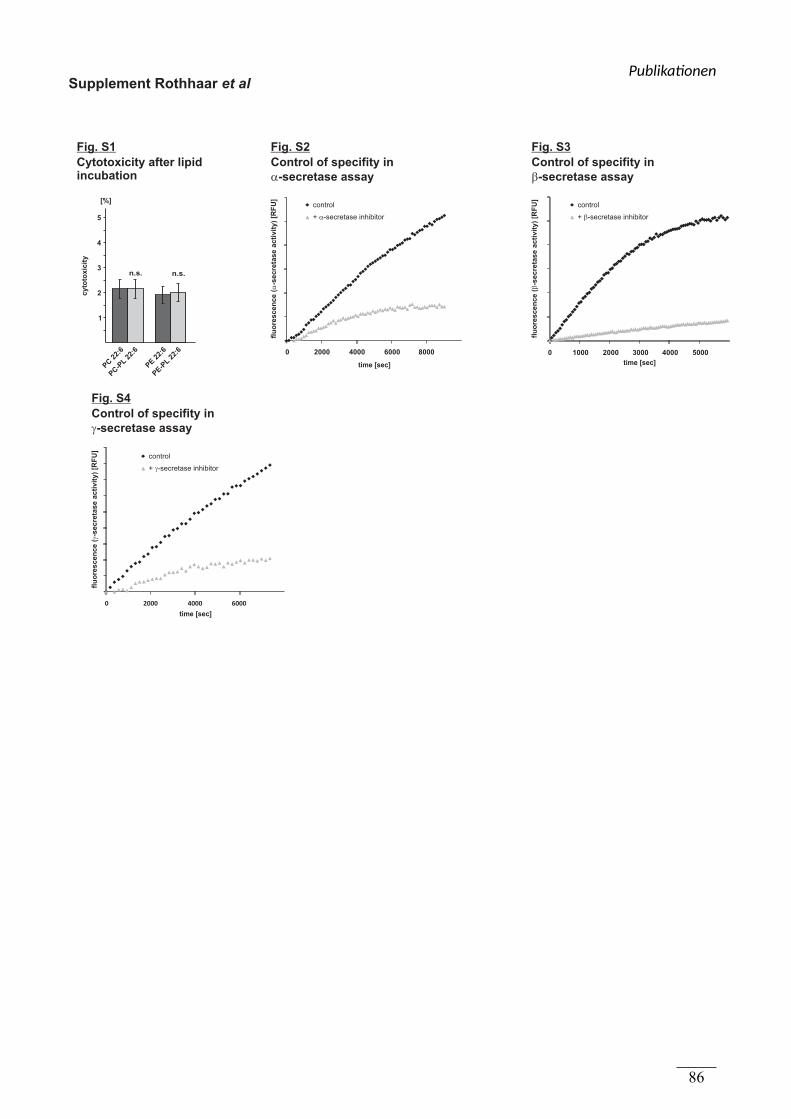
Supplement Rothhaar et al
Fig. S1
Cytotoxicity after lipidincubation
cyto
toxic
ity
PC-P
L22
:6
PC 2
2:6
PE 2
2:6
PE-P
L22
:6
5
1
2
3
4
[%]
n.s. n.s.
Fig. S4
Control of specifity in
-secretase assay�
0 2000 4000 6000
control
+ -secretase inhibitor�
flu
ore
sc
en
ce
(-s
ec
reta
se
ac
tiv
ity
) [R
FU
]�
time [sec]
Fig. S3
Control of specifity in
-secretase assay�
control
+ -secretase inhibitor�
0 1000 2000 3000 4000 5000
flu
ore
sc
en
ce
(-s
ec
reta
se
ac
tiv
ity
) [R
FU
]�
time [sec]time [sec]
Fig. S2
Control of specifity in
-secretase assay�
control
+ -secretase inhibitor�
flu
ore
sc
en
ce
(-s
ec
reta
se
ac
tiv
ity
) [R
FU
]�
0 2000 4000 6000 8000
Publika onen
86
Publikationen
87
(4.3) Zusammenfassung von Publikation 3 und Beschreibung des
Eigenanteils
Oxidized Docosahexaenoic Acid Species and Lipid Peroxidation Products Increase
Amyloidogenic Amyloid Precursor Protein Processing
Haupenthal VJ*, Grimm MO*, Mett J, Stahlmann CP, Blümel T, Mylonas NT, Endres K,
Grimm HS, Hartmann T
* equally contributed
In Publikation 3 wurde der Effekt von oxidierten Docosahexaensäure (DHA) -Derivaten und
Lipid-Peroxidationsprodukten der ω-3 und ω-6 Fettsäuren, 4-Hydroxyhexenal (HHE) und 4-
Hydroxynonenal (HNE), auf die APP-Prozessierung untersucht. Die Lipide sind in figure 1b
der Publikation 3 dargestellt.
Der Einsatz von DHA in der Prävention und Therapie der AD führte in epidemiologischen
und klinischen Studien zu kontroversen Ergebnissen; einerseits wurde DHA ein protektiver
Effekt zur Vorbeugung oder Behandlung der AD bescheinigt, andererseits gibt es Studien, die
keinen positiven Effekt von DHA nachweisen konnten (Chiu et al. 2008; Quinn et al. 2010;
Lee et al. 2013) (zusammengefasst in table 1 der Publikation 3).
Da DHA durch seine sechs Doppelbindungen für Oxidation anfällig ist und dadurch oxidierte
Derivate entstehen können, wurde in dieser Publikation der Einfluss von 7 oxidierten Lipiden
auf die APP-Proteolyse analysiert (VanRollins & Murphy 1984; Roberts et al. 1998).
Es konnte gezeigt werden, dass die Lipid-Peroxidation in humanen post-mortem Gehirnen
von Personen mit AD im Vergleich zu Personen ohne AD signifikant erhöht war. Zusätzlich
war der Gehalt an HNE in diesen Gehirnen sogar signifikant verdoppelt. Um den Einfluss von
oxidierten Lipiden auf die APP Proteolyse zu bestimmen, wurden SH-SY5Y Zellen und
murine, kortikale Primärneurone mit 5 oxidierten DHA-Derivaten, sowie den beiden Lipid-
Peroxidationsprodukten HNE und HHE inkubiert. Die Behandlung der SH-SY5Y Zellen mit
oxidierten Lipiden verursachte keine vermehrte Zytotoxizität.
4 von 5 oxidierte DHA-Derivate, HHE und HNE steigerten signifikant die Bildung von Aβ in
SH-SY5Y Zellen. Die sAPPβ Produktion wurde ebenfalls durch diese Lipide erhöht, jedoch
unterschied sich der Gehalt an sAPPβ nur bei 5 der 7 oxidierten Lipide signifikant von der
Publikationen
88
Kontrolle. Der Gehalt an sAPPα wurde durch die oxidierten Lipide tendenziell reduziert, der
Effekt war im Vergleich zur Kontrolle bei 2 der 7 oxidierten Lipide signifikant reduziert.
Im Gegenzug wurde der Aβ Gehalt durch nicht oxidiertes DHA signifikant im Vergleich zur
Kontrolle reduziert. Die Stabilität von DHA über den Inkubationszeitraum wurde durch
massenspektrometrische Analyse kontrolliert. Die Supplementierung von DHA mit einer
Mischung aller verwendeten, oxidierten Lipide invertierte den protektiven Effekt von DHA,
indem bereits 1% der oxidierten Lipidmischung den Aβ-Gehalt signifikant steigerte.
Der Anstieg des Aβ-Gehalts durch die oxidierten Lipide, wurde durch die Steigerung der
amyloidogenen APP-Prozessierung hervorgerufen. Es wurde in dieser Publikation
nachgewiesen, dass die Enzymaktivität der β-Sekretase direkt durch die verwendeten Lipide
erhöht wird. Zusätzlich wirkten die oxidierten Lipide auf die Expression von BACE-1, indem
sie diese steigerten. Im Gegensatz dazu, wurde die Steigerung der γ-Sekretase Aktivität in
lebenden Zellen ausschließlich durch eine vermehrte Expression der γ-Sekretase Bestandteile,
unter anderem PS1, dem aktiven Zentrum der γ-Sekretase, hervorgerufen. Es wurde kein
direkter Effekt der oxidierten DHA-Derivate oder der Lipid-Peroxidationsprodukte auf die γ-
Sekretase Aktivität festgestellt. Die steigernden Wirkung der oxidierten Lipide auf die
Aktivität der β- und γ-Sekretase wurde auch in kortikalen Primärneuronen der Maus
nachgewiesen.
Diese Ergebnisse verdeutlichen die Notwendigkeit einer Betrachtung der oxidierten DHA-
Derivate hinsichtlich der Prävention und möglicher Therapieansätze in der AD.
Publikation 3 ist in geteilter Erstautorenschaft mit Marcus O.W. Grimm entstanden. Ich habe
für diese Publikation die Versuche koordiniert und in Zusammenarbeit mit Marcus Grimm
geplant. Für diese Publikation wurden von mir die Lipid-Peroxidation und der Gehalt an HNE
in humanen post-mortem Gehirnen (figure 1a) bestimmt. Die Aufarbeitung der dafür
benötigten humanen post-mortem Gehirnproben wurde von Nadine T. Mylonas durchgeführt.
Die Analyse des Aβ-, sAPPβ-, sowie des sAPPα-Spiegels nach Inkubation mit oxidierten
Lipiden und DHA wurde von mir durchgeführt (figure 1c-f). Für diese Versuche habe ich die
Inkubation mit Lipiden, die Probenaufarbeitung und die statistische Analyse der gewonnenen
Daten durchgeführt. Die Analyse der Zytotoxizität der oxidierten Lipide wurde nach
Inkubation mit 2 der 7 Lipide von J. Mett durchgeführt, nach Inkubation der restlichen Lipide
wurde diese Messung von mir durchgeführt (Ergebnisse im Text).
Weiterhin habe ich die Aktivität der β- und γ-Sekretase, sowohl in lebenden SH-SY5Y Zellen
(figure 2a+b), als auch in lebenden kortikalen Primärneuronen der Maus gemessen (table 2).
Auch hier wurde von mir die Inkubation und statistische Datenanalyse durchgeführt.
Publikationen
89
Neben der Analyse in lebenden Zellen, habe ich die Inkubation von Membranen aus SH-
SY5Y Zellen und die nachfolgende Messung der β-Sekretase Aktivität durchgeführt und die
Daten statistisch ausgewertet (figure 2c). Die Bestimmung der Expression der Sekretasen
wurde von J. Mett im Anschluss an die von mir durchgeführte Inkubation der Zellen mit
oxidierten Lipiden (figure 2d) durchgeführt. T. Blümel bestimmte die Aktivität der β- und γ-
Sekretase nach Inkubation mit DHA (Ergebnisse im Text) und C. Stahlmann die DHA-
Stabilität (Ergebnisse im Text); zu beiden Analysen habe ich die Inkubation mit Lipiden
durchgeführt. Die statistische Auswertung der Aβ-Spiegel nach Inkubation mit DHA und
oxidierten Lipiden wurde von N. Mylonas durchgeführt (Ergebnisse im Text).
Nachfolgend ist Publikation 3 zu finden. Das Original der Publikation ist auf der
Internetseite des Verlages S. Karger AG (Basel, Schweiz), www.karger.com/ndd, unter
der Nummer doi:10.1159/000440839 zu finden.
E-Mail [email protected]
Original Paper
Neurodegener Dis DOI: 10.1159/000440839
Oxidized Docosahexaenoic Acid Species and Lipid Peroxidation Products Increase Amyloidogenic Amyloid Precursor Protein Processing
Marcus O.W. Grimm a–c Viola J. Haupenthal a Janine Mett a
Christoph P. Stahlmann a Tamara Blümel a Nadine T. Mylonas a
Kristina Endres d Heike S. Grimm a Tobias Hartmann a–c
a Experimental Neurology, b Neurodegeneration and Neurobiology and c Deutsches Institut für Demenzprävention (DIDP), Saarland University, Homburg/Saar , and d Department of Psychiatry and Psychotherapy, Clinical Research Group, University Medical Centre Johannes Gutenberg, University of Mainz, Mainz , Germany
neurons. In the presence of oxidized lipids Aβ and soluble β-secreted APP levels were elevated, whereas soluble α-secreted APP was decreased, suggesting a shift from the nonamyloidogenic to the amyloidogenic pathway of APP processing. Furthermore, β- and γ-secretase activity was in-creased by oxidized lipids via increased gene expression and additionally by a direct effect on β-secretase activity. Impor-tantly, only 1% oxidized DHA was sufficient to revert the pro-tective effect of DHA and to significantly increase Aβ produc-tion. Therefore, our results emphasize the need to prevent DHA from oxidation in nutritional approaches and might help explain the divergent results of clinical DHA studies.
© 2015 S. Karger AG, Basel
Introduction
Alzheimer’s disease (AD) is the most common neuro-degenerative disorder in the aged population. Main path-ological features of AD include β-amyloid (Aβ) accumu-lation and hyperphosphorylation of the microtubule-as-sociated protein tau, leading to the neuropathological
Key Words
Oxidized docosahexaenoic acid · Lipid peroxidation · Amyloid precursor protein processing · β-Amyloid
Abstract
One of the main characteristics of Alzheimer’s disease (AD) is the β-amyloid peptide (Aβ) generated by β- and γ-secretase processing of the amyloid precursor protein (APP). Previous-ly it has been demonstrated that polyunsaturated fatty acids (PUFAs), especially docosahexaenoic acid (DHA), are associ-ated with a reduced risk of AD caused by decreased Aβ pro-duction. However, in epidemiological studies and nutrition-al approaches, the outcomes of DHA-dependent treatment were partially controversial. PUFAs are very susceptible to reactive oxygen species and lipid peroxidation, which are in-creased during disease pathology. In line with published re-sults, lipid peroxidation was elevated in human postmortem AD brains; especially 4-hydroxy-nonenal (HNE) was in-creased. To investigate whether lipid peroxidation is only a consequence or might also influence the processes leading to AD, we analyzed 7 different oxidized lipid species includ-ing 5 oxidized DHA derivatives and the lipid peroxidation products of ω–3 and ω–6 PUFAs, HNE and 4-hydroxy-hexe-nal, in human neuroblastoma cells and mouse mixed cortical
Received: June 2, 2015 Accepted after revision: September 3, 2015 Published online: December 8, 2015 D i s e a s e s
Dr. Marcus O.W. Grimm Experimental Neurology, Saarland University Kirrberger Strasse 1, Gebäude 61.4 DE–66421 Homburg/Saar (Germany) E-Mail marcus.grimm @ uks.eu
© 2015 S. Karger AG, Basel1660–2854/15/0000–0000$39.50/0
www.karger.com/ndd
Marcus O.W. Grimm and Viola J. Haupenthal contributed equally to this work.
Dow
nloa
ded
by:
Saa
rländ
isch
e U
nive
rsitä
ts u
nd L
ande
sbib
lioth
ek
149.
126.
78.6
5 -
12/8
/201
5 1:
48:5
7 P
M
Publika onen
90
Grimm et al.
Neurodegener DisDOI: 10.1159/000440839
2
hallmarks of AD, senile plaques and neurofibrillary tan-gles [1, 2] . Amyloidogenic Aβ peptides are generated by sequential proteolytic processing of the amyloid precur-sor protein (APP), a type-I transmembrane protein. Ini-tial cleavage by the β-secretase BACE1 results in release of soluble β-secreted APP (sAPPβ) and the generationof a membrane-tethered C-terminal stub called β-CTF [3] . Further cleavage of this C-terminal fragment by γ-secretase, a heterotetrameric protein complex compris-ing presenilin-1 or -2 (PS1, PS2), nicastrin, anterior phar-ynx defective 1 (Aph1) and presenilin enhancer 2 gener-ates Aβ peptides [4] . Beside amyloidogenic processing of APP involving β- and γ-secretase activity, APP can be cleaved by α-secretases in a nonamyloidogenic pathway, preventing Aβ generation. The α-secretases cleave APP within the Aβ domain, releasing α-secreted APP (sAPPα) [3] . The remaining C-terminal fragment is also cleaved by the γ-secretase complex, liberating nonamyloidogenic p3 peptides [3] . Mounting evidence arises that the lipid mi-croenvironment of the membrane modulates APP pro-cessing. Beside the fact that APP itself and its processing secretases are all transmembrane proteins, amyloidogen-ic APP processing is discussed to be dependent on par-ticular membrane microdomains, called lipid rafts, en-riched in cholesterol and sphingolipids [5] . Furthermore, several lipids have been shown to alter Aβ generation, in-cluding gangliosides, sphingomyelin, trans -fatty acids, phytosterols, cholesterol and docosahexaenoic acid (DHA) [6–12] . DHA, an ω–3 polyunsaturated fatty acid (PUFA) predominantly found in marine fish and algae, was shown to reduce Aβ production in vitro and in ani-mal models of AD [12–15] . Therefore, DHA has become of major interest for nutritional intervention in AD. Moreover, DHA is decreased in postmortem AD brains, and AD patients have reduced serum DHA levels [16, 17] . DHA, as an important component of neuronal mem-branes, is involved in neurogenesis, neuronal differentia-tion, neurotransmission, synaptogenesis, synaptic plas-ticity and neurite outgrowth. DHA is therefore suggested to be crucial in learning, memory and cognitive processes [18] . However, epidemiological studies and clinical trials addressing the relationship between ω–3/DHA uptake and AD incidence or cognitive decline revealed diverging results; examples are given in table 1 . Although ω–3 fatty acids have several biological properties that might be ben-eficial in AD, DHA is very susceptible to lipid peroxida-tion and might auto-oxidize and induce lipid peroxida-tion in vivo, resulting in oxidative stress, known to be involved in AD pathogenesis [19] . Interestingly, it is known that DHA derivatives like neuroprotectins also af-
fect the mechanisms leading to AD [20] . However, up to now very little has been known about the effect of the oxidation products of DHA on APP processing.
In gray matter, 25–30% of all fatty acids in phospho-lipids are DHA. The oxidation products of PUFAs are increased in CSF of AD patients [21] . In human postmor-tem AD brains, Subbarao et al. [22] report a concentra-tion of 2 nmol oxidized DHA per milligram protein. Sim-ilar results were found by Musiek et al. [23] reporting 4.9 ng oxidized DHA in C57BL/6 mouse brain and 8.7 ng oxidized DHA in human brain per gram tissue (wet weight). In our study, we used a comparable concentra-tion range of oxidized PUFAs (0.3–3.3 nmol/mg protein).
In the present study, we addressed whether different oxidized DHA and lipid species affect APP processing and Aβ generation. Considering that DHA, which is highly susceptible to oxidation, and its oxidized products might have different effects on APP processing, our re-sults are not only important for nutritional approaches, but might also help understand the diverging outcomes of different DHA-based clinical trials.
Materials and Methods
If not stated otherwise, chemicals were purchased from Sigma-Aldrich (Taufkirchen, Germany). Oxidized lipids – 4-hydroxy-2E-hexenal (HHE), 4-hydroxy-nonenal (HNE), 17S-hydroxy-4Z,7Z,10Z,13Z,15E,19Z-docosahexaenoic acid (17-OH-DHA), 7, 17-dihydroxy-8E,10Z,13Z,15E,19Z-docosapentaenoic acid (7,17-OH-DPA), 17-keto-DHA, 17-hydroperoxy-DHA, 19,20-epoxy-DPA – were purchased from Biomol (Hamburg, Germany).
Cell Culture and Lipid Incubations SH-SY5Y cells were cultivated as described earlier [9]. For lipid incubations SH-SY5Y cells were plated on 96-well or
6-well plates (Thermo Scientific, Waltham, Mass., USA/Corning, Tewksbury, Mass., USA). When confluent, cells were cultivated in medium containing 0.1% fetal calf serum and 0.1% bovine serum albumin for 16 h. Incubation with lipids was carried out for 8 +16 h with 2 μ M oxidized DHA/20 μ M DHA. Measurement of cyto-toxicity was performed using the lactate dehydrogenase assay (Roche, Mannheim, Germany) according to the manufacturer’s protocol.
Preparation of Primary Cortical Neurons C57BL/6 mice (E13) were used for preparation as described
earlier [9] . Lipid incubation was carried out as described above.
Measurement of Aβ, sAPPα and sAPPβ Levels Analyses of Aβ levels were performed as described before [9] .
Briefly, Aβ in conditioned cell culture media was immunoprecipi-tated using W02 antibody (epitope: amino acid 2–8 of Aβ) [24] and detected by Western blot analysis. For sAPPα/β detection, condi-tioned media were adjusted to equal protein amounts using the
Dow
nloa
ded
by:
Saa
rländ
isch
e U
nive
rsitä
ts u
nd L
ande
sbib
lioth
ek
149.
126.
78.6
5 -
12/8
/201
5 1:
48:5
7 P
M
Publika onen
91
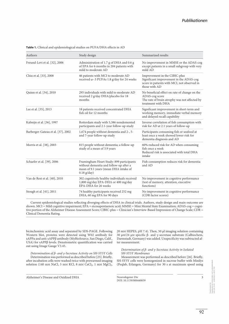
Alzheimer’s Disease and Oxidized DHA Neurodegener DisDOI: 10.1159/000440839
3
bicinchoninic acid assay and separated by SDS-PAGE. Following Western blot, proteins were detected using W02 antibody for sAPPα and anti-sAPPβ antibody (MyBioSource, San Diego, Calif., USA) for sAPPβ levels. Densitometric quantification was carried out using Image Gauge V3.45.
Determination of β- and γ-Secretase Activity on SH-SY5Y Cells Determination was performed as described before [25] . Briefly,
after incubation cells were washed twice with prewarmed imaging solution (140 m M NaCl, 5 m M KCl, 8 m M CaCl 2 , 1 m M MgCl 2 ,
20 m M HEPES, pH 7.4). Then, 50 μl imaging solution containing 30 μ M /24 μ M specific β- and γ-secretase substrate (Calbiochem, Darmstadt, Germany) was added. Unspecificity was subtracted af-ter measurement.
Determination of β- and γ-Secretase Activity in IsolatedSH-SY5Y Membranes Measurement was performed as described before [26] . Briefly,
SH-SY5Y cells were homogenized in sucrose buffer with Minilys (Peqlab, Erlangen, Germany) for 30 s at maximum speed using
Table 1. Clinical and epidemiological studies on PUFA/DHA effects in AD
Authors Study design Summarized results
Freund-Levi et al. [32], 2006 Administration of 1.7 g of DHA and 0.6 g of EPA for 6 months in 204 patients with mild to moderate AD
No improvement in MMSE or the ADAS-cog except patients in a small subgroup with very mild AD
Chiu et al. [33], 2008 46 patients with MCI to moderate AD received ω–3 PUFAs 1.8 g/day for 24 weeks
Improvement in the CIBIC-plusSignificant improvement in the ADAS-cog score in patients with MCI, not observed in those with AD
Quinn et al. [34], 2010 295 individuals with mild to moderate AD received 2 g/day DHA/placebo for 18 months
No beneficial effect on rate of change on the ADAS-cog scoreThe rate of brain atrophy was not affected by treatment with DHA
Lee et al. [35], 2013 18 patients received concentrated DHA fish oil for 12 months
Significant improvement in short-term and working memory, immediate verbal memory and delayed recall capability
Kalmijn et al. [36], 1997 Rotterdam study with 5,386 nondemented participants and 2.1-year follow-up study
Inverse correlation of fish consumption with risk for AD at 2.1 years of follow-up
Barberger-Gateau et al. [37], 2002 1,674 people without dementia and 2-, 5- and 7-year follow-up study
Participants consuming fish or seafood at least once a week showed lower risk for dementia diagnosis and AD
Morris et al. [38], 2003 815 people without dementia; a follow-up study of a mean of 3.9 years
60% reduced risk for AD when consuming fish once a weekReduced risk is associated with total DHA intake
Schaefer et al. [39], 2006 Framingham Heart Study: 899 participants without dementia and follow-up after a mean of 9.1 years (mean DHA intake of 0.18 g/day)
Fish consumption reduces risk for dementia and AD
Van de Rest et al. [40], 2010 302 cognitively healthy individuals received 1,800 mg/day EPA-DHA or 400 mg/day EPA-DHA for 26 weeks
No improvement in cognitive performance (test of memory, attention, executive functions)
Stough et al. [41], 2011 74 healthy participants received 252 mg DHA, 60 mg EPA for 90 days
No improvement in cognitive performance (CDR factor scores)
Current epidemiological studies reflecting diverging effects of DHA in clinical trials. Authors, study design and main outcome are shown. MCI = Mild cognitive impairment; EPA = eicosapentaenoic acid; MMSE = Mini Mental State Examination; ADAS-cog = cogni-tive portion of the Alzheimer Disease Assessment Score; CIBIC-plus = Clinician’s Interview-Based Impression of Change Scale; CDR = Clinical Dementia Rating.
Dow
nloa
ded
by:
Saa
rländ
isch
e U
nive
rsitä
ts u
nd L
ande
sbib
lioth
ek
149.
126.
78.6
5 -
12/8
/201
5 1:
48:5
7 P
M
Publika onen
92
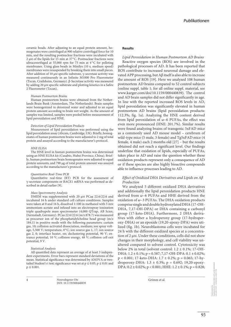
Grimm et al.
Neurodegener DisDOI: 10.1159/000440839
4
ceramic beads. After adjusting to an equal protein amount, ho-mogenates were centrifuged at 900 relative centrifugal force for 10 min, and the resulting postnuclear fractions were incubated with 2 μ M of the lipids for 15 min at 37 ° C. Postnuclear fractions were ultracentrifuged at 55,000 rpm for 75 min at 4 ° C for pelleting membranes. Using glass beads in Minilys (10 s, medium speed) membranes were resuspended by breaking them into small pieces. After addition of 10 μ M specific substrate, γ-secretase activity was measured continuously in an Infinite M1000 Pro Fluorometer (Tecan, Crailsheim, Germany). β-Secretase activity was measured by adding 20 μ M specific substrate and plotting kinetics in a Safire 2 Fluorometer (Tecan).
Human Postmortem Brains Human postmortem brains were obtained from the Nether-
lands Brain Bank (Amsterdam, The Netherlands). Brain samples were homogenized in deionized water and adjusted to an equal protein amount according to brain wet weight. As the amount of samples was limited, samples were pooled before measurement of lipid peroxidation and HNE.
Detection of Lipid Peroxidation Products Measurement of lipid peroxidation was performed using the
lipid peroxidation assay (Abcam, Cambridge, UK). Briefly, homog-enates of human postmortem brains were adjusted to 2 mg of total protein and assayed according to the manufacturer’s protocol.
HNE ELISA The HNE level in human postmortem brains was determined
using an HNE ELISA kit (Cusabio, Hubei Province, China). Brief-ly, human postmortem brain homogenates were adjusted to equal protein amounts, and 790 μg of total protein amount was assayed according to the manufacturer’s protocol.
Quantitative Real-Time PCR Quantitative real-time (RT) PCR for the assessment of
γ-secretase components or BACE1 mRNA was performed as de-scribed in detail earlier [9] .
Mass Spectrometry Analysis DMEM was supplemented with 20 μ M PCaa 22: 6/22: 6 and
incubated 16 h under standard cell culture conditions. Samples were taken at 0 and 16 h, dissolved 1: 100 in methanol with 5 m M ammonium acetate and infused into an electrospray ionization triple quadrupole mass spectrometer (4,000 QTrap, AB Sciex, Darmstadt, Germany). PCaa 22: 6/22: 6 (m/z 878.7) was measured as precursor ion of the phosphatidylcholine head group (m/z 184.1) in positive mode with the following parameters: curtain gas, 10; collision-activated dissociation, medium; ion spray volt-age, 5,500 V; temperature, 0 ° C; ion source gas 1, 17; ion source gas 2, 0; interface heater, on; declustering potential, 90 V; en-trance potential, 10 V; collision energy, 48 V; collision cell exit potential, 9 V.
Statistical Analysis All quantified data represent an average of at least 3 indepen-
dent experiments. Error bars represent standard deviations of the mean. Statistical significance was determined by ANOVA or two-tailed Student’s t test; significance was set at p ≤ 0.05, p ≤ 0.01 and p ≤ 0.001.
Results
Lipid Peroxidation in Human Postmortem AD Brains Reactive oxygen species (ROS) are involved in the
pathological processes of AD. It has been reported that ROS contribute to increased neuronal damage and ele-vated APP processing, but Aβ itself is also able to increase the amount of ROS [19] . Here we analyzed 108 human postmortem AD brains compared to 52 control subjects (online suppl. table 1; for all online suppl. material, see www.karger.com/doi/10.1159/000440839). The control and AD brain samples did not differ significantly in age. In line with the reported increased ROS levels in AD, lipid peroxidation was significantly elevated in human postmortem AD brains (lipid peroxidation products: 112.3%; fig. 1 a). Analyzing the HNE content derived from lipid peroxidation of ω–6 PUFAs, the effect was even more pronounced (HNE: 201.7%). Similar results were found analyzing brains of transgenic 5xFAD mice as a commonly used AD mouse model – cerebrum of wild-type mice (5 male, 3 female) and Tg5xFAD mice (4 female, 4 male) each 2 months old [27] – but the results obtained did not reach a significant level. Our findings underline that oxidation of lipids, especially of PUFAs, takes place in AD and raise the question whether these oxidation products represent only a consequence of AD or if these species are also highly bioactive compounds able to influence processes leading to AD.
Effect of Oxidized DHA Derivatives and Lipids on Aβ Production We analyzed 5 different oxidized DHA derivatives
and additionally the lipid peroxidation products HNE derived from ω–6 PUFAs and HHE derived from the oxidation of ω–3 PUFAs. The DHA oxidation products comprise single and double hydroxylated DHA (17-OH-DHA, 7,17-OH-DPA) or DHA containing a carbonyl group (17-keto-DHA). Furthermore, 2 DHA deriva-tives with either a hydroperoxy group (17-hydroper-oxy-DHA) or an epoxide (19,20-epoxy-DPA) were uti-lized ( fig. 1 b). Neuroblastoma cells were incubated for 24 h with the different oxidized species at a concentra-tion of 2 μ M . Under these conditions, cells did not show changes in their morphology, and cell viability was un-altered compared to solvent control. Cytotoxicity was below 2% in total (solvent control: 1.2 ± 0.1%; 17-OH-DHA: 1.2 ± 0.1%; p = 0.587; 7,17-OH-DPA: 0.1 ± 0.02%; p < 0.001; 17-keto-DHA: 1.7 ± 0.2%; p = 0.065; 17-hy-droperoxy-DHA: 1.3 ± 0.3%; p = 0.692; 19,20-epoxy-DPA: 0.2 ± 0.02%; p < 0.001; HHE: 1.2 ± 0.1%; p = 0.828;
Dow
nloa
ded
by:
Saa
rländ
isch
e U
nive
rsitä
ts u
nd L
ande
sbib
lioth
ek
149.
126.
78.6
5 -
12/8
/201
5 1:
48:5
7 P
M
Publika onen
93
Alzheimer’s Disease and Oxidized DHA Neurodegener DisDOI: 10.1159/000440839
5
100
Non-AD human postmortem brains
Lipi
d pe
roxi
datio
n (%
of c
ontr
ol)
AD human postmortem brains**
200
100
HN
E le
vel (
% o
f con
trol
)
***
a
100
Oxidized DHA species
A (%
of c
ontr
ol)
Solvent control
Solvent controln.s.
Unoxidized DHA
Average effectof ox. lipids
Solve
nt co
ntrol
17-O
H-DHA
7,17-
OH-DPA
17-K
eto-D
HA
17-H
ydro
peroxy
-DHA
19,20
-Epox
y-DPA HHE
HNE
Lipid peroxidation product
*** *** ***
***
*** ***
c
100
Oxidized DHA speciessA
PP (%
of c
ontr
ol)
Solvent control
Solvent control
n.s.n.s.
Average effectof ox. lipids
Solve
nt co
ntrol
17-O
H-DHA
7,17-
OH-DPA
17-K
eto-D
HA
17-H
ydro
peroxy
-DHA
19,20
-Epox
y-DPA HHE
HNE
Lipid peroxidation product
*** ******
*
*
d
100
Oxidized DHA species
sA (%
of c
ontr
ol)
Solvent control
Solvent controln.s. n.s.n.s. n.s.
n.s.
Average effectof ox. lipids
Solve
nt co
ntrol
17-O
H-DHA
7,17-
OH-DPA
17-K
eto-D
HA
17-H
ydro
peroxy
-DHA
19,20
-Epox
y-DPA HHE
HNE
Lipid peroxidation product
**
***
e
100
DHA + oxidized DHA species
A (%
of c
ontr
ol)
Solvent control
Solvent control
Solve
nt co
ntrol
1% 5% 10%
***
*
f
COOH
17-OH-DHA
COOH
OH
17-Keto-DHA
O
COOH COOH
OH
COOH
O
7,17-OH-DPAOH 17-Hydroperoxy-DHA
O-O-H
19,20-Epoxy-DPA HNE
H
O
OH
HHE
H
O
OH
b
(For legend see next page.) 1
Colo
r ver
sion
ava
ilabl
e on
line
Dow
nloa
ded
by:
Saa
rländ
isch
e U
nive
rsitä
ts u
nd L
ande
sbib
lioth
ek
149.
126.
78.6
5 -
12/8
/201
5 1:
48:5
7 P
M
Publika onen
94
Grimm et al.
Neurodegener DisDOI: 10.1159/000440839
6
HNE: 1.3 ± 0.1%; p = 0.557). As an additional control, 20 μ M DHA was incubated on cells. To investigate if DHA is oxidized under the incubation conditions used in this study, the DHA stability was determined by mass spectrometry. After 16 h the DHA amount was not al-tered (0 h: 100.0 ± 17.9%; 16 h: 100.9 ± 17.0%, p = 0.9697). As previously shown, DHA significantly de-creased Aβ production (DHA: 85.9 ± 2.5%; p < 0.001). In contrast, 6 out of 7 oxidized species significantly el-evated the Aβ levels compared to solvent control (17-OH-DHA: 102.0%; 7,17-OH-DPA: 125.7%; 17-keto-DHA: 120.0%; 17-hydroperoxy-DHA: 115.2%; 19,20-epoxy-DPA: 137.8%; HHE: 112.6%; HNE: 112.3%; average: 118%; fig. 1 c). Significant elevation of Aβ levels compared to nonoxidized DHA was found for all oxi-dized lipids (17-OH-DHA: p = 0.001; 7,17-OH-DPA:p < 0.001; 17-keto-DHA: p < 0.001; 17-hydroperoxy-DHA: p < 0.001; 19,20-epoxy-DPA: p < 0.001; HHE:p < 0.001; HNE: p < 0.001).
To substantiate the observed effect, we measured sAPPβ. sAPPβ levels were increased in all cells incubat-ed with oxidized lipids (17-OH-DHA: 107.2%; 7,17-OH-DPA: 127.8%; 17-keto-DHA: 124.3%; 17-hydroperoxy-DHA: 105.0%; 19,20-epoxy-DPA: 115.3%; HHE: 105.3%; HNE: 126.3%; fig. 1 d). On average the sAPPβ levels were increased to 115.9% and showed a correlation with the Aβ level (r = 0.5427). In contrast, the nonamyloidogen-ic α-secretase-derived sAPPα fragment was decreased to 96.5% on average, when cells were incubated with oxi-dized lipids as shown in figure 1 e (17-OH-DHA: 92.3%; 7,17-OH-DPA: 92.1%; 17-keto-DHA: 79.0%; 17-hydro-
peroxy-DHA: 93.7%; 19,20-epoxy-DPA: 105.3%; HHE: 100.9%; HNE: 111.8%). In our previous study we could show that 100 μ M DHA caused an increase in sAPPα levels (131.2 ± 3.5%, p < 0.001 [12] ). In line with these results, the sAPPα/sAPPβ ratio was changed, which was also published by Sahlin et al. [28] showing a 40% al-teration.
Taking into consideration that a certain proportion of DHA is oxidized under physiological conditions, we in-cubated unoxidized DHA with a certain percentage of oxidized DHA. In total 20 μ M of lipid was used with a to-tal of 1% (0.2 μ M ), 5% (1 μ M ) and 10% (2 μ M ) oxidized DHA species. 1% of oxidized DHA was able not only to attenuate the protective effect of DHA, but significantly increased the Aβ levels. The Aβ levels were dose-depen-dently increased with increasing fractions of oxidized DHA (1% oxidized DHA: 114.9%; 5% oxidized DHA: 123.3%; 10% oxidized DHA: 133.0%; fig. 1 f). To sum up, our results clearly show that oxidized DHA not only in-creases the amyloidogenic and decreases the nonamy-loidogenic processing, but it also reverts the beneficial ef-fect of DHA under the tested conditions in very low amounts.
Effect of Oxidized DHA Derivatives and Lipids on Secretases To elucidate the underlying mechanism of oxidized
lipid species on APP processing, we measured β- and γ-secretase activity after treatment with oxidized lipids. Six oxidized species showed a significant increase in β-secretase activity in neuroblastoma cells (17-OH-
Fig. 1. Lipid peroxidation in human postmortem brains and effects of oxidized lipids on APP processing in SH-SY5Y cells. a Oxidized lipids (AD brain: 112.3 ± 2.2%; p = 0.004; left) and HNE levels (AD: 201.7 ± 3.5%, p < 0.001; right) were increased in human AD post-mortem brains compared to nondemented control brains. b Struc-ture of oxidized DHA species and lipid peroxidation products. c–f Effects of oxidized DHA (17-OH-DHA, 7,17-OH-DPA, 17-keto-DHA, 17-hydroperoxy-DHA, 19,20-epoxy-DPA) and lipid peroxidation products (HHE, HNE) on APP processing: SH-SY5Y-APP695 cells were treated for 24 h with oxidized DHA de-rivatives or lipid peroxidation products with a final concentration of 2 μ M or solvent, respectively. c Aβ levels after incubation with oxidized lipids (17-OH-DHA: 102.0 ± 1.7%; p = 0.389; 7,17-OH-DPA: 125.7 ± 2.1%; p < 0.001; 17-keto-DHA: 120.0 ± 1.2%; p < 0.001; 17-hydroperoxy-DHA: 115.2 ± 2.4%; p < 0.001; 19,20-ep-oxy-DPA: 137.8 ± 2.5%; p < 0.001; HHE: 112.6 ± 1.7%; p < 0.001; HNE: 112.3 ± 1.6%; p < 0.001). d sAPPβ levels (17-OH-DHA: 107.2 ± 3.5%; p = 0.222; 7,17-OH-DPA: 127.8 ± 0.8%; p < 0.001; 17-keto-DHA: 124.3 ± 1.1%; p < 0.001; 17-hydroperoxy-DHA:
105.0 ± 2.4%; p = 0.224; 19,20-epoxy-DPA: 115.3 ± 0.5%; p < 0.001; HHE: 105.3 ± 1.0%; p = 0.019; HNE: 126.3 ± 6.5%; p = 0.044). e sAPPα levels were measured compared to solvent control (17-OH-DHA: 92.3 ± 3.5%; p = 0.197; 7,17-OH-DPA: 92.1 ± 4.9%; p = 0.317; 17-keto-DHA: 79.0 ± 2.4%; p = 0.004; 17-hydroperoxy-DHA: 93.7 ± 0.5%; p < 0.001; 19,20-epoxy-DPA: 105.3 ± 1.9%; p = 0.114; HHE: 100.9 ± 1.7%; p = 0.760; HNE: 111.8 ± 2.8%; p = 0.476). f SH-SY5Y-APP695 cells were treated with in total 20 μ M DHA containing a proportionately 1, 5 and 10% mix of all oxidized DHA species, and Aβ levels were measured compared to solvent control (1% oxidized DHA: 114.9 ± 3.2%; p = 0.029; 5% oxidized DHA: 123.3 ± 2.8%; p = 0.005; 10% oxidized DHA: 133.0 ± 8.4%; p = 0.021). Solvent control is indicated by a dashed line, the aver-age effect of all oxidized DHA derivatives is indicated by a dottedline. All quantified data represent an average of at least 3 indepen-dent experiments. Error bars represent standard deviations of the mean. Asterisks show the statistical significance compared to sol-vent control ( * p ≤ 0.05, * * p ≤ 0.01 and * * * p ≤ 0.001).
Dow
nloa
ded
by:
Saa
rländ
isch
e U
nive
rsitä
ts u
nd L
ande
sbib
lioth
ek
149.
126.
78.6
5 -
12/8
/201
5 1:
48:5
7 P
M
Publika onen
95
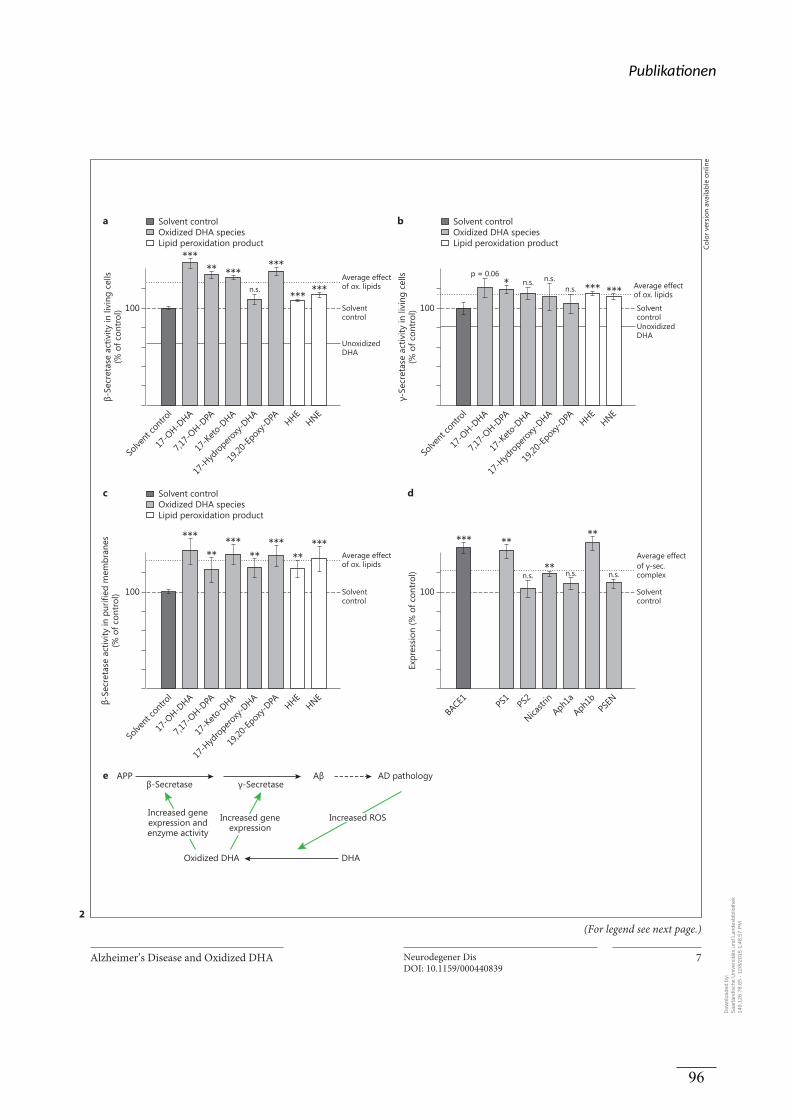
Alzheimer’s Disease and Oxidized DHA Neurodegener DisDOI: 10.1159/000440839
7
100
Oxidized DHA species
DHA
***** ***
***
*** ***
a
100
Oxidized DHA species
DHA
* *** ***
b
100
Oxidized DHA species
***
*****
*****
*****
c
e
100
of
*****
**
**
d
Oxidized DHA DHA
2
(For legend see next page.)
Colo
r ver
sion
ava
ilabl
e on
line
Dow
nloa
ded
by:
Saa
rländ
isch
e U
nive
rsitä
ts u
nd L
ande
sbib
lioth
ek
149.
126.
78.6
5 -
12/8
/201
5 1:
48:5
7 P
M
Publika onen
96
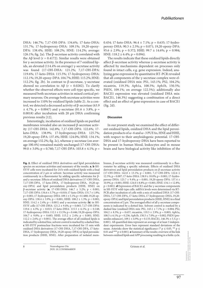
Grimm et al.
Neurodegener DisDOI: 10.1159/000440839
8
DHA: 146.7%; 7,17-OH-DPA: 134.6%; 17-keto-DHA: 131.7%; 17-hydroperoxy-DHA: 109.1%; 19,20-epoxy-DPA: 138.4%; HHE: 108.2%; HNE: 114.2%; average:126.1%; fig. 2 a). The β-secretase activity correlated with the Aβ level (r = 0.4172). Similar results were obtained for γ-secretase activity. In the presence of 7 oxidized lip-ids, an elevated (114.4% on average) γ-secretase activity was found (17-OH-DHA: 121.2%; 7,17-OH-DPA: 119.6%; 17-keto-DHA: 115.5%; 17-hydroperoxy-DHA: 112.1%; 19,20-epoxy-DPA: 104.7%; HHE: 115.2%; HNE: 112.2%; fig. 2 b). In contrast to β-secretase, γ-secretase showed no correlation to Aβ (r = 0.0282). To clarify whether the observed effects were cell-type specific, we measured both secretase activities in mixed cortical pri-mary neurons. On average both secretase activities were increased to 110% by oxidized lipids ( table 2 ). As a con-trol, we detected a decreased activity of β-secretase (63.9 ± 4.7%, p = 0.0047) and γ-secretase (81.6 ± 3.8%, p = 0.0018) after incubation with 20 μ M DHA confirming previous results [12] .
Interestingly, incubation of oxidized lipids on purified membranes revealed also an increased β-secretase activ-ity (17-OH-DHA: 142.8%; 7,17-OH-DPA: 122.6%; 17-keto-DHA: 138.9%; 17-hydroperoxy-DHA: 125.7%; 19,20-epoxy-DPA: 137.4%; HHE: 124.0%; HNE: 134.1%; on average 132.2%; fig. 2 c), whereas γ-secretase (on aver-age 100.4%) remained mainly unchanged (17-OH-DHA: 99.0 ± 3.0%; p = 0.760; 7,17-OH-DPA: 103.8 ± 4.1%; p =
0.454; 17-keto-DHA: 96.4 ± 7.1%; p = 0.635; 17-hydro-peroxy-DHA: 90.3 ± 2.5%; p = 0.071; 19,20-epoxy-DPA: 95.6 ± 2.9%; p = 0.372; HHE: 99.7 ± 14.6%; p = 0.984; HNE: 118.2 ± 6.4%; p = 0.094).
The results indicate that these oxidized lipids directly affect β-secretase activity whereas γ-secretase activity is affected by mechanisms dependent on processes only found in intact cells, e.g. gene expression. Indeed, ana-lyzing gene expression by quantitative RT-PCR revealed that all components of the γ-secretase complex were el-evated (oxidized DHA mix: PS1, 143.1%; PS2, 104.2%; nicastrin, 119.3%; Aph1a, 108.5%; Aph1b, 150.5%; PSEN, 109.1%; on average 122.5%); additionally also BACE1 expression was elevated (oxidized DHA mix: BACE1, 146.3%) suggesting a combination of a direct effect and an effect of gene expression in case of BACE1 ( fig. 2 d).
Discussion
In our present study we examined the effect of differ-ent oxidized lipids, oxidized DHA and the lipid peroxi-dation products of ω–6 and ω–3 PUFAs, HNE and HHE, with respect to their amyloidogenic potential. 17-OH-DHA and 17-hydroperoxy-DHA have been reported to be present in human blood, leukocytes and in mouse brain and have biological activity like inhibition of the
Fig. 2. Effect of oxidized DHA derivatives and lipid peroxidation species on secretase activities and summary of the results. a , b SH-SY5Y cells were incubated for 24 h with oxidized lipids with a final concentration of 2 μ M or solvent. Secretase activity was measured continuously in a fluorometer by adding specific substrates for β- and γ-secretase. Effects of oxidized DHA derivatives (17-OH-DHA, 7,17-OH-DPA, 17-keto-DHA, 17-hydroperoxy-DHA, 19,20-ep-oxy-DPA) and lipid peroxidation products (HHE, HNE) on β-secretase activity ( a ; 17-OH-DHA: 146.7 ± 5.2%; p < 0.001; 7,17-OH-DPA: 134.6 ± 3.7%; p = 0.010; 17-keto-DHA: 131.7 ± 1.8%; p < 0.001; 17-hydroperoxy-DHA: 109.1 ± 5.1%; p = 0.1480; 19,20-ep-oxy-DPA: 138.4 ± 3.9%; p < 0.001; HHE: 108.2 ± 1.3%; p < 0.001; HNE: 114.2 ± 2.4%; p < 0.001) and γ-secretase activity ( b ) in SH-SY5Y cells (17-OH-DHA: 121.2 ± 9.4%; p = 0.065; 7,17-OH-DPA: 119.6 ± 4.3%; p = 0.013; 17-keto-DHA: 115.5 ± 6.1%; p = 0.148; 17-hydroperoxy-DHA: 112.1 ± 13.5%; p = 0.446; 19,20-epoxy-DPA: 104.7 ± 9.8%; p = 0.683; HHE: 115.2 ± 2.4%; p < 0.001; HNE:112.2 ± 2.6%; p < 0.001). The average effect of all oxidized lipids is indicated by a dotted line, solvent control is marked by a dashed line. c SH-SY5Y postnuclear fractions were incubated for 15 min with oxidized DHA derivatives (17-OH-DHA, 7,17-OH-DPA, 17-keto-DHA, 17-hydroperoxy-DHA, 19,20-epoxy-DPA) or lipid peroxida-tion products (HHE, HNE). After preparation of isolated mem-
branes, β-secretase activity was measured continuously in a fluo-rometer by adding a specific substrate. Effects of oxidized DHA derivatives and lipid peroxidation products on β-secretase activity (17-OH-DHA: 142.8 ± 13.1%; p < 0.001; 7,17-OH-DPA: 122.6 ± 12.5%; p = 0.007; 17-keto-DHA: 138.9 ± 10.0%; p < 0.001; 17-hydro-peroxy-DHA: 125.7 ± 9.4%; p = 0.001; 19,20-epoxy-DPA: 137.4 ± 10.9%; p < 0.001; HHE: 124.0 ± 8.8%; p = 0.001; HNE: 134.1 ± 12.8%; p < 0.001). d Expression of BACE1 and the γ-secretase components in SH-SY5Y wild-type cells. mRNA levels were determined via RT-PCR after incubation of cells with a mix of oxidized DHA (17-OH-DHA, 7,17-OH-DPA, 17-keto-DHA, 17-hydroperoxy-DHA, 19,20-epoxy-DPA) and lipid peroxidation products (HHE, HNE) in a final concentration of 2 μ M . The averaged effect of all γ-secretase compo-nents is indicated by a dotted line. Solvent control is marked by a dashed line (oxidized DHA mix: PS1, 143.1 ± 7.1%; p = 0.004; PS2, 104.2 ± 8.3%; p = 0.637; nicastrin, 119.3 ± 2.9%; p = 0.003; Aph1a, 108.5 ± 6.1%; p = 0.236; Aph1b, 150.5 ± 7.0%; p = 0.002; PSEN (pre-senilin enhancer), 109.1 ± 4.9%; p = 0.133; BACE1, 146.3% ± 5.3; p < 0.001). All quantified data represent an average of at least 3 indepen-dent experiments. Error bars represent standard deviations of the mean. Asterisks show the statistical significance ( * p ≤ 0.05, * * p ≤ 0.01 and * * * p ≤ 0.001). e Summary of the results: overview of the link between oxidized lipids and APP processing resulting in a futile cycle.
Dow
nloa
ded
by:
Saa
rländ
isch
e U
nive
rsitä
ts u
nd L
ande
sbib
lioth
ek
149.
126.
78.6
5 -
12/8
/201
5 1:
48:5
7 P
M
Publika onen
97
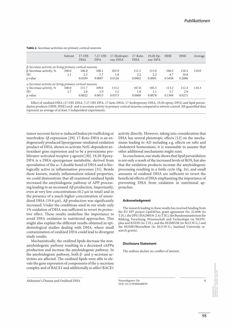
Alzheimer’s Disease and Oxidized DHA Neurodegener DisDOI: 10.1159/000440839
9
tumor necrosis factor α-induced leukocyte trafficking or interleukin-1β expression [29] . 17-Keto-DHA is an en-dogenously produced lipoxygenase-mediated oxidation product of DHA, shown to activate Nrf2-dependent an-tioxidant gene expression and to be a peroxisome pro-liferator-activated receptor γ agonist [30] . 19,20-Epoxy-DPA is a DHA epoxygenase metabolite, derived from epoxidation of the ω–3 double bond of DHA and is bio-logically active in inflammation processes [31] . Beside these known, mainly inflammation-related properties, we could demonstrate that all examined oxidized lipids increased the amyloidogenic pathway of APP process-ing leading to an increased Aβ production. Importantly, even at very low concentrations (0.2 μ M in total) and in the presence of a much higher concentration of unoxi-dized DHA (19.8 μ M ), Aβ production was significantly increased. Under the conditions used in our study only 1% oxidation of DHA was sufficient to revert its protec-tive effect. These results underline the importance to avoid DHA oxidation in nutritional approaches. This might also explain the different results obtained in epi-demiological studies dealing with DHA, where small contamination of oxidized DHA could lead to divergent study results.
Mechanistically, the oxidized lipids decrease the non-amyloidogenic pathway resulting in a decreased sAPPα production and increase the amyloidogenic pathway. In the amyloidogenic pathway, both β- and γ-secretase ac-tivities are affected. The oxidized lipids were able to ele-vate the gene expression of components of the γ-secretase complex and of BACE1 and additionally to affect BACE1
activity directly. However, taking into consideration that DHA has several pleiotropic effects [12] on the mecha-nisms leading to AD including e.g. effects on rafts and cholesterol homeostasis, it is reasonable to assume that other additional mechanisms might exist.
In conclusion, our study shows that lipid peroxidation is not only a result of the increased levels of ROS, but also that the oxidation products increase the amyloidogenic processing resulting in a futile cycle ( fig. 2 e), and small amounts of oxidized DHA are sufficient to revert the beneficial effects of DHA emphasizing the importance of preventing DHA from oxidation in nutritional ap-proaches.
Acknowledgment
The research leading to these results has received funding from the EU FP7 project LipiDiDiet, grant agreement No. 211696 (to T.H.), the DFG (HA2985/6-2; to T.H.), the Bundesministerium fü r Bildung, Forschung, Wissenschaft und Technologie via NGFN-plus and KNDD (to T.H.), and the HOMFOR (to M.O.W.G.) and the HOMFORexzellent (to M.O.W.G.; Saarland University re-search grants).
Disclosure Statement
The authors declare no conflict of interest.
Table 2. Secretase activities on primary cortical neurons
Solvent 17-OH-DHA
7,17-OH-DPA
17-Hydroper-oxy-DHA
17-Keto-DHA
19,20-Ep-oxy-DPA
HHE HNE Average
β-Secretase activity on living primary cortical neuronsβ-Secretase activity, % 100.0 106.8 108.1 105.9 111.5 113.0 106.5 118.2 110.0SD 1.1 2.3 1.7 1.8 2.2 2.2 4.7 10.8p value 0.0189 0.0007 0.0126 0.0002 0.0001 0.3458 0.2886
γ-Secretase activity on living primary cortical neuronsγ-Secretase activity, % 100.0 111.7 109.0 113.2 107.0 105.5 113.2 112.4 110.3SD 1.7 2.8 1.9 5.2 1.8 1.1 5.7 2.9p value 0.0022 0.0013 0.0373 0.0068 0.0078 0.1369 0.0271
Effect of oxidized DHA (17-OH-DHA, 7,17-OH-DPA, 17-keto-DHA, 17-hydroperoxy-DHA, 19,20-epoxy-DPA) and lipid peroxi-dation products (HHE, HNE) on β- and γ-secretase activity in primary cortical neurons compared to solvent control. All quantified data represent an average of at least 3 independent experiments.
Dow
nloa
ded
by:
Saa
rländ
isch
e U
nive
rsitä
ts u
nd L
ande
sbib
lioth
ek
149.
126.
78.6
5 -
12/8
/201
5 1:
48:5
7 P
M
Publika onen
98
Grimm et al.
Neurodegener DisDOI: 10.1159/000440839
10
References
1 Masters CL, Simms G, Weinman NA, Multhaup G, McDonald BL, Beyreuther K: Amyloid plaque core protein in Alzheimer disease and Down syndrome. Pro Natl Acad Sci USA 1985; 82: 4245–4249.
2 Grundke-Iqbal I, Iqbal K, Tung YC, Quinlan M, Wisniewski HM, Binder LI: Abnormal phosphorylation of the microtubule-associat-ed protein tau (tau) in Alzheimer cytoskeletal pathology. Proc Natl Acad Sci USA 1986; 83: 4913–4917.
3 Zhang YW, Thompson R, Zhang H, Xu H: APP processing in Alzheimer’s disease. Mol Brain 2011; 4: 3.
4 Kimberly WT, LaVoie MJ, Ostaszewski BL, Ye W, Wolfe MS, Selkoe DJ: Gamma-secre-tase is a membrane protein complex com-prised of presenilin, nicastrin, APH-1, and presenilin enhancer 2. Proc Natl Acad Sci USA 2003; 100: 6382–6387.
5 Ehehalt R, Keller P, Haass C, Thiele C, Simons K: Amyloidogenic processing of the Alzhei-mer beta-amyloid precursor protein depends on lipid rafts. J Cell Biol 2003; 160: 113–123.
6 Grimm MO, Zinser EG, Grosgen S, Hunds-dorfer B, Rothhaar TL, Burg VK, Kaestner L, Bayer TA, Lipp P, Muller U, Grimm HS, Hart-mann T: Amyloid precursor protein (APP) mediated regulation of ganglioside homeo-stasis linking Alzheimer’s disease pathology with ganglioside metabolism. PLoS One 2012; 7:e34095.
7 Zha Q, Ruan Y, Hartmann T, Beyreuther K, Zhang D: GM1 ganglioside regulates the pro-teolysis of amyloid precursor protein. Mol Psychiatry 2004; 9: 946–952.
8 Grimm MO, Grimm HS, Patzold AJ, Zinser EG, Halonen R, Duering M, Tschape JA, De Strooper B, Muller U, Shen J, Hartmann T: Regulation of cholesterol and sphingomyelin metabolism by amyloid-beta and presenilin. Nat Cell Biol 2005; 7: 1118–1123.
9 Grimm MO, Rothhaar TL, Grosgen S, Burg VK, Hundsdorfer B, Haupenthal VJ, Friess P, Kins S, Grimm HS, Hartmann T: Trans fatty acids enhance amyloidogenic process-ing of the Alzheimer amyloid precursor pro-tein (APP). J Nutr Biochem 2012; 23: 1214–1223.
10 Burg VK, Grimm HS, Rothhaar TL, Grosgen S, Hundsdorfer B, Haupenthal VJ, Zimmer VC, Mett J, Weingartner O, Laufs U, Broersen LM, Tanila H, Vanmierlo T, Lutjohann D, Hartmann T, Grimm MO: Plant sterols the better cholesterol in Alzheimer’s disease? A mechanistical study. J Neurosci 2013; 33: 16072–16087.
11 Grimm MO, Grimm HS, Tomic I, Beyreuther K, Hartmann T, Bergmann C: Independent inhibition of Alzheimer disease beta- and gamma-secretase cleavage by lowered choles-terol levels. J Biol Chem 2008; 283: 11302–11311.
12 Grimm MO, Kuchenbecker J, Grosgen S, Burg VK, Hundsdorfer B, Rothhaar TL, Friess
P, de Wilde MC, Broersen LM, Penke B, Peter M, Vigh L, Grimm HS, Hartmann T: Docosa-hexaenoic acid reduces amyloid beta produc-tion via multiple pleiotropic mechanisms. J Biol Chem 2011; 286: 14028–14039.
13 Lim GP, Calon F, Morihara T, Yang F, Teter B, Ubeda O, Salem N Jr, Frautschy SA, Cole GM: A diet enriched with the omega-3 fatty acid docosahexaenoic acid reduces amyloid burden in an aged Alzheimer mouse model. J Neurosci 2005; 25: 3032–3040.
14 Oksman M, Iivonen H, Hogyes E, Amtul Z, Penke B, Leenders I, Broersen L, Lutjohann D, Hartmann T, Tanila H: Impact of differ-ent saturated fatty acid, polyunsaturated fat-ty acid and cholesterol containing diets on beta-amyloid accumulation in APP/PS1 transgenic mice. Neurobiol Dis 2006; 23: 563–572.
15 Perez SE, Berg BM, Moore KA, He B, Counts SE, Fritz JJ, Hu YS, Lazarov O, Lah JJ, Mufson EJ: DHA diet reduces AD pathology in young APPSWE/PS1 delta E9 transgenic mice: pos-sible gender effects. J Neurosci Res 2010; 88: 1026–1040.
16 Soderberg M, Edlund C, Kristensson K, Dall-ner G: Fatty acid composition of brain phos-pholipids in aging and in Alzheimer’s disease. Lipids 1991; 26: 421–425.
17 Tully AM, Roche HM, Doyle R, Fallon C, Bruce I, Lawlor B, Coakley D, Gibney MJ: Low serum cholesteryl ester-docosahexaenoic acid levels in Alzheimer’s disease: a case-control study. Br J Nutr 2003; 89: 483–489.
18 Bazinet RP, Laye S: Polyunsaturated fatty ac-ids and their metabolites in brain function and disease. Nat Rev Neurosci 2014; 15: 771–785.
19 Cai Z, Zhao B, Ratka A: Oxidative stress and beta-amyloid protein in Alzheimer’s disease. Neuromol Med 2011; 13: 223–250.
20 Lukiw WJ, Cui JG, Marcheselli VL, Bodker M, Botkjaer A, Gotlinger K, Serhan CN, Bazan NG: A role for docosahexaenoic acid-derived neuroprotectin D1 in neural cell survival and Alzheimer disease. J Clin Invest 2005; 115: 2774–2783.
21 Roberts LJ 2nd, Montine TJ, Markesbery WR, Tapper AR, Hardy P, Chemtob S, Dettbarn WD, Morrow JD: Formation of isoprostane-like compounds (neuroprostanes) in vivo from docosahexaenoic acid. J Biol Chem 1998; 273: 13605–13612.
22 Subbarao KV, Richardson JS, Ang LC: Autop-sy samples of Alzheimer’s cortex show in-creased peroxidation in vitro. J Neurochem 1990; 55: 342–345.
23 Musiek ES, Cha JK, Yin H, Zackert WE, Terry ES, Porter NA, Montine TJ, Morrow JD: Quantification of F-ring isoprostane-like compounds (F4-neuroprostanes) derived from docosahexaenoic acid in vivo in humans by a stable isotope dilution mass spectromet-ric assay. J Chromatogr B Anal Technol Biomed Life Sci 2004; 799: 95–102.
24 Ida N, Hartmann T, Pantel J, Schroder J, Zer-fass R, Forstl H, Sandbrink R, Masters CL, Beyreuther K: Analysis of heterogeneous A4 peptides in human cerebrospinal fluid and blood by a newly developed sensitive Western blot assay. J Biol Chem 1996; 271: 22908–22914.
25 Grimm MO, Stahlmann CP, Mett J, Haupen-thal VJ, Zimmer VC, Lehmann J, Hundsdor-fer B, Endres K, Grimm HS, Hartmann T: Vi-tamin E: curse or benefit in Alzheimer’s dis-ease? A systematic investigation of the impact of α-, γ- and δ-tocopherol on Aß generation and degradation in neuroblastoma cells. J Nutr Health Aging 2015; 19: 646–656.
26 Rothhaar TL, Grosgen S, Haupenthal VJ, Burg VK, Hundsdorfer B, Mett J, Riemen-schneider M, Grimm HS, Hartmann T, Grimm MO: Plasmalogens inhibit APP pro-cessing by directly affecting gamma-secretase activity in Alzheimer’s disease. Sci World J 2012; 2012: 141240.
27 Oakley H, Cole SL, Logan S, Maus E, Shao P, Craft J, Guillozet-Bongaarts A, Ohno M, Dis-terhoft J, Van Eldik L, Berry R, Vassar R: In-traneuronal beta-amyloid aggregates, neuro-degeneration, and neuron loss in transgenic mice with five familial Alzheimer’s disease mutations: potential factors in amyloid plaque formation. J Neurosci 2006; 26: 10129–10140.
28 Sahlin C, Pettersson FE, Nilsson LN, Lannfelt L, Johansson AS: Docosahexaenoic acid stim-ulates non-amyloidogenic APP processing re-sulting in reduced Abeta levels in cellular models of Alzheimer’s disease. Eur J Neurosci 2007; 26: 882–889.
29 Marcheselli VL, Hong S, Lukiw WJ, Tian XH, Gronert K, Musto A, Hardy M, Gimenez JM, Chiang N, Serhan CN, Bazan NG: Noveldocosanoids inhibit brain ischemia-reperfu-sion-mediated leukocyte infiltration and pro-inflammatory gene expression. J Biol Chem 2003; 278: 43807–43817.
30 Groeger AL, Cipollina C, Cole MP, Wood-cock SR, Bonacci G, Rudolph TK, Rudolph V, Freeman BA, Schopfer FJ: Cyclooxygenase-2 generates anti-inflammatory mediators from omega-3 fatty acids. Nat Chem Biol 2010; 6: 433–441.
31 Serhan CN, Hong S, Gronert K, Colgan SP, Devchand PR, Mirick G, Moussignac RL: Re-solvins: a family of bioactive products of ome-ga-3 fatty acid transformation circuits initi-ated by aspirin treatment that counter proin-flammation signals. J Exp Med 2002; 196: 1025–1037.
32 Freund-Levi Y, Eriksdotter-Jonhagen M, Ce-derholm T, Basun H, Faxen-Irving G, Gar-lind A, Vedin I, Vessby B, Wahlund LO, Palmblad J: Omega-3 fatty acid treatment in 174 patients with mild to moderate Alzhei-mer disease: Omegad study: a randomized double-blind trial. Arch Neurol 2006; 63: 1402–1408.
Dow
nloa
ded
by:
Saa
rländ
isch
e U
nive
rsitä
ts u
nd L
ande
sbib
lioth
ek
149.
126.
78.6
5 -
12/8
/201
5 1:
48:5
7 P
M
Publika onen
99
Alzheimer’s Disease and Oxidized DHA Neurodegener DisDOI: 10.1159/000440839
11
33 Chiu CC, Su KP, Cheng TC, Liu HC, Chang CJ, Dewey ME, Stewart R, Huang SY: The ef-fects of omega-3 fatty acids monotherapy in Alzheimer’s disease and mild cognitive im-pairment: a preliminary randomized double-blind placebo-controlled study. Prog Neuro-psychopharmacol Biol Psychiatry 2008; 32: 1538–1544.
34 Quinn JF, Raman R, Thomas RG, Yurko-Mauro K, Nelson EB, Van Dyck C, Galvin JE, Emond J, Jack CR Jr, Weiner M, Shinto L, Aisen PS: Docosahexaenoic acid supplemen-tation and cognitive decline in Alzheimer dis-ease: a randomized trial. JAMA 2010; 304: 1903–1911.
35 Lee LK, Shahar S, Chin AV, Yusoff NA:Docosahexaenoic acid-concentrated fish oil supplementation in subjects with mild cogni-tive impairment (MCI): a 12-month ran-domised, double-blind, placebo-controlled trial. Psychopharmacology 2013; 225: 605–612.
36 Kalmijn S, Launer LJ, Ott A, Witteman JC, Hofman A, Breteler MM: Dietary fat intake and the risk of incident dementia in the Rot-terdam Study. Ann Neurol 1997; 42: 776–782.
37 Barberger-Gateau P, Letenneur L, Deschamps V, Peres K, Dartigues JF, Renaud S: Fish, meat, and risk of dementia: cohort study. BMJ 2002; 325: 932–933.
38 Morris MC, Evans DA, Bienias JL, Tangney CC, Bennett DA, Wilson RS, Aggarwal N, Schneider J: Consumption of fish and n–3 fat-ty acids and risk of incident Alzheimer dis-ease. Arch Neurol 2003; 60: 940–946.
39 Schaefer EJ, Bongard V, Beiser AS, Lamon-Fava S, Robins SJ, Au R, Tucker KL, Kyle DJ, Wilson PW, Wolf PA: Plasma phosphatidyl-choline docosahexaenoic acid content and risk of dementia and Alzheimer disease: the Framingham Heart Study. Arch Neurol 2006; 63: 1545–1550.
40 Van de Rest O, van der Zwaluw N, Beekman AT, de Groot LC, Geleijnse JM: The reliability of three depression rating scales in a general population of Dutch older persons. Int J Geri-atr Psychiatry 2010; 25: 998–1005.
41 Stough C, Downey L, Silber B, Lloyd J, Kure C, Wesnes K, Camfield D: The effects of 90-day supplementation with the omega-3 essen-tial fatty acid docosahexaenoic acid (DHA) on cognitive function and visual acuity in a healthy aging population. Neurobiol Aging 2012; 33: 824. e821–e823.
Dow
nloa
ded
by:
Saa
rländ
isch
e U
nive
rsitä
ts u
nd L
ande
sbib
lioth
ek
149.
126.
78.6
5 -
12/8
/201
5 1:
48:5
7 P
M
Publika onen
100
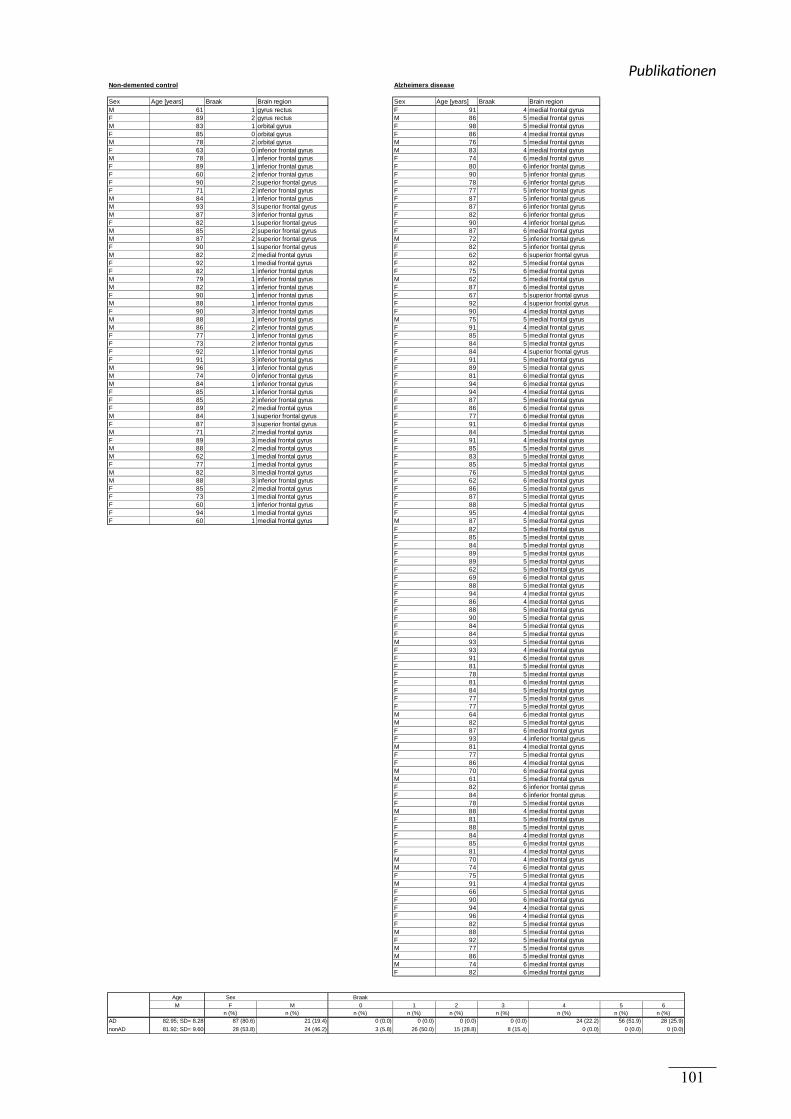
Non-demented control Alzheimers disease
Sex Age [years] Braak Brain region Sex Age [years] Braak Brain regionM 61 1 gyrus rectus F 91 4 medial frontal gyrusF 89 2 gyrus rectus M 86 5 medial frontal gyrusM 83 1 orbital gyrus F 98 5 medial frontal gyrusF 85 0 orbital gyrus F 86 4 medial frontal gyrusM 78 2 orbital gyrus M 76 5 medial frontal gyrusF 63 0 inferior frontal gyrus M 83 4 medial frontal gyrusM 78 1 inferior frontal gyrus F 74 6 medial frontal gyrusF 89 1 inferior frontal gyrus F 80 6 inferior frontal gyrusF 60 2 inferior frontal gyrus F 90 5 inferior frontal gyrusF 90 2 superior frontal gyrus F 78 6 inferior frontal gyrusF 71 2 inferior frontal gyrus F 77 5 inferior frontal gyrusM 84 1 inferior frontal gyrus F 87 5 inferior frontal gyrusM 93 3 superior frontal gyrus F 87 6 inferior frontal gyrusM 87 3 inferior frontal gyrus F 82 6 inferior frontal gyrusF 82 1 superior frontal gyrus F 90 4 inferior frontal gyrusM 85 2 superior frontal gyrus F 87 6 medial frontal gyrusM 87 2 superior frontal gyrus M 72 5 inferior frontal gyrusF 90 1 superior frontal gyrus F 82 5 inferior frontal gyrusM 82 2 medial frontal gyrus F 62 6 superior frontal gyrusF 92 1 medial frontal gyrus F 82 5 medial frontal gyrusF 82 1 inferior frontal gyrus F 75 6 medial frontal gyrusM 79 1 inferior frontal gyrus M 62 5 medial frontal gyrusM 82 1 inferior frontal gyrus F 87 6 medial frontal gyrusF 90 1 inferior frontal gyrus F 67 5 superior frontal gyrusM 88 1 inferior frontal gyrus F 92 4 superior frontal gyrusF 90 3 inferior frontal gyrus F 90 4 medial frontal gyrusM 88 1 inferior frontal gyrus M 75 5 medial frontal gyrusM 86 2 inferior frontal gyrus F 91 4 medial frontal gyrusF 77 1 inferior frontal gyrus F 85 5 medial frontal gyrusF 73 2 inferior frontal gyrus F 84 5 medial frontal gyrusF 92 1 inferior frontal gyrus F 84 4 superior frontal gyrusF 91 3 inferior frontal gyrus F 91 5 medial frontal gyrusM 96 1 inferior frontal gyrus F 89 5 medial frontal gyrusM 74 0 inferior frontal gyrus F 81 6 medial frontal gyrusM 84 1 inferior frontal gyrus F 94 6 medial frontal gyrusF 85 1 inferior frontal gyrus F 94 4 medial frontal gyrusF 85 2 inferior frontal gyrus F 87 5 medial frontal gyrusF 89 2 medial frontal gyrus F 86 6 medial frontal gyrusM 84 1 superior frontal gyrus F 77 6 medial frontal gyrusF 87 3 superior frontal gyrus F 91 6 medial frontal gyrusM 71 2 medial frontal gyrus F 84 5 medial frontal gyrusF 89 3 medial frontal gyrus F 91 4 medial frontal gyrusM 88 2 medial frontal gyrus F 85 5 medial frontal gyrusM 62 1 medial frontal gyrus F 83 5 medial frontal gyrusF 77 1 medial frontal gyrus F 85 5 medial frontal gyrusM 82 3 medial frontal gyrus F 76 5 medial frontal gyrusM 88 3 inferior frontal gyrus F 62 6 medial frontal gyrusF 85 2 medial frontal gyrus F 86 5 medial frontal gyrusF 73 1 medial frontal gyrus F 87 5 medial frontal gyrusF 60 1 inferior frontal gyrus F 88 5 medial frontal gyrusF 94 1 medial frontal gyrus F 95 4 medial frontal gyrusF 60 1 medial frontal gyrus M 87 5 medial frontal gyrus
F 82 5 medial frontal gyrusF 85 5 medial frontal gyrusF 84 5 medial frontal gyrusF 89 5 medial frontal gyrusF 89 5 medial frontal gyrusF 62 5 medial frontal gyrusF 69 6 medial frontal gyrusF 88 5 medial frontal gyrusF 94 4 medial frontal gyrusF 86 4 medial frontal gyrusF 88 5 medial frontal gyrusF 90 5 medial frontal gyrusF 84 5 medial frontal gyrusF 84 5 medial frontal gyrusM 93 5 medial frontal gyrusF 93 4 medial frontal gyrusF 91 6 medial frontal gyrusF 81 5 medial frontal gyrusF 78 5 medial frontal gyrusF 81 6 medial frontal gyrusF 84 5 medial frontal gyrusF 77 5 medial frontal gyrusF 77 5 medial frontal gyrusM 64 6 medial frontal gyrusM 82 5 medial frontal gyrusF 87 6 medial frontal gyrusF 93 4 inferior frontal gyrusM 81 4 medial frontal gyrusF 77 5 medial frontal gyrusF 86 4 medial frontal gyrusM 70 6 medial frontal gyrusM 61 5 medial frontal gyrusF 82 6 inferior frontal gyrusF 84 6 inferior frontal gyrusF 78 5 medial frontal gyrusM 88 4 medial frontal gyrusF 81 5 medial frontal gyrusF 88 5 medial frontal gyrusF 84 4 medial frontal gyrusF 85 6 medial frontal gyrusF 81 4 medial frontal gyrusM 70 4 medial frontal gyrusM 74 6 medial frontal gyrusF 75 5 medial frontal gyrusM 91 4 medial frontal gyrusF 66 5 medial frontal gyrusF 90 6 medial frontal gyrusF 94 4 medial frontal gyrusF 96 4 medial frontal gyrusF 82 5 medial frontal gyrusM 88 5 medial frontal gyrusF 92 5 medial frontal gyrusM 77 5 medial frontal gyrusM 86 5 medial frontal gyrusM 74 6 medial frontal gyrusF 82 6 medial frontal gyrus
Age Sex BraakM F M 0 1 2 3 4 5 6
n (%) n (%) n (%) n (%) n (%) n (%) n (%) n (%) n (%)AD 82.95; SD= 8.28 87 (80.6) 21 (19.4) 0 (0.0) 0 (0.0) 0 (0.0) 0 (0.0) 24 (22.2) 56 (51.9) 28 (25.9)
nonAD 81.92; SD= 9.60 28 (53.8) 24 (46.2) 3 (5.8) 26 (50.0) 15 (28.8) 8 (15.4) 0 (0.0) 0 (0.0) 0 (0.0)
Publika onen
101
Erweiterte Zusammenfassung
102
(5) Erweiterte Zusammenfassung
(5.1) Lipide in der AD
Lipide sind ein essentieller Bestandteil aller Nervenzellen und übernehmen neben den
strukturellen Funktionen wichtige zelluläre Aufgaben bei der Signaltransduktion,
Energieversorgung, Aktivierung von Rezeptoren und der Bereitstellung bioaktiver
Lipidmediatoren (Adibhatla & Hatcher 2007; Wenk 2005; Hullin-Matsuda et al. 2014; Purves
et al. 2006). Darüber hinaus sind einige Lipide in der Lage modulierend in die Prozesse
einzugreifen, die der AD Pathogenese zugrunde liegen (Grimm, Zinser, et al. 2012; Grimm,
Rothhaar, Grösgen, et al. 2012; Grimm, Lehmann, et al. 2014; Markesbery & Lovell 1998;
Gomez-Brouchet et al. 2007). Das schließt Lipide wie z.B. Cholesterin und Phytosterole, viele
Vitamine, Sphingolipide und Ganglioside mit ein (Lim et al. 2014; Grimm, Rothhaar &
Hartmann 2012; Grimm et al. 2008; Simons et al. 2002; Grimm, Hundsdörfer, et al. 2014;
Burg et al. 2013; Grimm, Grimm, Patzold, et al. 2005; Grimm et al. 2006).
Die Lipiddoppelschicht der Plasmamembran beeinflusst durch ihre biochemischen
Eigenschaften wie die Ladungsverteilung, Fluidität und Dichte der Membran, die
Funktionalität von membranständigen Proteinen (Haeffner & Privett 1975; Lenaz 1987;
Lahdo et al. 2002). Aus diesem Grund nehmen die biochemischen Eigenschaften der
Plasmamembran auch Einfluss auf die Prozessierung des APP. Da APP als
Transmembranprotein in oder nahe an der Membran von den Sekretasen gespalten wird, und
die Sekretasen selbst Transmembranproteine sind, beeinflusst die direkte Lipidumgebung die
Art der Prozessierung (Yang et al. 2014; Ehehalt et al. 2003; Yang et al. 2011). Einflüsse auf
die APP Prozessierung können direkt oder über zusätzliche zelluläre Mechanismen APP
selbst (Yoon et al. 2007; Ehehalt et al. 2003) oder die beteiligten Sekretasen (Takasugi et al.
2015; Zhou et al. 2008) betreffen.
Apo E, ein Lipidtransporter, der nicht nur im peripheren Kreislauf, sondern auch im Gehirn
eine essentielle Rolle bei der Versorgung mit Lipiden spielt, verdeutlicht die Rolle von
Lipiden für die AD (Hauser et al. 2011; Polazzi et al. 2015; Ulrich et al. 2013). In klinischen
Studien hat sich der Apo E Status als genetischer Risikofaktor für die SAD
herauskristallisiert. Insbesondere das Apo E ɛ4 Allel steigert das Risiko an AD zu erkranken,
Erweiterte Zusammenfassung
103
während Apo E ɛ3 keinen Einfluss auf das AD Risiko nimmt. Apo ɛ2 vermindert sogar das
Risiko an der AD zu erkranken (Corder et al. 1993; Strittmatter & Roses 1996). Ein weiterer
Risikofaktor für die AD ist ein zu hoher Serumcholesterinspiegel im mittleren
Lebensabschnitt. Ein reduzierter Gehalt an HDL-Cholesterin (Merched et al. 2000) ist für ein
erhöhtes AD-Risiko entscheidend. Ein erhöhter LDL- sowie ein reduzierter HDL-Gehalt
korrelieren zudem mit der Menge an Aβ-Plaques im humanen Gehirn (Reed et al. 2014;
Bonarek et al. 2000; Reitz et al. 2010; Zuliani et al. 2010). Ein zu hoher Cholesterinspiegel
resultiert oft aus einer pathologischen Aktivität der 3-Hydroxymethylglutaryl – Coenzym A –
Reduktase (HMGCR) (Shinohara et al. 2014; Endo 2008), deren Aktivität unter anderem
durch Aβ gesteuert wird. Die Aβ-Spezies Aβ40 inhibiert die HMGCR und reduziert dadurch
die Cholesterin de novo-Synthese. Im Gegenzug erhöht Cholesterin in vitro die Aβ –
Produktion durch Aktivierung der amyloidogenen APP Prozessierung (Grimm, Grimm,
Pätzold, et al. 2005; Wahrle et al. 2002; Simons et al. 1998). Dieser Effekt ist auch in vivo
nachgewiesen (Fassbender et al. 2001). Neben Cholesterin steigern z.B. trans-Fette, wie sie in
der Nahrung, z.B. in Milchprodukten und industriell gehärteten Fetten vorkommen, die
Produktion von Aβ (Grimm, Rothhaar, Grösgen, et al. 2012). Ein Mangel an Vitamin D,
einem Bestandteil von fetten Seefisch, kann ebenfalls den Gehalt an Aβ im Gehirn erhöhen
(Grimm, Lehmann, et al. 2014). Dies ist im Kontext von AD-Patienten umso interessanter, da
bei diesen oft eine Vitamin D-Hypovitaminose auftritt (Llewellyn et al. 2010).
Veränderungen des Lipidstoffwechsels in post mortem Gehirnen von an AD erkrankten
Personen, sowie im Blut von AD-Patienten wurden bereits nachgewiesen. So ist der Gehalt an
Sulfatiden, Ceramiden und Sphingolipiden in humanen post mortem Gehirnen von AD-
Patienten im Vergleich zu den Kontrollpersonen ohne AD verändert (Han 2007; Cutler et al.
2004). Zusätzlich ist der Gehalt an Phytosterinen im CSF (Vanmierlo et al. 2011) und im
Blutserum von AD-Patienten (Teunissen et al. 2016) verändert. Der Gehalt an
Phospholipiden, darunter im Speziellen PL, und ω-3 Fettsäuren, die in dieser Arbeit
verwendet wurden, unterscheidet sich zwischen dem Gehirn von AD-Patienten und dem
Gehirn ohne AD Pathologie.
Erweiterte Zusammenfassung
104
(5.2) Im Kontext: Phospholipide, DHA und oxidierte Lipide in der
APP Prozessierung und ihre Rolle für die AD
Phospholipide und DHA (C 22:6), dessen oxidierten Derivate in dieser Arbeit untersucht
wurden, stellen zusammen einen wesentlichen Teil der Lipide des humanen Gehirns dar.
Phospholipide sind, neben Cholesterin, die zweithäufigste Lipidklasse im humanen Gehirn.
Der Anteil von PC liegt im humanen Gehirn zwischen 14,4 und 17,5% des
Gesamtlipidgehalts. Der Gehalt an Phospholipiden variiert je nach Hirnregion (Chan et al.
2012). Der Gehalt an DHA im humanen Gehirn beträgt innerhalb der Phospholipide PC und
PE bis zu 24% des Gesamtfettsäuregehalts (Söderberg et al. 1990; Singh 2005). DHA stellt
einen wesentlichen Teil der ω-3 Fettsäuren des humanen Gehirns dar, zusammengefasst in
(Grimm, Zimmer, et al. 2013). Im Vergleich zur ω-3 Fettsäure Eicosapentaensäure (EPA) ist
die Menge von DHA im Gehirn um ein Vielfaches größer (Tassoni & Weisinger 2008). Damit
ist DHA im Gehirn die wichtigste ω-3 Fettsäure. Die Membranen von Neuronen sind
angereichert mit langkettigen, ungesättigten Fettsäuren, v.a. mit DHA und Arachidonsäure
(AA, ω-6 20:4), und die Konzentration dieser Fettsäuren ist in den Synapsen der Neuronen
am höchsten (Sun & Sun 1972; Kosicek & Hecimovic 2013; Kishimoto et al. 1969). Oxidierte
Derivate von ω-3 Fettsäuren, sowie Lipid-Peroxidationsprodukte wurden bereits im humanen
Gehirn nachgewiesen (Marcheselli et al. 2003; Serhan et al. 2002). PL kommen laut einer
Studie von Ginsberg et al. in der weißen Substanz des humanen Gehirns in einer
Konzentration von 28µmol pro Gramm Nassgewicht vor. Dieser Wert bezieht sich auf den
Frontallappen (Ginsberg et al. 1995). Zusätzlich wird das Phospholipid
Phosphatidylethanolamin (PE) im Gehirn zu 60-80% durch das korrespondierende
Plasmalogen dargestellt (Guan et al. 1999).
In dieser Arbeit wurde die Wirkung von Phospholipiden und oxidierten Derivaten der ω-3 und
ω-6 Fettsäuren auf die proteolytische Prozessierung des APP analysiert. Im Vordergrund
stand dabei der Einfluss der Lipide auf die Sekretasen und die Aβ Produktion. Um die
Wirkung von Phospholipiden auf die APP Prozessierung zu analysieren, wurden in
Publikation 1 und 2 unterschiedliche Phospholipide verwendet. Phospholipide sind eine
heterogene Gruppe, die sich durch eine polare, spezifische Kopfgruppe, z.B. Cholin oder
Ethanolamin, mit Bindung an Glycerin, Phosphat und meist eine oder zwei Fettsäuren
auszeichnen (Yadav & Tiwari 2014). Die Verknüpfung zwischen den Fettsäuren und Glycerin
Erweiterte Zusammenfassung
105
kann über eine Vinyletherbindung, wie sie im Falle von Plasmalogenen vorliegt, oder eine
Esterbindung erfolgen (Yadav & Tiwari 2014; Fahy et al. 2005). Im Gegensatz zu frei
vorliegenden Fettsäuren machen Phospholipide einen Großteil der Plasmamembran aus. Die
Art der gebundenen Fettsäuren reguliert die Fluidität und beeinflusst die biochemischen
Eigenschaften der Membran (Haeffner & Privett 1975). Im Gehirn von AD-Patienten sind der
Gehalt und die Zusammensetzung der Phospholipide im Vergleich zu gesunden Personen
verändert. Unterteilt man Phospholipide nach ihrer spezifischen Kopfgruppe, so stellen PC,
PE und Phosphatidylserin (PS) die Hauptspezies im humanen Gehirn dar (Söderberg et al.
1991; Chan et al. 2012). Veränderungen des PC-Gehaltes im Gehirn von AD-Patienten
werden diskutiert. Einerseits existieren Studien, die eine geringfügige Reduktion des PC-
Gehalts im Vergleich zu Personen, welche nicht an AD erkrankt waren, nachweisen konnten.
In diesen Studien wurde ein reduzierter PC-Spiegel nur bei wenigen PC-Spezies in
bestimmten Gehirnregionen nachgewiesen (Grimm, Grösgen, Riemenschneider, Tanila, et al.
2011; Nitsch et al. 1992). Im Gegensatz dazu war laut Wells et al. der Gehalt an PC im
Gehirn von AD-Patienten signifikant reduziert (Wells et al. 1995). Zusätzlich zur PC-
Reduktion wurde ein signifikant reduzierter PE-Gehalt im Gehirn von AD-Patienten
nachgewiesen (Prasad et al. 1998; Wells et al. 1995). Die Veränderungen im PC- und PE-
Gehalt könnten durch eine Reduktion ihrer Vorläufer, Cholin und Ethanolamin, hervorgerufen
werden. Diese Vorläufer sind im frontalen und parietalen Kortex des Gehirns von AD-
Patienten reduziert. Zudem ist das Abbauprodukt von PC, Glycerophosphocholin im Gehirn
von AD-Patienten vermehrt vorhanden, was auf einen kombinierten Effekt aus Phospholipid-
Synthese und -Abbau schließen lässt. Im Gegensatz dazu ist der Gehalt an Serin, dem
Vorläufer von PS, im Gehirn von AD-Patienten unverändert, während der PS–Spiegel selbst
erhöht ist (Nitsch et al. 1992). Der Gehalt an Phosphatidylinositol (PI), einem wichtigen Lipid
für die choline Rezeptorfunktion, ist im Gehirn von AD-Patienten signifikant reduziert
(Stokes & Hawthorne 1987; Pettegrew et al. 2001; Nitsch et al. 1992).
Die beschriebenen Veränderungen des Phospholipidgehalts im Gehirn variieren bei AD-
Patienten je nach betrachteter Hirnregion. Die signifikante Reduktion des PE-Gehalts zeigte
sich im Hippocampus und dem unteren Parietallappen von AD-Patienten im Vergleich zu
Personen ohne AD. Hingegen gibt es keine Unterschiede im PE-Gehalt des mittleren
temporalen Gyrus (Prasad et al. 1998). Die Reduktion des Gehalt an PC wird diskutiert und
konnte z.B. im okzipitalen Kortex nachgewiesen werden (Wells et al. 1995), während im
temporalen, frontalen und parietalen Kortex nur geringfügige oder keine Veränderungen
Erweiterte Zusammenfassung
106
nachgewiesen wurden (Grimm, Grösgen, Riemenschneider, Tanila, et al. 2011; Nitsch et al.
1992). Der Gehalt an PC ist im Cerebellum von AD-Patienten reduziert, dies gilt wurde
allerdings nur für PC 22:6 gezeigt, während der Gehalt an PC mit anderen Fettsäuren
unverändert war (Prasad et al. 1998). Das Cerebellum ist bei AD erst im späten
Krankheitsverlauf betroffen, während der Kortex und Hippocampus schon früh pathologische
Veränderungen im Lipidprofil zeigen (Padurariu et al. 2012; Mann 1991; Wegiel et al. 1999).
Demzufolge kann möglicherweise eine Reduktion von DHA im Gehirn als Anzeichen für
nachfolgende, weitere Lipidveränderungen gesehen werden. Dabei ist zu beachten, dass DHA
essentiell für die Funktion von Neuronen bei der Bildung von Axonen und der synaptischen
Plastizität ist (Janssen et al. 2015; Liu et al. 2015; Kidd 2007). Interessanterweise zeigen
speziell Membranen aus neuralen Synaptosomen eine Veränderung im Phospholipidgehalt.
Die beschriebene Reduktion von PE zeigt sich in den Synaptosomen deutlich und zwar für
fast alle Hirnregionen, einschließlich des Hippocampus und des frontalen Kortex (Wells et al.
1995). Da APP, die β- und γ-Sekretase in Neuronen gehäuft an den Axonen und Synapsen
vorkommen (Pliássova et al. 2015) könnten diese Veränderungen im Phospholipidgehalt auf
die Prozessierung wirken.
Phospholipide und die AD
Einige Phospholipide steigern die Aβ Bildung und damit die amyloidogene APP
Prozessierung durch direkte Aktivierung der γ-Sekretase (Holmes et al. 2012). Zusätzlich
steigern langkettige, ungesättigte Lipide die sAPPα Produktion (Yang et al. 2011) im nicht
amyloidogenen Weg und die Bedeutung der Lipidumgebung auf die α-Sekretase Schnittstelle
im APP Protein wurde gezeigt (Marenchino et al. 2008).
Um den Einfluss von Phospholipiden auf die nicht amyloidogene APP Prozessierung zu
untersuchen, wurde in Publikation 1 dieser Arbeit die Aktivität der α-Sekretase nach
Inkubation mit verschiedenen Phospholipiden gemessen. Phospholipide mit der Kettenlänge
10:0 und 12:0 werden in dieser Arbeit als kurzkettige, Phospholipide mit Fettsäuren ab 20:0
als langkettige Lipide bezeichnet. 14:0, 16:0 und 18:0 können als mittellange Fettsäuren
angesehen werden.
Es konnte in dieser Publikation gezeigt werden, dass die kurzkettigen, gesättigten
Phospholipide PC 10:0 und 12:0 die Aktivität der α-Sekretase signifikant im Vergleich zu PC
18:0 steigerten. Dieser Effekt konnte für PC 12:0 sowohl in isolierten Plasmamembranen von
Erweiterte Zusammenfassung
107
neuronalen SH-SY5Y Zellen als auch in lebenden SH-SY5Y Zellen nachgewiesen werden.
Für PC 10:0 zeigte sich dieser Effekt nur in lebenden SH-SY5Y Zellen. Außerdem wurde in
Publikation 1 die in vivo Relevanz durch Nachweis des Effekts der kurzkettigen, gesättigten
Phospholipide auf die α-Sekretase in Membranen von humanen post-mortem Gehirnen
verdeutlicht.
PC ist laut Literatur eines der häufigsten Phospholipide des humanen Gehirns (van Meer & de
Kroon 2010), dies wurde auch in Publikation 1 dieser Arbeit durch Analyse des PC Gehalts in
humanen post-mortem AD-Gehirnen gezeigt. Die gebundenen Fettsäuren in PC können die
Eigenschaften der Plasmamembran, wie z.B die Membranfluidität, die auch durch Cholesterin
reguliert wird (Buffone et al. 2009), verändern. Eine verringerte Fluidität reduziert zudem die
Bildung des neuroprotektiven sAPPα (Galbete et al. 2000). Im Gegensatz zu Cholesterin
steigert Kokosnussöl, welches reich an der kurzkettigen Fettsäure Laurinsäure (C12:0) ist, die
Membranfluidität (Tappia et al. 1997). Der Effekt auf die Aktivität der α-Sekretase wurde in
Publikation 1 dieser Arbeit durch Verwendung des korrespondierenden Phospholipids PC12:0
gezeigt und lässt Rückschlüsse auf die Bedeutung der Membranfluidität für die Aktivität der
α-Sekretase zu. Die Fluidität der Plasmamembran kann auch durch den Sättigungsgrad der
Phospholipide moduliert werden (Suzuki et al. 1998). Es wurde bereits nachgewiesen, dass
ungesättigte Fettsäuren die Membranfluidität erhöhen und dies in einer vermehrten Bildung
des sAPPα resultiert (Yang et al. 2011). Diese Studien untermauern die Ergebnisse dieser
Arbeit, in der der aktivierende Einfluss von den langkettigen, ungesättigten Phospholipiden
PC 20:4 (PC-AA), PC 20:5 (PC- EPA) und PC 22:6 (PC-DHA) auf die Aktivität der α-
Sekretase demonstriert werden konnte. Es wurde in Publikation 1 demonstriert, dass die
ungesättigten Phospholipide PC 22:6 und PC 20:5 eine aktivierende Wirkung auf die α-
Sekretase Aktivität in SH-SY5Y Zellen, deren isolierten Membranen und in Membranen von
humanen post-mortem Gehirnen haben. PC20:4 steigerte die Aktivität der α-Sekretase nur in
lebenden SH-SY5Y Zellen. Es ist wichtig hervorzuheben, dass der positive Einfluss der
Lipide auf die α-Sekretase erst durch vier oder mehr Doppelbindungen in PC hervorgerufen
wurde. Eine geringere Anzahl an Doppelbindungen zeigte in Publikation 1 dieser Arbeit
keinen vergleichbaren Effekt auf die α-Sekretase. Dieses Ergebnis wird durch eine Studie von
untermauert, nach der die sAPPα Produktion durch Inkubation mit EPA, DHA und AA
gesteigert wird (Yang et al. 2011). Schließlich wurde in Publikation 1 dieser Arbeit durch
Messung der Aktivität von ADAM-10, der hauptsächlichen α-Sekretase in kultivierten,
neuronalen Zellen (Kuhn et al. 2010), gezeigt, dass die gemessene Aktivierung der α-
Sekretase durch kurzkettige, gesättigte und langkettige, ungesättigte Phospholipide aus einer
Erweiterte Zusammenfassung
108
direkten Aktivierung von ADAM-10 resultiert. In einer anderen Studie wurde gezeigt, dass
ungesättigte Fettsäuren nicht etwa die Expression der α-Sekretasen ADAM-10 und ADAM-
17 oder von APP beeinflussen (Yang et al. 2011), sondern die direkt die Aktivität der α-
Sekretase steuern, wodurch die Bedeutung der Lipidumgebung für die α-Sekretase
verdeutlicht wird.
Die nicht amyloidogene Prozessierung von APP findet außerhalb der Lipid rafts statt. Das
Modell der Lipid rafts bescheibt dicht gepackte, Sphingomyelin- und Cholesterin-reiche
Abschnitte der Plasmamembran. Die α-Sekretase ADAM-10 ist außerhalb der Lipid raft-
Bereiche lokalisiert (Reid et al. 2007). Im Gegensatz dazu hängt die amyloidogene
Prozessierung direkt von den Lipid rafts ab (Ehehalt et al. 2003). Während nun die Sekretion
von sAPPα durch eine gesteigerte Membranfluidität begünstigt wird, erhöhen im Gegensatz
dazu Aβ-Oligomere durch Bindung an die Membran ihre Rigidität. Die dadurch resultierende
Abnahme der Membranfluidität fördert wiederum die Aβ-Produktion indem es die
amyloidogene APP Prozessierung steigert, dieser Mechanismus kann in einem Kreislauf aus
vermehrter Produktion von Aβ und dessen Wirkung auf die Membranfluidität resultieren
(Peters et al. 2009).
Es gibt bereits Fütterungsstudien mit ungesättigten und kurzkettigen Fettsäuren im Tiermodel.
Im einem AD-Rattenmodell konnte die Membranfluidität der Neuronen im Gehirn durch
Gabe der ω-6 Fettsäure AA (C20:4) verbessert werden (Fukaya et al. 2007). Kokosnussöl
besteht vorwiegend aus kurzkettigen Fettsäuren (Fernando et al. 2015), die in der Lage sind
die Bluthirnschranke leicht zu passieren (Ebert et al. 2003). Die Anwendung von Kokosnussöl
wirkt sich positiv auf das Herz-/ Kreislaufsystem aus und kann so Krankheiten verhindern, die
wiederum Risikofaktoren für AD darstellen (Fernando et al. 2015). Die Gabe von
Kokosnussöl im Tiermodell wirkte sich auch positiv auf die Blutfettwerte aus. Kokosnussöl
reduzierte zum Einen den Gehalt an Gesamtserum- und LDL-Cholesterin, und steigerte zum
Anderem den HDL-Cholesterinspiegel (Nevin & Rajamohan 2004). Bei Patienten mit
moderater bis schwerer AD führte eine Diät mit nativem Kokosnussöl zur Verbesserung der
kognitiven Fähigkeiten, wie in einer Studie von Gandotra et al. gezeigt wurde (Gandotra S
2014).
Da die molekularen Veränderungen im Gehirn lange vor dem Auftreten erster klinischer
Symptome beginnen, ist es wichtig grundlegende Ernährungsansätze auch bei Personen ohne
Demenz zu analysieren. Im frontalen Kortex von Patienten im frühen AD-Stadium war bereits
die Membranintegrität im Vergleich zu der Kontrollgruppe verändert, was zur Akkumulation
Erweiterte Zusammenfassung
109
der β-Sekretase innerhalb der Lipid raft der Plasmamembran führte (Fabelo et al. 2014). In
diesem Zusammenhang wurde auch gezeigt, dass eine mit kurzkettigen Fettsäuren
angereicherte Diät bei Probanden ohne Demenz die kognitiven Funktionen, insbesondere das
Kurzzeitgedächtnis, verbesserte (Page et al. 2009).
PL und die AD
In einer Studie von Grimm et al., wurde der Einfluss der APP Prozessierung auf die PL-
Synthese gezeigt (Grimm, Kuchenbecker, Rothhaar, et al. 2011). Demzufolge reguliert AICD
das Enzym, welches in der PL-Synthese den geschwindigkeitsbestimmenden Schritt
übernimmt. PL sind im Gegensatz zu den Esterphospholipiden gekennzeichnet durch eine
Vinyletherbindung an der sn1-Position des Glycerin zwischen der polaren Kopfgruppe und
den Fettsäureresten (Braverman & Moser 2012; Fahy et al. 2005). PL sind im frontalen
Kortex von post-mortem Gehirnen (Igarashi et al. 2011) und im Serum von AD-Patienten
reduziert. Diese Reduktion kann zudem mit der Schwere der AD korrelieren, wie in einer
Studie von Goodenowe et al. mittels kognitiver Tests gezeigt wurde (Goodenowe et al. 2007).
PE liegt, wie bereits erwähnt, in post-mortem Gehirnen von AD-Patienten deutlich reduziert
vor. Es wurde in einer Studie gezeigt, dass dieser Effekt durch die signifikante Reduktion des
korrespondierenden PE-PL hervorgerufen werden kann, diese Tatsache spiegelte sich in
einem verschobenen Verhältnis von PE zu PE-PL wider (Guan et al. 1999). PE-PL ist in
Gehirnbereichen wie dem temporalen Kortex, der in besonderem Maße von der AD
Pathologie betroffen ist, reduziert. In anderen Gehirnregionen, wie dem Cerebellum, die
wenig oder gar nicht von AD betroffen sind, wurde keine Veränderung des PE-PL-Gehalts
beobachtet (Ginsberg et al. 1995). Zusätzlich wurde nachgewiesen, dass der Gehalt an PE-PL
sowohl in der grauen und als auch in der weißen Gehirnsubstanz reduziert war, allerdings
nahm die Reduktion im Verlauf der AD nur in der grauen Substanz progressiv zu (Han et al.
2001). Auch die an PE-PL gebundenen Fettsäuren unterscheiden sich in post-mortem
Gehirnen von AD-Patienten im Vergleich zu Gehirnen von Personen, die nicht an AD
erkrankt waren. Vor allem mehrfach ungesättigte Fettsäuren, z.B. AA (C20:4), sowie die
einfach ungesättigte Ölsäure (C18:1) sind im Gehirn von AD-Patienten reduziert (Guan et al.
1999).
Die Reduktion des PL-Gehalts in humanen post-mortem Gehirnen von AD-Patienten wurde in
Publikation 2 dieser Arbeit bestätigt. Innerhalb von PC-PL waren die ungesättigten
Erweiterte Zusammenfassung
110
Fettsäuren, wie Ölsäure in PC-PL 16:0/18:1, 18:0/18:1 und 18:1/18:1, die mehrfach
ungesättigte ω-6 Fettsäure AA in PC-PL 18:0/20:4 und die mehrfach ungesättigte ω-3
Fettsäure DHA in PC-PL 18:0/22:6 im Vergleich zur Kontrollgruppe verringert. Hingegen
waren die analysierten PE-PL-Spezies in dieser Arbeit tendenziell, jedoch nicht signifikant
reduziert. Weiterhin wurde in Publikation 2 dieser Arbeit gezeigt, dass alle analysierten PL
die Aktivität der γ-Sekretase in SH-SY5Y Zellen und isolierten Membranen von
Mausgehirnen im Vergleich zum korrespondierenden Ester-Phospholipid signifikant
reduzierten. Darüber hinaus verringerte PC-PL 18:1 und PE-PL 20:4, die Aktivität der γ-
Sekretase sogar in humanen post-mortem Gehirnproben von AD-Patienten.
Die Bedeutung der Ergebnisse aus Publikation 2 wird umso klarer, wenn man berücksichtigt,
dass bis zu 30% aller Phospholipide und bis zu 80% des PE in neuronalen Membranen durch
PL dargestellt werden (Han et al. 2001; Horrocks & Sharma 1982). Die Effekte von PE-PL
auf die Aktivität der γ-Sekretase, können sich auf den Aβ-Spiegel auswirken, wie bereits in
einer Studie von Onodera et al. gezeigt wurde (Onodera et al. 2015). In Publikation 2 dieser
Arbeit wurde außerdem gezeigt, dass die Reduktion der γ-Sekretase Aktivität durch PL
unabhängig von Expressionseffekten auf die Bestandteile der γ-Sekretase, PS1 und PS2,
Aph1a und Aph1b, PSEN und Nicastrin war. PL reduzieren demnach die Aktivität der γ-
Sekretase durch direkte Interaktion. In einer Studie wurde eine Aktivierung der γ-Sekretase
durch Esther-Phospholipide mit den gleichen Fettsäuren wie sie in dieser Arbeit verwendet
wurden gezeigt. In dieser Studie wurde demonstriert, dass die Aktivität der γ-Sekretase durch
langkettige, ungesättigte Phospholipide, zum Beispiel PC 18:1 und PC 22:6 im Vergleich zur
Kontrolle gesteigert wird (Holmes et al. 2012). Diese Ergebnisse weisen auf die Bedeutung
des strukturellen Unterschieds zwischen PL und Ester-Phospholipiden für die Aktivität der γ-
Sekretase hin.
Im Gegensatz zu dem gemessenen Einfluss der PL auf die γ-Sekretase Aktivität zeigte sich in
dieser Arbeit kein Effekt der PL auf die Aktivität und Expression der β-Sekretase. Da die
nicht amyloidogene Prozessierung des APP durch die α-Sekretase die Aβ-Bildung verhindern
kann (Esch et al. 1990), wurde in Publikation 2 dieser Arbeit nach Inkubation mit PL die
Aktivität der α-Sekretase bestimmt. Im Vergleich zum korrespondierenden Ester-
Phospholipid war diese nach Behandlung mit PC-PL 20:4 und PE-PL 22:6 in SH-SY5Y
Zellen gesteigert. Der Effekt konnte in Mausgehirnen nicht nachgewiesen werden.
Zusammengefasst trat damit eine nur eine Aktivierung der α-Sekretase durch PL, welche mit
mehrfach ungesättigten Fettsäuren verknüpft sind, auf. Wie bereits zuvor in Publikation 1
Erweiterte Zusammenfassung
111
beschrieben, aktivieren auch Ester-Phospholipide mit mehrfach ungesättigten Fettsäuren die
α-Sekretase. Interessanterweise sind es genau die gleichen Fettsäuren, nämlich C20:4 und
C22:6 die einen Effekt auf die α-Sekretase haben. Im Zusammenspiel mit den inhibierenden
Effekten der PL auf die γ-Sekretase könnte die Aktivierung der α-Sekretase durch
ungesättigte Fettsäuren innerhalb von Phospholipiden einen wichtigen Schritt zur Prävention
der Aβ-Bildung darstellen. Ein weiterer Aspekt ist die Art der Regulation der α-Sekretase
durch Phospholipide im Vergleich zur γ-Sekretase. Die α-Sekretase wurde durch den
Sättigungsgrad der Fettsäuren innerhalb der Phospholipide beeinflusst während die
Regulation der γ-Sekretase stärker von der Bindung der Fettsäuren an das Glycerin der
Phospholipide beeinflusst wird.
Die Wirkung von PL wurde auch im Tiermodell in einer Studie von Ifuku et al. analysiert. Im
verwendeten Mausmodell, welches die schwedische APP-Mutation trug, konnte eine
Reduktion des PL-Gehalts festgestellt werden. Des Weiteren wurde nach intraperitonealer
Injektion oder systemischer Verabreichung von PL die Aβ-Dichte in Neuronen des Gehirns
der Mäuse reduziert. Interessant ist auch, dass inflammatorische Prozesse, wie z.B. aktivierte
Gliazellen dadurch abgemildert wurden (Ifuku et al. 2012). Umgekehrt trat bei Mäusen, die
neuroinflammatorische Prozesse im Gehirn aufwiesen, laut einer anderen Studie, eine
Reduktion der PL im Hippocampus auf. Dieser Effekt konnte durch Fütterung von PL wieder
aufgehoben werden (Katafuchi et al. 2012). Der Einfluss auf die Aβ Aggregate im
Mausgehirn könnte zusätzlich zu der gesteigerten Aβ-Produktion auch durch eine direkte
Interaktion von Aβ mit den Lipiden hervorgerufen werden, da Aβ mit anionischen
Phospholipiden wie PI und PS interagieren kann und in Verbindung mit diesen eine stärkere
Fibrillenbildung aufwiese (Chauhan et al. 2000). Diese Resultate deuten in vivo auf eine
Kombination aus Neuroinflammation und der neurotoxischen Wirkung von Aβ, bzw. Aβ-
Aggregaten hin, die schließlich die pathologischen Veränderungen im AD-Gehirn hervorrufen
können. Diese Effekte könnten zusammen mit Modifikationen im Plasmalogenhaushalt
auftreten. Ob diese Veränderungen Auslöser oder eine Folge der AD sind, ist noch nicht
geklärt.
Da im Gehirn von AD-Patienten im Krankheitsverlauf oxidativer Stress auftritt, ist zudem zu
beachten, dass, neben ungesättigten Fettsäuren, die Vinyletherbindung der PL ein
Hauptangriffspunkt für Oxidation darstellt (Guan et al. 1999). Des Weiteren kann oxidativer
Stress zu Bildung von Lipid-Peroxidationsprodukten führen, die neben Phospholipiden und
PL auch eine Rolle für die APP Proteolyse und die molekularen Grundlagen von AD spielen,
was in Publikation 3 gezeigt wurde. In dieser Publikation wurden Lipid-
Erweiterte Zusammenfassung
112
Peroxidationsprodukte der ω-3 und ω-6 Fettsäuren, sowie oxidierte DHA Derivate
hinsichtlich ihres amyloidogenen Potentials untersucht.
DHA und oxidierte Lipide in der AD
DHA ist eine mehrfach ungesättigte ω-3 Fettsäure, die angereichert in fettem Kaltwasserfisch
wie z.B. Lachs und Hering, sowie in marinen Algen vorkommt (Horrocks & Yeo 1999). In
einigen klinischen und epidemiologischen Studien wurde ein protektiver Effekt von DHA für
AD-Patienten oder die Probanten nachgewiesen. Eine DHA-reiche Diät oder eine künstliche
Supplementierung von DHA reduziert demnach das Risiko an AD zu erkranken. Allerdings
existieren auch Studien mit gegenteiligen Resultaten, in denen DHA keinen positiven Effekt
auf die Entstehung oder den Fortschritt der AD aufweisen konnte. Die Studienlage ist in table
1 der Publikation 3 zusammengefasst. Die Ursachen für diese gegensätzliche Studienlage
können im Studiendesign (Kohortenstudien oder longitudinale Studien, Korrelationsstudien),
der Probantenauswahl (Einteilung z.B. nach Alter oder Geschlecht) oder der
Diagnosemethoden liegen (Kotani et al. 2006). Zwei randomisierte, Placebo-kontrollierte
Doppelblindstudien über die Wirkung einer DHA-Gabe zeigten keine oder nur sehr schwache
Verbesserungen für die AD-Patienten (Quinn et al. 2010; Freund-Levi et al. 2006). Zudem
muss zwischen klinischen Studien, in denen Patienten eine definierte DHA Menge verabreicht
wurde, und epidemiologischen Studien, in denen die Ernährung der Probanten dokumentiert
wird, differenziert werden. Möglicherweise stellt auch die chemische Struktur von DHA einen
zusätzlichen Aspekt dar. DHA gilt durch seine sechs Doppelbindungen innerhalb der
Fettsäure als sehr anfällig für Oxidation (VanRollins & Murphy 1984; Roberts et al. 1998).
Diese Tatsache könnte zu unerwünschten Nebenprodukten bei der diätischen Aufnahme und
im wissenschaftlichen Experiment führen. Die Autooxidation von ω-3 Fettsäuren kann über
eine radikale Kettenreaktion verlaufen. Diese wird von freien Radikalen induziert und im
Verlauf der Reaktion treten Zwischenprodukte, z.B. Hydroperoxide, auf. Die Stabilisierung
von DHA mittels Antioxidanzien, wie z.B. Tocopherol wird untersucht (Kamal-Eldin &
Yanishlieva 2002; Frankel et al. 2002). Die Oxidation von ungesättigten Fettsäuren kann auch
durch Enzyme mediiert werden. In diesem Zusammenhang sind Mitglieder der Cox-
(Cyclooxygenase) und Lox- (Lipoxygenase) - Proteinfamilie zu erwähnen, die an definierten
Stellen der Kohlenwasserstoffkette Hydroxylgruppen einführen (Lee et al. 2001; Reis &
Erweiterte Zusammenfassung
113
Spickett 2012). Durch diese Reaktion werden z.B. aus AA bioaktive Lipidmediatoren gebildet
(Hamberg et al. 1974; Hamberg & Samuelsson 1973; Bergstroem et al. 1964).
Wie bereits in Publikation 1 und 2 beschrieben, wirkt sich nicht oxidiertes DHA innerhalb
von Phospholipiden positiv auf die Prozessierung des APP aus. Es aktiviert die α-Sekretase
und reduziert die Aktivität der γ-Sekretase. In Publikation 3 wurde nun demonstriert, dass
DHA die Aβ-Produktion signifikant reduziert. Die Daten werden durch andere in vitro- und
Tiermodell- Studien unterstützt. Holmes et al. zeigten, dass DHA in vitro die Aktivität der γ-
Sekretase reduziert (Holmes et al. 2012; Mulder et al. 1998) und Grimm et al. führten die
Reduktion des Aβ-Gehalts durch DHA auf einen pleiotropen Wirkmechanismus zurück.
Dieser Mechanismus beinhaltet eine Steigerung der nicht amyloidogenen, sowie eine
Reduktion der amyloidogenen Prozessierung des APP durch nicht oxidiertes DHA (Grimm,
Kuchenbecker, Grösgen, et al. 2011). Zusätzlich zur Aβ-Produktion, spielt die Aβ-
Aggegation eine Rolle für die AD. In einer Studie am Tiermodell wurde gezeigt, dass die
Aggregation des Aβ, die der Bildung von Plaques im Gehirn von AD-Patienten vorrausgeht,
durch DHA reduziert und der Abbau von Aβ im Blut durch DHA verbessert wurde. In diesem
Zusammenhang wurde auch ein Anstieg der Membranfluidität von Blutzellen durch DHA
beobachtet. Dies resultierte in eine Abnahme von oxidativen Stress, welcher durch reaktive
oxidative Spezies ausgelöst wird. Interessanterweise war DHA innerhalb der
Blutzellmembranen an die Lipid Rafts, welche die amyloidogene Prozessierung des APP
beeinflussen, gebunden (Hashimoto et al. 2015).
Ob DHA in Form von Phospholipiden oder als freie Fettsäure vorliegt, kann die
Wirkungsweise von DHA beeinflussen. Unter physiologischen Bedingungen wird DHA im
Blut als veresterte Fettsäure durch Lipoproteine transportiert. Für den Transport ins Gehirn
passiert DHA die Blut-Hirn-Schranke nach rezeptormediierter Endozytose. Dabei lösen
Lipasen der Endothelzellen an der Blut-Hirn-Schranke DHA vom Lipoprotein, wodurch es
mittels eines Transporters (Nguyen et al. 2014) ins Gehirn gelangen kann. Passive Diffusion
von DHA über die Blut-Hirn-Schranke ist zudem auch möglich. Nach Aufnahme in die
Gehirnzellen kann DHA in Phospholipide integriert werden, wodurch es ein Bestandteil der
Membran wird (Pan & Nicolazzo 2015). Dieser Transportmechanismus könnte auch für
andere ungesättigte Fettsäuren oder oxidierte Lipide, die ähnliche biochemische
Eigenschaften aufweisen, gelten. Jedenfalls wurden bereits oxidierte Derivate ungesättigter
Fettsäuren im Blut und innerhalb des Gehirns nachgewiesen (Marcheselli et al. 2003; Serhan
et al. 2002).
Erweiterte Zusammenfassung
114
In Publikation 3 dieser Arbeit wurde die Steigerung der Aβ-Bildung durch oxidierte DHA-
Derivate und Lipid-Peroxidationsprodukte gezeigt. Es wurden 5 oxidierte DHA Derivate und
die beiden Lipid-Peroxidationsprodukte HHE und HNE in zellkulturbasierten Versuchen an
SH-SY5Y Zellen und murinen, kortikalen Primärneuronen analysiert. HHE stammt aus dem
Abbau von ω-3 Fettsäuren, während HNE von ω-6 Fettsäuren abgeleitet wird (Gladine et al.
2012; Bruce-Keller et al. 1998). Der in Publikation 3 nachgewiesene Anstieg von Aβ durch
die oxidierten Lipide wurde von einer gesteigerten der Aktivität der β- und γ-Sekretase
hervorgerufen. In Publikation 3 wurde weiterhin gezeigt, dass die Aktivität der β-Sekretase
direkt durch die oxidierten Lipide beeinflusst wird, während die Aktivierung der γ-Sekretase
indirekt über eine gesteigerte Expression der γ-Sekretase Bestandteile verursacht wird. Diese
Effekte resultieren in einer vermehrten amyloidogenen Prozessierung des APP und sind
schliesslich sogar in der Lage den positiven Effekt von nicht oxidiertem DHA auf die Aβ-
Produktion zu revertieren. Nicht oxidiertes DHA reduzierte den Aβ Spiegel in Publikation 3
signifikant auf 85,9 % der Kontrolle. Bereits der Zusatz von 1% der oxidierten Lipide zu nicht
oxidiertem DHA war ausreichend um die Aβ-Produktion im Vergleich zu DHA um 29%
ansteigen zu lassen. Der Zusatz von 5% und 10% oxidierter Lipide liess diesen Effekt deutlich
auf 37,4% bzw. 47,1% im Vergleich zu nicht oxidiertem DHA ansteigen. Diese Resultate
unterstreichen die Bedeutung der Oxidation von ungesättigen Fettsäuren für die APP
Prozessierung.
Für HHE und HNE wurde bereits eine Wirkung auf die Proteolyse des APP nachgewiesen.
Dabei steigerte HNE die Expression und die Aktivität der β-Sekretase BACE-1 in neuronalen
Zellen. Dieser Effekt wurde durch oxidativen Stress hervorgerufen (Tamagno 2002).
Außerdem wurde in einer Stufie gezeigt, dass die γ-Sekretase Aktivität über HNE-Bindung an
Nicastrin erhöht wird und die Bildung von Aβ42 in neuronalen Zellen dadurch ansteigt (Gwon
et al. 2012). Diese Studie untermauert die Ergebnisse von Publikation 3 dieser Arbeit. In einer
weiteren Studie wurde neben dem Einfluss der Lipid-Peroxidationsprodukte auf die
Produktion von Aβ, auch ein Effekt auf den Aβ-Abbau gezeigt. HNE reduzierte die
Expression und Aktivität des Endothelin-konvertierenden Enzyms (Endothelin converting
enzyme, ECE). Darüber hinaus wurde eine Modifikation von ECE und des Insulin-
degradierenden Enzyms (Insulin-degrading enzyme, IDE) durch HNE in neuronalen Zellen
nachgewiesen (Wang et al. 2009). Es besteht die Möglichkeit, dass der amyloidogene Effekt
neben den Lipid-Peroxidationsprodukten HHE und HNE, auch durch andere
Oxidationsprodukte der ungesättigten Fettsäuren hervorgerufen werden kann, wie in
Publikation 3 dieser Arbeit gezeigt werden konnte. Das Auftreten von oxidierten Derivaten
Erweiterte Zusammenfassung
115
nach Gabe von DHA hängt von der Form der Supplementierung ab. DHA als Phospholipid ist
in vitro weniger anfällig für Oxidation als DHA-Triglyceride und verestertes DHA. Dies gilt
auch für andere mehrfach ungesättigte Fettsäuren, allerdings wird DHA im Vergleich zur ω-6
Fettsäure AA schneller oxidiert. In einer Fütterungstudie am Mausmodell wurde gezeigt, dass
DHA-Triglyceride und verestertes DHA die Lipidperoxidation im Blut und der Leber
steigerten, während DHA als Phospholipid keinen derartigen Effekt verursachte (Song &
Miyazawa 2001). In Retinazellen wurde demonstriert, dass DHA unter physiologischen
Bedingungen protektiv wirkt, bei oxidativem Stress allerdings selbst die Lipidperoxidation
und den ROS-Spiegel im Vergleich zur Kontrolle erhöht. Dieser Stress wirkte sich auch auf
die Integrität der Plasmamembran aus, diese Tatsache könnte die Prozessierung des APP
beeinflussen (Liu et al. 2014). Im Gegensatz dazu ist nicht oxidiertes DHA in der Lage Zellen
vor oxidativem Stress zu schützen. In hippocampalen Mauszellen steigerte DHA die
Expression der Glutathionperoxidase (PH-GPx/ GPx4), ein Enzym, das die Lipide der
Plasmamembran vor Oxidation bewahrt. Zudem besitzt DHA die Fähigkeit neurotoxische
Effekte durch Aβ, die auch oxidativen Stress hervorrufen können, in bestimmten
Konzentrationen zu reduzieren (Casañas-Sánchez et al. 2014). Im Tierversuch führte die
Fütterung von Mäusen mit einer DHA-reichen Diät zur Reduktion des Aβ-Gehalts im Kortex
und Hippocampus, den Regionen des Gehirns die in der AD häufig betroffen sind. Marker für
oxidativen Stress waren im Serum dieser Mäuse signifikant reduziert. Allerdings wurde in
dieser Studie bei der Supplementierung des Futters mit DHA auf den Einsatz von
Antioxidantien wie z.B. Zink, Selen und Vitamin E gesetzt, diese Vorgehensweise
unterstreicht die Bedeutung der DHA Stabilisierung in Fütterungsstudien. Letztlich führte die
Fütterung in dieser Studie auch zu einer Verbesserung der kognitiven Fähigkeiten der Mäuse
(Wang et al. 2015).
Der Schutz vor Lipidperoxidation kann auch durch andere ω-3 Fettsäuren gewährleistet
werden. Dabei spielt die Anzahl der Doppelbindungen eine entscheidende Rolle. So ist im
Rattenmodell EPA mit fünf Doppelbindungen protektiv, während α-LA mit drei
Doppelbindungen keinen Schutz vor Lipidperoxidation bot (Saito & Kubo 2003). Im
Vergleich zu EPA ist DHA länger und beinhaltet eine zusätzliche Doppelbindung, allerdings
ist DHA im Gehirn die weitaus häufigere und wichtigste ω-3 Fettsäure, zusammengefasst in
(Tassoni & Weisinger 2008; Bradbury 2011). Zuletzt bleibt zu erwähnen, dass auch oxidierte
DHA Derivate existieren, die sich positiv auf den Aβ-Gehalt und die Tau-Phosphorylierung
auswirken können. Im Gehirn eines AD-Mausmodells (5xFAD) wurde gezeigt, dass mit der
Nahrung aufgenommenes, oxidiertes DHA im Gehirn in Phospholipide, besonders PE,
Erweiterte Zusammenfassung
116
eingebaut wird und dort auf biologische Prozesse wirkt (Torres et al. 2014). Ein anderes
Derivat von DHA ist Neuroprotektin - D1 (NPD-1). Diesem Lipid wird eine anti-oxidative
und anti-apoptotische Wirkung zugeschrieben und es unterdrückt z.B. auch die Aβ42
induzierte Expression von pro-inflammatorischen Genen (Lukiw et al. 2005; Mukherjee et al.
2004).
Zusammenfassung & Ausblick
117
(6) Zusammenfassung und Ausblick
In dieser Arbeit wurde der Einfluss von Lipiden auf die proteolytische Prozessierung des APP
gezeigt und die daraus resultierenden Konsequenzen für die AD beschrieben.
In Publikation 1 wurde demonstriert, dass Phospholipide mit langkettigen, ungesättigten
Fettsäuren und kurzkettige Phospholipide die α-Sekretase aktivieren. Dieser Effekt wurde in
Zellkultur-basierten Versuchen und ex vivo in Membranen von humanen post-mortem
Gehirnen nachgewiesen.
Publikation 2 verdeutlichte den Einfluss von PL auf die γ-Sekretase. Durch direkte Interaktion
der PL mit der γ-Sekretase wurde die Aktivität des Enzyms reduziert und die
Supplementierung von humanen post-mortem Gehirnproben von AD-Patienten ergab zudem
eine Abnahme der γ-Sekretase Aktivität. Einzelne PL-Spezies mit ungesättigten Fettsäuren
aktivierten außerdem die α-Sekretase.
Zuletzt wurde die Steigerung der Aβ-Produktion durch oxidierte DHA-Derivate und Lipid-
Peroxidationsprodukte der ω-3 und ω-6 Fettsäuren in Publikation 3 gezeigt. Dieser Effekt
resultiert aus einer Aktivierung der β- und γ-Sekretase im amyloidogenen Weg der APP
Proteolyse. Verschiedene oxidierte Lipidspezies riefen diesen Effekt hervor und eine
Supplementierung von DHA mit einer Mischung aus oxidierten Lipiden, kehrte sogar den
protektiven Effekt von DHA für die AD um und ließ die Aβ-Produktion signifikant ansteigen.
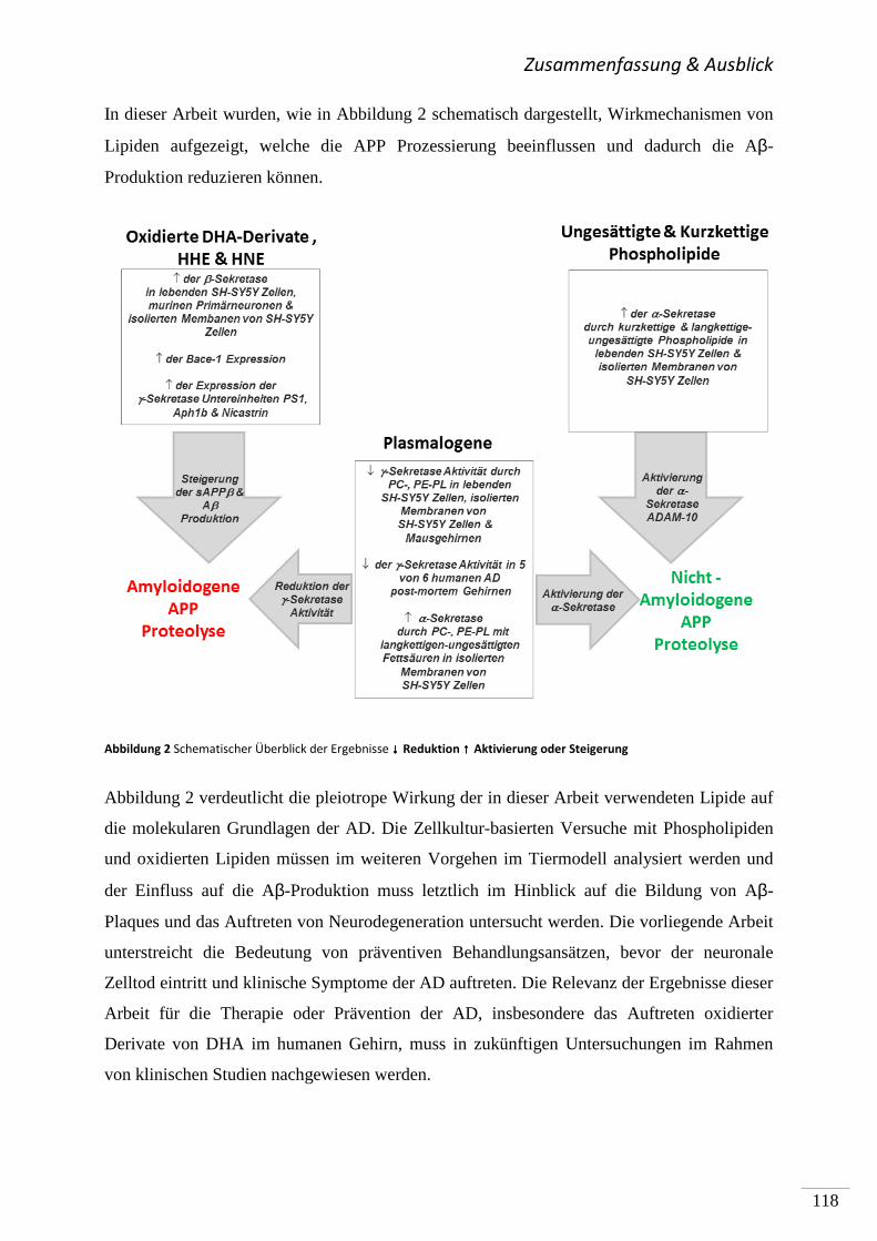
Zusammenfassung & Ausblick
118
In dieser Arbeit wurden, wie in Abbildung 2 schematisch dargestellt, Wirkmechanismen von
Lipiden aufgezeigt, welche die APP Prozessierung beeinflussen und dadurch die Aβ-
Produktion reduzieren können.
Abbildung 2 Schematischer Überblick der Ergebnisse ↓↓↓↓ Reduktion ↑↑↑↑ Aktivierung oder Steigerung
Abbildung 2 verdeutlicht die pleiotrope Wirkung der in dieser Arbeit verwendeten Lipide auf
die molekularen Grundlagen der AD. Die Zellkultur-basierten Versuche mit Phospholipiden
und oxidierten Lipiden müssen im weiteren Vorgehen im Tiermodell analysiert werden und
der Einfluss auf die Aβ-Produktion muss letztlich im Hinblick auf die Bildung von Aβ-
Plaques und das Auftreten von Neurodegeneration untersucht werden. Die vorliegende Arbeit
unterstreicht die Bedeutung von präventiven Behandlungsansätzen, bevor der neuronale
Zelltod eintritt und klinische Symptome der AD auftreten. Die Relevanz der Ergebnisse dieser
Arbeit für die Therapie oder Prävention der AD, insbesondere das Auftreten oxidierter
Derivate von DHA im humanen Gehirn, muss in zukünftigen Untersuchungen im Rahmen
von klinischen Studien nachgewiesen werden.
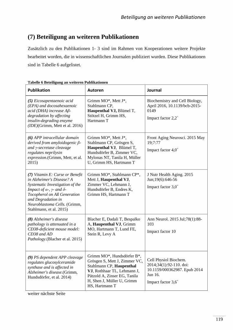
Beteiligung an weiteren Publikationen
119
(7) Beteiligung an weiteren Publikationen
Zusätzlich zu den Publikationen 1- 3 sind im Rahmen von Kooperationen weitere Projekte
bearbeitet worden, die in wissenschaftlichen Journalen publiziert wurden. Diese Publikationen
sind in Tabelle 6 aufgelistet.
Tabelle 6 Beteiligung an weiteren Publikationen
Publikation Autoren Journal
(5) Eicosapentaenoic acid (EPA) and docosahexaenoic acid (DHA) increase Aβ-degradation by affecting insulin-degrading enzyme (IDE)(Grimm, Mett et al. 2016)
Grimm MO*, Mett J*, Stahlmann CP, Haupenthal VJ, Blümel T, Stötzel H, Grimm HS, Hartmann T
Biochemistry and Cell Biology, April 2016, 10.1139/bcb-2015-0149
Impact factor 2,2+
(6) APP intracellular domain derived from amyloidogenic β- and γ-secretase cleavage regulates neprilysin expression.(Grimm, Mett, et al. 2015)
Grimm MO*, Mett J*, Stahlmann CP, Grösgen S, Haupenthal VJ, Blümel T, Hundsdörfer B, Zimmer VC, Mylonas NT, Tanila H, Müller U, Grimm HS, Hartmann T
Front Aging Neurosci. 2015 May 19;7:77
Impact factor 4,0+
(7) Vitamin E: Curse or Benefit in Alzheimer's Disease? A Systematic Investigation of the Impact of α-, γ- and δ-Tocopherol on Aß Generation and Degradation in Neuroblastoma Cells. (Grimm, Stahlmann, et al. 2015)
Grimm MO*, Stahlmann CP*, Mett J, Haupenthal VJ, Zimmer VC, Lehmann J, Hundsdörfer B, Endres K, Grimm HS, Hartmann T
J Nutr Health Aging. 2015 Jun;19(6):646-56
Impact factor 3,0+
(8) Alzheimer's disease pathology is attenuated in a CD38-deficient mouse model: CD38 and AD Pathology.(Blacher et al. 2015)
Blacher E, Dadali T, Bespalko A, Haupenthal VJ, Grimm MO, Hartmann T, Lund FE, Stein R, Levy A
Ann Neurol. 2015 Jul;78(1):88-103
Impact factor 10
(9) PS dependent APP cleavage regulates glucosylceramide synthase and is affected in Alzheimer's disease.(Grimm, Hundsdörfer, et al. 2014)
Grimm MO*, Hundsdörfer B*, Grösgen S, Mett J, Zimmer VC, Stahlmann CP, Haupenthal VJ, Rothhaar TL, Lehmann J, Pätzold A, Zinser EG, Tanila H, Shen J, Müller U, Grimm HS, Hartmann T
Cell Physiol Biochem. 2014;34(1):92-110. doi: 10.1159/000362987. Epub 2014 Jun 16.
Impact factor 3,6+
weiter nächste Seite
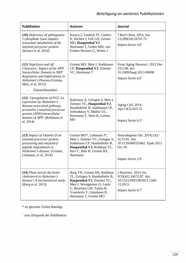
Beteiligung an weiteren Publikationen
120
Publikation Autoren Journal
(10) Deficiency of sphingosine-1-phosphate lyase impairs lysosomal metabolism of the amyloid precursor protein. (Karaca et al. 2014)
Karaca I, Tamboli IY, Glebov K, Richter J, Fell LH, Grimm MO, Haupenthal VJ, Hartmann T, Gräler MH, van Echten-Deckert G, Walter J
J Biol Chem, 2014, Jun 13;289(24):16761-72
Impact factor 4,6+
(11) Neprilysin and Aβ Clearance: Impact of the APP Intracellular Domain in NEP Regulation and Implications in Alzheimer's Disease.(Grimm, Mett, et al. 2013)
- Übersichtsartikel -
Grimm MO, Mett J, Stahlmann CP, Haupenthal VJ, Zimmer VC, Hartmann T
Front Aging Neurosci. 2013 Dec 23;5:98. doi: 10.3389/fnagi.2013.00098.
Impact factor 4,0+
(12) Upregulation of PGC-1α expression by Alzheimer's disease-associated pathway: presenilin 1/amyloid precursor protein (APP)/intracellular domain of APP. (Robinson et al. 2014)
Robinson A, Grösgen S, Mett J, Zimmer VC, Haupenthal VJ, Hundsdörfer B, Stahlmann CP, Slobodskoy Y, Müller UC, Hartmann T, Stein R, Grimm MO
Aging Cell, 2014, Apr;13(2):263-72
Impact factor 6,3+
(13) Impact of Vitamin D on amyloid precursor protein processing and amyloid-β peptide degradation in Alzheimer's disease. (Grimm, Lehmann, et al. 2014)
Grimm MO*, Lehmann J*, Mett J, Zimmer VC, Grösgen S, Stahlmann CP, Hundsdörfer B, Haupenthal VJ, Rothhaar TL, Herr C, Bals R, Grimm HS, Hartmann
Neurodegener Dis. 2014;13(2-3):75-81. doi: 10.1159/000355462. Epub 2013 Oct 30.
Impact factor 2,9+
(14) Plant sterols the better cholesterol in Alzheimer's disease? A mechanistical study. (Burg et al. 2013)
Burg VK, Grimm HS, Rothhaar TL, Grösgen S, Hundsdörfer B, Haupenthal VJ, Zimmer VC, Mett J, Weingärtner O, Laufs U, Broersen LM, Tanila H, Vanmierlo T, Lütjohann D, Hartmann T, Grimm MO
J Neurosci. 2013 Oct 9;33(41):16072-87. doi: 10.1523/JNEUROSCI.1506-13.2013.
Impact factor 6,7+
* zu gleichen Teilen beteiligt
+ zum Zeitpunkt der Publikation
Literaturverzeichnis
121
(8) Literaturverzeichnis
1. ADAPT Research Group, Lyketsos CG, Breitner JC, Green RC, Martin BK, Meinert C, Piantadosi S,
Sabbagh M (2007) Naproxen and celecoxib do not prevent AD in early results from a randomized
controlled trial. Neurology 68, 1800–1808.
2. Adibhatla RM , Hatcher JF (2007) Role of Lipids in Brain Injury and Diseases. Future Lipidol. 2, 403–422.
3. Alonso A del C, Grundke-Iqbal I, Iqbal K (1996) Alzheimer’s disease hyperphosphorylated tau
sequesters normal tau into tangles of filaments and disassembles microtubules. Nat Med 2, 783–787.
4. Alzheimer´s Association (2014) Alzheimer’s disease facts and figures. Alzheimer’s Dement. 10, 47–92.
5. Anand R, Gill K, Mahdi A (2014) Therapeutics of Alzheimer ’ s disease : Past , present and future. NP 76,
27–50.
6. Anstey KJ, von Sanden C, Salim A , O’Kearney R (2007) Smoking as a Risk Factor for Dementia and
Cognitive Decline: A Meta-Analysis of Prospective Studies. Am. J. Epidemiol. 166 , 367–378.
7. Anttila T, Helkala E-L, Viitanen M, Kåreholt I, Fratiglioni L, Winblad B, Soininen H, Tuomilehto J,
Nissinen A , Kivipelto M (2004) Alcohol drinking in middle age and subsequent risk of mild cognitive
impairment and dementia in old age: a prospective population based study. BMJ Br. Med. J. 329, 539.
8. Belyaev ND, Kellett K a B, Beckett C, Makova NZ, Revett TJ, Nalivaeva NN, Hooper NM , Turner AJ
(2010) The transcriptionally active amyloid precursor protein (APP) intracellular domain is
preferentially produced from the 695 isoform of APP in a β-secretase-dependent pathway. J. Biol.
Chem. 285, 41443–41454.
9. Bergstroem S, Danielsson H , Samuelsson B (1964) The enzymatic formation of prostaglandin E2 from
arachidonic acid prostaglandins and related factors 32. Biochim. Biophys. Acta 90, 10.
10. Bharadwaj PR, Dubey AK, Masters CL, Martins RN , Macreadie IG (2009) Abeta aggregation and
possible implications in Alzheimer’s disease pathogenesis. J. Cell. Mol. Med. 13, 412–21.
11. Biedler JL, Helson L , Spengler BA (1973) Morphology and Growth, Tumorigenicity, and Cytogenetics of
Human Neuroblastoma Cells in Continuous Culture. Cancer Res. 33 , 2643–2652.
12. Blacher E, Dadali T, Bespalko A, Haupenthal VJ, Grimm MOW, Hartmann T, Lund FE, Stein R , Levy A
(2015) Alzheimer’s disease pathology is attenuated in a CD38-deficient mouse model. Ann. Neurol. 78,
88–103.
13. Black RA , White JM (1998) ADAMs: focus on the protease domain. Curr. Opin. Cell Biol. 10, 654–659.
14. Blennow K, Leon MJ De , Zetterberg H (2006) Alzheimer ’ s disease. 368, 387–403.
15. Bligh EG , Dyer WJ (1959) A rapid method of total lipid extraction and purification. Can. J. Biochem.
Physiol. 37, 911–917.
16. Bonarek M, Barberger-Gateau P, Letenneur L, Deschamps V, Iron A, Dubroca B , Dartigues JF (2000)
Relationships between Cholesterol, Apolipoprotein E Polymorphism and Dementia: A Cross-Sectional
Analysis from the PAQUID Study. Neuroepidemiology 19, 141–148.
17. Bradbury J (2011) Docosahexaenoic Acid (DHA): An Ancient Nutrient for the Modern Human Brain. 3,
529–554.
18. Braverman NE , Moser AB (2012) Functions of plasmalogen lipids in health and disease. Biochim.
Biophys. Acta - Mol. Basis Dis. 1822, 1442–1452.
19. Brown MS, Ye J, Rawson RB , Goldstein JL (2000) Regulated Intramembrane Proteolysis. Cell 100, 391–
398.
20. Bruce-Keller AJ, Li YJ, Lovell MA, Kraemer PJ, Gary DS, Brown RR, Markesbery WR , Mattson MP (1998)
4-Hydroxynonenal, a product of lipid peroxidation, damages cholinergic neurons and impairs
visuospatial memory in rats. J. Neuropathol. Exp. Neurol. 57, 257–267.
21. Buffone MG, Verstraeten S V, Calamera JC , Doncel GF (2009) High Cholesterol Content and Decreased
Membrane Fluidity in Human Spermatozoa Are Associated With Protein Tyrosine Phosphorylation and
Functional Deficiencies. J. Androl. 30, 552–558.
Literaturverzeichnis
122
22. Burg VK, Grimm HS, Rothhaar TL, Gro S, Hundsdo B, Haupenthal VJ, Zimmer VC, Mett J, Weinga O,
Laufs U, Broersen LM, Tanila H, Vanmierlo T, Lu D, Hartmann T , Grimm MOW (2013) Plant Sterols the
Better Cholesterol in Alzheimer ’ s Disease ? A Mechanistical Study. 33, 16072–16087.
23. Buxbaum JD, Liu KN, Luo Y, Slack JL, Stocking KL, Peschon JJ, Johnson RS, Castner BJ, Cerretti DP , Black
R a (1998) Evidence that tumor necrosis factor alpha converting enzyme is involved in regulated alpha-
secretase cleavage of the Alzheimer amyloid protein precursor. J. Biol. Chem. 273, 27765–27767.
24. Caccamo A, Oddo S, Billings LM, Green KN, Martinez-Coria H, Fisher A , LaFerla FM (2016) M1
Receptors Play a Central Role in Modulating AD-like Pathology in Transgenic Mice. Neuron 49, 671–
682.
25. Casañas-Sánchez V, Pérez J a., Fabelo N, Herrera-Herrera A V., Fernández C, Marín R, González-
Montelongo MC , Díaz M (2014) Addition of docosahexaenoic acid, but not arachidonic acid, activates
glutathione and thioredoxin antioxidant systems in murine hippocampal HT22 cells: potential
implications in neuroprotection. J. Neurochem. 131, 470–483.
26. Chan RB, Oliveira TG, Cortes EP, Honig LS, Duff KE, Small SA, Wenk MR, Shui G , Di Paolo G (2012)
Comparative Lipidomic Analysis of Mouse and Human Brain with Alzheimer Disease. J. Biol. Chem.
287, 2678–2688.
27. Chasseigneaux S , Allinquant B (2012) Functions of Aβ, sAPPα and sAPPβ : similarities and differences.
J. Neurochem. 120, 99–108.
28. Chauhan A, Ray I , Chauhan VS (2000) Interaction of Amyloid Beta-Protein with Anionic Phospholipids:
Possible Involvement of Lys28 and C-Terminus Aliphatic Amino Acids. Neurochem. Res. 25, 423–429.
29. Chiu C-C, Su K-P, Cheng T-C, Liu H-C, Chang C-J, Dewey ME, Stewart R , Huang S-Y (2008) The effects of
omega-3 fatty acids monotherapy in Alzheimer’s disease and mild cognitive impairment: a preliminary
randomized double-blind placebo-controlled study. Prog. Neuropsychopharmacol. Biol. Psychiatry 32,
1538–1544.
30. Choy RW, Cheng Z, Schekman R (2012) Amyloid precursor protein ( APP ) traffics from the cell surface
via endosomes for amyloid β ( A β ) production in the trans -Golgi network. Proc Natl Acad Sci U S A
109, 2077-2082
31. Corder EH, Saunders a M, Strittmatter WJ, Schmechel DE, Gaskell PC, Small GW, Roses a D, Haines JL ,
Pericak-Vance M a (1993a) Gene dose of apolipoprotein E type 4 allele and the risk of Alzheimer’s
disease in late onset families. Science 261, 921–923.
32. Cutler RG, Kelly J, Storie K, Pedersen WA, Tammara A, Hatanpaa K, Troncoso JC , Mattson MP (2004)
Involvement of oxidative stress-induced abnormalities in ceramide and cholesterol metabolism in
brain aging and Alzheimer’s disease. Proc. Natl. Acad. Sci. U. S. A. 101, 2070–2075.
33. de la Monte SM, Wands JR (2008) Alzheimer ’ s Disease Is Type 3 Diabetes — Evidence Reviewed. J
Diabetes Sci Technol. 2, 1101–1113.
34. Ebert D, Haller RG , Walton ME (2003) Energy Contribution of Octanoate to Intact Rat Brain
Metabolism Measured by 13C Nuclear Magnetic Resonance Spectroscopy. J. Neurosci. 23 , 5928–
5935.
35. Ehehalt R, Keller P, Haass C, Thiele C , Simons K (2003) Amyloidogenic processing of the Alzheimer β-
amyloid precursor protein depends on lipid rafts. J. Cell Biol. 160, 113–123.
36. Endo A (2008) A gift from nature: the birth of the statins. Nat Med 14, 1050–1052.
37. Endres K , Fahrenholz F (2010) Upregulation of the α-secretase ADAM10-risk or reason for hope? FEBS
J. 277, 1585–1596.
38. Esch FS, Keim PS, Beattie EC, Blacher RW, Culwell AR, Oltersdorf T, McClure D , Ward PJ (1990)
Cleavage of amyloid beta peptide during constitutive processing of its precursor. Sci. 248 , 1122–1124.
39. Fabelo N, Martín V, Marín R, Moreno D, Ferrer I , Díaz M (2014) Altered lipid composition in cortical
lipid rafts occurs at early stages of sporadic Alzheimer’s disease and facilitates APP/BACE1
interactions. Neurobiol. Aging 35, 1801–1812.
40. Fahy E, Subramaniam S, Brown HA, Glass CK, Merrill AH, Murphy RC, Raetz CRH, Russell DW, Seyama Y,
Shaw W, Shimizu T, Spener F, van Meer G, VanNieuwenhze MS, White SH, Witztum JL , Dennis EA
(2005) A comprehensive classification system for lipids. J. Lipid Res. 46 , 839–862.
Literaturverzeichnis
123
41. Farooqui AA, Horrocks LA (2001) Plasmalogens , Phospholipase A 2 , and Docosahexaenoic Acid
Turnover in Brain Tissue. 16, 263–272.
42. Fassbender K, Simons M, Bergmann C, Stroick M, Lütjohann D, Keller P, Runz H, Kühl S, Bertsch T, von
Bergmann K, Hennerici M, Beyreuther K , Hartmann T (2001) Simvastatin strongly reduces levels of
Alzheimer’s disease β-amyloid peptides Aβ42 and Aβ40 in vitro and in vivo. Proc. Natl. Acad. Sci. U. S.
A. 98, 5856–5861.
43. Fernando WM, Martins IJ, Goozee KG, Brennan CS, Jayasena V , Martins RN (2015) The role of dietary
coconut for the prevention and treatment of Alzheimer’s disease: potential mechanisms of action. Br.
J. Nutr., 1–14.
44. Ferri CP, Prince M, Brayne C, Brodaty H, Fratiglioni L, Ganguli M, Hall K, Hasegawa K, Hendrie H, Huang
Y, Jorm A, Mathers C, Menezes PR, Rimmer E , Scazufca M (2005) Global prevalence of dementia: A
Delphi consensus study. Lancet 366, 2112–2117.
45. Finckh U (2006) Genetische Faktoren bei Alzheimer-Demenz. Dtsch. Arztebl. 103, A1010–A1016.
46. Frankel EN, Satué-Gracia T, Meyer AS , German JB (2002) Oxidative Stability of Fish and Algae Oils
Containing Long-Chain Polyunsaturated Fatty Acids in Bulk and in Oil-in-Water Emulsions. J. Agric.
Food Chem. 50, 2094–2099.
47. Freude KK, Penjwini M, Davis JL, LaFerla FM , Blurton-Jones M (2011) Soluble Amyloid Precursor
Protein Induces Rapid Neural Differentiation of Human Embryonic Stem Cells. J. Biol. Chem. 286 ,
24264–24274.
48. Freund-Levi Y, Eriksdotter-Jönhagen M, Cederholm T , al et (2006) Ω-3 fatty acid treatment in 174
patients with mild to moderate alzheimer disease: Omegad study: a randomized double-blind trial.
Arch. Neurol. 63, 1402–1408.
49. Fukaya T, Gondaira T, Kashiyae Y, Kotani S, Ishikura Y, Fujikawa S, Kiso Y , Sakakibara M (2007)
Arachidonic acid preserves hippocampal neuron membrane fluidity in senescent rats. Neurobiol. Aging
28, 1179–1186.
50. Galasko DR, Peskind E, Clark CM, Quinn JF, Ringman JM, Jicha GA, Cotman C, Cottrell B, Montine TJ,
Thomas RG , Aisen P (2012) Antioxidants for Alzheimer Disease: A Randomized Clinical Trial With
Cerebrospinal Fluid Biomarker Measures. Arch. Neurol. 69, 836–841.
51. Galbete JL, Martin TR, Peressini E, Modena P, Bianchi R , Forloni G (2000) Cholesterol decreases
secretion of the secreted form of amyloid precursor protein by interfering with glycosylation in the
protein secretory pathway. Biochem. J. 348 Pt 2, 307–13.
52. Gandotra S KJ and VDWA (2014) Efficacy of Adjunctive Extra Virgin Coconut Oil Use in Moderate to
Severe Alzheimer’s Disease. Int. J. Sch. Cogn. Psychol. 1, 1–10.
53. Ginsberg L, Rafique S, Xuereb JH, Rapoport SI , Gershfeld NL (1995) Disease and anatomic specificity of
ethanolamine plasmalogen deficiency in Alzheimer’s disease brain. Brain Res. 698, 223–226.
54. Gladine C, Roy NC, Rigaudière J-P, Laillet B, Da Silva G, Joly C, Pujos-Guillot E, Morio B, Feillet-Coudray
C, McNabb WC, Chardigny J-M , Comte B (2012) Increasing intake of long-chain n-3 PUFA enhances
lipoperoxidation and modulates hepatic gene expression in a dose-dependent manner. Br. J. Nutr.
107, 1254–73.
55. Gomez-Brouchet A, Pchejetski D, Brizuela L, Garcia V, Altié M-F, Maddelein M-L, Delisle M-B , Cuvillier
O (2007) Critical Role for Sphingosine Kinase-1 in Regulating Survival of Neuroblastoma Cells Exposed
to Amyloid-β Peptide. Mol. Pharmacol. 72 , 341–349.
56. Goodenowe DB, Cook LL, Liu J, Lu Y, Jayasinghe DA, Ahiahonu PWK, Heath D, Yamazaki Y, Flax J,
Krenitsky KF, Sparks DL, Lerner A, Friedland RP, Kudo T, Kamino K, Morihara T, Takeda M , Wood PL
(2007) Peripheral ethanolamine plasmalogen deficiency: a logical causative factor in Alzheimer’s
disease and dementia. J. Lipid Res. 48 , 2485–2498.
57. Grimm HS, Beher D, Lichtenthaler SF, Shearman MS, Beyreuther K , Hartmann T (2003) γ-Secretase
Cleavage Site Specificity Differs for Intracellular and Secretory Amyloid β. J. Biol. Chem. 278 , 13077–
13085.
Literaturverzeichnis
124
58. Grimm MOW, Grimm HS, Patzold AJ, Zinser EG, Halonen R, Duering M, Tschape J-A, Strooper B De,
Muller U, Shen J , Hartmann T (2005) Regulation of cholesterol and sphingomyelin metabolism by
amyloid-[beta] and presenilin. Nat Cell Biol 7, 1118–1123.
59. Grimm MOW, Grimm HS, Tomic I, Beyreuther K, Hartmann T , Bergmann C (2008) Independent
Inhibition of Alzheimer Disease beta- and gamma-Secretase Cleavage by Lowered Cholesterol Levels. J
Biol Chem. 283, 11302–11311.
60. Grimm MOW, Grösgen S, Riemenschneider M, Tanila H, Grimm HS , Hartmann T (2011) From brain to
food: Analysis of phosphatidylcholins, lyso-phosphatidylcholins and phosphatidylcholin–plasmalogens
derivates in Alzheimer’s disease human post mortem brains and mice model via mass spectrometry. J.
Chromatogr. A 1218, 7713–7722.
61. Grimm MOW, Grösgen S, Rothhaar TL, Burg VK, Hundsdörfer B, Haupenthal VJ, Friess P, Müller U,
Fassbender K, Riemenschneider M, Grimm HS , Hartmann T (2011) Intracellular APP Domain
Regulates Serine-Palmitoyl-CoA Transferase Expression and Is Affected in Alzheimer’s Disease. Int. J.
Alzheimers. Dis. 2011, 695413.
62. Grimm MOW , Hartmann T (2013) Recent Understanding of the Molecular Mechanisms of Alzheimer´s
Disease. J. Addict. Res. Ther. 0, 1–15.
63. Grimm MOW, Hundsdörfer B, Grösgen S, Mett J, Zimmer VC, Stahlmann CP, Haupenthal VJ, Rothhaar
TL, Lehmann J, Pätzold A, Zinser EG, Tanila H, Shen J, Müller U, Grimm HS , Hartmann T (2014) PS
Dependent APP Cleavage Regulates Glucosylceramide Synthase and is Affected in Alzheimer’s Disease.
Cell. Physiol. Biochem. 34, 92–110.
64. Grimm MOW, Kuchenbecker J, Grösgen S, Burg VK, Hundsdörfer B, Rothhaar TL, Friess P, de Wilde MC,
Broersen LM, Penke B, Péter M, Vígh L, Grimm HS , Hartmann T (2011) Docosahexaenoic Acid Reduces
Amyloid β Production via Multiple Pleiotropic Mechanisms. J. Biol. Chem. 286, 14028–14039.
65. Grimm MOW, Kuchenbecker J, Rothhaar TL, Grösgen S, Hundsdörfer B, Burg VK, Friess P, Müller U,
Grimm HS, Riemenschneider M , Hartmann T (2011) Plasmalogen synthesis is regulated via alkyl-
dihydroxyacetonephosphate-synthase by amyloid precursor protein processing and is affected in
Alzheimer’s disease. J. Neurochem. 116, 916–925.
66. Grimm MOW, Lehmann J, Mett J, Zimmer VC, Grösgen S, Stahlmann CP, Hundsdörfer B, Haupenthal
VJ, Rothhaar TL, Herr C, Bals R, Grimm HS , Hartmann T (2014) Impact of Vitamin D on Amyloid
Precursor Protein Processing and Amyloid-β Peptide Degradation in Alzheimer’s Disease.
Neurodegener. Dis. 13, 75–81.
67. Grimm MOW, Mett J, Stahlmann CP, Grösgen S, Haupenthal VJ, Blümel T, Hundsdörfer B, Zimmer VC,
Mylonas NT, Tanila H, Müller U, Grimm HS , Hartmann T (2015) APP intracellular domain derived from
amyloidogenic β- and γ-secretase cleavage regulates neprilysin expression. Front. Aging Neurosci. 7,
77.
68. Grimm MOW, Mett J, Stahlmann CP, Haupenthal VJ, Blümel T, Stötzel H, Grimm HS, Hartmann T (2016)
Eicosapentaenoic acid (EPA) and docosahexaenoic acid (DHA) increase Aβ-degradation by affecting
insulin-degrading enzyme (IDE). Biochem Cell Biol. 2016 April [Epub ahead of print].
69. Grimm MOW, Mett J, Stahlmann CP, Haupenthal VJ, Zimmer VC , Hartmann T (2013) Neprilysin and Aβ
Clearance: Impact of the APP Intracellular Domain in NEP Regulation and Implications in Alzheimer’s
Disease. Front. Aging Neurosci. 5, 98.
70. Grimm MOW, Rothhaar TL, Grösgen S, Burg VK, Hundsdörfer B, Haupenthal VJ, Friess P, Kins S, Grimm
HS , Hartmann T (2012) Trans fatty acids enhance amyloidogenic processing of the Alzheimer amyloid
precursor protein (APP). J. Nutr. Biochem. 23, 1214–1223.
71. Grimm MOW, Stahlmann CP, Mett J, Haupenthal VJ, Zimmer VC, Lehmann J, Hundsdörfer B, Endres K,
Grimm HS , Hartmann T (2015) Vitamin E: Curse or benefit in Alzheimer’s disease? A systematic
investigation of the impact of α-, γ- and δ-tocopherol on Aβ generation and degradation in
neuroblastoma cells. J. Nutr. Health Aging 19, 646–654.
72. Grimm MOW, Tschäpe J-A, Grimm HS, Zinser EG , Hartmann T (2006) Altered membrane fluidity and
lipid raft composition in presenilin-deficient cells. Acta Neurol. Scand. 114, 27–32.
73. Grimm MOW, Zimmer VC, Lehmann J, Grimm HS , Hartmann T (2013) The Impact of Cholesterol , DHA,
and Sphingolipids on Alzheimer ’ s Disease. Biomed Res Int. 2013,814390
Literaturverzeichnis
125
74. Grimm MOW, Zinser EG, Grösgen S, Hundsdörfer B, Rothhaar TL, Burg VK, Kaestner L, Bayer TA, Lipp P,
Müller U, Grimm HS , Hartmann T (2012) Amyloid Precursor Protein (APP) Mediated Regulation of
Ganglioside Homeostasis Linking Alzheimer’s Disease Pathology with Ganglioside Metabolism. PLoS
One 7, e34095.
75. Grimm MOW, Rothhaar TL, Hartmann T (2012) The role of APP proteolytic processing in lipid
metabolism. Exp. Brain Res. 217, 365–375.
76. Group AR (2007) Naproxen and celecoxib do not prevent AD in early results from a randomized
controlled trial. Neurology 68, 1800–1808.
77. Guan Z, Wang Y, Cairns NJ, Lantos PL, Dallner G , Sindelar PJ (1999) Decrease and Structural
Modifications of Phosphatidylethanolamine Plasmalogen in the Brain with Alzheimer Disease. J.
Neuropathol. Exp. Neurol. 58.
78. Gwon a. R, Park JS, Arumugam T V., Kwon YK, Chan SL, Kim SH, Baik SH, Yang S, Yun YK, Choi Y, Kim S,
Tang SC, Hyun DH, Cheng A, Dann CE, Bernier M, Lee J, Markesbery WR, Mattson MP , Jo DG (2012)
Oxidative lipid modification of nicastrin enhances amyloidogenic γ-secretase activity in Alzheimer’s
disease. Aging Cell 11, 559–568.
79. Haass C, Hung AY, Schlossmacher MG, Oltersdorf T, Teplow DB , Selkoe DJ (1993) Normal Cellular
Processing of the β-Amyloid Precursor Protein Results in the Secretion of the Amyloid β Peptide and
Related Moleculesa. Ann. N. Y. Acad. Sci. 695, 109–116.
80. Haass C, Koo EH, Mellon A, Hung AY , Selkoe DJ (1992) Targeting of cell-surface [beta]-amyloid
precursor protein to lysosomes: alternative processing into amyloid-bearing fragments. Nature 357,
500–503.
81. Haass C, Schlossmacher MG, Hung AY, Vigo-Pelfrey C, Mellon A, Ostaszewski BL, Lieberburg I, Koo EH,
Schenk D, Teplow DB , Selkoe DJ (1992) Amyloid beta-peptide is produced by cultured cells during
normal metabolism. Nature 359, 322–325.
82. Haeffner EW , Privett OS (1975) Influence of dietary fatty acids on membrane properties and enzyme
activities of liver mitochondria of normal and hypophysectomized rats. Lipids 10, 75–81.
83. Hamberg M , Samuelsson B (1973) Detection and Isolation of an Endoperoxide Intermediate in
Prostaglandin Biosynthesis. Proc. Natl. Acad. Sci. 70 , 899–903.
84. Hamberg M, Svensson J , Samuelsson B (1974) Prostaglandin Endoperoxides. A New Concept
Concerning the Mode of Action and Release of Prostaglandins. Proc. Natl. Acad. Sci. 71 , 3824–3828.
85. Han X (2007) Potential mechanisms contributing to sulfatide depletion at the earliest clinically
recognizable stage of Alzheimer’s disease: a tale of shotgun lipidomics. J. Neurochem. 103, 171–179.
86. Han X, Holtzman DM , McKeel DW (2001) Plasmalogen deficiency in early Alzheimer’s disease subjects
and in animal models: molecular characterization using electrospray ionization mass spectrometry. J.
Neurochem. 77, 1168–1180.
87. Hartmann T, Bieger SC, Bruhl B, Tienari PJ, Ida N, Allsop D, Roberts GW, Masters CL, Dotti CG, Unsicker
K , Beyreuther K (1997) Distinct sites of intracellular production for Alzheimer’s disease Abeta40/42
amyloid peptides. Nat Med 3, 1016–1020.
88. Hashimoto M, Hossain S, Katakura M, Al Mamun A , Shido O (2015) The binding of Aβ1–42 to lipid rafts
of RBC is enhanced by dietary docosahexaenoic acid in rats: Implicates to Alzheimer’s disease.
Biochim. Biophys. Acta - Biomembr. 1848, 1402–1409.
89. Hauser PS, Narayanaswami V , Ryan RO (2011) Apolipoprotein E: From lipid transport to neurobiology.
Prog. Lipid Res. 50, 62–74.
90. Hauser PS , Ryan RO (2013) Impact of apolipoprotein E on Alzheimer´s disease. Curr Alzheimer Res. 18,
1199–1216.
91. Holmes O, Paturi S, Ye W, Wolfe MS , Selkoe DJ (2012) Effects of membrane lipids on the activity and
processivity of purified γ-secretase. Biochemistry 51, 3565–75.
92. Horrocks LA , Sharma M (1982) Plasmalogens and O-alkyl glycerophospholipids. In Phospholipids.
Hawthorne, JN, Ansell, GB, eds. pp. 51–93.
Literaturverzeichnis
126
93. Horrocks LA , Yeo YK (1999) Health benefits of docosahexaenoic acid (DHA). Pharmacol. Res. 40, 211–
225.
94. Huang W, Qiu C, Winblad B , Fratiglioni L (2002) Alcohol consumption and incidence of dementia in a
community sample aged 75 years and older. J. Clin. Epidemiol. 55, 959–964.
95. Hullin-Matsuda F, Taguchi T, Greimel P , Kobayashi T (2014) Lipid compartmentalization in the
endosome system. Semin. Cell Dev. Biol. 31, 48–56.
96. Ida N, Hartmann T, Pantel J, Schröder J, Zerfass R, Förstl H, Sandbrink R, Masters CL , Beyreuther K
(1996) Analysis of Heterogeneous βA4 Peptides in Human Cerebrospinal Fluid and Blood by a Newly
Developed Sensitive Western Blot Assay. J. Biol. Chem. 271 , 22908–22914.
97. Ifuku M, Katafuchi T, Mawatari S, Noda M, Miake K, Sugiyama M , Fujino T (2012) Anti-
inflammatory/anti-amyloidogenic effects of plasmalogens in lipopolysaccharide-induced
neuroinflammation in adult mice. J. Neuroinflammation 9, 197.
98. Igarashi M, Ma K, Gao F, Kim H-W, Rapoport SI , Rao JS (2011) Disturbed choline plasmalogen and
phospholipid fatty acid concentrations in Alzheimer disease prefrontal cortex. J. Alzheimers. Dis. 24,
507–517.
99. Janssen CI, Zerbi V, Mutsaers MP, de Jong BS, Wiesmann M, Arnoldussen IA, Geenen B, Heerschap A,
Muskiet FA, Jouni ZE, van Tol EA, Gross G, Homberg JR, Berg BM , Kiliaan AJ (2015) Impact of dietary n-
3 polyunsaturated fatty acids on cognition, motor skills and hippocampal neurogenesis in developing
C57BL/6J mice. J Nutr Biochem 26, 24–35.
100. Kamal-Eldin A , Yanishlieva N V (2002) N-3 fatty acids for human nutrition: stability considerations. Eur.
J. Lipid Sci. Technol. 104, 825–836.
101. Karaca I, Tamboli IY, Glebov K, Richter J, Fell LH, Grimm MO, Haupenthal VJ, Hartmann T, Gräler MH,
Echten-deckert G Van , Walter J (2014) Deficiency of Sphingosine-1-phosphate Lyase Impairs
Lysosomal Metabolism of the Amyloid Precursor Protein. 289, 16761–16772.
102. Karnovsky MJ, Kleinfeld AM, Hoover RL , Klausner RD (1982) The concept of lipid domains in
membranes. J. Cell Biol. 94, 1–6.
103. Katafuchi T, Ifuku M, Mawatari S, Noda M, Miake K, Sugiyama M , Fujino T (2012) Effects of
plasmalogens on systemic lipopolysaccharide-induced glial activation and β-amyloid accumulation in
adult mice. Ann. N. Y. Acad. Sci. 1262, 85–92.
104. Kidd PM (2007) Omega-3 DHA and EPA for cognition, behavior, and mood: clinical findings and
structural-functional synergies with cell membrane phospholipids. Altern. Med. Rev. 12, 207–27.
105. Kim W-K , Pae Y-S (1996) Involvement of N-methyl-d-aspartate receptor and free radical in
homocysteine-mediated toxicity on rat cerebellar granule cells in culture. Neurosci. Lett. 216, 117–
120.
106. Kishimoto Y, Agranoff BW, Radin NS , Burton RM (1969) Comparison of the fatty acids of lipids of
subcellular brain fractions. J. Neurochem. 16, 397–404.
107. Kivipelto M, Helkala E-L, Laakso MP, Hänninen T, Hallikainen M, Alhainen K, Soininen H, Tuomilehto J ,
Nissinen A (2001) Midlife vascular risk factors and Alzheimer`s disease in later life: longitudinal,
population based study. BMJ 322, 1447–1451.
108. Kivipelto M, Ngandu T, Fratiglioni L , al et (2005) Obesity and vascular risk factors at midlife and the
risk of dementia and alzheimer disease. Arch. Neurol. 62, 1556–1560.
109. Koivisto H, Grimm MO, Rothhaar TL, Berkecz R, Lütjohann D, Giniatullina R, Takalo M, Miettinen PO,
Lahtinen H-M, Giniatullin R, Penke B, Janáky T, Broersen LM, Hartmann T , Tanila H (2016) Special lipid-
based diets alleviate cognitive deficits in the APPswe/PS1dE9 transgenic mouse model of Alzheimer’s
disease independent of brain amyloid deposition. J. Nutr. Biochem. 25, 157–169.
110. Kojro E, Gimpl G, Lammich S, Marz W , Fahrenholz F (2001) Low cholesterol stimulates the
nonamyloidogenic pathway by its effect on the alpha -secretase ADAM 10. Proc. Natl. Acad. Sci. U. S.
A. 98, 5815–5820.
111. Kojro E, Postina R, Buro C, Meiringer C, Gehrig-Burger K , Fahrenholz F (2006) The neuropeptide PACAP
promotes the α-secretase pathway for processing the Alzheimer amyloid precursor protein. FASEB J.
20 , 512–514.
Literaturverzeichnis
127
112. Koo EH , Squazzo SL (1994) Evidence that production and release of amyloid beta-protein involves the
endocytic pathway. J. Biol. Chem. 269 , 17386–17389.
113. Kosicek M , Hecimovic S (2013) Phospholipids and Alzheimer’s Disease: Alterations, Mechanisms and
Potential Biomarkers. Int. J. Mol. Sci. 14, 1310–1322.
114. Kotani S, Sakaguchi E, Warashina S, Matsukawa N, Ishikura Y, Kiso Y, Sakakibara M, Yoshimoto T, Guo J,
Yamashima T (2006) Dietary supplementation of arachidonic and docosahexaenoic acids improves
cognitive dysfunction. Neurosci. Res. 56, 159–164.
115. Kuhn P-H, Wang H, Dislich B, Colombo A, Zeitschel U, Ellwart JW, Kremmer E, Roßner S , Lichtenthaler
SF (2010) ADAM10 is the physiologically relevant, constitutive α-secretase of the amyloid precursor
protein in primary neurons. EMBO J. 29, 3020–3032.
116. Lahdo R, Coillet-Matillon S, Chauvet J-P , de La Fournière-Bessueille L (2002) The amyloid precursor
protein interacts with neutral lipids. Eur. J. Biochem. 269, 2238–2246.
117. Lammich S, Kojro E, Postina R, Gilbert S, Pfeiffer R, Jasionowski M, Haass C , Fahrenholz F (1999)
Constitutive and regulated α-secretase cleavage of Alzheimer’s amyloid precursor protein by a
disintegrin metalloprotease. Proc. Natl. Acad. Sci. 96 , 3922–3927.
118. LaPointe NE, Morfini G, Pigino G, Gaisina IN, Kozikowski AP, Binder LI , Brady ST (2009) The Amino
Terminus of Tau Inhibits Kinesin-Dependent Axonal Transport: Implications for Filament Toxicity. J.
Neurosci. Res. 87, 440–451.
119. Launer LJ, Ross GW, Petrovitch H, Masaki K, Foley D, White LR , Havlik RJ (2015) Midlife blood pressure
and dementia: the Honolulu,#x2013;Asia aging study,#x2606;
Neurobiol. Aging 21, 49–55.
120. Lee JY, Sohn KH, Rhee SH , Hwang D (2001) Saturated Fatty Acids, but Not Unsaturated Fatty Acids,
Induce the Expression of Cyclooxygenase-2 Mediated through Toll-like Receptor 4. J. Biol. Chem. 276 ,
16683–16689.
121. Lee LK, Shahar S, Chin A-V , Yusoff NAM (2013) Docosahexaenoic acid-concentrated fish oil
supplementation in subjects with mild cognitive impairment (MCI): a 12-month randomised, double-
blind, placebo-controlled trial. Psychopharmacology (Berl). 225, 605–612.
122. Lenaz G (1987) Lipid fluidity and membrane protein dynamics. Biosci. Rep. 7, 823–837.
123. Llewellyn DJ, Lang IA, Langa KM, Muniz-Terrera G, Phillips CL, Cherubini A, Ferrucci L, Melzer D (2010)
VItamin d and risk of cognitive decline in elderly persons. Arch. Intern. Med. 170, 1135–1141.
124. Lichtenthaler SF (2011) Alpha-secretase in Alzheimer’s disease: molecular identity, regulation and
therapeutic potential. J. Neurochem. 116, 10–21.
125. Lim WLF, Martins IJ , Martins RN (2014) The Involvement of Lipids in Alzheimer’s Disease. J. Genet.
Genomics 41, 261–274.
126. Lin N, Qin S, Luo S, Cui S, Huang G , Zhang X (2014) Homocysteine induces cytotoxicity and
proliferation inhibition in neural stem cells via DNA methylation in vitro. 281, 2088–2096.
127. Liu Y, Zhang D, Wu Y , Ji B (2014) Docosahexaenoic acid aggravates photooxidative damage in retinal
pigment epithelial cells via lipid peroxidation. J. Photochem. Photobiol. B Biol. 140, 85–93.
128. Liu Z-H, Yip PK, Adams L, Davies M, Lee JW, Michael GJ, Priestley J V. , Michael-Titus AT (2015) A Single
Bolus of Docosahexaenoic Acid Promotes Neuroplastic Changes in the Innervation of Spinal Cord
Interneurons and Motor Neurons and Improves Functional Recovery after Spinal Cord Injury. J.
Neurosci. 35, 12733–12752.
129. Livak KJ , Schmittgen TD (2001) Analysis of Relative Gene Expression Data Using Real-Time
Quantitative PCR and the 2−ΔΔCT Method. Methods 25, 402–408.
130. Lue L-F, Kuo Y-M, Roher AE, Brachova L, Shen Y, Sue L, Beach T, Kurth JH, Rydel RE , Rogers J (1999)
Soluble Amyloid β Peptide Concentration as a Predictor of Synaptic Change in Alzheimer’s Disease.
Am. J. Pathol. 155, 853–862.
131. Lukiw WJ, Cui J, Marcheselli VL, Bodker M, Botkjaer A, Gotlinger K, Serhan CN , Bazan NG (2005) A role
for docosahexaenoic acid – derived neuroprotectin D1 in neural cell survival and Alzheimer disease. J.
Clin. Invest. 115, 2774–2783.
Literaturverzeichnis
128
132. Mann DMA (1991) The topographic distribution of brain atrophy in Alzheimer’s disease. Acta
Neuropathol. 83, 81–86.
133. Marcheselli VL, Hong S, Lukiw WJ, Tian XH, Gronert K, Musto A, Hardy M, Gimenez JM, Chiang N,
Serhan CN , Bazan NG (2003) Novel Docosanoids Inhibit Brain Ischemia-Reperfusion-mediated
Leukocyte Infiltration and Pro-inflammatory Gene Expression. J. Biol. Chem. 278 , 43807–43817.
134. Marenchino M, Williamson PTF, Murri S, Zandomeneghi G, Wunderli-Allenspach H, Meier BH , Krämer
SD (2008) Dynamics and Cleavability at the α-Cleavage Site of APP(684-726) in Different Lipid
Environments. Biophys. J. 95, 1460–1473.
135. Markesbery WR , Lovell MA (1998) Four-Hydroxynonenal, a Product of Lipid Peroxidation, is Increased
in the Brain in Alzheimer’s Disease. Neurobiol. Aging 19, 33–36.
136. Mclean CA, RA C, Fraser FW, Fuller SJ, Smith MJ, Vbeyreuther K, Bush Al , CL M (1999) Soluble Pool of
Abeta Amyloid as a Determinant of Severity of Neurodegeneration in Alzheimer’s Disease. Ann.
Neurol. 46, 860–866.
137. van Meer G , de Kroon AIPM (2010) Lipid map of the mammalian cell. J. Cell Sci. 124, 5–8.
138. Merched A, Xia Y, Visvikis S, Serot JM , Siest G (2000) Decreased high-density lipoprotein cholesterol
and serum apolipoprotein AI concentrations are highly correlated with the severity of Alzheimer’s
disease. Neurobiol Aging 21, 27–30.
139. Mezyk R, Bzowska M , Bereta J (2003) Structure and functions of tumor necrosis factor-alpha
converting enzyme. Acta Biochim. Pol. 50, 625–645.
140. Mukherjee PK, Marcheselli VL, Serhan CN , Bazan NG (2004) Neuroprotectin D1: a docosahexaenoic
acid-derived docosatriene protects human retinal pigment epithelial cells from oxidative stress. Proc.
Natl. Acad. Sci. U. S. A. 101, 8491–6.
141. Mulder M, Ravid R, Swaab DF, de Kloet ER, Haasdijk ED, Julk J, van der Boom J , Havekes LM (1998)
Reduced Levels of Cholesterol, Phospholipids, and Fatty Acids in Cerebrospinal Fluid of Alzheimer
Disease Patients Are Not Related to Apolipoprotein E4. Alzheimer Dis. Assoc. Disord. 12.
142. Müller T, Schrötter A, Loosse C, Pfeiffer K, Theiss C, Kauth M, Meyer HE , Marcus K (2013) A ternary
complex consisting of AICD, FE65, and TIP60 down-regulates Stathmin1. Biochim. Biophys. Acta -
Proteins Proteomics 1834, 387–394.
143. Müller U , Wild K (2013) Structure and Function of the APP Intracellular Domain in Health and Disease.
Intech, Rijeka, 3–22. Isbn 978-953-51-1009-5
144. Nevin KG , Rajamohan T (2004) Beneficial effects of virgin coconut oil on lipid parameters and in vitro
LDL oxidation. Clin. Biochem. 37, 830–835.
145. Nguyen LN, Ma D, Shui G, Wong P, Cazenave-Gassiot A, Zhang X, Wenk MR, Goh ELK , Silver DL (2014)
Mfsd2a is a transporter for the essential omega-3 fatty acid docosahexaenoic acid. Nature 509, 503–
506.
146. Nitsch RM, Blusztajn JK, Pittas AG, Slack BE, Growdon JH , Wurtman RJ (1992b) Evidence for a
membrane defect in Alzheimer disease brain. Proc. Natl. Acad. Sci. 89 , 1671–1675.
147. Nitsch RM, Deng M, Tennis M, Schoenfeld D , Growdon JH (2000) The selective muscarinic M1 agonist
AF102B decreases levels of total Aβ in cerebrospinal fluid of patients with Alzheimer’s disease. Ann.
Neurol. 48, 913–918.
148. Obeid R , Herrmann W (2006) Mechanisms of homocysteine neurotoxicity in neurodegenerative
diseases with special reference to dementia. 580, 2994–3005
149. Onodera T, Futai E, Kan E, Abe N, Uchida T, Kamio Y , Kaneko J (2015) Phosphatidylethanolamine
plasmalogen enhances the inhibiting effect of phosphatidylethanolamine on γ-secretase activity. J.
Biochem. 157, 301–9.
150. Padurariu M, Ciobica A, Mavroudis I, Fotiou D , Baloyannis S (2012) Hippocampal neuronal loss in the
CA1 and CA3 areas of Alzheimer’s disease patients. Psychiatr Danub 24, 152–158.
151. Page KA, Williamson A, Yu N, McNay EC, Dzuira J, McCrimmon RJ , Sherwin RS (2009) Medium-Chain
Fatty Acids Improve Cognitive Function in Intensively Treated Type 1 Diabetic Patients and Support In
Vitro Synaptic Transmission During Acute Hypoglycemia. Diabetes 58 , 1237–1244.
Literaturverzeichnis
129
152. Pan Y , Nicolazzo HK and JA (2015) The Impact of Docosahexaenoic Acid on Alzheimer’s Disease: Is
There a Role of the Blood-Brain Barrier? Curr. Clin. Pharmacol. 10, 222–241.
153. Peters I, Igbavboa U, Schütt T, Haidari S, Hartig U, Rosello X, Böttner S, Copanaki E, Deller T, Kögel D,
Wood WG, Müller WE , Eckert GP (2009) The interaction of beta-amyloid protein with cellular
membranes stimulates its own production. Biochim. Biophys. Acta - Biomembr. 1788, 964–972.
154. Pettegrew JW, Panchalingam K, Hamilton RL , Mcclure RJ (2001) Brain membrane phospholipid
alterations in Alzheimer’s disease. Neurochem. Res. 26, 771–782.
155. Pliássova A, Lopes JP, Lemos C, Oliveira CR, Cunha RA , Agostinho P (2015) The Association of Amyloid-
β Protein Precursor With α- and β-Secretases in Mouse Cerebral Cortex Synapses Is Altered in Early
Alzheimer’s Disease. Mol. Neurobiol.
156. Polazzi E, Mengoni I, Peña-Altamira E, Massenzio F, Virgili M, Petralla S , Monti B (2015) Neuronal
Regulation of Neuroprotective Microglial Apolipoprotein E Secretion in Rat In Vitro Models of Brain
Pathophysiology. J. Neuropathol. Exp. Neurol. 74, 818–834.
157. Postina R, Schroeder A, Dewachter I, Bohl J, Schmitt U, Kojro E, Prinzen C, Endres K, Hiemke C, Blessing
M, Flamez P, Dequenne A, Godaux E, van Leuven F , Fahrenholz F (2004) A disintegrin-
metalloproteinase prevents amyloid plaque formation and hippocampal defects in an Alzheimer
disease mouse model. J. Clin. Invest. 113, 1456–1464.
158. Prasad MR, Lovell M a, Yatin M, Dhillon H , Markesbery WR (1998) Regional membrane phospholipid
alterations in alzheimers-disease. Neurochem. Res. 23, 81–88.
159. Prince M, Wimo A, Guerchet M, Gemma-Claire A, Wu Y-T , Prina M (2015) World Alzheimer Report
2015: The Global Impact of Dementia - An analysis of prevalence, incidence, cost and trends.
Alzheimer’s Disease International, London
160. PS A, KA S, Grundman M , Al E (2003) Effects of rofecoxib or naproxen vs placebo on alzheimer disease
progression: A randomized controlled trial. JAMA 289, 2819–2826.
161. Purves WK, Sadava D, Orians GH , Heller CH (2006) Biologie. Elsevier, München
162. Qiu C, Kivipelto M , von Strauss E (2009) Epidemiology of Alzheimer’s disease: occurrence,
determinants, and strategies toward intervention. Dialogues Clin. Neurosci. 11, 111–128.
163. Quinn JF, Raman R, Thomas RG, Yurko-Mauro K, Nelson EB, Dyck C Van, Galvin JE, Emond J, Jack CR Jr,
Weiner M, Shinto L , Aisen PS (2010) Docosahexaenoic Acid Supplementation and Cognitive Decline in
Alzheimer Disease. JAMA 304, 1903–1911.
164. Ramirez-Bermudez J (2012) Alzheimer’s disease: critical notes on the history of a medical concept.
Arch. Med. Res. 43, 595–9.
165. Reed B, Villeneuve S, Mack W, DeCarli C, HC C , Jagust W (2014) Associations between serum
cholesterol levels and cerebral amyloidosis. JAMA Neurol. 71, 195–200.
166. Reid PC, Urano Y, Kodama T , Hamakubo T (2007) Alzheimer’s Disease: cholesterol, membrane rafts,
isoprenoids and statins. J. Cell. Mol. Med. 11, 383–392.
167. Reis A , Spickett CM (2012) Chemistry of phospholipid oxidation. Biochim. Biophys. Acta - Biomembr.
1818, 2374–2387.
168. Reitz C, Tang M-X, Schupf N, Manly JJ, Mayeux R , Luchsinger JA (2010) Association of Higher Levels of
High-Density Lipoprotein Cholesterol in Elderly Individuals and Lower Risk of Late-Onset Alzheimer
Disease. Arch. Neurol. 67, 1491–1497.
169. Renner M, Lacor PN, Velasco PT, Xu J, Contractor A, Klein WL , Triller A (2010) Deleterious Effects of
Amyloid β Oligomers Acting as an Extracellular Scaffold for mGluR5. Neuron 66, 739–754.
170. Roberts LJ, Montine TJ, Markesbery WR, Tapper AR, Hardy P, Chemtob S, Dettbarn WD , Morrow JD
(1998) Formation of Isoprostane-like Compounds (Neuroprostanes) in Vivo from Docosahexaenoic
Acid . J. Biol. Chem. 273 , 13605–13612.
171. Robinson A, Mett J, Grösgen S, Zimmer VC, Haupenthal VJ, Stahlmann CP, Hundsdörfer B, Hartmann T,
Slobodskoy Y, Müller, Ulrike CM, Stein R , Grimm MOW (2014) Aging Cell. , 263–272.
172. Ruitenberg A, van Swieten JC, Witteman JCM, Mehta KM, van Duijn CM, Hofman A , Breteler MMB
(2002) Alcohol consumption and risk of dementia: the Rotterdam Study. Lancet 359, 281–286.
Literaturverzeichnis
130
173. Saito M , Kubo K (2003) Relationship between tissue lipid peroxidation and peroxidizability index after
α-linolenic, eicosapentaenoic, or docosahexaenoic acid intake in rats. Br. J. Nutr. 89, 19–28.
174. Satpute-Krishnan P, DeGiorgis JA, Conley MP, Jang M , Bearer EL (2006) A peptide zipcode sufficient for
anterograde transport within amyloid precursor protein. Proc. Natl. Acad. Sci. 103 , 16532–16537.
175. Selkoe DJ , Wolfe MS (2016) Presenilin: Running with Scissors in the Membrane. Cell 131, 215–221.
176. Serhan CN, Hong S, Gronert K, Colgan SP, Devchand PR, Mirick G , Moussignac R-L (2002) Resolvins: A
Family of Bioactive Products of Omega-3 Fatty Acid Transformation Circuits Initiated by Aspirin
Treatment that Counter Proinflammation Signals. J. Exp. Med. 196, 1025–1037.
177. Seshadri S, Beiser A, Selhub J, Jacques PF, Rosenberg IH, D’Agostino RB, Wilson PWF , Wolf PA (2002)
Plasma Homocysteine as a Risk Factor for Dementia and Alzheimer’s Disease. N. Engl. J. Med. 346,
476–483.
178. Shen B, Wang Y, Wang X, Du Y, Guo S , Cong L (2016) Estrogen induced the expression of ADAM9
through estrogen receptor α but not estrogen receptor β in cultured human neuronal cells. Gene 576,
823–827.
179. Shinohara M, Sato N, Shimamura M, Kurinami H, Hamasaki T, Chatterjee A, Rakugi H , Morishita R
(2014) Possible modification of Alzheimer’s disease by statins in midlife: interactions with genetic and
non-genetic risk factors. Front. Aging Neurosci. 6, 71.
180. Simons K , Van Meer G (1988) Lipid sorting in epithelial cells. Biochemistry 27, 6197–6202.
181. Simons M, Keller P, De Strooper B, Beyreuther K, Dotti CG , Simons K (1998) Cholesterol depletion
inhibits the generation of β-amyloid in hippocampal neurons. Proc. Natl. Acad. Sci. U. S. A. 95, 6460–
6464.
182. Simons M, Schwärzler F, Lütjohann D, Von Bergmann K, Beyreuther K, Dichgans J, Wormstall H,
Hartmann T , Schulz JB (2002) Treatment with simvastatin in normocholesterolemic patients with
Alzheimer’s disease: A 26-week randomized, placebo-controlled, double-blind trial. Ann. Neurol. 52,
346–350.
183. Sims NR, Smith CCT, Davison AN, Bowen DM, Flack RHA, Snowden JS , Neary D (1980) Glucose
metabolism and acetylcholine synthesis in relation to neuronal activity in Alzheimer's disease. Lancet
315, 333–336.
184. Singh M (2005) Essential fatty acids, DHA and human brain. Indian J. Pediatr. 72, 239–242.
185. Sisodia SS (1992) Beta-amyloid precursor protein cleavage by a membrane-bound protease. Proc. Natl.
Acad. Sci. U. S. A. 89, 6075–9.
186. Słomnicki ŁP , Leśniak W (2008) A putative role of the Amyloid Precursor Protein Intracellular Domain
(AICD) in transcription. Acta Neurobiol. Exp. (Wars). 68, 219–228.
187. Smith PK, Krohn RI, Hermanson GT, Mallia AK, Gartner FH, Provenzano MD, Fujimoto EK, Goeke NM,
Olson BJ , Klenk DC (1985) Measurement of protein using bicinchoninic acid. Anal. Biochem. 150, 76–
85.
188. Söderberg M, Edlund C, Kristensson K , Dallner G (1991) Fatty acid composition of brain phospholipids
in aging and in Alzheimer’s disease. Lipids 26, 421–425.
189. Söderberg M, Edlund C, Kristensson K , Dallner G (1990) Lipid Compositions of Different Regions of the
Human Brain During Aging. J. Neurochem. 54, 415–423.
190. Song JH , Miyazawa T (2001) Enhanced level of n-3 fatty acid in membrane phospholipids induces lipid
peroxidation in rats fed dietary docosahexaenoic acid oil. Atherosclerosis 155, 9–18.
191. Sonnino S, Aureli M, Grassi S, Mauri L, Prioni S , Prinetti A (2014) Lipid Rafts in Neurodegeneration and
Neuroprotection. Mol Neurobiol. 50, 130-148.
192. Stokes CE , Hawthorne JN (1987) Reduced phosphoinositide concentrations in anterior temporal
cortex of Alzheimer-diseased brains. J. Neurochem. 48, 1018–1021.
193. Strittmatter WJ , Roses AD (1996) Apolipoprotein E and Alzheimer´s disease. (1996) Annu Rev
Neurosci. 19, 53-77.
Literaturverzeichnis
131
194. De Strooper B, Saftig P, Craessaerts K, Vanderstichele H, Guhde G, Annaert W, Von Figura K , Van
Leuven F (1998) Deficiency of presenilin-1 inhibits the normal cleavage of amyloid precursor protein.
Nature 391, 387–390.
195. Sun GY , Sun AY (1972) Phospholipids and acyl groups of synaptosomal and myelin membranes
isolated from the cerebral cortex of squirrel monkey (Saimiri sciureus). Biochim. Biophys. Acta - Lipids
Lipid Metab. 280, 306–315.
196. Suzuki H, Park SJ, Tamura M , Ando S (1998) Effect of the long-term feeding of dietary lipids on the
learning ability, fatty acid composition of brain stem phospholipids and synaptic membrane fluidity in
adult mice: a comparison of sardine oil diet with palm oil diet. Mech. Ageing Dev. 101, 119–128.
197. Swardfager W, Lanctôt K, Rothenburg L, Wong A, Cappell J , Herrmann N (2015) A Meta-Analysis of
Cytokines in Alzheimer’s Disease. Biol. Psychiatry 68, 930–941.
198. Takasugi N, Sasaki T, Shinohara M, Iwatsubo T , Tomita T (2015) Synthetic ceramide analogues increase
amyloid-β 42 production by modulating γ-secretase activity. Biochem. Biophys. Res. Commun. 457,
194–199.
199. Tamagno E (2002) Oxidative Stress Increases Expression and Activity of BACE in NT2 Neurons.
Neurobiol. Dis. 10, 279–288.
200. Tappia P, Ladha S, Clark D , Grimble R (1997) The influence of membrane fluidity, TNF receptor
binding, cAMP production and GTPase activity on macrophage cytokine production in rats fed a variety
of fat diets. Mol. Cell. Biochem. 166, 135–143.
201. Tassoni D, Kaur G, Weisinger RS, Sinclair AJ (2008) The role of eicosanoids in the brain. Asia Pac J Clin
Nutr. 17, 220–228.
202. Teunissen CE, Lütjohann D, von Bergmann K, Verhey F, Vreeling F, Wauters A, Bosmans E, Bosma H,
van Boxtel MPJ, Maes M, Delanghe J, Blom HJ, Verbeek MM, Rieckmann P, De Bruijn C, Steinbusch
HWM , de Vente J (2016) Combination of serum markers related to several mechanisms in Alzheimer´s
disease. Neurobiol. Aging 24, 893–902.
203. Torres M, Price SL, Fiol-Deroque M a., Marcilla-Etxenike A, Ahyayauch H, Barceló-Coblijn G, Terés S,
Katsouri L, Ordinas M, López DJ, Ibarguren M, Goñi FM, Busquets X, Vitorica J, Sastre M, Escribá P V.
(2014) Membrane lipid modifications and therapeutic effects mediated by hydroxydocosahexaenoic
acid on Alzheimer’s disease. Biochim. Biophys. Acta 1838, 1680–92.
204. Ulrich J, Burchett J, Restivo J, Schuler D, Verghese P, Mahan T, Landreth G, Castellano J, Jiang H, Cirrito
J , Holtzman D (2013) In vivo measurement of apolipoprotein E from the brain interstitial fluid using
microdialysis. Mol. Neurodegener. 8, 1–7.
205. Vanmierlo T, Popp J, Kölsch H, Friedrichs S, Jessen F, Stoffel-Wagner B, Bertsch T, Hartmann T, Maier
W, von Bergmann K, Steinbusch H, Mulder M , Lütjohann D (2011) The plant sterol brassicasterol as
additional CSF biomarker in Alzheimer’s disease. Acta Psychiatr. Scand. 124, 184–192.
206. VanRollins M , Murphy RC (1984) Autooxidation of docosahexaenoic acid: analysis of ten isomers of
hydroxydocosahexaenoate. J. Lipid Res. 25 , 507–517.
207. Vassar R, Bennett BD, , Babu-Khan S, Kahn S, Mendiaz EA, Denis P, Teplow DB, Ross S, Amarante P,
Loeloff R, Luo Y, Fisher S, Fuller J, Edenson S, Lile J, Jarosinski MA, Biere AL, Curran E, Burgess T, Louis
JC, Collins F, Treanor J, Rogers G, Citron M. (1999) ß-Secretase Cleavage of Alzheimer’s Amyloid
Precursor Protein by the Transmembrane Aspartic Protease BACE. Science 286, 735–741.
208. Vina J, LLoret A, Giraldo E , Alonso MCB and MD (2011) Antioxidant Pathways in Alzheimers Disease:
Possibilities of Intervention. Curr. Pharm. Des. 17, 3861–3864.
209. Wahrle S, Das P, Nyborg AC, McLendon C, Shoji M, Kawarabayashi T, Younkin LH, Younkin SG , Golde
TE (2002) Cholesterol-Dependent γ-Secretase Activity in Buoyant Cholesterol-Rich Membrane
Microdomains. Neurobiol. Dis. 9, 11–23.
210. Wang R, Wang S, Malter JS , Wang D-S (2009) Effects of 4-Hydroxy-Nonenal and Amyloid-β on
Expression and Activity of Endothelin Converting Enzyme and Insulin Degrading Enzyme in SH-SY5Y
Cells. J. Alzheimers. Dis. 17, 489.
Literaturverzeichnis
132
211. Wang S, Cu Y, Wang C, Xie W, Ma L, Zhu J, Zhang Y, Dang R, Wang D, Wu Y , Wu Q (2015) Protective
Effects of Dietary Supplementation with a Combination of Nutrients in a Transgenic Mouse Model of
Alzheimer’s Disease. PLoS One 10, e0143135..
212. Wegiel J, Wisniewski HM, Dziewiatkowski J, Badmajew E, Tarnawski M, Reisberg B, Mlodzik B, De Leon
MJ , Miller DC (1999) Cerebellar atrophy in Alzheimer’s disease—clinicopathological correlations. Brain
Res. 818, 41–50.
213. Wells K, Farooqui A, Liss L , Horrocks L (1995) Neural membrane phospholipids in alzheimer disease.
Neurochem. Res. 20, 1329–1333.
214. Wenk MR (2005) The emerging field of lipidomics. Nat Rev Drug Discov 4, 594–610.
215. Wolfe MS , Guénette SY (2007) APP at a glance. J. Cell Sci. 120, 3157–3161.
216. Wolfe MS, Xia W, Ostaszewski BL, Diehl TS, Kimberly WT , Selkoe DJ (1999) Two transmembrane
aspartates in presenilin-1 required for presenilin endoproteolysis and gamma-secretase activity.
Nature 398, 513–517.
217. Wu L, Rosa-Neto P, Hsiung G-YR, Sadovnick AD, Masellis M, Black SE, Jia J , Gauthier S (2012) Early-
Onset Familial Alzheimer’s Disease (EOFAD). Can. J. Neurol. Sci. / J. Can. des Sci. Neurol. 39, 436–445.
218. Yadav R , Tiwari N (2014) Lipid Integration in Neurodegeneration: An Overview of Alzheimer’s Disease.
Mol. Neurobiol. 50, 168–176.
219. Yang X, Sheng W, Sun GY , Lee JC-M (2011) Effects of fatty acid unsaturation numbers on membrane
fluidity and α-secretase-dependent amyloid precursor protein processing. Neurochem. Int. 58, 321–
329.
220. Yang X, Sun GY, Eckert GP , Lee JC-M (2014) Cellular Membrane Fluidity in Amyloid Precursor Protein
Processing. Mol. Neurobiol. 50, 119–129.
221. Yoon I-S, Chen E, Busse T, Repetto E, Lakshmana MK, Koo EH , Kang DE (2007) Low-density lipoprotein
receptor-related protein promotes amyloid precursor protein trafficking to lipid rafts in the endocytic
pathway. FASEB J. 21 , 2742–2752.
222. Zhou Y, Suram A, Venugopal C, Prakasam A, Lin S, Su Y, Li B, Paul SM, Sambamurti K (2008)
Geranylgeranyl pyrophosphate stimulates γ-secretase to increase the generation of Aβ and APP-CTFγ.
FASEB J. 22 , 47–54.
223. Zuliani G, Cavalieri M, Galvani M, Volpato S, Cherubini a, Bandinelli S, Corsi a M, Lauretani F, Guralnik
JM, Fellin R, Ferrucci L (2010) Relationship between low levels of high-density lipoprotein cholesterol
and dementia in the elderly. The InChianti study. J. Gerontol. A. Biol. Sci. Med. Sci. 65, 559–64.
224. Zylberstein DE, Lissner L, Björkelund C, Mehlig K, Thelle DS, Gustafson D, Östling S, Waern M, Guo X,
Skoog I (2015) Midlife homocysteine and late-life dementia in women. A prospective population study.
Neurobiol. Aging 32, 380–386.
133
(C) Danksagung
Großer Dank gilt meinem Doktorvater Tobias Hartmann für seine Bereitschaft mich in seine Arbeitsgruppe aufzunehmen und die Bereitstellung des finanziellen Rahmens,
durch den die Durchführung dieser Arbeit erst möglich wurde. Vielen Dank für das mir entgegengebrachte Vertrauen.
Marcus Grimm möchte ich besonders für die wissenschaftliche Begleitung dieser Arbeit danken. Durch die unermüdliche und zielgerichtete Unterstützung seinerseits, durch die er mir immer mit fundiertem Rat zur Seite stand, wurde diese Arbeit maßgeblich beeinflusst. Nicht
zuletzt möchte ich mich für das kritische Lesen dieser Arbeit bedanken.
Mein besonderer Dank gilt auch Heike Grimm, Janine Mett und Sven Grösgen für die gute Zusammenarbeit im Labor und das sorgfältige Lesen dieser Arbeit.
Allen ehemaligen Kollegen der Arbeitsgruppe Experimentelle Neurologie danke ich, für die angenehme, freundliche und kollegiale Arbeitsatmosphäre im Labor. Besonders herzlich
bedanke ich mich bei Tatjana Rothhaar, Sven Grösgen, Janine Mett, Christoph Stahlmann und Tamara Blümel für ihre uneingeschränkte Unterstützung, ohne die vieles nicht möglich gewesen wäre. Ich bedanke mich auch herzlich bei Inge Tomic für ihre freundliche und
unterhaltsame Art und nicht zuletzt für ihre Unterstützung im Labor.
Ich möchte persönlich meinen Freunden und besonders meiner Familie danken. Meinen Eltern Maria Magdalena Haupenthal und Manfred Norbert Haupenthal
danke ich für all die Unterstützung auf meinem Lebensweg und ihre offene Art. Meiner Schwester Frauke Denise Haupenthal danke ich außerdem für die aufmunternden Worte in
allen Lebenslagen und ihr Interesse an meiner Arbeit. Klaus Huthmacher möchte ich zudem für seine unendliche Geduld und nicht zuletzt
die Unterstützung danken, die mir in all den Jahren zuteilwurde. Ohne euch wäre ich nicht dahin gekommen wo ich jetzt bin. Von Herzen vielen Dank.