Embed Size (px)
Citation preview
NeuroImage 237 (2021) 118150
Contents lists available at ScienceDirect
NeuroImage
journal homepage: www.elsevier.com/locate/neuroimage
Differences between the child and adult brain in the local functional
structure of the cerebral cortex
Jesus Pujol a , b , ∗ , Laura Blanco-Hinojo
a , b , Didac Macia
a , Gerard Martínez-Vilavella
a , Joan Deus a , c ,
Víctor Pérez-Sola
b , d , e , Narcís Cardoner b , f , g , Carles Soriano-Mas b , h , i , Jordi Sunyer j , k , l
a MRI Research Unit, Department of Radiology, Hospital del Mar, Passeig Marítim 25-29, 08003 Barcelona, Spain b Centro Investigación Biomédica en Red de Salud Mental, CIBERSAM, Barcelona, Spain c Department of Clinical and Health Psychology, Autonomous University of Barcelona, Spain d Institute of Neuropsychiatry and Addictions, Hospital del Mar-IMIM, Spain e Department of Psychiatry, Autonomous University of Barcelona, Barcelona, Spain f Mental Health Department, Parc Taulí Sabadell University Hospital, Spain g Department of Psychiatry and Forensic Medicine, Autonomous University of Barcelona, Spain h Department of Psychiatry, Bellvitge University Hospital, Bellvitge Biomedical Research Institute (IDIBELL), Spain i Department of Psychobiology and Methodology in Health Sciences, Autonomous University of Barcelona, Spain j ISGlobal, Barcelona, Spain k Pompeu Fabra University, Barcelona, Catalonia, Spain l Ciber on Epidemiology and Public Health (CIBERESP), Madrid, Spain
a r t i c l e i n f o
Keywords:
Brain development
Cortical maturation
Frontal lobe
Functional structure
Neuronal network
Functional connectivity
a b s t r a c t
Imaging studies on neuronal network formation provide relevant information as to how the brain matures during
adolescence. We used a novel imaging approach combining well-established MRI measures of local functional
connectivity that jointly provide qualitatively different information relating to the functional structure of the
cerebral cortex. To investigate the adolescent transition into adulthood, we comparatively assessed 169 preado-
lescents aged 8–12 years and 121 healthy adults. Whole-brain functional connectivity maps were generated using
multi-distance measures of intracortical neural activity coupling defined within iso-distant local areas. Such Iso-
Distant Average Correlation (IDAC) measures therefore represent the average temporal correlation of a given
brain unit, or voxel, with other units situated at increasingly separated iso-distant intervals. The results indicated
that between-group differences in the functional structure of the cerebral cortex are extensive and implicate part
of the lateral prefrontal cortex, a medial frontal/anterior cingulate region, the superior parietal lobe extending to
the somatosensory strip and posterior cingulate cortex, and local connections within the visual cortex, hippocam-
pus, amygdala and insula. We thus provided detail of the cerebral cortex functional structure maturation during
the transition to adulthood, which may serve to establish more accurate links between adolescent performance
gains and cerebral cortex maturation. Remarkably, our study provides new information as to the cortical matu-
ration processes in prefrontal areas relevant to executive functioning and rational learning, medial frontal areas
playing an active role in the cognitive appraisal of emotion and anxiety, and superior parietal cortices strongly
associated with bodily self-consciousness in the context of body image formation.
1
t
t
i
t
r
t
t
K
u
c
w
t
2
h
R
A
1
(
. Introduction
Developmental changes in brain structure and function implicate
he progressive sculpting of a multi-scale pattern of neural connec-
ions. Neuroimaging research increasingly contributes to characteriz-
ng neuronal network maturation. Previous studies focusing on iden-
ifying developmental changes based on functional connectivity met-
ics or functional anatomy mapping do indeed coincide in indicating
hat brain maturation evolves with a combination of network integra-
∗ Corresponding author at: MRI Research Unit, Department of Radiology, Hospital
E-mail address: [email protected] (J. Pujol).
ttps://doi.org/10.1016/j.neuroimage.2021.118150 .
eceived 8 December 2020; Received in revised form 2 May 2021; Accepted 3 May 2
vailable online 10 May 2021.
053-8119/© 2021 The Author(s). Published by Elsevier Inc. This is an open access
http://creativecommons.org/licenses/by-nc-nd/4.0/ )
ion and segregation (e.g., see reviews Cao et al., 2017 ; Menon, 2013 ;
hundrakpam et al., 2016 ; Oldham and Fornito, 2019 ).
Moving beyond the identification of anatomical change location, we
sed a novel approach designed to map the spatial structure of local
ortical functional connections using a composite measure. Essentially,
e expanded well-established MRI measures of local functional connec-
ivity ( Sepulcre et al., 2010 ; Tomasi and Volkow, 2010 ; Zang et al.,
004 ) by combining the connectivity measures of varying distances.
del Mar, Passeig Marítim 25-29, 08003 Barcelona, Spain.
021
article under the CC BY-NC-ND license
J. Pujol, L. Blanco-Hinojo, D. Macia et al. NeuroImage 237 (2021) 118150
O
e
w
(
c
(
c
c
i
r
t
P
t
p
i
d
(
g
i
p
c
L
t
e
T
t
S
e
r
c
b
i
c
c
c
n
t
t
m
o
t
v
m
d
t
fi
s
2
A
t
b
s
w
t
d
t
K
H
t
f
S
m
c
u
2
2
p
d
A
h
f
(
f
B
y
f
b
1
t
9
(
(
c
2
F
t
R
t
s
M
a
m
2
s
k
a
c
t
o
(
t
w
t
l
b
a
t
w
s
1
p
c
2
M
ur Iso-Distant Average Correlation (IDAC) measures represent the av-
rage functional MRI temporal correlation of a given brain unit, or voxel,
ith other units situated at increasingly separated iso-distant intervals
Macia et al., 2018 ). Three IDAC measures are combined to generate
olor-coded RGB maps that inform not only of the location of changes
functional anatomy), but also the functional structure of local cortical
onnections (a kind of “functional histology ” by using analogies).
We comparatively analyzed the functional structure of the cerebral
ortex in preadolescence and adulthood. The child brain transforms
nto an adult brain during adolescence through a sensitive period of
apid maturation and adaptation, which makes the individual cogni-
ively more capable, albeit emotionally vulnerable ( Davey et al., 2008 ;
aus et al., 2008 ; Dahl et al., 2018 ). Differences in the functional struc-
ure of the cerebral cortex between adults and preadolescents may thus
rovide us with useful information as to the maturational changes evolv-
ng during the developmental period of adolescence.
During development, the cerebral cortex specializes in functionally
istinct areas largely according to the pattern of neural connections
Cao et al., 2017 ; Khundrakpam et al., 2016 ; Long et al., 2017 ). The pro-
ression of local connections is based on morphological and neurochem-
cal changes known to be highly active during adolescence. Synaptic
runing, myelination of intra-cortical axons and the increase in axonal
aliber are all indeed accelerated during adolescence ( Paus et al., 2008 ;
arsen and Luna, 2018 ). The synaptic architecture is lastly stabilized by
he “perineuronal nets ”, a component of the extracellular matrix that
nsheaths cell bodies and proximal dendrites ( Larsen and Luna, 2018 ;
akesian and Hensch, 2013 ). Gamma-aminobutyric acid (GABA) neuro-
ransmission also matures during adolescence ( Larsen and Luna, 2018 ;
ilveri et al., 2013 ). Interestingly, GABA signaling promotes synapse
limination and axon pruning in developing cortical inhibitory interneu-
ons ( Wu et al., 2012 ). Such developmental changes in the structure of
onnections may affect both local and distant functional connectivity
etween regions.
Our composite local functional connectivity measure may uniquely
nform the connectivity-related specialization of the cerebral cortex. The
ombination of three measures provides a more complete functional
haracterization of local connections than previous approaches, as lo-
al connectivity is distance-specific to a large extent. Indeed, the con-
ectivity mapping based on combining three distance effects proved
o discriminate well between major classical anatomo-functional cor-
ical areas ( Macia et al., 2018 ). An illustration of the capability of IDAC
easures to capture local distance-specific differences was observed in
bsessive-compulsive disorder patients in orbitofrontal and sensory cor-
ices ( Pujol et al., 2019a ). Multi-distance local measures may thus pro-
ide a more complete characterization of the local spatial structure than
easures based on a single distance and, in turn, may serve to identify
ifferences between the child and adult brain in cortical area specializa-
ion.
The most notable performance gains in adolescence include the re-
nement of executive functioning skills, learning to reason about ab-
tract concepts and creating a mature understanding of self ( Dahl et al.,
018 ; Paus, 2005 ; Blakemore and Choudhury, 2006 ; Davey et al., 2019 ).
ccordingly, we hypothesized that the largest differences in the func-
ional structure of the cortex between adults and preadolescents would
e identified in brain areas integrating different information sources
uch as the prefrontal cortex and posterior multimodal areas. Thus,
e tested whether IDAC measures may capture developmental func-
ional specialization in these areas. Existing developmental data are in-
eed consistent with the notion that the associative, higher-order cor-
ex maturates later than primary areas in childhood ( Menon, 2013 ;
hundrakpam et al., 2016 ; Oldham and Fornito, 2019 ; Chomiak and
u, 2017 ; Pujol et al., 1993 ). Interestingly, some studies have indicated
hat adolescence may be critical for developing sexual dimorphism in
unctional connectivity between associative areas ( Ernst et al., 2019 ;
haw et al., 2011 ; Wu et al., 2013 ; Satterthwaite et al., 2015 ). Our study
ay contribute to previous research by providing a detailed depiction of
2
erebral cortex functional structure maturation and thus enable a better
nderstanding of the adolescent transition to adulthood.
. Methods
.1. Participants
The children sample was selected from participants of a large-scale
roject designed to assess the effects of environmental factors on brain
evelopment (BREATHE, The European Commission: FP7-ERC-2010-
dG, ID 268479) ( Pujol et al., 2016 ). The comparative sample involved
ealthy participants from a previously reported study characterizing the
unctional structure of the cerebral cortex with IDAC measures in adults
Pujol et al., 2019a ). Both children and adults were volunteers selected
rom the same healthy general population of the metropolitan area of
arcelona.
A total of 169 typically developing children with a mean age of 9.8
ears, SD of 0.9, range 8.0 to 12.0 and male/female: 80/89 were selected
rom a primary sample of 263 participants ( Pujol et al., 2016 ) on the
asis of strict imaging quality (see below). The adult sample included
21 participants with a mean age of 34.6 years, SD of 10.2, range 16
o 61, male/female: 66/55 and the level of education was primary in
participants (7%), secondary in 52 (43%) and university level in 60
50%). Sex distribution did not differ between children and adult groups
𝜒2 = 0.15, p = 0.236).
For the child sample, all parents or tutors signed the informed
onsent form approved by the Research Ethical Committee (No.
010/41221/I) of the IMIM-Parc de Salut Mar, Barcelona, Spain and the
P7-ERC-2010-AdG Ethics Review Committee (268479-22022011). For
he adult sample, the study protocol was approved by the Institutional
eview Board of the University Hospital of Bellvitge (Barcelona). Writ-
en informed consent was obtained from each participant. The whole
tudy was conducted in accordance with The Code of Ethics of the World
edical Association (Declaration of Helsinki). Data will be available via
request to the Authors with no particular restrictions, although a for-
al data sharing agreement will be considered.
.2. MRI acquisition
All study participants were uniformly assessed with a single MRI
canner. We used a 1.5-T Signa Excite system (General Electric, Milwau-
ee, Wisconsin) equipped with an eight-channel phased-array head coil
nd single-shot echo-planar imaging software. The functional sequence
onsisted of gradient recalled acquisition in the steady state (repetition
ime, 2000 ms; echo time, 50 ms; and pulse angle, 90°) in a 24-cm field
f view, with a 64 × 64 pixel matrix and a slice thickness of 4 mm
interslice gap, 1.5 mm). Twenty-two interleaved sections, parallel to
he anterior-posterior commissure line, were acquired to generate 120
hole-brain volumes (total duration of 4 min), excluding 4 initial addi-
ional dummy volumes. In the child sample, the functional sequence
asted 6 min, but only the first 4-min acquisition was used to make
oth samples comparable. All participants were instructed to relax, stay
wake, and to lie still without moving, while keeping their eyes closed
hroughout the procedure.
In both children and adults, high-resolution 3D anatomical images
ere also obtained using an axial T1-weighted three-dimensional fast
poiled gradient inversion recovery-prepared sequence (repetition time
1.9 ms; echo time 4.2 ms; flip angle 15 o ; field of view 30 cm; 256 × 256-
ixel matrix; slice thickness 1.2 mm), which served to assist functional
onnectivity image preprocessing.
.3. Image processing
Imaging data were processed using MATLAB version 2014b (The
athWorks Inc, Natick, Mass) and Statistical Parametric Mapping soft-

J. Pujol, L. Blanco-Hinojo, D. Macia et al. NeuroImage 237 (2021) 118150
w
d
p
c
a
n
e
2
q
>
t
a
v
c
p
w
e
c
t
a
m
g
r
o
h
I
m
m
I
t
n
e
C
n
C
0
s
m
i
c
2
t
s
t
S
i
2
t
D
c
m
b
t
s
t
d
Fig. 1. One-sample Iso-Distant Average Correlation (IDAC) brain maps. The
gray images correspond to individual distance IDAC maps. The color images
show the result of superimposing the three IDAC maps using RGB (red, green
and blue). The composite images are thus made up of primary RGB colors and
their secondary combinations. Note that such a multi-distance map is able to
discriminate between various cortical areas.
s
p
o
R
R
G
o
i
a
t
s
S
v
g
2
e
(
3
e
p
are (SPM8; The Wellcome Department of Imaging Neuroscience, Lon-
on).
Anatomical and functional images were visually inspected to detect
ossible acquisition artifacts. Functional MRI images were slice-time
orrected, realigned and then smoothed by convolving the image with
4 × 4 × 4 mm
3 full width at half maximum (FWHM) Gaussian kernel.
The resulting realignment parameters were used for scrubbing,
amely, discarding motion-affected volumes ( Power et al., 2014 ). For
ach subject, mean inter-frame motion measurements ( Pujol et al.,
014 ) served as an index of data quality to flag volumes of suspect
uality across the run. At time points with mean inter-frame motion
0.2 mm, the corresponding volume, the immediately preceding and
he succeeding two volumes were all discarded. Using this procedure,
mean of 3.66 (SD, 5.2; range, 0–19) volumes from the total of 120
olumes included in the functional MRI sequence were removed in the
hild sample, and 3.02 (SD, 8.9; range 0–53) volumes in the adult sam-
le ( p = 0.441, ns).
Image volumes were then co-registered to their anatomical images
ith an affine transformation. A warping matrix was also estimated for
very subject to match a group template created from the 3D anatomi-
al individual acquisitions and then to the Montreal Neurological Insti-
ute (MNI) space using DARTEL normalization ( Ashburner, 2007 ). Im-
ge volumes were re-sliced to 3 × 3 × 3 mm. Estimated DARTEL nor-
alizations to the MNI space were applied to the IDAC results to enable
roup inferences.
Analyses were conducted in a gray matter mask split into left and
ight hemispheres, so that no adjacent voxels from the medial regions
f one hemisphere would be locally associated with those from the other
emisphere. The two hemispheres were brought back together once the
DAC values had been calculated. The left and right hemisphere gray
atter masks were obtained by setting a threshold of p > 0.4 on the gray
atter probability maps obtained from the DARTEL group template. As
DAC value estimations were carried out in every subject’s native space,
he template masks were back-transformed with the inverse estimated
ormalization.
All time series were regressed on the 6 rigid body realignment param-
ters and their first-order derivatives, and on the average white matter,
SF and global brain signals extracted from the native tissue masks. Fi-
ally, all functional MRI time series were band-passed with a Discrete
osine Transform (DCT) filter letting through frequencies in the 0.01–
.1 Hz interval.
In the child sample, 94 participants were excluded from an initial
ample of 263 subjects on the grounds of imaging quality and the strict
ean inter-frame motion ( Pujol et al., 2014 ) criterion of > 0.1 mm,
n addition to sub-optimal 3D anatomical image quality. The excluded
hildren were slightly younger (9.5 ± 0.9 years vs 9.8 ± 0.9 years, t =.2 and = 0.027) and showed a marginal difference in sex distribu-
ion (male/female: 55/39 vs 80/89, 𝜒2 = 3.0 and p = 0.095). In the adult
ample, three participants (2 women) were also excluded from an ini-
ial sample of 124 subjects due to head motion during MRI acquisition.
upplementary Fig. 1 shows frequency distribution histograms of mean
nter-frame motion for included children and adults.
.4. Iso-Distant Average Correlation (IDAC) maps
The study aimed at describing local connectivity in the cortex and
ightly integrated structures such as the hippocampus and amygdala.
ata from the basal ganglia, brainstem and cerebellum were not in-
luded in the analysis. Whole-cortex IDAC maps were generated by esti-
ating the average temporal correlation of each voxel with all its neigh-
oring voxels placed at increasingly separated Euclidean iso-distant in-
ervals. IDAC was computed in native space separately for each hemi-
phere after realignment and smoothing. Three IDAC maps were ob-
ained at distance intervals 5–10 mm, 15–20 mm and 25–30 mm. The
efinition and mathematical formulation of IDAC measures are exten-
3
ively described in our early report ( Macia et al., 2018 ) and in the Sup-
lementary material.
Multi-distance IDAC color maps were generated from the overlay
f the three IDAC maps using an RGB color codification (e.g., Fig. 1 ).
GB color channels enabled the display of three values simultaneously.
ED corresponding to the results from 5 to 10 mm IDAC map analyses,
REEN from 15 to 20 mm and BLUE from 25 to 30 mm. The overlapping
f these primary colors produces a full range of secondary colors, which
llustrate the variations in the functional structure of the cerebral cortex
nd group differences.
To establish a color-coding, each gray image corresponding to the
hree individual IDAC maps (5–10 mm, 15–20 mm and 25–30 mm) was
eparately scaled to its maximal t value using conventional, automated
PM tools (see Fig. 1 ). Composite RGB maps were generated from indi-
idual (three distances) one-sample IDAC maps and individual between-
roup comparison t -maps.
.5. Statistical analysis
IDAC connectivity maps were included in SPM group-wise random-
ffects analyses adopting a 2 × 3 mixed design ANOVA (ANCOVA) model
group [children, adults] by distance [5–10 mm, 15–20 mm and 25–
0 mm]). A motion summary measure (mean inter-frame motion) for
ach participant was included as a covariate. To provide a complete de-
iction of the differences between the child and adult cerebral cortex,

J. Pujol, L. Blanco-Hinojo, D. Macia et al. NeuroImage 237 (2021) 118150
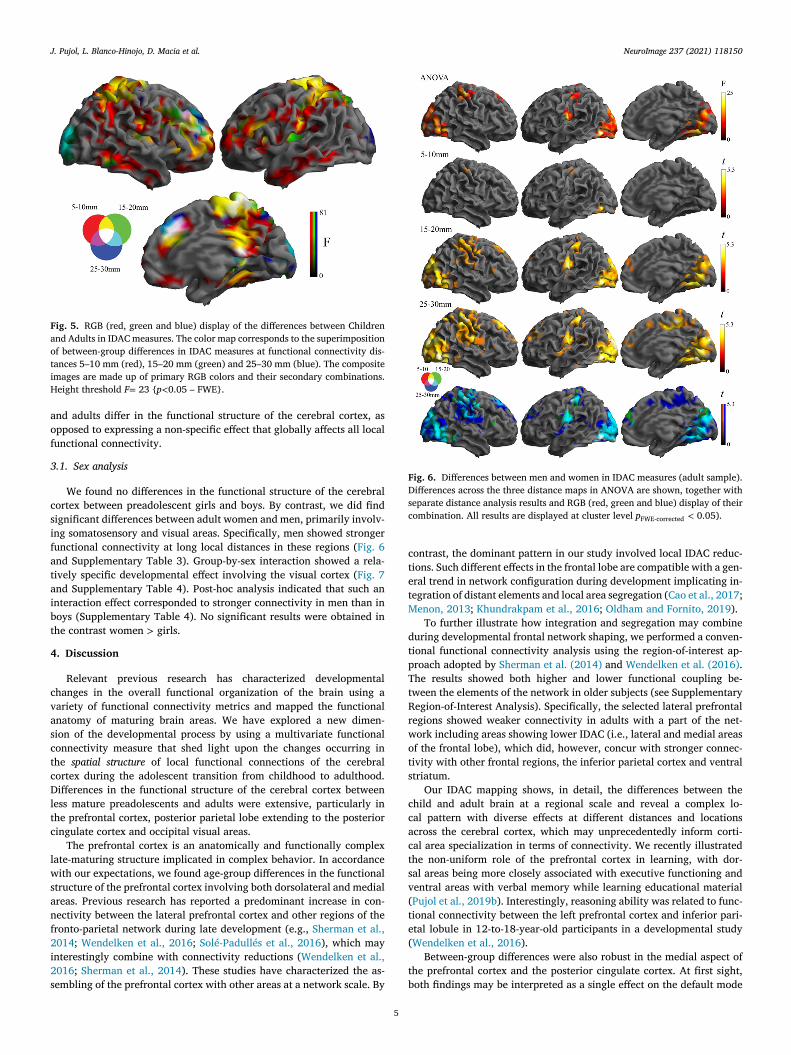
Fig. 2. Main effect of group illustrating the differences between children and
adults in IDAC measures across the three distance maps (ANOVA). Height
threshold F = 23 { p < 0.05 – FWE}.
w
i
A
b
a
f
r
a
i
d
o
e
3
c
m
s
m
b
fi
i
p
d
t
l
c
i
t
g
p
t
a
i
s
f
t
t
t
t
Fig. 3. Single effects illustrating differences between children and adults in
IDAC measures at three functional connectivity distances. Height threshold T = 4.7 { p < 0.05 – FWE}.
Fig. 4. ANOVA group (children vs adults) by distance (5–10 mm, 15–20 mm
and 25–30 mm) interactions as to local functional connectivity IDAC measures.
Height threshold F = 13.6 { p < 0.05 – FWE}.
b
i
a
c
2
s
o
e tested for main group effects, post-hoc between-group differences
n connectivity for each IDAC map and group-by-distance interactions.
n ANOVA model was also used to assess differences in IDAC measures
etween males and females within age groups, and group by sex inter-
ctions to identify potential differential development effects.
In all the analyses, results were considered significant when clusters
ormed at a threshold of p < 0.005 survived whole-brain family-wise er-
or (FWE) correction ( p < 0.05), calculated using SPM, which was further
djusted at p < 0.006 using Bonferroni correction for multiple compar-
sons (6 simple effects, 2 main effects and the interaction). The overall
ifferences between both study samples were displayed at higher thresh-
lds (equivalent to single-voxel FWE-correction) to emphasize brain ar-
as showing the most robust developmental effects.
. Results
Whole-brain maps of the local functional structure of the cerebral
ortex were generated from combined IDAC functional connectivity
easures in both adults and preadolescents. The RGB composition of
hort (5–10 mm), middle (15–20 mm) and long-distance (25–30 mm)
aps is illustrated in Fig. 1 . The maps were able to parcellate the cere-
ral cortex into regions arguably resembling the parcellations we can
nd in traditional brain atlases (e.g., Brodmann, 2006 ). For example,
n the adult sample, the angular and supramarginal gyri of the inferior
arietal lobule are both mostly connected at short and medium local
istances (typically yellow in the maps), whereas in the lateral occipi-
al cortex the dominant pattern involves high connectivity at the longer
ocal distance ranges.
The analysis of the main effect of group comparing IDAC functional
onnectivity measures across the three distances did demonstrate signif-
cant differences in a number of regions. The differences were robust in
he lateral and medial prefrontal cortex extending to the anterior cin-
ulate cortex, the posterior/superior parietal cortex extending to the
aracentral lobule, somatosensory strip and posterior cingulate cortex,
he occipital visual cortex, hippocampus, amygdala, and insula ( Fig. 2
nd Supplementary Table 1).
A map of between-group differences in local functional connectiv-
ty was then generated for each of the three IDAC distances reporting
imple effects ( Fig. 3 ). Overall, this set of analyses showed that dif-
erences between both age groups were not homogeneous across the
hree distances. For instance, parietal cortices showed stronger func-
ional connectivity in children than adults predominantly at short dis-
ances, whereas the occipital cortex showed weaker functional connec-
ivity in children at long local distances. Statistical testing of group-
4
y-distance interactions confirmed such distance effects for most of the
dentified areas. We would mention, however, that the significant inter-
ction effect was partial in frontal lobe areas and marginal in the visual
ortex, in terms of anatomical extension ( Fig. 4 , Supplementary Table
).
The details of distance effects may be jointly appreciated in Fig. 5
howing differences across the three distances in integrated RGB maps
f IDAC differences. These results would indicate that preadolescents
J. Pujol, L. Blanco-Hinojo, D. Macia et al. NeuroImage 237 (2021) 118150
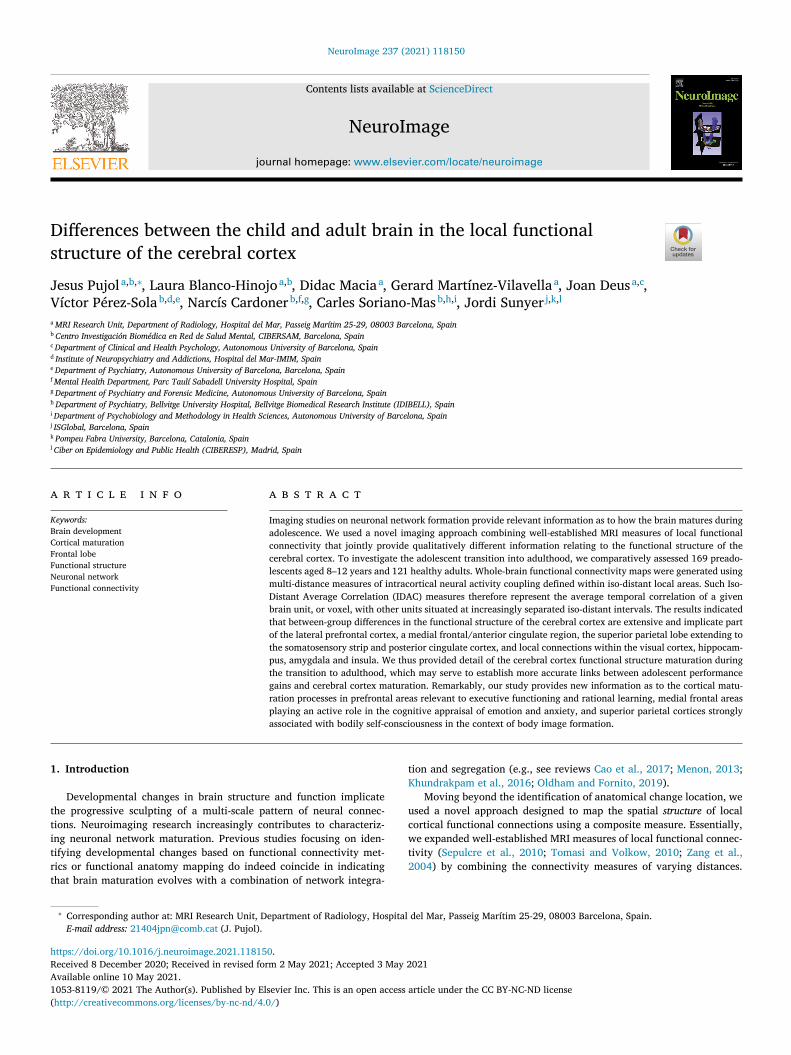
Fig. 5. RGB (red, green and blue) display of the differences between Children
and Adults in IDAC measures. The color map corresponds to the superimposition
of between-group differences in IDAC measures at functional connectivity dis-
tances 5–10 mm (red), 15–20 mm (green) and 25–30 mm (blue). The composite
images are made up of primary RGB colors and their secondary combinations.
Height threshold F = 23 { p < 0.05 – FWE}.
a
o
f
3
c
s
i
f
a
t
a
i
b
t
4
c
v
a
s
c
t
c
D
l
t
c
l
w
s
a
n
f
2
i
2
s
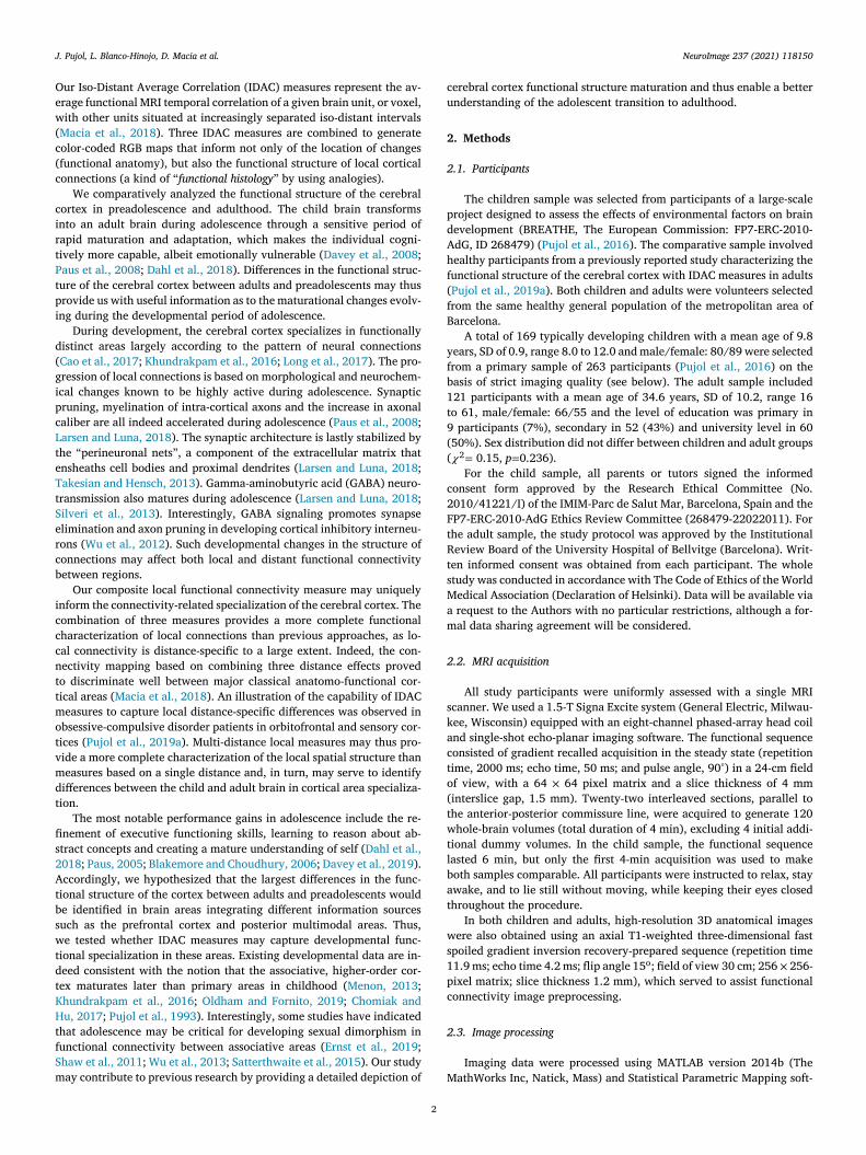
Fig. 6. Differences between men and women in IDAC measures (adult sample).
Differences across the three distance maps in ANOVA are shown, together with
separate distance analysis results and RGB (red, green and blue) display of their
combination. All results are displayed at cluster level p FWE-corrected < 0.05).
c
t
e
t
M
d
t
p
T
t
R
r
w
o
t
s
c
c
a
c
t
s
v
(
t
e
(
t
b
nd adults differ in the functional structure of the cerebral cortex, as
pposed to expressing a non-specific effect that globally affects all local
unctional connectivity.
.1. Sex analysis
We found no differences in the functional structure of the cerebral
ortex between preadolescent girls and boys. By contrast, we did find
ignificant differences between adult women and men, primarily involv-
ng somatosensory and visual areas. Specifically, men showed stronger
unctional connectivity at long local distances in these regions ( Fig. 6
nd Supplementary Table 3). Group-by-sex interaction showed a rela-
ively specific developmental effect involving the visual cortex ( Fig. 7
nd Supplementary Table 4). Post-hoc analysis indicated that such an
nteraction effect corresponded to stronger connectivity in men than in
oys (Supplementary Table 4). No significant results were obtained in
he contrast women > girls.
. Discussion
Relevant previous research has characterized developmental
hanges in the overall functional organization of the brain using a
ariety of functional connectivity metrics and mapped the functional
natomy of maturing brain areas. We have explored a new dimen-
ion of the developmental process by using a multivariate functional
onnectivity measure that shed light upon the changes occurring in
he spatial structure of local functional connections of the cerebral
ortex during the adolescent transition from childhood to adulthood.
ifferences in the functional structure of the cerebral cortex between
ess mature preadolescents and adults were extensive, particularly in
he prefrontal cortex, posterior parietal lobe extending to the posterior
ingulate cortex and occipital visual areas.
The prefrontal cortex is an anatomically and functionally complex
ate-maturing structure implicated in complex behavior. In accordance
ith our expectations, we found age-group differences in the functional
tructure of the prefrontal cortex involving both dorsolateral and medial
reas. Previous research has reported a predominant increase in con-
ectivity between the lateral prefrontal cortex and other regions of the
ronto-parietal network during late development (e.g., Sherman et al.,
014 ; Wendelken et al., 2016 ; Solé-Padullés et al., 2016 ), which may
nterestingly combine with connectivity reductions ( Wendelken et al.,
016 ; Sherman et al., 2014 ). These studies have characterized the as-
embling of the prefrontal cortex with other areas at a network scale. By
5
ontrast, the dominant pattern in our study involved local IDAC reduc-
ions. Such different effects in the frontal lobe are compatible with a gen-
ral trend in network configuration during development implicating in-
egration of distant elements and local area segregation ( Cao et al., 2017 ;
enon, 2013 ; Khundrakpam et al., 2016 ; Oldham and Fornito, 2019 ).
To further illustrate how integration and segregation may combine
uring developmental frontal network shaping, we performed a conven-
ional functional connectivity analysis using the region-of-interest ap-
roach adopted by Sherman et al. (2014) and Wendelken et al. (2016) .
he results showed both higher and lower functional coupling be-
ween the elements of the network in older subjects (see Supplementary
egion-of-Interest Analysis). Specifically, the selected lateral prefrontal
egions showed weaker connectivity in adults with a part of the net-
ork including areas showing lower IDAC (i.e., lateral and medial areas
f the frontal lobe), which did, however, concur with stronger connec-
ivity with other frontal regions, the inferior parietal cortex and ventral
triatum.
Our IDAC mapping shows, in detail, the differences between the
hild and adult brain at a regional scale and reveal a complex lo-
al pattern with diverse effects at different distances and locations
cross the cerebral cortex, which may unprecedentedly inform corti-
al area specialization in terms of connectivity. We recently illustrated
he non-uniform role of the prefrontal cortex in learning, with dor-
al areas being more closely associated with executive functioning and
entral areas with verbal memory while learning educational material
Pujol et al., 2019b ). Interestingly, reasoning ability was related to func-
ional connectivity between the left prefrontal cortex and inferior pari-
tal lobule in 12-to-18-year-old participants in a developmental study
Wendelken et al., 2016 ).
Between-group differences were also robust in the medial aspect of
he prefrontal cortex and the posterior cingulate cortex. At first sight,
oth findings may be interpreted as a single effect on the default mode

J. Pujol, L. Blanco-Hinojo, D. Macia et al. NeuroImage 237 (2021) 118150
Fig. 7. Group (children vs adults) by sex (males vs females) interactions in Iso-
Distance Average Correlations (IDAC) functional connectivity measures. Inter-
action effects across the three distance maps are shown, together with separate
distance analysis results and RGB (red, green and blue) display of their combi-
nation. All results are displayed at cluster level p FWE-corrected < 0.05).
n
m
(
c
m
r
o
H
l
e
c
R
p
d
p
t
a
E
e
a
l
r
p
a
s
w
2
c
c
2
h
p
S
h
m
s
2
f
r
c
c
G
a
H
a
2
t
e
c
s
t
a
l
r
t
d
n
l
d
p
f
o
y
a
s
t
i
s
c
m
p
S
f
o
m
i
t
t
(
o
s
w
d
d
w
i
s
i
m
t
etwork. However, a closer inspection would suggest that the involved
edial frontal area is not the core element of the default mode network
Harrison et al., 2008 ) and implicates the adjacent anterior cingulate
ortex. Supplementary Fig. 2 confirms that the area showing a develop-
ental effect in the current study is posteriorly located. Among other
oles, this medial frontal region is implicated in the cognitive appraisal
f emotion and anxiety ( Somerville et al., 2013 ; Pujol et al., 2013 ;
arrison et al., 2015 ). These results are therefore consistent with the
ate maturation of self-appraisal processes ( Davey et al., 2019 ). Inter-
stingly, this medial frontal region showed reduced cortical functional
onnectivity differentiation in individuals with psychopathy ( Contreras-
odríguez et al., 2015 ), for whom a developmental origin has been pro-
osed ( Pujol et al., 2019c ).
The posterior cingulate cortex is the posterior core element of the
efault mode network. In contrast with the anterior medial frontal as-
ect of the default mode network, we found robust group differences in
he posterior cingulate cortex during the transition to adulthood, which
re consistent with other developmental studies ( Sherman et al., 2014 ;
rnst et al., 2019 ; Fair et al., 2008 ). However, the anterior and posterior
lements of the default mode network may not be completely coupled
t rest until late adolescence ( Fair et al., 2008 ), particularly during chal-
enging tasks ( Pujol et al., 2008 ).
In addition to the perhaps more predictable prefrontal and poste-
ior cingulate cortex changes, we found highly robust differences in the
arietal lobe involving virtually all its posterior (and superior) aspect
nd extending to the somatosensory strip. All these areas have been
trongly linked to body image formation or bodily self-consciousness,
hich is the most somatic facet of self-consciousness ( Blanke et al.,
015 ; Ronchi et al., 2018 ). Bodily self-consciousness refers to body-
entered perception based on the multimodal integration of proprio-
eptive, vestibular, visual and interoceptive bodily inputs ( Blanke et al.,
6
015 ; Ronchi et al., 2018 ). Previous functional connectivity research
as also shown that adolescence is an active period in the formation of
arietal lobe networks ( Marcos-Vidal et al., 2018 ; O’Rawe et al., 2019 ;
haw et al., 2011 ; Vinette and Bray, 2015 ). Interestingly, other studies
ave indicated that a functional breakdown in the parietal connections
ay be a risk for developing psychiatric and neurological disorders as-
ociated with distorted body perception ( Ronchi et al., 2018 ; Via et al.,
018 ).
The visual cortex was also sensitive to age effects. In this case, we
ound an inverse pattern showing an increase in adults, as opposed to
eduction, in local functional connectivity involving the longest local
onnections (15–30 mm). Coherent visual perception combines recipro-
al interactions between striate and extrastriate visual areas ( Prasad and
aletta, 2011 ). The bulk of vision maturation takes place at early ages,
nd mostly under the age of 5 years ( Saygin et al., 2016 ; Kiorpes, 2015 ).
owever, our data are consistent with other studies indicating that some
spects of the visual function may maturate later (e.g., Gomez et al.,
018 ; Kiorpes, 2016 ; Kovács et al., 1999 ).
Developmental imaging studies indicate that visual cortex connec-
ivity between occipital visual areas and other distant (extra-occipital)
lements of the visual system indeed progresses until and during adoles-
ence ( Shaw et al., 2011 ; Vinette and Bray, 2015 ). What is perhaps more
urprising, in our study, is the connectivity progress observed within
he occipital lobe (i.e., strengthening the coupling between extrastriate
nd striate visual areas). It remains to be established, however, whether
ate visual striate and extrastriate cortex functional connectivity matu-
ation parallels the relatively slow performance refinement of vision, in
erms of, for instance, stereopsis —our ability to appreciate distance and
epth —or two-dimensional motion perception ( Kiorpes, 2015 ).
Previous studies have shown that differentiation in functional con-
ectivity between boys and girls is active during adolescence in parietal
obe connections related to complex visual function ( Shaw et al., 2011 ),
efault mode network ( Ernst et al., 2019 ) and for some global network
roperties ( Wu et al., 2013 ). Other studies demonstrated strong sex dif-
erences in global brain connectivity measures in a large 9-to-22 year-
ld sample ( Satterthwaite et al., 2015 ), but weak differences in 6-to-10
ear old children ( Langen et al., 2018 ). We tested for sex differences
nd did observe their presence in the adult sample involving visual and
omatosensory cortices. In addition, our analysis captured developmen-
al differences between males and females in the visual cortex. Signif-
cant interaction was not observed in the somatosensory cortex at the
tudy’s threshold, perhaps indicating a more subtle effect. It has been
ontemplated that the sensory and perceptual processes differ between
en and women ( Hamilton, 2008 ), with male or female advantages de-
ending on the specific operation tested (e.g., Satterthwaite et al., 2015 ;
pies and Sevincer, 2018 ; Goyette et al., 2012 ). It may be of interest in
uture study to test whether sex differences in the functional structure
f sensory areas are related to differences in sensory processing perfor-
ance.
Finally, differences between children and adults were also observed
n the hippocampus, amygdala and insula. These structures are part of
he limbic (and paralimbic) system, which includes a series of primi-
ive formations surrounding the boundary (or limbus ) of the neocortex
Morgane et al, 2005 ). Therefore, our results suggest that the shaping
f connections may similarly be active during late development in ba-
ic systems regulating emotional and motivational processes. However,
e would like to point out that although the hippocampus and amyg-
ala are closely integrated to the cerebral cortex, they have a markedly
istinct architecture and pattern of connections ( Morgane et al, 2005 ),
hich arguably could be captured only in part by our IDAC approach in
ts current form (i.e., limited to three measures within a 30-mm radius).
A limitation of our study may be the absence of a longitudinal as-
essment in the same cohort. Although cross-sectional designs are not
deal for the study of developmental changes, such a limitation may be
itigated by the large sample size. A similarly important limitation is
he use of only four minutes of resting-state data to compute our con-

J. Pujol, L. Blanco-Hinojo, D. Macia et al. NeuroImage 237 (2021) 118150
n
o
t
o
b
u
l
t
o
n
a
i
M
p
a
4
t
t
a
i
a
f
a
i
a
f
F
u
C
D
r
e
D
t
C
e
e
M
J
S
C
S
i
e
A
m
o
D
t
S
t
R
A
B
B
B
B
B
C
C
C
D
D
D
E
F
G
G
G
H
H
H
K
K
K
K
L
L
L
ectivity measures. Although prior studies have shown that acquisitions
f three to five minutes result in stable estimates of functional connec-
ivity measures ( Braun et al., 2012 ; Van Dijk et al, 2010 ), the reliability
f measuring individual differences in the strength of connectivity can
e greatly improved by increasing the scan lengths from five minutes
p to 13 min ( Birn et al., 2013 ). Interestingly, by using exceptionally
arge acquisitions (e.g., 5 h), functional connectomes become reliable
o the level of individual humans ( Gordon et al., 2017 ). Therefore, rec-
mmendations for future work should include the use of both longitudi-
al designs and longer acquisitions. Our study was also limited in that
complete demographic characterization and a comprehensive behav-
oral assessment covering general brain functioning were not obtained.
oreover, the study design, based on assessing children and adult sam-
les, renders the control for potential confounders less optimal in the
bsence of similar behavioral testing.
.1. Conclusions
We have provided some detail relating to the cerebral cortex func-
ional structure during the transition to adulthood, which may serve
o establish more accurate links between adolescent performance gains
nd cerebral cortex maturation. Remarkably, our study provides new
nformation as to the cortical maturation processes in lateral prefrontal
reas relevant to executive functioning and rational learning, the medial
rontal/anterior cingulate area playing an active role in the cognitive
ppraisal of emotion and anxiety, and the superior parietal lobe extend-
ng to the somatosensory strip and posterior cingulate cortex strongly
ssociated with bodily self-consciousness in the context of body image
ormation.
unding
This work was supported in part by the European Research Council
nder the ERC [grant number 268479 ] – the BREATHE project and the
arlos III Health Institute grants PI13/01958 and PI16/00889 .
ata and code availability statement
Data will be available via a request to the authors with no particular
estrictions, although a formal data sharing agreement will be consid-
red.
eclaration of Competing Interest
The authors report no financial interests or potential conflicts of in-
erest.
redit authorship contribution statement
Jesus Pujol: Conceptualization, Data curation, Writing - review &
diting. Laura Blanco-Hinojo: Formal analysis, Writing - review &
diting. Didac Macia: Software, Writing - review & editing. Gerard
artínez-Vilavella: Formal analysis, Writing - review & editing.
oan Deus: Data curation, Writing - review & editing. Víctor Pérez-
ola: Conceptualization, Writing - review & editing. Narcís Cardoner:
onceptualization, Investigation, Writing - review & editing. Carles
oriano-Mas: Conceptualization, Investigation, Writing - review & edit-
ng. Jordi Sunyer: Conceptualization, Supervision, Writing - review &
diting.
cknowledgments
We thank the Agency of University and Research Funding Manage-
ent of the Catalonia Government for their participation in the context
f Research Groups SGR2017_134, SGR2017_1198 and SGR2017-1247.
r. Soriano-Mas thanks the support of the Miguel Servet contract from
he Carlos III Health Institute (CPII16/00048).
7
upplementary materials
Supplementary material associated with this article can be found, in
he online version, at doi:10.1016/j.neuroimage.2021.118150 .
eferences
shburner, J. , 2007. A fast diffeomorphic image registration algorithm. NeuroImage 38
(1), 95–113 Oct 15 .
irn, R.M. , Molloy, E.K. , Patriat, R. , Parker, T. , Meier, T.B. , Kirk, G.R. , Nair, V.A. ,
Meyerand, M.E. , Prabhakaran, V. , 2013. The effect of scan length on the reliability of
resting-state fMRI connectivity estimates. NeuroImage 83, 550–558 Dec .
lakemore, S.J. , Choudhury, S. , 2006. Development of the adolescent brain: implica-
tions for executive function and social cognition. J Child Psychol Psychiatry 47 (3-4),
296–312 Mar-Apr .
lanke, O. , Slater, M. , Serino, A. , 2015. Behavioral, neural, and computational principles
of bodily self-consciousness. Neuron 88 (1), 145–166 Oct 7 .
raun, U. , Plichta, M.M. , Esslinger, C. , Sauer, C. , Haddad, L. , Grimm, O. , Mier, D. ,
Mohnke, S. , Heinz, A. , Erk, S. , Walter, H. , Seiferth, N. , Kirsch, P. , Meyer-Linden-
berg, A. , 2012. Test-retest reliability of resting-state connectivity network character-
istics using fMRI and graph theoretical measures. NeuroImage 59 (2), 1404–1412 Jan
16 .
rodmann, K. , 2006. Brodmann’s Localisation in the Cerebral Cortex. Springer US, New
York .
ao, M. , Huang, H. , He, Y. , 2017. Developmental connectomics from infancy through early
childhood. Trends Neurosci. 40 (8), 494–506 Aug .
homiak, T. , Hu, B. , 2017. Mechanisms of hierarchical cortical maturation. Front. Cell
Neurosci. 11, 272 Sep 11 .
ontreras-Rodríguez, O. , Pujol, J. , Batalla, I. , Harrison, B.J. , Soriano-Mas, C. , Deus, J. ,
López-Solà, M. , Macià, D. , Pera, V. , Hernández-Ribas, R. , Pifarré, J. , Menchón, J.M. ,
Cardoner, N. , 2015. Functional connectivity bias in the prefrontal cortex of psy-
chopaths. Biol. Psychiatry 78 (9), 647–655 Nov 1 .
ahl, R.E. , Allen, N.B. , Wilbrecht, L. , Suleiman, A.B. , 2018. Importance of investing in
adolescence from a developmental science perspective. Nature 554 (7693), 441–450
Feb 21 .
avey, C.G. , Fornito, A. , Pujol, J. , Breakspear, M. , Schmaal, L. , Harrison, B.J. , 2019.
Neurodevelopmental correlates of the emerging adult self. Dev. Cogn. Neurosci. 36,
100626 Apr .
avey, C.G. , Yücel, M. , Allen, N.B. , 2008. The emergence of depression in adolescence: de-
velopment of the prefrontal cortex and the representation of reward. Neurosci. Biobe-
hav. Rev. 32 (1), 1–19 .
rnst, M. , Benson, B. , Artiges, E. , Gorka, A.X. , Lemaitre, H. , Lago, T. , et al. , 2019. and IM-
AGEN Consortium. Pubertal maturation and sex effects on the default-mode network
connectivity implicated in mood dysregulation. Transl. Psychiatry 9 (1), 103 Feb 25 .
air, D.A. , Cohen, A.L. , Dosenbach, N.U. , Church, J.A. , Miezin, F.M. , Barch, D.M. ,
Raichle, M.E. , Petersen, S.E. , Schlaggar, B.L. , 2008. The maturing architecture of the
brain’s default network. Proc. Natl. Acad. Sci. U.S.A. 105 (10), 4028–4032 Mar 11 .
omez, J. , Natu, V. , Jeska, B. , Barnett, M. , Grill-Spector, K. , 2018. Development differ-
entially sculpts receptive fields across early and high-level human visual cortex. Nat.
Commun. 9 (1), 788 Feb 23 .
ordon, E.M. , Laumann, T.O. , Gilmore, A.W. , Newbold, D.J. , Greene, D.J. , Berg, J.J. ,
Ortega, M. , Hoyt-Drazen, C. , Gratton, C. , Sun, H. , Hampton, J.M. , Coalson, R.S. ,
Nguyen, A.L. , McDermott, K.B. , Shimony, J.S. , Snyder, A.Z. , Schlaggar, B.L. , Pe-
tersen, S.E. , Nelson, S.M. , Dosenbach, N.U.F. , 2017. Precision functional mapping of
individual human brains. Neuron 95 (4), 791–807 Aug 16.e7 .
oyette, S.R. , McCoy, J.G. , Kennedy, A. , Sullivan, M. , 2012. Sex differences on the judg-
ment of line orientation task: a function of landmark presence and hormonal status.
Physiol. Behav. 105 (4), 1045–1051 Feb 28 .
amilton, C. , 2008. Cognition and Sex Differences. Palgrave MacMillan, New York .
arrison, B.J. , Fullana, M.A. , Soriano-Mas, C. , Via, E. , Pujol, J. , Martínez-Zalacaín, I. ,
Tinoco-Gonzalez, D. , Davey, C.G. , López-Solà, M. , Pérez Sola, V. , Menchón, J.M. , Car-
doner, N. , 2015. A neural mediator of human anxiety sensitivity. Hum. Brain Mapp.
36 (10), 3950–3958 Oct .
arrison, B.J. , Pujol, J. , López-Solà, M. , Hernández-Ribas, R. , Deus, J. , Ortiz, H. , Sori-
ano-Mas, C. , Yücel, M. , Pantelis, C. , Cardoner, N. , 2008. Consistency and functional
specialization in the default mode brain network. Proc. Natl. Acad. Sci. U. S. A. 105
(28), 9781–9786 Jul 15 .
hundrakpam, B.S. , Lewis, J.D. , Zhao, L. , Chouinard-Decorte, F. , Evans, A.C. , 2016.
Brain connectivity in normally developing children and adolescents. NeuroImage 134,
192–203 Jul 1 .
iorpes, L. , 2015. Visual development in primates: neural mechanisms and critical periods.
Dev. Neurobiol. 75 (10), 1080–1090 Oct .
iorpes, L. , 2016. The puzzle of visual development: behavior and neural limits. J. Neu-
rosci. 36 (45), 11384–11393 Nov 9 .
ovács, I. , Kozma, P. , Fehér, A. , Benedek, G. , 1999. Late maturation of visual spatial
integration in humans. Proc. Natl. Acad. Sci. U. S. A. 96 (21), 12204–12209 Oct 12 .
angen, C.D. , Muetzel, R. , Blanken, L. , van der Lugt, A. , Tiemeier, H. , Verhulst, F. ,
Niessen, W.J. , White, T. , 2018. Differential patterns of age-related cortical and sub-
cortical functional connectivity in 6-to-10 year old children: a connectome-wide as-
sociation study. Brain Behav. 8 (8), e01031 Aug .
arsen, B. , Luna, B. , 2018. Adolescence as a neurobiological critical period for the devel-
opment of higher-order cognition. Neurosci. Biobehav. Rev. 94, 179–195 Nov .
ong, X. , Benischek, A. , Dewey, D. , Lebel, C. , 2017. Age-related functional brain changes
in young children. NeuroImage 155, 322–330 Jul 15 .

J. Pujol, L. Blanco-Hinojo, D. Macia et al. NeuroImage 237 (2021) 118150
M
M
M
M
O
O
P
P
P
P
P
P
P
P
P
P
P
P
R
S
S
S
S
S
S
S
S
S
T
T
V
V
V
W
W
W
Z
acia, D. , Pujol, J. , Blanco-Hinojo, L. , Martínez-Vilavella, G. , Martín-Santos, R. , Deus, J. ,
2018. Characterization of the spatial structure of local functional connectivity using
multi-distance average correlation measures. Brain Connect. 8 (5), 276–287 Jun .
arcos-Vidal, L. , Martínez-García, M. , Pretus, C. , Garcia-Garcia, D. , Martínez, K. ,
Janssen, J. , Vilarroya, O. , Castellanos, F.X. , Desco, M. , Sepulcre, J. , Carmona, S. , 2018.
Local functional connectivity suggests functional immaturity in children with atten-
tion-deficit/hyperactivity disorder. Hum. Brain Mapp. 39 (6), 2442–2454 Jun .
enon, V. , 2013. Developmental pathways to functional brain networks: emerging prin-
ciples. Trends Cogn. Sci. 17 (12), 627–640 Dec .
organe, P.J. , Galler, J.R. , Mokler, D.J. , 2005. A review of systems and networks of the
limbic forebrain/limbic midbrain. Prog. Neurobiol. 75 (2), 143–160 Feb .
ldham, S. , Fornito, A. , 2019. The development of brain network hubs. Dev. Cogn. Neu-
rosci. 36, 100607 Apr .
’Rawe, J.F. , Huang, A.S. , Klein, D.N. , Leung, H.C. , 2019. Posterior parietal influences
on visual network specialization during development: an fMRI study of functional
connectivity in children ages 9 to 12. Neuropsychologia 127, 158–170 Apr .
aus, T. , Keshavan, M. , Giedd, J.N. , 2008. Why do many psychiatric disorders emerge
during adolescence? Nat. Rev. Neurosci. 9 (12), 947–957 Dec .
aus, T. , 2005. Mapping brain maturation and cognitive development during adolescence.
Trends Cogn. Sci. 9 (2), 60–68 Feb .
ower, J.D. , Mitra, A. , Laumann, T.O. , Snyder, A.Z. , Schlaggar, B.L. , Petersen, S.E. , 2014.
Methods to detect, characterize, and remove motion artifact in resting state fMRI.
NeuroImage 84, 320–341 Jan 1 .
rasad, S. , Galetta, S.L. , 2011. Anatomy and physiology of the afferent visual system.
Handb. Clin. Neurol. 102, 3–19 .
ujol, J. , Blanco-Hinojo, L. , Maciá, D. , Alonso, P. , Harrison, B.J. , Martínez-Vilavella, G. ,
Deus, J. , Menchón, J.M. , Cardoner, N. , Soriano-Mas, C. , 2019a. Mapping alterations
of the functional structure of the cerebral cortex in obsessive-compulsive disorder.
Cereb. Cortex 29 (11), 4753–4762 Dec 17 .
ujol, J. , Blanco-Hinojo, L. , Martínez-Vilavella, G. , Canu-Martín, L. , Pujol, A. , Pérez–
Sola, V. , Deus, J. , 2019b. Brain activity during traditional textbook and audiovisu-
al-3D learning. Brain Behav. 9 (10), e01427 Oct .
ujol, J. , Giménez, M. , Ortiz, H. , Soriano-Mas, C. , López-Solà, M. , Farré, M. , Deus, J. ,
Merlo-Pich, E. , Harrison, B.J. , Cardoner, N. , Navinés, R. , Martín-Santos, R. , 2013.
Neural response to the observable self in social anxiety disorder. Psychol. Med. 43
(4), 721–731 Apr .
ujol, J. , Harrison, B.J. , Contreras-Rodriguez, O. , Cardoner, N. , 2019c. The contribution
of brain imaging to the understanding of psychopathy. Psychol. Med. 49 (1), 20–23
Jan .
ujol, J. , Macià, D. , Blanco-Hinojo, L. , Martínez-Vilavella, G. , Sunyer, J. , de la Torre, R. ,
Caixàs, A. , Martín-Santos, R. , Deus, J. , Harrison, B.J. , 2014. Does motion-related brain
functional connectivity reflect both artifacts and genuine neural activity? NeuroImage
101, 87–95 Nov 1 .
ujol, J. , Martínez-Vilavella, G. , Macià, D. , Fenoll, R. , Alvarez-Pedrerol, M. , Rivas, I. ,
Forns, J. , Blanco-Hinojo, L. , Capellades, J. , Querol, X. , Deus, J. , Sunyer, J. , 2016.
Traffic pollution exposure is associated with altered brain connectivity in school chil-
dren. NeuroImage 129, 175–184 Apr 1 .
ujol, J. , Reixach, J. , Harrison, B.J. , Timoneda-Gallart, C. , Vilanova, J.C. , Pérez-Al-
varez, F. , 2008. Posterior cingulate activation during moral dilemma in adolescents.
Hum. Brain Mapp. 29 (8), 910–921 Aug .
ujol, J. , Vendrell, P. , Junqué, C. , Martí-Vilalta, J.L. , Capdevila, A. , 1993. When does
human brain development end? Evidence of corpus callosum growth up to adulthood.
Ann. Neurol. 34 (1), 71–75 Jul .
onchi, R. , Park, H.D. , Blanke, O. , 2018. Bodily self-consciousness and its disorders.
Handb. Clin. Neurol. 151, 313–330 .
8
atterthwaite, T.D. , Wolf, D.H. , Roalf, D.R. , Ruparel, K. , Erus, G. , Vandekar, S. , Gen-
natas, E.D. , Elliott, M.A. , Smith, A. , Hakonarson, H. , Verma, R. , Davatzikos, C. ,
Gur, R.E. , Gur, R.C. , 2015. Linked sex differences in cognition and functional con-
nectivity in youth. Cereb. Cortex 25 (9), 2383–2394 Sep .
aygin, Z.M. , Osher, D.E. , Norton, E.S. , Youssoufian, D.A. , Beach, S.D. , Feather, J. ,
Gaab, N. , Gabrieli, J.D. , Kanwisher, N. , 2016. Connectivity precedes function in the
development of the visual word form area. Nat. Neurosci. 19 (9), 1250–1255 Sep .
epulcre, J. , Liu, H. , Talukdar, T. , Martincorena, I. , Yeo, B.T. , Buckner, R.L. , 2010. The
organization of local and distant functional connectivity in the human brain. PLoS
Comput. Biol. 6 (6), e1000808 .
haw, D.J. , Grosbras, M.H. , Leonard, G. , Pike, G.B. , Paus, T. , 2011. Development of func-
tional connectivity during adolescence: a longitudinal study using an action-observa-
tion paradigm. J. Cogn. Neurosci. 23 (12), 3713–3724 Dec .
herman, L.E. , Rudie, J.D. , Pfeifer, J.H. , Masten, C.L. , McNealy, K. , Dapretto, M. , 2014.
Development of the default mode and central executive networks across early adoles-
cence: a longitudinal study. Dev. Cogn. Neurosci. 10, 148–159 Oct .
ilveri, M.M. , Sneider, J.T. , Crowley, D.J. , Covell, M.J. , Acharya, D. , Rosso, I.M. ,
Jensen, J.E. , 2013. Frontal lobe 𝛾-aminobutyric acid levels during adolescence: as-
sociations with impulsivity and response inhibition. Biol. Psychiatry 74 (4), 296–304
Aug 15 .
olé-Padullés, C. , Castro-Fornieles, J. , de la Serna, E. , Calvo, R. , Baeza, I. , Moya, J. ,
Lázaro, L. , Rosa, M. , Bargalló, N. , Sugranyes, G. , 2016. Intrinsic connectivity networks
from childhood to late adolescence: effects of age and sex. Dev. Cogn. Neurosci. 17,
35–44 Feb .
omerville, L.H. , Jones, R.M. , Ruberry, E.J. , Dyke, J.P. , Glover, G. , Casey, B.J. , 2013. The
medial prefrontal cortex and the emergence of self-conscious emotion in adolescence.
Psychol. Sci. 24 (8), 1554–1562 Aug .
pies, M. , Sevincer, A.T. , 2018. Women outperform men in distinguishing between au-
thentic and nonauthentic smiles. J. Soc. Psychol. 158 (5), 574–579 .
akesian, A.E. , Hensch, T.K. , 2013. Balancing plasticity/stability across brain develop-
ment. Prog. Brain Res. 207, 3–34 .
omasi, D. , Volkow, N.D. , 2010. Functional connectivity density mapping. Proc. Natl.
Acad. Sci. U. S. A. 107 (21), 9885–9890 .
an Dijk, K.R. , Hedden, T. , Venkataraman, A. , Evans, K.C. , Lazar, S.W. , Buckner, R.L. ,
2010. Intrinsic functional connectivity as a tool for human connectomics: theory,
properties, and optimization. J. Neurophysiol. 103 (1), 297–321 Jan .
ia, E. , Goldberg, X. , Sánchez, I. , Forcano, L. , Harrison, B.J. , Davey, C.G. , Pu-
jol, J. , Martínez-Zalacaín, I. , Fernández-Aranda, F. , Soriano-Mas, C. , Cardoner, N. ,
Menchón, J.M. , 2018. Self and other body perception in anorexia nervosa: the role of
posterior DMN nodes. World J. Biol. Psychiatry 19 (3), 210–224 Apr .
inette, S.A. , Bray, S. , 2015. Variation in functional connectivity along anterior-to-poste-
rior intraparietal sulcus, and relationship with age across late childhood and adoles-
cence. Dev. Cogn. Neurosci. 13, 32–42 Jun .
endelken, C. , Ferrer, E. , Whitaker, K.J. , Bunge, S.A. , 2016. Fronto-parietal network re-
configuration supports the development of reasoning ability. Cereb. Cortex 26 (5),
2178–2190 May .
u, K. , Taki, Y. , Sato, K. , Hashizume, H. , Sassa, Y. , Takeuchi, H. , Thyreau, B. , He, Y. ,
Evans, A.C. , Li, X. , Kawashima, R. , Fukuda, H. , 2013. Topological organization of
functional brain networks in healthy children: differences in relation to age, sex, and
intelligence. PLoS One 8 (2), e55347 .
u, X. , Fu, Y. , Knott, G. , Lu, J. , Di Cristo, G. , Huang, Z.J. , 2012. GABA signaling promotes
synapse elimination and axon pruning in developing cortical inhibitory interneurons.
J. Neurosci. 32 (1), 331–343 Jan 4 .
ang, Y. , Jiang, T. , Lu, Y. , He, Y. , Tian, L. , 2004. Regional homogeneity approach to fMRI
data analysis. NeuroImage 22 (1), 394–400 .