Embed Size (px)
Citation preview
ORIGINAL ARTICLE
Differences in striatal dopamine transporter densitybetween tremor dominant and non-tremor Parkinson’s disease
Valtteri Kaasinen & Maija Kinos & Juho Joutsa &
Marko Seppänen & Tommi Noponen
Received: 24 February 2014 /Accepted: 2 May 2014# Springer-Verlag Berlin Heidelberg 2014
AbstractPurpose Parkinson’s disease (PD) canmanifest with a tremor-dominant or a non-tremor (akinetic-rigid) phenotype. Al-though the tremor-dominant subtype may show a better prog-nosis, there is limited information on the phenotypic differ-ences regarding the level of striatal dopamine transmission.The present study investigated striatal dopamine transporter(DAT) binding characteristics in a large sample of patientswith and without tremor.Methods [123I]FP-CIT SPECT scans of 231 patients with aclinical diagnosis of PD and abnormal FP-CIT binding (157with tremor, 74 without tremor) and 230 control patients withnormal FP-CIT binding (148 with tremor, 82 without tremor)were analysed using an automated region-of-interest analysisof the scans (BRASS). Specific striatal binding ratios werecompared between phenotypes and groups using age, sex, andsymptom duration, predominant side of symptoms, dopami-nergic medications and scanner as covariates.Results Patients with PD had 28.1 – 65.0 % lower binding inall striatal regions compared to controls (p<0.001). The meanFP-CIT caudate nucleus uptake and the left caudate nucleusuptake were higher in PD patients with tremor than in PDpatients without tremor (mean 9.0 % higher, left 10.5 %higher; p<0.05), whereas there were no differences betweentremor and non-tremor control patients. No significant effects
of tremor on DAT binding were observed in the anterior orposterior putamen.Conclusion The motor phenotype is associated with the extentof caudate dopamine terminal loss in PD, as dopamine functionis relativelymore preserved in tremor patients. Symptom type isrelated to caudate dopamine function only in association withParkinsonian dopaminergic degeneration, not in intact dopa-mine systems in patients with non-PD tremor.
Keywords Dopamine transporter . Parkinson’s disease .
Tremor . SPECT
Introduction
Tremor is a symptom characterized by involuntary, rhythmicand sinusoidal alternating movements of one or more bodyparts [1]. It is one of the cardinal symptoms of Parkinson’sdisease (PD) but seems to differ from bradykinesia and rigid-ity both clinically and pathophysiologically [2, 3]. Severallines of evidence indicate that tremor is a marker of relativelybenign PD [3]. Motor progression [4–6] and cognitive pro-gression [7] of tremor-dominant PD appear to be slower thanprogression of non-tremor PD, i.e. postural instability gaitdisorder (PIGD) or akinetic-rigid PD. The slower rate ofprogression in tremor-dominant PD is correlated with lesssevere nigral cell loss based on post-mortem examination[8]. Non-tremor PD patients show reduced brain activity inthe prefrontal cortex and globus pallidus [9], more prominentgrey matter atrophy in motor-related regions and decreasedfunctional connectivity [10] compared to tremor-dominantpatients. From a treatment-related perspective, non-tremorPD seems to be associated with an increased risk oflevodopa-induced dyskinesias [11].
Although lower CSF concentrations ofβ-amyloid 1–42, T-tau, P-tau181 and α-synuclein have been reported in patients
V. Kaasinen (*) :M. Kinos : J. JoutsaDivision of Clinical Neurosciences, University of Turku and TurkuUniversity Hospital, POB 52, 20521 Turku, Finlande-mail: [email protected]
V. Kaasinen :M. Kinos : J. Joutsa :M. SeppänenTurku PET Centre, University of Turku and Turku UniversityHospital, Turku, Finland
M. Seppänen : T. NoponenDepartment of Clinical Physiology and Nuclear Medicine,University of Turku and Turku University Hospital, Turku, Finland
Eur J Nucl Med Mol ImagingDOI 10.1007/s00259-014-2796-5
with the non-tremor PD subtype compared to tremor-dominant patients [12], data on possible imaging biomarkersin the nigrostriatal dopaminergic system are inconclusive.Previous studies with [123I]FP-CIT ([123I]N-ω-fluoropropyl-2β-carbomethoxy-3β-(4-iodophenyl)nortropane) SPECThave indicated that tremor-dominant patients may have higherstriatal dopamine transporter (DAT) binding [13–16], al-though there are also opposite [17] and negative [18] results.The sample sizes in these studies have varied between 20 and62 patients, of whom 10 to 24 patients have been tremor-dominant. In the largest study to date, Eggers et al. studied 122PD patients (23 with tremor) and found no significant differ-ences between subtypes when the effects of age, diseaseduration, disease severity and medication were excluded, al-though visual image analysis showed differences betweengroups [19].
It therefore appears that while there are clinical differencesbetween tremor-dominant and non-tremor PD patients, theresults of functional imaging studies of the DAT system aremixed but suggest a greater degree of neurodegeneration innon-tremor PD. It is possible that the discrepant results reflectmethodological differences together with small sample sizes.
In the present study, we investigated differences in DATdensity between tremor and non-tremor PD patients in thelargest reported sample of patients, and conducted an auto-mated analysis that was free from operator-induced bias. Toinvestigate the role of neurodegeneration of the brain dopa-mine system in PD, we also included tremor and non-tremorcontrol patients who had normal DAT scans and no evidenceof PD.
Materials and methods
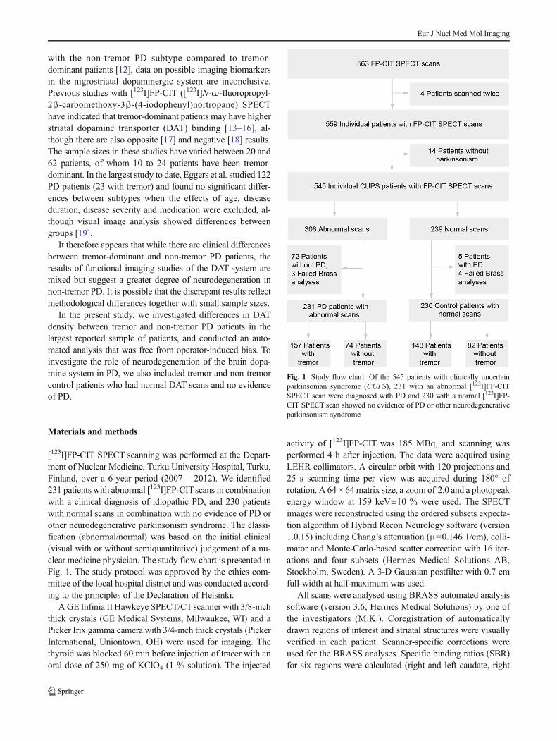
[123I]FP-CIT SPECT scanning was performed at the Depart-ment of Nuclear Medicine, Turku University Hospital, Turku,Finland, over a 6-year period (2007 – 2012). We identified231 patients with abnormal [123I]FP-CITscans in combinationwith a clinical diagnosis of idiopathic PD, and 230 patientswith normal scans in combination with no evidence of PD orother neurodegenerative parkinsonism syndrome. The classi-fication (abnormal/normal) was based on the initial clinical(visual with or without semiquantitative) judgement of a nu-clear medicine physician. The study flow chart is presented inFig. 1. The study protocol was approved by the ethics com-mittee of the local hospital district and was conducted accord-ing to the principles of the Declaration of Helsinki.
A GE Infinia II Hawkeye SPECT/CTscanner with 3/8-inchthick crystals (GE Medical Systems, Milwaukee, WI) and aPicker Irix gamma camera with 3/4-inch thick crystals (PickerInternational, Uniontown, OH) were used for imaging. Thethyroid was blocked 60 min before injection of tracer with anoral dose of 250 mg of KClO4 (1 % solution). The injected
activity of [123I]FP-CIT was 185 MBq, and scanning wasperformed 4 h after injection. The data were acquired usingLEHR collimators. A circular orbit with 120 projections and25 s scanning time per view was acquired during 180° ofrotation. A 64 × 64matrix size, a zoom of 2.0 and a photopeakenergy window at 159 keV±10 % were used. The SPECTimages were reconstructed using the ordered subsets expecta-tion algorithm of Hybrid Recon Neurology software (version1.0.15) including Chang’s attenuation (μ=0.146 1/cm), colli-mator and Monte-Carlo-based scatter correction with 16 iter-ations and four subsets (Hermes Medical Solutions AB,Stockholm, Sweden). A 3-D Gaussian postfilter with 0.7 cmfull-width at half-maximum was used.
All scans were analysed using BRASS automated analysissoftware (version 3.6; Hermes Medical Solutions) by one ofthe investigators (M.K.). Coregistration of automaticallydrawn regions of interest and striatal structures were visuallyverified in each patient. Scanner-specific corrections wereused for the BRASS analyses. Specific binding ratios (SBR)for six regions were calculated (right and left caudate, right
Fig. 1 Study flow chart. Of the 545 patients with clinically uncertainparkinsonian syndrome (CUPS), 231 with an abnormal [123I]FP-CITSPECT scan were diagnosed with PD and 230 with a normal [123I]FP-CIT SPECT scan showed no evidence of PD or other neurodegenerativeparkinsonism syndrome
Eur J Nucl Med Mol Imaging
and left anterior putamen, and right and left posterior puta-men) using the occipital cortex as the reference region:SBR = (VOIcaudate or putamen − VOIoccipital)/VOIoccipital [20].
All data were collected by one of the investigators (V.K.)using individual clinical patient histories, hospital records andreferrals together with the reanalysed SPECT images. Thefollowing factors were categorized: possible presence of Par-kinsonism at the time of imaging, presence of any tremor typeat the time of imaging, age and sex of the patient, duration ofmotor symptoms before scanning, possible dopaminergicmedication at the time of imaging, predominant side of motorsymptoms and the scanner. The clinical neurological diagno-ses of the patients were collected in March 2013(0.25 – 6.25 years after SPECT imaging) using the most recentpatient files from the treating neurologist.
The differences between the tremor and non-tremor pa-tients were compared between PD patients and controls (twogroups, four subgroups) using a two-way analysis of variance(ANOVA) with age, sex, symptom duration, symptom side,scanner and medication as covariates. The primary analysiswas performed with mean regional values [(left+right)/2], andindividual hemispheric values (left, right, ipsilateral or contra-lateral) were compared if a significant difference was ob-served in the primary analysis. The level of statistical signif-icance was set at p<0.05.
Results
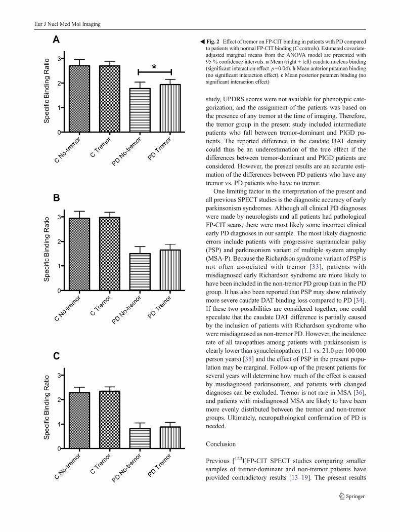
PD patients with tremor had a 9.0 % higher mean caudatetracer uptake than PD patients without tremor, whereas therewere no differences between the tremor and non-tremor con-trol patients (interaction effect group × subgroup F=4.23, df=1, p=0.04; Table 1, Fig. 2). This effect was also observed inthe left caudate (10.5 % difference in the PD group, 0 %difference in the control group; interaction F=4.59, df=1,p=0.03) but not in the right caudate. No significant interac-tions were detected in the anterior putamen (F=1.76, df=1,p=0.19) or the posterior putamen (F=0.03, df=1, p=0.85). Inthe patients with lateralized symptoms (202 PD patients, 164controls; patients with symmetrical symptoms excluded), nosignificant differences between tremor and non-tremor pa-tients were seen in the ipsilateral or contralateral caudate(p>0.17). The demographic characteristics of the patientstogether with the main ANOVA results are presented inTable 1. The difference in caudate tracer uptake betweentremor dominant and non-tremor PD patients was alsosignificant without any covariates (independent samples ttest p=0.004).
In patients with PD, FP-CIT uptake was 28.1 – 34.3 %lower in the caudate (F= 359.5, df = 1, p< 0.001),43.8 – 49.7 % lower in the anterior putamen (F=792.8, df=
1, p<0.001) and 61.0 – 65.0 % lower in the posterior putamen(F=1323.4, df=1, p<0.001) compared to uptake in controls.
Discussion
The results of the present large retrospective study indicatethat the extent of caudate nucleus dopamine terminal loss isgreater in non-tremor PD patients than in tremor PD patients,as investigated using DAT imaging approximately 2 yearsafter symptom onset. The association was selectively relatedto Parkinsonian tremor and not to tremor in patients withnormal striatal DAT binding. No significant differences wereobserved in the posterior putamen, which is the striatal regionthat is most severely affected by PD.
Loss of nigrostriatal dopamine neurons is the hallmark ofPD, and the extent of degeneration correlates well with theseverity of bradykinesia, and posture, gait and other midlinesymptoms [21]. However, one of the paradoxical aspects ofParkinsonian tremor is the weak relationship with dopaminedeficiency [22]. The pathophysiology of Parkinsonian tremoris poorly understood, but there are data to support the theorythat it is mediated primarily by cerebellothalamocortical path-ways [14, 23], although basal ganglia networks are also in-volved [23, 24]. It is possible that a combination lesion in thenigrostriatal and cerebellothalamocortical circuits is requiredfor PD tremor to emerge, and nigral loss alone is insufficient toproduce resting tremor [22]. Thus, the lesser degree of dopa-mine neuron loss in tremor patients in the present study couldbe viewed as a reflection of dysfunctional circuitry that has itsmajor lesion outside the striatum, such as in the thalamus.Caudate dopamine function may not primarily be reflected inthe motor symptoms, but rather in the non-motor differencesbetween the two motor phenotypes. PD patients with the non-tremor phenotype seem to be at higher risk of cognitiveimpairment compared to tremor-dominant PD patients [7,25], and they seem to have a greatly increased risk of dementia[26] compared to tremor-dominant patients. Non-tremor pa-tients also tend to be more depressed than patients with thetremor-dominant phenotype [27]. Indeed, impairments in cog-nitive tests have been reported to correlate with losses incaudate dopamine function in PD [28], and an inverse corre-lation between depression scores and caudate DATavailabilityin PD has been reported [29]. Future SPECT studies thatcombine neuropsychological and neuropsychiatric measuresshould be conducted to investigate this theory in detail.
A possible explanation for the lesser degree of dopamineterminal cell loss in tremor patients is that they or their next ofkin may become aware of motor symptoms earlier than pa-tients with non-tremor early PD. Therefore, although theduration of manifested symptoms may be the same, the dura-tion of premanifest disease could be longer in non-tremorpatients. This difference in the underlying level of progression
Eur J Nucl Med Mol Imaging
could explain why non-tremor patients show lower DATdensity in the early stages of the disease with seeminglysimilar symptom durations. However, the anatomical distribu-tion of the DAT binding difference between tremor and non-tremor patients does not support this theory. One could expectto see the largest difference in the posterior putamen, not in thecaudate nucleus, if non-tremor patients are more advanced atthe time of SPECT imaging. Furthermore, there do not seemto be differences in the ages of onset between tremor-dominant and akinetic-rigid patients [30], which do not sup-port a considerably longer period of unrecognized disease innon-tremor patients. Eggers et al. [19] investigated visualinterpretation of the scans together with BRASS. They foundno significant differences between tremor-dominant and non-tremor patients using BRASS after covariate correction. In-stead they found that tremor patients (n=23) showed a more“eagle-wing shaped” striatal configuration (more normal, with
some putaminal binding) than non-tremor patients whoshowed a more “egg-shaped” striatal configuration (moreabnormal, without putaminal binding). It is possible that notonly the average regional tracer binding but the diagnosticallytypical appearance of the striatum or the shape of the hot spotis also important. Further comparative studies (visual vs.semiquantitative) are needed.
Several subtypes of PD have been suggested to exist, butone of the most commonly used phenotypic categorization ofPD is tremor-dominant vs. non-tremor-dominant disease (ei-ther PIGD or akinetic-rigid) [6, 31]. Tremor-dominant, non-tremor-dominant and intermediate subtypes are most com-monly defined as the sum of Unified Parkinson’s DiseaseRating Scale (UPDRS) tremor items divided by the sum ofUPDRS PIGD items [5, 32]. Patients have also been assignedto groups based on UPDRS item score distributions or expertjudgement [31]. Due to the retrospective nature of the present
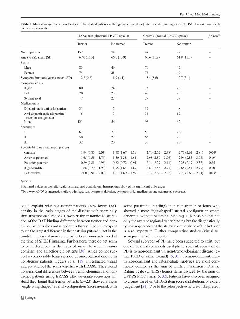
Table 1 Main demographic characteristics of the studied patients with regional covariate-adjusted specific binding ratios of FP-CIT uptake and 95 %confidence intervals
PD patients (abnormal FP-CIT uptake) Controls (normal FP-CIT uptake) p valuea
Tremor No tremor Tremor No tremor
No. of patients 157 74 148 82 –
Age (years), mean (SD) 67.0 (10.5) 66.0 (10.9) 65.6 (11.2) 61.8 (13.1) –
Sex, n
Male 83 49 70 42 –
Female 74 25 78 40 –
Symptom duration (years), mean (SD) 2.2 (2.8) 1.9 (2.1) 5.4 (8.6) 2.7 (3.1) –
Symptom side, n
Right 80 24 73 23 –
Left 70 28 48 20 –
Symmetrical 7 22 27 39 –
Medication, n
Dopaminergic antiparkinsonian 31 15 19 8 –
Anti-dopaminergic (dopaminereceptor antagonists)
5 3 33 12 –
None 121 56 96 62 –
Scanner, n
I 67 27 50 28 –
II 58 27 63 29 –
III 32 20 35 25 –
Specific binding ratio, mean (range)
Caudate 1.94 (1.86 – 2.03) 1.78 (1.67 – 1.89) 2.70 (2.62 – 2.78) 2.71 (2.61 – 2.81) 0.04*
Anterior putamen 1.65 (1.55 – 1.74) 1.50 (1.38 – 1.61) 2.98 (2.89 – 3.06) 2.94 (2.83 – 3.06) 0.19
Posterior putamen 0.89 (0.81 – 0.96) 0.82 (0.72 – 0.91) 2.34 (2.27 – 2.41) 2.28 (2.19 – 2.37) 0.85
Right caudate 1.88 (1.79 – 1.98) 1.75 (1.64 – 1.87) 2.63 (2.55 – 2.71) 2.65 (2.54 – 2.76) 0.10
Left caudate 2.00 (1.91 – 2.09) 1.81 (1.69 – 1.92) 2.77 (2.69 – 2.85) 2.77 (2.66 – 2.88) 0.03*
*p<0.05
Putaminal values in the left, right, ipsilateral and contralateral hemispheres showed no significant differencesa Two-way ANOVA interaction-effect with age, sex, symptom duration, symptom side, medication and scanner as covariates
Eur J Nucl Med Mol Imaging
study, UPDRS scores were not available for phenotypic cate-gorization, and the assignment of the patients was based onthe presence of any tremor at the time of imaging. Therefore,the tremor group in the present study included intermediatepatients who fall between tremor-dominant and PIGD pa-tients. The reported difference in the caudate DAT densitycould thus be an underestimation of the true effect if thedifferences between tremor-dominant and PIGD patients areconsidered. However, the present results are an accurate esti-mation of the differences between PD patients who have anytremor vs. PD patients who have no tremor.
One limiting factor in the interpretation of the present andall previous SPECT studies is the diagnostic accuracy of earlyparkinsonism syndromes. Although all clinical PD diagnoseswere made by neurologists and all patients had pathologicalFP-CIT scans, there were most likely some incorrect clinicalearly PD diagnoses in our sample. The most likely diagnosticerrors include patients with progressive supranuclear palsy(PSP) and parkinsonism variant of multiple system atrophy(MSA-P). Because the Richardson syndrome variant of PSP isnot often associated with tremor [33], patients withmisdiagnosed early Richardson syndrome are more likely tohave been included in the non-tremor PD group than in the PDgroup. It has also been reported that PSP may show relativelymore severe caudate DAT binding loss compared to PD [34].If these two possibilities are considered together, one couldspeculate that the caudate DAT difference is partially causedby the inclusion of patients with Richardson syndrome whoweremisdiagnosed as non-tremor PD. However, the incidencerate of all tauopathies among patients with parkinsonism isclearly lower than synucleinopathies (1.1 vs. 21.0 per 100 000person years) [35] and the effect of PSP in the present popu-lation may be marginal. Follow-up of the present patients forseveral years will determine how much of the effect is causedby misdiagnosed parkinsonism, and patients with changeddiagnoses can be excluded. Tremor is not rare in MSA [36],and patients with misdiagnosed MSA are likely to have beenmore evenly distributed between the tremor and non-tremorgroups. Ultimately, neuropathological confirmation of PD isneeded.
Conclusion
Previous [123I]FP-CIT SPECT studies comparing smallersamples of tremor-dominant and non-tremor patients haveprovided contradictory results [13–19]. The present results
�Fig. 2 Effect of tremor on FP-CIT binding in patients with PD comparedto patients with normal FP-CIT binding (C controls). Estimated covariate-adjusted marginal means from the ANOVA model are presented with95 % confidence intervals. aMean (right + left) caudate nucleus binding(significant interaction effect. p=0.04). bMean anterior putamen binding(no significant interaction effect). c Mean posterior putamen binding (nosignificant interaction effect)
Eur J Nucl Med Mol Imaging
indicate that PD patients with tremor have a less severedopaminergic defect in the caudate nucleus compared to PDpatients without tremor. This finding is consistent with theclinically more favourable prognosis of tremor PD. The local-ization of the findings to the caudate nucleus rather than to theposterior putamen suggests that the effect is not related to alonger unrecognized disease progression in non-tremor PD orto other major motor symptoms of the disease. The resultscontribute significantly to the understanding of the neurobio-logical basis of phenotypic differences in patients with PD.Possible therapeutic implications in different PD phenotypes,such as the risk of dyskinesias, remain unknown and could bea focus of further studies.
Ethical standards
The study protocol was approved by the ethics committee ofthe local hospital district and was conducted according to theprinciples of the Declaration of Helsinki.
Acknowledgments The contribution of the staff of the Department ofNuclear Medicine, Turku University Hospital, is gratefully acknowl-edged. This study was financially supported by the Academy of Finland(decision # 256836) and Turku University Hospital (ERVA funds).
Conflicts of interest None.
References
1. Abdo WF, van de Warrenburg BP, Burn DJ, Quinn NP, Bloem BR.The clinical approach to movement disorders. Nat Rev Neurol.2010;6:29–37. doi:10.1038/nrneurol.2009.196.
2. Hallett M. Parkinson’s disease tremor: pathophysiology.Parkinsonism Relat Disord. 2012;18 Suppl 1:S85–6. doi:10.1016/S1353-8020(11)70027-X.
3. Helmich RC, Hallett M, Deuschl G, Toni I, Bloem BR. Cerebralcauses and consequences of Parkinsonian resting tremor: a tale of twocircuits? Brain. 2012;135:3206–26. doi:10.1093/brain/aws023.
4. Clarimón J, Pagonabarraga J, Paisán-Ruíz C, Campolongo A,Pascual-Sedano B, Martí-Massó JF, et al. Tremor dominantParkinsonism: clinical description and LRRK2 mutation screening.Mov Disord. 2008;23:518–23. doi:10.1002/mds.21771.
5. Jankovic J, McDermott M, Carter J, Gauthier S, Goetz C, Golbe L,et al. Variable expression of Parkinson’s disease: a base-line analysisof the DATATOP cohort. The Parkinson Study Group. Neurology.1990;40:1529–34.
6. Zetusky WJ, Jankovic J, Pirozzolo FJ. The heterogeneity ofParkinson’s disease: clinical and prognostic implications.Neurology. 1985;35:522–6.
7. Williams-Gray CH, Foltynie T, Brayne CE, Robbins TW, Barker RA.Evolution of cognitive dysfunction in an incident Parkinson’s diseasecohort. Brain. 2007;130:1787–98. doi:10.1093/brain/awm111.
8. Selikhova M, Kempster PA, Revesz T, Holton JL, Lees AJ.Neuropathological findings in benign tremulous Parkinsonism.Mov Disord. 2013;28:145–52. doi:10.1002/mds.25220.
9. Prodoehl J, Planetta PJ, Kurani AS, Comella CL, Corcos DM,Vaillancourt DE. Differences in brain activation between tremor-
and nontremor-dominant Parkinson diseases. JAMA Neurol.2013;70:100–6. doi:10.1001/jamaneurol.2013.582.
10. Rosenberg-Katz K, Herman T, Jacob Y, Giladi N, Hendler T,Hausdorff JM. Gray matter atrophy distinguishes betweenParkinson disease motor subtypes. Neurology. 2013;80:1476–84.doi:10.1212/WNL.0b013e31828cfaa4.
11. Zhang YH, Tang BS, Song CY, Xu Q, Lou MX, Liu ZH, et al. Therelationship between the phenotype of Parkinson’s disease andlevodopa-induced dyskinesia. Neurosci Lett. 2013;556:109–12. doi:10.1016/j.neulet.2013.10.018.
12. Kang JH, Irwin DJ, Chen-Plotkin AS, Siderowf A, Caspell C, CoffeyCS, et al. Association of cerebrospinal fluidβ-amyloid 1-42, T-tau, P-tau181, and α-synuclein levels with clinical features of drug-naivepatients with early Parkinson disease. JAMANeurol. 2013;70:1277–87. doi:10.1001/jamaneurol.2013.3861.
13. Spiegel J, Hellwig D, Samnick S, JostW,MöllersMO, Fassbender K,et al. Striatal FP-CIT uptake differs in the subtypes of earlyParkinson’s disease. J Neural Transm. 2007;114:331–5. doi:10.1007/s00702-006-0518-2.
14. Helmich RC, Janssen MJ, Oyen WJ, Bloem BR, Toni I. Pallidaldysfunction drives a cerebellothalamic circuit into Parkinson tremor.Ann Neurol. 2011;69:269–81. doi:10.1002/ana.22361.
15. Schillaci O, Chiaravalloti A, Pierantozzi M, Di Pietro B, Koch G,Bruni C, et al. Different patterns of nigrostriatal degeneration intremor type versus the akinetic-rigid and mixed types ofParkinson’s disease at the early stages: molecular imaging with123I-FP-CIT SPECT. Int J Mol Med. 2011;28:881–6. doi:10.3892/ijmm.2011.764.
16. Rossi C, Frosini D, Volterrani D, De Feo P, Unti E, Nicoletti V, et al.Differences in nigro-striatal impairment in clinical variants of earlyParkinson’s disease: evidence from a FP-CIT SPECT study. Eur JNeurol. 2010;17:626–30. doi:10.1111/j.1468-1331.2009.02898.x.
17. Isaias IU, Benti R, Cilia R, Canesi M, Marotta G, Gerundini P, et al.[123I]FP-CIT striatal binding in early Parkinson’s disease patientswith tremor vs. akinetic-rigid onset. Neuroreport. 2007;18:1499–502.
18. Song IU, Chung YA, Oh JK, Chung SW. An FP-CIT PET compar-ison of the difference in dopaminergic neuronal loss in subtypes ofearly Parkinson’s disease. Acta Radiol. 2014:55:366–71. doi:10.1177/0284185113498075.
19. Eggers C, Kahraman D, Fink GR, Schmidt M, Timmermann L.Akinetic-rigid and tremor-dominant Parkinson’s disease patientsshow different patterns of FP-CIT single photon emission computedtomography. Mov Disord. 2011;26:416–23. doi:10.1002/mds.23468.
20. Varrone A, Dickson JC, Tossici-Bolt L, Sera T, Asenbaum S, Booij J,et al. European multicentre database of healthy controls for [(123)I]FP-CIT SPECT (ENC-DAT): age-related effects, gender differences andevaluation of different methods of analysis. Eur J Nucl Med MolImaging. 2013;40:213–27. doi:10.1007/s00259-012-2276-8.
21. Pirker W. Correlation of dopamine transporter imaging withParkinsonian motor handicap: how close is it? Mov Disord.2003;18 Suppl 7:S43–51. doi:10.1002/mds.10579.
22. Fishman PS. Paradoxical aspects of Parkinsonian tremor. MovDisord. 2008;23:168–73. doi:10.1002/mds.21736.
23. Mure H, Hirano S, Tang CC, Isaias IU, Antonini A, Ma Y, et al.Parkinson’s disease tremor-related metabolic network: characteriza-tion, progression, and treatment effects. Neuroimage. 2011;54:1244–53. doi:10.1016/j.neuroimage.2010.09.028.
24. Hallett M. Tremor: pathophysiology. Parkinsonism Relat Disord.2014;20 Suppl 1:S118–22. doi:10.1016/S1353-8020(13)70029-4.
25. Burn DJ, Rowan EN, Allan LM, Molloy S, O’Brien JT, McKeith IG.Motor subtype and cognitive decline in Parkinson’s disease,Parkinson’s disease with dementia, and dementia with Lewy bodies.J Neurol Neurosurg Psychiatry. 2006;77:585–9. doi:10.1136/jnnp.2005.081711.
26. Alves G, Larsen JP, Emre M, Wentzel-Larsen T, Aarsland D.Changes in motor subtype and risk for incident dementia in
Eur J Nucl Med Mol Imaging
Parkinson’s disease. Mov Disord. 2006;21:1123–30. doi:10.1002/mds.20897.
27. BurnDJ, Landau S, Hindle JV, SamuelM,Wilson KC, Hurt CS, et al.Parkinson’s disease motor subtypes and mood. Mov Disord.2012;27:379–86. doi:10.1002/mds.24041.
28. Kaasinen V, Rinne JO. Functional imaging studies of dopaminesystem and cognition in normal aging and Parkinson’s disease.Neurosci Biobehav Rev. 2002;26:785–93.
29. Di Giuda D, Camardese G, Bentivoglio AR, Cocciolillo F,Guidubaldi A, Pucci L, et al. Dopaminergic dysfunction and psychi-atric symptoms in movement disorders: a 123I-FP-CIT SPECTstudy. Eur J Nucl Med Mol Imaging. 2012;39:1937–48. doi:10.1007/s00259-012-2232-7.
30. Korchounov A, Schipper HI, Preobrazhenskaya IS, KesslerKR, Yakhno NN. Differences in age at onset and familialaggregation between clinical types of idiopathic Parkinson’sdisease. Mov Disord. 2004;19:1059–64. doi:10.1002/mds.20061.
31. Marras C, Lang A. Parkinson’s disease subtypes: lost in translation? JNeurol Neurosurg Psychiatry. 2013;84:409–15. doi:10.1136/jnnp-2012-303455.
32. Stebbins GT, Goetz CG, Burn DJ, Jankovic J, Khoo TK, Tilley BC.How to identify tremor dominant and postural instability/gait diffi-culty groups with the movement disorder society unified Parkinson’sdisease rating scale: comparison with the unified Parkinson’s diseaserating scale. Mov Disord. 2013;28:668–70. doi:10.1002/mds.25383.
33. Liscic RM, Srulijes K, Gröger A, Maetzler W, Berg D.Differentiation of progressive supranuclear palsy: clinical, imagingand laboratory tools. Acta Neurol Scand. 2013;127:362–70. doi:10.1111/ane.12067.
34. Oh M, Kim JS, Kim JY, Shin KH, Park SH, Kim HO, et al.Subregional patterns of preferential striatal dopamine transporter lossdiffer in Parkinson disease, progressive supranuclear palsy, andmultiple-system atrophy. J Nucl Med. 2012;53:399–406. doi:10.2967/jnumed.111.095224.
35. Savica R, Grossardt BR, Bower JH, Ahlskog JE, Rocca WA.Incidence and pathology of synucleinopathies and tauopathies relatedto Parkinsonism. JAMA Neurol. 2013;70:859–66. doi:10.1001/jamaneurol.2013.114.
36. Kaindlstorfer C, Granata R, Wenning GK. Tremor in multiple systematrophy – a review. Tremor Other Hyperkinet Mov (N Y). 2013;3:http://tremorjournal.org/article/view/165.
Eur J Nucl Med Mol Imaging