Embed Size (px)
Citation preview
REVIEW Open Access
Different fundus imaging modalities andtechnical factors in AI screening fordiabetic retinopathy: a reviewGilbert Lim1,2†, Valentina Bellemo2,3†, Yuchen Xie2, Xin Q. Lee2, Michelle Y. T. Yip3 and Daniel S. W. Ting2,3,4,5*
Abstract
Background: Effective screening is a desirable method for the early detection and successful treatment for diabeticretinopathy, and fundus photography is currently the dominant medium for retinal imaging due to its convenienceand accessibility. Manual screening using fundus photographs has however involved considerable costs for patients,clinicians and national health systems, which has limited its application particularly in less-developed countries. Theadvent of artificial intelligence, and in particular deep learning techniques, has however raised the possibility ofwidespread automated screening.
Main text: In this review, we first briefly survey major published advances in retinal analysis using artificial intelligence.We take care to separately describe standard multiple-field fundus photography, and the newer modalities of ultra-wide field photography and smartphone-based photography. Finally, we consider several machine learning conceptsthat have been particularly relevant to the domain and illustrate their usage with extant works.
Conclusions: In the ophthalmology field, it was demonstrated that deep learning tools for diabetic retinopathy showclinically acceptable diagnostic performance when using colour retinal fundus images. Artificial intelligence models areamong the most promising solutions to tackle the burden of diabetic retinopathy management in a comprehensivemanner. However, future research is crucial to assess the potential clinical deployment, evaluate the cost-effectivenessof different DL systems in clinical practice and improve clinical acceptance.
Keywords: Artificial intelligence, Deep learning, Diabetic retinopathy, Fundus photographs, Retinal imaging modalities,Survey
BackgroundA growing global health problem related to diabetesmellitus, one of the world’s fastest growing chronic dis-eases, is diabetic retinopathy (DR). This condition hasbeen projected to affect 700 million people across theworld within the next two decades [1]. Since one-third
of diabetic patients have underlying DR, this wouldtranslate to approximately 250 million people sufferingfrom DR by the year 2035 [2–4]. To meet this rapidlyevolving and growing crisis, tools that are able to dealwith this heavy workload quickly and efficiently areparamount in overcoming and tackling this leadingcause of blindness across the world [5, 6].Early detection of DR via population screening – asso-
ciated with timely treatment – has been shown to havethe potential to prevent visual loss in patients with dia-betic retinal complications [7]. Many computer-aidedalgorithms for automated retina image analysis havebeen explored [8–12]. Since before the deep learning
© The Author(s). 2020 Open Access This article is licensed under a Creative Commons Attribution 4.0 International License,which permits use, sharing, adaptation, distribution and reproduction in any medium or format, as long as you giveappropriate credit to the original author(s) and the source, provide a link to the Creative Commons licence, and indicate ifchanges were made. The images or other third party material in this article are included in the article's Creative Commonslicence, unless indicated otherwise in a credit line to the material. If material is not included in the article's Creative Commonslicence and your intended use is not permitted by statutory regulation or exceeds the permitted use, you will need to obtainpermission directly from the copyright holder. To view a copy of this licence, visit http://creativecommons.org/licenses/by/4.0/.The Creative Commons Public Domain Dedication waiver (http://creativecommons.org/publicdomain/zero/1.0/) applies to thedata made available in this article, unless otherwise stated in a credit line to the data.
* Correspondence: [email protected]†Gilbert Lim and Valentina Bellemo contributed equally to this work.2Singapore Eye Research Institute, Singapore National Eye Centre, Singapore,Singapore3Duke-NUS Medical School, National University of Singapore, 11 ThirdHospital Road Avenue, Singapore 168751, SingaporeFull list of author information is available at the end of the article
Lim et al. Eye and Vision (2020) 7:21 https://doi.org/10.1186/s40662-020-00182-7
(DL) era, the development and application of such tech-niques has produced cost-effective tools for DR screen-ing, [13, 14] and were crucial in the care of patients withDR and other diseases detectable from the retina such asglaucoma, age-related macular degeneration and retinop-athy of prematurity [6, 15–17]. Several international re-search groups have worked on automatic retinal imageanalysis methods to detect, localize, or measure retinal fea-tures and properties, [18–20] such as automated segmenta-tion and diameters measurement of retinal vessels [21].In this review paper, we present some state-of-the-art
DL systems for DR classification using fundus retinalimages. We further aim to explain the machine learning(ML) techniques and concepts involved alongside abroad overview of major published works.
Artificial intelligence in retinal analysisArtificial Intelligence (AI) is an attractive solution fortackling DR burden. ML is the subfield of AI that fo-cuses on techniques and algorithms that learn to per-form tasks without providing specific instructions, andthe subset of ML that is DL has garnered particularlyhuge interest in the last decade [5, 22]. DL was initiallyinspired by the neuronal connectivity of the brain, allow-ing it to process large amounts of data and extractmeaningful patterns based on past experiences with thesame input. Moreover, DL improved on prior and shal-lower artificial neural networks by being able to modeldata at various scale abstractions [23]. Specifically, deepconvolutional neural networks (CNN) has been at theforefront of this new wave of DL in medical analysis dueto its remarkable ability to analyse images and speechwith high accuracy. This has resulted in widespread ap-plications in multiple medical specialties, including butnot limited to ophthalmology, radiology and pathology[24–28]. CNNs have found particular success in thesespecialties due to their reliance on imaging data such asfundus photographs, radiological films and pathologicalslides [24–27].The validation of such methods is key for demonstrat-
ing the robustness and applicability of DL technologiesamong clinicians, eye care providers, and biomedicalscientists [15, 29]. Large and rich sets of testing data arerequired for the development, as well as comprehensiveexpert annotations as reference gold standards [30]. Tobe effective, a high level of confidence in the agreementbetween the computer system and expert human readersis required. Sensitivity, specificity, accuracy, positive andnegative predictive value, and AUC are common statis-tical analysis to assess the algorithm’s output validity.Also, DL-based systems might serve as a promising solu-tion to reduce human grading workload, and also serveas a cost-effective screening alternative for both high-and low-resource countries [31–33].
Ophthalmology has been at the forefront of this revo-lution, and DL-based methods are expected to increas-ingly influence routine clinical patient care in the future[16, 33]. In particular, Abràmoff et al. was the first groupto obtain United States (US) Food and Drug Administra-tion (FDA) approval for the use of a DL system in the diag-nosis of DR from retinal images [34]. As for Google AIHealthcare, Gulshan et al. demonstrated high diagnosticability for detecting DR whilst optimizing and minimizingthe size of the training dataset required to achieve these re-sults [35]. Ting et al. was able to translate this clinically bydemonstrating the high performance of a DL-based systemacross multi-ethnic populations, despite not originally beingtrained with eyes of differential phenotypical characteristics,while being subject to non-optimal real-world image cap-ture settings [26]. DL has also found success in detectingother ocular diseases from colour fundus photographs suchas age-related macular degeneration, [36] glaucoma [37]and retinopathy of prematurity [38].Despite many publications attesting to the robustness, re-
liability and accuracy of these DL systems in the detectionof pathological states, and the support garnered from fed-eral agencies such as the US FDA, translation into clinicalpractice has not been without its challenges [16, 39]. Resist-ance to implementation has been largely due to the inscrut-ability of these algorithms [33]. This is due to the ‘blackbox’ concept that is evident in DL methods describing theambiguity as to how these networks arrive at their conclu-sion [5]. Although this is a phrase commonly put forth dur-ing the analysis of the applications of DL systems, it holdssignificant weight in the field of medicine, where account-ability for incorrect decisions weigh heavily, and where thepatients’ and physicians’ trust is necessary for acceptance ofa novel method [16]. That said, there exist methods thatare introduced that help to address this issue, includingsaliency heatmaps that provide a visual representation of re-gions that DL systems consider in making a decision, orfeature attributions where values are assigned to featuresand those with higher values suggest areas that are criticalto the prediction by the model [40–43]. Such methods pro-vide a certain reassurance with DL implementations, andallow for further translational progress.
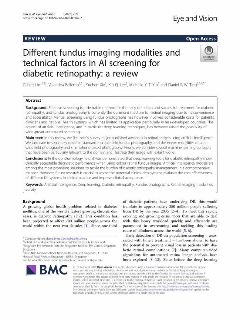
Main textRetina fundus imaging modalitiesFundus imaging is an established modality for retinalimaging, and the detection of DR from fundus imageshas a long and rich history in retinal analysis [44]. Fun-dus imaging is defined as the process whereby reflectedlight is used to form a two dimensional representation ofthe three dimensional retina, the semi-transparent, lay-ered tissue lining the interior of the eye projected ontoan imaging plane [45]. Figure 1 shows different levels ofDR severity from retinal colour fundus images and Fig. 2
Lim et al. Eye and Vision (2020) 7:21 Page 2 of 13
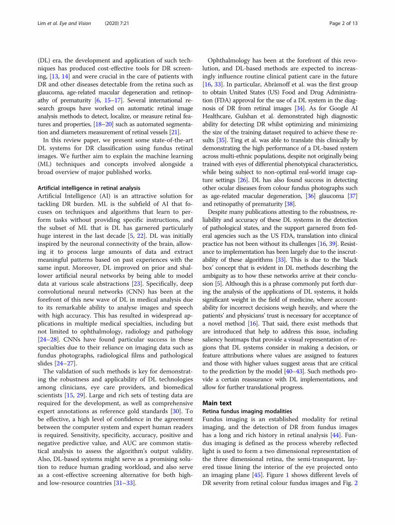
provides a comparison of retinal photographs obtainedfrom different types of devices and capturing views.Table 1 summarises the major publications in retinalanalysis using DL, separately describing standardmultiple-field colour fundus photography, and the newersub-modalities of ultra-wide field photography and
smartphone-based photography. The approaches usedfor the various studies are also included in the table.
Standard viewStandard colour fundus photography provides a 30 to50-degree image which includes the macula and optic
Fig. 1 Examples of retinal fundus images
Fig. 2 Comparison of standard view and ultra-wide field retinal images with and without referable diabetic retinopathy
Lim et al. Eye and Vision (2020) 7:21 Page 3 of 13
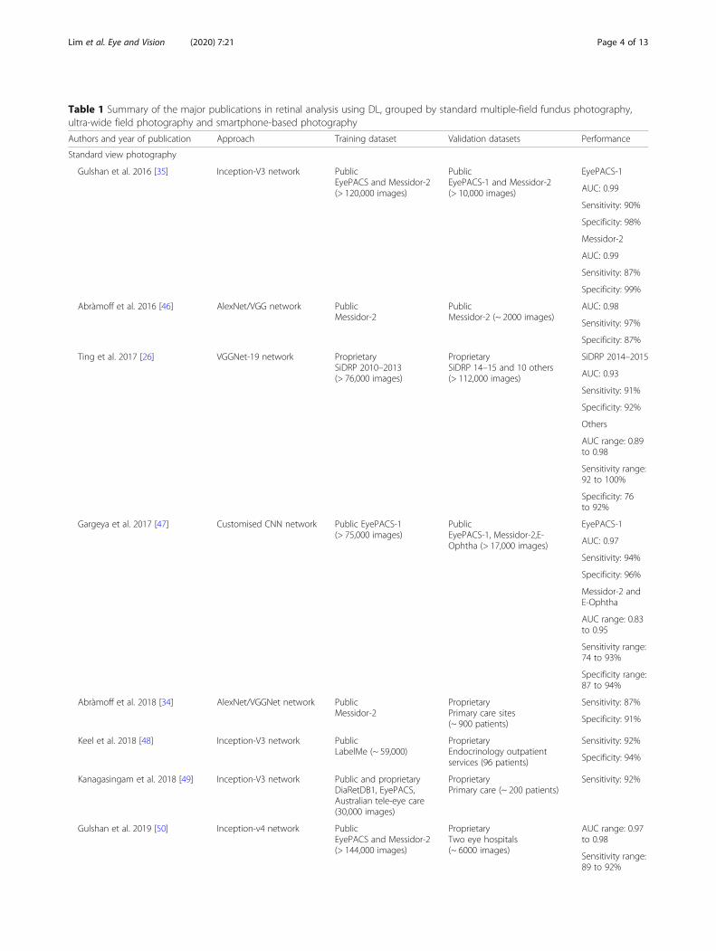
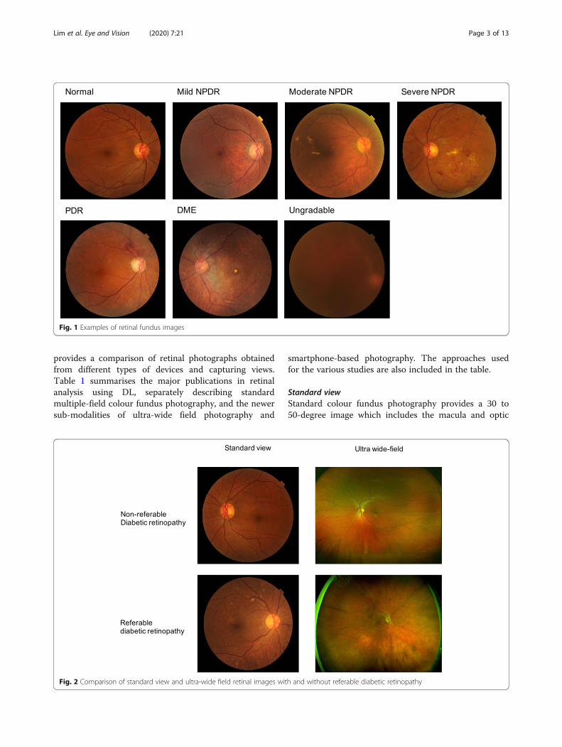
Table 1 Summary of the major publications in retinal analysis using DL, grouped by standard multiple-field fundus photography,ultra-wide field photography and smartphone-based photography
Authors and year of publication Approach Training dataset Validation datasets Performance
Standard view photography
Gulshan et al. 2016 [35] Inception-V3 network PublicEyePACS and Messidor-2(> 120,000 images)
PublicEyePACS-1 and Messidor-2(> 10,000 images)
EyePACS-1
AUC: 0.99
Sensitivity: 90%
Specificity: 98%
Messidor-2
AUC: 0.99
Sensitivity: 87%
Specificity: 99%
Abràmoff et al. 2016 [46] AlexNet/VGG network PublicMessidor-2
PublicMessidor-2 (~ 2000 images)
AUC: 0.98
Sensitivity: 97%
Specificity: 87%
Ting et al. 2017 [26] VGGNet-19 network ProprietarySiDRP 2010–2013(> 76,000 images)
ProprietarySiDRP 14–15 and 10 others(> 112,000 images)
SiDRP 2014–2015
AUC: 0.93
Sensitivity: 91%
Specificity: 92%
Others
AUC range: 0.89to 0.98
Sensitivity range:92 to 100%
Specificity: 76to 92%
Gargeya et al. 2017 [47] Customised CNN network Public EyePACS-1(> 75,000 images)
PublicEyePACS-1, Messidor-2,E-Ophtha (> 17,000 images)
EyePACS-1
AUC: 0.97
Sensitivity: 94%
Specificity: 96%
Messidor-2 andE-Ophtha
AUC range: 0.83to 0.95
Sensitivity range:74 to 93%
Specificity range:87 to 94%
Abràmoff et al. 2018 [34] AlexNet/VGGNet network PublicMessidor-2
ProprietaryPrimary care sites(~ 900 patients)
Sensitivity: 87%
Specificity: 91%
Keel et al. 2018 [48] Inception-V3 network PublicLabelMe (~ 59,000)
ProprietaryEndocrinology outpatientservices (96 patients)
Sensitivity: 92%
Specificity: 94%
Kanagasingam et al. 2018 [49] Inception-V3 network Public and proprietaryDiaRetDB1, EyePACS,Australian tele-eye care(30,000 images)
ProprietaryPrimary care (~ 200 patients)
Sensitivity: 92%
Gulshan et al. 2019 [50] Inception-v4 network PublicEyePACS and Messidor-2(> 144,000 images)
ProprietaryTwo eye hospitals(~ 6000 images)
AUC range: 0.97to 0.98
Sensitivity range:89 to 92%
Lim et al. Eye and Vision (2020) 7:21 Page 4 of 13
nerve. It is widely used in clinical and trial settings as itprovides relatively good documentation of DR. Multipleimages can be manually overlapped to create a montagefor example, 7 standard 30 degree colour fundus imagesmay be combined to produce a 75 degree horizontalfield of view [58]. With the addition of mydriasis, theproportion of ungradable photographs may be reducedfrom 26 to 5% (p < 0.001) [59].AI systems have generally been shown to be able to
accurately detect DR from colour fundus photographs.During the early development and validation of thescreening performance of DL systems, most scientificgroups evaluated their CNN performances in developedcountries, mostly on the United States population [35,46, 47]. In 2016, Abràmoff et al. developed and en-hanced a DL system which achieved a AUC of 0.98 andan achievable sensitivity and specificity of 96.8 and87.0% in detecting referable DR (defined as moderatenon-proliferative DR or worse, including diabetic
macular oedema) on a publicly available colour fundusdataset (Messidor-2) [46]. Gulshan et al. also reportedpromising diagnostic performances of their DL systemwith an AUC of 0.99, and an achievable sensitivity andspecificity of above 96 and 93%, respectively, on two pub-licly available colour fundus datasets (EyePACS-1 andMessidor-2) [35]. Several other notable studies were con-ducted in the same year, as awareness of the promisingabilities of DL in DR screening aroused the interest of thevision science and medical research communities [60–62].In 2017, Gargeya and Leng customized a CNN model
that achieved an AUC of 0.97 with 94% sensitivity and98% specificity, on five-fold cross-validation using theEyePACS dataset [47]. They further tested it on two ex-ternal datasets, achieving AUC scores of 0.94 and 0.95,respectively. Ting et al. then evaluated the performanceof their DL system in detecting DR, using colour fundusimages collected from a Singaporean national DRscreening program, and achieved an AUC of 0.94 with
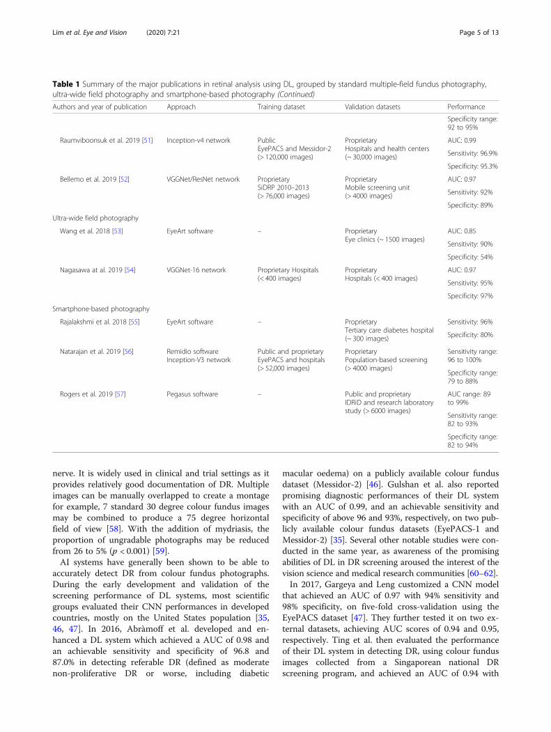
Table 1 Summary of the major publications in retinal analysis using DL, grouped by standard multiple-field fundus photography,ultra-wide field photography and smartphone-based photography (Continued)
Authors and year of publication Approach Training dataset Validation datasets Performance
Specificity range:92 to 95%
Raumviboonsuk et al. 2019 [51] Inception-v4 network PublicEyePACS and Messidor-2(> 120,000 images)
ProprietaryHospitals and health centers(~ 30,000 images)
AUC: 0.99
Sensitivity: 96.9%
Specificity: 95.3%
Bellemo et al. 2019 [52] VGGNet/ResNet network ProprietarySiDRP 2010–2013(> 76,000 images)
ProprietaryMobile screening unit(> 4000 images)
AUC: 0.97
Sensitivity: 92%
Specificity: 89%
Ultra-wide field photography
Wang et al. 2018 [53] EyeArt software – ProprietaryEye clinics (~ 1500 images)
AUC: 0.85
Sensitivity: 90%
Specificity: 54%
Nagasawa at al. 2019 [54] VGGNet-16 network Proprietary Hospitals(< 400 images)
ProprietaryHospitals (< 400 images)
AUC: 0.97
Sensitivity: 95%
Specificity: 97%
Smartphone-based photography
Rajalakshmi et al. 2018 [55] EyeArt software – ProprietaryTertiary care diabetes hospital(~ 300 images)
Sensitivity: 96%
Specificity: 80%
Natarajan et al. 2019 [56] Remidio softwareInception-V3 network
Public and proprietaryEyePACS and hospitals(> 52,000 images)
ProprietaryPopulation-based screening(> 4000 images)
Sensitivity range:96 to 100%
Specificity range:79 to 88%
Rogers et al. 2019 [57] Pegasus software – Public and proprietaryIDRiD and research laboratorystudy (> 6000 images)
AUC range: 89to 99%
Sensitivity range:82 to 93%
Specificity range:82 to 94%
Lim et al. Eye and Vision (2020) 7:21 Page 5 of 13
an achievable sensitivity and specificity of 91 and 92%[26]. They further validated the system on 10 additionalmulti-ethnic multi-cohort multi-settings datasets withdiabetes and achieved AUCs ranging from 0.89 to 0.98.Concurrently, interest in DL continued to grow, withmany noteworthy studies published [53, 63–68].In 2018, IDX-DR software utilizing Alex/VGGNet fea-
tures was validated with an external dataset [69] thatwas also approved for use by the US FDA, [34] havingreported a sensitivity of 91% and specificity of 87% in areal-world clinical setting. Other pilot studies have alsoshown the applicability of such technologies in real-world settings and primary care [48, 49, 70].There has thus been much sustained interest regarding
the application of DL systems for DR. [71–76] The mostnotable research direction in 2019 was arguably towardsassessing the transferability of AI to other less-exploredsettings, particularly in developing countries. The GoogleAI group extended their works to Thailand and India.Ruamviboonsuk et al. reported promising sensitivity andspecificity of 97 and 96%, respectively, (AUC of 0.99) in anational screening program from local hospitals andhealth in Thailand [51]. In India, their DL system achieveda sensitivity and specificity of 89 and 92%, respectively,(AUC of 0.96) on data from the Aravind Eye Hospital, and92 and 95%, respectively, (AUC of 0.98) on data from San-kara Nethralaya [50]. Bellemo et al. reported a promisingsensitivity and specificity (92 and 89%, respectively, withAUC of 0.97) for diagnosis in Zambia, a low middle-income African country [52]. In all the above developingcountries, the DL systems’ performance was either super-ior or comparable to that of human graders. This mightprovide an impetus for other countries of similar incomelevels to adopt DL systems for their routine national DRscreening programmes [75].Another notable trend has been the use of a DL system
as an assistive tool for human graders. Sayres et al. investi-gated the use of heat maps generated by a DL system as aguidance system for human graders, which led to a signifi-cant improvement in diagnostic accuracy as compared tounassisted humans [77]. Keel et al. investigated a method tovisualize the areas where their DL system focused indiagnosing DR. [78] Other applications concern the predic-tion of cardiovascular risk factors from colour fundus im-ages, as well as the estimation of DR prevalence [79, 80]. Inaddition, a promising field that might be explored is the useof DL for the generation of synthetic retinal images to over-come legal concerns and low disease prevalence [81].
Ultra-wide fieldUltra-wide field imaging allows examination of not onlythe central retinal area but also the peripheral zones, forup to a 200-degree view of the retina [82]; more than80% of the total retinal surface can be captured in a
single image. With its wide coverage, ultra-wide field im-aging is able to detect predominantly peripheral lesionsin eyes with DR, with more than 50% of the graded le-sions present outside the seven standard Early Treat-ment Diabetic Retinopathy Study fields [83, 84]. Thepresence and increasing extent of predominantly per-ipheral lesions have been associated with an increasedrisk of DR progression. Therefore, the automated ana-lysis of ultra-wide field images could be of value inDR screening, given the prognostic importance ofperipheral lesions in predicting the progression to ad-vanced disease [84].In 2017, Levenkova et al. developed an algorithm for the
automatic recognition of DR features, including bright(cotton wool spots and exudates) and dark lesions (microa-neurysms and blot, dot and flame haemorrhages) in ultra-wide field images [85]. The algorithm extracted DR fea-tures from grayscale and colour-composite UWF images,including intensity, histogram-of-gradient and local binarypatterns. The best AUCs for bright and dark lesions are 94and 95%, respectively, achieved by a Support Vector Ma-chine classifier. Wang et al. also evaluated performance ofan automated AI algorithm for detecting referable DR, with92%/90% sensitivity with 50%/54% specificity achieved fordetecting referral-warranted retinopathy at the patient andeye levels, respectively [53]. More recently in 2019, Naga-sawa et al. used ultra-wide field fundus images to detecttreatment-naïve proliferative DR. Utilizing 378 photo-graphic images to train the DL model, a high AUC of 0.97with promising sensitivity of 94.7% and specificity of 97.2%was achieved [54].
Smartphone-basedEven though fundus cameras are commonly used indeveloped regions for DR screening, due to the high costof equipment and lack of adequate number of trainedophthalmic technicians, deployment in rural areas withmedically underserved patient populations remains lim-ited [86]. In recent years, several solutions incorporatingadditional lens elements to smartphone cameras havebeen developed to provide affordable solutions andscalable approaches to widespread care.In 2013, Prasanna et al. developed a smartphone-based
decision support system attached to a handheld ophthal-moscope, for screening DR using sophisticated imageanalysis and ML techniques. It achieved an average sen-sitivity of 86% [87]. After a preliminary study [88], Raja-lakshmi et al. assessed the role of an AI system fordetection of DR and sight-threatening DR by colour fun-dus photography taken using smartphone-based retinalimaging system in 2018, and validated it against gradingby ophthalmologists [55]. The AI system achieved 96%sensitivity and 80% specificity in detecting any DR, and99% sensitivity and 80% specificity in detecting sight-
Lim et al. Eye and Vision (2020) 7:21 Page 6 of 13
threatening DR with a kappa agreement of 0.78 and0.75, respectively. In 2019, Wei et al. presented a real-time implementation of CNNs as a smartphone app toprovide a low-cost alternative to fundus camerasequipped with lenses [89]. Natarajan et al. also evaluatedthe performance of another offline, smartphone-basedAI system, for the detection of referable DR by using theimages taken by the same smartphone-based retinal im-aging system on different patient groups [56]. The sensi-tivity and specificity in diagnosing referable DR were 100and 88%, respectively, and in diagnosing any DR were 85and 92%, respectively, compared with ophthalmologistgrading. Finally, Rogers et al. evaluated the performanceof an AI system from images captured by a handheldportable fundus camera collected during a real-worldclinical practice. Validation on the detection of prolifera-tive DR resulted in an AUC of 0.92, with an AUC of0.90 for referable DR. [57]
Machine Learning Techniques & ConceptsState-of-the-art DL systems for DR classification generallymay be understood in terms of the ML techniques andconcepts involved. In particular, contributions by differentgroups may be analysed according to the choices madepertaining to each technique/concept. Here, we provide abroad overview of common techniques/concepts, and thetrade-offs and considerations involved.
Model architectureThe DL model architecture is a major design choice, asthe evidence on natural images strongly suggests thatthe model architecture used affects the classificationperformance level that may be attained, on the sametraining and validation data [35]. There has been
constant innovation in terms of general-purpose end-to-end deep network architectures in recent years [90],with some notable examples being LeNet, AlexNet,VGGNet, Inception, ResNet, DenseNet and SENet,roughly in chronological order of publication (Table 2).However, for the medical imaging domain in particular,
the declared performance of these architectures on large-scale natural image classification may not always be themost relevant, due to other considerations. For one, therelatively small quantity of medical image data availablemay lead to overtraining and/or difficulties with trainingto convergence, with more-sophisticated and higher-capacity models. As such, other than the careful applica-tion of transfer learning (covered later), older and simplerarchitectures may sometimes be favoured for particularapplications. For example, the VGGNet architecture re-mains exceptionally suited for the extraction of intermedi-ate features [91], while requiring relatively more weightparameters than other popular architectures [90].Moreover, end-to-end classification is not the only para-
digm for DL in DR screening. For instance, a hybridapproach would be to deploy DL models as low-level de-tectors that directly target various classes of lesions. Limet al. trained models similar to LeNet on spatially-transformed representations of candidate lesions proposedby a maximally-stable extremal region detector, [10] whileAbràmoff et al.’s IDx-DR X2.1 used models inspired byAlexNet and VGGNet [46]. In these cases, the projectednumber and location of true lesions can either be directlymatched against clinical reference standards, or the de-tector output vectors may be used as the input to a fusionalgorithm that perfoms the final image-level classification.Another notable consideration for model architectures
would be the amount of computing resources required,
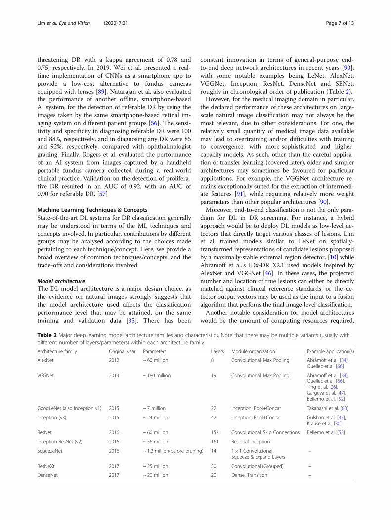
Table 2 Major deep learning model architecture families and characteristics. Note that there may be multiple variants (usually withdifferent number of layers/parameters) within each architecture family
Architecture family Original year Parameters Layers Module organization Example application(s)
AlexNet 2012 ~ 60 million 8 Convolutional, Max Pooling Abràmoff et al. [34],Quellec et al. [66]
VGGNet 2014 ~ 180 million 19 Convolutional, Max Pooling Abràmoff et al. [34],Quellec et al. [66],Ting et al. [26],Gargeya et al. [47],Bellemo et al. [52]
GoogLeNet (also Inception v1) 2015 ~ 7 million 22 Inception, Pool+Concat Takahashi et al. [63]
Inception (v3) 2015 ~ 24 million 42 Inception, Pool+Concat Gulshan et al. [35],Krause et al. [30]
ResNet 2016 ~ 60 million 152 Convolutional, Skip Connections Bellemo et al. [52]
Inception-ResNet (v2) 2016 ~ 56 million 164 Residual Inception –
SqueezeNet 2016 ~ 1.2 million(before pruning) 14 1 × 1 Convolutional,Squeeze & Expand Layers
–
ResNeXt 2017 ~ 25 million 50 Convolutional (Grouped) –
DenseNet 2017 ~ 20 million 201 Dense, Transition –
Lim et al. Eye and Vision (2020) 7:21 Page 7 of 13
which is relevant for deployment on consumer devicessuch as smartphones, embedded systems, and on possiblyless-powerful hardware in under-resourced regions. Ingeneral, the fewer the number of weight parameters in-volved in the model architecture, the quicker the infer-ence, ceteris paribus. If the inference time is sufficientlyquick, real-time analysis further becomes possible [92]. Tothis end, lightweight model architectures such as Mobile-Net [93] and ShuffleNet [94] have been designed for de-vices with limited computing power. Alternatively, modelcompression through pruning and parameter quantizationmay be done [95]. Given the medical implications of DRscreening, however, any such trade-offs of performancefor speed may need to be carefully considered.
EnsemblingEnsembling involves the combination of multiple inde-pendent ML classifier models, to produce a final classifiermodel that generally performs better than any of its con-stituent models. With DL models, ensembling is com-monly and easily implemented by training multiplemodels – not necessarily of the same network architectureor inputs – separately, and then combining the outputs ofthese models during inference. Although regularizationtechniques such as dropout may be utilized during modeltraining as an approximation to ensembling [96], modelstrained in this way nonetheless yield further performancegains when ensembled, in practice.
The number of models involved in the final ensemble is atrade-off between training/inference time and performance.Generally, the larger the number of independent modelsused, the better the performance, but with diminishingreturns. For example, Gulshan et al. used an ensemble often Inception-v3 models [35], Ting et al. used an ensembleof two VGGNet-based models, although with differentlypre-processed inputs [26], which was further extended witha ResNet model in Bellemo et al. [52]Various methods have been employed for integrating
the individual model outputs within an ensemble. Perhapsthe most straightforward would be to take a linear averageover these predictions, as was done for Gulshan et al. [35]and Ting et al. [26] More complex possibilities would in-clude weighted ensembles [25] and the training of a fur-ther classifier model over the ensemble output values.
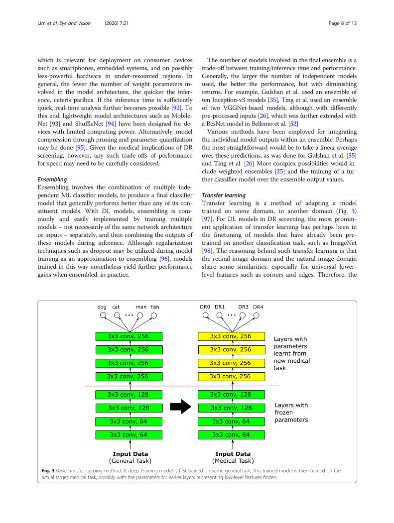
Transfer learningTransfer learning is a method of adapting a modeltrained on some domain, to another domain (Fig. 3)[97]. For DL models in DR screening, the most promin-ent application of transfer learning has perhaps been inthe finetuning of models that have already been pre-trained on another classification task, such as ImageNet[98]. The reasoning behind such transfer learning is thatthe retinal image domain and the natural image domainshare some similarities, especially for universal lower-level features such as corners and edges. Therefore, the
Fig. 3 Basic transfer learning method. A deep learning model is first trained on some general task. This trained model is then trained on theactual target medical task, possibly with the parameters for earlier layers representing low-level features frozen
Lim et al. Eye and Vision (2020) 7:21 Page 8 of 13
parameter weights from a natural image classificationtask should then serve as a good initialization for retinalimage classification.A major consideration for transfer learning with pre-
trained weights would be the policy by which thesepretrained weights are finetuned with new retinal data. Onepossible choice would be to consider the pretrained weightsmerely as an initialization and proceed with training as pernormal, allowing all weight values to be updated. At theother extreme, all pretrained weights are fixed, and the pre-trained model is effectively employed as a feature extractorwith only the output layer replaced, possibly by anotherclassifier such as a random forest [47] or support vectormachine [99]. Otherwise, the weights of any number oflayers within the model architecture may be fixed, with theremainder updated; if so, it is generally the layers corre-sponding to lower-level features that are fixed. A previoussurvey on transfer learning in the medical domain by Taj-bakhsh et al. suggests that although the use of pretrainedweights made DL models more robust to the size of train-ing sets, the optimal selection of layers to fix depends onthe task at hand and has to be empirically determined [98].
Weakly supervised and active learningA commonly encountered obstacle to training DL modelsfor DR classification is a lack of annotated image data, par-ticularly at the lesion level, since such detailed annotationwas not typically required in clinical screening workflows.This made gathering sufficient lesion-level ground truthfor hybrid DL implementations challenging. Althoughcoarse-grained image-level grades were more widely avail-able, it remained common to have large quantities of un-labelled retinal images for which no grades from humanexperts were available [100].In such situations, weakly-supervised transductive
learning becomes applicable. In transductive learning, aninitial model trained on the labelled training data is usedto classify the unlabelled training data. The originally-unlabelled training data now also becomes labelled, andmay be used together with the originally-labelled train-ing data to train an improved bootstrapped model [101].Whether or not such transductive learning is employed, it
is advisable to continually refine the trained model throughactive learning. Active learning presumes the presence of anoracle that can provide accurate answers to queries, whichin the case of DR screening would be a human expert. How-ever, there is an opportunity cost to consulting the oracle.As such, the goal of active learning is to intelligently selectthe most useful images for which to consult the oracle on,in the sense that the availability of accurate labels for theseimages would improve model performance to the greatestextent. One possible approach would be to select images forwhich the model is most uncertain [75].
Label ModellingAnother manifestation of weakly-supervised learning isthe presence of imperfect or noisy labels. The presence ofsuch imperfect labels is largely unavoidable in DR screen-ing, with qualified human graders sometimes disagreeingwith each other – or even themselves, from a previous ses-sion. Inter-grader kappa scores typically range from 0.40to 0.65 in DR grading [102], and the implied disagreementmay be resolved by majority decision, discussion betweenthe graders, or external adjudication. Krause et al. con-clude that rigorous adjudication of DR ground truth is im-portant in developing DR models, since it allows for theprincipled correction of subtle errors from image artefactsand missed microaneurysms [30].A further development by Guan et al. has been the
modelling of individual graders with independent DLmodels, following the observation that the labelling oflarge DR datasets usually involves a large number ofhuman graders, each of whom however grade only arelatively small subset of the dataset, with each imagemoreover also being graded by only a small subset of thehuman graders [102]. They found that modelling eachhuman grader separately and averaging the predictionsof these separate DL models in a weighted ensembleproduced better performance than modelling the ex-pected prediction of the average grader.
Joint LearningDR may co-occur with other related eye diseases, andthere is as such motivation to model its features to-gether with those of other eye diseases. This joint ormultitask learning involves training a DL model formultiple tasks simultaneously, and may induce benefi-cial regularization of intermediate representations, thusreducing overfitting [103]. González-Gonzalo et al.attempted the joint learning of referable DR and age-related macular degeneration, and concluded that ajointly-trained DL model could perform comparably tohuman graders [104].Joint learning may also be implemented for improving
mid-level representations, in terms of optimizing for vis-ual encodings and the final binary classifier at the sametime, for multiple-instance learning [105]. This multiple-instance learning framework also allows for a degree ofmodel interpretability by allowing the class of encodinginstances to be explicitly considered during training. Inthis case, two neural networks are utilized to generatethe mid-level representation encodings.
Hyperparameter search & optimizationOther than the model weight parameters themselves, DLmodels involve a large number of hyperparameters, suchas the initial learning rate, the learning rate decay sched-ule, the input batch size, etc. For DR screening
Lim et al. Eye and Vision (2020) 7:21 Page 9 of 13
applications, these hyperparameter settings are oftenborrowed directly from existing models, and whetherthese settings are the most appropriate for the DRscreening domain may not be systematically explored.Sahlsten et al. is an example of work that investigatesthe image resolution parameter in detail [106].The optimization of multiple hyperparameters is non-
trivial, due to the number of hyperparameter combina-tions increasing exponentially with the number of indi-vidual hyperparameters. Although grid search over thehyperparameter space is commonly attempted, when thenumber of relevant hyperparameters is relatively small,random search [107] and sequential optimization algo-rithms [108] may also be attempted to more thoroughlyexamine possible model performance.
RobustnessAlthough DL models may be trained and validated onlarge datasets, it is difficult to be certain whether the data-sets used can fully capture the potential variability of ret-inal images that may be encountered in future use.Differences may arise in the image acquisition process orpopulation demographics that can render a trained DL
model less effective on new data. Lim et al. demonstratedthat the uncertainty of a DL model could be estimated bythe standard deviation and entropy of the mean predictivedistribution, on the stochastic batch normalization layersof a ResNet architecture, and that prediction error is cor-related with high estimated uncertainty [75].
ExplainabilityA persistent obstacle against the uptake of AI systems inDR screening has been a lack of surface explainability[16]. In fact, the progression from handcrafted featuresand multi-stage classification to end-to-end deep learn-ing has been accompanied by a concurrent loss of inter-pretability, in that humans could no longer examine thereasoning of the classifier, unlike previously where animage kernel could be inspected to determine why it hadnot matched with a microaneurysm, for instance.This lack of interpretability has been mitigated some-
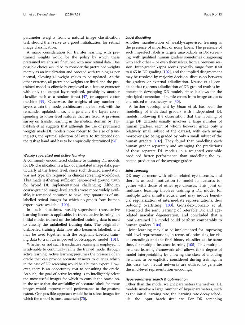
what through the development of various methods toextract saliency heatmaps from DL models, such asGrad-CAM [42] and integrated gradients [43]. These sa-liency heatmaps attempt to display the contribution ofeach image pixel or region to the final classification. This
Fig. 4 AI flow for diabetic retinopathy. In the diabetic retinopathy screening domain, the AI implementation allows automated diagnosis andsubsequent clinical decisions. In the example presented in the figure, the AI system would recommend referring the patient to the eye clinicbecause of the referable diagnosis for diabetic retinopathy. To allow researchers and clinicians determine how the AI model makes the decision,the heatmap attempts to display the contribution of each image pixel or region, to the final classification. Heatmaps open the ‘black box’highlighting the areas in which the AI system is focusing on to build trust among practitioners and patients. Abbreviations: DR; diabeticretinopathy; NPDR: non-proliferative diabetic retinopathy; PDR: proliferative diabetic retinopathy
Lim et al. Eye and Vision (2020) 7:21 Page 10 of 13
allows researchers to retrospectively determine whethertheir DL models are making their decisions based on theexpected image features, which in the DR screening do-main would be various lesions such as microaneurysms,haemorrhages and hard exudates (Fig. 4).A desire for greater interpretability has also seen
renewed interest in hybrid methods that expose theintermediate goals of the classifier [109]. For example,Yang et al. implemented a two-stage DL model, whichfirst classifies overlapping grid patches as containinglesions or not. The resulting weighted lesion map isthen used as input to a second global DL model, topredict the image-level DR severity [110]. Wang et al.introduced a Zoom-in-Net architecture that purportsto mimic the attentional behaviour of human graders,by allowing for suspicious regions to be focused onthrough additional learning on feature maps from themain network [111].
ConclusionsIn this paper, we provided a broad overview of the majorworks and technical implementations involving DL tech-niques for DR diagnosis as an alternative tool for screen-ing programmes. It emerged that, in the ophthalmologyfield, DL tools for DR show clinically acceptable diagnosticperformance when using colour retinal fundus images.DL-based AI models are among the most promising solu-tions to tackle the burden of DR management in a com-prehensive manner. However, future research is crucial toassess the potential clinical deployment, evaluate the cost-effectiveness of different DL systems in the clinical prac-tice and improve clinical acceptance.
AbbreviationsDR: Diabetic retinopathy; DL: Deep learning; AI: Artificial intelligence;CNN: Convolutional neural network; AUC: Area under the receiver operatingcharacteristic curve
AcknowledgementsNot applicable.
Authors' contributionsGL, VB, and DT contributed to the initial drafting of the manuscript. GL, VB,YX, XQ, MY and DT contributed to the critical review and final approval forthis manuscript.
FundingFunding from Research Grants Council-General Research Fund, Hong Kong (Ref:14102418); National Medical Research Council Health Service Research Grant,Large Collaborative Grant, Ministry of Health, Singapore; the SingHealthFoundation; and the Tanoto Foundation.
Availability for data and materialsNot applicable.
Ethics approval and consent to participateNot applicable.
Consent for publicationNot applicable.
Competing interestsDrs Daniel S.W. Ting and Gilbert Lim are co-inventors of a deep learning sys-tem for retinal diseases.
Author details1School of Computing, National University of Singapore, Singapore,Singapore. 2Singapore Eye Research Institute, Singapore National Eye Centre,Singapore, Singapore. 3Duke-NUS Medical School, National University ofSingapore, 11 Third Hospital Road Avenue, Singapore 168751, Singapore.4Vitreo-Retinal Service, Singapore National Eye Center, 11 Third Hospital RoadAvenue, Singapore 168751, Singapore. 5Artificial Intelligence inOphthalmology, Singapore Eye Research Institute, 11 Third Hospital RoadAvenue, Singapore 168751, Singapore.
Received: 12 December 2019 Accepted: 10 March 2020
References1. Moss SE, Klein R, Klein BE. The 14-year incidence of visual loss in a diabetic
population. Ophthalmology. 1998;105(6):998–1003.2. Yau JW, Rogers SL, Kawasaki R, Lamoureux EL, Kowalski JW, Bek T, et al.
Global prevalence and major risk factors of diabetic retinopathy. DiabetesCare. 2012;35(3):556–64.
3. Cheung N, Mitchell P, Wong TY. Diabetic retinopathy. Lancet. 2010;376(9735):124–36.
4. Ting DSW, Cheung GCM, Wong TY. Diabetic retinopathy: global prevalence,major risk factors, screening practices and public health challenges: areview. Clin Exp Ophthalmol. 2016;44(4):260–77.
5. Fogel AL, Kvedar JC. Artificial intelligence powers digital medicine. NPJ DigitMed. 2018;1:5.
6. Wong TY, Bressler NM. Artificial intelligence with deep learning technologylooks into diabetic retinopathy screening. JAMA. 2016;316(22):2366–7.
7. Group ETDRSR. Early photocoagulation for diabetic retinopathy: ETDRSreport number 9. Ophthalmology. 1991;98(Suppl 5):766–85.
8. Abràmoff MD, Niemeijer M, Suttorp-Schulten MS, Viergever MA, Russell SR,Van Ginneken B. Evaluation of a system for automatic detection of diabeticretinopathy from color fundus photographs in a large population ofpatients with diabetes. Diabetes Care. 2008;31(2):193–8.
9. Peto T, Tadros C. Screening for diabetic retinopathy and diabetic macularedema in the United Kingdom. Curr Diab Rep. 2012;12(4):338–45.
10. Lim G, Lee ML, Hsu W, Wong TY. Transformed representations forconvolutional neural networks in diabetic retinopathy screening. In: AAAIWorkshop: Modern Artificial Intelligence for Health Analytics. Quebec, 2014.pp. 21–5.
11. Lachure J, Deorankar A, Lachure S, Gupta S, Jadhav R. Diabetic retinopathyusing morphological operations and machine learning. In: 2015 IEEEInternational Advance Computing Conference (IACC). Banglore, 2015. p.617–22.
12. Prasad DK, Vibha L, Venugopal KR. Early detection of diabetic retinopathyfrom digital retinal fundus images. In: 2015 IEEE Recent Advances inIntelligent Computational Systems (RAICS). Trivandrum: IEEE; 2015. p. 240–5.
13. Scotland GS, McNamee P, Philip S, Fleming AD, Goatman KA, Prescott GJ,et al. Cost-effectiveness of implementing automated grading within thenational screening programme for diabetic retinopathy in Scotland. Br JOphthalmol. 2007;91(11):1518–23.
14. Scotland GS, McNamee P, Fleming AD, Goatman KA, Philip S, Prescott GJ,et al. Costs and consequences of automated algorithms versus manualgrading for the detection of referable diabetic retinopathy. Br J Ophthalmol.2010;94(6):712–9.
15. Trucco E, Ruggeri A, Karnowski T, Giancardo L, Chaum E, Hubschman JP,et al. Validating retinal fundus image analysis algorithms: issues and aproposal. Invest Ophthalmol Vis Sci. 2013;54(5):3546–59.
16. Ting DSW, Pasquale LR, Peng L, Campbell JP, Lee AY, Raman R, et al.Artificial intelligence and deep learning in ophthalmology. Br J Ophthalmol.2019;103(2):167–75.
17. Cheung CY, Tang F, Ting DSW, Tan GSW, Wong TY. Artificialintelligence in diabetic eye disease screening. Asia Pac J Ophthalmol(Phila). 2019;8(2):158–64.
18. Sinthanayothin C, Boyce JF, Williamson TH, Cook HL, Mensah E, Lal S, et al.Automated detection of diabetic retinopathy on digital fundus images.Diabet Med. 2002;19(2):105–12.
Lim et al. Eye and Vision (2020) 7:21 Page 11 of 13
19. Usher D, Dumskyj M, Himaga M, Williamson TH, Nussey S, Boyce J.Automated detection of diabetic retinopathy in digital retinal images: a toolfor diabetic retinopathy screening. Diabet Med. 2004;21(1):84–90.
20. Niemeijer M, Van Ginneken B, Staal J, Suttorp-Schulten MS, Abràmoff MD.Automatic detection of red lesions in digital color fundus photographs. IEEETrans Med Imaging. 2005;24(5):584–92.
21. Staal J, Abràmoff MD, Niemeijer M, Viergever MA, Van Ginneken B. Ridge-based vessel segmentation in color images of the retina. IEEE Trans MedImaging. 2004;23(4):501–9.
22. Lee A, Taylor P, Kalpathy-Cramer J, Tufail A. Machine learning has arrived!Ophthalmology. 2017;124(12):1726–8.
23. LeCun Y, Bengio Y, Hinton G. Deep learning. Nature. 2015;521(7553):436–44.24. Bejnordi BE, Veta M, Van Diest PJ, Van Ginneken B, Karssemeijer N, Litjens G,
et al. Diagnostic assessment of deep learning algorithms for detection oflymph node metastases in women with breast cancer. JAMA. 2017;318(22):2199–210.
25. Lakhani P, Sundaram B. Deep learning at chest radiography: automatedclassification of pulmonary tuberculosis by using convolutional neuralnetworks. Radiology. 2017;284(2):574–82.
26. Ting DSW, Cheung CY, Lim G, Tan GSW, Quang ND, Gan A, et al.Development and validation of a deep learning system for diabeticretinopathy and related eye diseases using retinal images from multiethnicpopulations with diabetes. JAMA. 2017;318(22):2211–23.
27. Ting DSW, Yi PH, Hui F. Clinical applicability of deep learning system indetecting tuberculosis with chest radiography. Radiology. 2018;286(2):729–31.
28. Esteva A, Robicquet A, Ramsundar B, Kuleshov V, DePristo M, Chou K, et al.A guide to deep learning in healthcare. Nat Med. 2019;25(1):24–9.
29. Kapetanakis VV, Rudnicka AR, Liew G, Owen CG, Lee A, Louw V, et al. Astudy of whether automated diabetic retinopathy image assessment couldreplace manual grading steps in the English National ScreeningProgramme. J Med Screen. 2015;22(3):112–8.
30. Krause J, Gulshan V, Rahimy E, Karth P, Widner K, Corrado GS, et al. Gradervariability and the importance of reference standards for evaluatingmachine learning models for diabetic retinopathy. Ophthalmology. 2018;125(8):1264–72.
31. Nguyen HV, Tan GSW, Tapp RJ, Mital S, Ting DSW, Wong HT, et al. Cost-effectiveness of a national telemedicine diabetic retinopathy screeningprogram in Singapore. Ophthalmology. 2016;123(12):2571–80.
32. Tufail A, Rudisill C, Egan C, Kapetanakis VV, Salas-Vega S, Owen CG, et al.Automated diabetic retinopathy image assessment software: diagnosticaccuracy and cost-effectiveness compared with human graders.Ophthalmology. 2017;124(3):343–51.
33. Xie Y, Nguyen Q, Bellemo V, Yip MY, Lee XQ, Hamzah H, et al. Cost-Effectiveness Analysis of an Artificial Intelligence-Assisted Deep LearningSystem Implemented in the National Tele-Medicine Diabetic RetinopathyScreening in Singapore. Invest Ophthalmol Vis Sci. 2019;60(9):5471.
34. Abràmoff MD, Lavin PT, Birch M, Shah N, Folk JC. Pivotal trial of anautonomous AI-based diagnostic system for detection of diabeticretinopathy in primary care offices. NPJ Digit Med. 2018;1(1):39.
35. Gulshan V, Peng L, Coram M, Stumpe MC, Wu D, Narayanaswamy A, et al.Development and validation of a deep learning algorithm for detection ofdiabetic retinopathy in retinal fundus photographs. JAMA. 2016;316(22):2402–10.
36. Grassmann F, Mengelkamp J, Brandl C, Harsch S, Zimmermann ME, LinkohrB, et al. A deep learning algorithm for prediction of age-related eye diseasestudy severity scale for age-related macular degeneration from color fundusphotography. Ophthalmology. 2018;125(9):1410–20.
37. Li Z, He Y, Keel S, Meng W, Chang RT, He M. Efficacy of a deep learningsystem for detecting glaucomatous optic neuropathy based on colorfundus photographs. Ophthalmology. 2018;125(8):1199–206.
38. Brown JM, Campbell JP, Beers A, Chang K, Ostmo S, Chan RP, et al.Automated diagnosis of plus disease in retinopathy of prematurity usingdeep convolutional neural networks. JAMA Ophthalmol. 2018;136(7):803–10.
39. Ting DSW, Peng L, Varadarajan AV, Keane PA, Burlina PM, Chiang MF, et al.Deep learning in ophthalmology: the technical and clinical considerations.Prog Retin Eye Res. 2019;72:100759.
40. Ribeiro MT, Singh S, Guestrin C. Why should I trust you?: Explaining thepredictions of any classifier. In: Proceedings of the 2016 Conference ofthe North American Chapter of the Association for ComputationalLinguistics: Demonstrations. San Diego, 2016. p. 97–101.https://doi.org/10.18653/v1/N16-3020.
41. Lundberg SM, Lee SI. A unified approach to interpreting model predictions. In:Advances in Neural Information Processing Systems. Long Beach, 2017. p. 4765–74.
42. Selvaraju RR, Cogswell M, Das A, Vedantam R, Parikh D, Batra D. Grad-cam:visual explanations from deep networks via gradient-based localization. Int JComput Vis. 2020;128:336–59. https://doi.org/10.1007/s11263-019-01228-7 .
43. Sundararajan M, Taly A, Yan Q. Axiomatic attribution for deep networks.arXiv:1703.01365, 2017.
44. Goh JKH, Cheung CY, Sim SS, Tan PC, Tan GSW, Wong TY. Retinal imagingtechniques for diabetic retinopathy screening. J Diabetes Sci Technol. 2016;10(2):282–94.
45. Abràmoff MD, Garvin MK, Sonka M. Retinal imaging and image analysis. IEEERev Biomed Eng. 2010;3:169–208.
46. Abràmoff MD, Lou Y, Erginay A, Clarida W, Amelon R, Folk JC, et al. Improvedautomated detection of diabetic retinopathy on a publicly available dataset throughintegration of deep learning. Invest Ophthalmol Vis Sci. 2016;57(13):5200–6.
47. Gargeya R, Leng T. Automated identification of diabetic retinopathy usingdeep learning. Ophthalmology. 2017;124(7):962–9.
48. Keel S, Lee PY, Scheetz J, Li Z, Kotowicz MA, MacIsaac RJ, et al. Feasibilityand patient acceptability of a novel artificial intelligence-based screeningmodel for diabetic retinopathy at endocrinology outpatient services: a pilotstudy. Sci Rep. 2018;8(1):4330.
49. Kanagasingam Y, Xiao D, Vignarajan J, Preetham A, Tay-Kearney ML,Mehrotra A. Evaluation of artificial intelligence–based grading of diabeticretinopathy in primary care. JAMA Netw Open. 2018;1(5):e182665.
50. Gulshan V, Rajan RP, Widner K, Wu D, Wubbels P, Rhodes T, et al. Performance of adeep-learning algorithm vs manual grading for detecting diabetic retinopathy inIndia. JAMA Ophthalmol. 2019. https://doi.org/10.1001/jamaophthalmol.2019.2004.
51. Raumviboonsuk P, Krause J, Chotcomwongse P, Sayres R, Raman R, Widner K,et al. Deep learning versus human graders for classifying diabetic retinopathyseverity in a nationwide screening program. NPJ Digit Med. 2019;2:25.
52. Bellemo V, Lim ZW, Lim G, Nguyen QD, Xie Y, Yip MY, et al. Artificialintelligence using deep learning to screen for referable and vision-threatening diabetic retinopathy in Africa: a clinical validation study. LancetDigital Health. 2019;1(1):e35–44.
53. Wang K, Jayadev C, Nittala MG, Velaga SB, Ramachandra CA, BhaskaranandM, et al. Automated detection of diabetic retinopathy lesions onultrawidefield pseudocolour images. Acta Ophthalmol. 2018;96(2):e168–73.
54. Nagasawa T, Tabuchi H, Masumoto H, Enno H, Niki M, Ohara Z, et al.Accuracy of ultrawide-field fundus ophthalmoscopy-assisted deep learningfor detecting treatment-naïve proliferative diabetic retinopathy. IntOphthalmol. 2019;39(10):2153–9.
55. Rajalakshmi R, Subashini R, Anjana RM, Mohan V. Automated diabeticretinopathy detection in smartphone-based fundus photography usingartificial intelligence. Eye (Lond). 2018;32(6):1138–44.
56. Natarajan S, Jain A, Krishnan R, Rogye A, Sivaprasad S. Diagnostic accuracyof community-based diabetic retinopathy screening with an offline artificialintelligence system on a smartphone. JAMA Ophthalmol. 2019. https://doi.org/10.1001/jamaophthalmol.2019.2923.
57. Rogers T, Gonzalez-Bueno J, Franco RG, Star EL, Marín DM, Vassallo J, et al.Evaluation of an AI system for the detection of diabetic retinopathy fromimages captured with a handheld portable fundus camera: the MAILOR AIstudy. arXiv preprint arXiv:190806399. 2019.
58. Baumal CR, Duker JS. Current management of diabetic retinopathy: ElsevierHealth Sciences; 2017.
59. Murgatroyd H, Ellingford A, Cox A, Binnie M, Ellis J, MacEwen C, et al. Effectof mydriasis and different field strategies on digital image screening ofdiabetic eye disease. Br J Ophthalmol. 2004;88(7):920–4.
60. Pratt H, Coenen F, Broadbent DM, Harding SP, Zheng Y. Convolutional neuralnetworks for diabetic retinopathy. Procedia Computer Science. 2016;90:200–5.
61. Colas E, Besse A, Orgogozo A, Schmauch B, Meric N, Besse E. Deep learningapproach for diabetic retinopathy screening. Acta Ophthalmol. 2016;94.https://doi.org/10.1111/j.1755-3768.2016.0635 .
62. Doshi D, Shenoy A, Sidhpura D, Gharpure P. Diabetic retinopathy detection usingdeep convolutional neural networks. In: 2016 International Conference onComputing, Analytics and Security Trends (CAST). Pune: IEEE; 2016. p. 261–6.
63. Takahashi H, Tampo H, Arai Y, Inoue Y, Kawashima H. Applying artificialintelligence to disease staging: deep learning for improved staging ofdiabetic retinopathy. PLoS One. 2017;12(6):e0179790.
64. Gegundez-Arias ME, Marin D, Ponte B, Alvarez F, Garrido J, Ortega C, et al. Atool for automated diabetic retinopathy pre-screening based on retinalimage computer analysis. Comput Biol Med. 2017;88:100–9.
Lim et al. Eye and Vision (2020) 7:21 Page 12 of 13
65. Xu K, Feng D, Mi H. Deep convolutional neural network-based earlyautomated detection of diabetic retinopathy using fundus image.Molecules. 2017;22(12). https://doi.org/10.3390/molecules22122054 .
66. Quellec G, Charrière K, Boudi Y, Cochener B, Lamard M. Deep image miningfor diabetic retinopathy screening. Med Image Anal. 2017;39:178–93.
67. Abbas Q, Fondon I, Sarmiento A, Jiménez S, Alemany P. Automaticrecognition of severity level for diagnosis of diabetic retinopathy usingdeep visual features. Med Biol Eng Comput. 2017;55(11):1959–74.
68. Choi JY, Yoo TK, Seo JG, Kwak J, Um TT, Rim TH. Multi-categorical deeplearning neural network to classify retinal images: a pilot study employingsmall database. PLoS One. 2017;12(11):e0187336.
69. Van Der Heijden AA, Abramoff MD, Verbraak F, van Hecke MV, Liem A,Nijpels G. Validation of automated screening for referable diabeticretinopathy with the IDx-DR device in the Hoorn Diabetes Care System.Acta Ophthalmol. 2018;96(1):63–8.
70. Verbraak FD, Abramoff MD, Bausch GC, Klaver C, Nijpels G, Schlingemann RO,et al. Diagnostic accuracy of a device for the automated detection of diabeticretinopathy in a primary care setting. Diabetes Care. 2019;42(4):651–6.
71. de La Torre J, Valls A, Puig D. A deep learning interpretable classifier fordiabetic retinopathy disease grading. Neurocomputing. 2019. In press.https://doi.org/10.1016/j.neucom.2018.07.102.
72. Li T, Gao Y, Wang K, Guo S, Liu H, Kang H. Diagnostic assessment of deeplearning algorithms for diabetic retinopathy screening. Inf Sci. 2019;501:511–22.
73. Liu YP, Li Z, Xu C, Li J, Liang R. Referable diabetic retinopathy identificationfrom eye fundus images with weighted path for convolutional neuralnetwork. Artif Intell Med. 2019;99:101694.
74. Raman R, Srinivasan S, Virmani S, Sivaprasad S, Rao C, Rajalakshmi R. Fundusphotograph-based deep learning algorithms in detecting diabeticretinopathy. Eye (Lond). 2019;33(1):97–109.
75. Lim ZW, Lee ML, Hsu W, Wong TY. Building Trust in Deep Learning Systemtowards automated disease detection. In: Proceedings of the AAAIConference on Artificial Intelligence. 2019. p. 9516-21. https://doi.org/10.1609/aaai.v33i01.33019516.
76. Yip MY, Lim G, Bellemo V, Xie Y, Lee XQ, Nguyen Q, et al. Effect of imagecompression and number of fields on a deep learning system for detectionof diabetic retinopathy. Invest Ophthalmol Vis Sci. 2019;60(9):1438.
77. Sayres R, Taly A, Rahimy E, Blumer K, Coz D, Hammel N, et al. Using a deeplearning algorithm and integrated gradients explanation to assist gradingfor diabetic retinopathy. Ophthalmology. 2019;126(4):552–64.
78. Keel S, Wu J, Lee PY, Scheetz J, He M. Visualizing deep learning models forthe detection of referable diabetic retinopathy and glaucoma. JAMAOphthalmol. 2019;137(3):288–92.
79. Poplin R, Varadarajan AV, Blumer K, Liu Y, McConnell MV, Corrado GS, et al.Prediction of cardiovascular risk factors from retinal fundus photographs viadeep learning. Nat Biomed Eng. 2018;2(3):158–64.
80. Ting DS, Cheung CY, Nguyen Q, Sabanayagam C, Lim G, Lim ZW, et al.Deep learning in estimating prevalence and systemic risk factors fordiabetic retinopathy: a multi-ethnic study. NPJ Digit Med. 2019;2:24.
81. Bellemo V, Burlina P, Yong L, Wong TY, Ting DSW. Generative adversarialnetworks (GANs) for retinal fundus image synthesis. In: Asian Conference onComputer Vision. 2018. p. 28-302.
82. Aiello LP, Odia I, Glassman AR, Melia M, Jampol LM, Bressler NM, et al.Comparison of early treatment diabetic retinopathy study standard 7-fieldimaging with ultrawide-field imaging for determining severity of diabeticretinopathy. JAMA Ophthalmol. 2019;137(1):65–73.
83. Ghasemi Falavarjani K, Wang K, Khadamy J, Sadda SR. Ultra-wide-field imagingin diabetic retinopathy; an overview. J Curr Ophthalmol. 2016;28(2):57–60.
84. Silva PS, Cavallerano JD, Haddad NMN, Kwak H, Dyer KH, Omar AF, et al.Peripheral lesions identified on ultrawide field imaging predict increasedrisk of diabetic retinopathy progression over 4 years. Ophthalmology. 2015;122(5):949–56.
85. Levenkova A, Sowmya A, Kalloniatis M, Ly A, Ho A. Automatic detection ofdiabetic retinopathy features in ultra-wide field retinal images. In: MedicalImaging 2017: Computer-Aided Diagnosis; International Society for Opticsand Photonics. 2017: 101341M. https://doi.org/10.1117/12.2253980.
86. Fenner BJ, Wong RLM, Lam WC, Tan GSW, Cheung GCW. Advances inretinal imaging and applications in diabetic retinopathy screening: a review.Ophthalmol Ther. 2018;7(2):333–46.
87. Prasanna P, Jain S, Bhagat N, Madabhushi A. Decision support system fordetection of diabetic retinopathy using smartphones. In: 2013 7th
International Conference on Pervasive Computing Technologies forHealthcare and Workshops. Venic: IEEE; 2013. p. 176–9.
88. Rajalakshmi R, Arulmalar S, Usha M, Prathiba V, Kareemuddin KS, Anjana RM,et al. Validation of smartphone based retinal photography for diabeticretinopathy screening. PLoS One. 2015;10(9):e0138285.
89. Wei H, Sehgal A, Kehtarnavaz N. A deep learning-based smartphone app forreal-time detection of retinal abnormalities in fundus images. In: Real-TimeImage Processing and Deep Learning 2019. Int Soc Opt Photonics. 2019;10996:1099602.
90. Canziani A, Paszke A, Culurciello E. An analysis of deep neural networkmodels for practical applications. arXiv preprint arXiv:160507678. 2016.
91. Jing Y, Yang Y, Feng Z, Ye J, Yu Y, Song M. Neural style transfer: a review. IEEETrans Vis Comput Graph. 2019. https://doi.org/10.1109/TVCG.2019.2921336.
92. Chen P, Gadepalli K, MacDonald R, Liu Y, Kadowaki S, Nagpal K, et al. Anaugmented reality microscope with real-time artificial intelligenceintegration for cancer diagnosis. Nat Med. 2019;25(9):1453–7.
93. Howard AG, Zhu M, Chen B, Kalenichenko D, Wang W, Weyand T, et al.Mobilenets: Efficient convolutional neural networks for mobile visionapplications. arXiv preprint arXiv:170404861. 2017.
94. Zhang X, Zhou X, Lin M, Sun J. Shufflenet: an extremely efficientconvolutional neural network for mobile devices. In: Proceedings of the IEEEConference on Computer Vision and Pattern Recognition. Salt Lake City,2018. p. 6848–56.
95. Cheng Y, Wang D, Zhou P, Zhang T. A survey of model compression andacceleration for deep neural networks. arXiv preprint arXiv:171009282. 2017.
96. Baldi P, Sadowski PJ. Understanding dropout. In: Advances in neuralinformation processing systems. 2013.
97. Pan SJ, Yang Q. A survey on transfer learning. IEEE Trans Knowl Data Eng.2009;22(10):1345–59.
98. Tajbakhsh N, Shin JY, Gurudu SR, Hurst RT, Kendall CB, Gotway MB, et al.Convolutional neural networks for medical image analysis: full training orfine tuning? IEEE Trans Med Imaging. 2016;35(5):1299–312.
99. Burlina PM, Joshi N, Pekala M, Pacheco KD, Freund DE, Bressler NM. Automatedgrading of age-related macular degeneration from color fundus images usingdeep convolutional neural networks. JAMA Ophthalmol. 2017;135(11):1170–6.
100. Lim G, Hsu W, Lee ML, Ting DSW, Wong TY. Technical and clinical challengesof A.I. in retinal image analysis. In: Trucco E, MacGillivray T, Xu Y, editors.Computational retinal image analysis:tools, applications, and perspectives,Elsevier-MICCAI Society Book Series. Academic Press; 2019. p. 445–66.
101. Arnold A, Nallapati R, Cohen WW. A comparative study of methods fortransductive transfer learning. In: Seventh IEEE International Conference onData Mining Workshops (ICDM). Omaha, 2007. p. 77–82.
102. Guan MY, Gulshan V, Dai AM, Hinton GE. Who said what: Modelingindividual labelers improves classification. In: Thirty-Second AAAIConference on Artificial Intelligence. 2018. arXiv:1703.08774v2.
103. Caruana R. Multitask learning. Mach Learn. 1997;28(1):41–75.104. González-Gonzalo C, Sánchez-Gutiérrez V, Hernández-Martínez P, Contreras I,
Lechanteur YT, Domanian A, et al. Evaluation of a deep learning system forthe joint automated detection of diabetic retinopathy and age-relatedmacular degeneration. arXiv preprint arXiv:190309555. 2019.
105. Costa P, Galdran A, Smailagic A, Campilho A. A weakly-supervisedframework for interpretable diabetic retinopathy detection on retinalimages. IEEE Access. 2018;6:18747–58.
106. Sahlsten J, Jaskari J, Kivinen J, Turunen L, Jaanio E, Hietala K, et al. DeepLearning Fundus Image Analysis for Diabetic Retinopathy and MacularEdema Grading. arXiv preprint arXiv:190408764. 2019.
107. Bergstra J, Bengio Y. Random search for hyper-parameter optimization. JMach Learn Res. 2012;13(1):281–305.
108. Bergstra JS, Bardenet R, Bengio Y, Kégl B. Algorithms for hyper-parameteroptimization. In: Advances in neural information processing systems. 2011.p. 2546-54.
109. Lim G, Hsu W, Lee ML. Intermediate goals in deep learning for retinal imageanalysis. In: Asian Conference on Computer Vision. Cham: Springer; 2018. p. 276-81. .
110. Yang Y, Li T, Li W, Wu H, Fan W, Zhang W. Lesion detection and grading ofdiabetic retinopathy via two-stages deep convolutional neural networks. In:International Conference on Medical Image Computing and Computer-Assisted Intervention. Cham: Springer; 2017. p. 533-40.
111. Wang Z, Yin Y, Shi J, Fang W, Li H, Wang X. Zoom-in-net: deep mininglesions for diabetic retinopathy detection. In: International Conference onMedical Image Computing and Computer-Assisted Intervention. Cham:Springer; 2017. p. 267-75.
Lim et al. Eye and Vision (2020) 7:21 Page 13 of 13