Embed Size (px)
Citation preview
CLINICAL CARE CONUNDRUMS
‘‘Different Strokes for Different Folks’’
The approach to clinical conundrums by an expert clinician is revealed through the presentation of an actual patient’s case in anapproach typical of a morning report. Similarly to patient care, sequential pieces of information are provided to the clinician, who isunfamiliar with the case. The focus is on the thought processes of both the clinical team caring for the patient and the discussant.
This icon represents the patient’s case. Each paragraph that follows represents the discussant’s thoughts.
Eric Wallace, MD1*, Christopher Douglas, MD2, Nasia Safdar, MD, PhD3, Sanjay Saint, MD, MPH4,5, Robert M. Centor, MD2
1Department of Medicine, Vanderbilt University, Nashville, Tennessee; 2Department of Medicine, University of Alabama at Birmingham, Birmingham,
Alabama; 3Department of Medicine, University of Wisconsin, Madison, Wisconsin; 4Department of Veterans Affairs, Health Services Research and
Development Center of Excellence, Ann Arbor, Michigan; 5Department of Internal Medicine, University of Michigan, Ann Arbor, Michigan
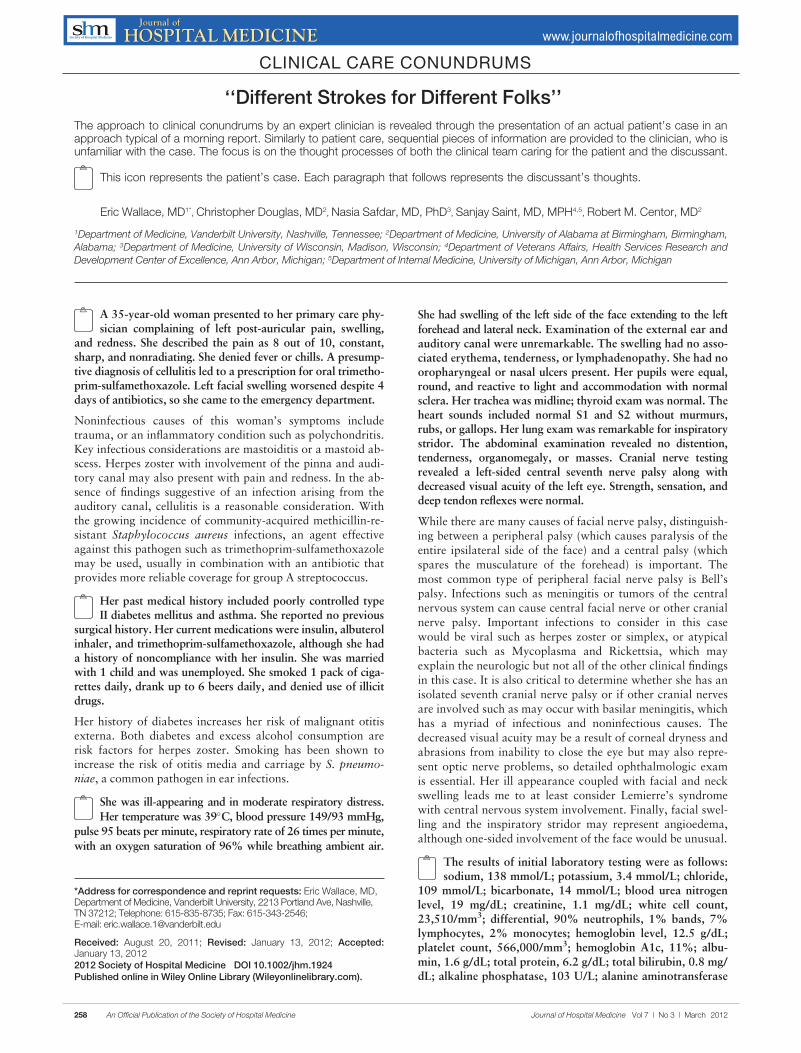
A 35-year-old woman presented to her primary care phy-sician complaining of left post-auricular pain, swelling,
and redness. She described the pain as 8 out of 10, constant,sharp, and nonradiating. She denied fever or chills. A presump-tive diagnosis of cellulitis led to a prescription for oral trimetho-prim-sulfamethoxazole. Left facial swelling worsened despite 4days of antibiotics, so she came to the emergency department.
Noninfectious causes of this woman’s symptoms includetrauma, or an inflammatory condition such as polychondritis.Key infectious considerations are mastoiditis or a mastoid ab-scess. Herpes zoster with involvement of the pinna and audi-tory canal may also present with pain and redness. In the ab-sence of findings suggestive of an infection arising from theauditory canal, cellulitis is a reasonable consideration. Withthe growing incidence of community-acquired methicillin-re-sistant Staphylococcus aureus infections, an agent effectiveagainst this pathogen such as trimethoprim-sulfamethoxazolemay be used, usually in combination with an antibiotic thatprovides more reliable coverage for group A streptococcus.
Her past medical history included poorly controlled typeII diabetes mellitus and asthma. She reported no previous
surgical history. Her current medications were insulin, albuterolinhaler, and trimethoprim-sulfamethoxazole, although she hada history of noncompliance with her insulin. She was marriedwith 1 child and was unemployed. She smoked 1 pack of ciga-rettes daily, drank up to 6 beers daily, and denied use of illicitdrugs.
Her history of diabetes increases her risk of malignant otitisexterna. Both diabetes and excess alcohol consumption arerisk factors for herpes zoster. Smoking has been shown toincrease the risk of otitis media and carriage by S. pneumo-niae, a common pathogen in ear infections.
She was ill-appearing and in moderate respiratory distress.
Her temperature was 39�C, blood pressure 149/93 mmHg,
pulse 95 beats per minute, respiratory rate of 26 times per minute,
with an oxygen saturation of 96% while breathing ambient air.
She had swelling of the left side of the face extending to the left
forehead and lateral neck. Examination of the external ear andauditory canal were unremarkable. The swelling had no asso-ciated erythema, tenderness, or lymphadenopathy. She had nooropharyngeal or nasal ulcers present. Her pupils were equal,round, and reactive to light and accommodation with normalsclera. Her trachea was midline; thyroid exam was normal. Theheart sounds included normal S1 and S2 without murmurs,rubs, or gallops. Her lung exam was remarkable for inspiratorystridor. The abdominal examination revealed no distention,tenderness, organomegaly, or masses. Cranial nerve testingrevealed a left-sided central seventh nerve palsy along withdecreased visual acuity of the left eye. Strength, sensation, anddeep tendon reflexes were normal.
While there are many causes of facial nerve palsy, distinguish-ing between a peripheral palsy (which causes paralysis of theentire ipsilateral side of the face) and a central palsy (whichspares the musculature of the forehead) is important. Themost common type of peripheral facial nerve palsy is Bell’spalsy. Infections such as meningitis or tumors of the centralnervous system can cause central facial nerve or other cranialnerve palsy. Important infections to consider in this casewould be viral such as herpes zoster or simplex, or atypicalbacteria such as Mycoplasma and Rickettsia, which mayexplain the neurologic but not all of the other clinical findingsin this case. It is also critical to determine whether she has anisolated seventh cranial nerve palsy or if other cranial nervesare involved such as may occur with basilar meningitis, whichhas a myriad of infectious and noninfectious causes. Thedecreased visual acuity may be a result of corneal dryness andabrasions from inability to close the eye but may also repre-sent optic nerve problems, so detailed ophthalmologic examis essential. Her ill appearance coupled with facial and neckswelling leads me to at least consider Lemierre’s syndromewith central nervous system involvement. Finally, facial swel-ling and the inspiratory stridor may represent angioedema,although one-sided involvement of the face would be unusual.
The results of initial laboratory testing were as follows:sodium, 138 mmol/L; potassium, 3.4 mmol/L; chloride,
109 mmol/L; bicarbonate, 14 mmol/L; blood urea nitrogenlevel, 19 mg/dL; creatinine, 1.1 mg/dL; white cell count,23,510/mm3; differential, 90% neutrophils, 1% bands, 7%lymphocytes, 2% monocytes; hemoglobin level, 12.5 g/dL;platelet count, 566,000/mm3; hemoglobin A1c, 11%; albu-
min, 1.6 g/dL; total protein, 6.2 g/dL; total bilirubin, 0.8 mg/dL; alkaline phosphatase, 103 U/L; alanine aminotransferase
*Address for correspondence and reprint requests: Eric Wallace, MD,Department of Medicine, Vanderbilt University, 2213 Portland Ave, Nashville,TN 37212; Telephone: 615-835-8735; Fax: 615-343-2546;E-mail: [email protected]
Received: August 20, 2011; Revised: January 13, 2012; Accepted:January 13, 20122012 Society of Hospital Medicine DOI 10.1002/jhm.1924Published online in Wiley Online Library (Wileyonlinelibrary.com).
258 An Official Publication of the Society of Hospital Medicine Journal of Hospital Medicine Vol 7 | No 3 | March 2012
level, 14 U/L; international normalized ratio of 1.2; partialthromboplastin time, 29 seconds (normal value, 24–34 sec-onds); erythrocyte sedimentation rate, 121 mm/hr; creatinekinase, 561 U/L (normal value 25–190). Arterial blood gasmeasurements with the patient breathing 50% oxygenrevealed a pH of 7.34, a partial pressure of carbon dioxide of28 mmHg, and a partial pressure of oxygen of 228 mmHg.
I am concerned that this patient has sepsis, likely due to an in-fectious trigger. With her clinical presentation localized to thehead and neck, her history of diabetes, and the acceleratedsedimentation rate, malignant otitis externa would explainmany of her findings. Empiric anti-infective therapy directedtoward Pseudomonas aeruginosa should be initiated, andimaging of the head and ear should be undertaken.
The patient required intubation due to increased respira-tory distress and stridor. Her physicians used intrave-
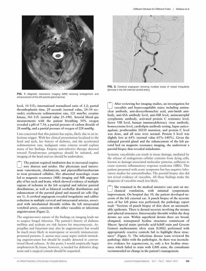
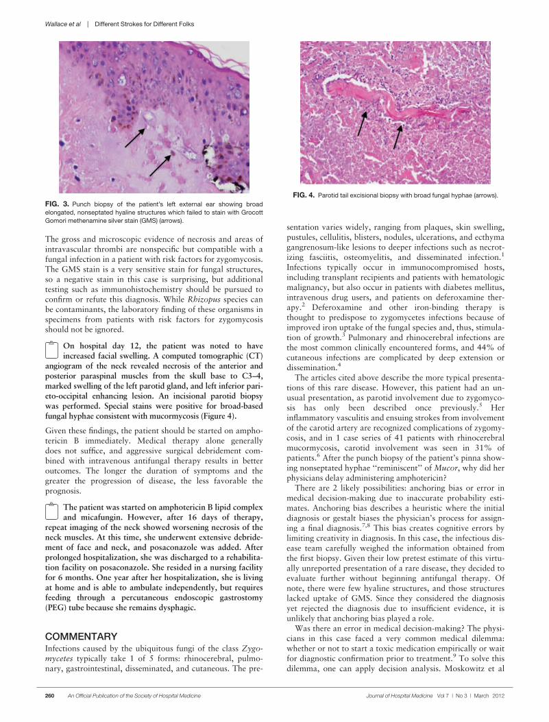
nous vancomycin, clindamycin, and piperacillin/tazobactamto treat presumed cellulitis. Her abnormal neurologic examled to magnetic resonance (MR) imaging and MR angiogra-phy of her neck and brain, which showed evidence of multipleregions of ischemia in the left occipital and inferior parietaldistributions, as well as bilateral cerebellar distributions andenhancement of the parotid gland and mastoid air cells (Fig-ure 1). A cerebral angiogram revealed irregularity and caliberreduction in multiple cervical and intracranial arteries, associ-ated with intraluminal thrombi within the left intracranialvertebral artery, consistent with either vasculitis or infectiousangioinvasion (Figure 2).
The angioinvasive nature of the findings on imaging leads meto suspect fungal infection. The patient’s history of diabetesmellitus and acidosis are risk factors for mucormycosis. As-pergillus and Fusarium may also be angioinvasive but wouldbe much more likely in neutropenic or severely immunocom-promised patients. S. aureus may cause septic emboli mimick-ing angioinvasion but should be readily detected in conven-tional blood cultures. At this point, I would empirically beginamphotericin B; tissue, however, is needed for definitive diag-nosis and a surgical consult should be requested.
After reviewing her imaging studies, an investigation forvasculitis and hypercoagulable states including antinu-
clear antibody, anti-deoxyribonucleic acid, anti-Smith anti-body, anti-SSA antibody level, anti-SSB level, antineutrophilcytoplasmic antibody, activated protein C resistance level,factor VIII level, human immunodeficiency virus antibody,homocysteine level, cardiolipin antibody testing, lupus antico-agulant, prothrombin 20210 mutation, and protein C levelwas done, and all tests were normal. Protein S level wasslightly low at 64% (normal value 65%–140%). Given theenlarged parotid gland and the enhancement of the left pa-rotid bed on magnetic resonance imaging, she underwent aparotid biopsy that revealed sialadenitis.
Systemic vasculitides can result in tissue damage, mediated bythe release of endogenous cellular contents from dying cells,known as damage-associated molecular patterns, sufficient tocause systemic inflammatory response syndrome (SIRS). Thispatient presented with acute symptoms but has negative labo-ratory studies for autoantibodies. The parotid biopsy also didnot reveal evidence of vasculitis. All these findings make thediagnosis of vasculitis much less likely.
She remained in the medical intensive care unit on me-chanical ventilation, with minimal symptomatic
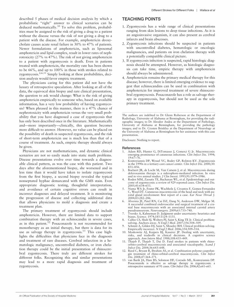
improvement. On hospital day 10, the patient developed ne-crosis of the left external ear. A punch biopsy of the necroticarea of her left pinna was performed; the pathology reportread: ‘‘Sections of punch biopsy of skin show an unremark-able epidermis. There is dermal necrosis involving the stromaand adnexal structures. Intravascular thrombi within the deepdermis are seen. Within superficial dermis there are broad,elongated, nonseptated hyaline structures reminiscent ofMucor. Special stains (periodic acid-Schiff stain and GrocottGomori methenamine silver stain [GMS]) performed withappropriately reactive controls fail to highlight these struc-tures’’ (Figure 3). The infectious disease team reviewed thepathology slides with the pathologist. As there was inconclu-sive evidence for zygomycosis, ie, only a few hyaline struc-tures which failed to stain with GMS stain, the consultants
recommended no change in the patient’s management.
FIG. 1. Magnetic resonance imaging (MRI) showing enlargement and
enhancement of the left parotid gland (arrow).
FIG. 2. Cerebral angiogram showing multiple areas of vessel irregularity
(arrows) in the left internal carotid artery.
An Official Publication of the Society of Hospital Medicine Journal of Hospital Medicine Vol 7 | No 3 | March 2012 259
Different Strokes for Different Folks | Wallace et al
The gross and microscopic evidence of necrosis and areas ofintravascular thrombi are nonspecific but compatible with afungal infection in a patient with risk factors for zygomycosis.The GMS stain is a very sensitive stain for fungal structures,so a negative stain in this case is surprising, but additionaltesting such as immunohistochemistry should be pursued toconfirm or refute this diagnosis. While Rhizopus species canbe contaminants, the laboratory finding of these organisms inspecimens from patients with risk factors for zygomycosisshould not be ignored.
On hospital day 12, the patient was noted to haveincreased facial swelling. A computed tomographic (CT)
angiogram of the neck revealed necrosis of the anterior andposterior paraspinal muscles from the skull base to C3–4,marked swelling of the left parotid gland, and left inferior pari-eto-occipital enhancing lesion. An incisional parotid biopsywas performed. Special stains were positive for broad-basedfungal hyphae consistent with mucormycosis (Figure 4).
Given these findings, the patient should be started on ampho-tericin B immediately. Medical therapy alone generallydoes not suffice, and aggressive surgical debridement com-bined with intravenous antifungal therapy results in betteroutcomes. The longer the duration of symptoms and thegreater the progression of disease, the less favorable theprognosis.
The patient was started on amphotericin B lipid complexand micafungin. However, after 16 days of therapy,
repeat imaging of the neck showed worsening necrosis of theneck muscles. At this time, she underwent extensive debride-ment of face and neck, and posaconazole was added. Afterprolonged hospitalization, she was discharged to a rehabilita-
tion facility on posaconazole. She resided in a nursing facilityfor 6 months. One year after her hospitalization, she is livingat home and is able to ambulate independently, but requiresfeeding through a percutaneous endoscopic gastrostomy(PEG) tube because she remains dysphagic.
COMMENTARYInfections caused by the ubiquitous fungi of the class Zygo-mycetes typically take 1 of 5 forms: rhinocerebral, pulmo-nary, gastrointestinal, disseminated, and cutaneous. The pre-
sentation varies widely, ranging from plaques, skin swelling,pustules, cellulitis, blisters, nodules, ulcerations, and ecthymagangrenosum-like lesions to deeper infections such as necrot-izing fasciitis, osteomyelitis, and disseminated infection.1
Infections typically occur in immunocompromised hosts,including transplant recipients and patients with hematologicmalignancy, but also occur in patients with diabetes mellitus,intravenous drug users, and patients on deferoxamine ther-apy.2 Deferoxamine and other iron-binding therapy isthought to predispose to zygomycetes infections because ofimproved iron uptake of the fungal species and, thus, stimula-tion of growth.3 Pulmonary and rhinocerebral infections arethe most common clinically encountered forms, and 44% ofcutaneous infections are complicated by deep extension ordissemination.4
The articles cited above describe the more typical presenta-tions of this rare disease. However, this patient had an un-usual presentation, as parotid involvement due to zygomyco-sis has only been described once previously.5 Herinflammatory vasculitis and ensuing strokes from involvementof the carotid artery are recognized complications of zygomy-cosis, and in 1 case series of 41 patients with rhinocerebralmucormycosis, carotid involvement was seen in 31% ofpatients.6 After the punch biopsy of the patient’s pinna show-ing nonseptated hyphae ‘‘reminiscent’’ of Mucor, why did herphysicians delay administering amphotericin?
There are 2 likely possibilities: anchoring bias or error inmedical decision-making due to inaccurate probability esti-mates. Anchoring bias describes a heuristic where the initialdiagnosis or gestalt biases the physician’s process for assign-ing a final diagnosis.7,8 This bias creates cognitive errors bylimiting creativity in diagnosis. In this case, the infectious dis-ease team carefully weighed the information obtained fromthe first biopsy. Given their low pretest estimate of this virtu-ally unreported presentation of a rare disease, they decided toevaluate further without beginning antifungal therapy. Ofnote, there were few hyaline structures, and those structureslacked uptake of GMS. Since they considered the diagnosisyet rejected the diagnosis due to insufficient evidence, it isunlikely that anchoring bias played a role.
Was there an error in medical decision-making? The physi-cians in this case faced a very common medical dilemma:whether or not to start a toxic medication empirically or waitfor diagnostic confirmation prior to treatment.9 To solve thisdilemma, one can apply decision analysis. Moskowitz et al
FIG. 3. Punch biopsy of the patient’s left external ear showing broad
elongated, nonseptated hyaline structures which failed to stain with Grocott
Gomori methenamine silver stain (GMS) (arrows).
FIG. 4. Parotid tail excisional biopsy with broad fungal hyphae (arrows).
260 An Official Publication of the Society of Hospital Medicine Journal of Hospital Medicine Vol 7 | No 3 | March 2012
Wallace et al | Different Strokes for Different Folks
described 5 phases of medical decision analysis by which aprobabilistic ‘‘right’’ answer to clinical scenarios can bededuced mathematically.10 To solve this problem, probabil-ities must be assigned to the risk of giving a drug to a patientwithout the disease versus the risk of not giving a drug to apatient with the disease. For example, amphotericin deoxy-cholate causes acute renal failure in 30% to 47% of patients.Newer formulations of amphotericin, such as liposomalamphotericin and lipid complex, result in lower rates of neph-rotoxicity (27% vs 47%). The risk of not giving amphotericinto a patient with zygomycosis is death. Even in patientstreated with amphotericin, the mortality rate has been shownto be 66%, and up to 100% in those with strokes related tozygomycosis.2,6,11 Simply looking at these probabilities, deci-sion analysis would favor empiric treatment.
The physicians caring for this patient did not have theluxury of retrospective speculation. After looking at all of thedata, the equivocal skin biopsy and rare clinical presentation,the question to ask would change: What is the risk of givingamphotericin empirically to someone who, based on availableinformation, has a very low probability of having zygomyco-sis? When phrased in this manner, there is a 47% chance ofnephrotoxicity with amphotericin versus the very small prob-ability that you have diagnosed a case of zygomycosis thathas only been described once in the literature. Mathematicallyand—more importantly—clinically, this question becomesmore difficult to answer. However, no value can be placed onthe possibility of death in suspected zygomycosis, and the riskof short-term amphotericin use is much less than that of acourse of treatment. As such, empiric therapy should alwaysbe given.
Physicians are not mathematicians, and dynamic clinicalscenarios are not so easily made into static math problems.Disease presentations evolve over time towards a diagnos-able clinical pattern, as was the case with this patient. Twodays after the aforementioned biopsy, she worsened and inless time than it would have taken to isolate zygomycosisfrom the first biopsy, a second biopsy revealed the typicalnonseptated hyphae demarcated with the GMS stain. Evenappropriate diagnostic testing, thoughtful interpretation,and avoidance of certain cognitive errors can result inincorrect diagnoses and delayed treatment. It is monitoringthe progression of disease and collecting additional datathat allows physicians to mold a diagnosis and create atreatment plan.
The primary treatment of zygomycosis should includeamphotericin. However, there are limited data to supportcombination therapy with an echinocandin in severe cases,as in this patient.12 Posaconazole is not recommended formonotherapy as an initial therapy, but there is data for itsuse as salvage therapy in zygomycosis.13 This case high-lights the difficulties that physicians face in the diagnosisand treatment of rare diseases. Cerebral infarction in a he-matologic malignancy, uncontrolled diabetes, or iron chela-tion therapy could be the initial presentation of rhinocere-bral zygomycosis. There truly are different strokes fordifferent folks. Recognizing this and similar presentationsmay lead to a more rapid diagnosis and treatment ofzygomycosis.
TEACHING POINTS
1. Zygomycosis has a wide range of clinical presentationsranging from skin lesions to deep tissue infections. As it isan angioinvasive organism, it can also present as cerebralinfarcts and brain abscesses.
2. Zygomycosis infections should be suspected in patientswith uncontrolled diabetes, hematologic or oncologicmalignancies, and patients on iron chelation therapy witha potentially compatible clinical picture.
3. If zygomycosis infection is suspected, rapid histologic diag-nosis should be attempted. However, as histologic diagno-sis can take time, empiric therapy with amphotericinshould always be administered.
4. Amphotericin remains the primary medical therapy for thisdisease; however, there is limited emerging evidence to sug-gest that echinocandins can be used in combination withamphotericin for improved treatment of severe rhinocere-bral zygomyocosis. Posaconazole has a role as salvage ther-apy in zygomycosis, but should not be used as the soleprimary treatment.
The authors are indebted to Dr Glenn Roberson at the Department ofRadiology, University of Alabama at Birmingham, for providing the radi-ographic images; to Dr Aleodor Andea at the Department of Pathology,University of Alabama at Birmingham, for providing the pathologyimages; and to Dr. Crysten Brinkley at the Department of Neurology atthe University of Alabama at Birmingham for her assistance with this casepresentation.
Disclosure: Nothing to report.
References1. Adam RD, Hunter G, DiTomasso J, Comerci G Jr. Mucormycosis:
emerging prominence of cutaneous infections. Clin Infect Dis. 1994;19:67–76.
2. Kontoyiannis DP, Wessel VC, Bodey GP, Rolston KV. Zygomycosisin the 1990s in a tertiary-care cancer center. Clin Infect Dis. 2000;30:851–856.
3. Boelaert JR, de Locht M, Van Cutsem J, et al. Mucormycosis duringdeferoxamine therapy is a siderophore-mediated infection. In vitroand in vivo animal studies. J Clin Invest. 1993;91:1979–1986.
4. Roden MM, Zaoutis TE, Buchanan WL, et al. Epidemiology and out-come of zygomycosis: a review of 929 reported cases. Clin Infect Dis.2005;41:634–653.
5. Numa WA Jr, Foster PK, Wachholz J, Civantos F, Gomez-FernandezC, Weed DT. Cutaneous mucormycosis of the head and neck with pa-rotid gland involvement: first report of a case. Ear Nose Throat J.2004;83:282–286.
6. Alvernia JE, Patel RN, Cai DZ, Dang N, Anderson DW, Melgar M.A successful combined endovascular and surgical treatment of a cra-nial base mucormycosis with an associated internal carotid arterypseudoaneurysm. Neurosurgery. 2009;65:733–740.
7. Tversky A, Kahneman D. Judgment under uncertainty: heuristics andbiases. Science. 1974;185:1124–1131.
8. Calfee CS, Shah SJ, Wolters PJ, Saint S, King TE Jr. Clinical problem-solving. Anchors away. N Engl J Med. 2007;356:504–509.
9. Schmitt A, Gilden DJ, Saint S, Moseley RH. Clinical problem-solving.Empirically incorrect. N Engl J Med. 2006;354:509–514.
10. Moskowitz AJ, Kuipers BJ, Kassirer JP. Dealing with uncertainty,risks, and tradeoffs in clinical decisions. A cognitive scienceapproach. Ann Intern Med. 1988;108:435–449.
11. Thajeb P, Thajeb T, Dai D. Fatal strokes in patients with rhino-orbito-cerebral mucormycosis and associated vasculopathy. Scand JInfect Dis. 2004;36:643–648.
12. Reed C, Bryant R, Ibrahim AS, et al. Combination polyene-caspofun-gin treatment of rhino-orbital-cerebral mucormycosis. Clin InfectDis. 2008;47:364–371.
13. van Burik JA, Hare RS, Solomon HF, Corrado ML, Kontoyiannis DP.Posaconazole is effective as salvage therapy in zygomycosis: aretrospective summary of 91 cases.Clin Infect Dis. 2006;42:e61–e65.
Different Strokes for Different Folks | Wallace et al
An Official Publication of the Society of Hospital Medicine Journal of Hospital Medicine Vol 7 | No 3 | March 2012 261