Embed Size (px)
Citation preview
Medical Clinic of the University, Ferrara, Italy (Director: Prof. A. Baserga)
DIFFERENTIAL ACRIDINE ORANGE STAINING OF HUMAN CHROMOSOMES
G.L. CASTOLDI, G.D. GRUSOVIN, G.L. SCAPOLI, R. SPANEDDA
SUMMARY
The acridine orange staining of metaphases previously treated with hot salt solutions, exhibits a differential banding pattern of the chromosomes. According to the physicochemical interpretation of the stained structures, the green and red fluorescent segments of the chromosomes should be considered as constituted respectively by double-stranded DNA and single-stranded DNA. The banding pattern is relatively consistent in different metaphases, although some occasional variations of the bands may be referred to the interference of chromosomal acid proteins. In general, the sequence of the bands is compatible with the picture of the reverse banding.
INTRODUCTION
Until now, several methods have been reported dealing with the demonstration of heterochromatic regions of the human chromosomes by means of both fluoro-chrome derivatives (Caspersson et al. 1968, 1971) and Giemsa procedures (Gagne" et al. 1971, Yunis et al. 1971, Dutrillaux and Lejeune 1971). A technique which combines hot salt salution treatment of the metaphases with acridine orange (AO) staining is here presented. This method shows, under suitable conditions, a metachromatic staining of different regions along the chromosomes.
AO staining is assumed to be sensitive to the steric complexity of the nucleic acid on the basis of a variable affinity of the dye for different physicochemical conditions (strandness) of the DNA. Helical content of nucleic-acid chain and availability of the phosphate groups are supposed to be essential for the differential staining (Bradley and Wolf 1959, Kasten 1966).
AO molecules are thought to attach to the DNA helix with the positively charged ring nitrogen close to the negatively charged phosphate groups. Bradley and Wolf (1959) have characterized two types of binding of acridines to DNA. Type I, which is common to all acridines, corresponds to the intercalation model showed by Ler-man (1961). In this condition, which is favoured by double-stranded DNA, the acridine molecules are bound to the DNA perpendicularly to the axis. Type II binding is a characteristic of only few dyes, such as AO, which is bound, in these conditions, externally to the helix. This binding is favoured by a single-stranded DNA.
319 Acta Genet. Med. Gemellol. (1972), 2 1 : 319-326
https://www.cambridge.org/core/terms. https://doi.org/10.1017/S0001566000010667Downloaded from https://www.cambridge.org/core. IP address: 65.21.228.167, on 22 Jan 2022 at 00:43:59, subject to the Cambridge Core terms of use, available at

ACTA GENETICAE MEDICAE ET GEMELLOLOGIAE
When chromosomal DNA is treated with hot salt solutions, it unwinds to some extent, allowing a different stacking of dye molecules along the chromosomes (Fig. i ) . As a consequence, there is an increase in dye stacking leading to a complete saturation of binding sites by dye molecules, which can interact in an associated molecular form leading to a metachromatic (red) staining of the DNA segment (Riegler 1966, Pachmann and Riegler 1972). After reassociation of the DNA, some regions of the chromosomes show a yellow green fluorescence indicating that individual
»/D >*I green
fluorescence
»/o=-red
fluorescence
cco M ( C H , ) 2
Ny^ ,K(CH, ) 2
COO'
ceo:
AFTER HEAT DENATURATION
IN 0,06 M NaaHP04
FIG. 1. A schematic representation of the binding of AO molecules to the DNA helix. When nucleic acid-AO complexes are inspected in ultraviolet light at 500 nm, the double-stranded DNA is green, whereas the single-stranded DNA is red. The former condition is related to the binding of AO in a monomer form to double-stranded DNA (number of polyanions P > than number of dye D molecules), and characterized by an absorption maximum at 500 nm and an emission maximum at 532 nm. The latter condition is related to a disordered structural organization of DNA (single-stranded) with binding of AO in an associated molecular form and characterized by a shift in the absorption maximum towards the shorter wavelength and the emission maximum towards a longer one: (PjD = 1).
320
https://www.cambridge.org/core/terms. https://doi.org/10.1017/S0001566000010667Downloaded from https://www.cambridge.org/core. IP address: 65.21.228.167, on 22 Jan 2022 at 00:43:59, subject to the Cambridge Core terms of use, available at
O.L. CASTOLDI ET AL. : DIFFERENTIAL STAINING OF HUMAN CHROMOSOMES
dye molecules are randomly bound to DNA, perhaps by intercalation between expanded helical coils (Lerman 1961).
In general, several factors are influencing the interaction of AO with nucleic acids, such as pH, ionic strenght, temperature, acidic proteins, availability of phosphate groups, and in situ " stacking coefficients ", which represent, according to Bradley and Wolf's definition, the inherent tendency of each polymer to promote stacking of dye molecules on its surface. However, under suitable conditions, the AO staining may be reliably employed to distinguish denaturated or single-stranded DNA from untreated or mildly denaturated double-stranded DNA by its red fluorescence instead of the typical yellow green fluorescence (De la Chapelle et al. 1971). Thus untreated or fixed mitotic cells show the chromosomes green and the cytoplasm red, indicating that the chromosomes, in these conditions, possess a double-stranded structure.
MATERIAL AND METHOD
In our experiments the following procedure proved to be the most useful for human metaphases obtained from blood cultures of different subjects (normal and mongol subjects) (Baserga and Castoldi 1972):
(a) After fixation in 3 : 1 methanol/acetic acid, the chromosome preparations were performed according to the usual air drying technique.
(b) The slides were subsequently immersed in 0.06M Na2HP04 solution at pH 6.9. The solution was warmed up to 96°G and the slides were kept for 10' at this temperature.
(c) The preparations were immediately transferred to a freezer at —20°G for 5'. id) Without washing, the slides were immersed in a solution of 0.01% acridine orange
(Calbiochem, Los Angeles) in 0.06M Na2HP04, pH 6.9, at room temperature, for 3-5'. (e) The preparations were washed for 3-5' in 0.06M Na2HP04, previously warmed
at 40°C, and mounted in the same solution for the fluorescence observation. For photomicroscopy, a Leitz Ortholux microscope with an HBO 200 W high-pressure
mercury vapor lamp, 3 mm BG 12 excitor filter, darkfield condenser, fluorite 95 X objective, K 510 barrier filter, and automatic exposure photographic attachment, was used.
RESULTS
As it can be shown by the application of this differential staining to different metaphases, the sequence of the bands metachromatically stained is relatively consistent.
During the various phases of the staining procedure, a progressive changing of the color patterns may be observed along the chromosomes. At the beginning of the staining, red or brownish-red color of the chromosomes indicate that a complete or almost complete denaturation of the chromosomes is achieved. With the progression of the reassociation, a yellow-brown color is appearing indicating a partial denaturation. For a longer time, the entire chromosomes take a green or a yellow-green fluorescence, indicating at least a partial renaturation of the DNA in all regions.
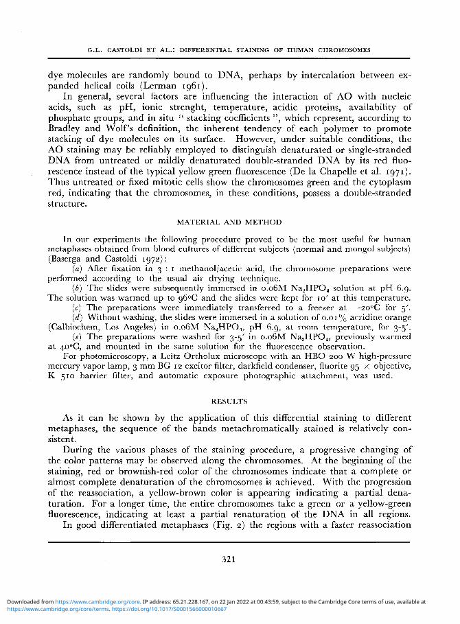
In good differentiated metaphases (Fig. 2) the regions with a faster reassociation
321
https://www.cambridge.org/core/terms. https://doi.org/10.1017/S0001566000010667Downloaded from https://www.cambridge.org/core. IP address: 65.21.228.167, on 22 Jan 2022 at 00:43:59, subject to the Cambridge Core terms of use, available at
ACTA GENETICAE MEDICAE ET GEMELLOLOGIAE
F I G . 2. Normal metaphase plate from blood culture, stained with AO after hot salt solution treatment. Light areas correspond to a green fluorescence, whereas
the darker regions appear to be stained red orange.
time are stained yellow-green, whereas the regions where the reassociation did not yet take place are stained red orange.
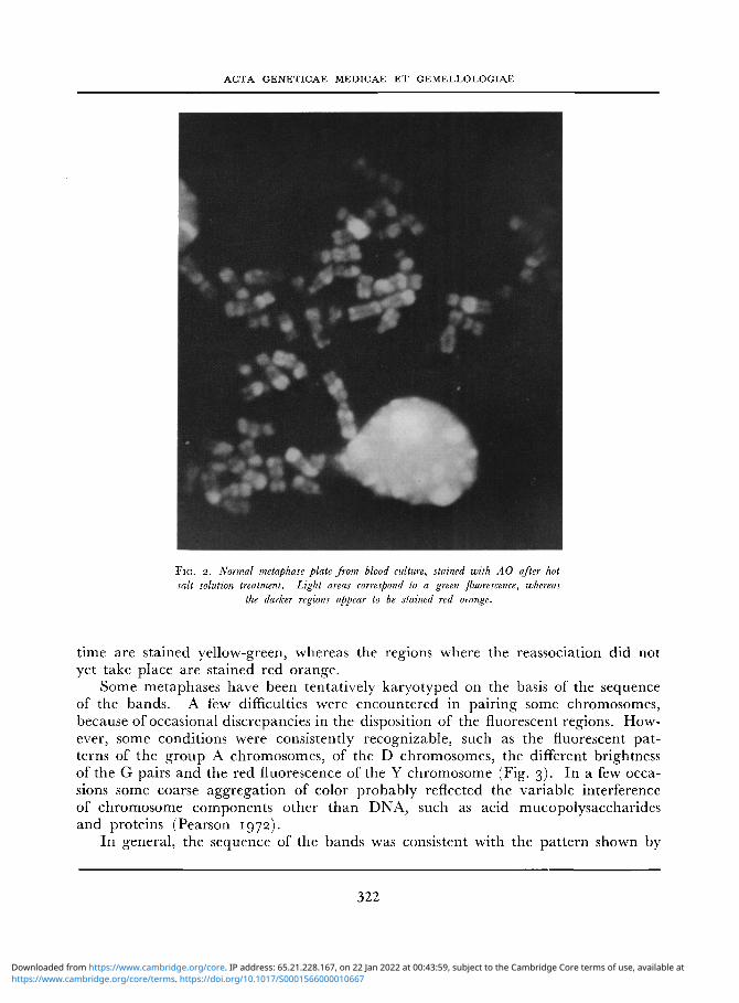
Some metaphases have been tentatively karyotyped on the basis of the sequence of the bands. A few difficulties were encountered in pairing some chromosomes, because of occasional discrepancies in the disposition of the fluorescent regions. However, some conditions were consistently recognizable, such as the fluorescent patterns of the group A chromosomes, of the D chromosomes, the different brightness of the G pairs and the red fluorescence of the Y chromosome (Fig. 3). In a few occasions some coarse aggregation of color probably reflected the variable interference of chromosome components other than DNA, such as acid mucopolysaccharides and proteins (Pearson 1972).
In general, the sequence of the bands was consistent with the pattern shown by
322
https://www.cambridge.org/core/terms. https://doi.org/10.1017/S0001566000010667Downloaded from https://www.cambridge.org/core. IP address: 65.21.228.167, on 22 Jan 2022 at 00:43:59, subject to the Cambridge Core terms of use, available at

G.L. CASTOLDI ET AL. : DIFFERENTIAL STAINING OF HUMAN CHROMOSOMES
FIG. 3. Selected chromosomes from different metaphase plates, stained with AO and showing a good similarity in the banding patterns. The sequence of the bands seems to correspond to
a reverse banding.
323
https://www.cambridge.org/core/terms. https://doi.org/10.1017/S0001566000010667Downloaded from https://www.cambridge.org/core. IP address: 65.21.228.167, on 22 Jan 2022 at 00:43:59, subject to the Cambridge Core terms of use, available at
ACTA GENETICAE MEDICAE ET GEMELLOLOGIAE
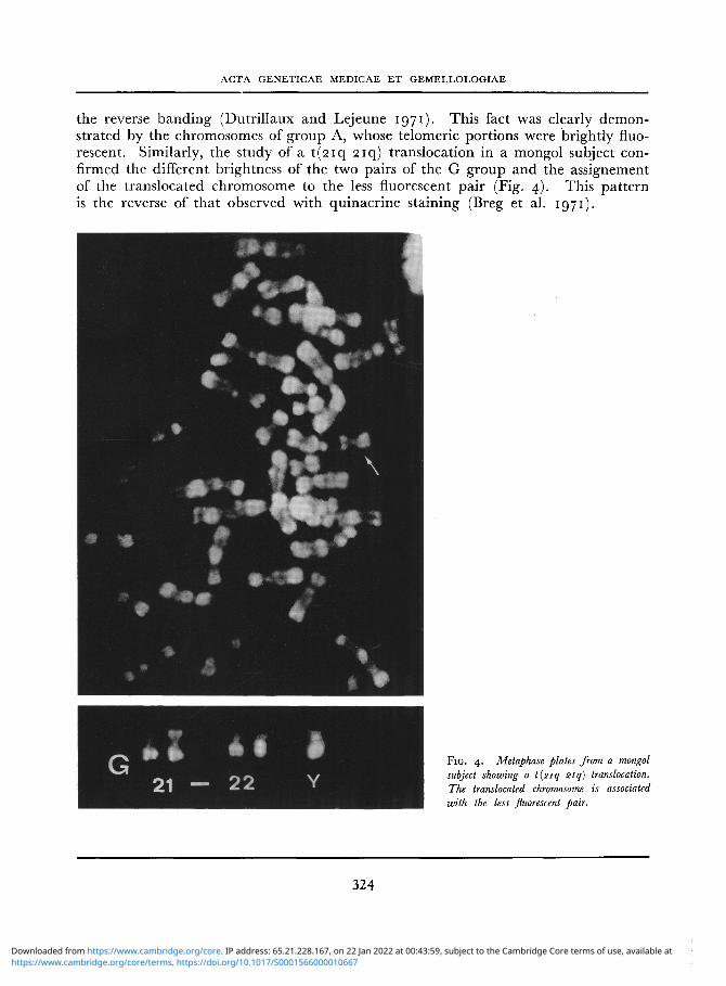
the reverse banding (Dutrillaux and Lejeune 1971). This fact was clearly demonstrated by the chromosomes of group A, whose telomeric portions were brightly fluorescent. Similarly, the study of a t (2iq 2iq) translocation in a mongol subject confirmed the different brightness of the two pairs of the G group and the assignement of the translocated chromosome to the less fluorescent pair (Fig. 4). This pattern is the reverse of that observed with quinacrine staining (Breg et al. 1971).
F I G . 4. Metaphase plates from a mongol subject showing a t(2iq 21q) translocation. The translocated chromosome is associated with the less fluorescent pair.
324
https://www.cambridge.org/core/terms. https://doi.org/10.1017/S0001566000010667Downloaded from https://www.cambridge.org/core. IP address: 65.21.228.167, on 22 Jan 2022 at 00:43:59, subject to the Cambridge Core terms of use, available at

G.L. CASTOLDI ET AL.: DIFFERENTIAL STAINING OF HUMAN CHROMOSOMES
DISCUSSION
The differential staining of the metaphases with AO appears to be useful in demonstrating regions of the chromosomes with a different rate of reassociation after denaturation. In our experiments the method showed a relatively consistent banding pattern in several metaphases. This fact and the reverse picture given by the sequence of the bands might be helpful in distiguishing among telomeric translocations, as shown by Bobrow et al. (1972).
Some bias in the interpretation of the staining may be due to the fact that this differential staining not only reflects abnormalities in reannealing (strandness) of the chromosomes after hot salt solution treatment, but also demonstrates both variation in the repetitious nature of the DNA (De la Chapelle et al. 1971) and the possible influence of acid mucopolysaccharides which interfere with the native state of the DNA (Clark and Felsenfeld 1971).
As a matter of fact, the problem whether the regions with a faster reassociation time do correspond in humans to heterochromatic zones with repetitive DNA sequences, still needs further investigation. Unlike the mouse satellite-DNA which is limited to the centromeric heterochromatin, whose differential staining with AO has been successfully applied by Stockert (1972), the human satellite-DNA seems to occur both in the centromeric heterochromatin areas and in the euchromatic regions (Corneo et al. 1970, Jones and Corneo 1971, Saunders et al. 1972). These facts render more complex the interpretation of the differentially stained regions by AO.
Despite these difficulties concerning the physicochemical background of the structures involved in the AO staining, this technique deserves further consideration for the analysis of the banding patterns of the chromosomes.
REFERENCES
Baserga A., Castoldi G.L. 1972. Applicazione alio studio delle zone eterocromatiche dei cromosomi umani della fluorescenza differenziale mediante arancio di acridina. Atti Simp. Int. sul " DNA satellite", Montecatini, 4-5 nov. 1972.
Bobrow M., Collacott H.E.A.C, Madan K. 1972. Chromosome banding with acridine orange. Lancet, 2: 1311.
Bradley D.F., Wolf M.K. 1959. Aggregation of dyes bound to polyanions. Proc. Natl. Acad. Sci. USA, 45: 944-952.
Breg W.R., Miller O.J., Miller D.A., Allerdice P.W., 1971. Distinctive fluorescence of quinacrine-label-led human G group chromosomes. Nature New Biol., 231: 276-277.
Caspersson T., Farber S., Foley G.E., Kudynowski J., Modest E.J., Simonsson E., Wagh U., Zech
L. 1968. Chemical differentiation along metaphase chromosomes. Exp. Cell Res., 49: 219-222.
Caspersson T., Lomakka G., Zech L. 1971. The 24 fluorescence patterns of the human metaphase chromosomes — distinguishing characters and variability. Hereditas, 67: 89-102.
Clark R.J., Felsenfeld G. 1971. Structure of chromatin. Nature New Biol., 229: 101-106.
Corneo G., Ginelli E., Polli E. 1970. Repeated sequences in human DNA. J. Mol. Biol., 48: 319-
327-De la Chapelle A., Schroeder J., Selander R.K.
1971. Repetitious DNA in mammalian chromosomes. Hereditas, 69: 149-153.
Dutrillaux B., Lejeune J. 1971. Sur une nouvelle technique d'analyse du caryotype humain. C.R. Acad. Sci. [D] (Paris), 272: 2638-2640.
325
https://www.cambridge.org/core/terms. https://doi.org/10.1017/S0001566000010667Downloaded from https://www.cambridge.org/core. IP address: 65.21.228.167, on 22 Jan 2022 at 00:43:59, subject to the Cambridge Core terms of use, available at
ACTA GENETICAE MEDICAE ET GEMELLOLOGIAE
Gagne R., Tanguay R., Laberge C. 1971- Differen- Pearson P. 1972. The use of new staining techniques tial staining patterns of heterochromatin in man. for human chromosome identification. J . Med. Nature, 232: 29-30. Genet., 9: 264-275.
Jones K.W., Corneo G. 1971. Location of satellite Riegler R. 1966. Micronuorometric characterization and homogeneous DNA sequences on human of intracellular nucleic acids and nucleoproteins chromosomes. Nature New Biol., 233: 268-271. by acridine orange. Acta Physiol. Scand., 67
Kasten F.H. 1966. Cytochemical studies with acri- (Suppl. 267): 1-121. dine orange and the influence of dye contami- Saunders G., Hsu T.C., Getz M.J., Simes E.L., nants in the staining of nucleic acids. Int. Rev. Arrighi F.E. 1972. Location of a human satellite Cytol., 21: 141-202. DNA in human chromosomes. Nature New Biol.,
Lerman L.S. 1961. Structural considerations in the 236: 244-246. interaction of DNA and acridines. J . Mol. Biol., Stockert J .C. 1972. Differential fluorescence in 3: 18-30. metaphase chromosomes stained by acridine
Pachmann U., Riegler R. 1972. Quantum yeld of orange. Stain Technol., 46: 103-105. acridines interacting with DNA of defined base Yunis J.J. , Roldan L., Yasmineh W.G., Lee J.C. sequence. A basis for the explanation of acridine ' 97 1 ' Staining of satellite DNA in metaphase bands in chromosomes. Exp. Cell Res., 67: 602- chromosomes. Nature, 231: 532-533. 608.
RlASSUNTO
L'applicazione della colorazione con arancio di acridina a metafasi precedentemente trattate con soluzioni saline a caldo fornisce, in fluorescenza, una colorazione differenziale di bande a livello dei cromosomi. L'inter-pretazione fisico-chimica delle modalita di estrinsecazione della colorazione consente di definire come com-poste da DNA double stranded le regioni colorate in verde e come composte da DNA single stranded quelle colo-rate metacromaticamente in rosso arancio. La dislocazione delle bande appare relativamente costante, seppure con occasionali deformazioni, per la possibile interferenza di strutture quali mucopolisaccaridi e proteine acide, nelle fasi di colorazione. II quadro presentato dalla sequenza delle bande lungo i singoli cromosomi sembra nel complesso sovrapporsi a quello del reverse banding.
RESUME
La coloration de metaphases prealablement traitees par des solutions salines a chaud, avec orange d'acri-dine, demontre la presence de bandes differemment fluorescentes a niveau des chromosomes. L'interpretation des modalites physico-chimiques de la coloration permet de considerer comme constituees par double-stranded DNA les regions chromosomiques qui donnent une fluorescence verte et par single-stranded DNA les regions me-tachromatiquement colorees en rouge-orange. La disposition des bandes est relativement constante dans des differentes metaphases, bien que des variations occasionnelles peuvent se manifester peut-etre par rapport a ['interference de proteines acides le long des chromosomes. Le tableau presente par les bandes fluorescentes au niveau des chromosomes est dans l'ensemble comparable a celui du reverse banding.
ZUSAMMENFASSUNG
Die durch AO Farbung von mit Salzlosungen im voraus behandelten Metaphasen zeigt eine Reihe unter-scheidlich fluoreszierender Streifen der Chromosomen. Die physico-chemische Grundlage der Farbung mit AO erlauben die griine fluoreszierende Streifen der Chromosomen als von double-stranded DNA und die rote fluoreszierende Streifen als von single-stranded DNA zusammengesetzt zu betrachten. Die Streifenanordnung scheint relativ konstant zu sein; einige Abweichungen sind in der Hauptsache zu der Interferenz saurer pro-teischen Strukturen zuriickzufuhren. Im allegemeinen ist die Streifenreihe vergleichbar mit dem Bild des reverse banding.
Prof. G.L. Castoldi, Clinica Medica dell'Universita, Ferrara, Italy.
326
https://www.cambridge.org/core/terms. https://doi.org/10.1017/S0001566000010667Downloaded from https://www.cambridge.org/core. IP address: 65.21.228.167, on 22 Jan 2022 at 00:43:59, subject to the Cambridge Core terms of use, available at


![SOME N- AND S-HETEROCYCLIC POLYCYCLIC AROMATIC … · ]acridine, benz[c] acridine, dibenz[a, j]acridine, dibenzo[c, h]acri dine and carbazole by gas chromatography from tobacco-smoke](https://img.pdfslide.net/doc/110x75/5e15aaf1fc75030377117681/some-n-and-s-heterocyclic-polycyclic-aromatic-acridine-benzc-acridine-dibenza.jpg)

![Acridine – a Promising Fluorescence Probe of Non-Covalent ... · [acridine-H]+BArF−, λ em =485 nm. Fig.3. Absorption spectra in CH 2 Cl 2 of: (1) acridine (2×10⁻⁵ mol/l) and](https://img.pdfslide.net/doc/110x75/5f4a49f4cafd5240686feade/acridine-a-a-promising-fluorescence-probe-of-non-covalent-acridine-hbarfa.jpg)