Embed Size (px)
Citation preview
JOURNAL OF CLINICAL MICROBIOLOGY, June 1994, p. 1537-1541 Vol. 32, No. 60095-1 137/94/$04.00+0Copyright © 1994, American Society for Microbiology
Differential Blocking of Coagulation-Activating Pathways ofLimulus Amebocyte Lysate
GUI-HANG ZHANG,1 LEIF BAEK,2 OLE BUCHARDT,3 AND CLAUS KOCHI*Department of Immunology, State Serum Institute, DK-2300 Copenhagen S,1 Department of Microbiology, Herlev
Hospital, Herlev Ringvej, DK-2730 Herlev,2 and Department of Chemistry, H.C. 0rsted Institute,University of Copenhagen, DK-2100 Copenhagen 0,3 Denmark
Received 15 December 1993/Returned for modification 25 January 1994/Accepted 1 March 1994
The coagulation of Limulus amebocyte lysate (LAL) can be activated through two pathways, one initiated byendotoxin and the other by ,-glucans. The two pathways join at the step of activation of the proclotting enzyme.We report here that the endotoxin-activated pathway can be differentially inhibited by two methods in aLimulus enzyme-linked immunosorbent assay (ELISA), either by the combined use of dimethyl sulfoxide andpolymyxin B or by a monoclonal antibody against Limulus factor C. LAL reactivities to 10 different endotoxinpreparations could be inhibited by the former method by a factor of 104 to 106 and could be blocked almosttotally by the latter method, irrespective of the source of endotoxin. The sensitivity of the assay wasapproximately 50 pg/ml both for curdlan from Alkaligenes faecalis and for laminarin from Laminaria digitata.We also found that the j-glucan-activated pathway could be totally blocked by laminarin (>1 ,ug/ml) withoutaffecting the endotoxin-activated pathway, allowing endotoxin to be quantitated specifically by the LimulusELISA with a detection limit of 0.005 endotoxin unit per ml. The use of uninhibited and differentially inhibitedELISAs demonstrated that different LAL preparations showed much greater variation in assaying I-glucansthan in assaying endotoxins. The LAL reactivity of normal human plasma was found to be due to the activationof the P-glucan pathway, but not the endotoxin pathway, of LAL.
Limulus amebocyte lysate (LAL) is known to contain acoagulation system that can be activated by a minute amountof bacterial endotoxin (lipopolysaccharide [LPS]). The coagu-lation cascade consists of three proenzymes (factor C, factor B,and proclotting enzyme) and one clottable protein (coagulo-gen) (7, 14). LPS activates factor C, which then activates factorB, which in turn converts proclotting enzyme to clottingenzyme. Clotting enzyme cleaves coagulogen to yield an insol-uble coagulin gel. The formation of gel clot and measurementof clotting enzyme activity by means of a synthetic chromoge-nic substrate constitute, respectively, the bases of the gel clotand chromogenic LAL assays for the detection of endotoxins(8, 15). We have recently developed sensitive endotoxin assaysbased on the immunochemical detection of Limulus coagulo-gen or its cleaved fragment, peptide C, with detection limits aslow as 0.001 endotoxin unit (EU) per ml (2, 24, 25).
Because of its extreme sensitivity to endotoxins, the LALtest has been considered a promising means of detectingendotoxin in clinical settings (9). However, its specificity forendotoxin appears to be compromised by certain ,B-glucans,originating from fungi (5, 6, 10) or hemodialysis membranes (3,23), which are also LAL reactive, although these glucans are atleast 1,000-fold less active than endotoxin. Fortunately, theactivation of LAL by 3-glucan occurs through an initiatingfactor different from that for endotoxin, namely, factor G (16).Separation of the two pathways by fractionation and reconsti-tution of the relevant factors has been reported recently (12,19). Addition of curdlan, a carboxymethylated 3-glucan, toLAL at high concentrations (>1 mg/ml) has also been shownto block the ,B-glucan pathway (11). Another study shows thatthe surfactant Zwittergent interferes with LAL reactivity to
* Corresponding author. Mailing address: Statens Seruminstitut,Division of Immunology, 5, Artillerivej, DK-2300 Copenhagen S,Denmark. Phone: +45 3268 3719. Fax: +45 3268 3149.
,B-glucan (20). However, the reconstituted LAL fractions arenot stable, Zwittergent also affects lipid A to some extent, andcurdlan tends to autopolymerize to form an insoluble gel athigh concentrations after storage for 3 months, even at 4°C(unpublished observation).
In this study, we have attempted to develop specific LALtests for endotoxin and P-glucan by differentially blocking thetwo activation pathways of LAL, using our newly developedLimulus peptide C enzyme-linked immunosorbent assay(ELISA) to detect reactivity. We have also assessed theinfluence of different formulations of LAL on the quantifica-tion of endotoxin or P-glucan activities of various laboratoryspecimens.
MATERIALS AND METHODS
All glassware was rendered pyrogen free by being heated to250°C for at least 3 h. Sterile, pyrogen-free tips and micro-plates were purchased from Eppendorf, Hamburg, Germany,and Nunc, Roskilde, Denmark, respectively. The LAL reagentwater used was confirmed to have less than 0.001 EU/ml by theLAL test.LAL preparations. Commercial LAL preparations included
Pyrotell (lot 42-99-541) from Associates of Cape Cod (ACC),Woods Hole, Mass., and LAL (lot 2L0860) from WhittakerBioproducts (WB), Walkersville, Md. The Tachypleus amebo-cyte lysate (TAL) was prepared from the horseshoe crabTachypleus tridentatus without chloroform extraction, as de-scribed previously (25).
LPS. The control standard endotoxin was NP-3, which is a
purified preparation of LPS from Salmonella abortus equipurchased from Pyroquant Diagnostik, Walldorf, Germany,and its potency was confirmed to be 10 EU/ng in comparisonwith USP reference standard endotoxin EC-5. The followingLPS preparations, obtained by phenol extraction, were pur-chased from Sigma Chemical Co., St. Louis, Mo.: Escherichia
1537
on July 20, 2019 by guesthttp://jcm
.asm.org/
Dow
nloaded from
J. CLIN. MICROBIOL.1538 ZHANG ET AL.
coli 01 11:B4 (determined potency, 2.5 EU/ng), E. coli 055:B5,E. coli 0127:B8, E. coli 0128:B12, Klebsiella pneumoniae,Pseudomonas aeruginosa, S. abortus equi, Salmonella enteritidis,Salmonella minnesota, Salmonella typhimurium, and Shigellaflexneni.
Glucan preparations. Laminarin isolated from Laminariadigitata was obtained from Sigma. Highly purified curdlan fromAlcaligenes faecalis was purchased from Wako Pure ChemicalIndustries, Osaka, Japan. Both laminarin and curdlan were
dissolved in 0.2 N NaOH at 5 mg/ml and incubated at 56°C for6 h to inactivate contaminating endotoxin (25).
Polymyxin B. Polymyxin B sulfate was purchased fromCalbiochem, La Jolla, Calif. The endotoxin and ,-glucan-likeactivities found in polymyxin B were removed by adsorptionwith activated charcoal (Merck, Darmstadt, Germany); 0.1 gwas mixed with 10 ml of polymyxin B solution (20 mg/ml) at37°C for 1 h, and this was followed by centrifugation to removethe charcoal.MAb against factor C. Partially purified factor C was
obtained from TAL by dextran sulfate-Sepharose CL-6Bchromatography as described by Nakamura et al. (17) and was
further purified by sodium dodecyl sulfate-polyacrylamide gelelectrophoresis (SDS-PAGE) carried out with a 12% gel,essentially as described by Laemmli (13). The band at 120 kDawas cut out and eluted with an electroelutor (Bio-Rad Labo-ratories, Richmond, Calif.). Purified factor C was adsorbed toaluminium hydroxide suspension (2 mg/ml in normal saline)and injected intraperitoneally (0.5 ml, containing 25 ,ug offactor C, per mouse) into five female CF1 x BALB/c mice.Booster doses were given at 2-week intervals, and animals weretest bled 10 days after each boost. When high antibody titerswere found by ELISA in factor C-coated microtiter plates,spleen cells were fused by a standard procedure (4) withmyeloma cells from line X63/Ag8.6.5.3. Selected monoclonalantibodies (MAbs) against factor C were purified from hybrid-oma supernatants by affinity chromatography on proteinA-Sepharose CL-4B columns (Pharmacia, Uppsala, Sweden).One of the MAbs (MAb-B2) was found to react with bothTachypleus factor C and Limulus factor C, since it recognizedthe 120-kDa band of the partially purified factor C prepara-
tions from both LAL and TAL in SDS-PAGE immunoblotting(unpublished observation), and was chosen for use in thepresent study. The endotoxin contamination of preparationscontaining 1 mg of MAb-B2 per ml was about 100 ng/ml andwas removed by Triton X-1 14 extraction (1).
Preparation and pretreatment of plasma. Blood from hu-man donors was drawn into pyrogen-free glass tubes contain-ing pyrogen-free heparin (final concentration, 4 IU/ml).Plasma was separated by centrifugation at 500 x g for 15 min.Perchloric acid (PCA) precipitation of plasma to remove
factors that interfere with LAL was carried out essentially as
described by Obayashi (18) but with one minor modification:the neutralized supernatant was further diluted twofold in theLAL reagent water.Limulus peptide C ELISA. Unless otherwise stated, the
Limulus peptide C ELISA was performed as previously de-scribed (25). When MAb-B2 was used as a blocking agent,
stock solution (1 mg/ml) was added to diluted LAL andincubated at 4°C for 20 min before the ELISA was performed.When dimethyl sulfoxide (DMSO) and polymyxin B were usedas blocking agents, the ordinary LAL buffer was replaced bybuffer containing 20% (vol/vol) DMSO, 100 ,ug of polymyxin Bper ml, 60 mM MgCI2, and 100 mM Tris-HCl, pH 8.
1.4
1.2
1
0.8
0.6
0.4
0.2
0
OD490-650
-3 -2 -1 0 1 2 3 4 5
Concentration of LPS or Curdlan, log ng/mI
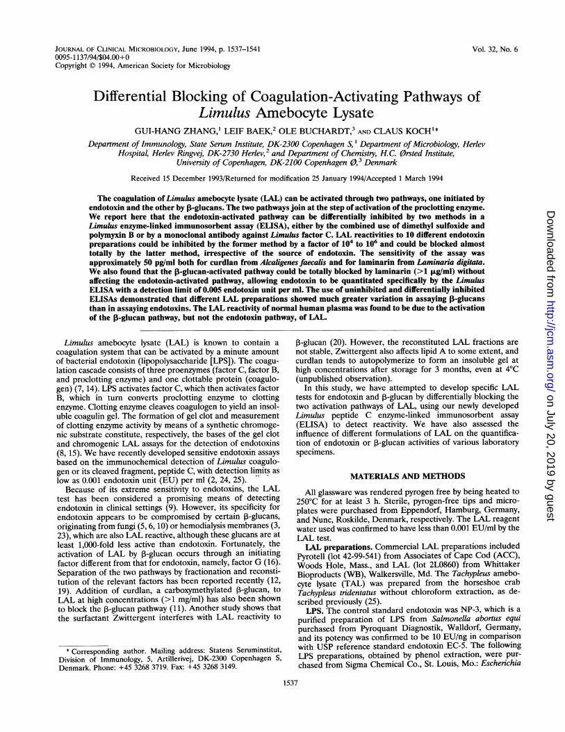
FIG. 1. Differential inhibition of the LPS-activated pathway ofLAL by MAb-B2 (10 ,ug/ml). OD490-650, optical density at 490 nm
corrected by optical density at 650 nm.
RESULTS
Differential blocking of the endotoxin-activated LAL path-way. MAb-B2 is a murine MAb raised against factor C fromTAL and was found to cross-react with factor C from LAL (seeMaterials and Methods). Figure 1 shows the effect of MAb-B2on the reactivity of LAL to LPS (from E. coli O111:B4) and tocurdlan. LAL (from ACC) became almost unreactive to LPS inthe presence of MAb-B2 at 10 ,ug/ml, while the reactivity ofLAL to curdlan was unaltered. The same effect was alsoobserved with other LPS preparations, as shown in Table 1.
Figure 2 shows the dose-dependent inhibition of the endo-toxin-activated pathway of LAL by MAb-B2. When the con-centration of the antibody in LAL was at or above 10 ,ug/ml,the reactivity of LAL to LPS was almost totally inhibited. Nofurther inhibition was observed when the MAb-B2 concentra-tion was increased above 20 ,ug/ml. The residual LAL reactivityof LPS at concentrations of >100 ,ug/ml was presumed to bedue to the contamination of the LPS preparations with 3-glu-can-like substances, since it could not be abolished even bytreatment with 0.2 N NaOH at 60°C for 24 h.
TABLE 1. Inhibition of the LAL-LPS reaction by DMSO pluspolymyxin B (D-P) or by MAb-B2
Detection limit (ng/ml) with:Source of LPS
LAL only LAL + D-Pa LAL + MAb-B2bE. coli 055:B5 0.001 100 >10,000E. coli 0127:B8 0.001 2,500 >10,000E. coli 0128:B12 0.0005 1,000 >10,000K pneumoniae 0.001 1,250 >10,000P. aeruginosa 0.001 250 >10,000S. abortus equi 0.002 20 >10,000S. enteritidis 0.002 250 >10,000S. minnesota 0.002 100 >10,000S. typhimurium 0.003 1,000 >10,000Shigella flexneni 0.005 2,500 >10,000
a The LAL buffer containing 20% DMSO, 100 ,ug of polymyxin B per ml, 60mM MgCl2, and 100 mM Tris-HCI (pH 8) was used to dilute LAL in the LimuluspepFtide C ELISA.
The concentration of MAb-B2 in LAL was 10 Kg/ml.
on July 20, 2019 by guesthttp://jcm
.asm.org/
Dow
nloaded from

BLOCKING OF LIMULUS COAGULATION 1539
OD490-6501.4
1.2
0.8
0.6
0.4
0.2
0
OD490-650
-3 -2 -1 0 1 2 3 4 5 6
Concentration of LM, CL or LPS, log ng/ml
-3 -2 -1 0 1 2 3 4 5 6 FIG. 4. Differential inhibition of the 3-glucan-activated pathway ofLAL by laminarin (LM) or curdlan (CL). #, Concentration of LM in
Concentration of LPS, log ng/ml LAL was 5 ,Lg/ml; *, concentration of CL in LAL was 1 mg/ml.
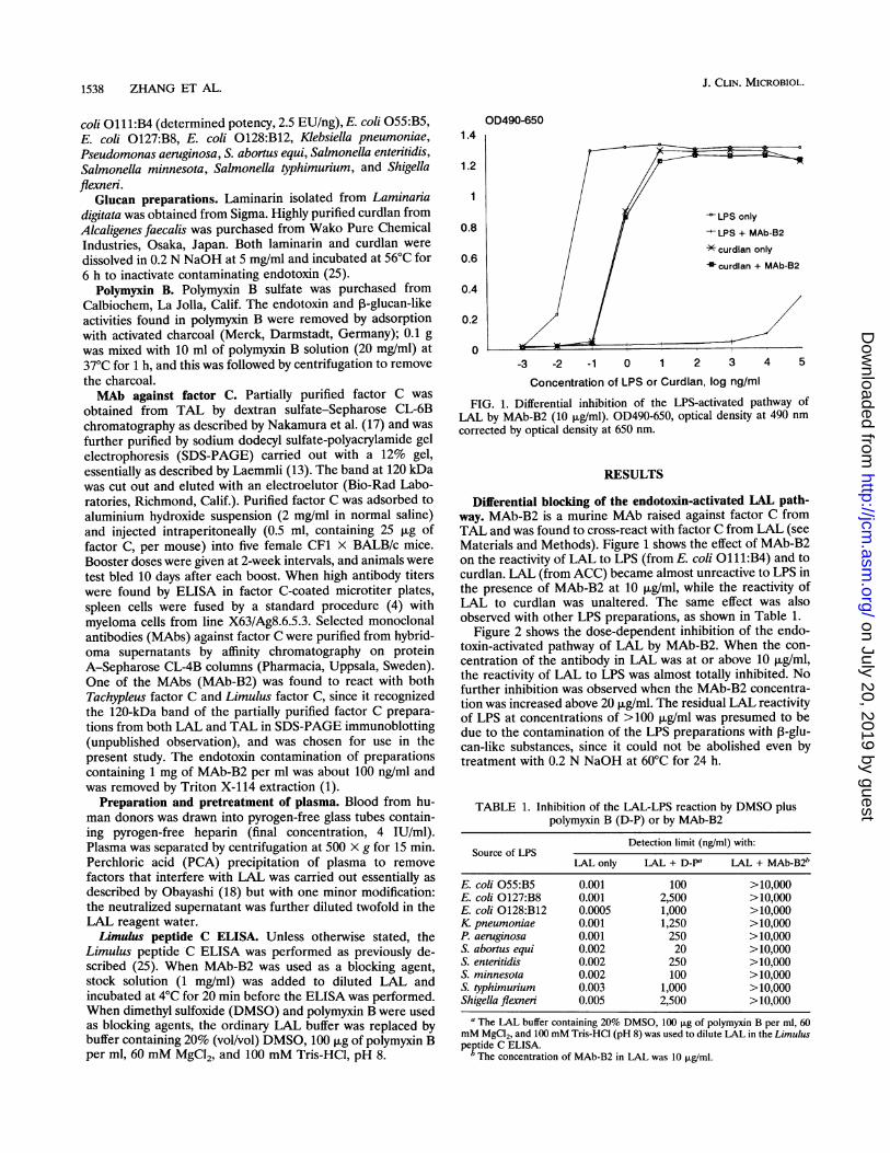
FIG. 2. Dose-dependent inhibition of the LPS-activated pathway of OD490-650, optical density at 490 nm corrected by optical density atLAL by MAb-B2 at 0 (L1), 2.5 (+), 5 (*), 10 (-), and 20 (X) ,ug/ml. 650 nm.OD490-650, optical density at 490 nm corrected by optical density at650 nm.
Figure 3 shows the effect of the combined use of 20%DMSO and polymyxin B (100 ,ug/ml) on the reactivity of LALto LPS (from E. coli 011 1:B4). The reaction of LAL with LPSwas inhibited by a factor of more than 105 (increasing thedetection limit from 2.5 pg/ml to 1 jLg/ml), but the reactivity ofLAL to curdlan was not significantly altered. The inhibitoryeffect of DMSO and polymyxin B on the reactivity of LAL toother LPS preparations was variable, ranging from a factor of104 to 106', as shown in Table 1.
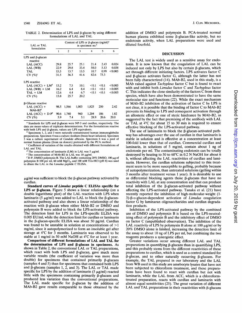
Differential blocking of the j-glucan-activated LAL path-way. Figure 4 shows the reaction curves of LAL with LPS,laminarin, and curdlan and the effects of laminarin and curdlan
2
1.5
0.5
0
OD490-650
LPS only
-curdlan only
LPS + D-P
-curdlan + D-P
-3 -2 -1 0 1
Concentration of LPS or curdlan,
FIG. 3. Differential inhibition of the LPS-aLAL by the combined use of 20% DMSO and 14
per ml (D-P). OD490-650, optical density at ioptical density at 650 nm.
on the 3-glucan-activated pathway. The concentration ranges
in which laminarin and curdlan reacted with LAL were from0.05 ng/ml to 1 .ig/ml and from 0.1 ng/ml to 500 Fg/ml,respectively. Addition of laminarin at 5 jig/ml or curdlan at I
mg/ml to LAL totally blocked the ,B-glucan-activated pathwaywithout compromising the reactivity of LAL to LPS.The reactions of TAL with LPS and curdlan were found to
be similar to those of LAL, but TAL did not react withlaminarin at any concentration from 1 pg/ml to 1 mg/ml (25).Addition of laminarin to TAL at a concentration as low as 0.1
OD490-650
X/ assayforassay for
LPS (LM)#- CL (MAb-B2)*
0.1
0.010 0.006 0.012 0.025 0.05 0.25
Concentration of LPS (EU/mI) or curdlan (CL,ng/ml)
1.25
2 3 4 FIG. 5. Standard curves of specific Limulus peptide C ELISAs for
log ng/ml LPS or curdlan. #, the concentration of laminarin (LM) in LAL was
n9/ml ,ug/ml. *, the concentration of MAb-B2 in LAL was 10 ,ug/ml. **, the
activated pathway of LAL buffer containing 20% DMSO, 100 p.g of polymyxin B per ml, 60
00 ,ug of polymyxin B mM MgCl2, and 100 mM Tris-HCI (pH 8) was used to dilute LAL in
490 nm corrected by the Limulus peptide C ELISA. OD490-650, optical density at 490 nm
corrected by optical density at 650 nm.
1.4
1.2
0.8
0.6
0.4
0.2
0
VOL. 32, 1994
on July 20, 2019 by guesthttp://jcm
.asm.org/
Dow
nloaded from
1540 ZHANG ET AL.
TABLE 2. Determination of LPS and I-glucan by using differentformulations of LAL and TAL
Content of LPS or p-glucan (ng/ml)aLAL or TAL in specimen no.bformulation
1 2 3 4 5 6
LPS and ,B-glucanreactive
LAL (ACC) 29.8 25.7 25.1 21.4 2.43 0.024LAL (WB) 22.9 29.8 33.4 58.0 5.13 0.038TAL 15.5 13.7 13.5 10.7 0.99 <0.005CV (%)' 31.5 36.3 41.6 82.6 73.3
LPS reactiveLAL (ACC) + LMd 13.2 7.5 10.1 <0.1 <0.1 <0.005LAL (WB) + LM 16.2 6.4 8.4 <0.1 <0.1 <0.005TAL + LM 12.6 4.4 6.7 <0.1 <0.1 <0.005CV (%) 13.8 25.7 20.2
,-Glucan reactiveLAL (ACC) + 98.5 1,566 1,003 1,825 250 2.42MAb-B2e
LAL (ACC) + D-Pf 90.6 1,740 960 1,204 186 1.82CV (%) 5.9 7.4 3.1 28.9 20.6 20.0
a Standards for LPS and ,B-glucan were NP-3 and curdlan, respectively. Thedata are mean values of triplicate determinations. For the formulations reactivewith both LPS and ,B-glucan, values are LPS equivalents.
b Specimens 1, 2, and 3 were naturally contaminated human immunoglobulinpreparations. Specimen 4 was a culture medium (brain heart infusion). Specimen5 was a culture supernatant of Candida albicans. Specimen 6 was a pool ofnormal human plasma (from six donors) pretreated by the PCA method.
c Coefficient of variation of the results obtained with different formulations ofLAL and TAL.
d The concentration of laminarin (LM) in LAL was 5 ,ug/ml.eThe concentration of MAb-B2 in LAL was 10 ,ug/ml.f D-P, DMSO-polymyxin B. The LAL buffer containing 20% DMSO, 100 ,ug of
polymyxin B 100 per ml, 60 mM MgCl2, and 100 mM Tris-HCI (pH 8) was usedto dilute the LAL in the Limulus peptide C ELISA.
,ug/ml was sufficient to block the f-glucan pathway activated bycurdlan.
Standard curves of Limulus peptide C ELISAs specific forLPS or 0-glucan. Figure 5 shows a linear relationship (on adouble logarithmic plot) of the LAL reaction with LPS whenlaminarin (5 ,ug/ml) was added to LAL to block the P-glucan-activated pathway and also shows a linear relationship of thereaction with ,B-glucan when either MAb-B2 or DMSO andpolymyxin B were added to block the LPS-activated pathway.The detection limit for LPS in the LPS-specific ELISA was0.005 EU/ml, while the detection limit for curdlan or laminarinin the ,B-glucan-specific ELISA was about 50 pg/ml. Curdlanwas found to be unstable in solution at concentrations of >1mg/ml, since it autopolymerized to form an insoluble gel afterstorage at 4°C for 3 months. Laminarin was observed to bestable at 1 mg/ml in 50 mM NaOH at 4°C for at least 1 year.Comparison of different formulations of LAL and TAL for
the determination of LPS and 13-glucan in specimens. Asshown in Table 2, the conventional LAL or TAL preparations,which react with both LPS and ,B-glucan, gave much morevariable results (the coefficient of variation was more thandouble) for specimens that contained primarily 3-glucans(samples 4 and 5) than for specimens that contained both LPSand ,B-glucans (samples 1, 2, and 3). The LAL or TAL madespecific for LPS by the addition of laminarin (5 ,ug/ml) reactedlittle with the specimens containing primarily 3-glucans andproduced less variation than the conventional preparations.The LAL made specific for ,3-glucan by the addition ofMAb-B2 gave results comparable to those obtained by the
addition of DMSO and polymyxin B. PCA-treated normalhuman plasma exhibited some ,B-glucan-like activity, but noendotoxin activity, when LAL preparations were used anddiluted fourfold.
DISCUSSION
The LAL test is widely used as a sensitive assay for endo-toxin. It is now known that the coagulation of LAL can beinduced not only by LPS but also by certain 3-glucans, whichact through different initiating factors. LPS activates factor Cand 3-glucan activates factor G, although the latter has notbeen fully characterized (14). MAb-B2, used in this study, is aMAb raised against Tachypleus factor C but is found to reactwith and inhibit both Limulus factor C and Tachypleus factorC. This indicates the close similarity of the factors C from thesespecies, which have also been demonstrated to have the samemolecular size and functions (22). While the exact mechanismof MAb-B2 inhibition of the activation of factor C by LPS isnot clear, it is possible that the binding of factor C to MAb-B2prevents its binding to LPS and consequent activation throughan allosteric effect or one of steric hindrance by MAb-B2, assuggested by the fact that premixing of the antibody with LALor TAL at 4°C for about 15 to 30 min is required to ensureeffective blocking of the LPS-activated pathway.The use of laminarin to block the 3-glucan-activated path-
way has advantages over the use of curdlan in that laminarin isstable in solution and is effective at a concentration at least100-fold lower than that of curdlan. Commercial curdlan andlaminarin, in solutions of 5 mg/ml, contain about 1 ng ofendotoxin per ml. The contaminating endotoxin can be totallydestroyed by heating to 50 to 60°C in 0.2 N NaOH for about 6h, without affecting the LAL reactivities of curdlan and lami-narin. However, the curdlan solutions subjected to this treat-ment seem to be more susceptible to gelling, probably becauseof autopolymerization, than untreated solutions (gelling within3 months after treatment versus 1 year). It is desirable to useas differential blocking agents those ,B-glucans that have noreactivity with LAL at any concentration and that can producetotal inhibition of the 3-glucan-activated pathway withoutaffecting the LPS-activated pathway. Tanaka et al. (21) haverecently reported the inhibition of high-molecular-weight (1-3)-p-D-glucan-dependent activation of Limulus coagulationfactor G by laminaran oligosaccharides and curdlan degrada-tion products.
Inhibition of the LPS-activated pathway by the combineduse of DMSO and polymyxin B is based on the LPS-neutral-izing effect of polymyxin B and the inhibitory effect of DMSOon factor C (unpublished observation). The inhibition of theLAL reactivity of LPS by polymyxin B alone at 100 ,ug/ml or by20% DMSO alone is limited, increasing the detection limit ofthe assay to about 10 ng of LPS per ml, but combining the tworeagents produces a synergistic effect.
Greater variations occur among different LAL and TALpreparations in quantifying ,-glucans than in quantifying LPS,and this probably stems from the different reactivities of thesepreparations to curdlan, which is used as a control standard for3-glucan, and to other naturally occurring 3-glucans. For
example, the TAL prepared in our laboratory and the LALfrom WB used in this study are amebocyte lysates that have notbeen subjected to chloroform treatment, and these prepara-tions have been found to react with curdlan but not withlaminarin, while the LAL from ACC, which is a chloroform-treated preparation, reacts with curdlan and laminarin withalmost equal sensitivities (25). The great variation of differentLAL and TAL preparations in their reactivities with 3-glucans
J. CLIN. MICROBIOL.
on July 20, 2019 by guesthttp://jcm
.asm.org/
Dow
nloaded from
BLOCKING OF LIMULUS COAGULATION 1541
may complicate the standardization of a Limulus ,3-glucanassay.Normal human plasma pretreated with PCA exhibits some
3-glucan-like activity when the LAL is diluted more thantwofold but shows no endotoxin activity regardless of thedilution of the LAL. The greater the dilution of LAL used, themore the ,-glucan activity is observed. A similar phenomenonhas been observed when normal human plasma is pretreatedby the diluting-heating method (the plasma is diluted 10-fold inwater and heated to 75°C for 5 min). Whether the observedf-glucan-like activity is a genuine property of normal plasmaor is caused by denaturation of plasma constituents duringPCA or heat treatment remains to be investigated.
In conclusion, this study has established specific Limuluspeptide C ELISA methods for determination of either endo-toxin or ,B-glucan by differentially blocking the coagulation-activating pathways of LAL. The blocking agents developed inthis study may in principle be applied to other quantitativeLAL assays, such as the chromogenic and turbidimetric assays,although their effective concentrations may well be different inview of the fact that the LAL is diluted at least fourfold in theLimulus peptide C ELISA. Optimization studies and assess-ment of the clinical applications of these assays for thediagnosis of endotoxemia and fungemia are being carried out.
ACKNOWLEDGMENTS
This study was supported by the Danish Biotechnology Centre (agrant to G.-H. Zhang) and the Danish Blood Donor ResearchFoundation.We are indebted to Lars Otto Uttenthal for his valuable suggestions
during preparation of the manuscript.
REFERENCES1. Aida, Y., and M. Pabst. 1990. Removal of endotoxin from protein
solutions by phase separation using Triton X-114. J. Immunol.Methods 132:191-195.
2. Baek, L. 1983. New, sensitive rocket immunoelectrophoretic assayfor measurement of the reaction between endotoxin and Limulusamebocyte lysate. J. Clin. Microbiol. 17:1013-1020.
3. Carson, L. A., and N. J. Petersen. 1982. LAL-reactive materialassociated with hemodialysis membranes, p. 217-230. In S. W.Watson, J. Levin, and T. J. Novitsky (ed.), Endotoxins and theirdetection with the Limulus amebocyte lysate test. Alan R. Liss,Inc., New York.
4. Harlow, E., and D. Lane. 1988. Antibodies: a laboratory manual.Cold Spring Harbor Laboratory, Cold Spring Harbor, N.Y.
5. Hodes, D. S., D. Heon, A. Hass, A. C. Hyatt, and H. L. Hodes. 1987.Reaction of fungal products with amebocyte lysates of the Japa-nese horseshoe crab, Tachypleus tridentatus. J. Clin. Microbiol.25:1701-1704.
6. Ikemura, K., K. Ikegami, T. Shimazu, T. Yoshioka, and T.Sugimoto. 1989. False-positive result in Limulus test caused byLimulus amebocyte lysate-reactive material in immunoglobulinproducts. J. Clin. Microbiol. 27:1965-1968.
7. Iwanaga, S., T. Miyata, F. Tokunaga, and T. Muta. 1992. Molec-ular mechanism of hemolymph clotting system in Limulus.Thromb. Res. 68:1-32.
8. Iwanaga, S., T. Morita, T. Harada, S. Nakamura, M. Niwa, K.Takada, T. Kimura, and S. Sakakibara. 1978. Chromogenicsubstrates for horseshoe crab clotting enzyme. Its application forthe assay of bacterial endotoxins. Haemostasis 7:183-188.
9. Jorgensen, J. H. 1986. Clinical application of the Limulus amebo-cyte lysate test, p. 127-160. In R. A. Proctor (ed.), Handbook ofendotoxin, vol. 4. Elsevier Science Publishing, Inc., New York.
10. Kakinuma, A., T. Asano, H. Torii, and Y. Sugino. 1981. Gelationof Limulus amebocyte lysate by an antitumor (1,3)-p-D-glucan.Biochem. Biophys. Res. Commun. 101:434-439.
11. Kambayashi, J., M. Yokota, M. Sakon, E. Shiba, T. Kawasaki, T.Mori, M. Tsuchiya, H. Oishi, and S. Matsuura. 1991. A novelendotoxin-specific assay by turbidimetry with Limulus amebocytelysate containing beta-glucan. J. Biochem. Biophys. Methods22:93-100.
12. Kitagawa, T., I. Tsuboi, S. Kimura, and Y. Sasamoto. 1991. Rapidmethod for preparing a beta-glucan-specific sensitive fraction fromTachypleus tridentatus amebocyte lysate. J. Chromatogr. 567:267-273.
13. Laemmli, U. K. 1970. Cleavage of structural proteins during theassembly of the head of bacteriophage T4. Nature (London)227:680-685.
14. Levin, J. 1985. The history of the development of the Limulusamebocyte lysate test, p. 3-28. In J. W. Cate, H. R. Buller, A.Sturk, and J. Levin (ed.), Bacterial endotoxin: structure, biochem-ical significance, and detection with the Limulus amebocyte lysatetest. Alan R. Liss, Inc., New York.
15. Levin, J., and F. B. Bang. 1968. Clottable protein in Limulus: itslocalization and kinetics of its coagulation by endotoxin. Thromb.Diath. Haemmorrh. 19:186-197.
16. Morita, T., S. Tanaka, T. Nakamura, and S. Iwanaga. 1981. A new1,3-3-D-glucan-mediated coagulation pathway found in Limulusamebocytes. FEBS Lett. 129:318-321.
17. Nakamura, T., T. Morita, and S. Iwanaga. 1986. Lipopolysaccha-ride-sensitive serine-protease zymogen (factor C) found in Limu-lus hemocytes. Eur. J. Biochem. 154:511-521.
18. Obayashi, T. 1984. Addition of perchloric acid to blood samplesfor colorimetric Limulus test using chromogenic substrate: com-parison with conventional procedure and clinical applications. J.Lab. Clin. Med. 104:321-330.
19. Obayashi, T., H. Tamura, and S. Tanaka. 1985. A new chromo-genic endotoxin-specific assay using recombined Limulus coagula-tion enzymes and its clinical applications. Clin. Chim. Acta149:55-65.
20. Roslansky, P., and T. J. Novitsky. 1991. Sensitivity of Limulusamebocyte lysate (LAL) to LAL-reactive glucans. J. Clin. Micro-biol. 29:2477-2483.
21. Tanaka, S., J. Aketagawa, S. Takahashi, Y. Shibata, Y. Tsumu-raya, and Y. Hashimoto. 1993. Inhibition of high-molecular-weight-(1-3)-p-D-glucan-dependent activation of a Limulus coag-ulation factor G by laminaran oligosaccharides and curdlandegradation products. Carbohydr. Res. 244:115-127.
22. Tokunaga, F., H. Nakajima, and S. Iwanaga. 1991. Purificationand characterization of lipopolysaccharide-sensitive serine pro-tease zymogen (factor C) isolated from Limulus polyphemushemocytes: a newly identified intracellular zymogen activated bya-chymotrypsin, not by trypsin. J. Biochem. (Tokyo) 109:150-157.
23. Yoshioka, T., K. Ikegami, K. Ikemura, S. Shiono, M. Uenishi, H.Sugimoto, and T. Sugimoto. 1989. A study on Limulus amebocytelysate (LAL) reactive material derived from dialyzers. Jpn. J. Surg.19:38-41.
24. Zhang, G. H., L. Baek, and C. Koch. 1988. New microassay forquantitation of endotoxin using Limulus amebocyte lysate com-bined with enzyme-linked immunosorbent assay. J. Clin. Micro-biol. 26:1464-1470.
25. Zhang, G. H., L. Baek, P. E. Nielsen, 0. Buchardt, and C. Koch.1994. Sensitive quantitation of endotoxin by enzyme-linked immu-nosorbent assay with a monoclonal antibody against Limuluspeptide C. J. Clin. Microbiol. 32:416-422.
VOL. 32, 1994
on July 20, 2019 by guesthttp://jcm
.asm.org/
Dow
nloaded from