Embed Size (px)
Citation preview

Differential Effects of Voluntary and Forced Exerciseon Stress Responses after Traumatic Brain Injury
Grace S. Griesbach,1 Delia L. Tio,2 Jennifer Vincelli,1 David L. McArthur,1 and Anna N. Taylor2
Abstract
Voluntary exercise increases levels of brain-derived neurotrophic factor (BDNF) after traumatic brain injury(TBI) when it occurs during a delayed time window. In contrast, acute post-TBI exercise does not increase BDNF.It is well known that increases in glucocorticoids suppress levels of BDNF. Moreover, recent work from ourlaboratory showed that there is a heightened stress response after fluid percussion injury (FPI). In order todetermine if a heightened stress response is also observed with acute exercise, at post-injury days 0–4 and 7–11,corticosterone (CORT) and adrenocorticotropic hormone (ACTH) release were measured in rats running vol-untarily or exposed to two daily 20-min periods of forced running wheel exercise. Forced, but not voluntaryexercise, continuously elevated CORT. ACTH levels were initially elevated with forced exercise, but decreasedby post-injury day 7 in the control, but not the FPI animals. As previously reported, voluntary exercise did notincrease BDNF in the FPI group as it did in the control animals. Forced exercise did not increase levels of BDNFin any group. It did, however, decrease hippocampal glucocorticoid receptors in the control group. The resultssuggest that exercise regimens with strong stress responses may not be beneficial during the early post-injuryperiod.
Key Words: adrenocorticotropic hormone; corticosterone; brain-derived neurotrophic factor; exercise; glucocor-ticoid receptors
Introduction
Traumatic brain injury (TBI) is a serious health concernthat is not limited to those cases categorized as severe.
Mild TBI is associated with long-term affective and cognitiveimpairments, along with a significant decrease in quality oflife (Bazarian et al., 2006; McAllister et al., 2006; Niogi et al,2008; Rapoport et al., 2002). Both human and animal studieshave demonstrated that mild TBI results in axonal damage(MacDonald et al., 2011), changes in neuronal excitability(Avramescu and Timofeev, 2008; Tran et al., 2006), and met-abolic alterations (Golding et al., 1999; Vagnozzi et al., 2008).These diverse pathophysiological effects are likely to influ-ence activation responses to different stimuli. In addition, wehave recently shown, in an animal model of TBI, that theregulation of the hypothalamic-pituitary-adrenal (HPA) axisis altered in that the response to stress is heightened duringthe subacute post-injury period (Griesbach et al., 2011). It isduring the first post-injury weeks that the effects of mild TBIare most noticeable. During this period the brain is morevulnerable to sustaining further damage (Tavazzi et al., 2007).
Along these lines, earlier studies indicated that acute post-traumatic physical exertion performed in a voluntary run-ning wheel worsens cognitive outcome and decreases pro-teins associated with synaptic plasticity (Griesbach et al.,2004a,2004b). Normally exercise increases proteins such asbrain-derived neurotrophic factor (BDNF). BDNF facilitatesthe synapse (Tyler and Pozzo-Miller, 2001), and enhancesneurotransmitter release (Levine et al, 1995). An improvementin cognitive capability is also associated with exercise. Incontrast to acute post-traumatic exercise, the beneficial effectsof exercise are best seen when it is delayed following experi-mental brain injury (Griesbach et al., 2004b,2009).
Given that increases in glucocorticoids are known to sup-press levels of BDNF (Hansson et al., 2006), it is plausible thatinjury-induced disruptions in glucocorticoid regulation con-tribute to the negative effects of acute post-injury exercise.This raises the possibility that acute exercise elicits a pro-nounced stress response. Here we set out to determine if aheightened stress response is also observed with acute exer-cise. In addition we also explored if the type of exercise (forcedversus voluntary running wheel) would have differential
Departments of Neurosurgery,1 and Neurobiology,2 David Geffen School of Medicine at the University of California–Los Angeles,Los Angeles, California.
JOURNAL OF NEUROTRAUMA 29:1426–1433 (May 1, 2012)ª Mary Ann Liebert, Inc.DOI: 10.1089/neu.2011.2229
1426
effects on the stress and BDNF responses. The glucocorticoidreceptor (GR) was also measured. Different motivationalfactors are involved in forced exercise compared to voluntaryexercise (Leasure and Jones, 2008); thus it is predicted that thestress response will also differ. We utilized the fluid percus-sion injury (FPI) model for these studies, a type of TBI that inour hands results in no gross neuronal death (Gurkoff et al.,2006; Osteen et al., 2001).
Methods
Subjects
A total of 108 male Sprague-Dawley rats (mean weight289 – 3.37 standard error or the mean [SEM]) from CharlesRiver Breeding Labs (Hollister, CA) were utilized in theseexperiments. The animals were handled daily and habituatedto a reversed lighting schedule (lights off 09:30–21:30 h).During the experiments, the rats were single-housed in opa-que plastic bins (50.8 · 25.4 · 25.4 cm), which were lined withbedding material. Rats had ad libitum access to water and ratchow. All procedures were performed in accordance with theUnited States National Institutes of Health (NIH) Guide forthe Care and Use of Laboratory Animals, and were approvedby the UCLA Chancellor’s Animal Research Committee.
Transmitter implantation
One week prior to brain injury, the rats were implantedwith telemetry devices (Mini Mitter; Respironics, Bend, OR)to monitor heart rate, core body temperature, and gross motoractivity (results to be reported separately). The rats were an-esthetized with isofluorane (4% for induction and 2.0% formaintenance in 100% oxygen) via a nose mask, and trans-mitters were implanted according to the manufacturer’sprotocol.
Lateral fluid percussion injury
As previously done (Griesbach et al., 2004a,2007), the ratswere anesthetized with isoflurane via nose mask. The level ofanesthesia was monitored by level of respiration, muscularrelaxation, and pedal reflexes. After loss of pedal reflexes, thescalp and scapular regions were shaved, the animal was se-cured in a stereotaxic head frame, and the scalp was cleansedwith ethanol and povidone-iodine. Rectal temperature wasmonitored and maintained between 36.5 and 38.0�C with athermostatically-controlled heating pad (Braintree ScientificInc., Braintree, MA). The scalp and temporal muscle werereflected and a 3-mm-diameter circular craniotomy was madeover the left parietal cortex, centered 3 mm posterior to thebregma and 6 mm lateral to the midline. The bone flap wasremoved and the dura left intact in all animals to receive FPI(n = 42). The dura was inspected with the aid of a microscopein order to assure that it was intact and thus allow for animalinclusion. A plastic injury cap was placed over the craniotomywith silicone adhesive, cyanoacrylate, and dental cement.When the dental cement hardened, the cap was filled with0.9% NaCl solution. Anesthesia was discontinued and theanimal was removed from the stereotaxic device. The injurycap was attached to the fluid percussion device. At the firstsign of hindlimb withdrawal to a paw pinch, a mild/mod-erate fluid percussion pulse (1.5 atm) was administered. Ap-nea times were determined as the time from injury to the
return of spontaneous breathing. The time of unconsciousnesswas determined from the time of injury until the return of thehindlimb withdrawal reflex. Immediately upon responding toa paw pinch, anesthesia was restored, the injury cap removed,and the scalp was sutured. Anesthesia control and sham an-imals (n = 24 and 38 respectively) underwent an identicalpreparation with the exception of the FPI. Anesthesia controlswere placed under anesthesia for a period of time similar tothat of the rats that underwent FPI or sham surgery. A skinincision was also made, as described above, to assure thatthere would be experimenter blindness for the followingprocedures. After suturing, bupivacaine (0.25 mg) was in-jected into the margins of the scalp incision and triple anti-biotic ointment was applied over the incision. The rat wasplaced in a recovery chamber for approximately 1 h beforebeing returned to its cage. All injuries were performed before12:00 h.
Forced and voluntary wheel exercise
Animals were randomly assigned to either voluntary ex-ercise (vRW; FPI: n = 18, sham: n = 15, anesthesia control:n = 8), forced exercise (fRW; FPI: n = 9, sham: n = 8, anesthesiacontrol: n = 8), or sedentary (SED; FPI: n = 15, sham: n = 15,anesthesia control: n = 8) conditions. Exposure to exercisecommenced 3–5 h after the FPI. Rats in the vRW conditionwere placed in cages equipped with a running wheel (RW,diameter = 31.8 cm, width = 10 cm; Nalge Nunc International,Rochester, NY) that rotated against a resistance of 100 g. Theseanimals were allowed to exercise from post-injury days (PIDs)0–4 and PIDs 7–11. Exercise was quantified by recording thenumber of wheel revolutions per hour using VitalView DataAcquisition System software (Respironics). Rats under thefRW condition were exposed to similar wheels as those uti-lized for the vRW, with the exception that these had a motorattached (Pittman, Harleysville, PA), that allowed for speed tobe individually controlled. Rats under the fRW condition re-ceived two daily 20-min exercise sessions (10:00 h and 14:00 h)at PIDs 0–4 and PIDs 7–11.
Corticosterone and ACTH radioimmunoassays
Blood was collected by tail venipuncture prior to the injuryin order to obtain baseline values. Following injury, collec-tions were made on PID 0, 4, 7, and 11. All blood samples wereobtained between 14:00 and 16:00 h during the active darkphase. For those animals under the forced exercise condition(fRW), samples were obtained 5 min after exercise. Additionalsamples were obtained 1 h after exercise at PIDs 4 and 7. Ratsunder the voluntary exercise condition (vRW) were exercisingwhen removed from their cage to obtain blood samples. Afterthe last blood collection on PID 11, the rats were sacrificed andthe hippocampus was dissected for analysis. The thymus andadrenal glands were removed and weighed.
Blood samples (250 lL/sample) were collected in EDTA-micro collection tubes with added trasylol (200 kIU/mLaprotinin; Sigma-Aldrich, St. Louis). Blood samples were thencentrifuged at 2000 rpm for 20 min at 4�C, and plasma wasseparated, aliquotted, and stored at - 80�C until assayed forCORT and ACTH. Plasma CORT was assayed with a com-mercial rat CORT 125I-RIA kit (MP Biomedicals, Inc., Orange-burg, NY), according to the vendor’s instructions, as donepreviously (Griesbach et al., 2011). The reported detection limit
EXERCISE-INDUCED STRESS AFTER BRAIN INJURY 1427
for the CORT assay is 8 ng/mL, and intra- and inter-assaycoefficients of variation are lower than 10.3% and 7.2%, re-spectively. The results are expressed as nanograms per milli-liter of plasma. Plasma ACTH levels were assessed with ahuman ACTH 125I-RIA kit (DiaSorin Corp., Stillwater, MN),according to the vendor’s instructions, as done previously(Griesbach et al., 2011). The reported detection limit of thisassay is 15 g/mL, and intra- and inter-assay coefficients ofvariation are lower than 10.7% and 5.7%, respectively. Theresults are expressed as picograms per milliliter of plasma.
Western blots for BDNF and GR
Hippocampal tissue within the injured hemisphere wasdissected and immediately placed on dry ice. Tissue sampleswere weighed and homogenized in lysis buffer (100 mM Tris/HCL [pH 7], 1 M NaCl, 4 mM EDTA, 2% Triton X-100, 0.1%sodium azide, and protease inhibitor cocktail tablets ([RocheApplied Science, Indianapolis, IN]). Homogenates werecentrifuged at 14,000g for 30 min at 4�C. The resulting su-pernatants were collected and immediately processed for totalprotein concentration according to the Micro BCA procedure(Pierce Biotechnology, Rockford, IL) using bovine serum al-bumin as the standard. All chemicals were obtained fromSigma-Aldrich unless otherwise noted.
Homogenates were separated by SDS-page on 15% and10% Tris-HCl Criterion Precast Gels (Bio-Rad LaboratoriesInc., Hercules, CA). Proteins were transferred to PVDFmembranes for Western blot analysis and stained for totalprotein using Sypro Ruby protein blot stain (Invitrogen Mo-lecular Probes, Eugene, OR) according to the manufacturer’sinstructions. Non-specific binding sites were blocked in TBSwith 0.1% Tween-20 and 5% milk for 1 h at room temperature.The membranes were incubated at 4�C overnight with rabbitpolyclonal anti-BDNF N-20 (1:1000; Santa Cruz Biotechnol-ogy, Santa Cruz, CA), or 2 h at room temperature with rabbitpolyclonal anti-GR M-20 (1:500; Santa Cruz Biotechnology),followed by 1-h incubation at room temperature with anti-rabbit IgG horseradish peroxidase-conjugate secondary anti-body (1:10,000; Pierce Biotechnology). All washing steps werecarried out with TBS with 0.1% Tween-20. The blots weredeveloped using a chemiluminescence detection method withthe Super Signal West Femto Maximum Sensitivity Substratekit (Thermo-Pierce Biotechnology, Rockford, IL) according tothe manufacturer’s instructions. The blots were processed onthe Chemi Doc XRS Imaging System (Bio-Rad LaboratoriesInc.). Optical densities for all blots were standardized againstSypro values, and each experimental group was normalizedto Control-SED values within the same gel. The final datawere expressed as the percent change from the mean Control-SED values.
ELISA for BDNF
BDNF protein was quantified using an enzyme-linkedimmunosorbent assay (ELISA) kit (Chemikine BDNF; Milli-pore, Billerica, MA) according to the manufacturer’s instruc-tions. Absorbance at 450 nm was measured using anautomated microplate reader (Model 3550; Bio-Rad Labora-tories Inc.). Standards were assayed with the samples on eachplate. Duplicate measures were averaged and values werecorrected for the total amount of protein (as described above)
in the sample to derive the picograms of BDNF protein permilligram of total protein.
Statistical analysis
Mixed models analyses were conducted on all dependentvariables involving repeated assessments over time (Pinheiroand Bates, 2000). These models incorporate both fixed andrandom effects and are calculable even with missing entries(e.g., a case with a missing data point is not deleted from theanalysis as happens in older statistical routines). Conven-tional two-factor analyses of variance (ANOVA) were con-ducted for dependent measures obtained only once. Tukey’sHSD test was used to assess significant main or interactioneffects. The threshold for statistical significance was set atp < 0.05. All analyses were conducted using R version 2.13.0 (RDevelopment Core Team, 2011).
Results
Subjects
Injured rats had a mean ( – standard error of the mean[SEM]) unconsciousness duration of 145 – 10 sec, and a meanapnea duration of 41 – 10 sec. Four animals were deleted fromthe study after FPI because the severity of injury was deter-mined to be too high (over 300 sec unconsciousness), and itwas the purpose of these experiments to study mild/moder-ate TBI. Additionally, severely-injured animals have an in-creased risk of cardiac arrest (Neigh et al., 2009). Although wedid not measure cardiac output in our animals, by eliminatingseverely-injured subjects we eliminated cardiac arrest-induced increases in CORT. No gross motor impairments ofambulatory ability were observed in any of the injured rats.No significant differences in mobility, as detected by teleme-try, were observed during the dark (active) phase of the di-urnal cycle. No significant differences were observed betweenanesthesia controls and sham-injured rats in any of the as-sessment measures. Therefore these groups were pooled andwill be referred to as the control group.
A mixed-models analysis of the percent change in bodyweight of each animal during the first 11 days post-FPI indi-cated no effect of surgery or exercise, and no significant in-teractions except for the exercise · day interaction. Percentweight gain of the FPI groups tended to lag behind that of thecontrols on PID 4 and PID 7, and on PID 4, vRW produced asignificantly ( p < 0.05) greater effect in the controls. By PID 11weight gain was significantly ( p < 0.01) reduced by fRW inboth the FPI and control groups compared to their respectiveSED conditions.
Forced and voluntary exercise
No group differences were observed between groups whenfRW speed was analyzed. A significant time (i.e., PID) effectwas observed across all groups [F(1,15) = 44.05, p < 0.01]. Thenumber of wheel revolutions during the 12-h dark phase wasanalyzed. Results indicated that the FPI-vRW rats ran signif-icantly less compared to the control-vRW group [F(1,15) = 5.66,p < 0.05]. Post-hoc comparisons indicated significant differ-ences at PIDs 0, 1, 2, and 3. The amount of running increasedover time across groups, as indicated by a significant timeeffect [F(1,15) = 14.76, p < 0.01].
1428 GRIESBACH ET AL.
Effect of 20-min forced exercise or voluntary exerciseon CORT and ACTH in control and FPI rats
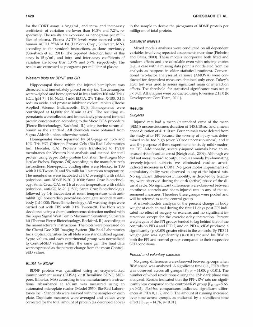
Tail vein samples were collected at 5 and 60 min after 20-min forced exercise, and randomly during the same 1400–1600 h period in voluntary exercising and sedentary rats.Figures 1 and 2 show the CORT and ACTH responses at the 5-min time point. All 60-min values had returned to pre-surgerylevels by this time (data not shown).
A mixed-models analysis of CORT (Fig. 1) shows no maineffect of group and no main effect of exercise, but strong( p < 0.01) effects due to time (PID), and strong effects due tothe exercise · time interaction. Tukey’s post-hoc comparisonsindicated that pre-surgery levels of CORT did not differamong the groups. On PID 0, within 3–5 h after FPI or controlsurgeries, significant ( p < 0.05) increases in CORT levelscompared to pre-surgery levels occurred in all groups exceptthe control-SED group; among the controls, CORT was sig-nificantly ( p < 0.01) increased by fRW and vRW compared tothe SED condition, while in the FPI groups, there were nodifferences among the elevated CORT levels regardless ofcondition (SED, vRW, or fRW). From PID 0 to PID 4, CORTlevels increased significantly ( p < 0.05) in the control-fRWgroup, and decreased significantly ( p < 0.05) in the control-vRW, FPI-SED, and FPI-vRW groups, with no further changeson PIDs 7 and 11. On PIDs 4, 7, and 11, CORT levels weresignificantly ( p < 0.01) higher in the control-fRW and FPI-fRWgroups than in their respective vRW and SED groups, withoutany differences in the elevated CORT levels between the twofRW groups.
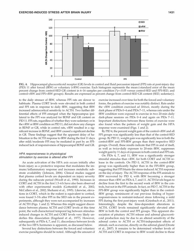
A mixed-models analysis of plasma ACTH levels (Fig. 2)showed no main effect of group, but significant effects of ex-ercise ( p < 0.05), and time (i.e., PID, p < 0.01), along with strong( p < 0.01) effects due to the exercise · time interaction, and tothe three-way interaction between group, exercise, and time.Tukey’s post-hoc comparisons indicated that on PID 0, within3–5 h after FPI or control surgeries, significant ( p < 0.05) in-creases in ACTH levels compared to pre-surgery levels oc-curred in all groups except the FPI-SED group; among thecontrols, ACTH was significantly ( p < 0.01) increased by fRWcompared to the vRW and SED conditions, resulting in sig-
nificantly ( p < 0.01) higher ACTH in the control-fRW groupthan in the FPI-fRW group. On PIDs 4, 7, and 11, ACTH levelswere significantly ( p < 0.05) higher in the control-fRW andFPI-fRW groups than in their respective vRW and SEDgroups. From PID 4 to PID 7, control-fRW ACTH decreasedsignificantly ( p < 0.05), resulting in significantly ( p < 0.01)lower ACTH levels in the control-fRW group than in the FPI-fRW group on PID 7.
Effect of 20-min forced exercise or voluntary exerciseon hippocampal BDNF in control and FPI rats
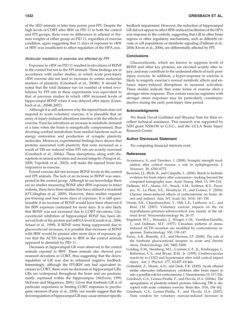
Two-way ANOVA of BDNF on PID 11 obtained with anELISA indicated no significant effects of surgery or exercise,but a significant interaction between the two factors[F(2,54) = 3.82, p < 0.05]. Tukey’s post-hoc analysis indicatedsignificantly higher BDNF content in the control-vRW groupthan in either the control-fRW ( p < 0.05) or control-SED( p < 0.01) groups (Fig. 3A). However, there were no significantdifferences among the three conditions in the FPI rats, or be-tween each FPI group and its corresponding control condi-tion.
Values for PID 11 mature BDNF (mBDNF) that were ob-tained with Western blot showed a similar pattern; however,they did not attain statistical significance (Fig. 3B). No sig-nificant effects were found when proBDNF was analyzed.
Effect of 20-min forced exercise or voluntaryexercise on hippocampal glucocorticoidreceptors in control and FPI rats
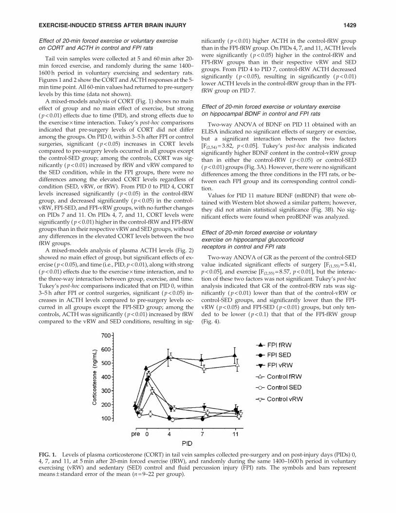
Two-way ANOVA of GR as the percent of the control-SEDvalue indicated significant effects of surgery [F(1,55) = 5.41,p < 0.05], and exercise [F(2,55) = 8.57, p < 0.01], but the interac-tion of these two factors was not significant. Tukey’s post-hocanalysis indicated that GR of the control-fRW rats was sig-nificantly ( p < 0.01) lower than that of the control-vRW orcontrol-SED groups, and significantly lower than the FPI-vRW ( p < 0.05) and FPI-SED ( p < 0.01) groups, but only ten-ded to be lower ( p < 0.1) that that of the FPI-fRW group(Fig. 4).
FIG. 1. Levels of plasma corticosterone (CORT) in tail vein samples collected pre-surgery and on post-injury days (PIDs) 0,4, 7, and 11, at 5 min after 20-min forced exercise (fRW), and randomly during the same 1400–1600 h period in voluntaryexercising (vRW) and sedentary (SED) control and fluid percussion injury (FPI) rats. The symbols and bars representmeans – standard error of the mean (n = 9–22 per group).
EXERCISE-INDUCED STRESS AFTER BRAIN INJURY 1429

Discussion
The current study demonstrates that mild/moderate FPIaffects HPA responses to fRW during the first 11 days post-injury. Daily fRW is a potent stimulus for the HPA axis, unlike
vRW, which is only a stimulus on its first presentation. Incontrast to the significant elevation of ACTH on PID 0, and thesubsequently lower response to daily fRW in the controls,ACTH levels continued to be elevated by daily fRW exposurein the FPI rats. It therefore appears that control rats habituate
FIG. 2. Levels of plasma adrenocorticotropic hormone ACTH in tail vein samples collected pre-surgery and on post-injurydays (PIDs) 0, 4, 7, and 11 at 5 min after 20-min forced exercise (fRW), and randomly during the same 1400–1600 h period involuntary exercising (vRW) and sedentary (SED) control and fluid percussion injury (FPI) rats. The symbols and bars presentmeans – standard error of the mean (n = 11–23 per group).
FIG. 3. Hippocampal brain-derived neurotrophic factor (BDNF) levels in control and fluid percussion injured (FPI) rats atpost-injury day (PID) 11 after forced (fRW) or voluntary (vRW) exercise. Each histogram represents the mean – standard errorof the mean for 6–16 samples per condition. (A) Total BDNF as detected by an enzyme-linked immunosorbent assayexpressed as pg/mg protein (*p < 0.05 versus the control-SED and control-fRW groups). (B) Mature BDNF (mBDNF) detectedwith Western blot, expressed as percent increase over control-SED content (SED, sedentary).
1430 GRIESBACH ET AL.
to the daily stressor of fRW, whereas FPI rats are slower tohabituate. Plasma CORT levels were elevated in both controland FPI rats in response to daily fRW, suggesting that fRWincreased adrenocortical sensitivity to ACTH. Two further dif-ferential effects of FPI emerged when the hippocampus ipsi-lateral to the FPI was analyzed for BDNF and GR content onPID 11. FPI rats, regardless of whether they were sedentary or inthe vRW or fRW condition on PID 11, did not show any changesin BDNF or GR, while in control rats, vRW resulted in a sig-nificant increase in BDNF, and fRW caused a significant declinein GR. These findings suggest that the apparent delay of ha-bituation in the ACTH response to fRW during the first 11 daysafter mild/moderate FPI may be mediated in part by an FPI-induced lack of responsiveness of hippocampal BDNF and GR.
HPA responsiveness to physiologicalstimulation by exercise is altered after FPI
An acute activation of the HPA axis occurs initially afterbrain injury as a protective response that modulates the im-mune/inflammatory response and increases metabolic sub-strate availability ( Johnson, 2006). Clinical studies suggestthat plasma cortisol levels are dependent on injury severityduring the subacute period (Woolf et al., 1990). Increases inCORT and ACTH in the first 1.5–6 h have also been observedwith other experimental models (Gottesfeld et al., 2002;McCullers et al., 2002; Shohami et al., 1995). Likewise, eleva-tions in CORT, which in the rodent is equivalent to cortisol,were found 3–5 h after FPI in the SED rats in the present ex-periments, although they were not accompanied by increasesof ACTH (Figs. 1 and 2). Whereas this might suggest discor-dance between plasma ACTH and CORT levels at this timepoint, the well-characterized temporal lag between stimulus-induced changes in ACTH and CORT levels very likely un-derlies this dissociation (Engeland et al., 1977). However,subsequently at PIDs 4, 7, and 11, discordance between levelsof the two HPA hormones is apparent, as discussed below.
Several key distinctions between the forced and voluntaryexercise paradigms should be noted. Although the amount of
exercise increased over time for both the forced and voluntaryforms, the pattern of exercise was notably distinct. Rats underthe vRW condition exercised ad libitum, mostly during thedark phase of PIDs 0–4 and PIDs 7–11, whereas rats under thefRW condition were exposed to exercise in two 20-min dailydark-phase sessions on PIDs 0–4 and again on PIDs 7–11.Important distinctions between these forms of exercise werealso found when the pattern of weight gain and the HPAresponse were examined (Figs. 1 and 2).
By PID 4, the percent weight gain of the control-vRW and allFPI groups was significantly less than that of the control-SEDgroup. By PID 11, weight gain was significantly less in both thecontrol-fRW and FPI-fRW groups than their respective SEDgroups. Overall, these results indicate that FPI in and of itself,as well as twice-daily exposure to 20-min fRW, suppressesweight gain by 11 days of exposure in both control and FPI rats.
On PIDs 4, 7, and 11, fRW was a significantly strongerstressful stimulus than vRW, for both CORT and ACTH re-lease in the controls. On PID 0, ACTH in the control-fRWgroup was significantly higher than in the FPI-fRW group,suggesting that FPI blunts the stress-induced ACTH responseon the day of injury. The ACTH response of the FPI animals tofRW recovered by PID 4, with fRW becoming a strongerstressor than vRW on PIDs 4, 7, and 11. ACTH levels began todecline from the first to the second week of fRW in the con-trols, but not in the FPI animals. In fact, on PID 7, ACTH in theFPI-fRW group was significantly higher than in the control-fRW group, reminiscent of our previous finding of hyper-responsiveness to restraint-induced stress following a mildFPI during the first post-injury week (Griesbach et al., 2011).Interestingly, despite the time-dependent alterations inACTH, CORT levels remained significantly elevated afterfRW in both the control and FPI groups. This apparent dis-sociation of pituitary ACTH release and adrenal glucocorti-coid production may be due to an altered sensitivity of theadrenal cortex to ACTH or ACTH-independent effects ofstimuli, such as exercise, directly on the adrenal gland (Drosteet al., 2007). It remains to be determined whether levels ofACTH and CORT in response to fRW would decline to those
FIG. 4. Hippocampal glucocorticoid receptor (GR) levels in control and fluid percussion injured (FPI) rats at post-injury day(PID) 11 after forced (fRW) or voluntary (vRW) exercise. Each histogram represents the mean – standard error of the meanpercent change from control-SED GR content in 6–16 samples per condition (*p < 0.05 versus control-SED and FPI-SED, andcontrol-vRW and FPI-vRW groups). Results are expressed as percent change from control-SED GR content (SED, sedentary).
EXERCISE-INDUCED STRESS AFTER BRAIN INJURY 1431
of the SED animals at later time points post-FPI. Despite thehigh levels of CORT after fRW on PID 11 in both the controland FPI groups, there were no differences in adrenal or thy-mus weights of either group on PID 11, regardless of exercisecondition, again suggesting that 11 days of exposure to vRWor fRW was insufficient to affect regulation of the HPA axis.
Molecular mediators of exercise are affected by FPI
Exposure to vRW on PID 11 resulted in elevations of BDNFin the control but not in the FPI animals. These findings are inaccordance with earlier studies, in which acute post-injuryvRW exercise did not lead to increases in certain molecularmarkers of plasticity (Griesbach et al., 2004b). It should benoted that the total distance run (or number of wheel revo-lutions) by FPI rats in these experiments was equivalent tothat of previous studies in which vRW increased levels ofhippocampal BDNF when it was delayed after injury (Gries-bach et al., 2004b,2007).
Although it is still unknown why the injured brain does notrespond to acute voluntary exercise, it is plausible that anarray of injury-induced alterations interfere with the effects ofexercise. Exercise introduces an increase in metabolic demandat a time when the brain is energetically compromised, thusdiverting cerebral metabolism from needed functions such asenergy restoration and production of synaptic plasticitymolecules. Moreover, experimental findings have shown thatproteins associated with plasticity that were increased as aresult of TBI are reduced when FPI rats are acutely exercised(Griesbach et al., 2004a). These disruptions, along with dis-ruptions in neural activation and axonal integrity (Niogi et al.,2008; Topolnik et al., 2003), will make the injured brain lessresponsive to exercise.
Forced exercise did not increase BDNF levels in the controland FPI animals. The lack of an increase in BDNF was unex-pected in the control group. Although to our knowledge thereare no studies measuring BDNF after fRW exposure in intactrodents, there have been studies that have utilized a treadmill(O’Callaghan et al., 2009). However, these studies requiredpre-training and had more days of exposure. It is still ques-tionable if an increase of BDNF would have been observed ifthe fRW exposure continued for more days. It is also likelythat BDNF was not increased due to CORT elevations. Glu-cocorticoid inhibition of hippocampal BDNF has been ob-served both at the protein and mRNA level (Gronli et al., 2006;Schaaf et al., 1998). If BDNF were being suppressed due toglucocorticoid increases, it is possible that increases of BDNFwith fRW would be present after more days of exposure, gi-ven that the ACTH response to fRW in the control animalsappeared to diminish by PID 11.
Decreases in hippocampal GR were observed in the controlanimals exposed to fRW. These animals also showed pro-nounced elevations in CORT, thus suggesting that the down-regulation of GR was due to enhanced negative feedback.Interestingly, although the FPI-fRW rats had equivalent in-creases in CORT, there were no decreases in hippocampal GRs.GRs are widespread throughout the brain and are predomi-nantly expressed within the hippocampus (McEwen, 1999;McEwen and Magarinos, 2001). Given that forebrain GR is ofparticular importance in limiting CORT responses to psycho-genic stressors (Furay et al., 2008), our results suggest that thedownregulation of hippocampal GR may cause stressor-specific
feedback impairment. However, the reduction of hippocampalGR did not appear to affect fRW-induced facilitation of the HPAaxis response in the controls, suggesting that GR in other brainregions or other regulatory mechanisms, such as different cir-cuits or cell populations or metabolic signaling (Dallman et al.,2004; Kwon et al., 2006), are differentially affected by FPI.
Conclusions
Glucocorticoids, which are known to suppress levels ofBDNF and other key proteins, are elevated acutely after in-jury, and may contribute to the undesired effects of early post-injury exercise. In addition, a hyper-response to exercise islikely to magnify exercise’s normal metabolic effects and en-hance injury-induced disruptions in neuronal activation.These studies indicate that some forms of exercise elicit astronger stress response. Thus certain exercise regimens withstronger stress responses may be particularly counterpro-ductive during the early post-injury time period.
Acknowledgments
We thank David Garfinkel and Shyama Nair for their ex-cellent technical assistance. This research was supported byNIH grant NS06190 to G.S.G., and the UCLA Brain InjuryResearch Center.
Author Disclosure Statement
No competing financial interests exist.
References
Avramescu, S., and Timofeev, I. (2008). Synaptic strength mod-ulation after cortical trauma: a role in epileptogenesis. J.Neurosci. 28, 6760–6772.
Bazarian, J.J., Blyth, B., and Cimpello, L. (2006). Bench to bedside:evidence for brain injury after concussion—looking beyond thecomputed tomography scan. Acad. Emerg. Med. 13, 199–214.
Dallman, M.F., Akana, S.F., Strack, A.M., Scribner, K.S., Pecor-aro, N., La Fleur, S.E., Houshyar, H., and Gomez, F. (2004).Chronic stress-induced effects of corticosterone on brain: di-rect and indirect. Ann. NY Acad. Sci. 1018, 141–150.
Droste, S.K., Chandramohan, Y., Hill, L.E., Linthorst, A.C., andReul, J.M. (2007). Voluntary exercise impacts on the rathypothalamic-pituitary-adrenocortical axis mainly at the ad-renal level. Neuroendocrinology 86, 26–37.
Engeland, W.C., Shinsako, J., Winget, C.M., Vernikos-Danellis,J., and Dallman, M.F. (1977). Circadian patterns of stress-induced ACTH secretion are modified by corticosterone re-sponses. Endocrinology 100, 138–147.
Furay, A.R., Bruestle, A.E., and Herman, J.P. (2008). The role ofthe forebrain glucocorticoid receptor in acute and chronicstress. Endocrinology 149, 5482–5490.
Golding, E.M., Steenberg, M.L., Contant, C.F., Jr., Krishnappa, I.,Robertson, C.S., and Bryan, R.M., Jr. (1999). Cerebrovascularreactivity to CO(2) and hypotension after mild cortical impactinjury. Am. J. Physiol. 277, H1457–H1466.
Gottesfeld, Z., Moore, A.N., and Dash, P.K. (2002). Acute ethanolintake attenuates inflammatory cytokines after brain injury inrats: a possible role for corticosterone. J. Neurotrauma 19, 317–326.
Griesbach, G.S., Gomez-Pinilla, F., and Hovda, D.A. (2004a). Theupregulation of plasticity-related proteins following TBI is dis-rupted with acute voluntary exercise. Brain Res. 1016, 154–162.
Griesbach, G.S., Gomez-Pinilla, F., and Hovda, D.A. (2007).Time window for voluntary exercise-induced increases in
1432 GRIESBACH ET AL.

hippocampal neuroplasticity molecules after traumatic braininjury is severity dependent. J. Neurotrauma 24, 1161–1171.
Griesbach, G.S., Hovda, D.A., and Gomez-Pinilla, F. (2009).Exercise-induced improvement in cognitive performance aftertraumatic brain injury in rats is dependent on BDNF activa-tion. Brain Res. 1288, 105–115.
Griesbach, G.S., Hovda, D.A., Molteni, R., Wu, A., and Gomez-Pinilla, F. (2004b). Voluntary exercise following traumaticbrain injury: brain-derived neurotrophic factor upregulationand recovery of function. Neuroscience 125, 129–139.
Griesbach, G.S., Hovda, D.A., Tio, D.L., and Taylor, A.N. (2011).Heightening of the stress response during the first weeks aftera mild traumatic brain injury. Neuroscience 178, 147–158.
Gronli, J., Bramham, C., Murison, R., Kanhema, T., Fiske, E.,Bjorvatn, B., Ursin, R., and Portas, C.M. (2006). Chronic mildstress inhibits BDNF protein expression and CREB activationin the dentate gyrus but not in the hippocampus proper.Pharmacol. Biochem. Behav. 85, 842–849.
Gurkoff, G.G., Giza, C.C., and Hovda, D.A. (2006). Lateral fluidpercussion injury in the developing rat causes an acute, mildbehavioral dysfunction in the absence of significant cell death.Brain Res. 1077, 24–36.
Hansson, A.C., Sommer, W.H., Metsis, M., Stromberg, I., Agnati,L.F., Fuxe, K. (2006). Corticosterone actions on the hippocampalbrain-derived neurotrophic factor expression are mediated byexon IV promoter. J. Neuroendocrinol. 18, 104–114.
Johnson, J.A. (2006). The hypothalmic-pituitary-adrenal axis incritical illness. AACN Clin. Issues 17, 39–49.
Kwon, M.S., Seo, Y.J., Shim, E.J., Choi, S.S., Lee, J.Y., and Suh,H.W. (2006). The effect of single or repeated restraint stress onseveral signal molecules in paraventricular nucleus, arcuatenucleus and locus coeruleus. Neuroscience 142, 1281–1292.
Leasure, J.L., and Jones, M. (2008). Forced and voluntary exercisedifferentially affect brain and behavior. Neuroscience 156,456–465.
Levine, E.S., Dreyfus, C.F., Black, I.B., and Plummer, M.R. (1995).Brain-derived neurotrophic factor rapidly enhances synaptictransmission in hippocampal neurons via postsynaptic tyrosinekinase receptors. Proc. Natl. Acad. Sci. USA 92, 8074–8077.
Mac Donald, C.L., Johnson, A.M., Cooper, D., Nelson, E.C.,Werner, N.J., Shimony, J.S., Snyder, A.Z., Raichle, M.E.,Witherow, J.R., Fang, R., Flaherty, S.F., and Brody, D.L. (2011).Detection of blast-related traumatic brain injury in U.S. mili-tary personnel. N. Engl. J. Med. 364, 2091–2100.
McAllister, T.W., Flashman, L.A., McDonald, B.C., and Saykin,A.J. (2006). Mechanisms of working memory dysfunction aftermild and moderate TBI: evidence from functional MRI andneurogenetics. J. Neurotrauma 23, 1450–1467.
McCullers, D.L., Sullivan, P.G., Scheff, S.W., and Herman, J.P.(2002). Traumatic brain injury regulates adrenocorticosteroidreceptor mRNA levels in rat hippocampus. Brain Res. 947, 41–49.
McEwen, B.S., and Magarinos, A.M. (2001). Stress and hippo-campal plasticity: implications for the pathophysiology of af-fective disorders. Hum. Psychopharmacol. 16., S7–S19.
McEwen BS. (1999). Stress and hippocampal plasticity. Ann RevNeurosci. 22., 105–122.
Neigh, G.N., Karelina, K., Zhang, N., Glasper, E.R., Owens, M.J.,Plotsky, P.M., Nemeroff, C.B., and Devries, A.C. (2009). Car-diac arrest and cardiopulmonary resuscitation dysregulatesthe hypothalamic-pituitary-adrenal axis. J. Cereb. Blood FlowMetab. 29, 1673–1682.
Niogi, S.N., Mukherjee, P., Ghajar, J., Johnson, C., Kolster, R.A.,Sarkar, R., Lee, H., Meeker, M., Zimmerman, R.D, Manley,G.T., and McCandliss, B.D. (2008). Extent of microstructural
white matter Injury in postconcussive syndrome correlateswith impaired cognitive reaction time: A 3T diffusion tensorimaging study of mild traumatic brain injury. Am. J. Neu-roradiol. 29, 967–973.
O’Callaghan, R.M., Griffin, E.W., and Kelly, A.M. (2009). Long-term treadmill exposure protects against age-related neuro-degenerative change in the rat hippocampus. Hippocampus19, 1019–1029.
Osteen, C.L., Moore, A.H., Prins, M.L., and Hovda, D.A. (2001).Age-dependency of 45calcium accumulation following lateralfluid percussion: acute and delayed patterns. J. Neurotrauma18, 141–162.
Pinheiro, J.C., and Bates, D.M. (2000). Mixed Effects Models in Sand S-Plus. Springer-Verlag: New York.
Rapoport, M., McCauley, S., Levin, H., Song, J., and Feinstein, A.(2002). The role of injury severity in neurobehavioral outcome3 months after traumatic brain injury. Neuropsychiatry Neu-ropsychol. Behav. Neurol. 15, 123–132.
R Development Core Team. (2011). R: A language and envi-ronment for statistical computing. R Foundation for StatisticalComputing: Vienna, Austria. ISBN 3-900051-07-0, URL http://www.R-project.org/.
Schaaf, M.J., de Jong, J., de Kloet, E.R., and Vreugdenhil, E.(1998). Downregulation of BDNF mRNA and protein in the rathippocampus by corticosterone. Brain Res. 813, 112–120.
Shohami, E, Bass, R., Trembovler, V., and Weidenfeld, J. (1995).The effect of the adrenocortical axis upon recovery fromclosed head injury. J. Neurotrauma 12, 1069–1077.
Tavazzi, B., Vagnozzi, R., Signoretti, S., Amorini, A.M., Belli, A.,Cimatti, M., Delfini, R., Di Pietro, V., Finocchiaro, A., andLazzarino, G. (2007). Temporal window of metabolic brainvulnerability to concussions: oxidative and nitrosative stresses—part II. Neurosurgery 61, 390–395; discussion 395x–396.
Topolnik, L., Steriade, M., and Timofeev, I. (2003). Hyperexcit-ability of intact neurons underlies acute development oftrauma-related electrographic seizures in cats in vivo. Eur. J.Neurosci. 18, 486–496.
Tran, L.D., Lifshitz, J., Witgen, B.M., Schwarzbach, E., Cohen,A.S., and Grady, M.S. (2006). Response of the contralateralhippocampus to lateral fluid percussion brain injury. J. Neu-rotrauma 23, 1330–1342.
Tyler, W.J., and Pozzo-Miller, L.D. (2001). BDNF enhancesquantal neurotransmitter release and increases the number ofdocked vesicles at the active zones of hippocampal excitatorysynapses. J. Neurosci. 21, 4249–4258.
Vagnozzi, R., Signoretti, S., Tavazzi, B., Floris, R., Ludovici, A.,Marziali, S., Tarascio, G., Amorini, A.M., Di Pietro, V., Delfini,R., and Lazzarino, G. (2008). Temporal window of metabolicbrain vulnerability to concussion: a pilot 1H-magnetic reso-nance spectroscopic study in concussed athletes—part III.Neurosurgery 62, 1286–1295; discussion 1295–1286.
Woolf, P.D., Cox, C., Kelly, M., Nichols, D., McDonald, J.V., andHamill, R.W. (1990). The adrenocortical response to brain in-jury: correlation with the severity of neurologic dysfunction,effects of intoxication, and patient outcome. Alcohol Clin. Exp.Res. 14, 917–921.
Address correspondence to:Grace S. Griesbach, Ph.D.
David Geffen School of Medicine at UCLADepartment of Neurosurgery
Box 957030Los Angeles, CA 90095-7039
E-mail: [email protected]
EXERCISE-INDUCED STRESS AFTER BRAIN INJURY 1433