Embed Size (px)
Citation preview
Dublin Institute of TechnologyARROW@DIT
Books/Book Chapters School of Food Science and Environmental Health
2011-01-01
Differential Precipitation and Solubilization ofProteins.Barry RyanDublin Institute of Technology, [email protected]
Follow this and additional works at: http://arrow.dit.ie/schfsehbk
Part of the Biochemistry Commons, Biotechnology Commons, Food Science Commons, and theMolecular Biology Commons
This Book Chapter is brought to you for free and open access by the Schoolof Food Science and Environmental Health at ARROW@DIT. It has beenaccepted for inclusion in Books/Book Chapters by an authorizedadministrator of ARROW@DIT. For more information, please [email protected], [email protected], [email protected].
This work is licensed under a Creative Commons Attribution-Noncommercial-Share Alike 3.0 License
Recommended CitationRyan, B.J. (2011). Differential precipitation and solubilization of proteins. IN Methods in Molecular Biology (Eds. Loughran, S.T. andWalls, D.) Springer Protocols/Humana Press, NY, USA, pp. 203-213.
Differential Precipitation and Solubilisation of Proteins.
Barry J. Ryan
School of Food Science and Environmental Health, Dublin Institute of Technology, Cathal
Brugha Street, Dublin 1, Republic of Ireland.
Email: [email protected]
Summary/Abstract
Differential protein precipitation is a rapid and economical step in protein purification and is
based on exploiting the inherent physico-chemical properties of the polypeptide. Precipitation of
recombinant proteins, lysed from the host cell, is commonly used to concentrate the protein of
choice before further polishing steps with more selective purification columns (e.g. His-Tag,
Size Exclusion etc.). Recombinant proteins can also precipitate naturally as inclusion bodies due
to various influences during over-expression in the host cell. Although this phenomenon permits
easier initial separation from native proteins, these inclusion bodies must carefully be
differentially solubilised so as to reform functional, correctly folded proteins. Here, a typical
protein extraction, precipitation and selective resolubilisation procedure is outlined, based on a
recombinantly expressed protein.
Key Words
Ammonium Sulphate precipitation, Trichloroacetic acid precipitation, inclusion body
solubilisation, protein refolding.
1. Introduction
Protein precipitation can be caused by the differential solubility between a protein-rich soluble
phase and a solid chemical precipitant. Soluble proteins can be insolubilised by interaction with a
suitable precipitant that decreases the protein’s attraction to the solvent and increases the
protein’s attraction to other protein molecules, resulting in protein accumulation and eventually
precipitation. The addition of low molecular weight substances, such as glycerol, polyethylene
glycol and sucrose, and high molecular weight substances such as serum albumin, can have
significant effects on protein structure and stability. Preferential hydration of a protein molecule
caused by the presence of these additives can increase the protein’s stability. Certain salts can
also exert a stabilising effect by ‘salting out’ hydrophobic residues of a protein, causing the
molecule to adapt a more compact, stable structure (1) frequently resulting in precipitation. The
use of such protein precipitating molecules is an empirical process, the effects of any given
substance on a protein must be determined experimentally. The use of additives can not only be
used as a simple approach to increase the stability of a given protein, but also to actively effect
protein precipitation. Protein precipitation can be used as a crude protein clean-up method from
cell lysates, readily employed after bacterial over-expression of recombinant proteins.
Differential solubilisation of proteins is often employed for proteomic analyses (2, 3), but it too
can offer an alternative purification technique for non-soluble recombinant proteins expressed in
heterologous hosts. Recombinant proteins expressed as inclusion bodies can be readily separated
from the host cell protein matrix, however careful solubilisation and refolding are critical for
obtaining suitable recombinant proteins for further downstream processes. Here a typical
recombinant protein precipitation and resolubilisation procedure is outlined.
2. Materials
Note: All consumables may be sourced from Sigma-Aldrich unless otherwise stated.
2.1 Recombinant / Native Protein Extraction.
1. Centrifuge (Bucket Type, e.g. J2-21, Beckman and microfuge, e.g. 5415D Eppendorf).
2. pH meter (e.g. M-240, Corning).
3. Sonicator (e.g. Vibra Cell, Sonics Scientific).
4. Water bath (temperature controlled).
5. Vortex.
6. Resuspension Buffer One: 50 mM Na2HPO4-NaH2PO4, pH 8.0, 0.3 M NaCl.
7. -80oC freezer
2.2 Protein Precipitation using Ammonium Sulphate.
1. Resuspension Buffer One: 50 mM Na2HPO4-NaH2PO4, pH 8.0, 0.3 M NaCl.
2. Saturated Ammonium Sulphate: Add 750 g of (NH4)2SO4 to 1 L of double distilled water
in a beaker or flask. Stir the solution at room temperature with a magnetic stirrer for 15
min or until saturation. Gently decant the clear supernatant solution after the undissolved
solids settle on the bottom of the flask.
3. Graduated Pipette (10 mL).
2.3 Protein Precipitation using Trichloroacetic acid.
1. 2% Deoxycholate (DOC): Add 2 g of DOC to 100 mL of double distilled H2O, mix well.
2. 100% Trichloroacetic acid (TCA): Add 1 g of TCA to 454 µL double distilled H2O and
mix carefully. Store in a light proof bottle at 4oC until required for use. TCA is a harmful
skin and eye irritant. Always use correct personal protective equipment when handling it.
3. Acetone (ice cold). Store acetone at –20oC. Use directly from -20oC.
4. Vacuum Dryer (e.g. DNA 110 Speed Vac, Savant)
5. Resuspension Buffer: 50 mM Na2HPO4-NaH2PO4, pH 8.0, 0.3 M NaCl.
6. Centrifuge (Bucket Type, e.g. J2-21, Beckman and microfuge, e.g. 5415D Eppendorf).
2.4 Protein Solubilisation.
1. DNase I (100 U/mL).
2. Resuspension Buffer Two: 50 mM Na2HPO4-NaH2PO4, pH 8.0, 0.3 M NaCl, 5mM DTT,
0.35 mg/mL Lysozyme, Proteinase Inhibitor Cocktail (see also Chapter 4). Make up as
fresh prior to use.
3. Triton X-100.
4. Solubilisation Buffer: 50 mM Na2HPO4-NaH2PO4, pH 8.0, 0.3 M NaCl, 25 mM DTT, 6
M Guanidine HCl.
5. PBS-T: Phosphate buffered saline (PBS, 1x) containing 1% Triton X-100.
6. Centrifuge (Bucket Type, e.g. J2-21, Beckman and microfuge, e.g. 5415D Eppendorf).
7. Vacuum Concentrator (Speed Vac, Savant)
2.5 Protein Refolding.
1. Refolding Buffer: 50 mM Na2HPO4-NaH2PO4, pH 8.0, 0.3 M NaCl, 2.5 mM reduced
Glutathione, 0.25 mM Oxidized Glutathione, 0.2 M Arginine.
2. Dialysis Buffer: 50 mM Na2HPO4-NaH2PO4, pH 8.0, 0.3 M NaCl.
3. Dialysis apparatus (dialysis tubing and clips, magnetic mixer, large clean container).
4. Guanidine Hydrochloride: 6 M stock, made in double distilled H2O.
5. Amicon protein concentration device (e.g. Ultra-15 Centrifugal Filter Units, Amicon).
6. Gradient maker apparatus (see Fig. 1).
3. Methods
3.1 Recombinant / Native Protein Extraction.
The source of the protein will determine the optimal technique to release the protein from
the tissue or cells in which it is contained. The typical freeze-thaw cell lysis procedure
(below) is generally sufficient to lyse most bacterial cell types although other options are
available (see Notes 1, 2, 3).
1. Collect the bacterial cells by transferring the bacterial culture to a pre-chilled
sterile centrifuge tube and centrifuge at low speed (5 min, 800 x g) in a previously
cooled centrifuge (4°C).
2. Carefully remove the culture media from the bacterial cell pellet, ensuring the
pellet is not disturbed.
3. Resuspend the cell pellet in Resuspension Buffer One, in 10% of the original
culture volume.
4. Freeze the resuspended cells to -80oC by placing the resuspension solution (still in
the plastic centrifuge tube) into a pre-equilibrated -80oC freezer, then warm the
cells to 37oC (using a pre-equilibrated water bath) for 10 min. Repeat this freeze
thaw process three times.
Sonication can also be used if the protein is not released during the freeze thaw steps. It is
crucial to maintain the cell suspension on ice during the sonication process (see Notes 4, 5,
6).
5. Sonicate at 10 amplitude microns for 10-20 s.
6. Allow the cell suspension to stand on ice for 30 s.
7. Repeat steps 5 and 6 three more times.
8. Check the recombinant protein induction/expression by loading and analysing a
representative sample (typically 50 µg protein) onto a SDS-PAGE gel.
3.2 Protein Precipitation using Ammonium Sulphate.
A common and inexpensive first step to isolate proteins during protein purification is
precipitation with an external additive. This additive alters the physicohemical properties of
the protein causing it to fall out of solution. Ammonium sulphate (see Note 7) is commonly
used for large scale precipitations.
1. Gently stir the protein mixture with the aid of a magnetic stirring bar at 4oC. Add,
using a graduated pipette, the saturated ammonium sulphate solution drop-wise to
the protein solution until precipitates start to form (see Notes 8, 9, 10).
2. Once sufficient saturated salt solution has been added to cause precipitation of the
protein of interest (indicated by collection of precipitate at the bottom of the
container), centrifuge the mixture at 10,000 g for 15 min. Collect the precipitate
by carefully discarding as much supernatant as possible (see Note 10).
3. Resuspend the protein pellet at 4oC in Resuspension Buffer One for further
downstream processes.
3.3 Protein Precipitation using Trichloroacetic acid.
Trichloroacetic acid (TCA) is routinely employed for small scale operations or
precipitations of protein preparations that are at low concentration; however, it should be
noted that this procedure is protein denaturing, and caution must be exercised when
working with TCA (see Notes 11, 12).
1. To one volume of protein solution, add 1/100 volume of 2% DOC (sodium
deoxycholate).
2. Vortex and incubate for 30 min at 4oC.
3. Add 1/10 volume of 100% Trichloroacetic acid (TCA). Vortex the solution and
incubate statically overnight at 4oC.
4. Centrifuge the sample for 15 min at 4oC (10,400 x g). Gently remove the
supernatant and retain the pellet. Carefully dry the tube by inversion on tissue
paper (note: the pellet may be difficult to see).
5. Optional: Wash the pellet twice with one volume of ice-cold acetone. Vortex and
re-pellet the samples by centrifugation 10,400 x g for 5 min at 4oC between
washes (see Note 13).
6. Dry the samples under vacuum (e.g. Speed Vac, Savant) or allow to air dry.
7. Resuspend the protein pellet in a buffer of choice for further downstream
processes.
3. 4 Protein Solubilisation.
Recombinant protein expression in a heterologous host frequently results in insoluble and
inactive proteins. Regularly, protein overexpression results in the production of inclusion
bodies, which are insoluble aggregates of misfolded protein. Although these inclusion
bodies can easily be purified, further characterisation of this protein mass is often
impossible without solubilisation of the protein of interest and refolding into an active form
(see Note 14). A typical inclusion body solubilisation and refolding protocol is outlined
below (see also Note 15):
1. Carry out steps 1-3 as outlined in 3.1, except resuspend the cell pellet in 10% of
the original culture volume of Resuspension Buffer Two.
2. Slowly add Triton X-100 (to a final concentration of 1% v/V), and mix gently.
3. Carry out the sonication procedure as outlined in 3.1, steps 5 -7.
4. Incubate the cell debris with DNase I (100 U/mL) for 1 h at 37oC.
5. Collect the inclusion bodies by centrifugation at 30,000 x g for 30 min at 4oC.
6. Wash the inclusion body pellet twice with PBS-T, followed by centrifugation at
30,000 g for 30 min at 4oC.
7. Solubilise the pelleted inclusion bodies in the solubilisation buffer and allow total
solubilisation to occur at 4oC for 1 h, with occasional gentle mixing.
8. After 1 h solubilisation, remove all remaining insoluble material by centrifugation
30,000 x g for 10 min at 4oC (see Note 16).
9. Determine the protein concentration and adjust to 1 mg/mL by dilution in
solubilisation buffer and proceed directly to re-folding at 4oC (Section 3.5).
3.5 Protein Refolding.
1. Dilute the solubilised proteins as quickly as possible (to yield a final protein
concentration of 0.1 mg/mL, see Note 17) into pre-chilled Refolding Buffer (see
Note 18).
2. Dialyze the diluted solubilised protein overnight against a 200-fold volume of
dialysis buffer with slowly decreasing concentrations of GuHCl (typically
decrease GuHCl concentration as follows: 6 M, 4 M, 2 M, 1 M, 0.5 M and then 0
M in a continual dialysis approach; see Fig. 1 and Notes 19, 20).
3. Centrifuge the dialysate at 4°C for 30 min at 30,000 x g.
4. Carefully remove the liquid protein rich layer, concentrate (e.g. Amicon filtration)
and store at an appropriate temperature (see Note 21).
Figure legend
Figure 1: Schematic diagram of a simple gradient maker.
Notes
1. There are several methods to achieve this, including repeated freezing and thawing,
sonication, homogenization at high pressures, enzymatic lysis or permeabilization by organic
solvents. The method of choice depends on how fragile the protein is and how robust the host
cell is.
2. Proteins can also be selectively released from the various compartments of a bacterial host, for
example proteins expressed in the periplasmic envelope can be selectively lysed by a method
similar to that described by French and co-workers (4). Pellet the bacterial cells to be disrupted
by centrifugation at 800 x g for 3 min. Resuspend the pellet in Fractionation Buffer (F1) buffer in
20% of the original culture volume. The F1 comprises (final concentrations): 500 µg/mL
lysozyme, 20% w/v sucrose, 1 mM EDTA, pH 8.0, 200 mM guanidine hydrochloride and 200
mM Tris-HCl, pH 8.0, at room temperature. Statically incubate the resuspended cells at room
temperature for 15 min, after which add an equal volume of ice-cold water. Stand the mixture at
room temperature for 15 min. Remove the cell debris by centrifugation at 10,400 x g for 10 min.
Transfer the supernatant (containing the periplasmic fraction) to a clean container for further
purification.
3. Proteins expressed and transported to the culture supernatant can also be conveniently
concentrated by a method outlined by Caldwell and Lattemann (5). In brief this method involves
adding an equal volume of a PRMM solution (0.05 mM pyrogallol red, 0.16 mM sodium
molybdate, 1.0 mM sodium oxalate, 50.0 mM succinic acid, 20% v/v methanol in H2O, adjusted
to pH 2.0 with HCl) to cleared (0.22 µm filtered) culture supernatant. Adjust the pH of the
solution to 2.8 (±0.1), and allow the proteins to precipitate for 1 to 2 h at room temperature,
followed by an overnight incubation at 4°C. Sediment the precipitate by centrifugation at 10,000
x g for 1 h, and carefully remove the supernatant. Repeatedly rinse the precipitate with 1 mL of
acetone. Remove all traces of acetone by evaporation at room temperature. Solubilise the
precipitate by adding 100 µL of 2x sodium dodecyl sulfate-polyacrylamide gel electrophoresis
(SDS-PAGE) sample buffer (25% glycerol, 8% SDS, 4% β-mercaptoethanol, 0.02%
bromophenol blue, 100 mM Tris- HCl, pH 6.8). These samples can be applied directly to an
SDS-PAGE gel, if this type of analysis is required.
4. The sonication process can generate large amounts of heat, which is why pulses are limited to
~20 s. In between pulses, cool the tube in ice or ice-water slurry for 30 s. If a large volume is
required to be sonicated, split the cell suspension into two tubes, and alternate the sonication and
cooling steps. Sonicate on ice where possible.
5. The extraction process also releases proteases, which will digest all proteins in the solution. If
the protein is sensitive to proteolysis, it is desirable to employ a protease inhibitor (see Chapter
4), to proceed quickly, and to keep the extract cooled to minimise proteolysis.
6. Lysozyme (500 µg/mL, to assist cell wall degradation) and DNase I (100 U/mL, to degrade
genomic DNA) can be added to the lysis buffer.
7. The addition of high concentrations of salt to a protein solution causes precipitation by
removing water from hydrophobic patches on the protein’s surface, resulting in these patches
aggregating together causing the protein to come out of solution. A number of salts can be used
for this process; NaCl, Na2SO4, KCl, CaCl2 and MgSO4, however, (NH4)2SO4 is by far the most
commonly used additive. This is due to several advantageous characteristics of the salt, including
the fact that it has a high solubility in water (4 M saturation) and it has a low density at saturation
allowing precipitated proteins to be collected by centrifugation. Hence the first proteins to be
purified during ammonium sulphate precipitation are water-soluble proteins (6).
8. Final concentrations of ammonium sulphate must be calculated using standard nomograms or
with online tools (“Ammonium Sulfate Calculator”, available at
www.encorbio.com/protocols/AM-SO4.htm). Adding increasing amounts of ammonium sulphate
causes the different fractions of a protein mixture to precipitate at different rates. One advantage
of this method is that it can be performed inexpensively with very large volumes. Additionally,
the high salt content of the precipitated protein permits its direct addition onto a hydrophobic
interaction chromatography (HIC) purification column, thus speeding up the overall purification
process.
9. Purification of integral membrane proteins requires the addition of a detergent such as sodium
dodecyl sulphate (1% SDS) to dissolve cell membranes and keep membrane proteins in solution
during purification. It should be noted that SDS causes protein denaturation, hence milder
detergents such as 1% Triton X-100 or 1% CHAPS can be used to retain the protein's native
conformation during cell membrane dissolution.
10. Ammonium sulphate salt can be added either in saturated solution (as described above) or
directly as salt crystals. It may be advantageous to add ammonium sulphate directly into the
protein mixture as powdered solids during large scale purification processes so that the effect of
dilution by the salt solution is minimized. If a saturated salt solution is employed the amount of
ammonium sulphate solution added must be recorded accurately, often this is achieved by
dispensing from a graduated pipette. It is critical to avoid the spatial non-uniformity in the salt
concentration during the addition of the salt solution. Localized concentration “hot-spots” will
prematurely initiate the precipitation of other proteins and inadvertently affect the precipitation
process. Record the volume of the saturated ammonium sulphate solution required to precipitate
the protein of interest. Also note that protein precipitation is not instantaneous; it may require 15
to 20 min to equilibrate.
11. TCA is a harmful skin and eye irritant. Always use correct personal protective equipment
when handling it.
12. There are numerous options to effect other types of protein precipitation including (but not
limited to) acetone precipitation (useful to simultaneously eliminate acetone soluble components
and increase protein concentration), ethanol precipitation (useful to simultaneously concentrate
proteins and remove traces of GuHCl prior to SDS-PAGE analysis), acidified acetone/methanol
(50/50 v/v; useful to simultaneously remove acetone and methanol soluble interferences such as
SDS prior to IEF analysis) and chloroform/methanol (50/50 v/v; useful to simultaneously remove
salt and detergents).
13. The presence of trace amounts of TCA, carried through from the precipitation, can acidify
the resuspension sample buffer. If further downstream processes are pH sensitive the sample
buffer should be titrated with 1 M NaOH or 1 M TrisHCl, pH 8.5, to obtain the desired pH for
the required process. Acidified SDS-PAGE sample buffer, for example, can give a yellow
colour. Correct titration will result in reversion to the typical blue sample buffer colour. Hint: A
simple method to overcome this is to resuspend the samples in a slightly basic SDS-PAGE
loading buffer (e.g. pH 9.0). Hence, any residual TCA left it will be neutralized by the basic
buffer allowing direct addition onto the SDS-PAGE gel. Additionally, excess TCA traces will
cause the Coomassie dye to precipitate during SDS-PAGE. If this is a problem repeat the
optional wash steps outlined.
14. The interactions between solvents and proteins, and also proteins and proteins, determine the
solubility of any given protein. Interactions can be classified as either attractive or repulsive. A
protein will be soluble in a particular solvent if the net free energy of the proteins interactions is
adequately negative (i.e. attractive). Additionally, protein solubility is improved if protein-
protein interactions have sufficiently positive net free energy (i.e. repulsive), although it should
be noted that protein-protein interaction is modulated by the chemical nature of the solvent of
choice. Conversely, insolubility typically results from net attractive forces between proteins and
net repulsive forces between the solvent of choice and the protein of interest. Furthermore, a
soluble protein can be insolubilised by a change in it’s free energy state in relation to the
proteins, or the solvent, it interacts with and, hence, additions/subtractions to a protein solution
should be carefully assessed on a small scale (7).
15. The separation of one protein, or family of proteins, from other proteins by means of
differential solubility with chemical reagents is based on the differential solubility between a
liquid phase and a solid phase. The optimisation of this procedure is empirical but, Lindwall and
colleagues (8) outline an optimisation procedure based on a sparse matrix approach;
solubilisation buffers are composed based on “solubility space” which is related to accepted
protein solubilisation theories. This method assists in identifying suitable solubilisation
conditions for most over-expressed proteins.
16. It is important to remove existing aggregates that can act as nuclei to trigger aggregation
during folding.
17. The final protein concentration should not exceed 0.05 to 0.1 mg/mL as dilute protein
mixtures refold optimally at this concentration. A rapid and efficient mix is essential at this step.
18. The addition of a mild solubilising agent [e.g. 1 M 3(1-pyridinio)-1-propane sulfonate]
during the refolding steps limits re-aggregation of re-folding proteins.
19. Continual dialysis can be set up by using a gradient maker. In its simplest form, this consists
of two containers of the same shape connected by a siphon. One container contains the low
concentration buffer, and the other contains high concentration buffer. The buffer is withdrawn
from the low concentration container to the high concentration container. This will produce a
linear gradient from high to low buffer concentrations over the total volume of the gradient.
Once the “low concentration buffer” supply has been depleted, the dialysis tubing is removed
from the larger vessel and placed in a similar, clean vessel containing fresh buffer at the same
concentration as the original “low concentration buffer”. The “low concentration buffer” vessel
is replaced with a vessel containing buffer at the next lower concentration level and the process
is allowed to continue until the “low concentration buffer” supply is depleted again. This process
is repeated until the vessel containing the dialysis tubing has reached the desired final
concentration, typically 0 M GuHCl.
20. See Rudolph and Lilie (9) for a comprehensive overview of protein refolding and (10) for an
industrial viewpoint on that topic.
21. Most proteins can be stored at 4°C, without significant denaturation, for up to 24 h. For
intermediate storage times (24 h to one week) the protein should be filter sterilised (through a
0.22 µm filter) and stored at 4°C. Additional supplements, such as a bacteriostatic agent (e.g.
0.1% sodium azide) can be included to avoid bacterial growth. For storage times greater than one
week (up to several months) it is advisable to freeze the protein preparation. Rapid freezing helps
reduce protein denaturation. It is useful to freeze the solution in small aliquots to avoid repeated
freeze/thaw cycles which may reduce the biological activity of the protein. Additional stabilizing
agents can also be added prior to freezing, such as glycerol (5-50% w/v), serum albumin (10
mg/mL), reducing agents (such as 1 mM DTT), and ligands/co-factors (depending on the nature
of the target protein). Extended protein storage (several months to years) should be carried out at
-80°C or in liquid nitrogen. The addition of 50% (w/v) glycerol is recommended for storage at
this temperature. Alternative strategies include storing the protein as an ammonium sulphate
precipitate at 4°C, or at lower temperatures in a lyophilized form (see also Chapter 10 for
protocols and discussion regarding the storage and lyophilisation of proteins).
References
1. O’Fágáin, C. (1997) Protein stability and its measurement, in Stabilising protein function
(Fágáin, C.O’., ed.), Springer Press, Berlin, pp 69-75.
2. Ramos, Y., García, Y., Llopiz, A., and Castellanos-Serra, L. (2008) Selectivity of bacterial
proteome fractionation based on differential solubility: A mass spectrometry evaluation Anal.
Biochem. 377, 134–140.
3. Leimgruber, R. M. (2005) Extraction and solubilisation of proteins for proteomic studies, in
The Proteomics Protocols Handbook (Walker J. M., ed.), Humana, Totowa, NJ, pp 1-18.
4. French, C.; Keshavarz-Moore, E., Ward, J.M. (1996) Development of a simple method for the
recovery of recombinant proteins from the E.coli periplasm. Enzyme Microb. Technol. 19, 332–
8.
5. Caldwell R.B., and Lattemann C.T. (2004) Simple and reliable method to precipitate proteins
from bacterial culture supernatant. Appl. Environ. Microb. 70, 610-12.
6. Doonan, S. (2004) Bulk Purification by Fractional Precipitation, in Protein Purification
Protocols, Methods in Molecular Biology (Cutler, P., ed.), Humana, Totowa, NJ, 244, pp 117-
125.
7. Rothstein F. (1994) Differential Precipitation of Proteins, in Protein Purification Process
Engineering (Harrison, R. G. ed), Marcel Dekker Inc., NY, pp 115-16.
8. Lindwall, G., Chau, M.-F., Gardner, S.R., and Kohlstaedt, L.A. (2000) A sparse matrix
approach to the solubilisation of overexpressed proteins. Protein Eng. 13, 67-71.
9. Rudolph, R., and Lilie, H. (1996) In vitro folding of inclusion body proteins. FASEB 10, 49-
56.
10. Jungbauer, A., and Kaar, W. (2007) Current status of technical protein refolding. J.
Biotechnol. 128, 587-96.
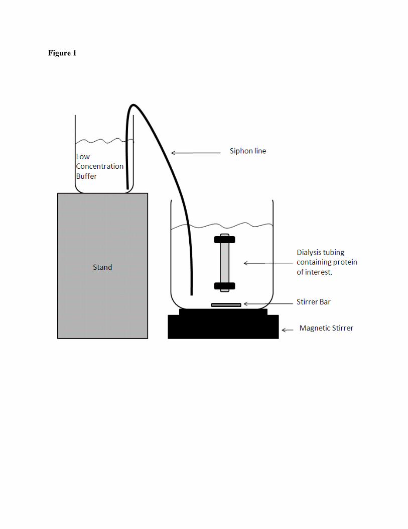
Figure 1