Embed Size (px)
Citation preview
Differential Survival of Solid Tumor Cells after Inoculationinto Established Ascites Tumors*
EVAKLEINANDGEORGEKLEIN
(Wallenberg Laboratory, Institute for CM Research, Karolinska Institutet, Stockholm 60, Sweden)
It has been shown (6) that only a limited number of transplantable mouse neoplasms can begrown in the form of typical "ascites tumors," if
the term is used to denote a condition where freetumor cells or cell complexes multiply in the peritoneal fluid, and, accompanied by the gradual accumulation of ascites, there attain a nearly pureculture. The 42 tumors surveyed showed three distinctly different patterns of behavior: (a) Six tumors, mainly malignant lymphomas, grew readilyin the ascites form when adequate numbers of cellsfrom the solid tumor were injected intraperitoneal-ly. (6) Six other neoplasms yielded exudates poorin tumor cells. Upon further serial intraperitonealtransfer of these exudates typical ascites tumorswere eventually obtained, (c) The majority, 30 tumors, did not lend themselves to transformationinto the ascites form under the conditions used.
The present investigation represents the firststep in a series of experiments designed to gain information about the factors concerned in determining whether a certain tumor can be grown inthe ascites form. It is possible that the differenceamong these three tumor categories is dependenton a differential capacity of the various cell populations to survive and multiply in the peritonealfluid. Alternatively, the findings could be due tovariations in the capacity of different tumors toprovoke primary exúdate formation after intraperitoneal inoculation. In the latter case, survivalof various tumor cell populations inoculated intoestablished peritoneal exudates would not be expected to be systematically different. To decideamong these possibilities, several neoplasms whichhave been studied previously with regard to theirascites tumor-producing capacity were inoculatedas cell suspensions into established peritonealexudates, and their survival in the fluid was testedby means of a biological assay.
* This investigation was supported by research grants fromthe Swedish Anti-Cancer Society, Lotten Bohman's Fund, andKonung Gustaf V's Jubileumsfond.
Received for publication October 14, 1953.
MATERIALS AND METHODSMice.—Thefollowing five inbred strains were used: C8H/
St, A/St, CS7BL, DBA, and CS7L. Breeding pairs of thefirst four strains were obtained from Dr. T. S. Hauschka (Institute for Cancer Research, Philadelphia) in 1950. The C57Lstrain was kindly sent us by Miss E. Fekete of the Jackson Memorial Laboratory. All five strains were maintained by strictbrother-sister mating. For part of the experiments, Fi hybridsproduced from various combinations of the five strains wereused. Animals of both sexes were employed, at an age of 2-3months and with a body weight of 18-25 gm. The mice werekept on a standard pellet diet, which, as well as drinking water,was available ad libitum at all times.
Tumors.—Seventeendifferent transplantable mouse tumorswere used (Table 1). Most of these neoplasms had been testedpreviously with regard to their capacity to grow in the ascitesform (4, 6) by passing them through serial intraperitonealtransfers; peritoneal exudates were used for inoculation whenever possible.
The tumors capable of growing in the ascites form (seecol. 7, Table 1) were maintained by serial intraperitonealtransfers of ascitic fluid. For the present experiments, 0.2 ml.was inoculated subcutaneously into mice of a compatiblestrain, and the developing solid tumors were used. The neoplasms that did not grow as ascites tumors were maintainedby serial subcutaneous passages of cell suspensions preparedby mechanical disruption of the tissue.
In the present work medium-sized solid tumors of bothkinds were removed, freed from grossly necrotic areas, minced,and disrupted by the centrifuge method of Hauschka andPoppe (3). Using a disc of stainless steel mesh, 60 wires/inch,and buffered physiological saline as medium, uniform suspensions were obtained which contained mainly single cells andsmall clumps of cells. The suspensions were diluted twenty-fold by Tyrode's solution containing 0.05 per cent eosin,
and the unstained cells were quickly counted in a Buerkerhemocytometer. According to Schrek (9), this represents thenumber of viable cells. More recent evidence (1) indicatesthat, while unstained cells are viable, staining does notnecessarily mean cell death. The number of unstained cellswould therefore represent the minimum number of viablecells. A predetermined number of unstained cells (usually1 million) was used for inoculation according to the experimental scheme as presented below.
The growth rate of the solid tumors was estimated on thebasis of daily caliper measurements according to the methoddescribed by Schrek (8). Three different tumor diameterswere measured, and the geometric mean was calculated andplotted against time. Approximately straight-line relationships were usually obtained on an arithmetic scale. Leastsquare lines were fitted to the experimental points, and theregression coefficient was taken to indicate the average dailylinear growth of the tumor.
139
on March 31, 2019. © 1954 American Association for Cancer Research.cancerres.aacrjournals.org Downloaded from
140 Cancer Research
RESULTSThe main technical problem was the production
of sufficient amounts of peritoneal exúdate whichcould be presumed to be to some degree similar incomposition to the fluid in which ascites tumorcells usually grow. Although there are several waysto produce sterile exudates in the peritoneal cavityof mice, large amounts of fluid are seldom obtained,and it is difficult to maintain the exúdateat a satisfactory level for an adequate period of time. Suchexudates are usually produced by means of mechanical or chemical irritation, and they are notnecessarily comparable to the medium of ascitestumor cells. It was therefore decided to utilize anestablished ascites tumor to produce and maintain asufficient volume of exúdateinto which cell suspensions from the test tumor could be inoculated. Advantage was taken of the high strain specificity oftwo ascites lymphomas which were used for thispurpose and which will be referred to as "carrier"
tumors. The solid neoplasms tested will be designated as "test tumors."
The two carrier tumors, 6C3HED lymphomaand DBA lymphoma, were found to grow progressively in their strains of origin only (C3H andDBA, respectively), or in FI hybrids between theoriginal and any other strain (2, 5). In each experiment, test tumors were selected that hadoriginated and could grow in a mouse strain different from that of the carrier tumor. Under suchcircumstances, the carrier and test tumors could begrown together in FI hybrids between their twoparent strains and separated by subsequent inoculation into animals of the two original pure lines.
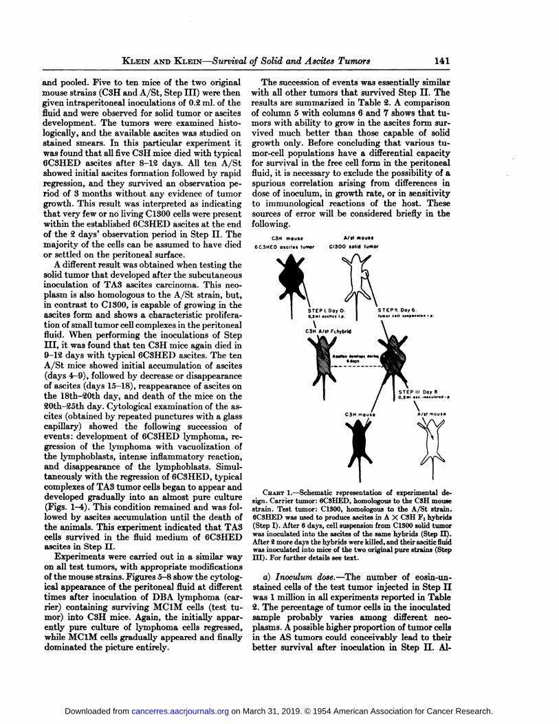
Chart 1 illustrates the experimental procedurefor carrier tumor 6C3HED and test tumor C1300.Five A X C3H FI hybrids were given an inoculationin Step I of 0.2 ml. 6C3HED ascites on day 0. Byday 6 the mice showed pronounced abdominal distension due to the accumulation of 3-4 ml. ofascites. Into this fluid a suspension containing 1million eosin-unstained cells from a C1300 tumorwas injected (Step II). On the 8th day the micewere killed, and all the ascitic fluid was collected
TABLE1LISTOFTUMORS
Designation16C3HEDDBA
lymphoma(Dalton)Ehrlich15091aKrebs
2TASMCIAMC1MLymphoma
EL.4SPÄHS2CS8ASSCC954SSDS6CC1SOOType*2ind.
lymphosarcomasp.lymphosarcomasp.
carcinomasp.
adenocarcinoma.(since1835, spin
dle-cellcarcinoma)sp.carcinoma?re-ticulosarcoma?sp.
adenocarcinomaind.
rhabdomyosar-comaind.
sarcomaind.
lymphomasp.
adenocarcinomasp.
adenocarcinomasp.
adenocarcinomasp.
adenocarcinomacarcinoma
ofliverparenchymacellssp.adenocarcinomasp.
adenocarcinomasp.
neuroblastoma?Year
oforiginS194119471806?19281933194819451946194519501951195219521940195219521940Derivation4thymusthymusmammaryglandmammarygland?mammaryglandmusclemuscleinfiltratedorgansmammaryglandmammaryglandmammaryglandmammaryglandlivermammaryglandmammaryglandregion
ofspinalcordMouse
strainoforiginSCSUDBAnonin-bredAnonin-bredAC3HC3HC57BLACSHAC3HC57LDBAC3HAProgressivegrowthinmousestrain6CSH
onlyDBAonlyall
strainsA;
also some foreignstrainsall
strainsA;
also some foreignstrainsC8HC3H;
alsosomeforeignstrainsC57BLACSHAC3HC57LDBAC3HA;
also some foreign strainsTransformation
intoascitestumort+i+i+i+g+g+g+g+g+i—
e—
e—e—e—e-0-0-07(1950)(1950)(1948)(1950)(1950)(1950)(1951)(1951)(1952)No.
of transfer generations beforethese experi
ments8»100»100>200»100»100134129>100>
7682342710>
60116>100
* Sp. ™spontaneous; ind. •=induced,t Successful immediately: +i; successful gradually:
lion: —0;in parentheses: year of transformation.+g; unsuccessful, with regular exúdate formation: —e; unsuccessful, with no regular exúdate forma*
on March 31, 2019. © 1954 American Association for Cancer Research.cancerres.aacrjournals.org Downloaded from
KLEIN ANDKLEIN—Survivalof Solid and Asdics Tumors 141
and pooled. Five to ten mice of the two originalmouse strains (C3H and A/St, Step III) were thengiven intraperitoneal inoculations of 0.2 ml. of thefluid and were observed for solid tumor or ascitesdevelopment. The tumors were examined histo-logically, and the available ascites was studied onstained smears. In this particular experiment itwas found that all five C3H mice died with typical6C3HED ascites after 8-12 days. All ten A/Stshowed initial ascites formation followed by rapidregression, and they survived an observation period of 3 months without any evidence of tumorgrowth. This result was interpreted as indicatingthat very few or no living C1300 cells were presentwithin the established 6C3HED ascites at the endof the 2 days' observation period in Step II. The
majority of the cells can be assumed to have diedor settled on the peritoneal surface.
A different result was obtained when testing thesolid tumor that developed after the subcutaneousinoculation of TA3 ascites carcinoma. This neoplasm is also homologous to the A/St strain, but,in contrast to Cl 300, is capable of growing in theascites form and shows a characteristic proliferation of small tumor cell complexes in the peritonealfluid. When performing the inoculations of StepIII, it was found that ten C3H mice again died in9-12 days with typical 6C3HED ascites. The tenA/St mice showed initial accumulation of ascites(days 4-9), followed by decrease or disappearanceof ascites (days 15-18), reappearance of ascites onthe 18th-20th day, and death of the mice on the20th-25th day. Cytological examination of the ascites (obtained by repeated punctures with a glasscapillary) showed the following succession ofevents: development of 6C3HED lymphoma, regression of the lymphoma with vacuolization ofthe lymphoblasts, intense inflammatory reaction,and disappearance of the lymphoblasts. Simultaneously with the regression of 6C3HED, typicalcomplexes of TA3 tumor cells began to appear anddeveloped gradually into an almost pure culture(Figs. \-4t). This condition remained and was followed by ascites accumulation until the death ofthe animals. This experiment indicated that TA3cells survived in the fluid medium of 6C3HEDascites in Step II.
Experiments were carried out in a similar wayon all test tumors, with appropriate modificationsof the mouse strains. Figures 5-8 show the cytolog-ical appearance of the peritoneal fluid at differenttimes after inoculation of DBA lymphoma (carrier) containing surviving MClM cells (test tumor) into C3H mice. Again, the initially apparently pure culture of lymphoma cells regressed,while MClM cells gradually appeared and finallydominated the picture entirely.
The succession of events was essentially similarwith all other tumors that survived Step II. Theresults are summarized in Table 2. A comparisonof column 5 with columns 6 and 7 shows that tumors with ability to grow in the ascites form survived much better than those capable of solidgrowth only. Before concluding that various tumor-cell populations have a differential capacityfor survival in the free cell form in the peritonealfluid, it is necessary to exclude the possibility of aspurious correlation arising from differences indose of inoculum, in growth rate, or in sensitivityto immunological reactions of the host. Thesesources of error will be considered briefly in thefollowing.
C3H mouse
6C3HEO osóles tumor
A/It moute
CI300 (Olid tumor
CHART1.—Schematic representation of experimental design. Carrier tumor: 6C8HED, homologous to the C8H mousestrain. Test tumor: ClSOO, homologous to the A/St strain.6C3HED was used to produce ascites in A X CSH FI hybrids(Step I). After 6 days, cell suspension from ClSOOsolid tumorwas inoculated into the ascites of the same hybrids (Step II).After 2 more days the hybrids were killed, and their ascitic fluidwas inoculated into mice of the two original pure strains (StepIII). For further details see text.
a) Inoculum dose.—The number of eosin-un-stained cells of the test tumor injected in Step IIwas 1 million in all experiments reported in Table2. The percentage of tumor cells in the inoculatedsample probably varies among different neoplasms. A possible higher proportion of tumor cellsin the AS tumors could conceivably lead to theirbetter survival after inoculation in Step U. Al-
on March 31, 2019. © 1954 American Association for Cancer Research.cancerres.aacrjournals.org Downloaded from
142 Cancer Research
though systematic differences between the twogroups of neoplasms with regard to the proportionbetween stroma and tumor parenchyma were notapparent histologically or upon examination ofstained smears of the cell suspensions, the following experiments were carried out to exclude thispossibility: Three representative "negative" tu
mors were selected, namely, SPÄH, S3A, andC1300, and their survival in 6C3HED ascites wastested by using 2 and 4 million unstained cells asinocula. Survival was not improved despite theincrease in the size of the inoculum. It is therefore
of the fact that they are intimately intermixedwith the cells of the carrier lymphoma.
The group of "positive" tumors includes several
neoplasms of low strain specificity, while the majority of "negative" tumors are highly strain spe
cific (see Tables 1 and 2). It could be argued thatthe "positive" group might consist of tumors that
are insensitive to the immunological reaction directed against cells of the foreign carrier tumor,while the "negative" ones are damaged by this re
action in a nonspecific way. Contrary to this assumption, it could be demonstrated that two of the
TABLE 2
SURVIVALOFTHETESTTUMORSIN THE ASCITESPRODUCEDBYTHECARRIERLYMPHOMASSURVIVAL AFTEBINOCULATIONINTO ESTABLISHEDASCITESOF
DBA M .illl.l)lymphoma lymphoma
6 7
15/158/107/105/5
7/10 10/106/105/1011/159/10
TESTTUMOR1DBA
lymphomaEhrlich15091aKrebs
2TASMCIAMC1AMC1MMC1MEL.
4SPÄHS2CS3AS5CC954SSDsecC1SOOTRANS-PLANTA
TIONLINE*tASASASASASASSSASSSASSSSSssssssssssssNo.EXPERIMENTSs:i21132232241222116DAILY
AV.LINEARGROWTH(MM.)4not
measured§1.50not
measured§1.480.860.810.741.121.06not
measured!1.491.401.151.08not
measured1.310.971.15GROWTH
INTHEASCITESFORM
t5++++++#+#+—e—e—e—e—e-0-0-0
0/100/60/100/105/100/60/51/16
6/103/10
0/14
* AS: solid tumor, produced by subcutaneous inoculation of ascitic fluid; SS: solid tumor, kept by serial subcutaneous passages, never transferred to the peritoneal cavity.
t —e:regular exúdateformation, but without growth of free tumor cells; —0:no regular exúdateformation.; Number of mice with progressively growing test tumors/number of mice inoculated in Step III of test tumor's own strain.
§Diffuse ¡nfUtrativegrowth made caliper measurements impossible.i Only after serial intraperitoneal transfers of peritoneal exúdate (4).
very improbable that the results could be due tosystematic differences in tumor cell dose.
b) Growth rate.—Caliper measurements were
carried out to study whether there is any majorsystematic difference between the two groups oftest tumors with regard to growth rate. No suchdifference could be found (col. 4, Table 2).
c) Differential sensitivity to the immunologicalreaction directed against the carrier tumor.—In
Step III, cells from the carrier lymphoma, intermixed with surviving cells of the test tumor, wereinoculated into mice incompatible with the carrierbut compatible with the test tumor. The carriertumor began to grow in the foreign strain, but wassoon attacked and finally destroyed by the immunological reaction of the host. To obtain a positive result with this experimental design, cells ofthe test tumor have to survive this reaction in spite
highly strain-specific "negative" tumors were left
undamaged in compatible hosts by immunologicalreactions directed against imcompatible tumorcells with which they were closely intermixed. Thiswas done in the following way:
S3D and S6C were selected as representativesof the negative group. Both neoplasms are highlystrain-specific. When tested in DBA/C3H F2 hybrids in our laboratory, tumor S3D (of DBA origin) showed progressive growth in 14.0 per cent,which corresponds to a requirement of about sevenhistocompatibility (H) factors. Tested in A/C3HF2 hybrids, carcinoma S6C (of C3H origin) grewprogressively in 56.5 per cent, which fact can beinterpreted to mean that this tumor needs thepresence of two H-factors for progressive growthwith reference to the genotypes used (7). S3D doesnot grow progressively in C3H mice, and the same
on March 31, 2019. © 1954 American Association for Cancer Research.cancerres.aacrjournals.org Downloaded from
KLEIN ANDKLEIN—Survivalof Solid and Ascites Tumors 143
applies for the combination of S6C and DBAmice.
S3D was tested in the main experiments byinoculation into 6C3HED as carrier (Step II),with DBA X C3H Fj hybrids. In Step III, theascites was inoculated into pure DBA animalswhich destroyed the carrier cells of C3H origin.Conversely, tumor S6C of the C3H strain wastested by being mixed with the DBA lymphoma ascarrier. In Step III, the surviving cells of S6C hadto be left unharmed by the reaction of the C3Hanimals against the "carrier" cells of DBA origin.
It was therefore questioned whether S3D cellscould survive in DBA mice when, closely intermixed with tumor cells of C3H origin that werebeing gradually destroyed by the host; and, conversely, whether S6C cells could survive in C3Hhosts that react against tumor cells of the DBAstrain. To answer this question, the following experiment was performed :
Cell suspensions of S3D and S6C were preparedin the usual way and the number of viable cellscounted. About 20 million cells from each tumorwere mixed and inoculated subcutaneously to tenDBA/C3H FI hybrids which support the progressive growth of both tumors. All ten animals developed large neoplasms 3-5 weeks after inoculation.Their tumors were further implanted into tenDBA and ten C3H mice. All animals developedtumors. The neoplasms that appeared in the DBAmice had the characteristic morphology of S3Dand did not grow progressively in C3H mice. Conversely, the tumors of the C3H mice showed themorphological features of S6C and did not grow inpure line DBA mice. This proves that both tumors survived the procedure and were left unharmed by the immunological reaction directedagainst the admixed cells of the other, geneticallyincompatible neoplasm.
DISCUSSIONThe present experiments have demonstrated
that tumors unable to grow in the ascites form arealso largely incapable of survival in the free cellform for 2 days within a pre-established peritonealexúdate. Only occasional survival could be demonstrated in a few cases (see Table 2). The refractoriness of these neoplasms to conversion into ascitestumors is thus probably determined by some nutritional or other characteristic of their cells whichis incompatible with the environmental conditionsprevailing in the peritoneal fluid. The alternatehypothesis, which would ascribe the behavior ofthese tumors to a defective capacity to elicit theprimary accumulation of peritoneal exúdate, isimprobable.
The neoplasms that were adapted to growth inthe ascites form showed, in general, a very goodsurvival, as could be expected. The absence of tumors in a few animals in Step III can be explainedas a probable dilution effect. Assuming the presence of 3-5 ml. ascites in the mice used for Step IIand survival of the test tumor cells at inoculumlevel, the order of magnitude of the number of neo-plastic cells inoculated in Step III can be estimated as 4-6 X IO4at a maximum. Cell doses ofthis magnitude do not invariably lead to tumordevelopment in 100 per cent of the mice.
It was surprising that no difference in survivalcould be found between the SS and AS lines, respectively, of the two sarcomas MClA andMClM. SS designates the original solid line, keptby serial subcutaneous transplants, while AS symbolizes solid tumors produced by subcutaneousinoculation of fluid from the established ascitesform. Both sarcomas, although different with regard to morphological and growth characteristics,show similar behavior in the following respects:
The SS forms are not capable of growth in theascites form immediately. Serial intraperitonealpassage of peritoneal exúdate is necessary beforea typical ascites tumor can be obtained (4). Thenumber of such transfers was 24-26 for MClAand ten to twelve for MClM in our previousexperiments. The AS forms, on the other hand,can give rise to typical ascites tumors in all animals after the first intraperitoneal inoculation ofadequate cell numbers.
The production of ascites tumors is a matter ofgrowth of free tumor cells in the peritoneal fluid,while the present experiments were designed totest cell survival only. As they failed to demonstrate any difference between the survival of ASand SS cells in the exúdate, it must be assumedthat the contrasting behavior of these lines depends on a differential capacity to grow in thefluid, while their cells can survive equally well, atleast during the first 2 days after inoculation.
It would appear that there are at least threetypes of behavior with regard to the conversion ofsolid into ascites tumors. Certain tumor lines areapparently unable to survive within the peritonealfluid, others can survive for some time but notgrow, while still others can grow in the free cellform and reach a nearly pure tumor cell culture inthe ascitic fluid.
There are some findings which suggest that tumor cells with the capacity to grow in the peritoneal fluid may arise by mutation (in the broad sense,not necessarily gene mutation). They can bebriefly summarized as follows:
a) The neoplasms that could be converted into
on March 31, 2019. © 1954 American Association for Cancer Research.cancerres.aacrjournals.org Downloaded from
144 Cancer Research
ascites tumors generally had an earlier date oforigin than those that did not lend themselves totransformation (see Table 1). They are often moreanaplastic, and their cells have passed through amuch larger number of cell generations. Hence, theprobability for the accumulation of mutations isgreater.
6) -With the exception of lymphomas, most ofthe successfully converted tumors had to be passedthrough serial exúdate transfers (4) before theywould grow in the ascites form. A small number ofmutant cells possessing the capacity to grow in thefluid could be selected during such a procedure, asthe cells surviving in the fluid are always thetransmitters of the tumor.
c) Once established, the change is permanentwithout regard to the routine site of transplantation. The AS lines of the MClA and Krebs 2ascites tumors could be kept for at least eight andtwenty subcutaneous solid transfer generations,respectively, without losing or diminishing the capacity to induce ascites tumors immediately.
The question remains what properties make tumor cells of certain types better suited to grow inthe ascitic fluid than others. It can be speculatedthat at least part of the essential differences maybe due to variations in the nutritional requirements. Warburg and Heipler (10) have shown thatwell developed ascites is very poor in free oxygenand glucose. Determinations showed a large decrease in the glucose content of the peritoneal fluidwithin 2 days after the inoculation of 2 millionEhrlich ascites tumor cells (E. Klein, unpublished). There is probably a similar shortage ofmany other important nutrients as well. Cells thatare able to grow under such environmental circumstances may have less exacting nutritional requirements than the original cell population, andit is conceivable that they arise by an essentiallynutritional type of mutation.
SUMMARYWhen two transplanted tumors, specific for two
different inbred strains of mice, are mixed andinoculated into FI hybrid mice between the twooriginal strains, both tumors grow progressively.By transplanting the resulting mixed neoplasminto mice of the original strains, the incompatibletumor regresses in each case, while the compatibletumor grows progressively and is unharmed by the
reaction against its partner. This fact permittedthe development of a technic for comparing thesurvival of various tumor cell suspensions withinthe fluid medium of two highly strain-specificascites lymphomas.
Solid tumors that developed after the subcutaneous inoculation of eight different mouse ascites tumors were compared with eight othertransplantable mouse neoplasms that were unableto grow in the ascites form. A difference was foundbetween the two groups of tumors with regard tothe survival of their cells within the pre-established lymphoma ascites for a period of 2 days.While the first group of tumors survived hi mostcases, cells from the second group very seldom survived. It is concluded that conversion of a solidneoplasm into an ascites tumor primarily dependson the capacity of its cells to survive and grow inthe peritoneal fluid. The possible origin of suchcells as nutritional mutants in the broadest senseis indicated.
ACKNOWLEDGMENTS
The authors wish to thank Mrs. Ulla Lomakka for valuabletechnical assistance.
REFERENCES1. liKi n siL KMIMuK CANCEBCAMPAIGN,29th Annual Re
port, pp. 57-58, 1951.2. HAUSCHKA,T. S., and LEVAN,A. Inverse Relationship
between Chromosome Ploidy and Host-Specificity ofSixteen Transplantable Tumors. Exper. Cell Research,4:457-67, 1953.
S. HAUSCHKA,T. S., and POPPE.E. A Centrifugal Devicefor the Preparation of Embryo Extract and TissueMinces. Science, 114: IK;Mr,, 1951.
4. KLEIN, G. Comparative Studies of Mouse Tumors withRespect to Their Capacity for Growth as "Ascites Tumors" and Their Average Nucleic Acid Content per Cell.Exper. Cell Research, 11:518-73,1951.
5. . The Nature of Mammalian LymphosarcomaTransmission by Isolated Chromatin Fractions. CancerResearch, 12:589-90, 1952.
6. . Conversion of Solid into Ascites Tumors. Nature,171: 398-99, 1953.
7. LITTLE, C. C. The Genetics of Tumor Transplantation.In: Biology of the Laboratory Mouse, pp. 279-309. Philadelphia: The Blakiston Company, 1941.
8. SCEBEK,R. Quantitative Study of the Growth of theWalker Rat Tumor and the Flexner Jobling Rat Carcinoma. Am. J. Cancer, 24:807-22, 1935.
9. . A Method for Counting the Viable Cells in Normaland in Malignant Cell Suspensions. //','.•/.,28:389-92,1936.
10. WABBUBG,<).. and I|II:IM.I;U,E. Versuche mit Ascites-Tumorzellen. Ztschr. f. Naturforsch., Tb: 193-94, 1952.
on March 31, 2019. © 1954 American Association for Cancer Research.cancerres.aacrjournals.org Downloaded from
»i JB
«^ Wflff *
*
on March 31, 2019. © 1954 American Association for Cancer Research.cancerres.aacrjournals.org Downloaded from
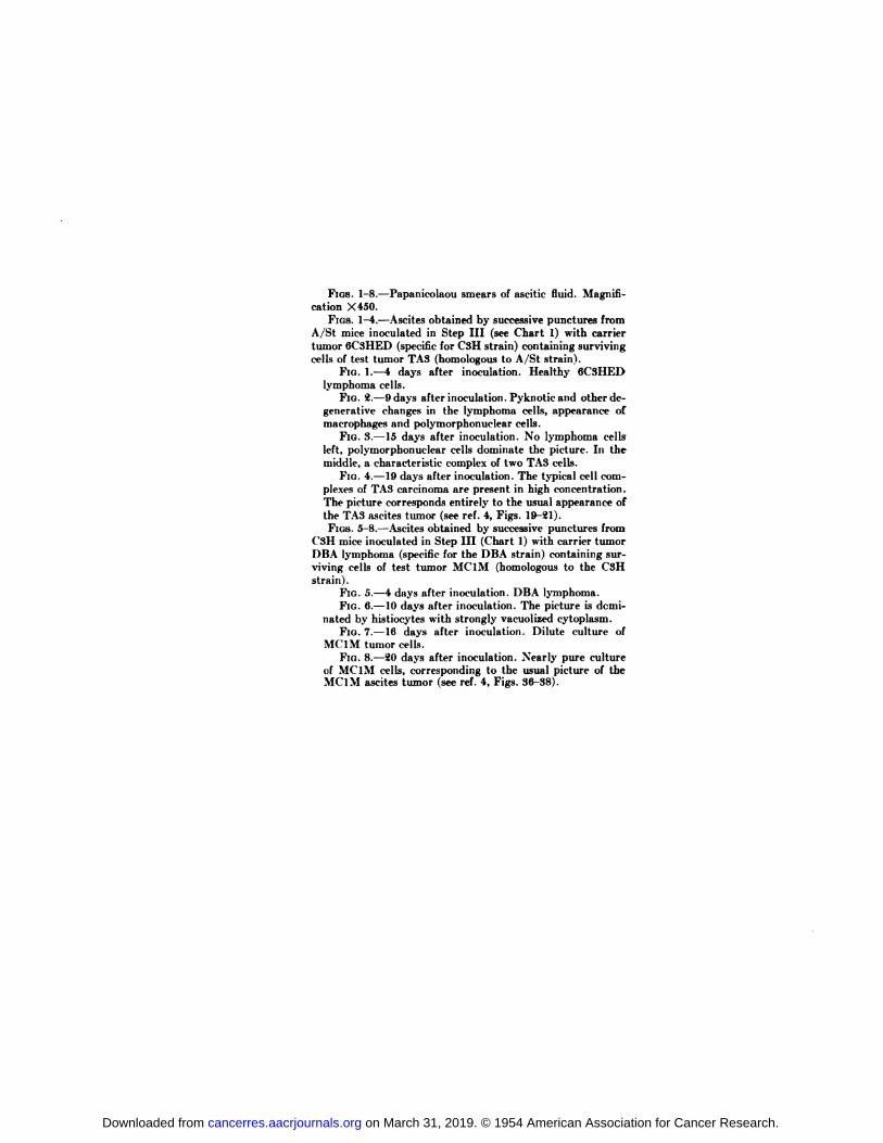
FIGS. 1-8.—Papanicolaou smears of ascitic fluid. Magnification X450.
FIGS. 1-4.—Ascitesobtained by successive punctures fromA/St mice inoculated in Step III (see Chart 1) with carriertumor 6C3HED (specific for CSH strain) containing survivingcells of test tumor TA3 (homologous to A/St strain).
FIG. 1.—4 days after inoculation. Healthy 6C3HEDlymphoma cells.
FIG. 2.—9days after inoculation. Pyknotie and other degenerative changes in the lymphoma cells, appearance ofmacrophages and polymorphonuclear cells.
FIG. 3.—15days after inoculation. No lymphoma cellsleft, polymorphonuclear cells dominate the picture. In themiddle, a characteristic complex of two TA3 cells.
FIG. 4.—19days after inoculation. The typical cell complexes of TA3 carcinoma are present in high concentration.The picture corresponds entirely to the usual appearance ofthe TA3 ascites tumor (see réf.4, Figs. 19-21).FIGS.5-8.—Ascitesobtained by successive punctures from
C3H mice inoculated in Step III (Chart 1) with carrier tumorDBA lymphoma (specific for the DBA strain) containing surviving cells of test tumor MC1M (homologous to the CSHstrain).
FIG. 5.—4days after inoculation. DBA lymphoma.FIG. 6.—10days after inoculation. The picture is domi
nated by histiocytes with strongly vacuolized cytoplasm.FIG. 7.—16 days after inoculation. Dilute culture of
MC1M tumor cells.FIG. 8.—20days after inoculation. Nearly pure culture
of MCI M cells, corresponding to the usual picture of theMCI M aseites tumor (see réf.4, Figs. 36-38).
on March 31, 2019. © 1954 American Association for Cancer Research.cancerres.aacrjournals.org Downloaded from
1954;14:139-144. Cancer Res Eva Klein and George Klein Established Ascites TumorsDifferential Survival of Solid Tumor Cells after Inoculation into
Updated version
http://cancerres.aacrjournals.org/content/14/2/139
Access the most recent version of this article at:
E-mail alerts related to this article or journal.Sign up to receive free email-alerts
Subscriptions
Reprints and
To order reprints of this article or to subscribe to the journal, contact the AACR Publications
Permissions
Rightslink site. Click on "Request Permissions" which will take you to the Copyright Clearance Center's (CCC)
.http://cancerres.aacrjournals.org/content/14/2/139To request permission to re-use all or part of this article, use this link
on March 31, 2019. © 1954 American Association for Cancer Research.cancerres.aacrjournals.org Downloaded from

![Surgical Case Reports - Treatment of tumor thrombus in the … · 2020. 12. 14. · complete surgical tumor resection and chemotherapy, some patients achieved long-term survival [4]](https://img.pdfslide.net/doc/110x75/61173aa9115ae12494473cdc/surgical-case-reports-treatment-of-tumor-thrombus-in-the-2020-12-14-complete.jpg)