Embed Size (px)
Citation preview

3954 Research Article
IntroductionIn cells, actin-filament-based structures control diverse activitiessuch as polarized intracellular trafficking, cytokinesis, migrationand adhesion (Mitchison and Cramer, 1996; Vasioukhin et al.,2000). Although cellular actin turnover is known to be spatiallyand temporally regulated by signaling cascades, many of theregulatory mechanisms remain to be characterized (Schmidt andHall, 1998). Enabled/vasodilator-stimulated phosphoprotein(Ena/VASP) proteins localize to regions of dynamic actinremodeling and promote filament formation (Bear and Gertler,2009; Trichet et al., 2008). In vertebrates, the evolutionaryconserved protein family is comprised of VASP, mammalianEnabled (Mena), and Ena-VASP-like (EVL) (Krause et al., 2003;Kwiatkowski et al., 2003; Sechi and Wehland, 2004). All threeproteins share a conserved structure consisting of an N-terminalEna/VASP homology (EVH)-1 domain, a central poly-prolineregion (PPR) and a C-terminal EVH2 domain. Whereas the EVH1domain mediates binding of Ena/VASP proteins to proline-richligands such as vinculin and zyxin (Ball et al., 2000), the PPRinteracts with the actin-binding protein profilin and with Srchomology 3 (SH3) domains (Benz et al., 2008; Lambrechts et al.,2000). Residues in EVH2 bind to G-actin and F-actin (Bachmannet al., 1999) and mediate tetramerization of Ena/VASP proteins(Kuhnel et al., 2004) (Fig. 1). In cells, Ena/VASP proteins promoteF-actin assembly, which involves anti-capping (Barzik et al., 2005;
Pasic et al., 2008), bundling (Applewhite et al., 2007; Bachmannet al., 1999; Kuhnel et al., 2004; Schirenbeck et al., 2006) andanti-branching (Bear et al., 2002; Skoble et al., 2001) activities.
The mammalian Ena/VASP proteins are known substrates ofserine/threonine kinases, and human VASP harbors the threephosphorylation sites serine 157 (S157), serine 239 (S239), andthreonine 278 (T278) (Blume et al., 2007; Butt et al., 1994; Gertleret al., 1996; Lambrechts et al., 2000). Whereas the first twophosphorylation sites are conserved in Mena, EVL contains only thefirst site (Fig. 1). In cells, the first and second site in VASP arephosphorylated in order by cAMP-dependent protein kinase (PKA).Conversely, cGMP-dependent protein kinase (PKG) initiallyphosphorylates the second site and then the first (Butt et al., 1994;Zhuang et al., 2004). Only recently, AMP-activated protein kinase(AMPK) was identified as the kinase responsible for phosphorylatingT278 (Blume et al., 2007) (Fig. 1). VASP is dephosphorylated byprotein phosphatase (PP)-1, PP2A, PP2B and PP2C (Abel et al.,1995), resulting in complex and dynamic phosphorylation patterns.
Evidence that phosphorylation might alter specific VASPproperties has come from studies of phosphomimetic VASP mutants,which have one or all three phosphorylation sites substituted withacidic amino acids or alanine residues to imitate a permanentlyphosphorylated or unphosphorylated protein, respectively (Barziket al., 2005; Geese et al., 2002; Grosse et al., 2003; Harbeck et al.,2000; Loureiro et al., 2002; Smolenski et al., 2000). Phosphorylation
Proteins of the Enabled/vasodilator-stimulated phosphoprotein(Ena/VASP) family link signal transduction pathways to actincytoskeleton dynamics. VASP is substrate of cAMP-dependent,cGMP-dependent and AMP-activated protein kinases thatprimarily phosphorylate the sites S157, S239 and T278,respectively. Here, we systematically analyzed functions ofVASP phosphorylation patterns for actin assembly andsubcellular targeting in vivo and compared the phosphorylationeffects of Ena/VASP family members. Methods used were thereconstitution of VASP-null cells with ‘locked’ phosphomimeticVASP mutants, actin polymerization of VASP mutants in vitroand in living cells, site-specific kinase-mediated VASPphosphorylation, and analysis of the endogenous protein withphosphorylation-status-specific antibodies. Phosphorylation atS157 influenced VASP localization, but had a minor impact on
F-actin assembly. Phosphorylation of the S157-equivalent sitein the Ena/VASP family members Mena and EVL had no effecton the ratio of cellular F-actin to G-actin. By contrast, VASPphosphorylation at S239 (and the equivalent site in Mena) orT278 impaired VASP-driven actin filament formation. The datashow that VASP functions are precisely regulated by differentialphosphorylation and provide new insights into cytoskeletalcontrol by serine/threonine kinase-dependent signalingpathways.
Supplementary material available online athttp://jcs.biologists.org/cgi/content/full/122/21/3954/DC1
Key words: Vasodilator-stimulated phosphoprotein (VASP),Ena/VASP family, Serine/threonine kinase, Actin turnover
Summary
Differential VASP phosphorylation controlsremodeling of the actin cytoskeletonPeter M. Benz1,*,‡, Constanze Blume1,*, Stefanie Seifert2, Sabine Wilhelm1, Jens Waschke3, Kai Schuh4,Frank Gertler5, Thomas Münzel6 and Thomas Renné2,§
1Institute of Clinical Biochemistry and Pathobiochemistry, 3Institute of Anatomy and 4Institute of Physiology, University of Würzburg, Würzburg,Germany2Department of Molecular Medicine and Surgery, and Center for Molecular Medicine, Karolinska Institutet, Stockholm, Sweden5Center for Cancer Research, Massachusetts Institute of Technology, Cambridge, MA, USA6Second Medical Clinic, Department of Cardiology and Angiology, Johannes-Gutenberg-University, Mainz, Germany*These authors contributed equally to this work‡Present address: Institute of Physiology, University of Würzburg, D-97080, Würzburg, Germany§Author for correspondence ([email protected])
Accepted 3 September 2009Journal of Cell Science 122, 3954-3965 Published by The Company of Biologists 2009doi:10.1242/jcs.044537
Jour
nal o
f Cel
l Sci
ence

3955VASP phosphorylation
affects the interaction of VASP with actin; however, the crucialphosphorylation sites and kinases involved are still unclear (Barziket al., 2005; Harbeck et al., 2000; Laurent et al., 1999; Lindsay etal., 2007). VASP phosphorylation also modulates other protein-protein interactions. Whereas S157 phosphorylation abrogatesinteraction of VASP with the SH3 domains of Abl, nSrc and aII-spectrin (Benz et al., 2008; Lambrechts et al., 2000), binding offocal adhesion proteins to the EVH1 domain, and of profilin to thePPR, is independent of the VASP phosphorylation status (Ferronet al., 2007; Harbeck et al., 2000). Additionally, VASPphosphorylation status appears to modulate subcellular proteindistribution. It has been postulated that VASP phosphorylation byPKA controls protein targeting to the cell-cell junctions of adherent
cells (Benz et al., 2008; Comerford et al., 2002), but others haveshown that PKG activity might control VASP localization (Lindsayet al., 2007; Smolenski et al., 2000).
In the present study, we analyzed VASP phosphorylation patternsfor their impact on subcellular protein localization and actinfilament assembly in cells and compared the phosphorylation-mediated regulation of VASP, Mena and EVL. We used a systematicstrategy, combining phosphomimetic mutants, site-specific kinase-mediated phosphorylation and phosphorylation-status-specificantibodies. Our findings demonstrate that S157 phosphorylationcontrols subcellular VASP distribution, whereas phosphorylationsat S239 and T278 impair F-actin accumulation. Similarly, thecellular F-actin to G-actin ratio was independent of thephosphorylation status at the S157 conserved site in Mena and EVL,whereas phosphorylation of the PKG homologous site in Menainterfered with F-actin assembly. This is the first comprehensivestudy of VASP phosphorylation that links serine/threonine-kinasesignaling to F-actin levels in vivo.
ResultsEna/VASP phosphomimetic mutants imitate ‘locked’phosphorylation patternsHuman VASP is a substrate for PKA, PKG and AMPK, whichphosphorylate residues S157, S239 and T278, respectively. The firsttwo phosphorylation sites are conserved in Mena (S236 and S376in the murine protein), whereas EVL only contains the first site(S160 in the human protein) (Fig. 1A,C,E). We generatedphosphomimetic Ena/VASP mutants by systematically exchangingphosphorylation sites for acidic amino acids to imitate aconstitutively phosphorylated residue. Substitution with alaninearrested the remaining phosphorylation sites into a non-phosphorylated state. For VASP, constructs were denominated witha three-letter code according to the amino acids at positions 157,239 and 278 (using A, alanine; D, aspartic acid; E, glutamic acid).For example, DAA is a VASP mutant bearing substitutions S157D,S239A and T278A. The phosphomimetic mutants covered all VASP
Fig. 1. Arrested phosphomimetic Ena/VASP mutants. (A)Domainorganization and phosphorylation sites of N-terminally 6�His-tagged humanwild-type VASP (wt) and phosphomimetic VASP mutants. VASP is composedof an N-terminal EVH1 domain, a PPR and a C-terminal EVH2 domain. Theblack square with white ‘T’ indicates the tag. PKA phosphorylates S157 (boxshaded with horizontal lines) and S239 in order; S239 (box shaded withdiagonal lines) is the primary target for PKG, which might also phosphorylateS157. T278 (box shaded with squares) is exclusively phosphorylated byAMPK. Phosphorylation sites were substituted with alanines (black boxes) oracidic residues (light boxes). Sites for G-actin binding (GAB, residues 234-237) and F-actin binding (FAB, residues 259-276), are located upstream of thepreferred PKG and AMPK phosphorylation sites, respectively. The preferredPKA phosphorylation site is located upstream of the PPR. (B)Western blotwith anti-6�His antibodies to confirm expression in EC_VASP–/– cellstransiently transfected with cDNAs coding for VASP, arrestedphosphomimetic mutants or vector without insert (MOCK). (C)Domainorganization and phosphorylation sites of N-terminally 6�His-tagged murinewild-type Mena and phosphomimetic Mena mutants. LERER indicates the lowcomplexity region harboring LERER repeats. Mena S236 and S376correspond to VASP S157 and S239, respectively. Shading as for VASP.(D)Western blot with anti-6�His antibodies shows expression of wild-typeand mutant Mena. (E)Domain organization and phosphorylation sites of N-terminally 6�His-tagged human wild-type EVL and phosphomimetic EVLmutants. EVL phosphorylation site S160 corresponds to VASP S157.(D)Western blot with anti-6�His antibodies confirms expression of wild-typeEVL and arrested phosphomimetic mutants A and D. Note that(pseudo)phosphorylation at the first phosphorylation site leads to a mobilityshift in SDS-PAGE of all mammalian Ena/VASP proteins.
Jour
nal o
f Cel
l Sci
ence

3956
phosphorylation patterns ranging from non-phosphorylated (AAA),single-phosphorylated (DAA, ADA, AAE), double-phosphorylated(DDA, DAE, ADE), up to triple-phosphorylated (DDE) proteins,each in a fixed state of phosphorylation (Fig. 1A). Mena and EVLconstructs were named accordingly, using a two- or one-letter codefor Mena or EVL, respectively (Fig. 1C,E).
It was previously shown that phosphorylation of VASP at S157,but not S239 or T278, leads to an electrophoretic mobility shiftfrom 46 to 50 kDa in SDS PAGE (Butt et al., 1994; Krause et al.,2003; Kwiatkowski et al., 2003). We transfected microvascularendothelial cells from VASP-null mice (EC_VASP–/–) with cDNAscoding for phosphomimetic mutants or wild-type protein andanalyzed the migration of VASP variants by western blotting.Hexahistidine-tag-specific antibodies detected the characteristicdoublet of S157 phosphorylated and non-phosphorylated wild-typeVASP (Fig. 1B, lane 1). Phosphomimetic mutants with a negativecharge substitution at position 157 (DDE, DDA, DAE, DAA; Fig.1B, lanes 6-9) migrated with a higher apparent molecular mass (49-50 kDa) compared with mutants with alanine at this position (46kDa; AAA, ADA, AAE, ADE; Fig. 1B, lanes 2-5). Similar to thephosphorylation of S239 and/or T278 in wild-type VASP (Blumeet al., 2007), the migration of VASP mutants was not altered by thepresence of acidic residues at the second or third phosphorylationsite. Phosphorylation of Mena and EVL at their first phosphorylationsites also leads to a small shift in apparent molecular mass in SDS-
Journal of Cell Science 122 (21)
PAGE (Gertler et al., 1996; Lambrechts et al., 2000).Phosphomimetic Mena and EVL mutants with an aspartic acid atthe first phosphorylation site (Mena: DD and DA; EVL: D),migrated with a slightly higher apparent molecular mass thancorresponding mutants with alanine at this position (Mena: AA andAD; EVL: A) (Fig. 1D and F). Expression levels of all Ena/VASPmutants were largely independent of the point mutations, asindicated by signal intensities in the western blots.
VASP targeting to the endothelial plasma membrane and focaladhesions depends on S157 phosphorylationTo analyze the impact of S157 phosphorylation on the localizationof endogenous VASP, microvascular endothelial cells from VASPwild-type mice (EC_VASP+/+) were treated with the PKA activatorforskolin (5 M) or buffer control. Forskolin increased S157phosphorylation, as indicated by phospho-S157 (S157-P; also knownas pS157)–VASP-specific antibodies (Fig. 2A, upper panel; S157-P)and anti-VASP antibodies (Fig 2A, middle panel; S157-P–VASP isthe upper and S157-VASP the lower signal of the doublet) in westernblotting. We compared the localization of total-VASP and S157-P–VASP in buffer- (Fig. 2B-D) and forskolin-treated EC_VASP+/+
(Fig. 2E-G). In unstimulated cells, total-VASP (Fig. 2B)predominantly localized to stress fibers (black arrowheads) and smallportions of total cellular protein enriched at punctuated structures,probably representing focal adhesions (black arrows). VASP at focaladhesions was strongly S157-phosphorylated and readily stained byphosphorylation-specific antibodies (Fig. 2C, black arrows). Bothantigens co-localized at these sites (Fig. 2D). PKA-stimulationlargely increased S157-P signal intensity and substantially changedthe subcellular VASP distribution. Following a 10 minute forskolintreatment, total-VASP disappeared from stress fibers (compare total-VASP staining in stimulated versus unstimulated cells) and localizedto focal adhesions (black arrows) and the plasma membrane (whitearrowheads, Fig. 2E). VASP at these sites was S157-phosphorylated(Fig. 2F,G).
In addition to S157, PKA activation might also phosphorylate S239and T278 (Butt et al., 1994) or modulate VASP localization by otherpathways. Therefore, we confirmed the contribution of S157 phos-phorylation to subcellular VASP targeting with a kinase-independentapproach. VASP-deficient endothelial cells (EC_VASP–/–) werereconstituted with phosphomimetic mutants. Except for VASP proteinlevels, EC_VASP–/– are indistinguishable from EC_VASP+/+ and havesimilar Mena and EVL expression (Fig. 2H).
To analyze localization of phosphomimetic VASP mutants inEC_VASP–/–, cells were transiently transfected with cDNAs codingfor the eight VASP mutants AAA (Fig. 3A-C), DAA (Fig. 3D-F),ADA (Fig. 3G-I), DDA (Fig. 3J-L), AAE (Fig. 3M-O), DAE (Fig.3P-R), ADE (Fig. 3S-U), and DDE (Fig. 3V-X). At 24 hours aftertransfection, cells were trypsinized and replated for 3 hours beforefixing. Polyclonal anti-VASP antibodies were used for visualizingphosphomimetic mutants (green) and fluorescent phalloidinto stain for F-actin (red). For a semi-quantitative analysis ofpseudophosphorylation-mediated VASP localization, twoindependent investigators determined the localization of mutantsat the leading edge of lamellipodia or the accumulation at focaladhesions. VASP accumulation at these sites was scored weak (–),moderate (o) or strong (+) in >40 cells with mutant expressionlevels similar to endogenous VASP in EC_VASP+/+ (Table 1). TheVASP accumulation score was consistent with the length ratio ofVASP-positive leading edges relative to the cell circumference.Mutants DAA, DDA, DAE and DDE (which share a negative
Fig. 2. VASP translocation to the cell periphery depends on S157phosphorylation. Wild-type endothelial cells (EC_VASP+/+) were incubatedwith forskolin (5M) or buffer and analyzed using antibodies against S157-P–VASP and total-VASP. (A)Western blots of cell lysates normalized forGAPDH. (B-G)Immunofluorescence images of fixed and permeabilized cells.The merge of total-VASP (red; B,E) and S157-P–VASP (green; C,F) is givenin yellow (D,G). Black arrowheads indicate stress fibers, black arrows indicatefocal adhesions, and white arrowheads indicate plasma membranes. Imageswere taken with a 100� objective and are representative of a series of eightexperiments. Scale bar: 15m. (H)Comparison of VASP, Mena and EVLexpression in EC_VASP–/– and EC_VASP+/+ cells by western blotting. The celllysates are normalized for GAPDH.
Jour
nal o
f Cel
l Sci
ence

3957VASP phosphorylation
charge at the first site) were largely enriched at the leading edgeof lamellipodia-like structures as compared with correspondingmutants with an alanine at this specific position (compare DAAvs AAA, DDA vs ADA, DAE vs AAE, and DDE vs ADE; Table1 and Fig. 3, arrowheads). An acidic residue at position S157 alsoaccumulated phosphomimetic mutants at focal-adhesion-likestructures (compare DAA vs AAA, DDA vs ADA and DAE vs
AAE; Table 1) and increased the number and size of focaladhesions.
In cells, VASP forms homotetramers but might also formheterotetramers with Mena and EVL (Bachmann et al., 1999;Grosse et al., 2003; Kuhnel et al., 2004; Loureiro et al., 2002;Vasioukhin et al., 2000; Zhuang et al., 2004). To exclude thepossibility that tetramerization of transfected VASP mutants with
Fig. 3. Subcellular localization of phosphomimetic VASP mutants in EC_VASP–/– cells. Confocal immunofluorescence images of EC_VASP–/– transientlytransfected with phosphomimetic VASP mutants AAA (A-C), DAA (D-F), ADA (G-I), DDA (J-L), AAE (M-O), DAE (P-R), ADE (S-U), and DDE (V-X). At 24hours after transfection, cells were trypsinized and replated on gelatinized chamberslides for 3 hours before fixation and staining. Reconstituted VASP mutantswere immunolocalized with anti-VASP antibodies (green) and F-actin with fluorescent phalloidin (red). Arrowheads indicate the leading edge of lamellipodia.Scale bar: 15m.
Table 1. Subcellular localization of phosphomimetic VASPmutants in EC_VASP–/– cells
VASP mutant Lamellipodia Focal adhesions
AAA – oDAA + +
ADA – oDDA + +
AAE – oDAE + +
ADE – oDDE + o
In EC_VASP–/– cells, subcellular localization of transiently expressedVASP mutants at the leading edge of lamellipodia and focal adhesions wasscored as weak (–), moderate (o), or strong (+). 40 cells each with mutantexpression levels similar to endogenous VASP in EC_VASP–/– were randomlyselected and analyzed.
Table 2. Subcellular localization of phosphomimetic VASPmutants in MVD7 cells
VASP mutant Lamellipodia Focal adhesions
AAA o oDAA o +
ADA o oDDA + +
AAE o oDAE + +
ADE – –DDE + o
In MVD7 cells, subcellular localization of transiently expressed VASPmutants at lamellipodia or focal adhesions was scored as weak (–), moderate(o), or strong (+). Per mutant, 40 randomly selected cells with comparableVASP expression levels were analyzed.
Jour
nal o
f Cel
l Sci
ence
3958
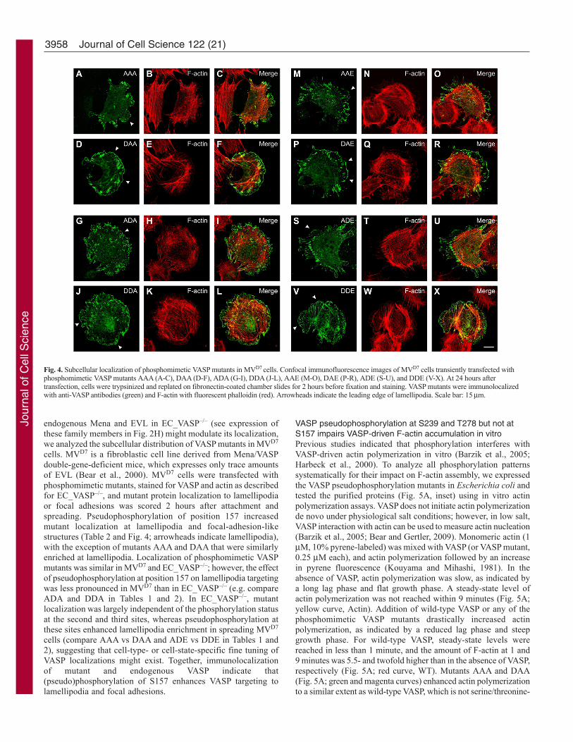
endogenous Mena and EVL in EC_VASP–/– (see expression ofthese family members in Fig. 2H) might modulate its localization,we analyzed the subcellular distribution of VASP mutants in MVD7
cells. MVD7 is a fibroblastic cell line derived from Mena/VASPdouble-gene-deficient mice, which expresses only trace amountsof EVL (Bear et al., 2000). MVD7 cells were transfected withphosphomimetic mutants, stained for VASP and actin as describedfor EC_VASP–/–, and mutant protein localization to lamellipodiaor focal adhesions was scored 2 hours after attachment andspreading. Pseudophosphorylation of position 157 increasedmutant localization at lamellipodia and focal-adhesion-likestructures (Table 2 and Fig. 4; arrowheads indicate lamellipodia),with the exception of mutants AAA and DAA that were similarlyenriched at lamellipodia. Localization of phosphomimetic VASPmutants was similar in MVD7 and EC_VASP–/–; however, the effectof pseudophosphorylation at position 157 on lamellipodia targetingwas less pronounced in MVD7 than in EC_VASP–/– (e.g. compareADA and DDA in Tables 1 and 2). In EC_VASP–/–, mutantlocalization was largely independent of the phosphorylation statusat the second and third sites, whereas pseudophosphorylation atthese sites enhanced lamellipodia enrichment in spreading MVD7
cells (compare AAA vs DAA and ADE vs DDE in Tables 1 and2), suggesting that cell-type- or cell-state-specific fine tuning ofVASP localizations might exist. Together, immunolocalizationof mutant and endogenous VASP indicate that(pseudo)phosphorylation of S157 enhances VASP targeting tolamellipodia and focal adhesions.
Journal of Cell Science 122 (21)
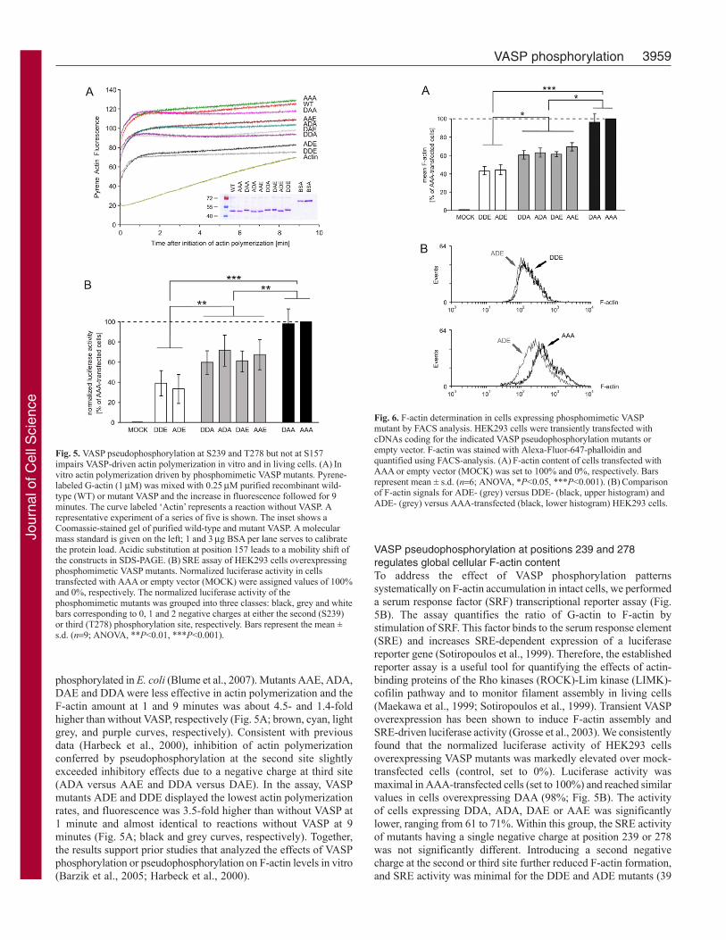
VASP pseudophosphorylation at S239 and T278 but not atS157 impairs VASP-driven F-actin accumulation in vitroPrevious studies indicated that phosphorylation interferes withVASP-driven actin polymerization in vitro (Barzik et al., 2005;Harbeck et al., 2000). To analyze all phosphorylation patternssystematically for their impact on F-actin assembly, we expressedthe VASP pseudophosphorylation mutants in Escherichia coli andtested the purified proteins (Fig. 5A, inset) using in vitro actinpolymerization assays. VASP does not initiate actin polymerizationde novo under physiological salt conditions; however, in low salt,VASP interaction with actin can be used to measure actin nucleation(Barzik et al., 2005; Bear and Gertler, 2009). Monomeric actin (1M, 10% pyrene-labeled) was mixed with VASP (or VASP mutant,0.25 M each), and actin polymerization followed by an increasein pyrene fluorescence (Kouyama and Mihashi, 1981). In theabsence of VASP, actin polymerization was slow, as indicated bya long lag phase and flat growth phase. A steady-state level ofactin polymerization was not reached within 9 minutes (Fig. 5A;yellow curve, Actin). Addition of wild-type VASP or any of thephosphomimetic VASP mutants drastically increased actinpolymerization, as indicated by a reduced lag phase and steepgrowth phase. For wild-type VASP, steady-state levels werereached in less than 1 minute, and the amount of F-actin at 1 and9 minutes was 5.5- and twofold higher than in the absence of VASP,respectively (Fig. 5A; red curve, WT). Mutants AAA and DAA(Fig. 5A; green and magenta curves) enhanced actin polymerizationto a similar extent as wild-type VASP, which is not serine/threonine-
Fig. 4. Subcellular localization of phosphomimetic VASP mutants in MVD7 cells. Confocal immunofluorescence images of MVD7 cells transiently transfected withphosphomimetic VASP mutants AAA (A-C), DAA (D-F), ADA (G-I), DDA (J-L), AAE (M-O), DAE (P-R), ADE (S-U), and DDE (V-X). At 24 hours aftertransfection, cells were trypsinized and replated on fibronectin-coated chamber slides for 2 hours before fixation and staining. VASP mutants were immunolocalizedwith anti-VASP antibodies (green) and F-actin with fluorescent phalloidin (red). Arrowheads indicate the leading edge of lamellipodia. Scale bar: 15m.
Jour
nal o
f Cel
l Sci
ence
3959VASP phosphorylation
phosphorylated in E. coli (Blume et al., 2007). Mutants AAE, ADA,DAE and DDA were less effective in actin polymerization and theF-actin amount at 1 and 9 minutes was about 4.5- and 1.4-foldhigher than without VASP, respectively (Fig. 5A; brown, cyan, lightgrey, and purple curves, respectively). Consistent with previousdata (Harbeck et al., 2000), inhibition of actin polymerizationconferred by pseudophosphorylation at the second site slightlyexceeded inhibitory effects due to a negative charge at third site(ADA versus AAE and DDA versus DAE). In the assay, VASPmutants ADE and DDE displayed the lowest actin polymerizationrates, and fluorescence was 3.5-fold higher than without VASP at1 minute and almost identical to reactions without VASP at 9minutes (Fig. 5A; black and grey curves, respectively). Together,the results support prior studies that analyzed the effects of VASPphosphorylation or pseudophosphorylation on F-actin levels in vitro(Barzik et al., 2005; Harbeck et al., 2000).
VASP pseudophosphorylation at positions 239 and 278regulates global cellular F-actin contentTo address the effect of VASP phosphorylation patternssystematically on F-actin accumulation in intact cells, we performeda serum response factor (SRF) transcriptional reporter assay (Fig.5B). The assay quantifies the ratio of G-actin to F-actin bystimulation of SRF. This factor binds to the serum response element(SRE) and increases SRE-dependent expression of a luciferasereporter gene (Sotiropoulos et al., 1999). Therefore, the establishedreporter assay is a useful tool for quantifying the effects of actin-binding proteins of the Rho kinases (ROCK)-Lim kinase (LIMK)-cofilin pathway and to monitor filament assembly in living cells(Maekawa et al., 1999; Sotiropoulos et al., 1999). Transient VASPoverexpression has been shown to induce F-actin assembly andSRE-driven luciferase activity (Grosse et al., 2003). We consistentlyfound that the normalized luciferase activity of HEK293 cellsoverexpressing VASP mutants was markedly elevated over mock-transfected cells (control, set to 0%). Luciferase activity wasmaximal in AAA-transfected cells (set to 100%) and reached similarvalues in cells overexpressing DAA (98%; Fig. 5B). The activityof cells expressing DDA, ADA, DAE or AAE was significantlylower, ranging from 61 to 71%. Within this group, the SRE activityof mutants having a single negative charge at position 239 or 278was not significantly different. Introducing a second negativecharge at the second or third site further reduced F-actin formation,and SRE activity was minimal for the DDE and ADE mutants (39
Fig. 5. VASP pseudophosphorylation at S239 and T278 but not at S157impairs VASP-driven actin polymerization in vitro and in living cells. (A)Invitro actin polymerization driven by phosphomimetic VASP mutants. Pyrene-labeled G-actin (1M) was mixed with 0.25M purified recombinant wild-type (WT) or mutant VASP and the increase in fluorescence followed for 9minutes. The curve labeled ‘Actin’ represents a reaction without VASP. Arepresentative experiment of a series of five is shown. The inset shows aCoomassie-stained gel of purified wild-type and mutant VASP. A molecularmass standard is given on the left; 1 and 3g BSA per lane serves to calibratethe protein load. Acidic substitution at position 157 leads to a mobility shift ofthe constructs in SDS-PAGE. (B)SRE assay of HEK293 cells overexpressingphosphomimetic VASP mutants. Normalized luciferase activity in cellstransfected with AAA or empty vector (MOCK) were assigned values of 100%and 0%, respectively. The normalized luciferase activity of thephosphomimetic mutants was grouped into three classes: black, grey and whitebars corresponding to 0, 1 and 2 negative charges at either the second (S239)or third (T278) phosphorylation site, respectively. Bars represent the mean ±s.d. (n9; ANOVA, **P<0.01, ***P<0.001).
Fig. 6. F-actin determination in cells expressing phosphomimetic VASPmutant by FACS analysis. HEK293 cells were transiently transfected withcDNAs coding for the indicated VASP pseudophosphorylation mutants orempty vector. F-actin was stained with Alexa-Fluor-647-phalloidin andquantified using FACS-analysis. (A)F-actin content of cells transfected withAAA or empty vector (MOCK) was set to 100% and 0%, respectively. Barsrepresent mean ± s.d. (n6; ANOVA, *P<0.05, ***P<0.001). (B)Comparisonof F-actin signals for ADE- (grey) versus DDE- (black, upper histogram) andADE- (grey) versus AAA-transfected (black, lower histogram) HEK293 cells.
Jour
nal o
f Cel
l Sci
ence

3960
and 34%, respectively; Fig. 5B). SRE activity driven by wild-typeVASP varied from 49 to 81%, probably reflecting various non-defined phosphorylation patterns.
To confirm the effects of phosphorylation on F-actin assembly,we used quantitative fluorescence-activated cell sorting (FACS)(Fig. 6). The mean F-actin content of the cells expressing VASPmutants was measured relative to mock-transfected controls (0%value). The signal of the F-actin-specific dye, Alexa-Fluor-647-conjugated phalloidin, was normalized to the F-actin in HEK293cells expressing the phosphorylation-resistant AAA mutant (100%).Overexpression of mutants DDA, ADA, DAE or AAE (which havea single negative charge either at position 239 or at 278) reducedthe cellular F-actin amount to 60, 63, 61 and 68%, respectively;whereas DAA expression had a minor effect on actin fiber formation(97%). A second negative charge at positions 239 or 278 reducedF-actin formation to 44 and 47% (DDE, ADE; Fig. 6A). The F-actin signals in cells expressing DDE or ADE were almost identical(Fig. 6B, upper plot), but they differed in cells expressing ADE orAAA (Fig. 6B, lower plot). Together with the results from thepyrene-actin and SRE assays (Fig. 5A,B), the data demonstrate thatpseudophosphorylation at positions 239 or 278 interferes withVASP-induced actin filament nucleation in vitro and F-actinaccumulation in living cells, and that the inhibitory effects areadditive and maximal in the 239/278 double-pseudophosphorylatedmutants.
Pseudophosphorylation of Mena S376, but not Mena S236 orEVL S160, reduces F-actin levels in vivoVASP pseudophosphorylations at the second and third siteinterferes with actin filament assembly in living cells (Fig. 5B,Fig. 6). Only the second site is conserved in Mena (S376), andEVL lacks equivalents to both sites (Krause et al., 2003) (Fig.1). We investigated the function of Mena pseudophosphorylationfor actin assembly using the SRE-reporter assay (Fig. 7A).Transient transfection only slightly increased the high endogenousMena protein levels in HEK293 cells and degradation productsappeared in overexpressing cells (supplementary material Fig.S1A). Therefore, we established the SRE assay in adherent CHO-S cells (Suzuki et al., 2006), which have no detectable endogenousMena and allow high transient protein expression levels(supplementary material Fig. S1B). Normalized luciferase activityof CHO-S cells that overexpressed mutant Mena was markedlyelevated over mock-transfected cells (control, set to 0%).Luciferase activity was maximal in mutant AA-transfected cells(set to 100%) and reached similar values in cells overexpressingDA variant (96%; Fig. 7A). The activity of cells expressingmutants AD and DD, which have a negative charge at the 376position, was significantly lower and reached 39 and 38%,respectively. Luciferase activity induced by EVL A mutant (setto 100%) in HEK293 cells clearly exceeded levels of mock-transfected cells (control, set to 0%) and was almost identical toEVL D variant (99%; Fig. 7B).
S157 phosphorylation does not affect global cellular F-actincontentThe previous findings in this study suggest that thepseudophosphorylation status of S157 is important for VASPlocalization in spreading cells but does not affect overall F-actincontent. By contrast, pseudophosphorylation at positions 239 or278 inhibits the VASP-dependent F-actin accumulation, but hasa minor impact on subcellular targeting. To test this for kinase-
Journal of Cell Science 122 (21)
mediated VASP phosphorylation, we used partially arrestedmutants SAA, ASA and AAT (Fig. 8A). The mutants restrictphosphorylation to the first, second or third site, respectively, withthe other sites blocked from phosphorylation. Using unstimulatedreconstituted EC_VASP–/– cells, mutant SAA but not ASA or AATmigrated as a doublet in SDS-PAGE, indicating that S157phosphorylation is blocked in ASA and AAT but partiallyphosphorylated in SAA (Fig. 8B). To investigate the consequencesof PKA-mediated S157 phosphorylation on F-actin assembly, weused the SRE-based reporter assay (Fig. 8C). Overexpression ofSAA induced F-actin assembly in unstimulated HEK293 cells(102%), similar to the level observed in AAA-transfected cells.By contrast, overexpression of ADE reduced the signal to 33%(positive control). Treatment of SAA-transfected cells withforskolin (5 M) largely increased S157 phosphorylation, whichwas almost undetectable in unstimulated cells, as shown by S157-P-specific antibodies (Fig. 8D, upper panel). The relative signalintensities of the SAA doublet indicated that more than 75% oftotal SAA protein was phosphorylated at S157 (Fig. 8D, lowerpanel, anti-VASP). However, luciferase activity was notsignificantly changed by PKA stimulation for up to 80 minutes(99-107% versus 102%, n.s.; Fig. 8C), which was consistent withcells expressing DAA (98%). Higher forskolin concentrations oralternative PKA-activators, such as 8-Br-cAMP, did not affect SREsignals (not shown). Together, these results strongly suggest thatPKA-mediated VASP S157 phosphorylation does not affect globalF-actin content in cells.
Phosphorylation at S239 and T278 impairs cellular F-actinaccumulation in vivoNext, we analyzed the effects of PKG-mediated VASPphosphorylation using SRE assays in HEK293 cells (Fig. 8E,F).In the absence of transfected PKG, the luciferase activity of cellsexpressing ADA and ADE served as a reference value andinternal control, respectively (69 and 35% relative to the SREsignals of cells expressing AAA, set to 100%). Mutant ASA, withphosphorylation restricted to position 239, was co-transfected with
Fig. 7. Effect of Mena and EVL pseudophosphorylations for actin assemblyin cells. (A)SRE assay of CHO-S cells overexpressing phosphomimeticMena mutants. Normalized luciferase activity in cells transfected with AA orempty vector (MOCK) were assigned values of 100% and 0%, respectively.Mean values ± s.d. are given (n13; ANOVA, *** P<0.001). (B)SRE assayof HEK293 cells transfected with EVL mutants D and A. Normalizedluciferase activity in cells transfected with A or empty vector were assignedvalues of 100% and 0%, respectively. Mean ± s.d. (n8; ANOVA, P>0.05 Dversus A).
Jour
nal o
f Cel
l Sci
ence

3961VASP phosphorylation
plasmids coding for wild-type (PKGwt) or a catalytically inactiveforms (PKGCI) of PKG. Kinase activity was abolished in PKGCI
by an A405K substitution (Smolenski et al., 2000). Westernblotting with S239-P-specific antibodies revealed that co-expression of PKGwt increased ASA phosphorylation in a dose-dependent manner concomitant with a significant decrease inluciferase activity to 61%, compared to ASA without PKG(101%). By contrast, co-expression of PKGCI and ASA increasedSRE signals slightly, but not significantly (103 and 110% of AAA;Fig. 8E) in transfected cells. To address the function of AMPK-mediated phosphorylation at residue T278 in F-actin formation,we co-expressed mutant AAT with either a constitutively active(AMPKCAa) or a dominant negative (AMPKDNa) form of the
AMPK a1-subunit (Fig. 8G,H). Consistent with basal phospho-T278 (T278-P; also known as pT278)–VASP levels in endothelialcells (Blume et al., 2007), T278-P-specific anti-VASP antibodiesdetected T278-phosphorylated AAT mutants in the absence oftransfected AMPK (Fig. 8H, upper panel, lane 1). The luciferaseactivity in these cells was slightly lower than in AAA controls(92 versus 100%). The co-expression of increasing amounts ofAMPKCAa with AAT increased T278-P-levels and reduced SREsignals down to 61% in a dose-dependent manner, which wassimilar to the reduction observed in cells expressing AAE (62%,comparative value). By contrast, a reduction of T278-P levels byAMPKDNa expression increased SRE signals up to 133% in a dose-dependent manner. The data is consistent with the results obtained
Fig. 8. Phosphorylation at S239 and T278 but not at S157 impairs VASP-driven actin polymerization in living cells. (A)Schematic representation of partially arrestedVASP mutants. To analyze defined VASP phosphorylation patterns, we substituted two of the three phosphorylation sites with alanine residues, retaining one to allowkinase-mediated phosphorylation specifically at this residue. Black and open boxes indicate alanine and an accessible phosphorylation site, respectively. White ‘T’indicates the tag. (B)Western blot with anti-VASP antibodies to confirm expression of mutants SAA, ASA or AAT in transfected EC_VASP–/– cells. (C-D)PKA-mediated phosphorylation of S157-VASP does not affect global cellular F-actin content. Transfected HEK293 cells were incubated with forskolin (FSK, 5M) for 5,20, 40 and 80 minutes. (C)SRE assay. The luciferase activities in cells transfected with AAA or empty vector (MOCK) were set to 100% and 0%, respectively. Forcomparison, the luciferase activity of unstimulated cells expressing DAA is given (dashed line, n6; ANOVA, P>0.05 stimulated versus unstimulated SAA-transfectedcells). The transfection with ADE, which maximally blocked VASP-driven actin assembly (Fig. 5B), served as a positive control. (D)The expression of VASP mutantsand phosphorylation of SAA at S157 was confirmed by immunoblotting using antibodies against S157-P (upper panel) and total-VASP (lower panel). (E-F)PKG-mediated VASP phosphorylation at S239 reduces global actin polymerization. HEK239 cells were co-transfected with VASP mutant ASA and the indicated amounts ofPKGwt or PKGCI. (E)For comparison, the signal of unstimulated cells expressing ADA is given (dashed line). Normalized luciferase activities are plotted relative to theactivity in cells transfected with AAA or empty vector (MOCK) (set to 100% and 0%, respectively; n8; ANOVA, *P<0.05, ***P<0.001). ADE served as a positivecontrol. (F)Anti-S239-P antibodies indicated the phosphorylation of S239-ASA (upper panel) in western blots. The expression of VASP mutants in transfected cellswas confirmed by immunoblotting with anti-VASP antibodies (middle panel). The expression of PKG variants was probed using antibodies against PKG (lower panel).(G-H)AMPK-meditated phosphorylation of VASP at T278 interferes with F-actin assembly. HEK239 cells were transiently co-transfected with VASP-AAT andAMPKCAa or AMPKDNa. (G)For comparison, the activity of AAE-transfected cells is indicated (dashed line). Normalized luciferase activities are plotted relative to theactivity in cells transfected with AAA or empty vector (MOCK) (set to 100% and 0%, respectively; n8; ANOVA, *P<0.05). ADE served as a positive control.(H)Western blots using anti-T278-P (upper panel) and anti-VASP antibodies (second panel) confirmed T278 phosphorylation and AAT expression, respectively.AMPK variants were probed by myc-tag specific antibodies (lower panels).
Jour
nal o
f Cel
l Sci
ence

3962
with the phosphomimetic mutants and demonstrates thatphosphorylation at S239 and T278 interferes with global F-actinassembly in cells.
DiscussionOriginally, VASP was identified in platelets as a substrate for cyclic-nucleotide-dependent protein kinases PKA and PKG (Halbrugge etal., 1990). Its strategic localization at the intersection of importantkinase-driven signaling cascades established phospho-VASP as amarker for the integrity of cyclic-nucleotide-dependent pathwaysin cardiovascular cells. Today, analysis of phospho-VASP levels inplatelets is used in clinical diagnostics (reviewed in Munzel et al.,2003; Gachet and Aleil, 2008). Despite its importance, the cellularfunction and complexity of VASP phosphorylation has remaineduncertain.
VASP is a substrate of the protein kinases PKA, PKG and AMPKthat primarily phosphorylate the sites S157, S239 and T278,respectively. Whereas the first two phosphorylation sites areconserved in Mena, EVL contains only the first site. In vitro studieshave suggested that PKA-mediated VASP phosphorylation has anegative effect on both actin nucleation/G-actin binding and VASPinteraction with actin filaments (Harbeck et al., 2000). In order toclarify underlying molecular mechanisms, we analyzed all VASPphosphorylation patterns systematically for their impact on actinfilament assembly and subcellular protein targeting and comparedphosphorylation-mediated effects of Ena/VASP family members.The study correlates defined VASP phosphorylation patterns withdistinct functions in endothelial and HEK293 cells. The phospho-VASP-mediated regulation of actin dynamics appears to be ofgeneral importance and has been observed in endothelial cells (Benzet al., 2008; Price and Brindle, 2000; Rentsendorj et al., 2008;Rosenberger et al., 2007), leukocytes (Lawrence and Pryzwansky,2001), platelets (Pula et al., 2006) and fibroblasts (Grosse et al.,2003), and in glioma (Zhuang et al., 2004), smooth muscle (Chenet al., 2004) and epithelial cells (Lindsay et al., 2007).
One of our key findings is that subcellular VASP localization ispredominantly regulated by S157 phosphorylation in spreadingendothelial cells. In the endothelium, VASP is involved in sealingcell-cell contacts and permeability regulation (Benz et al., 2008;Furman et al., 2007; Rentsendorj et al., 2008; Rosenberger et al.,2007; Schlegel et al., 2008). S157-P or pseudophosphorylated VASPwas enriched at lamellipodia and focal adhesions in spreading cells(Figs 2, 3 and Table 1). The activation of PKA has been shown toincrease S157 phosphorylation and to regulate VASP accumulationat the cell periphery in human (Comerford et al., 2002) and murinemicrovascular endothelial cells (Benz et al., 2008). The PKA-mediated phosphorylation of S157 blocks VASP binding to the SH3domains of Abl (Howe et al., 2002), Src and aII-spectrin (Benz etal., 2008); interferes with complex formation; and coincides withS157-P–VASP accumulation at sites of high actin dynamics at thecell periphery. S157-P–VASP concentration in the cell periphery isalso observed in platelets (Wentworth et al., 2006), in part due toPKC-mediated VASP phosphorylation (Pula et al., 2006). Inaddition to S157-P, previous studies have suggested a role of S239phosphorylation for VASP localization. Thrombin-inducedphosphorylation of AST, but not SAT, resulted in the translocationof VASP to the cell periphery in human umbilical vein endothelialcells (HUVEC) (Profirovic et al., 2005). By contrast, the stimulationof VASP S239 phosphorylation in the same cell line was shown todetach the protein from focal adhesions and to translocate it in theopposite direction, from the cell periphery to the cytosol (Smolenski
Journal of Cell Science 122 (21)
et al., 2000). The two studies used different experimental conditions,making the results difficult to compare. Localization of ourphosphomimetic mutants indicates that the phosphorylation of S239and T278 has a minor effect on the subcellular distribution of VASPin endothelial cells (Fig. 3 and Table 1). However, cell-type-specificregulation of Ena/VASP proteins might exist because thephosphorylation status of S239 modulates VASP localization in renalepithelial cells in NO-dependent signaling pathways (Lindsay etal., 2007). Importantly, phosphorylation at S239 and T278 hassignificant effects on VASP interaction with G- and F-actin (Barziket al., 2005; Harbeck et al., 2000) as well as on the ability of VASPto bind the chemotactic receptor CXCR2 (Neel et al., 2009).
We also analyzed VASP mutants in Ena/VASP-deficientfibroblasts and consistently found that mutant subcellularlocalization depends on the pseudophosphorylation status of S157(MVD7; Fig. 4 and Table 2). The localization of Mena mutantsappeared to be independent of their pseudophosphorylation statusin this cell line (Loureiro et al., 2002); however, the cells wereanalyzed under steady-state conditions rather than in spreading cellsas in the current studies. MVD7 cells do not make filopodia andlamellipodia robustly once they are spread (Applewhite et al., 2007),and localization of actin-binding proteins might differ dependingon the cytoskeleton turnover rate. Indeed, localization patterns ofall phosphomimetic VASP mutants in MVD7 cells that were allowedto spread for 24 hours were indistinguishable (not shown), consistentwith the previous literature (Loureiro et al., 2002).
In contrast to its role in subcellular targeting, VASP S157phosphorylation had a minor impact on global cellular F-actincontent (Fig. 5B, Figs 6 and 8), although this site might play animportant role in regulating F-actin assembly at the plasmamembrane (e.g. in lamellipodia or filopodia) or focal adhesions.The phosphorylation-independent recruitment of profilin–G-actincomplexes (Harbeck et al., 2000; Reinhard et al., 1995) mightexplain the increased F-actin formation in cells expressing DDE orADE as compared to mock-transfected cells, and the similaractivities of DAA versus AAA and DDE versus ADE in the SREand FACS assays (Fig. 5B and Fig. 6). In agreement with VASPS157 phosphorylation, phosphomimetic substitution of theconserved PKA site in Mena (S236) and EVL (S160) had minoreffects on cellular F-actin content (Fig. 7).
Phosphorylation and phosphomimetic substitution of S239 andT278 synergistically impaired VASP-driven actin assembly in vitro(Fig. 5A) and in living cells (Fig. 5B, Fig. 6, Fig. 8E,G). Supportingthe inhibitory role of S239- and T278-phosphorylation in VASP-driven actin assembly, Barzik and colleagues (Barzik et al., 2005)demonstrated that PKA-mediated phosphorylation of SAT or SSAreduced VASP-driven actin assembly. In their actin polymerizationassay, the inhibitory effects on VASP anti-capping activity conferredby the phosphorylated VASP mutants were similar, suggesting thatphosphorylation at S239 and T278 blocks VASP-driven actinassembly to a similar extent. In the presence of barbed-end cappingproteins, blocking S157 phosphorylation did not affect phospho-VASP-regulated actin assembly. Together, the data suggest thatphosphorylation at S239 and T278 decreases VASP anti-cappingand F-actin bundling activity and impairs phospho-VASP-drivenactin polymerization in vitro (Barzik et al., 2005). Notably, the S239site is adjacent to the VASP G-actin binding site (residues 234-237),whereas T278 is adjacent to the F-actin binding site (residues 259-276) (Bachmann et al., 1999). Because actin filaments are negativelycharged, VASP phosphorylation and phosphomimetic substitutionsimpair its ability to bind actin and cause a reduction in VASP-driven
Jour
nal o
f Cel
l Sci
ence

3963VASP phosphorylation
actin filament assembly (Barzik et al., 2005), supporting ourpyrene-actin, FACS and SRE-based data (Figs 5, 6 and 8).Consistently, pseudophosphorylation at the VASP S239 homologoussite in Mena (S376) impaired F-actin accumulation (Fig. 7).
For fibroblast motility, the Mena phosphorylation status at thefirst site (S236; equivalent to S157 in VASP) is crucial. Though aphosphorylation-resistant Mena mutant (S236A) co-localized withwild-type protein, the mutant failed to rescue the hypermotilephenotype of MVD7 cells (Loureiro et al., 2002). By contrast, theinhibition of phosphorylation at the second site (S376A), which ispreferred by PKG, restored the motility similar to that of transfectedwild-type Mena, arguing against a role of PKG activity in phospho-Mena-driven actin-based processes, at least in the absence of moreglobal regulation (Loureiro et al., 2002). It has been shown thatDrosophila Ena, which lacks an equivalent to S157-VASP (Gertleret al., 1996), localizes properly but fails to complement the motilityphenotype of VASP/Mena-null fibroblasts. However, findings fromcell-motility models do not necessarily reflect the results from SRE-based systems. Interestingly, AMPK-mediated phosphorylation atT278-VASP is unique to the Ena/VASP protein family (Blume etal., 2007), allowing a more complex regulation of VASP than ofEna and Mena/EVL (Han et al., 2002).
In this study we focused on the three VASP phosphorylation sitesS157, S239 and T278, which were originally identified by 32P-peptide analysis of PKA-phosphorylated purified VASP (Butt et al.,1994). While this study was in progress, proteomic high-throughputscreenings identified nine novel phosphorylation sites in VASP(Dephoure et al., 2008; Rikova et al., 2007; Zahedi et al., 2008)that might modify VASP functions. This is reminiscent of cortactin,another actin-binding protein, for which proteomic andtranscriptomic analyses identified new serine/threoninephosphorylations and acetylations. Some of these sites are locatedwithin the F-actin-binding region of cortactin and, at least foracetylations, regulate interaction of the protein with actin filaments(Martin et al., 2006; Zhang et al., 2007). In VASP, the novelphosphorylation sites are located in the EVH1 domain (Y16 andY39) and are clustered upstream of the tetramerization motif(residues 336-380) in the EVH2 domain (T316, S322, S323, S324,T327, T328, T335). Whether these sites are dynamicallyphosphorylated, and whether these phosphorylations regulate VASPfunctions, remains to be shown.
In summary, we have shown how VASP phosphorylationregulates VASP-driven actin filament formation and subcellulartargeting of the protein in vivo. The PKG- and AMPK-mediatedphosphorylation of VASP-S239 and VASP-T278, respectively,synergistically interferes with F-actin accumulation but has a minorimpact on subcellular targeting. Only the PKG site is conserved inMena, and phosphorylation at this position impairs Mena-drivenactin-filament assembly. By contrast, the PKA site is conservedamong all mammalian Ena/VASP family members (VASP S157,Mena S236 and EVL S160) and phosphorylation at this site doesnot affect Ena/VASP-driven F-actin assembly, but favors VASPlocalization to focal adhesions and lamellipodia as cells spread.Therefore, this study characterizes mechanisms by which Ena/VASPproteins connect serine/threonine-kinase signaling cascades with theactin cytoskeleton.
Materials and MethodsPlasmids and transfection experimentsThe hexahistidine (6�His)-tagged Ena/VASP mutants were generated by site-directed mutagenesis using the QuikChange Multi kit (Stratagene) and designated
with: (i) three capital letters representing the residues at positions 157, 239 and 278in the human VASP protein in one-letter amino acid code; (ii) two capital lettersrepresenting the residues at positions 236 and 376 in the murine Mena protein; (iii)one capital letter representing the residue at position 160 in the human EVL protein.For example, VASP mutant AAE has the three mutations S157A, S239A and T278E.Point mutations were introduced using Ena/VASP cDNA in pcDNA3 vector(Invitrogen) and adequate primers (supplementary material Table 1). Wild-type andcatalytically inactive (CI) forms of human PKGI (Sandberg et al., 1989) and myc-tagged constitutively active (CAa), dominant negative (DNa), or wild-type (wta)forms of the AMPK a1-subunit were used as previously described (Blume et al.,2007; Woods et al., 2000). CAa is a truncated form of the wild-type aI-subunit (aminoacids 1-312) containing a T172D mutation that mimics phosphorylation.Phosphorylation of T172 of the aI-subunit is essential for enzyme activity.
Cell cultureImmortalized endothelial cells from wild-type (EC_VASP+/+) and VASP-deficient(EC_VASP–/–) mice backcrossed into the C57BL/6 background for more than eightgenerations, were generated and cultured as previously described (Schlegel et al.,2008). Culture and transfection of HEK293 cells was carried out as described (Renneet al., 1999; Renne et al., 2005). CHO-S cells were kindly provided by Stephan M.Feller (Weatherall Institute of Molecular Medicine, University of Oxford, Oxford,UK). Adherent cultures were grown in DMEM/Ham F12 supplemented with 10%fetal calf serum. MVD7 cells were cultured as described (Applewhite et al., 2007).
Western blottingTo analyze VASP phosphorylation, stimulated cells were immediately lysed in SDS-sample buffer, proteins were separated by 8% SDS-PAGE under reducing conditions,and electrotransferred to a nitrocellulose membrane (Schleicher and Schüll, Dassel,Germany). Blots were probed using antibodies against 6�His-tag (Calbiochem), total-VASP (M4, Immunoglobe), S157-P–VASP (5C6, Nanotools; identical with 3111, Cell-Signaling Technology), S157-P–VASP (16C2, Nanotools), or T278-P–VASP (Blumeet al., 2007) in 4% skimmed milk in PBS supplemented with 0.1% Tween-20, followedby horseradish-peroxidase-conjugated secondary antibodies (Dako) andchemiluminescence substrate solution (ECL Plus, Amersham Pharmacia Biotech).Expression of Mena and EVL was probed by 6�His-tag- or polyclonal Mena- orEVL-specific antibodies (Lambrechts et al., 2000). The expression of PKG variantswas probed using antibodies against PKG (Smolenski et al., 2000); AMPK variantswere probed by myc-tag-specific antibodies (Santa Cruz Biotechnology).
ImmunocytochemistryTo localize VASP mutants in reconstituted EC_VASP–/–, cells were cultivated ingelatinized six-well plates to 90% confluence and transiently transfected withLipofectamine 2000 (Invitrogen). After 24 hours, cells were trypsinized, replated ongelatinized two-well chamberslides (Nunc) and cultured for an additional 3 hours.To analyze phosphorylation of endogenous VASP, sparse EC_VASP+/+ were incubatedwith 5 M forskolin for 10 minutes (Benz et al., 2008; Blume et al., 2007). All cellswere washed with PBS, immediately fixed with ice-cold 4% paraformaldehyde inPBS for 5 minutes, permeabilized with 0.1% Triton X100 in PBS for 20 minutes,and blocked in 10% goat serum in PBS for 1 hour. The sections were incubatedovernight with primary antibodies against total-VASP (M4), and S157-P–VASP (5C6),followed by secondary anti-mouse and anti-rabbit antibodies conjugated to AlexaFluor 488 or 594 (Molecular Probes) for 1 hour. All antibody incubations wereperformed in 10% goat serum in PBS at room temperature. Note that the occasionalnuclear staining of anti-VASP has been previously described (Benz et al., 2008;Moeller et al., 2004) and most probably represents a fixation artefact. Forimmunolocalization of VASP mutants in MVD7, cells were transfected withLipofectamine 2000, trypsinized after 24 hours, and replated on fibronectin-coatedchamberslides for additional 2 hours before fixing and staining as detailed above.Stained sections were investigated using a NIKON Eclipse E600 microscopeequipped with a C1 confocal scanning head and 100� oil immersion objective. Imageswere acquired and prepared for presentation using the EZ-C1 software (Nikon, version3.00). In EC_VASP–/– and MVD7, subcellular localization of transiently expressedVASP mutants to the leading edge of lamellipodia and focal adhesions was scoredby visual inspection as weak (–), moderate (o), or strong (+). For each of the eightphosphomimetic VASP mutants, >40 cells with comparable VASP expression wererandomly selected and analyzed. Localization to lamellipodia was confirmed withARP3-specific antibodies. Ratios of VASP-positive lamellipodia and cellcircumference were determined using the ImageJ (National Institutes of Health)software.
Actin polymerization assayHis6-tagged wild-type and mutant VASP (VASP-AAA, -DAA, -ADA, -AAE, -DDA,-DAE, -ADE, and -DDE) was expressed in E. coli and purified as described(Bachmann et al., 1999). Proteins were quantified from Coomassie-blue-stainedpolyacrylamide gels by densitometric scans using bovine serum albumin (BSA) asstandard (ImageJ version 1.34s). Actin polymerization assays were performedessentially as described (Harbeck et al., 2000). Briefly, 1 M G-Actin (10% pyrene-labeled, cytoskeleton) in GA-buffer (5 mM Tris-HCl pH 8.0, 0.2 mM CaCl2, 0.2 mM
Jour
nal o
f Cel
l Sci
ence

3964 Journal of Cell Science 122 (21)
ATP and 0.2 mM DTT) was mixed with or without 0.25 M purified recombinantwild-type or mutant VASP and 58 mM imidazole, 2 mM MgCl2 and 15 mM KCl toinitiate actin polymerization (all final concentrations). Increase in fluorescence,representing F-actin assembly, was recorded for 9 minutes with an excitationwavelength of 366 nm and an emission wavelength of 384 nm using a PerkinElmerLife Science LS50B fluorescence photometer and the time-drive protocol of the FLWinlab 2.00 software.
SRE transcriptional reporter assaysWe utilized an SRF-based transactivation assay to quantify changes in ratio of cellularG-actin to F-actin (Geneste et al., 2002; Sotiropoulos et al., 1999). HEK293 cellswith low endogenous VASP and EVL expression were grown on six-well plates andtransiently transfected using Rotifect (Roth, Karlsruhe, Germany). Each transfectionmix (2 g total plasmid DNA) included cDNAs coding either VASP or EVL mutants(0.1 g), SRE reporter vector (0.5 g, SRE-P; Stratagene), 0.25 g Renilla luciferasetransfection control vector (phRL-TK, Promega), and 1.15 g pcDNA3 vector withoutinsert (Invitrogen). To address the effect of kinase-mediated VASP phosphorylation,partially arrested VASP mutants were co-transfected with cDNA coding for PKGI(wild-type or CI) or AMPK a1 subunits (wta, CAa, DNa). After 24 hours in serum-free medium, the cells were lysed and luciferase activity of the supernatant measuredusing the Dual Luciferase Reporter Assay System (Promega) with a luminometer(Lumat; Berthold Technologies, Bad Wildbad, Germany). Firefly luciferase activitywas normalized to Renilla luciferase activity and expressed relative to mock (set to0%) and AAA-VASP and A-EVL transfected cells (set to 100%), respectively. Renillaluciferase intensities did not significantly differ within cells expressing mutant VASP.To stimulate S157-VASP phosphorylation, transfected cells were incubated with 5M forskolin for 5, 20, 40 and 80 minutes before lysis. Transfection efficiency wascontrolled by immunofluorescence microscopy with a vector coding for green-fluorescent protein (GFP). Immunofluorescence microscopy revealed that more than60% of HEK293 cells stained positive for GFP after 24 hours of expression. SREtranscriptional reporter assays with Mena mutants were performed accordingly inCHO-S cells. For transfection, Lipofectamine 2000 (Invitrogen) and 0.2 g Menaplasmid were used. Firefly luciferase activity was normalized to Renilla luciferaseactivity and expressed relative to mock (set to 0%) and AA-Mena transfected cells(set to 100%).
Quantification of cellular F-actin content by FACS analysisHEK293 cells were transfected using Metafectene (Biontex Laboratories, Munich,Germany) with pcDNA3 vectors coding for arrested VASP mutants (AAA, AAE,DAA, ADA, DDA, DAE, ADE, DDE; 0.25 g each) or vector without insert (0.75g; Invitrogen). After 24 hours of expression in DMEM with all supplements,including serum, cells were washed with ice-cold PBS, trypsinized with ice-coldtrypsin/EDTA solution, and then transferred into ice-cold DMEM containing 10%FCS to stop the trypsin/EDTA reaction. Cells were washed twice with ice-cold PBS,fixed in 3.7% paraformaldehyde in PBS (5 minutes at 4°C), permeabilized with 0.1%Triton X-100 in PBS (5 minutes at 4°C), and then fixed again in 3.7%paraformaldehyde in PBS (5 minutes at 4°C). The cells were co-stained for F-actinusing Alexa-Fluor-647–phalloidin (1:250); for myc-tagged AMPK a1 subunit usinganti-myc antibody (1:250); or for 6�His-tagged VASP mutants using anti-6�Hisantibody (1:500) followed by FITC-conjugated anti-mouse antibody (1:500; Sigma).All incubations were performed in 5% goat serum in PBS in darkness at roomtemperature for 1 hour. The mean cellular F-actin content determined by phalloidinstaining of overexpressing cells was quantified using FACScan (BD Biosciences) aspreviously described (Grosse et al., 2003).
Statistical analysisResults are expressed as mean ± s.d. Data sets were analyzed by one-way analysisof variance (ANOVA) followed by Tukey’s Multiple Comparison Test to determinestatistical significance between groups (Prism 3.0, GraphPad Software). Degrees ofstatistical significance are defined as *P<0.05, **P<0.01 and ***P<0.001.
This work was supported in part by grants of the DeutscheForschungsgemeinschaft (NWG SFB355/688), the EU-funded ERAREprogram and IZKF Würzburg, E-40 (to T.R.). K.S. was supported bythe IZKF Würzburg, E-33 and the DFG (SCHU1600/2-1). F.G.acknowledges support from NIH grant GM58801. We are grateful toStephan M. Feller for providing CHO-S cells, Daniela Urlaub forexcellent technical support, and Nadja Niedermeier for help withEC_VASP cells. Deposited in PMC for release after 12 months.
ReferencesAbel, K., Mieskes, G. and Walter, U. (1995). Dephosphorylation of the focal adhesion
protein VASP in vitro and in intact human platelets. FEBS Lett. 370, 184-188.Applewhite, D. A., Barzik, M., Kojima, S., Svitkina, T. M., Gertler, F. B. and Borisy,
G. G. (2007). Ena/VASP proteins have an anti-capping independent function in filopodiaformation. Mol. Biol. Cell 18, 2579-2591.
Bachmann, C., Fischer, L., Walter, U. and Reinhard, M. (1999). The EVH2 domain ofthe vasodilator-stimulated phosphoprotein mediates tetramerization, F-actin binding, andactin bundle formation. J. Biol. Chem. 274, 23549-23557.
Ball, L. J., Kuhne, R., Hoffmann, B., Hafner, A., Schmieder, P., Volkmer-Engert, R.,Hof, M., Wahl, M., Schneider-Mergener, J., Walter, U. et al. (2000). Dual epitoperecognition by the VASP EVH1 domain modulates polyproline ligand specificity andbinding affinity. EMBO J. 19, 4903-4914.
Barzik, M., Kotova, T. I., Higgs, H. N., Hazelwood, L., Hanein, D., Gertler, F. B. andSchafer, D. A. (2005). Ena/VASP proteins enhance actin polymerization in the presenceof barbed end capping proteins. J. Biol. Chem. 280, 28653-28662.
Bear, J. E. and Gertler, F. B. (2009). Ena/VASP: towards resolving a pointed controversyat the barbed end. J. Cell Sci. 122, 1947-1953.
Bear, J. E., Loureiro, J. J., Libova, I., Fassler, R., Wehland, J. and Gertler, F. B. (2000).Negative regulation of fibroblast motility by Ena/VASP proteins. Cell 101, 717-728.
Bear, J. E., Svitkina, T. M., Krause, M., Schafer, D. A., Loureiro, J. J., Strasser, G.A., Maly, I. V., Chaga, O. Y., Cooper, J. A., Borisy, G. G. et al. (2002). Antagonismbetween Ena/VASP proteins and actin filament capping regulates fibroblast motility.Cell 109, 509-521.
Benz, P. M., Blume, C., Moebius, J., Oschatz, C., Schuh, K., Sickmann, A., Walter,U., Feller, S. M. and Renne, T. (2008). Cytoskeleton assembly at endothelial cell-cellcontacts is regulated by alphaII-spectrin-VASP complexes. J. Cell Biol. 180, 205-219.
Blume, C., Benz, P. M., Walter, U., Ha, J., Kemp, B. E. and Renne, T. (2007). AMP-activated protein kinase impairs endothelial actin cytoskeleton assembly byphosphorylating vasodilator-stimulated phosphoprotein. J. Biol. Chem. 282, 4601-4612.
Butt, E., Abel, K., Krieger, M., Palm, D., Hoppe, V., Hoppe, J. and Walter, U. (1994).cAMP- and cGMP-dependent protein kinase phosphorylation sites of the focal adhesionvasodilator-stimulated phosphoprotein (VASP) in vitro and in intact human platelets. J.Biol. Chem. 269, 14509-14517.
Chen, L., Daum, G., Chitaley, K., Coats, S. A., Bowen-Pope, D. F., Eigenthaler, M.,Thumati, N. R., Walter, U. and Clowes, A. W. (2004). Vasodilator-stimulatedphosphoprotein regulates proliferation and growth inhibition by nitric oxide in vascularsmooth muscle cells. Arterioscler Thromb. Vasc. Biol. 24, 1403-1408.
Comerford, K. M., Lawrence, D. W., Synnestvedt, K., Levi, B. P. and Colgan, S. P.(2002). Role of vasodilator-stimulated phosphoprotein in PKA-induced changes inendothelial junctional permeability. FASEB J. 16, 583-585.
Dephoure, N., Zhou, C., Villen, J., Beausoleil, S. A., Bakalarski, C. E., Elledge, S. J.and Gygi, S. P. (2008). A quantitative atlas of mitotic phosphorylation. Proc. Natl. Acad.Sci. USA 105, 10762-10767.
Ferron, F., Rebowski, G., Lee, S. H. and Dominguez, R. (2007). Structural basis for therecruitment of profilin-actin complexes during filament elongation by Ena/VASP.EMBO J 26, 4597-4606.
Furman, C., Sieminski, A. L., Kwiatkowski, A. V., Rubinson, D., Vasile, E., Bronson,R. T., Fässler, R. and Gertler, F. B. (2007). Ena/VASP is required for endothelial barrierfunction in vivo. J. Cell Biol. 179, 761-775.
Gachet, C. and Aleil, B. (2008). Testing antiplatelet therapy. Eur. Heart J. Suppl. 10 A28-A34.
Geese, M., Loureiro, J. J., Bear, J. E., Wehland, J., Gertler, F. B. and Sechi, A. S.(2002). Contribution of Ena/VASP proteins to intracellular motility of listeria requiresphosphorylation and proline-rich core but not F-actin binding or multimerization. Mol.Biol. Cell 13, 2383-2396.
Geneste, O., Copeland, J. W. and Treisman, R. (2002). LIM kinase and Diaphanouscooperate to regulate serum response factor and actin dynamics. J. Cell Biol. 157, 831-838.
Gertler, F. B., Niebuhr, K., Reinhard, M., Wehland, J. and Soriano, P. (1996). Mena,a relative of VASP and Drosophila Enabled, is implicated in the control of microfilamentdynamics. Cell 87, 227-239.
Grosse, R., Copeland, J. W., Newsome, T. P., Way, M. and Treisman, R. (2003). A rolefor VASP in RhoA-Diaphanous signalling to actin dynamics and SRF activity. EMBOJ. 22, 3050-3061.
Halbrugge, M., Friedrich, C., Eigenthaler, M., Schanzenbacher, P. and Walter, U.(1990). Stoichiometric and reversible phosphorylation of a 46-kDa protein in humanplatelets in response to cGMP- and cAMP-elevating vasodilators. J Biol. Chem. 265,3088-3093.
Han, Y. H., Chung, C. Y., Wessels, D., Stephens, S., Titus, M. A., Soll, D. R. and Firtel,R. A. (2002). Requirement of a vasodilator-stimulated phosphoprotein family memberfor cell adhesion, the formation of filopodia, and chemotaxis in dictyostelium. J. Biol.Chem. 277, 49877-49887.
Harbeck, B., Huttelmaier, S., Schluter, K., Jockusch, B. M. and Illenberger, S. (2000).Phosphorylation of the vasodilator-stimulated phosphoprotein regulates its interactionwith actin. J. Biol. Chem. 275, 30817-30825.
Howe, A. K., Hogan, B. P. and Juliano, R. L. (2002). Regulation of vasodilator-stimulatedphosphoprotein phosphorylation and interaction with Abl by protein kinase A and celladhesion. J. Biol. Chem. 277, 38121-38126.
Kouyama, T. and Mihashi, K. (1981). Fluorimetry study of N-(1-pyrenyl)iodoacetamide-labelled F-actin. Local structural change of actin protomer both on polymerization andon binding of heavy meromyosin. Eur. J. Biochem. 114, 33-38.
Krause, M., Dent, E. W., Bear, J. E., Loureiro, J. J. and Gertler, F. B. (2003). Ena/VASPproteins: regulators of the actin cytoskeleton and cell migration. Annu. Rev. Cell Dev.Biol. 19, 541-564.
Kuhnel, K., Jarchau, T., Wolf, E., Schlichting, I., Walter, U., Wittinghofer, A. andStrelkov, S. V. (2004). The VASP tetramerization domain is a right-handed coiled coilbased on a 15-residue repeat. Proc. Natl. Acad. Sci. USA 101, 17027-17032.
Jour
nal o
f Cel
l Sci
ence

3965VASP phosphorylation
Kwiatkowski, A. V., Gertler, F. B. and Loureiro, J. J. (2003). Function and regulationof Ena/VASP proteins. Trends Cell Biol. 13, 386-392.
Lambrechts, A., Kwiatkowski, A. V., Lanier, L. M., Bear, J. E., Vandekerckhove, J.,Ampe, C. and Gertler, F. B. (2000). cAMP-dependent protein kinase phosphorylationof EVL, a Mena/VASP relative, regulates its interaction with actin and SH3 domains.J. Biol. Chem. 275, 36143-36151.
Laurent, V., Loisel, T. P., Harbeck, B., Wehman, A., Grobe, L., Jockusch, B. M.,Wehland, J., Gertler, F. B. and Carlier, M. F. (1999). Role of proteins of the Ena/VASPfamily in actin-based motility of Listeria monocytogenes. J. Cell Biol. 144, 1245-1258.
Lawrence, D. W. and Pryzwansky, K. B. (2001). The vasodilator-stimulatedphosphoprotein is regulated by cyclic GMP-dependent protein kinase during neutrophilspreading. J. Immunol. 166, 5550-5556.
Lindsay, S. L., Ramsey, S., Aitchison, M., Renne, T. and Evans, T. J. (2007). Modulationof lamellipodial structure and dynamics by NO-dependent phosphorylation of VASPSer239. J. Cell Sci. 120, 3011-3021.
Loureiro, J. J., Rubinson, D. A., Bear, J. E., Baltus, G. A., Kwiatkowski, A. V. andGertler, F. B. (2002). Critical roles of phosphorylation and actin binding motifs, butnot the central proline-rich region, for Ena/vasodilator-stimulated phosphoprotein(VASP) function during cell migration. Mol. Biol. Cell 13, 2533-2546.
Maekawa, M., Ishizaki, T., Boku, S., Watanabe, N., Fujita, A., Iwamatsu, A., Obinata,T., Ohashi, K., Mizuno, K. and Narumiya, S. (1999). Signaling from Rho to the actincytoskeleton through protein kinases ROCK and LIM-kinase. Science 285, 895-898.
Martin, K. H., Jeffery, E. D., Grigera, P. R., Shabanowitz, J., Hunt, D. F. and Parsons,J. T. (2006). Cortactin phosphorylation sites mapped by mass spectrometry. J. Cell Sci.119, 2851-2853.
Mitchison, T. J. and Cramer, L. P. (1996). Actin-based cell motility and cell locomotion.Cell 84, 371-379.
Moeller, M. J., Soofi, A., Braun, G. S., Li, X., Watzl, C., Kriz, W. and Holzman, L. B.(2004). Protocadherin FAT1 binds Ena/VASP proteins and is necessary for actindynamics and cell polarization. EMBO J. 23, 3769-3779.
Munzel, T., Feil, R., Mulsch, A., Lohmann, S. M., Hofmann, F. and Walter, U. (2003).Physiology and pathophysiology of vascular signaling controlled by guanosine 3�,5�-cyclic monophosphate-dependent protein kinase. Circulation 108, 2172-2183.
Neel, N. F., Barzik, M., Raman, D., Sobolik-Delmaire, T., Sai, J., Ham, A. J., Mernaugh,R. L., Gertler, F. B. and Richmond, A. (2009). VASP is a CXCR2-interacting proteinthat regulates CXCR2-mediated polarization and chemotaxis. J. Cell Sci. 122, 1882-1894.
Pasic, L., Kotova, T. and Schafer, D. A. (2008). Ena/VASP proteins capture actin filamentbarbed ends. J. Biol. Chem. 283, 9814-9819.
Price, C. J. and Brindle, N. P. (2000). Vasodilator-stimulated phosphoprotein is involvedin stress-fiber and membrane ruffle formation in endothelial cells. Arterioscler ThrombVasc. Biol. 20, 2051-2056.
Profirovic, J., Gorovoy, M., Niu, J., Pavlovic, S. and Voyno-Yasenetskaya, T. (2005).A novel mechanism of G protein-dependent phosphorylation of vasodilator-stimulatedphosphoprotein. J. Biol. Chem. 280, 32866-32876.
Pula, G., Schuh, K., Nakayama, K., Nakayama, K. I., Walter, U. and Poole, A. W.(2006). PKCdelta regulates collagen-induced platelet aggregation through inhibition ofVASP-mediated filopodia formation. Blood 108, 4035-4044.
Reinhard, M., Giehl, K., Abel, K., Haffner, C., Jarchau, T., Hoppe, V., Jockusch, B.M. and Walter, U. (1995). The proline-rich focal adhesion and microfilament proteinVASP is a ligand for profilins. EMBO J. 14, 1583-1589.
Renne, T., Dedio, J., Meijers, J. C., Chung, D. and Müller-Esterl, W. (1999). Mappingof the discontinuous H-kininogen binding site of plasma prekallikrein. Evidence for acritical role of apple domain-2. J. Biol. Chem. 274, 25777-25784.
Renne, T., Schuh, K. and Muller-Esterl, W. (2005). Local bradykinin formation iscontrolled by glycosaminoglycans. J. Immunol. 175, 3377-3385.
Rentsendorj, O., Mirzapoiazova, T., Adyshev, D., Servinsky, L. E., Renne, T., Verin,A. D. and Pearse, D. B. (2008). Role of vasodilator-stimulated phosphoprotein in cGMP-
mediated protection of human pulmonary artery endothelial barrier function. Am. J.Physiol. Lung Cell Mol. Physiol. 294, L686-L697.
Rikova, K., Guo, A., Zeng, Q., Possemato, A., Yu, J., Haack, H., Nardone, J., Lee, K.,Reeves, C., Li, Y. et al. (2007). Global survey of phosphotyrosine signaling identifiesoncogenic kinases in lung cancer. Cell 131, 1190-1203.
Rosenberger, P., Khoury, J., Kong, T., Weissmuller, T., Robinson, A. M. andColgan, S. P. (2007). Identification of vasodilator-stimulated phosphoprotein (VASP)as an HIF-regulated tissue permeability factor during hypoxia. FASEB J. 21, 2613-2621.
Sandberg, M., Natarajan, V., Ronander, I., Kalderon, D., Walter, U., Lohmann, S. M.and Jahnsen, T. (1989). Molecular cloning and predicted full-length amino acid sequenceof the type I beta isozyme of cGMP-dependent protein kinase from human placenta.Tissue distribution and developmental changes in rat. FEBS Lett. 255, 321-329.
Schirenbeck, A., Arasada, R., Bretschneider, T., Stradal, T. E., Schleicher, M. andFaix, J. (2006). The bundling activity of vasodilator-stimulated phosphoprotein isrequired for filopodium formation. Proc. Natl. Acad. Sci. USA 103, 7694-7969.
Schlegel, N., Burger, S., Golenhofen, N., Walter, U., Drenckhahn, D. and Waschke, J.(2008). The role of VASP in regulation of cAMP- and Rac 1-mediated endothelial barrierstabilization. Am. J. Physiol. Cell Physiol. 294, C178-C188.
Schmidt, A. and Hall, M. N. (1998). Signaling to the actin cytoskeleton. Annu. Rev. CellDev. Biol. 14, 305-338.
Sechi, A. S. and Wehland, J. (2004). ENA/VASP proteins: multifunctional regulators ofactin cytoskeleton dynamics. Front Biosci. 9, 1294-1310.
Skoble, J., Auerbuch, V., Goley, E. D., Welch, M. D. and Portnoy, D. A. (2001). Pivotalrole of VASP in Arp2/3 complex-mediated actin nucleation, actin branch-formation, andListeria monocytogenes motility. J. Cell Biol. 155, 89-100.
Smolenski, A., Poller, W., Walter, U. and Lohmann, S. M. (2000). Regulation of humanendothelial cell focal adhesion sites and migration by cGMP-dependent protein kinaseI. J. Biol. Chem. 275, 25723-25732.
Sotiropoulos, A., Gineitis, D., Copeland, J. and Treisman, R. (1999). Signal-regulatedactivation of serum response factor is mediated by changes in actin dynamics. Cell 98,159-169.
Suzuki, J., Fukuda, M., Kawata, S., Maruoka, M., Kubo, Y., Takeya, T. and Shishido,T. (2006). A rapid protein expression and purification system using Chinese hamsterovary cells expressing retrovirus receptor. J. Biotechnol. 126, 463-474.
Trichet, L., Sykes, C. and Plastino, J. (2008). Relaxing the actin cytoskeleton for adhesionand movement with Ena/VASP. J. Cell Biol. 181, 19-25.
Vasioukhin, V., Bauer, C., Yin, M. and Fuchs, E. (2000). Directed actin polymerizationis the driving force for epithelial cell-cell adhesion. Cell 100, 209-219.
Wentworth, J. K., Pula, G. and Poole, A. W. (2006). Vasodilator-stimulatedphosphoprotein (VASP) is phosphorylated on Ser157 by protein kinase C-dependent and-independent mechanisms in thrombin-stimulated human platelets. Biochem. J. 393, 555-564.
Woods, A., Azzout-Marniche, D., Foretz, M., Stein, S. C., Lemarchand, P., Ferre, P.,Foufelle, F. and Carling, D. (2000). Characterization of the role of AMP-activatedprotein kinase in the regulation of glucose-activated gene expression using constitutivelyactive and dominant negative forms of the kinase. Mol. Cell. Biol. 20, 6704-6711.
Zahedi, R. P., Lewandrowski, U., Wiesner, J., Wortelkamp, S., Moebius, J., Schutz,C., Walter, U., Gambaryan, S. and Sickmann, A. (2008). Phosphoproteome of restinghuman platelets. J. Proteome Res. 7, 526-534.
Zhang, X., Yuan, Z., Zhang, Y., Yong, S., Salas-Burgos, A., Koomen, J., Olashaw, N.,Parsons, J. T., Yang, X. J., Dent, S. R. et al. (2007). HDAC6 modulates cell motilityby altering the acetylation level of cortactin. Mol. Cell 27, 197-213.
Zhuang, S., Nguyen, G. T., Chen, Y., Gudi, T., Eigenthaler, M., Jarchau, T., Walter,U., Boss, G. R. and Pilz, R. B. (2004). Vasodilator-stimulated phosphoprotein activationof serum-response element-dependent transcription occurs downstream of RhoA and isinhibited by cGMP-dependent protein kinase phosphorylation. J. Biol. Chem. 279, 10397-10407.
Jour
nal o
f Cel
l Sci
ence


![Review Actin-targeting natural products: structures ... · actin-binding proteins actively break or ‘sever’ actin filaments [e.g. actin-depolymerizing factor (ADF) and cofilin]](https://img.pdfslide.net/doc/110x75/5f0f85bd7e708231d44494d0/review-actin-targeting-natural-products-structures-actin-binding-proteins-actively.jpg)


![A two-step actin polymerization mechanism drives dendrite … · 2021. 7. 19. · Netrin receptor, DCC, [7–9] to promote polarized lopo - dia formation during axon guidance. Ena/VASP](https://img.pdfslide.net/doc/110x75/6138eaeda4cdb41a985b5e18/a-two-step-actin-polymerization-mechanism-drives-dendrite-2021-7-19-netrin.jpg)