Embed Size (px)
Citation preview

REVIEW ARTICLE
Difficult Lumbar Puncture: Pitfalls and Tips from the TrenchesX P.A. Hudgins, X A.J. Fountain, X P.R. Chapman, and X L.M. Shah
ABSTRACTSUMMARY: Lumbar puncture has, for many years, been the responsibility of the internal medicine physician or the neurologist. As morepatients have undergone spine surgery and with the current increase in body mass index of the general population, the radiologist has beenconsulted with increasing frequency to perform lumbar puncture with fluoroscopic guidance. Radiology, in fact, is now the dominantoverall provider of lumbar puncture procedures. The procedure is more difficult when the needle length increases, and if fluoroscopy isused, landmarks are more difficult to visualize with increasing subcutaneous fat. Our goal with this review was to describe our techniquesfor lumbar puncture in the difficult patient, with emphasis on using fluoroscopy in the obese patient and to suggest maneuvers that mightmake the procedure easier. Combining our experience from performing these procedures on an obese population, we would like to shareour tips, especially with trainees early in their career.
ABBREVIATIONS: BMI � body mass index; IIH � idiopathic intracranial hypertension; LP � lumbar puncture
Lumbar puncture (LP) has, for many years, been the responsi-
bility of the internal medicine physician or the neurologist. As
more patients have undergone spine surgery and with the current
increase in body mass index (BMI) of the general population, the
radiologist has been consulted with increasing frequency to per-
form the LP fluoroscopic guidance.1
Current estimates from the Centers for Disease Control and Pre-
vention are that more than one-third (34.9% or 78.6 million) of
adults in the United States are obese.2 BMI is calculated as weight in
kilograms divided by the square of the height in meters, and obesity is
defined as a BMI of �30. In our practice, patients often weigh �136
kg, with a BMI of �50. The financial impact of obesity on the health
care system is well-known, because this chronic condition contrib-
utes to the development of diabetes, cardiovascular issues, and, now
increasingly recognized, idiopathic intracranial hypertension (IIH),
previously termed “pseudotumor cerebri.”3,4
The impact of obesity on the radiology practice is now being
recognized. Patients are not able to fit on imaging equipment,
including procedural equipment, designed for the “average-
sized” patient. The obese patient who needs CSF sampling pres-
ents multiple potential difficulties. Equipment has weight limits
above which table function is not assured, and the fluoroscopy
table may not tilt. Increasing BMI has been shown to result in a
longer fluoroscopy time for LP access.5 LP is more difficult when
the needle length increases, and if fluoroscopy is used, landmarks
are more difficult to visualize with increasing subcutaneous fat.
Conventional imaging parameters are not sufficient to penetrate
extra layers of patient fat; the density properties of human tissue
become problematic in patients weighing �113 kg.6,7
Previously, LPs were typically performed without image guid-
ance; however, radiology is now the dominant overall provider of
LP procedures.1 Image guidance is often requested when there is
postoperative hardware and/or osseous fusion, extensive degen-
erative change, or scoliosis8; after multiple failed attempts with-
out imaging; and with the inability to identify or palpate spinous
processes or the iliac crest, osseous landmarks routinely used to
plan LP, such as in the obese patient. One of the most common
indications for LP in the obese patient is confirming or treating
IIH, a known complication of obesity. For diagnosis alone, CSF
pressure measurements may be all that are required. However, for
the symptomatic patient, especially with headache or fluctuating
vision loss, removing CSF is therapeutic.
Our goal in this review was to describe our techniques for LP in
the difficult patient, with emphasis on the obese patient, and sug-
gest maneuvers that might make the procedures easier. Because
fluoroscopically guided LP is a common procedure performed by
From the Department of Radiology and Imaging Sciences (P.A.H., A.J.F.), Division ofNeuroradiology, Emory University School of Medicine, Atlanta, Georgia; Depart-ment of Radiology (P.R.C.), University of Alabama, Tuscaloosa, Alabama; and De-partment of Radiology and Imaging Sciences (L.M.S.), University of Utah, Salt LakeCity, Utah.
Please address correspondence to Patricia A. Hudgins, MD, FACR, Emory UniversityHospital, 1364 Clifton Rd NE, Atlanta, GA 30322; e-mail: [email protected];@phudge54
Indicates open access to non-subscribers at www.ajnr.org
http://dx.doi.org/10.3174/ajnr.A5128
1276 Hudgins Jul 2017 www.ajnr.org

the neuroradiologist, experienced physicians likely have their own
tricks. Combining our experience in performing these procedures
on an obese population with those of other interventionalists with
similar challenges, we would like to share our tips, especially with
trainees early in their careers.
Fluoroscopy or CT?Most LPs in our practice are performed with fluoroscopy because
it is faster and does not tie up a CT scanner that might be needed
for critical inpatients or those in the emergency department. The
table load weight limit indicates the z-axis accuracy as the patient
goes through the scanner, ensuring the diagnostic quality of the
image.9 However, if the weight limit is exceeded, the table may
bend or break with possible injury to the patient. CT scanners
have variable table weight limits, and vendors now offer bariatric
tables to accommodate larger patients. Furthermore, the body
habitus of many of our patients does not allow them to fit in the
scanner with the needle in place. Thus, the usable portion of the
anteroposterior diameter is important to know, not just the gan-
try aperture.9 We have occasionally had to tie or tape patients’
hands together and loosely bind redundant skin folds to just get a
diagnostic CT scan (Fig 1). Additionally, the CT table usually
cannot tilt; this maneuver facilitates the flow of CSF. Using a flu-
oroscopic C-arm is the ideal method because the obese patient
often cannot lie prone or even slightly oblique. However, our
C-arm fluoroscopic machines are usually scheduled for interven-
tional use, and we initially attempt the LP with conventional flu-
oroscopy most of the time. It is obviously essential that the fluo-
roscopy tower clear the patient and the needle; otherwise, a C-arm
is essential.
Radiation doses from fluoroscopy versus CT are reportedly
comparable for LPs performed in obese patients.10 Data from
phantom studies indicate that obese patients receive higher radi-
ation doses from CT and radiography than nonobese pa-
tients.11,12 For severely ill or intubated patients in the hospital,
obese or not, CT or a biplane image intensifier is invaluable, with
the patient in the lateral decubitus position (Fig 2). This allows
greater comfort, less respiratory motion, better airway control by
the respiratory therapist, and assurance that the patient will fit in
the gantry with the LP needle in position.
Planning the ExaminationWe almost always have prior brain imaging available, ideally
within 30 days of the LP, to be sure there is no mass, hydroceph-
alus, or mass effect that may result in herniation when the spinal
pressure is lowered by removing CSF. A recent physical examina-
tion with normal findings documented by a neurosurgeon or neu-
rologist can also suffice. Especially if there has been a prior lumbar
spine operation, plain radiographs or postoperative cross-sec-
tional imaging is essential. This preprocedural image review helps
determine the best level for the procedure and can show surgical
complications such as hardware failure, arachnoiditis (Fig 3), or
spinal infection. A scar from a prior operation, if mature, is gen-
erally not sensitive, but lidocaine is still used subcutaneously. A
new postoperative scar tends to be exquisitely sensitive, and if
possible, we avoid these levels. Any bone window, whether a nor-
mal space or as a result of prior surgery, potentially can be used to
gain access.
FIG 1. Adult woman, BMI 55, in a serious motor vehicle collision.Scout CT scan for chest, abdomen, and pelvis shows the hands in themidline, loosely tied with tape so the patient could fit through the CTbore.
FIG 2. Adult patient, BMI 49, with bowel incarceration requiring anoperation, now with altered mental status. LP was requested to ex-clude meningitis. The patient is in the right lateral decubitus positionso that the respiratory technologist can control the tracheostomyand airway. Note the tip of the needle in the mid-spinal canal.
AJNR Am J Neuroradiol 38:1276 – 83 Jul 2017 www.ajnr.org 1277

If there is prior lumbar spine MR imaging or abdominal CT
imaging, the distance between skin and thecal sac can be mea-
sured and use of a longer needle can be planned. Measurement is
made typically from the left parasagittal region, the expected site
of the proposed LP. The depth of the thecal sac is variable over the
length of the lumbar spine but often decreases from inferior to
superior. For example, in a given patient, a 3.5-cm needle may be
sufficient at the L2–3 level but may be too short to reach at the
L5–S1 level. A pillow beneath the hips may decrease the lumbar
lordosis and slightly “stretch out” the subcutaneous fat. If there is
no imaging available, we palpate the lumbar spinous processes. If
they cannot be palpated, it is likely that the standard 3.5-cm nee-
dle will not be long enough. This is a relatively crude measure-
ment, but in our experience, it works well. Nayate et al13 have
proposed a formula [Skin-Canal Distance (inches) � 0.077 �
BMI � 0.88] to predict the appropriate needle length in oblique
interlaminar-approach LP using the BMI. In some obese patients,
there is a large “buttock shelf” with almost a right angle with the
back (Fig 4). Occasionally, we may need a second person to gently
push this large shelf inferiorly.
Review of prior spine imaging helps avoid levels of spinal ste-
nosis, where the thecal sac is narrowed and nerve roots are
clumped. Increased fat in the epidural space, especially at L4 or
lower, should also be avoided (Fig 5). At a stenotic level, the pa-
tient will likely experience radicular pain with the procedure, and
CSF return will be slow or nonexistent. Therefore, it will be ben-
eficial to approach the level above the stenosis to access the sub-
arachnoid space.
Patient Experience and ComfortWe have all heard about negative experiences with LP. The goal of
the radiologist should be to provide an LP procedure with as little
anxiety, discomfort, and pain as possible. While obtaining con-
sent, the radiologist should be calm, relaxed, and unhurried and
offer positive reassurance that the goal is to provide a pain-free
experience. The details of the procedure and the risks and benefits
should be conveyed in layman’s terms. The small risk of spinal
headache, infection, and nerve injury is important to communi-
cate but with reassurance that the risks are low. If the patient
FIG 3. Adult patient with multiple epidural lumbar steroid injec-tions, now recalcitrant to more injections. The patient experi-enced severe pain during an attempted LP. Note severe arachnoid-itis. A, T2 sagittal MR image shows marked clumping of the caudaequina in the central thecal sac, but it also adhered to the poste-rior dural wall. The conus appears irregular, also from arachnoiditis.B, T1 sagittal, postgadolinium image with fat saturation shows dif-fuse enhancement of the nerve roots and meninges. Arachnoiditisdoes not always enhance. Because meningitis could have a similarappearance, CSF must be obtained to exclude infection, despitethe arachnoiditis.
FIG 4. Adult female patients with morbid obesity and large fat shelfon the lower lumbar back. A, Sagittal reformation from a noncontrastabdominal and pelvis CT shows the deformity of the back, even withthe patient supine. B, Sagittal T2 MR image in another patient withmarked subcutaneous fat in the lower thoracic and lumbar back. Notean abrupt increase in fat thickness at the L3 level, which makes LPdifficult.
FIG 5. A man with epidural lipomatosis. T1 sagittal MR image showshigh-signal-intensity fat around the thecal sac, with marked narrowingat L5.
1278 Hudgins Jul 2017 www.ajnr.org
experiences what is likely a low-pressure headache, he or she may
be advised to call the radiology department and speak with a neu-
roradiologist. We discuss the headache with the patient, and ad-
vise increasing caffeine intake and bedrest for at least 2 additional
days before treatment by a blood patch is considered. Low-pres-
sure headache treatment is institutional-dependent.
The patient should be prone or prone oblique on the fluoros-
copy table, with only a thin pillow under the head, arms by the
side, and a small bolster at the abdominal level if there is a lordosis.
With the patient in the oblique position, we have the top leg,
usually the right leg, bent. Local anesthesia is important. While
some providers minimize the effects of local anesthesia and may
claim that the lidocaine injection is more painful than the lumbar
puncture itself, we consider it essential for patient comfort. Lido-
caine HCl 1% concentration itself can be painful, but the pain can
be reduced by a slower, gentler injection to create a cutaneous and
subcutaneous wheal. After we create a skin wheal, the 25-ga needle
is advanced through the subcutaneous tissues and several millili-
ters of lidocaine is injected. We have used 1 mL of bicarbonate
solution mixed with 4 –5 mL of lidocaine to mitigate the initial
sting.
One trick is to use a 25-ga 3.5-cm spinal needle in all patients
to deliver anesthesia to the deeper tissues and periosteum (if the
needle reaches it). Unlike larger needles, the 25-ga needle can be
advanced through the tissues with minimal or no discomfort for
the patient. After we numb the skin, the 25-ga needle is advanced
through the subcutaneous tissues to the fascia of the paraspinal
muscle, the stylet is removed, and several milliliters of lidocaine is
injected. The needle can be advanced with the syringe still at-
tached, injecting into the muscle. With fluoroscopy to be sure the
needle is not intraspinal, the needle can even be directed to the
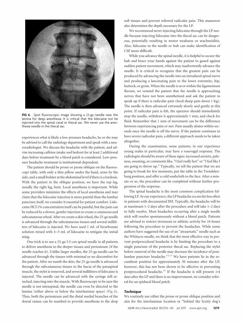
lamina (either above or below the interlaminar space) (Fig 6).
Thus, both the periosteum and the distal medial branches of the
dorsal ramus can be numbed to provide anesthesia to the deep
soft tissues and prevent referred radicular pain. This maneuver
also determines the depth necessary for the LP.
We recommend never injecting lidocaine through the LP nee-
dle because injecting lidocaine into the thecal sac can be danger-
ous, potentially resulting in motor weakness or arachnoiditis.
Also, lidocaine in the needle or hub can make identification of
CSF more difficult.
While you advance the spinal needle, it is helpful to secure the
hub and brace your hands against the patient to guard against
sudden patient movement, which may inadvertently advance the
needle. It is critical to recognize that the greatest pain can be
produced by advancing the needle into an intradural spinal nerve
and producing a lancinating pain to the lower extremity, hip,
buttock, or groin. When the needle is at or within the ligamentum
flavum, we remind the patient that the needle is approaching
nerves that have not been anesthetized and ask the patient to
speak up if there is radicular pain (focal sharp pain down 1 leg).
The needle is then advanced extremely slowly and gently at this
point. If radicular pain is felt, the operator should immediately
stop the needle, withdraw it approximately 1 mm, and check for
fluid. Remember that 1 mm of movement can be the difference
between experiencing pain or not. Pain usually abates within sec-
onds once the needle is off the nerve. If the patient continues to
have severe radicular pain, a different approach needs to be taken
altogether.
During the examination, some patients, in our experience
young males in particular, may have a vasovagal response. The
radiologist should be aware of these signs: increased anxiety, pale-
ness, sweating, or comments like, “I feel really hot” or “I feel like I
am going to throw up.” Typically, we tell the patient that we are
going to break for few moments, put the table in the Trendelen-
burg position, and offer a cold washcloth to the face. After a min-
ute or so, the procedure can be completed without further pro-
gression of the response.
The spinal headache is the most common complication fol-
lowing LP. In our experience, the LP headache occurs far less often
in patients with documented IIH. Typically, the headache will be
at maximum 1–2 days after the procedure and will take 1–2 days
to fully resolve. Most headaches occurring after a single needle
stick will resolve spontaneously without a blood patch. Patients
are advised to restrict strenuous or athletic activity for 24 hours
following the procedure to prevent the headaches. While some
authors have suggested the use of an “atraumatic” needle such as
the Whitacre needle, we think that the most effective way to pre-
vent postprocedural headache is by limiting the procedure to a
single puncture of the posterior thecal sac. Replacing the stylet
before removal of the needle may decrease the incidence of post-
lumbar puncture headache.14,15 We have patients lie in the re-
cumbent position for approximately 30 minutes after the LP;
however, this has not been shown to be effective in preventing
postprocedural headache.16 If the headache is still present �4
days after the LP and there is no improvement, we consider refer-
ral for an epidural blood patch.
TechniqueWe routinely use either the prone or prone oblique position and
aim for the interlaminar location or “behind the Scotty dog’s
FIG 6. Spot fluoroscopic image showing a 23-ga needle near thelamina for deep anesthesia. It is critical that the lidocaine not beinjected into the spinal canal or thecal sac. We never use the anes-thesia needle in the thecal sac.
AJNR Am J Neuroradiol 38:1276 – 83 Jul 2017 www.ajnr.org 1279

neck,” avoiding the disc space level. Because an LP needle can
easily extend through the disc itself, terminating in the preverte-
bral location or retroperitoneum (Fig 7), we avoid the disc space
level. If there is inadvertent extension through a disc with the
spinal needle, a rare event, we have found that replacing the stylet
and removing the needle at 1⁄2-cm intervals, with frequent checks
for blood return, is a method to deal with this complication. We
have never had venous or arterial blood return, but if that oc-
curred, we would plan to document the blood return, attempt to
differentiate venous from arterial blood, and consider a CT an-
giogram/venogram of the aorta and inferior vena cava after ter-
minating the unsuccessful LP. It is critical that when using fluo-
roscopy, the needle be centered in the image to avoid parallax
artifacts, an effect distinct to radiographs (Fig 8). Parallax is a
difference in the apparent position of an object along 2 different
lines of sight.17
Experienced radiologists are often comfortable with a 25-ga
spinal needle, but if a longer needle is needed, a 25-ga needle is
more difficult to steer. Our standard needle is 22 ga. For extremely
obese patients, we often use a 3.5-inch 18-ga introducer, but this
larger gauge should not enter the thecal sac.18 The introducer is
advanced using the same landmarks used in the LP and is hubbed.
Although the introducer needle is easy to steer, it is important to
have a properly planned trajectory because it can be difficult to
correct the path with this larger gauge coaxial needle. The needle
is much easier to steer through the introducer. The standard spi-
nal needle is 8.8 cm (3.5 inches). Needles are also available at 13-,
14-, 15-, and 18-cm or even longer lengths.
The 22-ga 19-cm spinal needle is then advanced through the
introducer. The needle hub has a notch, which is on the bevel side.
Therefore, the side opposite the notch is the “sharp” side. The
needle will go toward this sharper side and away from the notch.
Therefore, if you are steering the needle, the notch should be
opposite the direction you are attempting to steer. Some intro-
ducer needles may have the bevel on the opposite side of the notch
on the hub. Turning the needle 1⁄4 turn every 1 cm or so helps
direct the needle in a straight line. The introducer will help with
needle position, but it is still important to check periodically with
the fluoroscopy for an adequate trajectory. Even with the longer
spinal needle, the “pop” through the dura can often be felt. The
bevel should be directed cephalad, away from the dura/arachnoid
and perpendicular to the course of the nerves, to improve expo-
sure to the fluid, particularly if there is slow flow.
The obese patient usually cannot roll on the table for an open-
ing pressure in the lateral decubitus position, so we obtain the
pressure in the prone or prone oblique position, adding the needle
length to the final pressure measurement.19 The normal CSF pres-
sure range is 6 –20 cm H20 in adults and up to 25 cm H20 in obese
patients.20 An opening pressure of �25 cm H20 is diagnostic of
IIH, in the correct clinical setting.21
For the diagnostic LP, how much CSF to remove and put in
each tube varies on the basis of what has been requested by the
referring physician or provider. We routinely attach short tub-
ing to the needle and use a 10-mL syringe after the seal has been
broken, to gently remove CSF. The suction is applied very
gently, for just 2–3 seconds, then released to avoid affecting a
nerve root. This process is then repeated as long as CSF return
is present. One of the authors uses a unique technique that
allows air release into the tube-hub interface (Fig 9). The con-
nection between the tubing and hub is loose and not airtight,
thus preventing oversuction of fluid. For the standard diagnos-
tic LP, CSF is collected in 4 tubes for a total of 8 –15 mL.22 If
leptomeningeal carcinomatosis is suspected, we get the usual
specimen for the initial cell count, protein and glucose levels,
and culture; then, fluid for cytology can be placed either in a
black-top tube or in one of the standard LP tubes on the tray.
We always send the last 1–2 mL for the final cell count.
Flow cytometry in lymphoma or leukemia is unpredictable.
The laboratory needs “viable” cells to perform the test, and it is
impossible to know how much CSF is truly necessary. It can be
done with 3 mL of fluid, depending on the viable cell population.
FIG 7. Cross-table lateral film after failure to obtain CSF, thought tobe a dry tap. Note the LP approach at the L4 –5 disc space and theneedle extended through the disc into the retroperitoneum. At thispoint, the stylet was removed and no blood was returned. The needlewas withdrawn at 5-mm increments, each time checking for arterial orvenous blood return. There were no complications, but the diagnos-tic LP was cancelled until the following day.
FIG 8. Fluoroscopic spot films obtained during LP, showing parallaxartifacts. A, Note that on this image, the needle is not centered andthe tip appears to abut the inferior spinous process. B, The sameprocedure with no needle adjustment, but now the tip is centered inthe image. CSF flow was normal.
1280 Hudgins Jul 2017 www.ajnr.org

However, 10 mL of fluid might be inadequate at other times if the
load of abnormal cells in the CSF is small. We try to obtain a
separate vial of 6 –10 mL when flow cytometry is requested. Flow
cytometry should be distinguished from cytology.
Therapeutic LP: How Much CSF to Remove?The therapeutic LP generally implies removal of CSF to both con-
firm the diagnosis of IIH and treat the symptoms of intracranial
hypertension, which in some institutions can mean removal of
�30 mL of CSF. When LP is performed to remove CSF, we ask the
patient about a headache or visual obscurations before the study
and symptom changes during the procedure and then ask again
once CSF has been removed. In our experience, a high-volume tap
is rarely needed, and there is a risk of low-pressure headaches after
the LP. Most patients with IIH do well with a standard amount of
CSF removed (15–20 mL). This is likely because the dural defect
continues to leak after the tap. In addition, the high intracranial
pressure in IIH results in distal transverse venous sinus narrow-
ing; removing CSF decreases the intracranial pressure and reduces
venous sinus narrowing, improving venous return with a result-
ing decrease in symptoms. The opening and closing pressures, the
amount of CSF removed, and symptom resolution are docu-
mented in the final dictation. The closing pressure is only ob-
tained when performing a therapeutic tap.
Dry Tap“Dry tap” is defined as an LP with confirmed needle location in
the thecal sac, but no egress of CSF. Confirmation of the needle
position is by a cross-table lateral radiograph. In our experience,
the dry tap most commonly occurs in the hospitalized patient
who has not been eating or drinking and is relatively dehydrated.
Before any LP in the hospitalized patient, we ask the team to
hydrate the patient, ideally with intravenous fluids. For outpa-
tients, we use no sedating medications, and patients are not NPO
and are instructed to drink fluids liberally. Anecdotally, we have
noticed that the elderly patient will often have a dry tap and very
low CSF pressure, and CSF flow may be very slow. Dry taps can
occur in severe spinal stenosis, including thecal sac narrowing
from epidural lipomatosis (Fig 5) or arachnoiditis (Fig 3). If a
fluoroscopically guided LP has been a truly dry tap, repeating the
LP under CT guidance can assure definitive needle tip placement.
Before the LP, be sure the footplate is secured at the end of the
fluoroscopy table. After the pop into the thecal sac is felt, advance
the needle an additional 2–3 mm and then withdraw the stylet to
check for return of CSF. If the spinal needle is 25 ga, CSF return
may be very slow. Flow of CSF can be very unpredictable because
even tiny alterations in position can affect flow and the rate of
flow. It is critical that once CSF flow is confirmed, great care be
taken to maintain the same position of the needle throughout the
procedure. The hub can be held with the left hand during attach-
ment of the tubing or manometer. Be careful to not let the syringe
or tube torque and rotate the needle.
If CSF flow is confirmed and then stops, reinsert the stylet
fully, remove it again, and recheck it. Several maneuvers can help
with a dry tap. First, have the patient gently cough or perform a
Valsalva maneurver, and this will usually start CSF return. An-
other step is to raise the fluoroscopy table to about 45° to fill the
distal thecal sac. After several minutes in this position with pro-
vocative maneuvers, recheck the flow by removing the stylet. If
there is still no flow, with the stylet out, carefully turn the needle
90° and wait patiently. Continue with this maneuver until a full
circle has been achieved. If there is still no flow, the stylet should
be replaced and the needle advanced 1–2 mm, followed by a check
for CSF return. An additional maneuver is attaching a short tub-
ing and gentle suction with a 10-mL syringe. The suction should
be gentle and intermittent, not sustained. Adequately stretch the
tubing so that there are few “uphill” kinks through which the CSF
has to travel. It may be helpful to hold the tubing lower than the
table for gravitational assistance. This is particularly helpful when
the table cannot be tilted. In our experience, if CSF flow is initially
good, then stops, being patient is most important because usually
the flow will start again. Rarely, aspiration is ineffective, and CSF
has to be obtained by gravity flow only. This may take up to 30
minutes. The air-leak technique, described previously, can also be
attempted.
At this point, a cross-table lateral film if using fluoroscopy or
moving the C-arm to confirm the needle position is recom-
mended. If the needle position is confirmed within the canal and
there is still no flow, another level can be accessed in the cooper-
ative patient. If a myelography is being performed, 3–5 mL of
myelographic-safe contrast can be instilled to determine whether
the needle is intrathecal.
Cases that cannot be successfully accessed with fluoroscopy
may require biplane or CT guidance.
If another attempt does not result in CSF, there may be thick
FIG 9. Bubble technique to avoid sucking nerve roots into the spinalneedle. The short tubing is literally lying on the LP needle hub so thatthere is no manipulation of the needle. Both air and CSF fill the tubingand the syringe. With this technique, we have never had nerve rootpain reported, and CSF can be easily aspirated.
AJNR Am J Neuroradiol 38:1276 – 83 Jul 2017 www.ajnr.org 1281

tenacious secretions in the CSF as may be seen with meningitis,
especially in coccidioidomycosis or arachnoiditis ossificans.
If a second level results in no CSF, then it is truly a dry tap.
Our preference is to perform a lumbar spine MR imaging to see
whether there is a small thecal sac with a narrowed subarach-
noid space, such as in patients with epidural lipomatosis. De-
pending on the clinical setting and need for CSF, the LP can be
attempted another day after hydration or performed with CT
guidance or a C1–2 puncture can be considered. Before the
C1–2 tap, a cervical spine MR imaging is recommended to be
sure there is no cerebellar tonsillar ectopia, which can be pres-
ent in IIH. Particular attention should be paid to the course of
the vertebral and posterior inferior cerebellar arteries. Because
of low CSF volume in the subarachnoid space with intracranial
hypotension, a dry tap is not uncommon. Additional imaging
findings of intracranial hypotension include cerebellar tonsil-
lar ectopia, brain stem slumping, lack of CSF about the orbital
optic nerves, dural enhancement, venous engorgement, and
cerebral and spinal subdural collections.23-26 For patients with
intracranial hypotension or a prior dry tap, planning the initial
or repeat LP under CT guidance is another consideration. The
advantage is immediate confirmation of needle position in the
thecal sac.
The Patient with a Prior Lumbar Spine SurgeryIt is critical that prior imaging be reviewed in a patient with a
history of lumbar spine surgery. This review helps avoid bulky
osteophytes or osseous fusions; determine whether there is arach-
noiditis, which may impair CSF return; and finally, determine the
easiest site for the LP (Fig 10). We do not hesitate to go through
prior surgical sites. We have not experienced increased pain at
prior laminectomy sites, but this experience is patient-dependent.
3D surface-rendered images and models can help the procedur-
alist visualize the trajectory, especially if there are significant de-
generative changes.27 In general, a decompressive laminectomy
can make the LP easier. However, the procedure can be compli-
cated by arachnoiditis, especially if nerves are adherent to the
posterior thecal sac along the course of the needle tract. Postop-
erative images are also important in determining whether the
patient has a dorsal postoperative fluid collection, a seroma, or
pseudomeningocele (Fig 11). In general, avoid the fluid collection
if you know it is there, to avoid any chance of cross-contamina-
tion or confusing imaging or clinical issues with a CSF leak and
pseudomeningocele. One of us has inadvertently “sampled” a
FIG 10. Lateral plain film in a patient with recurrent back pain afterbilateral pedicle screws at nearly every level. Note dystrophic ossifi-cation from a fusion graft. In a patient with extensive postoperativechanges, cross-sectional imaging should be obtained to determinewhether there is a patent trajectory for the LP.
FIG 11. Adult patient with severe back pain following a lumbarspine operation. If LP or myelography is deemed necessary, theneedle should be above or below the dorsal intraspinal extraduralcollection.
1282 Hudgins Jul 2017 www.ajnr.org

dorsal seroma in a postoperative patient. If the operation was
recent, try to avoid the operative levels, again to prevent infection
(or blame for infection) or a confusing picture in a CSF leak.
Recent postoperative cases are probably better performed by us-
ing CT guidance.
Finally, the Dictation TemplateThe obese patient often undergoes serial LPs and knowing what
worked previously is helpful to the next proceduralist. We always
dictate whether fluoroscopy was adequate or whether the C-arm
was necessary. Of course, the level for the successful stick, types of
needles used, and whether coaxial technique was needed are in-
cluded in the report. A general estimate of the necessary needle
depth is useful to include in the report. Opening pressure, symp-
tom resolution with the LP, and any other technical facts are in-
cluded. The anesthetic used, whether bicarbonate was used, and
the appearance of the CSF, whether clear, cloudy, or blood-tinged
with clearing, are dictated. Finally, fluoroscopy time and radiation
dose are always mentioned (Table).
REFERENCES1. Kroll H, Duszak R Jr, Nsiah E, et al. Trends in lumbar puncture over
2 decades: a dramatic shift to radiology. AJR Am J Roentgenol 2015;204:15–19 CrossRef Medline
2. Ogden CL, Carroll MD, Kit BK, et al. Prevalence of childhood andadult obesity in the United States, 2011–2012. JAMA 2014;311:806 –14 CrossRef Medline
3. Hannerz J, Ericson K. The relationship between idiopathic intracra-nial hypertension and obesity. Headache 2009;49:178 – 84 CrossRefMedline
4. Mokdad AH, Ford ES, Bowman BA, et al. Prevalence of obesity,diabetes, and obesity-related health risk factors, 2001. JAMA 2003;289:76 –79 Medline
5. Boddu SR, Corey A, Peterson R, et al. Fluoroscopic-guided lumbarpuncture: fluoroscopic time and implications of body mass index–abaseline study. AJNR Am J Neuroradiol 2014;35:1475– 80 CrossRefMedline
6. Fetterly KA, Schueler BA. Experimental evaluation of fiber-inter-spaced antiscatter grids for large patient imaging with digital x-raysystems. Phys Med Biol 2007;52:4863– 80 CrossRef Medline
7. Bushberg JT. The AAPM/RSNA physics tutorial for residents: X-rayinteractions. Radiographics 1998;18:457– 68 CrossRef Medline
8. Eskey CJ, Ogilvy CS. Fluoroscopy-guided lumbar puncture: de-creased frequency of traumatic tap and implications for the assess-ment of CT-negative acute subarachnoid hemorrhage. AJNR Am JNeuroradiol 2001;22:571–76 Medline
9. Modica MJ, Kanal KM, Gunn ML. The obese emergency patient:imaging challenges and solutions. Radiographics 2011;31:811–23CrossRef Medline
10. Brook AD, Burns J, Dauer E, et al. Comparison of CT and fluoro-scopic guidance for lumbar puncture in an obese population withprior failed unguided attempt. J Neurointerv Surg 2014;6:324 –28CrossRef Medline
11. Schindera ST, Nelson RC, Toth TL, et al. Effect of patient size onradiation dose for abdominal MDCT with automatic tube currentmodulation: phantom study. AJR Am J Roentgenol 2008;190:W100 – 05 CrossRef Medline
12. Yanch JC, Behrman RH, Hendricks MJ, et al. Increased radiationdose to overweight and obese patients from radiographic examina-tions. Radiology 2009;252:128 –39 CrossRef Medline
13. Nayate AP, Nasrallah IM, Schmitt JE, et al. Using body mass index topredict needle length in fluoroscopy-guided lumbar punctures.AJNR Am J Neuroradiol 2016;37:572–78 CrossRef Medline
14. Strupp M, Brandt T, Muller A. Incidence of post-lumbar puncture syn-drome reduced by reinserting the stylet: a randomized prospectivestudy of 600 patients. J Neurol 1998;245:589–92 CrossRef Medline
15. Evans RW. Complications of lumbar puncture. Neurol Clin 1998;16:83–105 CrossRef Medline
16. Wu CL, Rowlingson AJ, Cohen SR, et al. Gender and post-dural punc-ture headache. Anesthesiology 2006;105:613–18 CrossRef Medline
17. Curry TS III, Dowdey JE, Murry RC Jr. Christensen’s Physics of Diag-nostic Radiology. 4th ed. Philadelphia: Lea & Febiger; 1990:258 –59
18. Johnson JC, Deeb ZL. Coaxial needle technique for lumbar punc-ture in the morbidly obese patient. Radiology 1991;179:874 CrossRefMedline
19. Abel AS, Brace JR, McKinney AM, et al. Practice patterns and open-ing pressure measurements using fluoroscopically guided lumbarpuncture. AJNR Am J Neuroradiol 2012;33:823–25 CrossRef Medline
20. Rando TA, Fishman RA. Spontaneous intracranial hypotension: re-port of two cases and review of the literature. Neurology 1992;42:481– 87 CrossRef Medline
21. Corbett JJ, Mehta MP. Cerebrospinal fluid pressure in normal obesesubjects and patients with pseudotumor cerebri. Neurology 1983;33:1386 – 88 CrossRef Medline
22. Cauley KA. Fluoroscopically guided lumbar puncture. AJR Am JRoentgenol 2015;205:W442–50 CrossRef Medline
23. Dillon WP, Fishman RA. Some lessons learned about the diagnosisand treatment of spontaneous intracranial hypotension. AJNRAm J Neuroradiol 1998;19:1001– 02
24. Mokri B, Piepgras DG, Miller GM. Syndrome of orthostatic head-aches and diffuse pachymeningeal gadolinium enhancement. MayoClin Proc 1997;72:400 –13 CrossRef Medline
25. Schievink WI, Meyer FB, Atkinson JL, et al. Spontaneous spinal ce-rebrospinal fluid leaks and intracranial hypotension. J Neurosurg1996;84:598 – 605 CrossRef Medline
26. Fishman RA, Dillon WP. Dural enhancement and cerebral displace-ment secondary to intracranial hypotension. Neurology 1993;43:609 –11 CrossRef Medline
27. Cramer J, Quigley E, Hutchins T, et al. Spine Procedures in 3D. Salt LakeCity: Practical Imaging; 2016: version 1.1, iTunes Bookstore. https://itunes.apple.com/us/book/spine-procedures-in-3d/id1119397210?ls�1&mt�13. Accessed February 25, 2017
LP dictation templateThe following are included in our LP template:
Reason for LP: ���Level of stick, if additional level was attempted: ���Fluoroscopy or C-arm: ���Coaxial technique vs single needle: ���Anesthetic used, amount, bicarbonate used: ���Gauge and length of needle: ���Opening pressure and position (prone or on side) when pressure
was measured: ���Closing pressure (not always performed):Amount of CSF obtained:Appearance of CSF:Symptoms when patient arrived in radiology department:Resolution of symptoms following LP: ���Fluoroscopy time: ���Radiation dose: ���
AJNR Am J Neuroradiol 38:1276 – 83 Jul 2017 www.ajnr.org 1283


![Diagnostic accuracy of diagnostic imaging for lumbar disc ...Approximately 5–15% of patients with low back pain suffer from lumbar disc herniation (LDH) [1, 2]. LDH is the most common](https://img.pdfslide.net/doc/110x75/5f8484d50a20a30a0d46ff1d/diagnostic-accuracy-of-diagnostic-imaging-for-lumbar-disc-approximately-5a15.jpg)