Embed Size (px)
Citation preview

8/3/2019 Digital Photography in Orthodon
http://slidepdf.com/reader/full/digital-photography-in-orthodon 1/48
Digital Photography in Orthodontics
Introduction
Photographs are an essential part of clinical documentation. Current
‘best practice’ is a full set of extra- and intra-oral photographs, both at the
start and completion of a course of orthodontic treatment and, ideally, some
mid-treatment photographs showing key-stages in treatment (Sandler, 2000).
Photographs are an essential part of clinical records for a number of
reasons:
1. Unreliable memories. Within a matter of months, patients and parents
tend to forget how severe the original malocclusion was. Having slides
available at every visit reminds both the orthodontist and the patient of
the original situation, against which all improvements can be judged.
2. Medico legal requirements. In our increasingly litigious society, it is
critical to have clinical photographs that indicate any preexisting
pathology or trauma to the teeth. Close-up photographs are strongly
advised for any marked decalcification or enamel fractures that are
evident from the outset.
The debonding appointment is often the first time patients or
parents really focus in on the labial enamel, and it may be the first time

8/3/2019 Digital Photography in Orthodon
http://slidepdf.com/reader/full/digital-photography-in-orthodon 2/48
they actually notice surface decalcification, fractures, or other
blemishes. Proper records will help avoid any post-treatment disputes.
3. Teaching needs. Slides are probably the most important teaching aidsin orthodontics. If cases are to be used in lectures, posters, papers, and
presentations, a high standard of clinical photography is required.
4. Treatment evaluations. A quick scan of sequential slides with patients
and parents during treatment will save lengthy explanations of
biomechanics or tooth movements.
Digital photography has been generally available since 1981. In 1991
‘Autotrader’ were the first mass market publication to move completely to
digital recording of images. Now, many trades and professions, including
estate agents, advertising agencies, police, and the media use digital
photography on a routine basis.
Digital images are made up of picture elements (‘pixels’) comprising
red, green, and blue light, each set at a level between 0 and 255. If all three
colours are set at 255 white is the result, while if all are set at zero, black
results (Figure 1 ). There are 256 grey shades that result from all three
colours being set at the same number. Varying the level of each of the three
colours results in the gamut of 16•7 million colours. Numerical values for
each of these colours are stored on the Charged Couple device (CCD). This
is made up of pixels, the number of which, combined with the degree of
compression, determines the quality of the final output.

8/3/2019 Digital Photography in Orthodon
http://slidepdf.com/reader/full/digital-photography-in-orthodon 3/48
FIG. 1 Digital images made up of Red, Green
and Blue light at levels between 0 and 255. 16•7 million colors in all.
In the 1990s a typical CCD would comprise 640 x 480
pixels resulting in acceptable images for snapshots, but lacking the quality
needed for high quality clinical photographs (Figure 2 ). By 1999 the first
‘mega-pixel’ cameras (over 1,000,000 pixels per image) were becoming
available, but above 1•5 mega pixels the law of diminishing returns kicked in
with a disproportionate price increase for only modest improvements in
quality.

8/3/2019 Digital Photography in Orthodon
http://slidepdf.com/reader/full/digital-photography-in-orthodon 4/48
FIG. 2
Low pixel count (left) results in poorer quality image than required for
orthodontics(right).
Bearing in mind a conventional 35-mm slide is thought to
contain the equivalent of 25–30 million pixels of information there was still a
long way to go for digital images to be serious competition. In addition,
conventional photographic equipment for orthodontic images produced good
image quality, was very reliable and user-friendly (Sandler and Murray,
1999 ), and was relatively inexpensive.
However, well-recognized problems with conventional
photographic techniques are the cost of developing and processing films, the
time required for processing and physical storage of all the patients slides or
prints.
Digital photography offers many advantages including:
1. rapid turn-around;
2. checkable exposure accuracy;
3. no ageing of photos;
4. dust and scratches are irrelevant;

8/3/2019 Digital Photography in Orthodon
http://slidepdf.com/reader/full/digital-photography-in-orthodon 5/48
5. built in white balance;
6. immediate viewing;
7. no film or processing costs;
8. inexpensive storage;
9. easy retrieval;
10. duplication easy;
11. Transmission around the world in seconds is entirely feasible.
‘Prosumer’ cameras
One type of digital camera (prosumer) falls into the mid-range
price bracket £500–1500 and lies between the consumer camera and the
professional models. They usually have a host of useful features including
macro-zoom lenses and potentially high image quality. The ‘piece de
resistance’ of digital cameras is undoubtedly the image preview facility in
that images can be immediately viewed on the LCD screen and accepted or,
if flawed, deleted and retaken.
The problems with the ‘prosumer’ cameras used for orthodontic
photography are three-fold.
First, the flash provided with most digital cameras is a point flash.
Experience has shown that for high quality intra-oral
images ring flashes are
essential to avoid unacceptable shadowing on most of the images (Figure 3 ).
Despite the use of deflectors and diffusers the results with the built-in point
flash tend to be disappointing. The point flashes are also not powerful enough
to allow the photos to be taken on very small apertures (F32). This is essential

8/3/2019 Digital Photography in Orthodon
http://slidepdf.com/reader/full/digital-photography-in-orthodon 6/48
as it greatly increases the depth of field and ensures most of the frame is in
focus. In addition, even if it is possible to add a ring flash to the ‘prosumer’
camera high quality consistently exposed images require through the lens
(TTL) metering, which is not available on these ‘lower
end’ systems. After 20
years of ever increasing quality of orthodontic photography using SLR
systems, TTL metering, and ring flashes, some of the orthodontic community
are accepting mediocre photographs, taken with substandard digital equipment
just for the facility of immediate viewing.
FIG. 3 Point flash intra-oral images are invariably inferior to ring flash due
to shadowing and variable exposure.
It is possible to overcome some of the shadowing problems of a
point flash by modifying the technique used. Taking the occlusal shots from
much further away may ensure adequate illumination, but will inevitably
waste pixels unnecessarily and focusing
will also be problematic (Figure 4 ).
Turning the camera around on buccal intra-oral shots may also reduce
shadowing to a degree.

8/3/2019 Digital Photography in Orthodon
http://slidepdf.com/reader/full/digital-photography-in-orthodon 7/48
FIG. 4 Slightly different technique used for occlusal shots with point
flash—shadows less but pixels wasted.
The second problem involves the viewfinders; some digital
cameras are available with a ‘Galilean telescope’ viewfinder that is very
suitable for snapshots, but totally unsuitable for high quality intra-oral
photography. The problem is that the viewfinder, when close to the subject,
doesn’t accurately represent what the lens will ‘see’. Live display on the
LCD screen is also possible, but they are again inaccurate if the ‘refresh rate’
is slow, and are very power hungry, making it an unsuitable method unless a
mains supply is utilized.
Thirdly, the focusing system can be problematic as the
auto-focus systems on the ‘prosumer’ cameras are frustrating to work with
when capturing intra-oral photographs. They often take three or four
attempts to get the system to focus adequately, and all the area of interest is
not always as sharp as it might be. The predetermined distance ‘macro’
settings available on some of the digital cameras also sometimes give
disappointing results.
Professional cameras

8/3/2019 Digital Photography in Orthodon
http://slidepdf.com/reader/full/digital-photography-in-orthodon 8/48
Top end cameras have always been available. Indeed,
Kodak teamed up with Nikon in the late 1990s to produce the Digital
Camera System (DCS), which was capable of very high quality images. The
problem with this system was that the camera body alone
was over £10,000.
FIG. 5 Basic price of Nikon/Kodak DCS £10,495
The Nikon Dl is one of the best digital cameras and is the one used
by a great many photojournalists worldwide. It has a vast array of features
required by professionals, and the body is built out of titanium to an
incredibly high specification, for use in sandstorms, typhoons, and war zones
throughout the world. The problem with the Dl is that it is a very heavy
camera and would be difficult to hold with one hand, a technique essential
for high quality intra-oral images (Sandler and Murray, 1999 ). Also The
SB29 ring flash does not work as TTL metering with this body. Finally, a
price tag for the whole package of close
to £5000 makes it unaffordable for many clinicians.
Another digital camera recently released is the Fuji FinePix S1
Pro, which may be the perfect digital camera for orthodontics (Figure 6 ).

8/3/2019 Digital Photography in Orthodon
http://slidepdf.com/reader/full/digital-photography-in-orthodon 9/48
The body is made by Nikon and is therefore built to a high specification. The
lens system required is the Nikon 105 mm/2•8 AF Macro and the flash
system is the Nikon SB29 Speedlight. The flash provides TTL metering and,
therefore,
the intra-oral photos taken at F32 are invariably perfectly
exposed
and in focus. The pictures are all taken on manual focus just by setting the
lens adjustment for intra-oral shots, then moving backwards and forwards to
focus. Using the ‘limit’ switch on the lens allows the same magnification to
be set for all intra-oral photos, thus allowing direct comparability between
photos.
FIG. 6 Fuji Si FinePix Pro plus Nikon SB29 flash.
Images may be stored on a 64 Mb storage card. The capacity of
this card means that 330 images can be stored, using the lowest pixel setting
(1440) and maximum compression, resulting in images of about 200 Kb. The
quality of these images is more than acceptable for most clinical situations
(Figure 7 ). The images can still be cropped and enlarged as necessary
retaining sufficient detail for most situations.

8/3/2019 Digital Photography in Orthodon
http://slidepdf.com/reader/full/digital-photography-in-orthodon 10/48
FIG. 7 Top pictures with point flash and no TTL metering, bottom with
FujiS1Pro.
The only adjustment the camera requires is from F32 to F11 for
extra-oral shots and to switch off the flash bulb behind the patients head on
the three-quarter and profile view to throw the shadow behind the head.
MANIPULATION OF DIGITAL PHOTOGRAPHS
The requirements for successful use of digital images are a high
quality digital camera, and a sufficiently powerful computer to allow easy
viewing and subsequent manipulation of the images.
Most digital images are stored within the camera on either a
Compact Flash card (43 x38x3.5mm in hard case) or a Smart Media card(thinner, lighter, and more flimsy).
The former storage medium is probably more appropriate for a busy
clinical environment, as the latter requires delicate handling. Once the

8/3/2019 Digital Photography in Orthodon
http://slidepdf.com/reader/full/digital-photography-in-orthodon 11/48
images have been captured they need to be 'read' by the computer. At
present, the most convenient method is use of an adapter to allow the card
to be inserted directly into an empty PCM CIA port. On a modem laptop
this is the one occupied by the computers removable fax / modem or
alternatively by the network card.
PCM CIA card slots into the laptop
I.EXIF VIEWER

8/3/2019 Digital Photography in Orthodon
http://slidepdf.com/reader/full/digital-photography-in-orthodon 12/48
• Modern digital cameras are often sold with 'bundled' software
to allow viewing of images.
• Exif viewer is provided with many cameras, and if loaded
correctly, the 'thumbnails' (small representations of each picture on
the computer screen) are automatically loaded on the computer
screen when the memory card is accessed.
• ‘Thumbnails’ automatically appear in the Exif viewer
• Exif viewer also allows inspection of individual images, to
check entire area of interest is included, as well as confirming
sufficient depth of field was available to ensure the whole picture is
in focus.

8/3/2019 Digital Photography in Orthodon
http://slidepdf.com/reader/full/digital-photography-in-orthodon 13/48
• The Instant preview facility, on all but the cheapest digital
cameras' LCD screen, certainly gives an overall impression of the
image.
• It is only the top range cameras, such as the Fuji S 1 Pro, that
have a sufficiently high quality LCD screen combined with the
facility to scan the entire image easily with a powerful optical
zoom. This will allow quality verification by viewing on the camera
alone.
Images taken with mid-range cameras may need quality confirming,
using Exif viewer, before the patients are sent home.
Each image can be checked for quality.

8/3/2019 Digital Photography in Orthodon
http://slidepdf.com/reader/full/digital-photography-in-orthodon 14/48
Once all the images are satisfactory, Dento facial Showcase is
opened alongside Exif viewer. The 'Restore Down' button is now used
(top right of the screen, button next to Close Program button) for both
programs, to allow them to be visible on the screen at the same time.
All of an individual patient's images are now selected in Exif
Viewer (left mouse button, whilst holding down the Ctrl key) and these
are dragged and dropped into a previously opened new file in Showcase.
In a busy clinic, particularly where images are not necessarily
downloaded from the card after every patient, it is important to write
down the patient's name on paper. This name should be photographed to
allow subsequent identification of patients.
This is essential if many clinicians use the camera, particularly if
the cards are filled to capacity before a backup is made.
II. DENTO FACIAL SHOWCASE

8/3/2019 Digital Photography in Orthodon
http://slidepdf.com/reader/full/digital-photography-in-orthodon 15/48
• Showcase is a popular program for storing, manipulating, and
showing orthodontic records of patients, in an informal setting.
• Thumb nails are stored under each patient's name, and
attributes such as type of photograph and stage of treatment can be
easily attached to individual images, or groups of images.
• Slides of particular interest can be selected within Showcase
to run a slide show to illustrate the features of the patient's
malocclusion.
• Individual slides can be presented in order to show
maximum detail of each view taken. Alternatively a selection of
views can be incorporated with in a single 'slide' for a less detailed,
but more comprehensive over view of the case
•
•

8/3/2019 Digital Photography in Orthodon
http://slidepdf.com/reader/full/digital-photography-in-orthodon 16/48

8/3/2019 Digital Photography in Orthodon
http://slidepdf.com/reader/full/digital-photography-in-orthodon 17/48
The image will probably need to be resized with in PowerPoint and then
the process is repeated for further images.
The advantage of the PowerPoint slides is that the relative size and
position of the individual images is infinitely variable and maximum
space can be occupied by material of interest.
Intra-oral slides are scanned into PowerPoint and all five views can
be incorporated into one slide to give the 'full picture' of the malocclusion
and treatment at that point in time.
Radiographic information should also be imported in to the
computer. The lateral cephalometric radiograph and OPG can either be
scanned, if the scanner has a transparency adaptor, or alternatively
photographed with the digital camera.
The flash is turned off and the camera aperture opened sufficiently
wide to reduce the shutter speed to 125 or faster, to eliminate camera
shake. The ideal background is an outside window, using daylight to
trans-illuminate the film avoiding the greenish hue inevitable when an X-
ray viewer is used for illumination.
Radiographs captured digitally & converted to gray scale

8/3/2019 Digital Photography in Orthodon
http://slidepdf.com/reader/full/digital-photography-in-orthodon 18/48

8/3/2019 Digital Photography in Orthodon
http://slidepdf.com/reader/full/digital-photography-in-orthodon 19/48
All the information on particular patients can be presented to
patients, individual colleagues or a large audience in a clear and concise
manner, which can serve as an aid to future diagnosis and treatment
planning.
SOURCES OF ERRORS IN CLINICAL PHOTOGRAPHY
Clinical photographs taken before, during and after orthodontic
treatment form an essential part of the patients' records. If correctly taken,
they offer more useful information about the malocclusion and treatment
than any other clinical record. There are, however, many potential sources of
errors whilst obtaining these invaluable records. Photographs of inadequate
quality may misrepresent the patients starting malocclusion, may
inaccurately reflect progress with treatment or may inaccurately record
dental anomalies and defects that may be present.
With both conventional and digital systems, many of these
errors, which involve use of mirrors and retractors and patient positioning
issues, are common to both methods. With the recent widespread use of
digital equipment a whole new range of possible errors has been introduced
and specific problems related to the digital system are discussed in detail.
There are a number of errors that are commonly seen and these
can be divided into two groups.
• The first group comprises errors due to inappropriate choice or
use of equipment including the camera, lens, flash, retractors, mirrors
or suction, or a lack of understanding of the digital technology resulting
in inadequate or inappropriate images.

8/3/2019 Digital Photography in Orthodon
http://slidepdf.com/reader/full/digital-photography-in-orthodon 20/48
• The second group of errors relates to any recording medium and
involves inappropriate positioning of the subjects.
Technical errors
1. Camera
2. Retractors
3. Mirrors
1.
Camera.The correct equipment is required for high quality clinical
photographs, which include a camera (either conventional or digital) with
a macro-facility (ability to produce I : I images) and, ideally, a ring flash,
an appropriate background, suitable lighting and well trained assistants.
Correct camera orientation is important, with extra-oral
photographs taken in portrait mode and intra-oral photographs taken in
landscape mode. To allow direct comparison of photographs taken at
different times consistent magnification of images is required.
To aid this with conventional equipment a label can be placed
on the barrel of the lens indicating the required lens setting (focal length)
for each of the standard views .

8/3/2019 Digital Photography in Orthodon
http://slidepdf.com/reader/full/digital-photography-in-orthodon 21/48
The magnification will therefore be preset for intra-oral, mirror and extra-
oral views allowing direct comparison of sequential shots. The lens barrel
is set to the predetermined position and the subject brought into focus by
moving the camera closer to or further from the patient.
With digital images this is not such a critical issue as they can
be resized at a later stage to allow comparison with previous or
subsequent images providing there is sufficient information on the image
to guarantee quality, once cropped and resized. This is determined by the
number of picture elements (pixels) on the charge-coupled device within
the digital camera and whether the area of interest completely fills the
recorded area. Most modern digital cameras record 3 mega pixels or
more, which is more than adequate for high quality clinical photography.
2.Retractors.
Two sizes of double-ended retractor are prerequisite to
obtaining a set of high quality intra-oral photographs.

8/3/2019 Digital Photography in Orthodon
http://slidepdf.com/reader/full/digital-photography-in-orthodon 22/48
The large ends of the larger retractor are used to obtain retraction for the
anterior intra-oral shot. The assistant should hold both retractors pulling
them both laterally and also forwards, which is the opposite to the natural
instincts of the assistants when retracting. By pulling the lips forwards
towards the photographer it makes it easier for the patient to bite together
in occlusion and pulls the soft tissues away from the teeth.
For the buccal shots, one retractor is turned through 180°, thus using
the smaller end of the larger retractor on the side of interest. The
photographer should hold this retractor themselves and, immediately before
capturing the image, pull it an extra 4-5 mm both distally and away from the

8/3/2019 Digital Photography in Orthodon
http://slidepdf.com/reader/full/digital-photography-in-orthodon 23/48
teeth to ensure at least the distal of the first molars is captured. To allow
optimal soft tissue retraction the assistant passively holds the large end of
the large retractor on the opposite side
.
For both occlusal shots the assistant inserts the small ends of
the small retractors under the respective lips and rotates them towards the
midline pulling the lips forward, as well as laterally. This is essential to
prevent obscuring the teeth with the lips. The direction of pull is away
from the teeth, and upwards for maxillary shots and downwards for
mandibular shots, thus ensuring a background of reflected mucosa rather
than stretched vermillion.

8/3/2019 Digital Photography in Orthodon
http://slidepdf.com/reader/full/digital-photography-in-orthodon 24/48
3. Mirrors.
Long-handled, front-silvered, glass mirrors are the ideal tool for
clinical photography, although they are significantly more expensive than
rear-silvered or metal mirrors. Long handles are held by the photographer to
allow complete control of the picture and to keeps assistants fingers out of
the shot.
Glass mirrors produce a far superior photograph compared to
polished metal mirrors as there is much greater reflection of the light and
they are more resistant to scratching. Silvering on the front side of the mirror
prevents double images, which occur due to a second reflection from the
glass surface when the silvering is on the back surface.
Ghost image from glass and image Sharp image when front silvered
from rear silver Surface is used
Prior to taking the photograph the mirror should either be warmed to
prevent misting of the mirror when it is inserted into the patients' mouth or the patient should be instructed to hold their breath for 10 seconds or so.
The occlusal mirrors are available in three different sizes; however, the
two smallest sizes are required in less than 10% of patients.

8/3/2019 Digital Photography in Orthodon
http://slidepdf.com/reader/full/digital-photography-in-orthodon 25/48
During occlusal photography light is never reflected 100%, and there is a
tendency for mirror photographs to be slightly underexposed.
It is therefore worth using an aperture compensation of + 1 F-stop, to ensure
good illumination of mirror shots. This adjustment can be usually made on
both conventional and modern digital camera systems.

8/3/2019 Digital Photography in Orthodon
http://slidepdf.com/reader/full/digital-photography-in-orthodon 26/48
Problems related to Digital Photography
1. Depth of field.*
2. Auto focus.*
3. Shadows.*
4. Constructing symmetrical images.
5. Image storage.
6. Digital image- fit for purpose?
*Problems frequently encountered when using midrange 'Prosumer' cameras.
Depth of field problems.
The depth of field represents the amount of the image that
is in sharp focus, and is dependant upon magnification and the aperture
selected. As the magnification increases and as the aperture through which
the picture is taken widens the depth of field reduces. Many mid-range
digital cameras that bridge the gap between consumer and professional
models, (known as 'Prosumer' cameras, e.g. Nikon Cool Pix 990/4500) will
only allow the aperture to be reduced to about Fll.
When taking intra-oral photographs with these mid-range
cameras the depth of field will be relatively small and on the anterior intra-
oral photograph part of the picture will inevitably be out of focus.

8/3/2019 Digital Photography in Orthodon
http://slidepdf.com/reader/full/digital-photography-in-orthodon 27/48
The depth of field is distributed approximately one-third in front and
two-thirds behind the focal plane. This disadvantage of small depth of field
with pictures taken with larger apertures can be minimized (but not avoided
completely) by focusing on the distal surface of the lateral incisors to at least
get central incisors to canines in focus.
With professional digital cameras, e.g. Fuji Sl FinePix Pro, combined with
the powerful Nikon SB29 flash, which allows through the lens metering a
perfect exposure is possible on F32. This tiny aperture allows sufficient
depth of field to include both incisor brackets and second premolar bracketsin sharp focus provided the focal plane is positioned correctly, i.e. on the
mesial of the canines.

8/3/2019 Digital Photography in Orthodon
http://slidepdf.com/reader/full/digital-photography-in-orthodon 28/48
With buccal shots and occlusal shots, provided the subject is correctly
positioned and retractors are appropriately used, all the area of interest is on
one plane; therefore, depth of field should not be an issue.
Auto-focus problems.
Digital cameras often allow the choice between auto-focus or
manual focus. Manual focus is by far the preferred option for the following
reasons. With Prosumer cameras focusing have to be on the lateral incisors
and with top end cameras on the canines, whilst still maintaining a centered
photograph. Because of the lack of sharply contrasting lines in the area of
interest many of these digital cameras have difficulty focusing using the
auto-focus setting for intraoral photographs. The result of this is attempt
after attempt to get the camera focus light (usually flashing green) to stop
flashing, indicating that the shot is in focus. This often proves fruitless
despite repeatedly moving the camera slightly between attempts at focusing.
All this is occurring whilst the assistant and the clinician are heaving on the
retractors to get maximum retraction of the soft tissues and some patients
may find this a little uncomfortable.

8/3/2019 Digital Photography in Orthodon
http://slidepdf.com/reader/full/digital-photography-in-orthodon 29/48
Frustrating attempts to get auto focus to work.
The solution to this problem is to use the manual focus setting
for all clinical photography. With top end cameras with through the lens
(TTL) facility focusing is done looking through the viewfinder. With the
Prosumer models, the clinician decides the appropriate distance between the
patient and the camera that fills the frame with the area of interest, This
focusing distance of, for example, 0.2 m, is set manually on the camera, and
the camera is then merely moved backwards and forwards until the image on
the LCD screen is in sharp focus, and the picture is taken. Twenty
centimeters is a good distance to start testing the cameras ability to take
sharp anterior intra-oral photographs on manual setting.
8/3/2019 Digital Photography in Orthodon
http://slidepdf.com/reader/full/digital-photography-in-orthodon 30/48
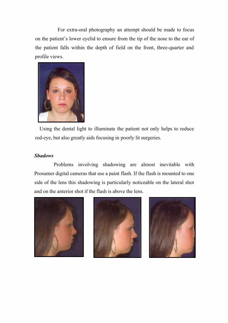
For extra-oral photography an attempt should be made to focus
on the patient’s lower eyelid to ensure from the tip of the nose to the ear of
the patient falls within the depth of field on the front, three-quarter and
profile views.
Using the dental light to illuminate the patient not only helps to reduce
red-eye, but also greatly aids focusing in poorly lit surgeries.
Shadows
Problems involving shadowing are almost inevitable with
Prosumer digital cameras that use a paint flash. If the flash is mounted to one
side of the lens this shadowing is particularly noticeable on the lateral shot
and on the anterior shot if the flash is above the lens.

8/3/2019 Digital Photography in Orthodon
http://slidepdf.com/reader/full/digital-photography-in-orthodon 31/48
Various mirrors, reflectors and diffusers have been suggested in the past to
reduce this problem; however, none provide the perfect solution and the
additions tend to make the set-up unwieldy to use.
The other alternatives are either to use an illuminated screen as the
backdrop to the patients when taking the extra-oral photographs, or use a
dark non-reflective background (preferably velvet) to maximize the quality
of the image.
With intra-oral views again the solution with a side mounted point
flash is to turn the camera upside down on the buccal view with the very
dark buccal corridor. This will ensure the flash illuminates the area that
would otherwise be in shadow due to the cheek. This digital photograph can
then be rotated 180º before the picture is saved in the patients file.
Dark right buccal corridor as Shadow overcome byCheek prevents light from Left turning Camera through 180 º
Mounted flash. So the flash is now on left.

8/3/2019 Digital Photography in Orthodon
http://slidepdf.com/reader/full/digital-photography-in-orthodon 32/48
High quality occlusal photographs are also difficult to obtain using
cameras with point flashes with the usual magnification, because of the
proximity of the camera to the patient; much of the area of interest is in
shadow
.
One solution to the problem of inadequate illumination is to focus further
away from the patient, which allows more light in and therefore reduces
shadowing. In this situation, the area of interest only fills about 20% of the
area captured by the camera so the charge couple device must be of high
enough quality to produce a good image after 80% of the information
captured has been discarded .

8/3/2019 Digital Photography in Orthodon
http://slidepdf.com/reader/full/digital-photography-in-orthodon 33/48
Constructing symmetrical images.
One major advantage of the very popular Dental Eye 3 camera,
over many of its competitors, was the presence of a graticule in the
viewfinder. This allowed very well constructed symmetrical and balanced
intra- and extra-oral photographs to be taken, even by relatively
inexperienced photographers using the occlusal plane the interpupillary line
and the Frankfort plane to construct reproducible photographs.
Most of the midrange digital cameras do not have the benefit
of a graticule to help with construction of the photographs, but some of the
top end cameras, e.g. the Fuji FinePix S2 Pro, have 'on-demand' grid lines,
which help significantly with construction of the extra-oral and intra-oral
images.
Card problems. The digital images are often recorded onto PCMCIA
cards. These cards have a series of 50 small holes that accept 50 tiny metal
pins within the camera. Small imperfections in the PCMCIA card may
damage the pins and once damaged will necessitate return of the camera to
the manufacturers for repair.

8/3/2019 Digital Photography in Orthodon
http://slidepdf.com/reader/full/digital-photography-in-orthodon 34/48
CCD problems. Even when the lenses on the digital cameras are
never changed dust may still eventually get onto the CCD of the cameras.
This will be seen as tiny 'in focus' black marks, at a specific spot on intra-
and extra-oral images.
Hairs and dust eventually get onto the CCD
On SLR type cameras it is often possible to get access to the CCD to
allow it to be cleaned with optic cleaning liquid on lint-free non-abrasive
cloths, but this must be done with extreme care. If in any doubt at all the
camera should be returned to the manufacturer for this to be carried out.
Digital image: fit for purpose?
Most digital cameras come with a variety of settings and it is
sometimes difficult to know which is the best setting to use in any particular
situation. The questions that need to be answered are what will the digital
image be used for, is memory card space at a premium and will the images
ever be used to produce hard copy?
When deciding upon the type of image there are choices about the
pixel dimensions. These may be 3040, 2048, or 1024 pixels across the wider
dimension of the image. (Cheaper cameras have even smaller dimensions of
images, but the quality of these is usually unacceptable for clinical

8/3/2019 Digital Photography in Orthodon
http://slidepdf.com/reader/full/digital-photography-in-orthodon 35/48
purposes). If the image is only ever to be viewed on a computer screen, there
is little point having more information available than can be exhibited on the
screen, or displayed using a laptop projector. The average screen has 1024
pixels across, so if a landscape image is going to occupy the whole screen
1000 pixels across will be the setting of choice, reduced proportionally as
the area of the slide occupied by the image is reduced.
.
Keeping images as small as possible will ensure that the
slideshows into which they are imported are a manageable size, and that the
computers do not struggle when displaying the slideshow. When creating an
orthodontic slideshow an image will often only occupy half of the screen so
the image size can be reduced further, to 500 pixels on its horizontal axis,
using any of the commonly available image manipulation programmes, prior
to insertion into the slide show.
This is preferable to grabbing the corners of a grossly oversized
image and 'squashing' it to within the dimensions of a PowerPoint slide, as
all the superfluous 'memory hungry' information is still within the file
making the slideshow unnecessarily large and often unwieldy. On most
digital cameras there is also a setting for image quality, as various degrees of

8/3/2019 Digital Photography in Orthodon
http://slidepdf.com/reader/full/digital-photography-in-orthodon 36/48
compression are used to reduce memory requirements. A common situation
is for the camera to save files at maximum quality with no compression as
TIFF files and to have 2 or 3 levels of JPEG compression represented by the
'fine', 'normal' and 'basic' settings. Roughly, the file sizes are reduced to 1/4,
1/8 and 1/16 of the original file size by successive compressions. The
'normal' setting produces images that are adequate for most purposes, and
the 'High', and 'Fine' settings are generally required when hard copy prints
are required.
If there is a possibility that the digital image will need to be
printed at some stage then for photographic quality printing a resolution of
approximately 300 pixels per inch is required. For a good quality 6 x 4 inch
print the image needs to be taken with the 2048 pixel setting across its
longer dimension. Images taken for publication purposes, therefore, need to
be of a higher size and ideally higher quality (less compression) than those
taken for routine patient records.
The typical setting for standard digital photographs using a Fuji
FinePix S2 Pro is the 1440 setting on 'normal' for the intra-oral photographs
and using a + 1 compensation for mirror shots. The aperture of the camera is
set at F32 for both types of intra-oral photographs and F5.6 for extra-oral
photographs.
Positioning errors
Both the patient and the clinician need to be positioned
correctly, in a standardized manner, to produce consistent photographs. All
features of the malocclusion should be demonstrated, and areas of interest
not obscured by clothing, hair, impression material, retractors or saliva.
Problems may be encountered where there is a height difference

8/3/2019 Digital Photography in Orthodon
http://slidepdf.com/reader/full/digital-photography-in-orthodon 37/48
between the patient and the clinician, and it may not be possible to get a
uniform background as the photographs may appear to be taken above or
below the patient. This problem can be solved by getting the patient or the
clinician, which ever is appropriate, to stand on a platform to raise them to
the same height.
The required photographs and the objectives for each shot have been
previously outlined.
I. Extra-oral photographs
include a full face view, a full face smiling view, a profile
view and a three-quarter profile view, and the intra-oral photographs include
an anterior view, and right and left buccal views of the teeth in occlusion,
and upper and lower occlusal views.With all cameras time must be spent calibrating the system to
determine the optimal settings for both intra and extra-oral photographs.
Intra-oral photographs should be taken with the smallest aperture possible to
maximize the depth of field.

8/3/2019 Digital Photography in Orthodon
http://slidepdf.com/reader/full/digital-photography-in-orthodon 38/48
Extra-oral photographs
Full face and full face smiling views
Ideally, this is a 'portrait' view with the face filling the frame
extending to just above the top of the head and just below the chin. The
photograph should be symmetrical with the interpupillary plane parallel to
the floor. If possible, the dental light is directed towards the patient to
constrict their pupils to minimize any 'red eye' effect.
The first photograph is taken with the lips at rest and the next
one with the patient grinning broadly showing their Teeth. Commonly seen
features of a poor extra-oral shot include the photograph taken in landscape
orientation, at the wrong magnification and too much of the patient's torso in
the photograph.
An appropriate and consistent background should be selected,
such as a blue non-reflective material, or alternatively to eliminate shadows
completely a light box. Soap containers, light switches, door handles and
edges of notice boards add 'noise' to the view and detract from the overall
quality of the final picture.
It is important to give clear and concise instructions to the patient.

8/3/2019 Digital Photography in Orthodon
http://slidepdf.com/reader/full/digital-photography-in-orthodon 39/48
Occasionally, when asked to stand in front of the background, patients will
take the instructions too literally and turn their back to the photographer,
highlighting the need for explicit patient instructions.
Profile and three-quarter profile views
Usually only one profile (the patients right profile to match up
with the lateral cephalograms and tracing) is taken. However, for patients
with facial asymmetries both right and left profiles should be taken. Again,
the face should fill the frame extending to above the top of the head, in front
of the nose and below the chin. The back of the head is not necessarily
required and it reduces the size of the frame occupied by areas of interest.
The patient's Frankfort plane should be horizontal. The dental light, if
required, should be directed so that the patient's shadow is thrown behind the
patient and the camera's flash, where possible, should be adjusted for similar
effect.
Errors with profile shots include a misrepresentation of the soft
tissue morphology or skeletal pattern and this may be due to patient
posturing or alternatively excessive tilting of the head forwards or
backwards.
8/3/2019 Digital Photography in Orthodon
http://slidepdf.com/reader/full/digital-photography-in-orthodon 40/48
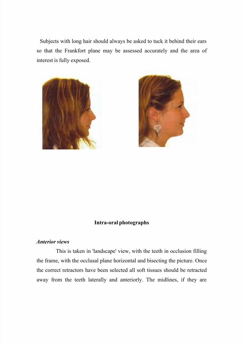
Subjects with long hair should always be asked to tuck it behind their ears
so that the Frankfort plane may be assessed accurately and the area of
interest is fully exposed.
Intra-oral photographs
Anterior views
This is taken in 'landscape' view, with the teeth in occlusion filling
the frame, with the occlusal plane horizontal and bisecting the picture. Once
the correct retractors have been selected all soft tissues should be retracted
away from the teeth laterally and anteriorly. The midlines, if they are

8/3/2019 Digital Photography in Orthodon
http://slidepdf.com/reader/full/digital-photography-in-orthodon 41/48
correct, should be in the centre of the frame. One possible error, although
relatively uncommon, is taking an intra-oral shot in portrait orientation.
Common errors include canted occlusal planes, inappropriate
selection and use of cheek retractors. Another totally preventable error is
saliva not aspirated or the tongue not retracted before the photograph is
taken, and bits of alginate left on the teeth. It is therefore worth familiarizing
the assistants with the retractors, always having good suction available and
taking photos before impressions when collecting records.
To aid focusing for intra-oral photographs the dental light should
always be shone directly into the patients' mouth. Adequate depth of field is
required particularly for the anterior photograph, so it is important to focus
on the level on the lateral incisors to ensure that the maximum numbers of
teeth are in focus.
Buccal views
Again the occlusal plane should be horizontal and bisect the frame.
The frame should be filled with teeth extending from the mesial surface of
the central incisor to at least the distal surface of the first permanent molars
and further posteriorly if possible. It is important to angle the camera so that
the lens is perpendicular to a tangent to the buccal surfaces of the posterior
teeth to avoid underestimation of the sagittal discrepancy, which occurs
through a 'parallax' effect.

8/3/2019 Digital Photography in Orthodon
http://slidepdf.com/reader/full/digital-photography-in-orthodon 42/48
Mirror views
The upper and lower mirror shots should ideally be
symmetrical views of the occlusal surfaces of the teeth, extending from just
in front of the incisors to at least the distal surfaces of the first molars and
ideally to include all the erupted teeth. There should be no direct view of the
mcisor teeth. Whilst setting up for the mirror shots move the patient by
tilting their head back so that the photographer doesn't have to stoop or twist
excessively. There is always a tendency for patients not to open their mouth
fully for these occlusal shots. To avoid this problem, after placing the mirror
and just prior to talking the shot ask the patient to open 'twice as wide',
which usually provides significantly better opening for the shot.
Remember that whatever is seen through the viewfinder
is invariably what will reproduced on the final photograph. Photographs
taken with a mirror require the aperture compensation setting on the camera
to be changed to + I to allow more light in. The differences between 0
setting and + I are small, but demonstrate slight underexposure of the shot
when mirrors are used with no compensation.

8/3/2019 Digital Photography in Orthodon
http://slidepdf.com/reader/full/digital-photography-in-orthodon 43/48
The effect aperture compensation for mirror shots.
With conventional slide photography never trust the last slide on the film as,
during processing, the ends of the films are joined together and this may
result in exposure to light thus spoiling the last frame. Therefore, always
settle for 36 shots per film and rewind at that stage, rather than attempting to
squeeze another I or 2 prints on the film.
Many of the aforementioned errors can be overcome with meticulous
attention to technique and the use of digital photography. Positioning errors
and camera errors are noticed immediately on the LCD screen, which is a
major advantage of digital photography.
Other errors can sometimes be compensated for by image

8/3/2019 Digital Photography in Orthodon
http://slidepdf.com/reader/full/digital-photography-in-orthodon 44/48
manipulation at a later date, but this is not without its disadvantages.
Rotation of images for example will lead to distortion of straight lines and
thus 'steps' in arch wires.
Resizing digital images is of course possible, but information is
unnecessarily sacrificed if the frame area is 'wasted' by filling it with areas
of no interest. Some programmes such as Dolphin TM allow guide lines to
be used when resizing images so consistent magnification is almost
guaranteed. The principles of use of retractors, mirrors and suction are
identical whether using conventional or digital equipment.
Conclusions
Good quality accurate clinical photographs can easily be
obtained using the correct equipment and appropriately trained staff. An
awareness of all the possible errors in extra- and intra-oral clinical
photography will increase the chances of obtaining high quality images.
References

8/3/2019 Digital Photography in Orthodon
http://slidepdf.com/reader/full/digital-photography-in-orthodon 45/48
1. SandlerJ,MurrayA. Digital photography in orthodontics.
JOrthod.2001Sep;28(3):197-201.
2. Palomo JM, Wolf GR, Hans MG. Use of digital photography in the
Case orthodontic clinic.Am J Orthod Dentofacial Orthop. 2004
Sep;126(3):381-5.
3.Sandler J. Digital records in orthodontics.Pa Dent J (Harrisb). 2001
Nov-Dec;68(6):29-33.
4.McKeown HF, Murray AM, Sandler PJ. How to avoid common errors
in clinical photography.J Orthod. 2005 Mar;32(1):43-54.
5.Coimbra O, Lomheim C. Digital imaging and orthodontics.
Am J Orthod Dentofacial Orthop. 1999 Jan;115(1):103-5.
6.Fiorelli G, Pupilli E, Patane B. Digital photography in the orthodontic
practice.J Clin Orthod. 1998 Nov;32(11):651-6.
7.Sandler J, Murray A. Recent developments in clinical
photography.Br J Orthod. 1999 Dec;26(4):269-72.
8.Abelson M. Digital imaging update.
Am J Orthod Dentofacial Orthop. 1999 Nov;116(5):587-90.
9.Mah J, Ritto AK. Imaging in othodontics: present and future.
J Clin Orthod. 2002 Nov;36(11):619-25.
10.Swartz ML. Digital photographs and powerpoint.
Am J Orthod Dentofacial Orthop. 2004 Nov;126(5):639
11.Halazonetis DJ. Making slides for orthodontic presentations.
Am J Orthod Dentofacial Orthop. 1998 May;113(5):586-9.

8/3/2019 Digital Photography in Orthodon
http://slidepdf.com/reader/full/digital-photography-in-orthodon 46/48
12.Burke JF. Video printing in orthodontic photography.
J Clin Orthod. 1987 Feb;21(2):118-22.
13.Juggins KJ. The bigger the better: can magnification aid
orthodontic clinical practice? J Orthod. 2006 Mar;33(1):62-6.
14.Meredith G. Facial photography for the orthodontic office.
Am J Orthod Dentofacial Orthop. 1997 May;111(5):463-70.
15.Scholz RP. Imaging in the orthodontic office (and beyond).
Am J Orthod Dentofacial Orthop. 1998 Oct;114(4):464-6.
16.Pappel JE. Lip retractor for occlusal photography.
J Clin Orthod. 1996 Nov;30(11):639.
17.Sandler J, Murray A. Clinical photography in orthodontics.
J Clin Orthod. 1997 Nov;31(11):729-39.
18.Redmond WR, Redmond WJ, Redmond MJ. Clinical
implications of digital orthodontics. Am J Orthod Dentofacial Orthop.
2000 Feb;117(2):240-1
19.Ackerman MB, Ackerman JL. Smile analysis and design in the
digital era. J Clin Orthod. 2002 Apr;36(4):221-36.
20.Scholz RP. Considerations in selecting a digital camera for
orthodontic records.
Am J Orthod Dentofacial Orthop. 1998 Nov;114(5):603-5.21.Abelson MN. Parameters for digital imaging: Part 1.
Am J Orthod Dentofacial Orthop. 2000 Nov;118(5):580-2.

8/3/2019 Digital Photography in Orthodon
http://slidepdf.com/reader/full/digital-photography-in-orthodon 47/48
22.Hatcher DC, Aboudara CL. Diagnosis goes digital.
Am J Orthod Dentofacial Orthop. 2004 Apr;125(4):512-5.
23.Sandler J, Murray A. Manipulation of digital photographs.
J Orthod. 2002 Sep;29(3):189-94.
24.Sandler J, Murray A. Clinical photographs--the gold standard.
J Orthod. 2002 Jun;29(2):158-61.

8/3/2019 Digital Photography in Orthodon
http://slidepdf.com/reader/full/digital-photography-in-orthodon 48/48