Embed Size (px)
Citation preview

Marquette Universitye-Publications@Marquette
Master's Theses (2009 -) Dissertations, Theses, and Professional Projects
Dimensional Changes of Facial Soft TissueAssociated with Rapid Palatal ExpansionPeter Charles LongoMarquette University
Recommended CitationLongo, Peter Charles, "Dimensional Changes of Facial Soft Tissue Associated with Rapid Palatal Expansion" (2014). Master's Theses(2009 -). Paper 271.http://epublications.marquette.edu/theses_open/271

DIMENSIONAL CHANGES OF FACIAL SOFT TISSUE ASSOCIATED WITH RAPID PALATAL EXPANSION
by
Peter C Longo, DDS
A Thesis submitted to the Faculty of the Graduate School, Marquette University,
in Partial Fulfillment of the Requirements for the Degree of Master of Science
Milwaukee, Wisconsin
August 2014
ABSTRACT DIMENSIONAL CHANGES OF FACIAL SOFT TISSUE ASSOCIATED
WITH RAPID PALATAL EXPANSION
Peter C Longo, DDS
Marquette University, 2014
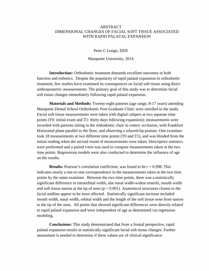
Introduction: Orthodontic treatment demands excellent outcomes in both function and esthetics. Despite the popularity of rapid palatal expansion in orthodontic treatment, few studies have examined its consequences on facial soft tissue using direct anthropometric measurements. The primary goal of this study was to determine facial soft tissue changes immediately following rapid palatal expansion.
Materials and Methods: Twenty-eight patients (age range, 8-17 years) attending Marquette Dental School Orthodontic Post-Graduate Clinic were enrolled in the study. Facial soft tissue measurements were taken with digital calipers at two separate time points (T0: initial exam and T1: thirty days following expansion); measurements were recorded with patients sitting in the orthodontic chair in centric occlusion, with Frankfurt Horizontal plane parallel to the floor, and observing a relaxed-lip posture. One examiner took 18 measurements at two different time points (T0 and T1), and was blinded from the initial reading when the second round of measurements were taken. Descriptive statistics were performed and a paired t-test was used to compare measurements taken at the two time points. Regressions models were also conducted to determine the influence of age on the results.
Results: Pearson’s correlation coefficient, was found to be r = 0.998. This indicates nearly a one-to-one correspondence in the measurements taken at the two time points by the same examiner. Between the two time points, there was a statistically significant difference in intraorbital width, alar nasal width-widest nostrils, mouth width and soft tissue nasion at the tip of nose (p < 0.001). Anatomical structures closest to the facial midline appear to be most affected. Statistically significant increase included mouth width, nasal width, orbital width and the length of the soft tissue nose from nasion to the tip of the nose. All points that showed significant differences were directly related to rapid palatal expansion and were independent of age as determined via regression modeling.
Conclusions: This study demonstrated that from a frontal perspective, rapid palatal expansion results in statistically significant facial soft tissue changes. Further assessment is needed to determine if these values are of clinical significance.

i
ACKNOWLEDGMENTS
Peter C Longo, DDS
I first wish to thank my loving wife, Danielle Longo. Without her support and most importantly, her patience, I wouldn’t have been able to complete this project. Furthermore, I would like to thank my family for their devotion, support, and humor during the more difficult portions of my thesis work. I would like to extend my deepest gratitude to Dr. Thomas G. Bradley for providing continued mentorship and guidance in completing my thesis. Dr. Bradley had several responsibilities yet still provided me with outstanding leadership and invaluable direction to finish this project. I would also like to thank Dr. Dawei Liu and Dr. William Lobb. Drs. Liu and Lobb worked extremely hard on editing and refining my project, as well as guiding me in proper research methodology.
Additionally, I thank Dr. Jose Bosio. Dr. Bosio holds my utmost respect and
gratitude, as he spent countless hours on data collection, and kept me focused on my tasks during his time at Marquette. I greatly appreciate the time and effort that Dr. Jessica Pruszynski has spent on statistical analysis. Her help was essential in the completion of this project. Lastly, I would like to acknowledge all the faculty and staff of the Marquette University Graduate Orthodontic Program for their support over the past two years. They are a solid foundation for all of Marquette’s previous, current, and future residents.

ii
TABLE OF CONTENTS
ACKNOWLEDGMENTS ................................................................................................... i
LIST OF TABLES……………………………………………………………………….iii LIST OF FIGURES………………………………………………………………………iv
CHAPTER I. INTRODUCTION ........................................................................................ 1
CHAPTER II. LITERATURE REVIEW ........................................................................... 4
A. The Role of Facial Esthetics In Orthodontics .................................................... 4
B. Review of Palatal Expansion............................................................................. 6
C. Effects of Palatal Expansion ........................................................................... 11
D. Soft Tissue Effects of Palatal Expansion ........................................................ 14
E. Soft Tissue Measurement ................................................................................ 15
F. Current State of the Problem ........................................................................... 16
CHAPTER III: MATERIALS AND METHODS ............................................................ 18
A. Measurement ................................................................................................... 19
B. Statistical Analysis .......................................................................................... 23 CHAPTER IV: RESULTS ................................................................................................ 24
A. Reliability of Measurements ........................................................................... 24
B. Soft Tissue Measurements .............................................................................. 25
CHAPTER V: DISCUSSION ........................................................................................... 29
A. Study Weaknesses and Future Direction ......................................................... 38 CHAPTER VI: CONCLUSION ....................................................................................... 40
BIBLIOGRAPHY ............................................................................................................. 41
ADDENDUM A – Soft Tissue Landmark Definitions ..................................................... 47
ADDENDUM B – Regression Models ............................................................................. 55

iii
ADDENDUM C – IRB Approval Form ........................................................................... 74

iv
LIST OF TABLES
Table 1. Description of the points utilized for the soft tissue measurements ....................25
Table 2. Soft tissue measurements. ....................................................................................30
Table 3. The paired t-test results (significant values<0.01). ..............................................31
Table 4. Regression model for alar nasal width at the base of the nose ............................32

v
LIST OF FIGURES
Figure 1. Soft tissue measurements. ................................................................................23 Figure 2. Examples of facial soft tissue measurements being recorded with caliper ......24
Figure 3. Pearson’s correlation coefficient measurements ..............................................28

1
CHAPTER 1 INTRODUCTION
Rapid palatal expansion (RPE) is a common treatment modality for orthodontic
patients (Proffit 2012). Indications for RPE treatment include narrow transverse
maxillary width, unilateral or bilateral posterior crossbite, and severe maxillary crowding
(Proffit 2012, Haas 1961). Several different designs exist for rapid palatal expanders,
including both banded and bonded appliances, and those with or without acrylic
coverage. Regardless of design, the majority of appliances employ much the same
mechanism of action. The primary goal of expansion is to orthopedically create a
separation of the midpalatal suture, thereby moving teeth into a broader arch form. The
older the patient the more likely the midpalatal suture is fused. Consequently, the use of
an expander in these patients more likely results in dental movement, including tipping of
teeth in a buccal direction (Krebs 1959, Haas 1961, Wertz 1970, Harberson 1978, Adkins
et al. 1990, McNamara 1993, Hesse 1997, McNamara 2000).
Orthodontists utilize rapid palatal expanders on a wide age range of patients,
although Proffit (2012) and others recommend their use primarily before the completion
of the adolescent growth spurt. Consequently, early treatment of children in the primary
or mixed dentition stages often involves palatal expansion, as practitioners aim to
capitalize on the patency of sutures in young patients. Yet, the determination of suture
patency can be difficult as suture closure can vary significantly amongst patients. A
recent study by Angelieri, et al, has focused on the utilization of CBCT to identify the
stage of suture maturation (Angelieri et al. 2013). This provides the practitioner with a
means of determining the potential success of palatal expansion. However, the benefits

2
of this method must be critically weighed against the potential disadvantage of radiation
exposure. Additionally, true separation of the maxillary sutures is not necessarily a
prerequisite to successful treatment in all patients, as the resultant increase in transverse
width, or “expansion” outcome can still be achieved without orthopedic change.
Anatomically, the midpalatal suture is located on the midline of the maxilla, or
the roof of the oral cavity. Subsequently, this roof is shared with the bony floor of the
nasal cavity. Indeed, the conclusion can be drawn that changes in the transverse
dimension of the oral cavity may have the concurrent effect of altering the nose. Several
studies support this conclusion (Derichsweiler 1953, and Haas 1961). Several studies
evaluating facial esthetics show that symmetry of the midface, and the nose in particular,
constitutes a great role in overall pleasant facial appearance (Naini 2006). The
orthodontist places a great deal of emphasis on facial esthetics and thus must be aware of
any potential facial changes produced by utilization of a palatal expander. Expected
changes in the nasal complex, and the overall net effect on the facial soft tissue drape,
must be considered and conveyed by professionals to patients prior to initiation of
treatment. The importance of identifying and treatment planning for changes in facial
soft tissues have been reinforced within current orthodontic literature (Sarver 2003).
Previous studies examined facial changes produced by palatal expansion via
lateral cephalometric radiographs. Such studies have shown that palatal expansion
impacts facial soft tissues. Kilic et al (2008), found a significant decrease in the soft
tissue facial angle and increases in both the H angle and profile convexity. They
hypothesized that these changes were due to a greater forward displacement of soft
tissues overlying the maxilla that overcame the negative effect of the flattening of the

3
nose. More recent studies have advanced on these foundations by utilizing novel
technologies. Several three-dimensional imaging modalities now exist, the most common
of which is the cone beam computed tomography (CBCT). Garett, et al, (2008) used
CBCT imaging to study immediate transverse changes occurring from rapid palatal
expansion. They showed that palatal expansion resulted in significant widening of the
nasal structures as well as narrowing of the maxillary sinus. Habeeb, Boucher, and
Chung’s (2013) CBCT studies revealed similar results as Kilic’s earlier study that rapid
palatal expansion results in significant forward movement of the maxilla. Again,
however, as discussed earlier, routine CBCT imaging involves exposure to relatively
large doses of radiation, and is not commonly used for soft tissue analysis. Therefore a
more practical means of determining soft tissue changes would be beneficial for both
patients and practitioners.
Beginning in the 1980s, orthodontic treatment goals have gradually placed a
greater emphasis on refinement and enhancement of facial soft tissues. As a result of this
paradigm shift, parameters and guidelines have been proposed to guide the orthodontist
in improving facial esthetics (Arnett 1993). Yet a review of the literature reveals little
information regarding changes in the facial soft tissues from palatal expansion.
Therefore, a study documenting soft tissue changes produced by rapid palatal expansion
would be valuable. Payne (2013) and Mollov et. al (2012) showed that several soft tissue
landmarks are both accurate and reliable, and therefore provide the orthodontist with an
economic and relatively easy method of analyzing patients in everyday practice. The
main goal of this study is to determine the extent of facial soft tissue changes following
rapid palatal expansion.

4
CHAPTER II LITERATURE REVIEW
The Role of Facial Esthetics in Orthodontics
Orthodontic treatment has the capability to alter facial shape and form, and thus
facial esthetics. Consequently, facial esthetics plays a foundational role in orthodontics
and should be addressed as early as the initial treatment planning. A review of facial
esthetic guidelines is necessary for orthodontists attempting to maximize treatment
outcomes. However, a review of literature reveals that several different methodologies
have been used for evaluation of facial esthetics. Therefore, a brief history underlining
the evolution of facial esthetics and its interrelationship with orthodontic treatment
decisions is beneficial.
Charles Tweed argued that a treatment approach focused solely on achieving ideal
arch form and occlusal relationships often resulted in poor facial esthetics, with a loss of
the balance and harmony of the face (Tweed 1945). Therefore, from examining
treatment “successes” and “failures”; he concluded that the most optimal facial esthetics
are a result of upright mandibular incisor position as determined by a 90 degree incisor
mandibular plane angle. This allowed for a dental and muscular balance that provided
pleasing facial results. Incisor position appears prominently in several discussions on
facial esthetics throughout orthodontic history. An early study of facial profile esthetics
showed that an establishment of balance between the upper and lower incisor position
resulted in optimal facial esthetics. The relative convexity of the face in profile guides
these final positions, with a more convex face requiring a more upright final incisor
position, and vice versa (Riedel 1950).

5
Increased availability of cephalometric radiology led to its widespread utilization
by practitioners as a means of guiding and interpreting treatment outcomes. As a result
of its popularity, analyses were developed as a tool to help guide the orthodontist in
determining successes or failures. An early cephalometric study demonstrated the
importance of a balance between several facial and dental structures and listed several
“norms” for achieving ideal facial esthetics. For example, once a balance between the
maxilla and mandible (ANB=2) is achieved, the upper and lower incisors can therefore be
related to the skeletal landmarks to achieve harmony (Steiner 1953). Some of the earliest
landmark studies on cephalometric facial analysis were completed by William B Downs,
who concluded that although individuals display a wide array of skeletal and dental types,
certain characteristics are common amongst those demonstrating more ideal facial
esthetics. Such requirements for ideal facial esthetics include a functionally balanced
occlusion, profile balance, and primarily an overall balance of dental and skeletal bases
(Downs 1956).
An overriding theme in several of these earlier cephalometric studies was that
optimal facial esthetics is a consequence of attaining proper facial balance. For example,
Merrifield concluded that lower face balance was of great importance and should not
simply be subjectively determined by practitioners. He offered that critical evaluation of
a patients’ Z angle and soft tissue profile line removes ambiguity and provides proper
determination of balance. Furthermore, he stated that a more esthetically pleasing profile
is attained by completing treatment with a chin position equal to or more protrusive than
the upper lip thickness (Merrifield 1966). As cephalometric studies advanced, an
increasing degree of attention was directed towards soft tissue changes. Clearly, soft

6
tissue, and not simply dental and skeletal balance, occupies a central role in facial
esthetics.
Review of Palatal Expansion
The utilization of palatal expanders by orthodontists began with the father of
orthodontics, Edward Angle, and their utilization has been extensively detailed within
orthodontic literature (Angle 1860). Much of the earliest work on expanders and their
indications was conducted by Dr. Andrew Haas. In one of his earliest publications, Haas
advocated for the use of palatal expanders in five clinical situations, and although not
entirely true today, several elements of this article published over 40 years ago are still
highly relevant (Haas 1970). Interestingly, as Haas pointed out, the use of palatal
expansion as indicated for Class III malocclusions is common, and other prominent
authors, such as McNamara, have relayed similar benefits of palatal expansion for Class
III correction (McNamara 1987). Indeed, the use of expanders as a treatment modality is
still commonly used today primarily to aid with the correction of crossbites, crowding,
and transverse maxillary deficiencies (Proffit 2012).
Despite this relatively common use of expansion as a treatment method, multiple
modalities and appliance designs exist. These modalities can be categorized by three
primary types of expansion: rapid maxillary expansion, slow maxillary expansion, and
surgically assisted maxillary expansion (Agarwal and Mathur 2010). Furthermore, four
types of expanders exist when categorized by activation protocol. These four groups are
screw-type, spring-type, magnetic, and Shape Memory Alloy (SMA) activation.

7
The screw-type expander category consists of expanders in which manual rotation
via a wrench or “key” by either the clinician or patient results in widening of the jack-
screw. This design has a well understood and historically common mechanical concept
of expansion via turning of a screw-jack. Simply, the amount of screw rotation directly
corresponds to the amount of expansion. An advantage of this category includes the
flexibility provided to the practitioner to prescribe a certain amount of expansion over a
certain period of time. Also, appliances can be adapted to fit a variety of palate sizes and
shapes, seemingly limited only by the size and placement of the jackscrew component.
However, several drawbacks to the jackscrew appliance do exist. Foremost among these
is that upon activation of the screw, a sudden, rapid increase in force is produced which
has been shown to possibly result in less physiologic expansion of the palatal suture.
Also, this method of expansion places a large responsibility on the patient to comply with
expansion protocols, which is a significant disadvantage of this design. Within this
category of appliances is the most commonly used Hyrax appliance design, named for its
trademarked Hyrax jackscrew (Romanyk et al 2010).
Spring-type expanders are defined as any appliance that funtions via mechanical
deformation of a body. This deformation subsequently results in elastic restoration forces
that are exerted on the palate and thus results in expansion. This design offers certain
advantages, such as less dependency upon the patient to manually activate the expander
at a regular interval. Furthermore, theoretically it applies a constant force over a period
of time and avoids sudden increases in force as seen in the screw appliances. This
predictable amount of force likely results in greater comfort for the patient following
initial delivery. The amount of force produced is inversely proportional to the amount of

8
expansion. Therefore, the more expansion produced the less force for further expansion
remains and the orthodontist may have to remove and re-activate the appliance if more
expansion is needed following treatment. Patient safety is also another concern, as the
appliance is delivered “active”. Perhaps the greatest drawback of this design is that
although springs provide predictable transverse forces, any deformation of the appliance
may result in unwanted force in all other planes of space (Romanyk et al 2010).
Magnetic expanders also exist, and have been referred to as Magnetic Expansion
Devices (MED). The goal of magnetic expansion devices is to provide continuous forces
of a lesser magnitude than traditional expansion devices. The magnets are applied so that
their directionality opposes each other, thus creating a repulsive and expansive force
(Romanyk et al 2010). Theoretically, this would result in a more biologically friendly
and less traumatic stimulation of maxillary suture growth, similar to spring type
appliances. A study by Darendeliler showed the greatest degree of skeletal expansion
occurred in banded appliances with four magnets. A set of two magnets were placed
apically to the central and lateral incisors and the second set placed between the second
bicuspid and first molar. Although the sample size was small, they did show that MEDs
provide a relatively effective method of palatal expansion, which did not rely on patient
compliance (Darendeliler 1994). Advantages and disadvantages of MEDs are similar to
those of the spring type of appliances. However, one significant advantage of MEDs is
that they are less prone to deformation and thus have less risk to produce undesirable and
unpredictable forces in dimensions other than the transverse. Similar to the spring type,
magnetic forces decrease with increasing expansion and therefore also require
adjustments like its spring counterparts. This problem can be avoided by placement of

9
magnets of greater strength; however this may result in less patient comfort and less
physiologic suture opening (Romanyk et al 2010).
Finally, the fourth category of expanders, shape memory alloy appliances, utilize
the properties of nickel titanium wires and are therefore dependent upon the properties of
the shape memory alloy incorporated into the device for expansion. Orthodontists have
become increasingly familiar with the properties of Ni-Ti wires and therefore it is prudent
to assume their incorporation into expansion devices is a natural progression. However,
the drawbacks of a conventional spring appliance still exist with these appliances. Of
primary concern is that any deformation of the appliance may result in uncontrolled
forces transverse to the direction of expansion. More recently, promising hybrid devices
have been constructed to include both Ni-Ti wires and expansion jackscrews; however
these appliances re-introduce the major disadvantage of patient compliance (Romanyk et
al 2010).
As previously mentioned, palatal expanders can also be categorized based on rate
of expansion into three broad categories: rapid, slow, and surgically assisted. Rapid
palatal expansion (RPE) typically involves two turns per day of a jackscrew expander
appliance, commonly a rate of 0.5 mm expansion per day. Indications for rapid palatal
expansion include transverse discrepancies of greater than 4mm with dental
compensation via buccally tipped maxillary molars, disruption of sutures to aid Class III
correction, and moderate maxillary crowding. RPE is contraindicated when there is
recession of the alveolar bone on maxillary molars or premolars, presence of anterior
open bite, high mandibular plane angle, convex profile and doubtful patient compliance
(Agarwal and Mathur 2010). Furthermore, RPE is contraindicated in mature patients

10
beyond the growth spurts, however many practitioners may still choose this treatment
modality on older patients. Clinically, patients are instructed to turn the expander once in
the morning and once in the evening, or only once a day, commonly for a period of two
to three weeks followed by a retention period of at least three months. This rapid rate of
activation and relatively large force application is thought to maximize orthopedic
skeletal expansion while minimizing dental movements (Agarwal and Mathur 2010).
RPEs can be designed as banded or bonded appliances, and can be tooth-borne, tissue-
borne, or tooth and tissue borne. Some examples of rapid palatal expanders are the Hyrax
expander, Issacson expander, and Haas expander (Agarwal and Mathur 2010).
Slow palatal expansion (SPE) is a process by which light, relatively continuous
force levels commonly in the range of 450-900 grams are applied. It is thought that the
lighter forces result in less resistance from sutural structures, thus allowing more bone
formation and activity in the intermaxillary suture, but possibly less suture opening.
Furthermore, post-expansion stability and retention may be greater with SPE. Appliance
designs vary greatly for SPE, with some examples being the Quadhelix, the Coffin
appliance, magnet expanders, W-arches, and spring jets. The hyrax expander can also be
used for SPE, with a maximum of 1 mm expansion per week applied. Finally, surgically
assisted palatal expansion occurs when expansion is aided with surgical intervention.
Surgical expansion involves either surgically assisted rapid palatal expansion (SARPE) or
segmental maxillary surgery such as a Lefort osteotomy. Indeed, this procedure allows
for expansion beyond skeletal maturation but is highly invasive and complex (Agarwal
and Mathur 2010).

11
Effects of Palatal Expansion
When palatal expanders are used, widening of the maxillary arch may occur via
either orthodontic or orthopedic movements, or a combination of both. Generally, the
initial increase in width occurs as teeth compress and stretch the periodontal tissue. This
is an orthodontic movement as the teeth are tipped laterally due to sudden force
application, and studies have shown that this gain is usually completed within one week
(Cotton 1978) (Hicks 1978). Compressive forces continue as long as the expander is
active. These forces result in resorption of the buccal alveolar plate at the interface of the
tooth root and periodontal attachment, resulting in further orthodontic tooth movement.
This movement is bodily movement of the teeth into a wider arch form. Orthopedic
expansion of the maxilla occurs via separation of the midpalatal suture. This separation
occurs when forces applied exceed the bioelastic tensile strength of the elements within
the suture, or the interdigitation of the bony spicules. As long as the transverse force of
expansion is greater than this tensile strength, further separation of the suture occurs.
Once the expander is locked, the suture reorganizes (Bell 1982).
Palatal expansion, similarly to several treatment modalities in orthodontics, can
produce profound changes in a patient’s overall facial appearance. Cephalometric studies
have historically been used to describe these changes. In one of the earliest studies
conducted by Haas, he found that cephalometric analysis revealed an increased internasal
width following expansion, concluding that there is “coincidental widening of the nose
and lowering of its floor”. Of course, these studies focused solely on skeletal changes via
radiographic appearance. He also found that there was a widening of the mandibular
arch, and postulated that this may be attributed to tongue pressure from inferior

12
displacement by the expansion appliance (Haas 1961). White (1972) examined
pretreatment and post treatment records of thirty patients undergoing palatal expansion.
He noted clockwise movement of the mandible following expansion treatment. Also of
note, however, was that he noticed significant widening of the internasal width on
posteroanterior cephalograms. Chung and Font examined pre and post-treatment records
on twenty children who were treated with Haas type expanders. The authors also found
statistically significant downward and forward displacement of the maxilla, and
clockwise rotation of the mandible following expansion treatment. Furthermore,
significant facial changes beyond the maxilla and mandible were noted. Anterior facial
height and the width of both the maxilla and the nasal cavity increased significantly
(Chung and Font 2004).
Similar results were published by Gabriel de Silva Fo, who studied the results of
rapid palatal expansion on thirty children, aged 5 to 10 years and 11 months. Haas type
expanders were used on all patients, and parents were prescribed 1 turn daily for 1 to 2
weeks. From their results, they concluded that the maxilla always dislocates in a
downward and backward manner with respect to the palatal plane, thus significantly
altering N-ANS, PNS-PNS’, A-A’, and SN-PP. Furthermore, B point is posteriorly
displaced following expansion due to clockwise rotation of the mandible in response to
palatal expansion changes. Finally, facial heights were increased significantly, which
was attributed to the vertical displacement of the maxilla and maxillary molars (Gabriel
de Silva Fo 1991). Similar results were found by Sarver who reported anterior and
inferior movement of the maxilla following expansion treatment; however he did note
that utilization of a bonded expander device limited this effect (Sarver 1989). This

13
information should be critically evaluated by the orthodontist, as these studies support
that expansion treatment changes extend beyond the dental arches.
The advancement of cone beam computed tomographic (CBCT) radiology has
allowed orthodontists and researchers to explore changes produced by palatal expansion
more precisely. Habeeb evaluated pre and post-treatment CBCT radiographs of twenty
eight patients who were treated with bonded Haas expander appliances. The investigator
found that both ANS and PNS moved significantly downward from expansion treatment.
In addition, they found that while the maxilla was anteriorly displaced, the maxillary
central incisors moved posteriorly from their initial position (Habeeb 2013). A similar
CBCT study was conducted using thirty patients, mean age of 13.8 +/- 1.7 years, who
underwent palatal expansion with a banded Hyrax appliance design. High-resolution
CBCT images were taken before and after expansion, which the authors claimed allowed
for more accuracy than previous studies. They found that rapid maxillary expansion
resulted in a statistically significant increase in nasal width (p<.0001). On average, nasal
width increased by 1.89 mm, or 37.2% of the mean expansion of the hyrax appliance
(Garrett 2008). It may be important to note that this change was due primarily to
expansion, independent of aging (Garret 2008). Similar findings occurred in a CBCT
study on fourteen children who underwent rapid palatal expansion. All patients were
treated with a four-banded Hyrax appliance design and instructed to activate the
appliance 1 turn/day for 28 days or until complete correction of pre-existing crossbites.
CBCT scans were taken prior to initiation of treatment and again 3 to 4 months following
expansion. From their data, the authors agreed with previous studies that RPE indeed
may cause significant expansion of the maxilla. Furthermore, they concluded that there is
14
a significant increase in the cross-sectional area of the upper airway following rapid
maxillary expansion (Chang et al. 2013). Therefore, recent CBCT studies show
agreement with earlier hard tissue studies on expansion effects.
Soft Tissue Effects of Palatal Expansion
Although the studies listed above discuss treatment outcomes that altered facial
characteristics of orthodontic patients, it is important to our work to also investigate
studies which examine effects of expansion on soft tissues. Mota dos Santos evaluated
twenty patients who underwent rapid palatal expansion. Lateral cephalograms were
taken prior to expansion treatment, immediately following expansion treatment, and
finally after a period of retention. Any conclusions on long-term soft tissue changes
based on their data were inconclusive. The authors did note statistically significant
changes in the following cephalometric measurements: S–li , H Line–Prn, E Line–Li, E
Line–Ls , ANS′–Me′. They concluded these changes were primarily due to the presence
of the expander appliance. However, they concluded that following a retention period of
six months, minimal soft tissue changes are seen following expansion (Mota dos Santos
2012).
A similar study was conducted which consisted of 18 patients with bilateral
posterior crossbite. All patients underwent rapid palatal expansion using bonded Hyrax
expanders, the same appliance used in the previous study. Cephalometric radiographs
were again taken at three separate time points, prior to treatment, following completion of
expansion, and retention (mean 5.95 +/- 0.35 months). All radiographs were taken by a
single operator, with caution taken to ensure lips in repose. Holdaway soft tissue

15
measurements were analyzed from the radiographs. The results were slightly different
than those of Mota dos Santo’s study, as soft tissue facial angle decreased (P < 0.05), and
H angle and skeletal profile convexity significantly increased (P < 0.001) following
expansion. Also, the authors noted that while some relapse did occur, both H angle and
profile convexity remained significantly altered following retention periods. (Kilic 2008).
In summary, the literature database supports the notion that rapid palatal expansion
produces soft tissue changes, however the question still remains on the most reliable and
efficient method of measuring such changes.
Soft Tissue Measurement
The method of soft tissue evaluation is critical to the overall quality of study
results. Therefore, a determination of the most accurate method is necessary prior to data
collection. It has been determined that intra-examiner measurements display a higher
degree of consistency, accuracy and reliability than inter-examiner measurements
(Mollov 2012). Furthermore, certain soft tissue facial landmarks are more accurately
identifiable than others. Of all measurements, nasal width (al-al, R=0.992), middle third
of the face (N’- Sn, R= 0.989), and upper lip length were the most accurately measured
(Mollov 2012).
Much of Farkas’ work on soft tissue anthropometry has shown that direct
anthropometric measurements on children vary in difficulty of acquiring accurate data.
Major factors of influence are age, duration of examination, and cooperation (Farkas
1996). However, much of Farkas’ difficulties came on children under the age of five.
No individuals below the age of five were included in our project, thus the challenge did

16
not affect our data acquisition. The majority of the measurements in this study were
centrally located on the face. Payne (2013) demonstrated that nose and mouth landmarks
were amongst the most reliable from frontal photographs. However, photographic
examination is time consuming and is only necessary as an adjunct to the initial clinical
exam. Yet direct examination and photographic examination are comparable enough that
it can be determined that equally reliable anthropometric measurements can be taken on
patients at rest position or via photographic examination.
Current State of the Problem
Despite the relatively large volume of literature describing palatal expansion, few
studies have reported the effect of rapid maxillary expansion using only facial soft tissue
landmarks. This is likely due to the difficulty of controlling large number of variables,
including soft tissue thickness, weight fluctuations, and accounting for natural growth of
patients. Many of the present studies available rely heavily on radiographic evaluation,
rather than photographic or direct patient evaluation. Thus, a void in knowledge is
available on a commonly used technique, palatal expansion, and its direct soft tissue
clinical effects on a patient. Therefore, a sample of 28 Marquette University School of
Dentistry patients were measured before rapid palatal expansion and again following
palatal expansion using a digital caliper. In all, eighteen soft tissue measurements were
obtained for evaluation. Included amongst these are mouth width, base of the nose, alar
of the nose, intraorbital width and other measurements which are of utmost importance to
final esthetic treatment outcomes. The outcome of this study will aid orthodontic
practitioners, as well as patients, in better understanding what facial soft tissue changes,
as well as degree of change, may occur as a result of palatal expansion.

17
The purpose of this study was to evaluate facial soft tissue changes following
rapid palatal expansion using only soft tissue landmarks measured by a digital caliper, at
two different time points.

18
CHAPTER III MATERIALS AND METHODS
This study was approved by the Institutional Review Board (protocol # HR-2083).
Twenty eight subjects (14 males and 14 females), with ages ranging from 7 years, 1
month to 15 years, 5 months, who were enrolled as patients at the Marquette University
School of Dentistry Orthodontic Clinic, were included in this study. Subjects were
excluded from the study if they (1) had history of any congenital abnormality, (2) had
undergone medical/pharmacological treatment that would affect the facial complex, (3)
had facial hair that would mask landmarks to be identified, or (4) were unable to follow
verbal instructions to allow measurements to be taken. Weight fluctuations were not
accounted for in this study.
The RPE appliance designs used in this study consisted of banded Hyrax
expanders (24 subjects), banded Haas expanders (3), and a bonded Hyrax expander (1
subject). Study participants turned the expander an average of 25 turns, or 6.25 mm of
total expansion. This consisted of a range of 14-38 turns, depending on the amount of
expansion needed to obtain a relationship of the maxillary lingual cusp in occlusal
contact with the mandibular buccal cusp. Of the 28 subjects, 22 were prescribed 1 turn of
expansion every day, 3 prescribed 1 turn every other day, 2 prescribed 1 turn every third
day, and 1 failed to report amount of turns per day.
The study did not create any restrictions to normal orthodontic care. Patient
treatment plans were determined by the resident and attending faculty members,
independently of the study investigators.

19
Measurement
At orthodontic records or at treatment planning appointments, the principal
investigator recorded the initial facial measurements (T0). Patients over 15 years of age
consented to participate in the study and patients under 15 assented to participate with
written authorization from their legal guardians.
Facial soft tissue measurements were taken in centric occlusion. The subjects
were seated in the orthodontic chair with Frankfort Horizontal plane parallel to the floor.
Their lips were then manipulated into a relaxed-lip posture, which was attained initially
by asking patients to pronounce the word “EMMA” and to hold their teeth together in
order to guide the mandible into maximum intercuspation, while keeping their eyes
opened. Measurements were recorded for all participants in the form created for this
study. (Fig. 1) A relaxed lip position was confirmed by the investigator, and if patients
needed assistance to relaxed lip position the investigator gently stroked the lips
(Burstone, 1967). Measurements were not recorded until relaxed lip position was
attained, and patients were not included in the study if unable to achieve this position.
Relaxed-lip posture was used because it is a reasonably reproducible position
independent of teeth and supporting alveolar processes.
All eighteen facial soft tissue measurements were taken by the same investigator.
A description on how to identify the facial landmarks can be seen on Table 1, Figure 1,
and Figure 2. Facial measurements were taken using a 1/8 inch (203.2 mm), sliding
digital Mitutoyo caliper (Aurora, IL). The measurement error for all Mitutoyo calipers
was identical to the company’s description (0.01 mm). The investigator was trained and

20
calibrated for using the Mitutoyo caliper. Ten subjects were re-measured two-weeks to
one month later, prior to treatment initiation (RPE cementation) solely to determine the
investigator’s intra-examiner reliability.
A second set of measurements (T1) was then performed when RPE treatment was
completed and the rapid palatal expander was locked.
Figure 1. Soft tissue measurements. (These drawings appeared in Arnett GW, Bergman
RT. Facial keys to orthodontic diagnosis and treatment planning. Part II. Am J Orthod Dentofac Orthoped 1993; 103(5):395e411 Elsevier, 1993, and in N. Mollov et al., Journal of the World Federation of Orthodontists 1 (2012)
e157ee161 Elsevier 2012.)

21
Figure 2. Examples of facial soft tissue measurements being recorded with caliper.

22
Table 1. Description of the points utilized for the soft tissue measurements.
trichion (tr) The point on the hairline in the midline of the forehead. Note: for this project, no
participants with visible hair loss or abnormally high hairline were selected for
participation
soft tissue nasion
(Na’)
The soft tissue covering the point located in the midline of both the nasal root and
the nasofrontal suture
endocanthion
(en)(Left or Right)
The point at the inner commissure of the eye fissure
zygion (zy)(L or R) The most lateral point of each zygomatic arch; identified by trial measurements.
pronasale(prn) The most protruded point of the apex nasion
alare (al)(L or R) The most lateral point on each alar contour
subnasale (sn) (L
or R)
The midpoint of the columnella base at the apex of the angle where the lower
border of nasal septum and the surface of the upper lip meet
subalare (sbal)(L
or R)
The point at the lower limit of each alar base, where the alar base disappears into
the skin of the upper lip
labiale superius
(ls)
The midpoint of the upper vermillion line
labiale inferius (li) The midpoint of the lower vermillion line
cheilion (ch)(L or
R)
The point located at each labial commissure
stomion (sto)
(Upper and Lower)
The imaginary point at the crossing of the vertical facial midline and the horizontal
labial fissure between the upper/lower lip and the oral cavity as seen from a frontal
view. Note: in this project the study participants were asked to relax their lips, hence
the visible border of each lip was used as the horizontal landmark
soft tissue B point
(B’)
The deepest curvature of the soft tissue between the lower lip and the chin point
gnathion (gn’) The lowest median landmark of the lower border of the mandible
menton (me) The lowest, or most inferior, point on the mandible from a frontal view

23
Statistical Analysis
To determine intra-examiner reliability, we calculated Pearson’s correlation
coefficient, and found it to be r = 0.998. This indicates nearly a one-to-one
correspondence in the measurements taken at the two time points by the same examiner.
For comparative measurements, we first considered descriptive statistics of the
age of the patients and of each of the measurements taken. A paired t-test was used to
compare the measurements taken at the two time points. Since conducting multiple t-tests
naturally inflates the Type I error, or the probability of finding a significant result simply
due to chance, we reduced the significance level from 0.05 to 0.01. (In other words, a
result is only significant if the p-value is found to be less than 0.01).

Reliability of Measurements
Pearson’s correlation coefficient was calculated and found to be r = 0.998. This
indicates nearly a one-to-
points by the examiner. The measurements can be seen in the scatterplot below (Figure
3).
Figure 3. Pearson’s correlation coefficient measurements
CHAPTER IV RESULTS
Reliability of Measurements
Pearson’s correlation coefficient was calculated and found to be r = 0.998. This
-one correspondence in the measurements taken at the two time
points by the examiner. The measurements can be seen in the scatterplot below (Figure
Pearson’s correlation coefficient measurements.
24
Pearson’s correlation coefficient was calculated and found to be r = 0.998. This
one correspondence in the measurements taken at the two time
points by the examiner. The measurements can be seen in the scatterplot below (Figure

25
Soft Tissue Measurements
We first consider descriptive statistics of the age of the patients and of each of the
measurements taken. At the initial time of measurement (T-0), the patient age range was
between 7 years, 1 month to 15 years, 5 months. The mean age of the patients was 12
years, 2 months, with a standard deviation of 3.1 years. The average age of the female
patients (n=14) was 11 years, 10 months. The average age of the male patients (n=14)
was 12 years, 8 months.
The eighteen soft tissue measurements were divided into two subgroups of
horizontal and vertical measurements (Table 2 and Table 3). For the horizontal group,
the results (Table 3) showed that intraorbital width, nasal width at the widest point of the
nostrils (p<.0001), and mouth width (p=0.0035) were significantly different following
rapid palatal expansion. For the vertical group, soft tissue nasion to pronasale, showed a
significant difference (p=0.0008).
The majority of measurements showed increased values following palatal
expansion, but most of them were not statistically significant (Table 3). The following
measurements showed a non-statistical decrease: nasal width at base of nose (sbalR –
sbalL), stomion lower – soft tissue B point (stL –B’), soft tissue B point – gnathion (B’ –
gn’), and subnasale – gnathion (sn – gn’)). One horizontal, zygomatic width, and one
vertical measurement, subnasale-gnathion, showed almost no variation (p>0.9).
Of the horizontal group, three of the five measurements demonstrated positive
change. Of these three, the alar nasal width at the widest point of the nostrils exhibited
the greatest difference after palatal expansion, expanding a mean distance of 1.1mm.

26
Negative change between T0 and T1 was noted for zygomatic width and alar nasal width
at the base of the nose, with alar nasal width at the base of the nose undergoing the
greatest change.
Of the vertical group, seven measurements exhibited positive changes, or
increases (greatest change: soft tissue nasion to the tip of the nose), five measurements
exhibited negative change, or decreases (greatest: soft tissue B point to Menton), and one
measurement exhibited no change (subnasale to upper lip vermillion border).
Table 2. Soft tissue measurements.
Horizontal Measurements
(mm)
Facial Landmark 1. Zygomatic Width (zyR – zyL) 2. Intraorbital Width (enR – enL) 3. Nasal width at widest nostrils (alR - alL) 4. Nasal width at Base of Nose (sbalR - sbalL) 5. Mouth Width (chR - chL)
Vertical
Measurements (mm)
6. Subnasale - right commissure (sn e chR) 7. Subnasale - left commissure (sn e chL) 8. Hairline - nasion (tr – Na’) 9. Nasion – Pronasale (Na’ – prn) 10. Nasion – SubNasale (Na’ – sn) 11. Pronasale – upper lip (prn - ls) 12. SubNasale - upper lip (sn- ls) 13. Mouth height (ls - li) 14. Interlabial Gap (stU – stL) - if lips are incompetent 15. Lower lip thickness (stL - li) 16. Stomion lower – soft tissue B point (stL –B’) 17. Soft tissue B point – gnathion (B’ – gn’) 18. Subnasale – gnathion (sn – gn’)

27
Table 3. The paired t-test results (significant values<0.01).
Measurement Time N Mean SD t
statistic p-value Significance
Age 28 11.9 3.1
Zygomatic width (zyR – zyL) 0 28 110.8 8.3
0.08 0.9337 1 28 110.7 8.1
Intraorbital width (enR – enL) 0 28 31.3 2.9
-5.07 <.0001 **** 1 28 32.6 2.7
Nasal width at widest nostrils (alR - alL) 0 28 33.8 3.8
-7.09 <.0001 **** 1 28 34.9 3.8
Nasal width at Base of Nose (sbalR -
sbalL)
0 28 18.0 3.6 2.04 0.0513
1 28 17.4 3.0
Mouth Width (chR - chL) 0 28 43.8 3.8
-3.21 0.0035 **** 1 28 45.9 4.0
Subnasale - right commissure (sn e chR) 0 28 35.8 2.6
-2.47 0.0200 1 28 36.6 2.7
Subnasale - left commissure (sn e chL) 0 28 36.2 3.5
-1.11 0.2784 1 28 36.6 3.1
Hairline - nasion (tr – Na’) 0 28 66.0 6.5
-1.38 0.1774 1 28 66.9 6.0
Nasion – Pronasale (Na’ – prn) 0 28 40.4 5.3
-3.79 0.0008 **** 1 28 42.7 4.4
Nasion – SubNasale (Na’ – sn) 0 28 46.0 4.6
-1.89 0.0691 1 28 46.8 4.2
Pronasale – upper lip (prn - ls) 0 28 25.8 4.8
1.08 0.2891 1 28 25.3 3.9
SubNasale - upper lip (sn- ls) 0 28 12.2 2.9
0.20 0.8459 1 28 12.2 2.1
Mouth height (ls - li) 0 28 23.1 5.5
-1.09 0.2851 1 28 23.9 4.3
Interlabial Gap (stU – stL) 0 28 4.1 3.0
-0.86 0.3991 1 28 4.5 2.3
Lower lip thickness (stL - li) 0 28 10.3 1.6
1.10 0.217 1 28 10.1 1.7
Stomion lower – soft tissue B point (stL
–B’)
0 28 16.2 2.6 0.39 0.6989
1 28 16.1 2.4
Soft tissue B point – gnathion (B’ – gn’) 0 28 27.0 3.8
1.62 0.1170 1 28 26.0 3.0
Subnasale – gnathion (sn – gn’) 0 28 67.8 6.6
0.08 0.9407 1 28 67.7 6.3

28
Regression models were performed for each of the eighteen measurements. These
were performed to determine the influence of age on the measurements. One of the
eighteen measurements, alar nasal width at the base of the nose, was determined to be
significantly influenced by age. For this value, the parameter estimate was -0.290 and the
t value -2.07, giving a significant p value (p<0.05) of Pr>|t|=0.048. The R-Square value
was 0.142 with an adjusted R-Square value of 0.109, indicating the proportion of the
variability in the data accounted for by the model. An R-Square value of 1 indicates a
perfect fit, with a value of 0 indicating a poor fit. This is shown in table 4 below.
Table 4. Regression model for alar nasal width at the base of the nose.
R-Square 0.1417
Adj R-Sq 0.1086
Parameter Estimates
Variable Label DF Parameter
Estimate Standard
Error t Value Pr > |t|
Intercept Intercept 1 2.93473 1.73972 1.69 0.1036
age age 1 -0.28958 0.13980 -2.07 0.0484

29
CHAPTER V DISCUSSION
Modern dentistry, and especially orthodontics, places an ever-increasing demand
to balance treatment goals between function and esthetics. Consequently, in order to
appropriately incorporate rapid palatal expansion into a comprehensive orthodontic plan,
a greater understanding of the effects of rapid palatal expansion on soft tissues
comprising the face is necessary. Therefore, this study was designed to evaluate facial
soft tissue changes following rapid palatal expansion.
Studies involving direct measurements of soft tissue require careful attention to
both the number of investigators and the timing of measurement. In this particular study,
data for both reliability testing and palatal expansion measurements was gathered at a
minimum of two weeks apart. This two week time period was chosen in order to
minimize the possibility of memory recall of previous measurements that would
introduce bias in location of landmarks. This interval is supported several times within
the literature (Fernandez-Riveiro 2002, 2003, Anic Milosevic 2008, 2011, Schimmel
2010, Lee 2010, Mollov 2012, Payne 2013).
Furthermore, the question of whether one or several investigators should be
recording measurements must be addressed. It is inherently challenging to construct
research projects that require measurements directly of facial soft tissues. It has been
stated that a major reason for this is that soft tissue contours, as compared to hard tissue
elements, have softer contours that are less distinct (Park 1986, Arnett 1999). Thus, it is
necessary that the examiner maximizes the reliability of measurement taking, in order to
counter the decreased reliability involved with identification of soft tissue landmarks.

30
One such strategy is to have all measurements recorded by only a single investigator.
Consequently, a single examiner, who was calibrated for anthropometric measurements
using the Mitutoyo calipers, recorded all measurements in this study. A recent study
compared inter- and intra-examiner reliability when attempting to localize soft tissue
landmarks on sagittal and frontal photographs. The author concluded that localization of
all landmarks had significantly greater intra-examiner reliability compared to inter-
examiner reliability (Payne 2013). Consequently, it can be determined that when a single
individual is recording all measurements that greater consistency results. It is thought
that this is from identification bias and repetition, compared to the rather inconsistent
analysis of a landmark by different examiners. This is supported by several other facial
soft tissue studies that also utilized only a single examiner for landmark identification
(Phillips 1984, Fernandez-Riveiro 2002, Ghoddousi 2007, Anic-Milosevic 2008, Ferring
2008 and Lee 2010).
When evaluating facial soft tissues, a decision must be made to investigate via
direct anthropometric measurements or photographs. For our study, direct
anthropometric measurements were made for twenty eight patients. In a study by Franke-
Gromberg (2010), the authors compared direct anthropometric measurements with
measurements made from two-dimensional photography of 27 classic facial anatomic
landmarks. The results showed only minor differences between the two modalities, and it
was determined that the two methods are similarly valid; however reliability depends on
the investigators ability. Aksu et al. (2010) also examined the differences between 2D-
photography measurements and direct anthropometric measurements taken with a digital
caliper. Caliper measurements were made for 100 subjects, who were placed in centric

31
relation, natural head position, relaxed lip posture and in a sitting position. A total of 16
distances were measured, 8 frontal and 8 sagittal. These results were then compared with
measurements taken from frontal and lateral photographs and it was determined that
direct measurements are more reliable than photography for frontal assessment.
Comparison of three-dimensional photographs with direct anthropometric caliper
measurements reveals different conclusions. Aynechi et al (2011) compared the
precision of anthropometric measurements when using labeled 3D images, unlabeled 3D
images, and direct caliper methods and found that caliper recordings were least precise.
Most variation between the three measurements was found for landmarks with less
distinct edges, as well as structures around the ears. Much of this variation was attributed
to shadows cast on landmarks by the ear. Measurements taken directly with a caliper
provide the advantage that the investigator is able to largely eliminate issues related to
facial hair, shadowing, and edge delineation.
It is important to note that certain soft tissue landmarks are easier to accurately
identify. Payne (2013) revealed that when identifying anthropometric landmarks from a
frontal perspective, trichion, soft tissue glabella, right soft tissue gonion, and right tragus
were most difficult to accurately locate. Conversely, pronasale, subnasale, philtrum
point, stomion inferius, labrale inferius, supramentale, soft tissue pogonion, and right
cheilion are more accurately delineated. Therefore, investigators should be aware of the
established difficulty to properly identify certain facial landmarks when designing
studies. Furthermore, similar difficulties in landmark identification have been noted
throughout previous studies (Phillips 1984, Lagravere 2010, Mollov 2012). During rapid
palatal expansion treatment, it can be assumed that midline structures will undergo

32
proportionately greater change due to proximity to the midpalatal suture. When
evaluating landmarks from frontal images, mouth and nose landmarks are significantly
more reliable for use in soft tissue analysis than lateral landmarks such as gonion and
trichion (Payne 2013). These results were consistent with those of Farkas (1980), as he
found increasing reliability with lip and mouth structures, likely due to the relatively
defined edge landmarks. This supports significant values identified in this study in the
mouth and lip areas, as well as those along the midline with similarly defined landmarks.
Several of the parameters used in this study were based on anthropometric
guidelines published by Farkas (1981). In his studies, Farkas argued that the majority of
measurement errors come from three sources. The first, improper measuring technique
was eliminated by using a trained and calibrated recorder. The second, problems with the
measuring equipment, was minimized by using a 1/8 inch (203.2 mm), sliding digital
Mitutoyo caliper (Aurora, IL). The measurement error for all Mitutoyo calipers was
identical to the company’s description (0.01 mm). Furthermore, all measurements were
made in the same manner and within the same clinical setting. The third primary source
of error according to Farkas is improper identification of facial landmarks. This was
again minimized by using only a single examiner rather than multiple examiners. Also,
Pearson’s correlation coefficient for this examiner was calculated at a near perfect r value
of 0.998, indicating a nearly 1-1 reliability factor. Another possible method for reducing
identification error issues is marking the landmarks on the face with some form of
identifier. This method has been used in previous studies (Shaner 1998), however it was
not utilized in this current study as the evaluator was already trained and clinical time
management was required.

33
The eighteen facial soft tissue points measured in this study were developed from
previous anthropometric guidelines established by Farkas (1981) and later adapted by
Mollov (2012). The most substantial alteration between the two measurement guidelines
is based on lip posture. Farkas had originally defined stomion as the intersection of the
facial midline and the horizontal labial fissure of the gently closed lips. In this study, a
relaxed lip posture was achieved prior to measurements, resulting in an interlabial gap
that excluded use of this definition. Therefore, Mollov adapted this definition to identify
stomion upper and stomion lower, respectively, as the intersection of the facial midline
with “the lower-most point of the upper lip” and “the upper-most point of the lower lip.”
This definition was used for identification of stomion in this study.
From the results, the first change of statistical significance resulting from palatal
expansion was the intraorbital width. In this study, we defined intraorbital width as the
distance between the two innermost points of the orbit (enR-enL). However, despite
statistical significance, the mean increase was only 1.3 mm per patient. Such a minor
amount on overall intraorbital width is likely not clinically significant. A literature
search failed to reveal data regarding soft tissue orbital effects following RPE.
Alterations in orbital dimensions, particularly volumetric changes, have been seen in
other studies investigating palatal expansion. Sicurezza (2011) studied thirty patients
who underwent rapid palatal expansion using a Hyrax expansion device. The authors
looked specifically at volumetric orbital dimensions as well as nasal aperture width, as
both were calculated using multidetector computerized tomography (MDCT). All
measurements were taken by a single trained and calibrated technician. From their
results, they discovered that both orbital volume and nasal aperture width increased

34
significantly following expansion treatment. Specifically, orbital volume increased from
a mean of 18.81 ±1.23 mL to 19.53 ± 1.26 mL, and nasal aperture width increased from
36.02 ± 1.24 mm to 37.11 ± 1.01mm following expansion. Such information is
particularly important to orthodontists hoping to achieve optimal facial esthetics
following expansion. Faure et al. (2002) performed a study in which frontal photographs
were manipulated and judged by a panel for overall esthetics using the Visual Analog
Scale (VAS). The panel included dental students who were trained to focus on the smile
and overall facial proportions. Photographs of thirty six different individuals were
shown, three sets of each, with one set having interocular distance increased by 20%.
The results demonstrated that increased inter-ocular distance was deemed less esthetically
pleasing overall. The authors note that 20% increased width may have affected the
results, and that a smaller increase of only 10% may have yielded differing results. As
stated earlier, an increase of 1.3 mm is likely not apparent when judging overall facial
esthetics. Despite this, the proportions and symmetry of the face may be effected by this
increased width and so clinicians should still consider the overall proportions of an
individual patient prior to performing palatal expansion.
The second statistically significant value in this study was increased alar nasal
width at the widest point of the nostrils. A mean increase of 1.1 mm was seen following
RPE treatment. This is likely not clinically significant, as that represents less than a 3%
increase of the overall width between these two facial landmarks (33.8 mm prior to
expansion). This point was defined in our assessment as the widest portion of the nose in
the nostril area (alR-alL). This result coincides with much of the literature present on
rapid palatal expansion. A recent meta-analysis on the effects of rapid palatal expansion

35
on facial structures revealed that the overall effect on nasal cavity dimension is “apparent
and indicates an enlargement between 17% and 33% of the total screw expansion”
(Bazargani et al. 2013). However, this number is more indicative of the overall
volumetric changes in the nasal cavity rather than strictly nasal width. Cross and
McDonald (2000) looked at the effects of rapid palatal expansion on dental, skeletal, and
nasal structures on twenty five patients and compared them to a control group of 25
individuals. Their results were similar to those of our study in that they found a
statistically significant mean increase of 1.06 mm (p<0.001) in the maximum nasal width.
One drawback to these studies is that both deal with evaluation of hard tissue changes
following palatal expansion, while our study focused primarily on soft tissues.
Conversely, a study performed by Johnson et al. (2010) focused solely on soft tissue
changes following palatal expansion, specifically nasal widths. In this study, they
examined 79 patients who underwent treatment with a Hyrax appliance. Direct facial
measurements were taken using a caliper (similar to this study) before and following
treatment. They measured alar base width and greater alar cartilage width. Their results
revealed a statistically significant increase in alar base width regardless of gender or
pubertal status, and a significant increase in greater alar cartilage width in post-pubertal
females and pre-pubertal males. Additionally, the authors concluded that the
demonstrated changes, although statistically significant, were not clinically relevant due
to all mean differences falling below 1.5mm. These results agreed with those of another
soft tissue study which examined the effects of rapid maxillary expansion on nasal
morphology (da Silva Filho 2011). They evaluated frontal and profile facial photographs
of 60 patients treated with Haas expanders and found no significant changes in nasal

36
width in the middle third or at the base of the nose. Moreover, Johnson (2010) also
concluded that pubertal status does not play a role in the overall effect of rapid palatal
expansion on nasal soft tissues. This conclusion may be unexpected, as several other
studies have found that more significant and successful transverse expansion occurs if
completed prior to the pubertal growth spurt (Cameron 2002, Landsberger 1910, Krebs
1964, Baccetti et al. 2001 and Lagravere et al. 2005). However, it does agree with the
CBCT study performed by Garrett (2008) who showed that there is significant increase in
nasal width but age had no direct effect on the changes. In our study, age was recorded
for participants however no data was gathered to determine pubertal growth status.
Regression models from the data gathered showed that age only may have played a
significant role in one of the eighteen measurements. Future studies may want to examine
facial soft tissue change variation amongst pre and post-pubertal groups to determine the
effects of intrinsic growth.
The third soft tissue measurement that was shown to have statistically
significance was mouth width. In this study, mouth width was defined as the straight line
distance between the right and left commissures (chR and chL). Our results showed that
mouth width increased a mean amount of 2.1 mm, making it the most affected
measurement of the horizontal group. This increase may, but is not likely to be, clinically
significant. It does surpass the 1.5 mm guideline as outlined by Johnson (2010), yet the
increase represents an overall increase in width of less than 5%. This may not be
identifiable in overall facial esthetics. However, as discussed earlier, landmarks
involving the lips and mouth are the most reliable and tend to undergo the most
significant change. It can be concluded therefore that this result is expected, and largely

37
due to the proximity of the mouth to the maxillary appliance. The drape of the mouth is
largely determined by position of the underlying skeletal and dental structures. Chang
(2013) compared cone beam computed tomographic (CBCT) scans of fourteen patients
treated with rapid palatal expansion. These high resolution, three-dimensional scans
were analyzed for effects on the maxillary arch, and confirmed previous studies that
width increases are expected following expansion. Specifically, they identified a
statistically significant increase at both the maxillary first molar and first premolar, with
greater change more anteriorly at the level of the premolar.
This information aids the practitioner, as more dramatic changes occur to the
maxillary dentition closer to the oral aperture. Kilic (2008) discusses how a complex
relationship exists between hard tissues and soft tissues. Definitive statements on the
changes of soft tissue mouth width are difficult to conclude, however, as there is a void in
literature on soft tissue mouth width changes following palatal expansion. It can be
concluded that soft tissue mouth width will likely increase due to expansion of
underlying skeletal and dental structures; however more evidence is certainly needed.
Finally, the fourth measure of statistical significance in this study related to the
length of the nose, or soft tissue nasion to tip of the nose. This point was defined as the
distance between Na’ and ToN, and showed the greatest change of all eighteen landmarks
over the two time points (mean=2.3mm). The 2.3 mm change represents a 6% overall
increase, which is likely not clinically significant. This was the only vertical
measurement that demonstrated statistical significance. Despite a clinically significant
overall change, the length of the nose has been shown to increase over time as a function
of age, largely due to the loss of elasticity within the fibers of the nasal drape (Sforza

38
2010). Combined with RPE treatment, the results of this study show that these age-
related effects may be accelerated by expansion.
These findings contradict those of Kilic (2008). In his study, he examined 18
patients who underwent rapid palatal expansion with bonded jackscrew appliances and
evaluated them for Holdaway soft tissue changes. The author concluded that there was
overall no effect of rapid maxillary expansion on nose prominence. However, different
results were yielded by a similar study by Karaman et al (2002). Twenty patients
undergoing rapid palatal expansion with a jackscrew appliance were evaluated and it was
determined that the nasal tip became more prominent and the length increased following
expansion.
Similar downward and forward movement of the nasal tip has been noted
following expansion in other studies (Subtelny, 1961, Nanda et al., 1990). This change,
similar to mouth width, can likely be attributed to the facial soft tissues following the
underlying hard dental and skeletal structures. As the maxilla relocates in a more
downward, forward position, and the skeletal complex rotates clockwise, the soft tissue
of the nose may also lengthen.
Study Weaknesses and Future Direction
Future studies involving soft tissue changes following RPE treatment are
necessary to properly address and determine clinical importance. This study provides
only a framework from which to build upon. Several limitations existed in this study
which must be addressed. First, no control group was incorporated. A control group
with matched age and genders without any orthodontic treatment would have provided

39
information for comparison on changes seen with expected growth, aging, and other
time-related alterations. Without a control, only superficial comparisons with previously
established norms can be communicated. Secondly, no information was gathered on the
immediate effects of placement of the rapid palatal expander appliance on the soft tissue.
RPE designs vary in width and bulk, and may cause displacement of soft tissue as well as
reposturing of musculature as patients adapt to the appliance. Without this information, it
cannot be determined what changes are attributable to orthopedic and dental expansion
versus secondary effects of appliance placement. Furthermore, the appliances used were
different and the expansion protocol was not standardized. Thirdly, height and weight
measurements were not recorded for participants. This information would have provided
a method of control for soft tissue changes due to fluctuations of patient weight. Lastly,
no retention records were taken. RPE treatment has been shown to have variable relapse
following removal of the expansion device. The soft tissue changes noted in this study
may have diminished following a retention period, and it would have been beneficial to
have measurements taken at a time point six or more months following completion of
active expansion treatment. Due to these limitations, few definitive conclusions can be
drawn from this study.

40
CHAPTER VI CONCLUSIONS
1. In a horizontal dimension, statistically significant increases in intraorbital
width, nasal width at the widest nostrils, and mouth width can be expected immediately
following rapid palatal expansion.
2. In a vertical dimension, a statistically significant increase in nasal length
between nasion and the tip of the nose can be expected immediately following rapid
palatal expansion.
3. Despite statistically significant changes produced by RPE, none of the
demonstrated changes are likely to be of clinical significance.

41
BIBLIOGRAPHY
Adkins, M. D., Nanda, R. S., & Currier, G. F. (1990). Arch perimeter changes on rapid palatal expansion. Am J Orthod Dentofacial Orthop, 97(3), 194-199. doi: 10.1016/S0889-5406(05)80051-4
Agarwal, A., & Mathur, R. (2010). Maxillary Expansion. International Journal of Clinical
Pediatric Dentistry, 3(3), 139-146. Aksu, M., Kaya, D., & Kocadereli, I. (2010). Reliability of reference distances used in
photogrammetry. Angle Orthod, 80(4), 482-489. doi: 10.2319/070309-372.1 Angelieri, F., Cevidanes, L. H., Franchi, L., Gonçalves, J. R., Benavides, E., &
McNamara, J. A. (2013). Midpalatal suture maturation: classification method for individual assessment before rapid maxillary expansion. Am J Orthod Dentofacial Orthop, 144(5), 759-769. doi: 10.1016/j.ajodo.2013.04.022
Angle E H. (1860) Treatment of irregularity of the permanent adult teeth. Dental Cosmos.
1:54-544, 599-600. Arnett, G. W., & Bergman, R. T. (1993a). Facial keys to orthodontic diagnosis and
treatment planning--Part II. Am J Orthod Dentofacial Orthop, 103(5), 395-411 Arnett, G. W., & Bergman, R. T. (1993b). Facial keys to orthodontic diagnosis and
treatment planning. Part I. Am J Orthod Dentofacial Orthop, 103(4), 299-312. doi: 10.1016/0889-5406(93)70010-L
Arnett, G. W., Jelic, J. S., Kim, J., Cummings, D. R., Beress, A., Worley, C. M., . . .
Bergman, R. (1999). Soft tissue cephalometric analysis: diagnosis and treatment planning of dentofacial deformity. Am J Orthod Dentofacial Orthop, 116(3), 239-253.
Aynechi, N., Larson, B. E., Leon-Salazar, V., & Beiraghi, S. (2011). Accuracy and
precision of a 3D anthropometric facial analysis with and without landmark labeling before image acquisition. Angle Orthod, 81(2), 245-252. doi: 10.2319/041810-210.1
Baccetti, T., Franchi, L., Cameron, C. G., & McNamara, J. A. (2001). Treatment timing
for rapid maxillary expansion. Angle Orthod, 71(5), 343-350. doi: 10.1043/0003-3219(2001)0712.0.CO;2
Bazargani, F., Feldmann, I., & Bondemark, L. (2013). Three-dimensional analysis of
effects of rapid maxillary expansion on facial sutures and bones. Angle Orthod, 83(6), 1074-1082. doi: 10.2319/020413-103.1

42
Bell, R. A. (1982). A review of maxillary expansion in relation to rate of expansion and patient's age. Am J Orthod, 81(1), 32-37.
Burstone, C. J. (1967). Lip posture and its significance in treatment planning. Am J
Orthod, 53(4), 262-284. Cameron, C. G., Franchi, L., Baccetti, T., & McNamara, J. A. (2002). Long-term effects
of rapid maxillary expansion: a posteroanterior cephalometric evaluation. Am J Orthod Dentofacial Orthop, 121(2), 129-135; quiz 193.
Chang, Y., Koenig, L. J., Pruszynski, J. E., Bradley, T. G., Bosio, J. A., & Liu, D. (2013). Dimensional changes of upper airway after rapid maxillary expansion: a prospective cone-beam computed tomography study. Am J Orthod Dentofacial Orthop, 143(4), 462-470. doi: 10.1016/j.ajodo.2012.11.019
Chung, C. H., & Font, B. (2004). Skeletal and dental changes in the sagittal, vertical, and
transverse dimensions after rapid palatal expansion. Am J Orthod Dentofacial Orthop, 126(5), 569-575. doi: 10.1016/S0889540604005992
Cotton, L. A. (1978). Slow maxillary expansion: skeletal versus dental response to low
magnitude force in Macaca mulatta. Am J Orthod, 73(1), 1-23. Cross, D. L., & McDonald, J. P. (2000). Effect of rapid maxillary expansion on skeletal,
dental, and nasal structures: a postero-anterior cephalometric study. Eur J Orthod, 22(5), 519-528.
da Silva Filho, O. G., Boas, M. C., & Capelozza Filho, L. (1991). Rapid maxillary
expansion in the primary and mixed dentitions: a cephalometric evaluation. Am J Orthod Dentofacial Orthop, 100(2), 171-179.
Darendeliler, M. A., Strahm, C., & Joho, J. P. (1994). Light maxillary expansion forces
with the magnetic expansion device. A preliminary investigation. Eur J Orthod, 16(6), 479-490.
Derichsweiler, H. (1953). [The removal of the palatine suture]. Fortschr Kieferorthop,
14(1), 5-23. Downs, W. B. (1956). Analysis of the Dentofacial Profile. The Angle Orthodontist,
26(4), 191-212. Farkas, L. (1981). Anthropometry of the Head and Face in Medicine. New York:
Elesvier. Farkas, L. G. (1996). Accuracy of anthropometric measurements: past, present, and
future. Cleft Palate Craniofac J, 33(1), 10-18; discussion 19-22. doi: 10.1597/1545-1569(1996)0332.3.CO;2

43
Farkas, L. G., Bryson, W., & Klotz, J. (1980). Is photogrammetry of the face reliable? Plast Reconstr Surg, 66(3), 346-355.
Faure, J. C., Rieffe, C., & Maltha, J. C. (2002). The influence of different facial
components on facial aesthetics. Eur J Orthod, 24(1), 1-7. Fernández-Riveiro, P., Smyth-Chamosa, E., Suárez-Quintanilla, D., & Suárez-Cunqueiro,
M. (2003). Angular photogrammetric analysis of the soft tissue facial profile. Eur J Orthod, 25(4), 393-399.
Fernández-Riveiro, P., Suárez-Quintanilla, D., Smyth-Chamosa, E., & Suárez-Cunqueiro,
M. (2002). Linear photogrammetric analysis of the soft tissue facial profile. Am J Orthod Dentofacial Orthop, 122(1), 59-66.
Franke-Gromberg, C., Schüler, G., Hermanussen, M., & Scheffler, C. (2010). Digital 2D-
photogrammetry and direct anthropometry--a comparing study on test accomplishment and measurement data. Anthropol Anz, 68(1), 11-20.
Garrett, B. J., Caruso, J. M., Rungcharassaeng, K., Farrage, J. R., Kim, J. S., & Taylor, G.
D. (2008). Skeletal effects to the maxilla after rapid maxillary expansion assessed with cone-beam computed tomography. Am J Orthod Dentofacial Orthop, 134(1), 8-9. doi: 10.1016/j.ajodo.2008.06.004
Haas, A. J. (1961). Rapid Expansion of the Maxillary Dental Arch and Nasal Cavity by
Opening the Midpalatal Suture. The Angle Orthodontist, 31(2), 73-90. Haas, A. J. (1970). Palatal expansion: just the beginning of dentofacial orthopedics. Am J
Orthod, 57(3), 219-255. Habeeb, M., Boucher, N., & Chung, C. H. (2013). Effects of rapid palatal expansion on
the sagittal and vertical dimensions of the maxilla: a study on cephalograms derived from cone-beam computed tomography. Am J Orthod Dentofacial Orthop, 144(3), 398-403. doi: 10.1016/j.ajodo.2013.04.012
Harberson, V. A., & Myers, D. R. (1978). Midpalatal suture opening during functional
posterior cross-bite correction. Am J Orthod, 74(3), 310-313. Hesse, K. L., Artun, J., Joondeph, D. R., & Kennedy, D. B. (1997). Changes in condylar
postition and occlusion associated with maxillary expansion for correction of functional unilateral posterior crossbite. Am J Orthod Dentofacial Orthop, 111(4), 410-418.
Hicks, E. P. (1978). Slow maxillary expansion. A clinical study of the skeletal versus
dental response to low-magnitude force. Am J Orthod, 73(2), 121-141.

44
Johnson, B. M., McNamara, J. A., Bandeen, R. L., & Baccetti, T. (2010). Changes in soft tissue nasal widths associated with rapid maxillary expansion in prepubertal and postpubertal subjects. Angle Orthod, 80(6), 995-1001. doi: 10.2319/033110-179.1
Karaman, A. I., Bascifti, F., Gelgor, I., & Demir, A. (2002). Examination of Soft Tissue
Changes After Rapid Maxillary Expansion. World Journal of Orthodontics, 3(3), 217-222.
Kiliç, N., & Oktay, H. (2008). Effects of rapid maxillary expansion on nasal breathing
and some naso-respiratory and breathing problems in growing children: a literature review. Int J Pediatr Otorhinolaryngol, 72(11), 1595-1601. doi: 10.1016/j.ijporl.2008.07.014
Krebs, A. (1964). Midpalatal suture expansion studies by the implant method over a
seven-year period. Rep Congr Eur Orthod Soc, 40, 131-142. Lagravere, M. O., Major, P. W., & Flores-Mir, C. (2005a). Long-term dental arch
changes after rapid maxillary expansion treatment: a systematic review. Angle Orthod, 75(2), 155-161. doi: 10.1043/0003-3219(2005)0752.0.CO;2
Lagravere, M. O., Major, P. W., & Flores-Mir, C. (2005b). Long-term skeletal changes
with rapid maxillary expansion: a systematic review. Angle Orthod, 75(6), 1046-1052. doi: 10.1043/0003-3219(2005)75[1046:LSCWRM]2.0.CO;2
Lagravère, M. O., Low, C., Flores-Mir, C., Chung, R., Carey, J. P., Heo, G., & Major, P.
W. (2010). Intraexaminer and interexaminer reliabilities of landmark identification on digitized lateral cephalograms and formatted 3-dimensional cone-beam computerized tomography images. Am J Orthod Dentofacial Orthop, 137(5), 598-604. doi: 10.1016/j.ajodo.2008.07.018
Lagravère, M. O., Major, P. W., & Flores-Mir, C. (2005). Skeletal and dental changes
with fixed slow maxillary expansion treatment: a systematic review. J Am Dent Assoc, 136(2), 194-199.
McNamara, J. A. (1987). An orthopedic approach to the treatment of Class III
malocclusion in young patients. J Clin Orthod, 21(9), 598-608. Merrifield, L. L. (1966). The profile line as an aid in critically evaluating facial esthetics.
Am J Orthod, 52(11), 804-822. Milosević, S. A., Varga, M. L., & Slaj, M. (2008). Analysis of the soft tissue facial
profile of Croatians using of linear measurements. J Craniofac Surg, 19(1), 251-258. doi: 10.1097/scs.0b013e31815c9446

45
Mollov, N., J.A. Bosio, J. Pruszynski, T. Wirtz. Intra- and inter-examiner Reliability of Direct Facial Soft Tissue Measurements Using Digital Calipers. Journal of the
World Federation of Orthodontists, Vol. 1, No. 4 December (2012) Mota dos Santos, B., Stuani, A. S., Stuani, A. S., Faria, G., Quintao, C. C., & Stuani, M.
B. (2012). Soft Tissue Profile Changes After Rapid Maxillary Expansion with a Bonded Expander. European Journal of Orthodontics, 34(3), 367-373.
Naini, F. B., Moss, J. P., & Gill, D. S. (2006). The enigma of facial beauty: esthetics,
proportions, deformity, and controversy. Am J Orthod Dentofacial Orthop, 130(3), 277-282. doi: 10.1016/j.ajodo.2005.09.027
Nanda, R. S., Meng, H., Kapila, S., & Goorhuis, J. (1990). Growth changes in the soft
tissue facial profile. Angle Orthod, 60(3), 177-190. doi: 10.1043/0003-3219(1990)0602.0.CO;2
Park, Y. C., & Burstone, C. J. (1986). Soft-tissue profile--fallacies of hard-tissue
standards in treatment planning. Am J Orthod Dentofacial Orthop, 90(1), 52-62. Payne, M. The Reliability Of Facial Soft Tissue Landmarks With Photogrammetry.
Masters Thesis Marquette University. (2013) Phillips, C., Greer, J., Vig, P., & Matteson, S. (1984). Photocephalometry: errors of
projection and landmark location. Am J Orthod, 86(3), 233-243. Riedel, R. A. (1950). Esthetics and its relation to orthodontic therapy. Angle Orthod,
20(3), 168-178. doi: 10.1043/0003-3219(1950)0202.0.CO;2 Romanyk, D. L., Lagravere, M. O., Toogood, R. W., Major, P. W., & Carey, J. P. (2010).
Review of maxillary expansion appliance activation methods: engineering and clinical perspectives. J Dent Biomech, 2010. doi: 10.4061/2010/496906
Sarver, D. M., & Ackerman, M. B. (2003). Dynamic smile visualization and
quantification: part 1. Evolution of the concept and dynamic records for smile capture. Am J Orthod Dentofacial Orthop, 124(1), 4-12. doi: 10.1016/S0889540603003068
Sarver, D. M., & Johnston, M. W. (1989). Skeletal changes in vertical and anterior
displacement of the maxilla with bonded rapid palatal expansion appliances. Am J Orthod Dentofacial Orthop, 95(6), 462-466.
Sforza, C., Mapelli, A., Galante, D., Moriconi, S., Ibba, T. M., Ferraro, L., & Ferrario, V.
F. (2010a). The effect of age and sex on facial mimicry: a three-dimensional study in healthy adults. Int J Oral Maxillofac Surg, 39(10), 990-999. doi: 10.1016/j.ijom.2010.05.011

46
Sforza, C., Mapelli, A., Galante, D., Moriconi, S., Ibba, T. M., Ferraro, L., & Ferrario, V. F. (2010b). The effect of age and sex on facial mimicry: a three-dimensional study in healthy adults. Int J Oral Maxillofac Surg, 39(10), 990-999. doi: 10.1016/j.ijom.2010.05.011
Shaner, D. J., Bamforth, J. S., Peterson, A. E., & Beattie, O. B. (1998). Technical note:
Different techniques, different results--a comparison of photogrammetric and caliper-derived measurements. Am J Phys Anthropol, 106(4), 547-552. doi: 10.1002/(SICI)1096-8644(199808)106:4<547::AID-AJPA9>3.0.CO;2-F
Sicurezza, E., Palazzo, G., & Leonardi, R. (2011). Three-dimensional computerized
tomographic orbital volume and aperture width evaluation: a study in patients treated with rapid maxillary expansion. Oral Surg Oral Med Oral Pathol Oral Radiol Endod, 111(4), 503-507. doi: 10.1016/j.tripleo.2010.11.022
Steiner, C. C. (1953). Cephalometrics For You And Me. American Journal of
Orthodontics and Dentofacial Orthopedics, 39, 729-755. Tweed, C. H. (1945). A Philosophy of Orthodontic Treatment. American Journal of
Orthodonitcs and Oral Surgery, 31, 30. Wertz, R. A. (1970). Skeletal and dental changes accompanying rapid midpalatal suture
opening. Am J Orthod, 58(1), 41-66. White, R. E. (1972). A Cephalometric Appraisal of Changes in the Maxillofacial
Complex Resulting From Palatal Suture Expansion Utilizing Fixed Appliance Therapy. American Journal of Orthodontics and Dentofacial Orthopedics, 61(5), 527-528.

47
ADDENDUM A
Soft Tissue Landmark Definitions
(Adapted from Previous Study (Mollov 2012))

48
1. Zygomatic Width. Defined as the straight-line distance between zyR and
zyL.
2. Mouth Width. Defined as the straight line distance between chR and chL
zyR/zyL are defined as the most
prominent points of the
cheekbone (zygoma) on either the
right or left side. Palpate the area
in order to select the point.
Hold the caliper horizontally. Use
the longer side to measure the
distance.
chR / chL are the commissures of
the mouth, or the end points of
the mouth in the transverse plane.
Locate the points by determining
where the upper and lower lip
vermillions intersect with the skin
of the face.
Hold the caliper horizontally. Use
the longer side to measure the
distance.

49
3. Alar Width. Defined as widest portion of the nose in the nostril area. (alR-alL)
4. Alar Base Width. Defined as the straight-line distance between the base of the two alae.(sbalR-sbalL)
5. Intra-orbital width. Defined as the distance between the two innermost points of the orbits (enR-enL)
alR/alL are the points of the alae
that yield the widest portion of the
nose in the nostril area. Locate
the points by determining the
most lateral points of the nostrils.
Connect the two points to obtain
the measurement.
Hold the caliper horizontally. Use
the longer side to measure the
distance.
sbalR/sbalL are the points where
the nostrils connect with the skin
of the upper lip. Locate the points
by determining the intersection of
the nostrils with the upper lip.
Hold the caliper horizontally. Use
the longer side to measure the
distance.
enR/enL are the innermost points
of the right and left orbits. Locate
the points by determining the
intersection of the orbits with skin
of the face.
Hold the caliper horizontally. Use
the longer side to measure the
distance. The tips of the longer
side should be pointing upward
and NOT toward the face. Do not
touch the points directly. Get as
close to the points as possible and
project the location of enR/enL.

50
6. Hairline (tr) – Soft Tissue Nasion (Na’). Defined as the distance between the hairline and Nasion.
7. Nasion (Na’)– SubNasale (sn). Defined as the distance between Nasion and SubNasale.
8. SubNasale (sn) – Soft Tissue Gnathion (gn’). Defined as the distance
between SubNasale and menton.
tr is located at the intersection of
the hair and the skin of the
forehead. Na’ is the soft tissue
point representing the bony
intersection between the frontal
and nasal bones. Locate Na’ by
palpating the innermost point
between the forehead and nose.
Hold the caliper vertically. Use the
short side to measure the
distance.
Na’ is defined as in (6). sn is
defined as the intersection of the
columnella with the philtrum.
Hold the caliper vertically. Use the
longer side to measure the
distance.
sn is defined as in (7). Me is
defined as the most inferior point
of the mandible in the midline. In
order to determine gn’ locate the
intersection of the most inferior
point of the chin/mandible and
midline. Use the philtrum as an
indicator for the midline.
Hold the caliper vertically. Use the
short side to measure the
distance.

51
9. Nasion (Na’) – Tip of Nose (prn). Defined as the distance between Na’ and ToN.
10. Stomion Lower (StL) – Soft tissue B point (B’). Defined as the distance
between the uppermost point of the lower lip and the innermost point between the lower lip and the chin.
11. Soft Tissue B Point (B’) – Menton (Me). Defined as the distance between B’ and Me.
Na’ is defined as in (6). prn is the
most anterior point of the nose in
the alar area. Palpate the nose in
order to determine prn.
Hold the caliper vertically. Use the
short side to measure the
distance.
StL is the uppermost point of the
lower lip that you can locate. Locate
B’ by examining the area below the
lower lip and the chin and
determining the innermost point.
Measure the distance between the
two
Hold the caliper vertically. Have the
tips of the caliper point toward the
participant. Use the short side to
measure the distance. NOTE: in order
to not touch the participant’s chest
have the tail of the caliper point
upward.
B’ is defined as in (10). Me is
defined as in (8). Measure the
distance between the two points.
Hold the caliper vertically. Have
the tips of the caliper point toward
the participant. Use the short side
to measure the distance. NOTE: in
order to not touch the
participant’s chest have the tail of
the caliper point upward.

52
12. SubNasale – Right Commisure (chR). Defined as the distance between SubNasale and the right outermost point of the mouth
13. Subnasale – Left Commissure (ch L). Defined as the distance between SubNasale and the left outermost point of the mouth
14. Tip of Nose (prn) – Upper Vermillion Border (ls). Defined as the distance between the tip of the nose and the line passing through the intersection points of the philtrum with the upper vermillion border
sn is defined as in (7). chR is
defined as the intersection of the
upper lip vermillion, lower lip
vermillion and right side of the
skin of the face. Measure the
distance between the two points.
Hold the caliper at an
angle/diagonally. Use the short
side to measure the distance.
sn is defined as in (7). chL is
defined as the intersection of the
upper lip vermillion, lower lip
vermillion and left side of the skin
of the face. Measure the distance
between the two points.
Hold the caliper at an
angle/diagonally. Use the short
side to measure the distance.
prn is defined as in (9). ls is
defined as the imaginary line
connecting the intersection of the
philtrum columns with the upper
vermillion border.
Hold the caliper vertically. Use the
short side to measure the
distance. Have the tail of the
caliper point upward.

53
15. Mouth Height. Defined as the distance between the upper vermillion border (ls) and the lower vermillion border (li).
16. Upper lip length. Defined as the distance between subnasale and ls.
17. Lower Lip Thickness. Defined as the distance between stomion lower
and labius inferius.
ls is defined as in (14). li is the
horizontal lowermost line that
passes through the intersection of
the lower lip with the skin of the
face. Measure the distance
between the two
Hold the caliper vertically. Use the
longer side to measure the
distance. Have the tail of the
caliper point upward.
sn is defined as in (7). ls is defined
as in (14). Measure the distance
between the two.
Hold the caliper vertically. Use the
longer side to measure the
distance. Have the tail of the
caliper point upward.
stL is defined as in (10). li is
defined as in (15). Measure the
distance between the two points.
Hold the caliper vertically. Use the
longer side to measure the
distance. Have the tail of the
caliper point upward.

54
18. Interlabial gap. Defined as any space present between Stomion Upper (stU) and Stomion Lower (stL) when the participant is in repose.
StL is defined as in (10). StU is the
lowermost point the you can
locate on the upper lip. Measure
the distance between the two.
Hold the caliper vertically. Use the
longer side to measure the
distance. Have the tail of the
caliper point upward.

55
ADDENDUM B
Regression Models

56

57

58

59

60

61

62

63

64

65

66

67

68

69

70

71

72

73

74
ADDENDUM C
IRB Approval

75