Embed Size (px)
Citation preview
IMPORTANT INFORMATION FOR CANDIDATES Please note that the RCVS Diploma for this subject has now been phased out in favour of the European College of Veterinary Diagnostic Imaging (ECVDI) Diploma. No more new applications for this subject will be accepted. Candidates who are already enrolled will still have the remainder of their enrolment in which to enter for the examination. Diploma in Veterinary Diagnostic Imaging
Please view the general documents to obtain copies of: Timetable Guidance Notes B.1 - for candidates on general requirements. Application form E.1 - for enrolment and initial approval of experience. Role of Advisers to Candidates The following papers are attached: Important information for candidates B.2: Specific Guidance Notes for the Diploma. These notes explain what is required
in terms of experience and in terms of the content of the Diploma examinations. B.3 Guidance on the preparations of Dissertations C: Syllabus and Commentary for the Diploma. E: Application Forms E.1A, E.1B and E.2. E.1A - for specific details of practice E.1B Proposed title for Dissertation E.2 - for final approval of experience and for permission to submit an entry to the
examination F. List of Advisers – also refer to Lists of Diploma and Specialist holders in Register
of Members. A copy of the most recent Examination Question Paper is enclosed for your information.
April 2008
B.2 - 2008
The Royal College of Veterinary Surgeons Specialisation and Further Education THE DIPLOMA IN VETERINARY DIAGNOSTIC IMAGING1 SPECIFIC GUIDANCE NOTES FOR CANDIDATES [These notes must be read in conjunction with the B1 General Guidance Notes to Candidates] MEMBERSHIP OF THE ROYAL COLLEGE OF VETERINARY SURGEONS 1. It is not a requirement of the Board for Veterinary Diagnostic Imaging that ALL Candidates
entering for the Diploma are Members of the Royal College of Veterinary Surgeons (MsRCVS); they will, however need to hold an approved veterinary qualification.
SPECIFIC EXPERIENCE 2. Candidates may gain experience for a Diploma at: a. an approved practice b. an approved centre Approved Practice Route There is no separate application form other than the candidate applications forms included
with this Information Pack. Practices are approved for each individual candidate. 3. Candidates following the approved practice route will not be permitted to enter for the
examination until they have been Members of the College or held an approved veterinary qualification for at least five years. They are required to offer experience in veterinary diagnostic imaging over at least five years including 200 days spent at an approved centre or other specialist facility.
Approved centre 4. Candidates following an approved training programme at an approved centre will not be
permitted to enter for the examination until they have been Members of the College or held an approved veterinary qualification for at least four years. They are required to offer experience in the subject over at least three years.
5. The Board has discretion to increase the requirements for experience, above the minimum
specified, for any candidate where it is considered that additional experience will be of benefit to them.
6. Experience accepted for the Certificate will count towards the experience required for the
Diploma, at the discretion of the Board, whether the candidate is at an approved centre or an approved practice.
1 Previously entitled the Diploma in Veterinary Radiology. The change of title is effective from 1 November 2003.
Applications for approval of a Centre must be made directly by the Centre to the RCVS and not by the candidate. An application form can be obtained from the RCVS.
APPROVED CENTRES FOR VETERINARY DIAGNOSTIC IMAGING
7. The following establishments have been granted approved centre status:
• Animal Health Trust • University of Bristol, Department of Clinical Veterinary Science • University of Cambridge Queen’s Veterinary School Hospital • Davies Veterinary Specialists, Hertfordshire • Dick White Referrals, Suffolk • Ecole Nationale Vétérinaire D’Alfort (Paris, France) • The Royal Veterinary College • University College Dublin, Faculty of Veterinary Medicine • University of Glasgow Veterinary School • University of Liverpool, Small Animal Teaching Hospital • Swedish University of Agricultural Sciences, Department of Clinical Radiology • University of Liege, Medical Imaging Department
Radiation Protection 8. Candidates should ensure that they have adequate knowledge of radiation protection
and must expect to be examined in some detail on the Ionising Radiations Regulations 1999 (SI 1999:3232 [IRR99 ]).
9. All candidates should also gain practical knowledge of radiotherapy at a veterinary or
medical centre. FINAL APPROVAL OF EXPERIENCE Large or small animal option 10. Candidates are asked to choose either the large or small animal option when they enrol,
and to confirm their choice when applying for final approval of experience and applying to enter the examination.
11. The second paper of the written examination is structured to offer candidates the choice of
answering two large and one small animal question, or two small animal and one large animal question.
12. The option chosen will determine the selection of films on which the candidate will be
examined in the practical film reading section. Candidates who have chosen the large animal option will be examined on 10 large and 5 small animal sets of images. Candidates who have chosen the small animal option will be examined on 10 small animal and 5 large animal sets of images.
THE EXAMINATION 13. The examination consists of three Sections: (a) A Dissertation OR 2 Published Papers (b) TWO x 3 hour written papers, and (c) A practical film reading, practical clinical and oral. SUBMITTED WORK FOR EXAMINATION Dissertation OR minimum of Two Published Papers 14. Candidates are required to submit a dissertation or a minimum of two published papers,
one of which must be original work. Candidates must seek approval from the Board for their proposed submission by 1 November preceding the planned examination date, or earlier. For dissertations, the proposed title and a brief summary of the project must be submitted for approval.
Dissertation 15. The dissertation should be clinically relevant and contain original material. The
dissertation should take the form of a report on a project, which involves critical radiological interpretation and judgement. It could, for example, be a review of the radiological features of a certain condition or group of conditions, or an original radiographic anatomical study and could be either prospective or retrospective in design. Candidates should discuss their proposed work with their adviser prior to submitting titles for the Board's approval.
16. You must apply for approval of the proposed subject of the dissertation by 1 November
on form E1B. No exemption is permitted. Word Count 17. A word count must be shown on the front cover of the Dissertation. This should be
between 5,000 and 10,000 words (excluding references and appendices) and should not exceed 10,000 words. Candidates who exceed the word limit will be disqualified. Candidates are asked to submit an electronic version of their submitted work together with their hard copy. This will be retained at RCVS unless requested by the examiners for purposes such as checking the word count. The electronic version should be Microsoft Office 2000 or XP compatible and should be submitted on CD. Please ensure that the disks are easily identifiable by placing them in an envelope with your name, and ‘Electronic version of submitted work for Diploma In Veterinary Diagnostic Imaging’ marked clearly on the front.
Published Papers 18. In the case of published papers, details of the titles of the papers and an outline of the
papers must be submitted. You must apply for approval of the proposed subject of the published papers by 1 November on Form E1B. No exemption is permitted. At least one of the papers submitted should have principal author status.
19. The published papers should have a common or linked theme and be presented bound, with both introductory commentary showing the relationships between the published papers submitted and with publications in related areas and concluding chapters. The candidate may elect to include additional data related to but not included in the papers.
20. Only papers published, or accepted for publication at the time of submission, in a
refereed journal may be used. If a paper is submitted for examination that has not already been published, then it MUST be accompanied by a letter from the Editor of the refereed journal confirming final acceptance of the paper for publication. A paper that is accepted for publication subject to minor changes being made is NOT ACCEPTABLE for the examinations.
21. A review article is acceptable as ONE of the published papers providing at least ONE of
the other published papers is original work. Short communications, such as brief case reports, are not acceptable. Reviews of a series of cases are acceptable.
22. If any multi-author papers are to be included, these must be accompanied by a statement
from the co-authors that the principal author (the candidate) was responsible for the majority of the work. The following statement should be included at the front of each copy of the published papers.
Published papers submitted in part fulfilment of the requirements for the RCVS Diploma
in.........…………………………………….by (name of candidate). Acknowledgements are due to: name………………......for (description of assistance given).
Grading Scheme 23. The submitted work will be graded “Good Pass”; “Pass” or “Fail”:
• Good Pass - The work will be lodged in the RCVS Library as a suitable example for
future candidates. • Pass – The work is adequate to enable the candidate to proceed to the remaining
sections of the examination, but the submitted work may need to be revised by the date of the clinical, oral and practical for lodging in the Library if the candidate is successful in the examination as a whole.
• Fail – The work is not adequate to enable a candidate to proceed to the remaining
sections of the examination for the year in question. WRITTEN EXAMINATION 24. Candidates are warned that answers should be given specifically and that illegible
handwriting may result in examiners being unable to award marks for information which candidates intended to convey. In addition, the Examiners will take into consideration spelling and whether or not the question has been answered in the form requested.
Format 25. Paper 1 - Advanced radiological physics, advanced techniques and interpretation.
Out of a total of six questions, at least two (and in some years three questions) will be on radiological physics. Candidates must answer four out of six questions.
Paper 2 - Radiological interpretation - three sections containing two questions each:
The second paper of the written examination is structured to offer candidates the choice of answering two large and one small animal question, or two small animal and one large animal question.
Section A - general Section B - large animal (equine and food animals) Section C - small animal Candidates answer four questions, including at least one question from each section.
Marks Scheme 26. Paper l will be marked out of 50 marks
Paper ll will be marked out of 50 marks Total Mark for this Section (b) = 100 marks CLINICAL, ORAL AND PRACTICAL EXAMINATION
27. Format – Practical film reading/practical clinical/oral. The practical film reading/practical clinical/oral examinations are usually held over two days. However, should the number of candidates entering for the examinations warrant it there will be a break of one day between the practical film reading on the first day and the practical clinical and oral examinations on the second day of the examination. If on the other hand, there are only one or two candidates, the examiners may decide to conduct the whole examination on one day only.
28. Practical Film Reading: The film reading section will last 3 hours. Candidates will be examined on 15 sets of images with 12 minutes allowed for each set. An image set may comprise radiographs, static or moving US images, MRI, CT or scintigrams. Candidates who have chosen the large animal option will be examined on 10 large and 5 small animal sets of images. Candidates who have chosen the small animal option will be examined on 10 small and 5 Large Animal cases.
29. Practical Clinical: Candidates are examined by each of the three examiners, including a
recognised radiological physicist and these sessions last half an hour each. These three sessions focus on clinical aspects of Physics, Small Animal diagnostic imaging and Large Animal diagnostic imaging respectively.
30. Oral: The oral section lasts half an hour and can cover all areas of Diagnostic Imaging.
Marks Scheme 31. The Film Reading section will be marked out of 45 marks
The Practical clinical section will be marked out of 30 marks The Oral section will be marked out of 25 marks TOTAL Mark for this Section (c) = 100 marks (Candidates must achieve 50% in each section to pass this component.) SYLLABUS AND READING LIST 32. A syllabus, with a brief commentary, and reading lists for the Certificate are provided for
reference. 33. There is no separate reading list for the Diploma as candidates at this level are expected to
be familiar with all literature in the area of their elective and most particularly so in the topic of their dissertation or submitted papers and are encouraged to seek advice on suitable reading matter from their advisers and the RCVS Library and Information Service.
34. Candidates are advised to make full use of the RCVS Library and Information Service,
which gives access to a range of on-line journals, as well as providing a regular update service. (www.rcvslibrary.org.uk, telephone 020 7222 2021, or email [email protected]).
ADVISERS 35. Candidates are advised to look in the back of the RCVS Register of Members for the
names of Certificate and Diploma holders of Veterinary Radiology and seek their agreement to act in this capacity, prior to enrolment.
ATTENDANCE AT SHORT COURSES 36. Candidates are asked to provide confirmation of attendance at an approved course in
radiological physics taken at a University Medical School, or Teaching Hospital (this course may have been undertaken prior to, and in preparation for, the Certificate examination).
MEMBERSHIP OF VETERINARY ASSOCIATIONS/SOCIETIES 37. All candidates are advised to join the European Association of Veterinary Diagnostic
Imaging (EAVDI). The British and Irish Division Chairman: Mr F J Llabres Diaz DVR DipECVDI MRCVS Davies Veterinary Specialists Manor Farm Business Park Higham Gobion Hertfordshire SG5 3HR
Tel: 01582 855015
Secretary: Miss N J Hayward BVM&S DVR DipECVDI MRCVS Great Western Referrals Unit 10, Country Park Business Park Shrivenham Road Swinden SN1 2NR Tel: 01793 603800 Email: [email protected]
ABBREVIATION FOR QUALIFICATION 38. Holders of the Diploma in Veterinary Diagnostic Imaging are permitted to use the
abbreviation “DVDI” after their names in the RCVS Register of Members, and on practice plate, stationery, etc. Certificate holders who obtain the Diploma in the same subject cease to use the Certificate abbreviation (CertVDI) or (CertVR) if the examination was taken prior to 2004.
November 1987
Revised May 1998, April 1999, March 2000, November 2001, Feb 2003. November 2003, November 2004, April 2006
GENERAL GUIDANCE NOTES FOR DIPLOMA CANDIDATES B.3 ON THE PREPARATION OF A DISSERTATION The dissertation should be presented in the normal format for a scientific article unless there are strong reasons why this is not appropriate: any different format should be approved by the supervisor before the first draft is produced. The normal sections are: Introduction This should include a brief review of the literature on the subject giving appropriate references. References may be cited in one of two ways e.g. 'Smith and Brown (1993) found that parasites increased in July' or 'Previous studies have shown that parasites increased in July (Smith and Brown, 1993)'. It should be a critical review to indicate what is already known and where the gaps are in our knowledge which you have set out to remove. At the end of the introduction, it should be possible to say ' In the light of the literature I have reviewed, the aims of this study are to plug the following gaps by carrying out the following work' or something to the same effect! Materials and methods This should include an account of the animals or flocks or specimens used and the experimental methods and techniques you have used in order to obtain your results. There is no need to give details of well-known techniques but it is important that a reader should be able to repeat the work and certainly be able to decide on the reliability of your techniques, which obviously affect the value of your results. If you use techniques developed by other people, you should refer to a book or journal where the details are published. Results There should be a logical description of what you have found by the techniques you have described. This section may benefit by the inclusion of tables, graphs, figures or photographs which should have captions which are sufficiently self-explanatory to stand alone, though they should also be referred to in the appropriate part of the text. This section should not contain any comments on the significance of the results or to any inconsistencies or problems encountered.
Discussion This section should contain a critical discussion of the significance of the results and of the extent to which the aims described in the introduction have been achieved. It should also relate the new findings to previous work and it may therefore be necessary to quote again some of the papers cited in the introduction but for a different purpose. In the introduction, it was to show where the gaps were, here it is to show how your results agree, disagree or add to the previous work. Any conclusions or new ways of tackling the problem should be indicated here. References Unfortunately, there are a number of different ways used by different journals to list references in this list, so it isn't possible to lay down the one correct way! Since you might also wish to write your work as a paper for a journal, however, it is best to use a style which can be adapted to any journal, which means that the reference should be cited in full as follows: Smith, M. J. and Jones, W. B. (1993). The seasonal fluctuations in parasite numbers in sheep in Britain. Veterinary Record, 134, 123 - 134. A very careful check should be made to ensure that the references in the text are exactly the same as those in the list. (Trying this out on articles in any journal will give you a good idea as to the care with which the articles are edited by the authors or the journal). Acknowledgements This section gives you the opportunity to thank anyone who has helped with the work or the dissertation. Appendices If there is a great deal of detailed data such as laboratory findings, it may be helpful to place most of it in appendices with only summaries such as mean values in the results section. Prepared by Professor M J Clarkson April 1997
C THE ROYAL COLLEGE OF VETERINARY SURGEONS Specialisation and Further Education
DIPLOMA IN VETERINARY DIAGNOSTIC IMAGING
SYLLABUS AND COMMENTARY Radiation Physics 1. In addition to the subject material covered in the Certificate examination a more detailed
knowledge of radiation physics will be expected; Bremstrahlung and characteristic radiation; linear and mass attenuation co-efficients; X-ray spectra; factors affecting tube rating; film/intensifying screen technology; automatic processing of radiographs.
2. Ultrasound - physics of ultrasound; different modes, types of equipment and image
recording systems available; applications and limitations of the various types of ultrasound equipment.
3. X-ray equipment - the advantages of more sophisticated generators, including 3-phase
units, solid state rectification, high speed anodes, etc. The principles of the function and the use of ancillary equipment such as serial cassettes and changers, and spot film cameras, automatic injectors, and video recording of the fluoroscopic image. The theory and practical application of tomographic equipment. The principles of moving grids and the advantages of more sophisticated methods of controlling scattered radiation.
4. The physical nature of ionisation - the methods available for measuring X-ray dosage;
cloud chambers and ionisation chambers; air-wall materials; the importance of incident and absorbed doses; the Gray and Sievert; film and TLD dosemeters; principles of film densitometry.
5. A basic knowledge of radioactive substances and of alpha, beta and gamma radiation;
radioactive disintegration; the exponential law of radioactive decay; half life. The nature and detection of alpha, beta and gamma rays; their relative penetrating powers. Radioactive isotopes. The principles and practice of diagnostic nuclear medicine and the nature and preparation of the isotopes used. Scintillation counting and photo-multipliers. The Becquerel; specific activity.
6. The biological effects of ionising radiation - genetic and somatic consequences of
exposure of individuals. Quantitative aspects of biological effectiveness of radiations; the quality factor; linear energy transfer; relative biological effectiveness.
7. The principles of radiotherapy and its application to veterinary medicine. 8. Advanced techniques - a basic understanding of other imaging techniques; CT scanning;
Digital Subtraction Radiography; Thermography, Magnetic Resonance Imaging.
Interpretation of Diagnostic Images 9. A comprehensive knowledge of the whole field of veterinary diagnostic imaging will be
expected together with familiarity with the current literature and developments in this field.
10. The use and application of contrast media - a detailed knowledge of all the contrast
techniques of the agents currently in use in veterinary radiology, and the relevant pharmacokinetics of the agents currently in use.
11. Practical aspects of Ultrasound - candidates will be expected to show competence in
performing ultrasound investigations of organs and structures routinely examined in all domestic species and to interpret the images they produce.
12. Basic interpretation of the principles and practice of advanced imaging techniques.
Radiation Protection 13. The hazards involved in the use of X-rays and other ionising radiations and in the
handling of radioactive substances. Methods of protection and safety devices and the experimental measurements and tests of the efficacy of these devices. Precautions to be taken in the handling of radioactive materials; the use of sealed and unsealed sources; the design of laboratories for handling such materials. A detailed knowledge and understanding of the Ionising Radiation Regulations 1999 and the Guidance Notes for the Protection of Persons Against Ionising Radiations Arising from Veterinary Use, including dose assessment and monitoring of radiation levels. Knowledge of the duties and responsibilities of Radiation Protection Advisers (RPAs).
November 1987 Amended November 1991 Amended November 1995 Amended January 1998 Amended November 2003
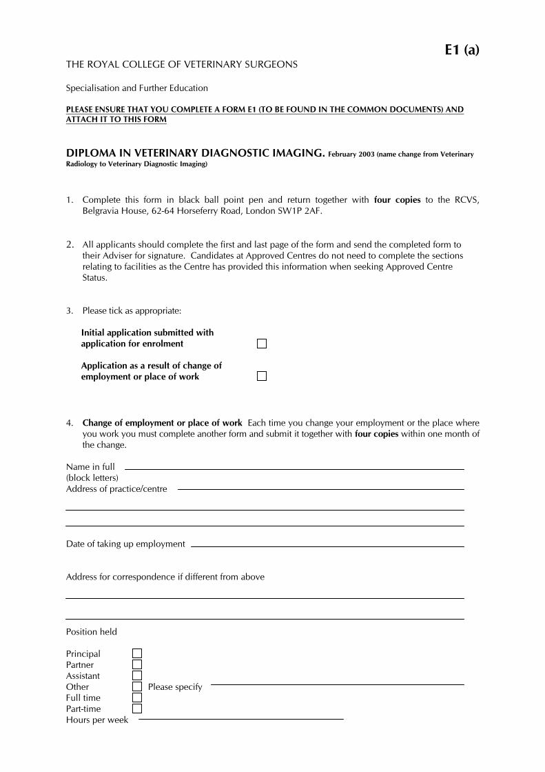
E1 (a) THE ROYAL COLLEGE OF VETERINARY SURGEONS Specialisation and Further Education PLEASE ENSURE THAT YOU COMPLETE A FORM E1 (TO BE FOUND IN THE COMMON DOCUMENTS) AND ATTACH IT TO THIS FORM
DIPLOMA IN VETERINARY DIAGNOSTIC IMAGING. February 2003 (name change from Veterinary Radiology to Veterinary Diagnostic Imaging)
1. Complete this form in black ball point pen and return together with four copies to the RCVS,
Belgravia House, 62-64 Horseferry Road, London SW1P 2AF. 2. All applicants should complete the first and last page of the form and send the completed form to
their Adviser for signature. Candidates at Approved Centres do not need to complete the sections relating to facilities as the Centre has provided this information when seeking Approved Centre Status.
3. Please tick as appropriate:
Initial application submitted with application for enrolment Application as a result of change of employment or place of work 4. Change of employment or place of work Each time you change your employment or the place where
you work you must complete another form and submit it together with four copies within one month of the change.
Name in full (block letters) Address of practice/centre Date of taking up employment Address for correspondence if different from above Position held Principal Partner Assistant Other Please specify Full time Part-time Hours per week
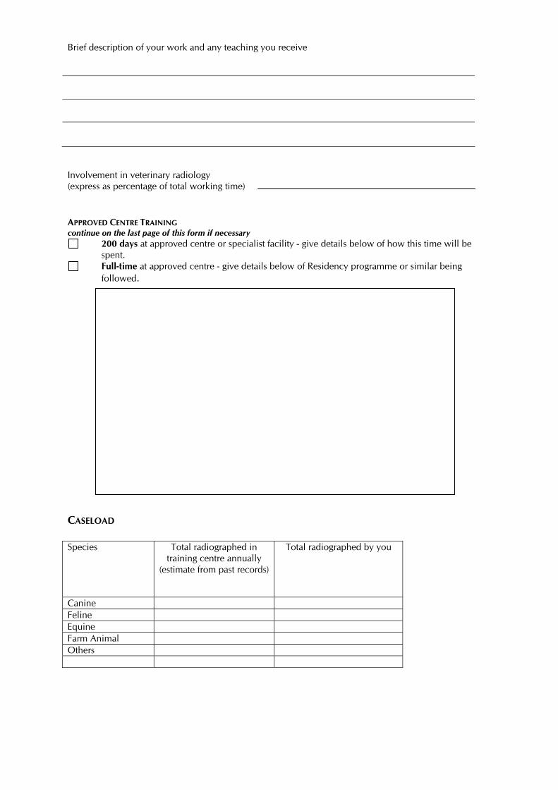
Brief description of your work and any teaching you receive Involvement in veterinary radiology (express as percentage of total working time) APPROVED CENTRE TRAINING continue on the last page of this form if necessary
200 days at approved centre or specialist facility - give details below of how this time will be spent.
Full-time at approved centre - give details below of Residency programme or similar being followed.
CASELOAD Species Total radiographed in
training centre annually (estimate from past records)
Total radiographed by you
Canine Feline Equine Farm Animal Others
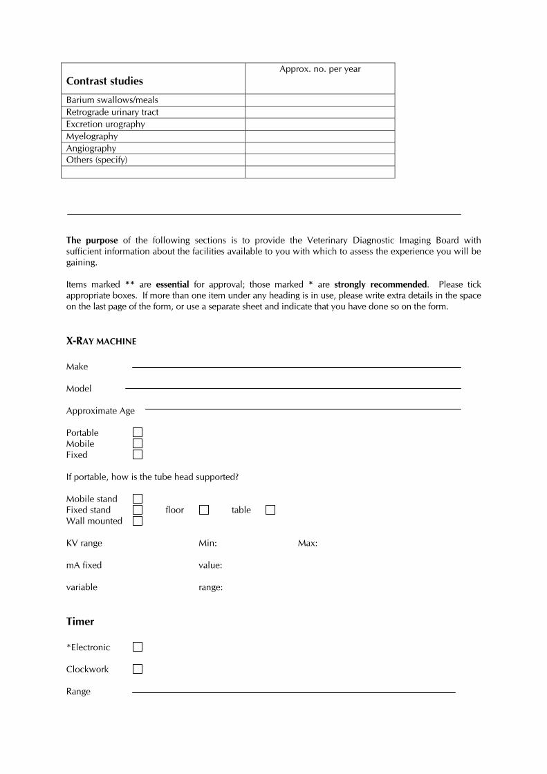
Contrast studies Approx. no. per year
Barium swallows/meals Retrograde urinary tract Excretion urography Myelography Angiography Others (specify) The purpose of the following sections is to provide the Veterinary Diagnostic Imaging Board with sufficient information about the facilities available to you with which to assess the experience you will be gaining. Items marked ** are essential for approval; those marked * are strongly recommended. Please tick appropriate boxes. If more than one item under any heading is in use, please write extra details in the space on the last page of the form, or use a separate sheet and indicate that you have done so on the form.
X-RAY MACHINE Make Model Approximate Age Portable Mobile Fixed If portable, how is the tube head supported? Mobile stand Fixed stand floor table Wall mounted KV range Min: Max: mA fixed value: variable range:
Timer *Electronic Clockwork Range
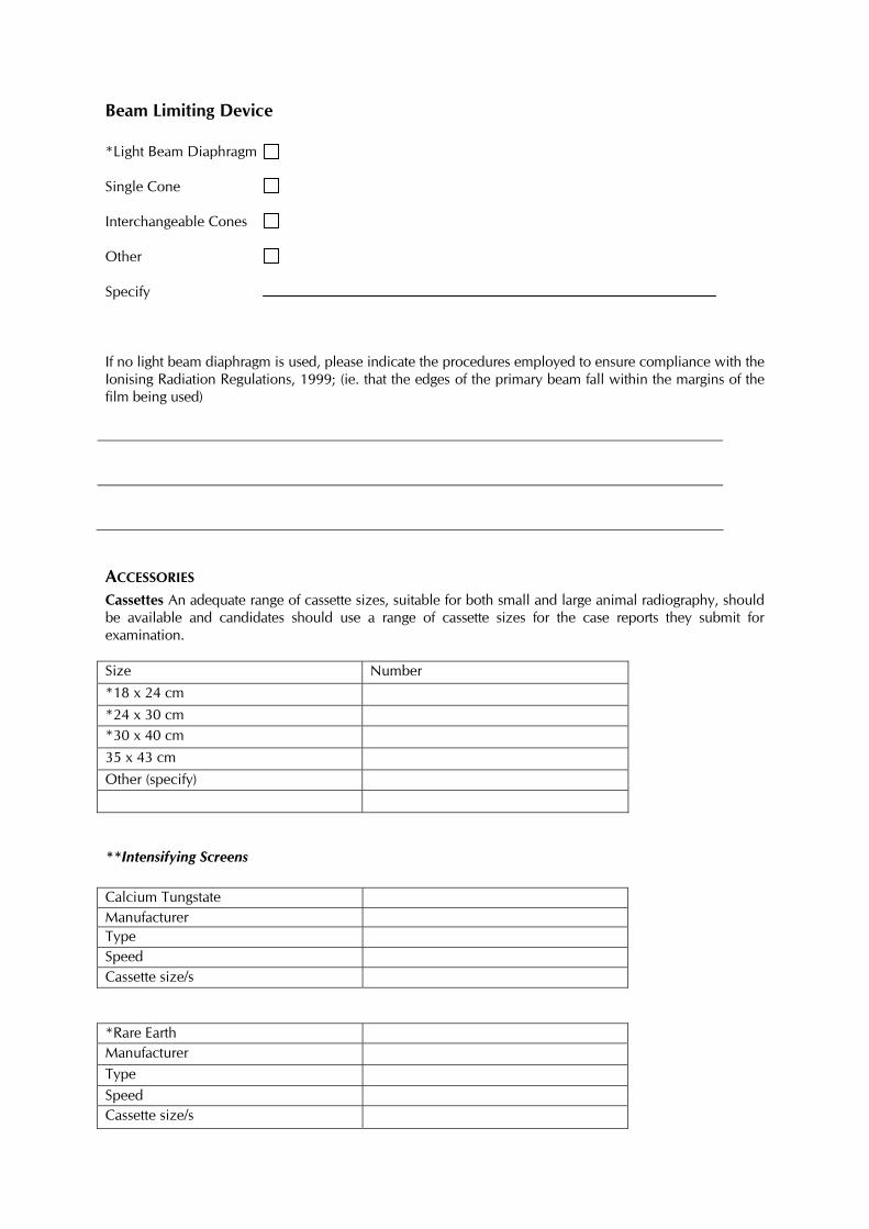
Beam Limiting Device *Light Beam Diaphragm Single Cone Interchangeable Cones Other Specify If no light beam diaphragm is used, please indicate the procedures employed to ensure compliance with the Ionising Radiation Regulations, 1999; (ie. that the edges of the primary beam fall within the margins of the film being used)
ACCESSORIES Cassettes An adequate range of cassette sizes, suitable for both small and large animal radiography, should be available and candidates should use a range of cassette sizes for the case reports they submit for examination. Size Number *18 x 24 cm *24 x 30 cm *30 x 40 cm 35 x 43 cm Other (specify)
**Intensifying Screens Calcium Tungstate Manufacturer Type Speed Cassette size/s *Rare Earth Manufacturer Type Speed Cassette size/s
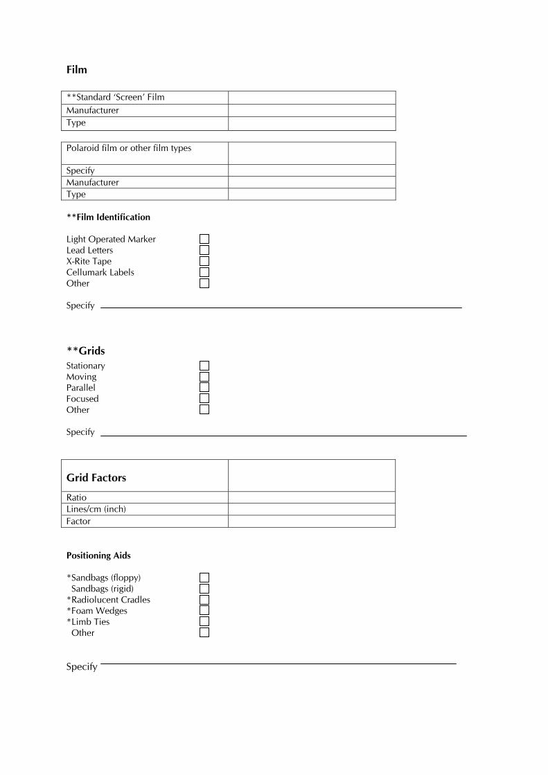
Film **Standard ‘Screen’ Film Manufacturer Type Polaroid film or other film types
Specify Manufacturer Type **Film Identification Light Operated Marker Lead Letters X-Rite Tape Cellumark Labels Other Specify
**Grids Stationary Moving Parallel Focused Other Specify
Grid Factors
Ratio Lines/cm (inch) Factor Positioning Aids *Sandbags (floppy) Sandbags (rigid) *Radiolucent Cradles *Foam Wedges *Limb Ties Other Specify
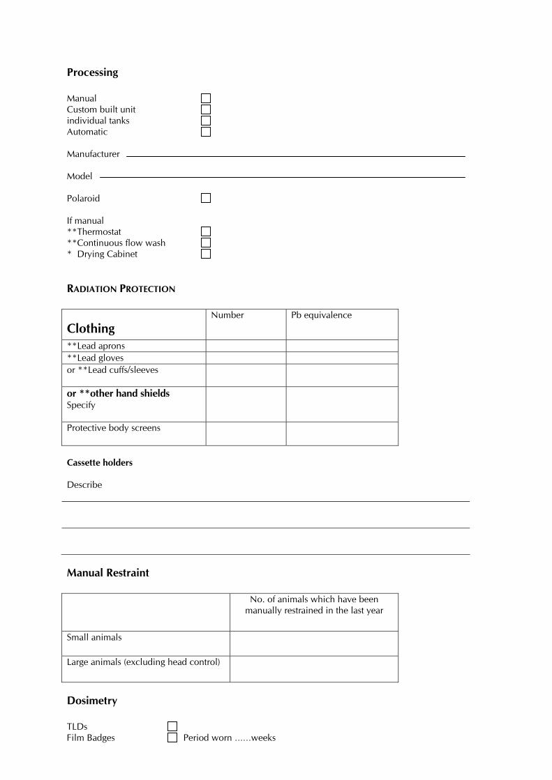
Processing Manual Custom built unit individual tanks Automatic Manufacturer Model Polaroid If manual **Thermostat **Continuous flow wash * Drying Cabinet
RADIATION PROTECTION
Clothing Number Pb equivalence
**Lead aprons **Lead gloves or **Lead cuffs/sleeves
or **other hand shields Specify
Protective body screens
Cassette holders Describe
Manual Restraint No. of animals which have been
manually restrained in the last year
Small animals
Large animals (excluding head control)
Dosimetry TLDs Film Badges Period worn ……weeks
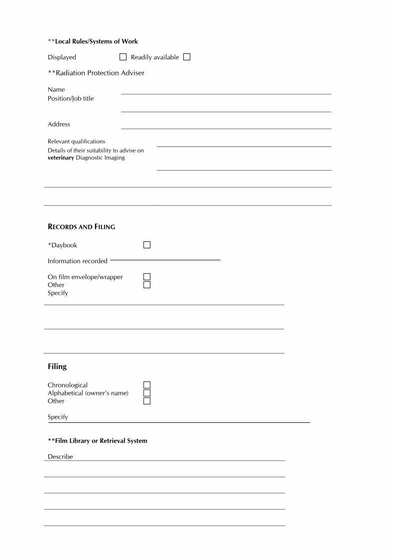
**Local Rules/Systems of Work Displayed Readily available **Radiation Protection Adviser Name
Position/Job title
Address
Relevant qualifications Details of their suitability to advise on veterinary Diagnostic Imaging
RECORDS AND FILING *Daybook Information recorded On film envelope/wrapper Other Specify
Filing Chronological Alphabetical (owner’s name) Other Specify **Film Library or Retrieval System Describe
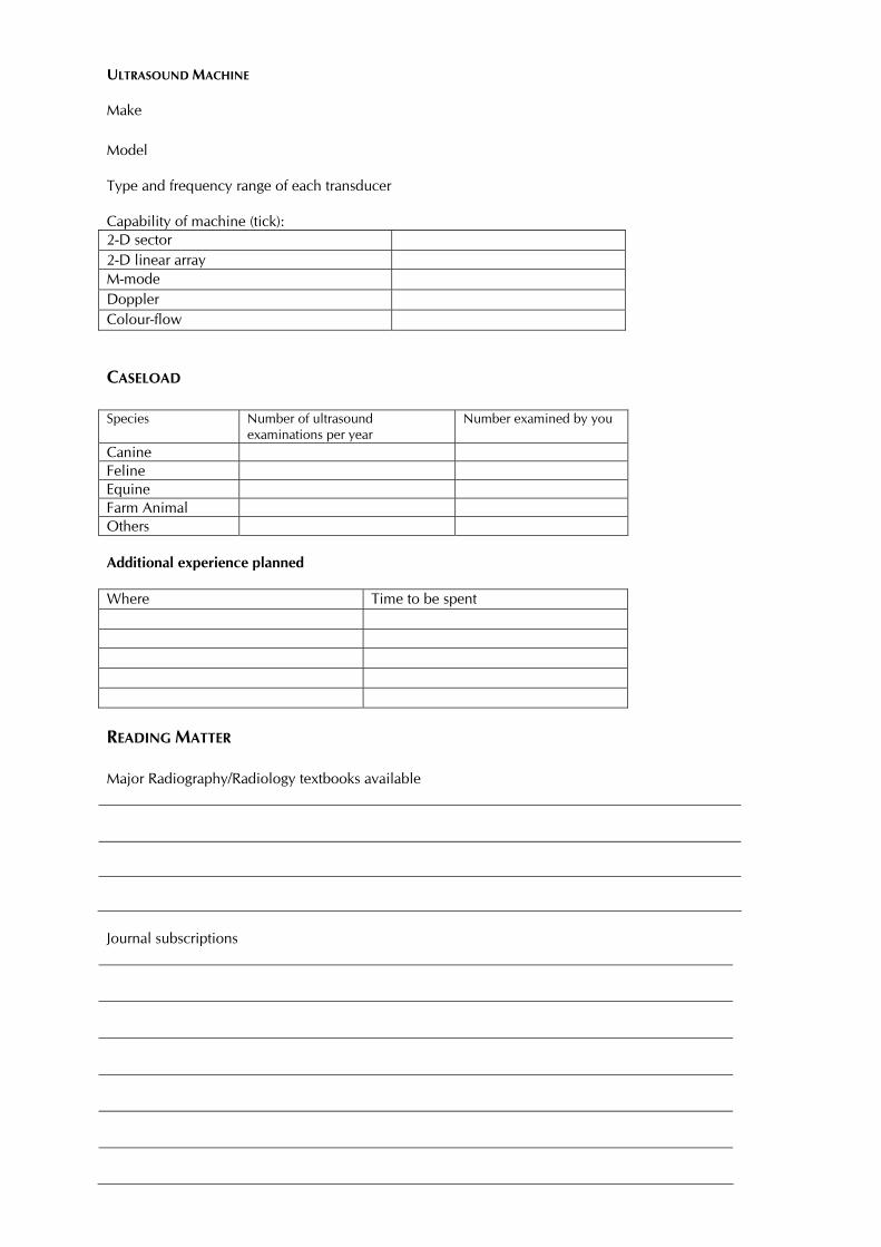
ULTRASOUND MACHINE
Make Model Type and frequency range of each transducer Capability of machine (tick): 2-D sector 2-D linear array M-mode Doppler Colour-flow
CASELOAD Species Number of ultrasound
examinations per year Number examined by you
Canine Feline Equine Farm Animal Others Additional experience planned Where Time to be spent
READING MATTER Major Radiography/Radiology textbooks available
Journal subscriptions
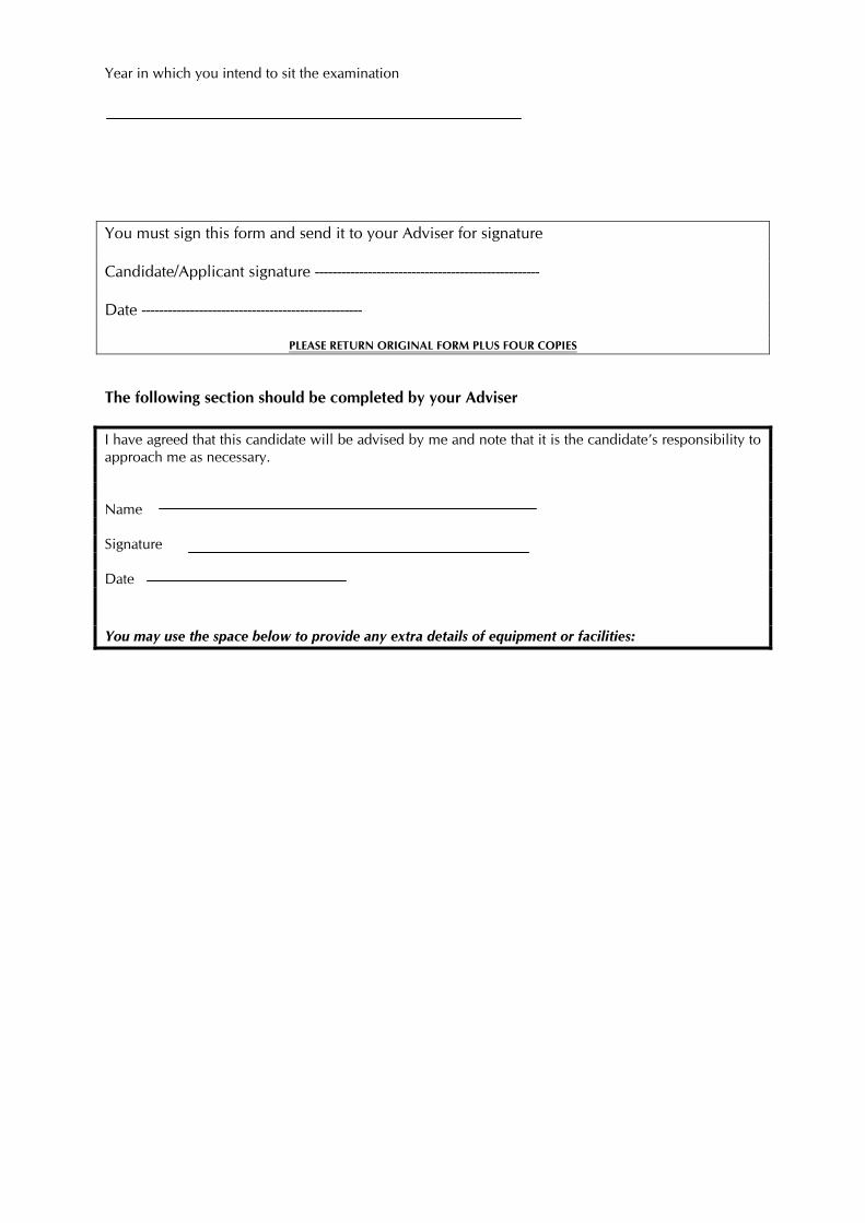
Year in which you intend to sit the examination You must sign this form and send it to your Adviser for signature Candidate/Applicant signature --------------------------------------------------- Date --------------------------------------------------
PLEASE RETURN ORIGINAL FORM PLUS FOUR COPIES
The following section should be completed by your Adviser I have agreed that this candidate will be advised by me and note that it is the candidate’s responsibility to approach me as necessary. Name Signature Date You may use the space below to provide any extra details of equipment or facilities:
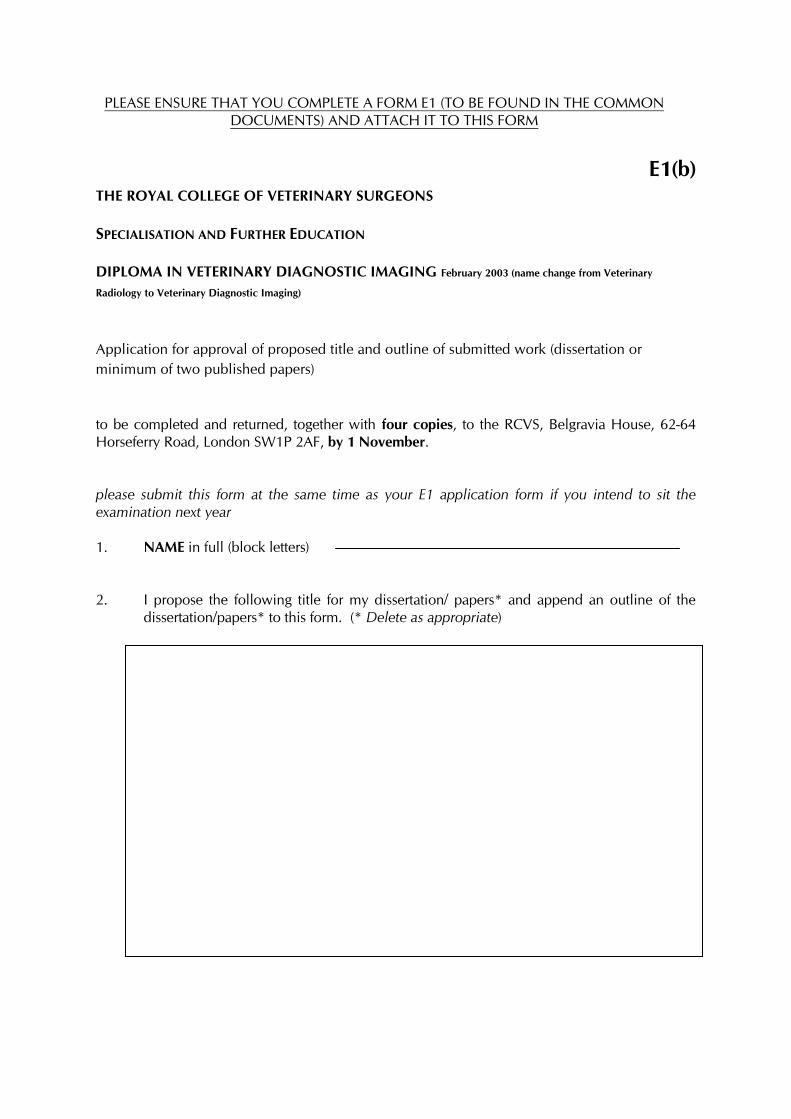
PLEASE ENSURE THAT YOU COMPLETE A FORM E1 (TO BE FOUND IN THE COMMON DOCUMENTS) AND ATTACH IT TO THIS FORM
E1(b)
THE ROYAL COLLEGE OF VETERINARY SURGEONS SPECIALISATION AND FURTHER EDUCATION DIPLOMA IN VETERINARY DIAGNOSTIC IMAGING February 2003 (name change from Veterinary
Radiology to Veterinary Diagnostic Imaging) Application for approval of proposed title and outline of submitted work (dissertation or minimum of two published papers) to be completed and returned, together with four copies, to the RCVS, Belgravia House, 62-64 Horseferry Road, London SW1P 2AF, by 1 November. please submit this form at the same time as your E1 application form if you intend to sit the examination next year 1. NAME in full (block letters) 2. I propose the following title for my dissertation/ papers* and append an outline of the
dissertation/papers* to this form. (* Delete as appropriate)
3. I have discussed the dissertation/ papers with my Adviser Signature Date
PLEASE RETURN ORIGINAL PLUS FOUR COPIES The following section should be completed by your Adviser Name (block letters) Signature Date
E2 THE ROYAL COLLEGE OF VETERINARY SURGEONS Specialisation and Further Education
DIPLOMA IN VETERINARY DIAGNOSTIC IMAGING February 2003 (name change from Veterinary Radiology to Veterinary Diagnostic Imaging)
Large or small animal option in the practical film reading Please tick your choice as appropriate
SMALL ANIMAL Practical film reading will consist of 10 small animal and 5 large animal sets of images
LARGE ANIMAL Practical film reading will consist of 10 large animal and 5 small animal sets of images Application for FINAL approval of experience and for permission to submit an entry to the next examination to be completed and returned, together with four copies, to the RCVS, Belgravia House, 62-64 Horseferry Road, London SW1P 2AF by 1 November in the year preceding the year in which you wish to enter the examination. NO LATE APPLICATIONS WILL BE ACCEPTED. Four copies of your CPD Record Card/s covering the period of experience offered must be submitted with this application form. 1. a) NAME in full (block letters) b) TITLE (please delete as appropriate) MR / MRS / MISS / DR / Other 2. DEGREES/DIPLOMAS/CERTIFICATES (in abbrev. form) 3. ADDRESS for all correspondence (block letters) 4. (a) TELEPHONE NO(s) for contact during day (b) FAX NO for contact during the day (c) EMAIL ADDRESS 5. QUALIFICATION FOR ENTRY Are you a member of the RCVS? YES/NO Please state veterinary degree obtained (full title and recognised abbreviation), name of awarding University, and date of graduation 6. DATE OF ENROLMENT (Month/Year) If application for enrolment is being submitted concurrently with this application please state 'concurrent'.
7. PERIODS OF EXPERIENCE BEING OFFERED to meet the requirements of the byelaws
Approved practice or approved centre (name and address)
Period(s) of employment (from/to)
Certified by Principal
If there has been any change in the work-load of the practice/centre, or in your personal work-load, since you applied for enrolment, please give details below: 8. (a) ATTENDANCE AT COMPULSORY RADIOLOGICAL PHYSICS COURSE
(Please note that the European association of Veterinary Diagnostic Imaging (EAVDI) no longer run the annual physics course)
Title of course attended Dates and venue
(b) ATTENDANCE AT RELEVANT SHORT COURSES Please give details of course title, dates and venue:
9. OTHER INVOLVEMENT IN RELATION TO VETERINARY RADIOLOGY List any
attendances at relevant congresses, conferences, meetings, symposia, etc., with dates. (Additional sheets may be attached if necessary)
10. PUBLICATIONS/ARTICLES/PAPERS/LECTURES (Additional sheets may be attached if necessary) 11. OTHER POSTGRADUATE STUDIES During the period of experience being offered,
have you been or are you studying for any other postgraduate qualification? YES/NO
If yes, please give brief details:
12. SUBMITTED WORK Title The subject and the title of my dissertation/published papers were approved by the Board on 13. I HEREBY APPLY FOR APPROVAL OF EXPERIENCE AND FOR PERMISSION TO
SUBMIT AN ENTRY TO THE NEXT DIPLOMA EXAMINATION IN VETERINARY RADIOLOGY.
14. CONFIRMATION OF INTENT TO SIT THE EXAMINATION
If approval of experience is granted, I do / do not (delete as applicable) intend to submit an entry to the next examination being held in ............. (year)
Signature Date
PLEASE RETURN ORIGINAL PLUS FOUR COPIES THE FOLLOWING SECTION SHOULD BE COMPLETED BY YOUR ADVISER. I confirm that I am acting as this candidate's Adviser Signature Name Date No acknowledgement will be sent. If an acknowledgement is required please enclose a stamped addressed envelope. You will be advised of the outcome of this application by early January. An examination entry form for the next examination will be sent to you at that time. In subsequent years, if you have not sat the examination or you have sat but failed, you will automatically receive an entry form
F DIPLOMA IN VETERINARY DIAGNOSTIC IMAGING ADVISER LIST Candidates should consult the RCVS Register of Members for a suitably willing and qualified individual (someone holding a Diploma or equivalent) who is familiar with the RCVS Diploma examination system and clinically active.
THE ROYAL COLLEGE OF VETERINARY SURGEONS
DIPLOMA IN VETERINARY DIAGNOSTIC IMAGING
MONDAY 9 JULY 2007
PAPER l Advanced Radiological Physics, Techniques and Interpretation
(3 hours)
This paper is in two Sections (A and B) Candidates are required to answer FOUR of the SIX questions which MUST INCLUDE ONE FROM EACH SECTION Allow 45 minutes per question. Please use a separate answer sheet for each question. Answers to the questions in Sections A and B should be put inside separate answer sheet covers. Illegible handwriting or failure to answer the question in the form requested may result in examiners being unable to award marks for information which candidates intended to convey. ________________________________________________________________________
SECTION A
Physics Questions 1. With reference to nuclear medicine imaging:
a) Briefly describe the TWO main physical processes by which gamma rays interact with tissue and with the gamma camera crystal.
b) For imaging with 99Tcm, draw a graph demonstrating the energies of the gamma photons detected by the gamma camera (the Z signal).
Using this graph, describe the function of the pulse height analyser in the gamma camera.
c) Describe the factors that can be optimised to improve image quality (contrast and resolution) in a 99Tcm –HDP or MDP bone scintigraphy image.
2.a) Name the two principal instruments of United Kingdom legislation which
apply to a veterinary practice carrying out nuclear medicine examinations. Give a brief outline of the purpose of both of these and state which government body polices this legislation.
b) Describe how you would decide whether Local Rules are required and outline their purpose.
c) Give a list of typical contents of Local Rules for a routine diagnostic X-ray room making it clear which items are mandatory and which cover local requirements.
P.T.O. FOR QUESTIONS 3 - 6
3. With reference to scanning the peripheral vasculature in any species
a. Briefly outline the Doppler effect for an ultrasound beam incident on a moving target.
b. Briefly describe the differences between colour flow imaging, power Doppler imaging and pulsed wave Doppler.
c. What is the practical trade-off between imaging a blood vessel and observing the blood flow within it? How is this trade-off solved in linear array scanners?
d. Briefly describe common artefacts which may lead to the impression that there is no flow in a blood vessel and briefly indicate how to correct for these artefacts.
SECTION B
(Advanced Techniques and Interpretation)
4. Discuss how diagnostic ultrasonography of the abdomen in a horse with colic and a dog with abdominal distension can be used to differentiate surgical from non-surgical conditions.
5. Describe your technique for an ultrasound examination of the palmar metacarpal and palmar pastern soft tissue structures of an adult Thoroughbred racehorse. Use diagrams to illustrate the ultrasonographic anatomy you would expect to find in a normal horse. Discuss how operator errors can lead to a poor quality study.
6. Discuss the use of contrast media in:
a) Radiography ( including CT).
b) MRI.
For each, use the following headings: indications for contrast studies; generic names of contrast media used; the physical principles underlying their use; examples in veterinary diagnostic imaging.
* * * *
THE ROYAL COLLEGE OF VETERINARY SURGEONS
DIPLOMA IN VETERINARY DIAGNOSTIC IMAGING
MONDAY 9 JULY 2007 PAPER ll
Radiological Interpretation (3 hours)
This paper is in three Sections (A, B and C)
Candidates are required to answer FOUR of the SIX questions which MUST INCLUDE ONE FROM EACH SECTION. Allow 45 minutes per question. Please use a separate answer sheet for each question. Answers to the questions in Sections A, B and C should be put inside separate answer sheet covers. Illegible handwriting or failure to answer the question in the form requested may result in examiners being unable to award marks for information which candidates intended to convey.
__________________________________________________________________________
SECTION A (GENERAL) 1. Discuss the role of radiology in monitoring the healing of fractures.
2. Discuss the statement ‘Selective and non selective cardiovascular angiography has been superseded by alternative imaging techniques’.
Illustrate the relevant points in your answer using the horse and dog as examples.
SECTION B (LARGE ANIMAL)
3. Discuss the use of diagnostic imaging in the diagnosis of periapical tooth root infection in the equine maxillary arcade. You should include in your answer what role modalities other than radiography have in this field.
4. Discuss the use of current diagnostic imaging techniques in diagnosing pathology
of the navicular bone.
P.T.O. FOR SECTION C
SECTION C – (SMALL ANIMAL) 5. Describe the typical imaging findings that may be seen in dogs and/or cats with
the following diseases:
Use diagrams where appropriate.
a) Air trapping. b) Tetralogy of Fallot. c) Renal dysplasia. d) Meningioma.
6. Briefly outline the types of portosystemic shunts seen in small animals.
Discuss the advantages and disadvantages of the various imaging modalities available to diagnose portosystemic shunts.
__________________


![Paged Veterinary Surgeons and Veterinary Para-Professionals … · · 2013-08-19No. 29 of 2011 [Rev. 2012] Veterinary Surgeons and Veterinary Para-professionals [Issue 1] 6 “inspector”](https://img.pdfslide.net/doc/110x75/5acc2a597f8b9a27628c3028/paged-veterinary-surgeons-and-veterinary-para-professionals-29-of-2011-rev.jpg)