Embed Size (px)
Citation preview
Department ofVeterans Affairs
Journal of Rehabilitation Research andDevelopment Vol . 34 No . 1, January 1997Pages 72—81
Direct bladder stimulation with suture electrodes promotesvoiding in a spinal animal model : A technical report
James S . Walter, PhD ; John S . Wheeler, MD; Wuying Cai, MD; Robert D. Wurster, PhDRehabilitation Research and Development Center, Edward Hines, Jr VA Hospital, Hines, IL, 60141 ; Departments of
Urology, Physiology, and Neurosurgery, Loyola Medical Center, Stritch School of Medicine, Maywood, IL 60153
Abstract—To determine the efficacy of a new electrode fordirect bladder stimulation, five male cats were instrumentedduring anesthesia . Multistranded, 316LVM, stainless-steel,wire electrodes were implanted on the bladder wall serosaabove the trigone area. The electrodes were made with a needleattached to the end that was cut off after suturing the electrodein place . Additional instrumentation included tubes for pressurerecording and filling, and hook electrodes for leg and pelvicfloor EMG recording. Bladder filling and stimulation studieswere conducted in tethered animals 1 to 2 weeks followingrecovery. Chronic studies were conducted following recoveryin tethered animals . To test these electrodes in a spinal cordinjury (SCI) model, a T-1 level complete lesion was performedon the above instrumented animals . Spinal animals had suc-cessful direct bladder stimulation that induced active contrac-tions and voiding both before and after SCI, but voiding rateswere higher more than 2 weeks after SCI and at larger initialbladder volumes . Optimum stimulation parameters consisted of40 pulses per second, 300 Rs to 1 ms pulse duration, a stimula-tion period of 3 to 4 s, and 10 to 40 mA . Urethral resistance,indicated by a urethral function measure, showed that stimula-tion had no adverse effect on urethral function, and fluoroscopyshowed an open membranous urethra during stimulation andvoiding . The cat has a small penile urethra that is the flow ratecontrolling zone . The suture electrode did not corrode, erode
This material is based upon work supported by the Department ofVeterans Affairs, Research and Development Service, Washington, DC20420 and the National Institutes of Health, National Center for MedicalRehabilitation Research, Bethesda, MD 20832Address all correspondence and requests for reprints to : James S . Walter, PhD,Edward Hines, Jr. VA Hospital, Rehabilitation R&D Center (151L), P .O. Box20, Hines . IL 60141 .
into the bladder, or become dislodged, and appears suitable forchronic implantation.
Key words : cystometry, electrical stimulation, functional elec-trical stimulation, neural prosthetics, neurogenic bladderspinal cord injury, urodynamic studies.
INTRODUCTION
Sacral ventral root stimulation in conjunction withsacral dorsal root rhizotomy has been effective in pro-moting voiding with minimal residual volume in spinalcord injured (SCI) patients (1–4) . However, extensivesurgery is required to implant the electrodes in the lowerlumbar or sacral canal . Direct bladder stimulation withimplantable stimulators has also been investigated forpromoting micturition . Direct bladder stimulation in ani-mal models has been shown to be effective (2) ; however,many clinical studies have shown poor results . Citedproblems in the clinical studies have included high ure-thral resistance due to activation of the striated urethralsphincter, poor bladder emptying, the need for high stim-ulating currents, and activation of lower limb muscles,pain, and electrode erosion into the bladder (2,5–10) . Onegroup (11,12) reported effective stimulation-driven void-ing in 29 of 32 patients . These subjects were unable tovoid on their own due to SCI and other neurologicaldeficits . Eight small platinum disk electrodes weresutured into the bladder wall, and the most effective loca-
72
73
WALTER et al . Direct Bladder mimu!ation in SCI Model
tion of the electrodes was adjacent to the ureters wherethe neurovascular bundle innervates the bladder.Concerns with this study (!! ' l2)include the large num-ber of electrodes on the bladder wall and the lack of long-term results . In addition, this work does not address thehigh urethral resistance problem cited in many other stud-ies (2 `5-/O).
We are investigating electrodes that might improvedirect bladder stimulation techniques. We reported thatsacral nerve electrodes and "woven-eye" bladder wallelectrodes were effective for activation of the bladder inanimal models (13,14) . Currently, we are evaluating asuture type electrode that might have the followingadvantages : 1) an extended length that could be placedacross the entire neurovascular bundle that innervates thebladder, 2) implantation in the outer serosal layer thatwould not erode into the bladder, and 3) a simple elec-trode requiring little additional implantation procedures,such as suturing, and that might be implanted through alaparoscope . Studies were conducted in male cats bothbefore and after SCI . Urodynamic responses to stimula-tion, electrode characteristics, and aspects of this animalmodel are described.
Instrumentation Surgery
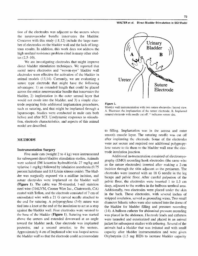
Five male cats 2 to 4 kg) were instrumentedfor subsequent direct bladder stimulation studies . Animalswere sedated (IM ketamine hydrochloride 27 mg/kg andxylazine 1 mg/kg) followed by inhalation anesthesia (0 .75porootba!oibaoe and 0 .8 lJudooibnnu oxide) . The blad-der was surgically exposed via a midline incision, andsuture electrodes were implanted on the bladder wall(Figure 1) . The cable was 50-stranded, 1-mil stainlesssteel wire (316LVM, Cooner Wire Inc ., Chatsworth, CA)coated with Teflon, and the electrode consisted of 7 cm ofuninsulated wire with a 21 G curved needle attached tothe end for suturing . A polypropylene (3-0) suture wastied into a knot at the end of the insulation to act as a stopagainst the bladder wall . Four electrodes were sutured tothe base of the bladder (Figure 1) . Suturing was startedabove the ureters and extended downward at an angletoward the bladder neck. One electrode was implantedposterior, and a second anterior, to the ureters.Approximately 4 cm of implanted wire was looped acrossthe bladder wall so that the electrode could accommodate
Figure 1.Bladder wall instrumentation with two suture electrodes : lateral view.A. Illustrates the implantation of the suture electrode : B. Implantedsutured electrode with needle cut off. * indicates suture site.
to filling . Implantation was in the serosa and outersmooth muscle layer . The suturing needle was cut offafter implanting the electrode . Some of the electrodeswere not secure and required one additional polypropy-lene suture to tie them to the bladder wall near the elec-trode insulation junction.
Additional instrumentation consisted of electromyo-graphy (EMG) recording hook electrodes (the same wireas the suture electrodes) inserted after making a 2-cmincision through the skin adjacent to the perineum . Theelectrodes were inserted with an 18 G needle in the legbiceps and pelvic floor. After careful palpation of thepelvic floor, the electrodes were inserted 1 to 1 .5 cmdeep, adjacent to the urethra in the bulbous urethral area.Additionally, two electrodes were placed under the skinin the back. These electrodes, with several inches ofstripped insulation, served as grounding wires . Two smalldiameter Silastic tubes were also sutured into the dome ofthe bladder for bladder filling and pressure recording(/4).A balloon catheter for abdominal pressure recordingwas placed in the abdomen . Electrode leads and catheterswere tunneled and exteriorized and placed in an animaljacket for subsequent studies with tethering . Several of theanimals had a bladder that was irritated and with smallcapacity after bladder instrumentation and were givenOxybutynin (1 .5 mg BD}) to increase bladder capacity
74
Journal of Rehabilitation Research and Development Vol . 34 No. 1 1997
during the first 2 weeks after the surgery. The bladdercatheters were also regularly flushed with antibiotic(Neosporin G.U. irrigant) . Stimulation and cystometricstudies were conducted 2–4 weeks after instrumentation.
Spinalization ProceduresA second survival surgery was conducted for spinal-
ization 4–5 weeks after the animal instrumentation . Theanimals were reanesthetized and given an antihyperten-sive agent (8 mg per mouth, Nifedipine ; 13–15) to reduceblood pressure increases associated with spinal injuryprocedures . The T-1 spinal cord dural space was surgical-ly exposed. A small balloon was used for crushing thecord in the first two animals . However, this procedurewas abandoned because the second cat developed somerecovery of back leg function. The final three animalswere spinalized using a hemostat compression for 20 s(13,14). These animals showed no recovery of back legfunction. The animals were healthy for the 8–10 weeksthat they were maintained in the study following spinalinjury. An initial set of cystometric and stimulation stud-ies were conducted 2–3 weeks, and a second set 4–7weeks, after injury. Fluoroscopy was used on one daywith radiopaque medium (Hypaque-76, Winthrop, NY) inthe bladder. The lower urinary tract was observed duringspontaneous and stimulation-induced voiding .
(cm H2O) and flow rate, Q (ml/s) . This factor has beenfound to be helpful in the evaluation of voiding difficul-ties due to obstruction (16,17) and stress urinary inconti-nence (18) . This formula might have validity in urody-namic evaluation after SCI where reduced voiding isoften seen due to membranous urethra contraction.
Direct Bladder Stimulation StudiesBoth bipolar and monopolar stimulation were evalu-
ated in all five animals . For bipolar stimulation, both neg-ative and positive electrodes were on the bladder wall,while for monopolar stimulation the negative electrodewas on the bladder wall and the positive electrode was inthe back (described above as implanted grounding elec-trodes) . Capacitor-coupled stimulation was conducted forbalanced charge injection pulses with two stimulators(S48, Grass, Quincy, MA) . The stimulators were isolatedfrom ground and were constant current units (13,15).Stimulating parameters such as current and pulse dura-tion were set with a dial on the front of the stimulator, andwere checked on an oscilloscope . A standard stimulationwas 40 pps for 3 s with a pulse duration of 1 ms (14).Stimulating parameters evaluated in this study includedthe period, frequency, current, and pulse duration. Eachparameter was studied independently, keeping other para-meters constant.
Additional studies included electrode corrosion andpostmortem evaluations. Two bladder wall electrodeswere pulsed at the end of the study in a bipolar configu-ration for 115 hours using 40 pps with 25 mA and 1 mspulse duration . Following euthanasia, pulsed electrodesand nonimplanted electrodes were viewed with lightmicroscopy and with scanning electron microscopy(SEM, by EIC Laboratories, Norwood MA) . Postmortembladder wall thickness was evaluated after fixation withthe installation of 20 ml formalin (HT50 Sigma, St.Louis, MO) . Histological sections at the electrode sitewere stained with H&E . Data are presented as mean -I-SD . Statistics were conducted using Student's t-test withpaired data.
RESULTS
Urodynamic StudiesUrodynamic studies were conducted with the ani-
mals tethered in a urodynamic recording cage both beforeand after SCI . Pressures, EMG, and volume voided wererecorded on an 8-channel recorder (Astromed, WestWarwick, RI) . Urine volume was collected in a funnelunder the animal's cage and the weight of the fluid wasrecorded as a measure of the volume. Urine flow rate wasassessed graphically by the slope of the volume voidedrecord . Stimulation studies were conducted with an initialbladder volume one-half to two-thirds of cystometriccapacity and isotonic saline at room temperature wasused for filling . Cystometry was perfonned at 5 ml/min,until strong spontaneous bladder contractions associatedwith micturition occurred . Detrusor pressure wasobtained by subtracting abdominal pressure from therecorded bladder pressure . Effects of stimulation on ure-thral functions were assessed with a urodynamic conduc-tance factor, Area Equivalent Factor male (AEFm) :
Cystometry and Spontaneous VoidingAEFm (mm2) = 3.7Q/P058 , where AEFm estimates
Five male cats were instrumented before SCI, andcross-sectional area of the flow controlling zone of the
urodynamic studies were conducted 2–4 weeks afterurethra during voiding in terms of detrusor pressure, P
surgery when oxybutynine administration had been
75
WALTER et al . Direct Bladder Stimulation in SCI Model
A. Before SCI
B. After SCI
oakSod io
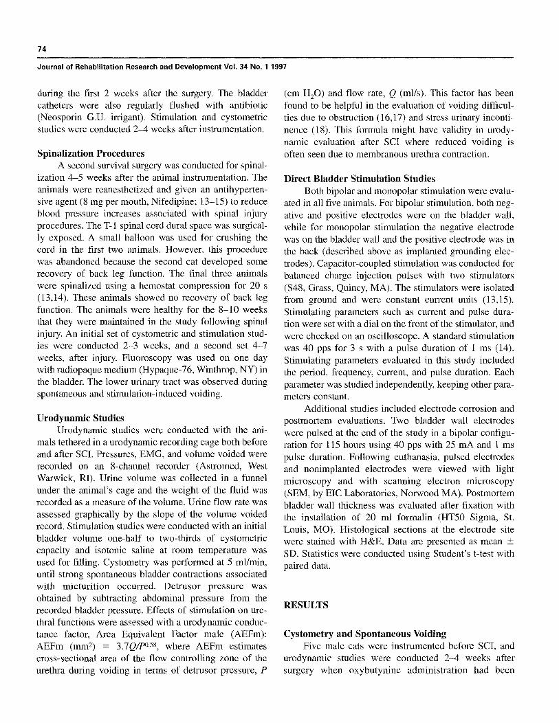
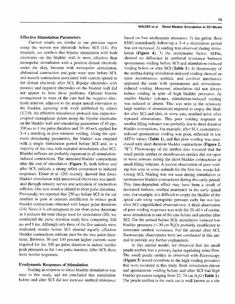
Figure 2.Comparison of cystometry-induced micturition before and after SCI.A. Before SCI, showing voiding at a high flow rate . Records alsoshow phasic pelvic floor activity during voiding (a) and strongerpelvic floor contractions at the end of voiding (b) . B . After SCI, show-ing small bladder contractions with voiding at a low flow rate . Arrowsindicate discharges of the pelvic floor. Record in B obtained 49 daysfollowing SCI.
stopped and bladder capacity had increased . In Figure 2,"A" shows cystometry-induced spontaneous micturitionfor one tethered cat with complete bladder emptying andat a high urine flow rate . A summary of micturitionresponses to cystometry for the five cats is shown inTable 1 . Filling volumes for micturition ranged from 10to 27 ml. Peak detrusor pressures during voiding were32–75 cm H 20, and the peak urine flow rates were0.6–2 .2 mils (Table 1) . Increased abdominal pressureduring voiding was usually associated with the animalstanding and assuming a voiding posture. Before SCI, thebulbous urethral EMG was unchanged during bladderfilling in all of the animals, although movement-relatedchanges occurred . Spiking in the EMG was seen duringmicturition, usually starting halfway through the voiding
period and continuing to the end of voiding . As the detru-sor pressure declined at the end of voiding, larger pelvicfloor discharges were seen.
After SCI, an initial cystometry was conducted inthe second to third week after injury. and a second cys-tometry was conducted after 5 weeks . For the first cys-tometry, numerous small bladder contractions occurredwith little voiding. By 5 weeks after injury, bladder con-tractile activity increased. In Figure 2, "B" shows thesestronger bladder contractions for one animal ; however,the bladder contractions are short in duration and littlevoiding is shown. Table 1 also shows this poor sponta-neous voiding after SCI with rates from 0 .1 to 0 .2 ml/sand voiding that left high residual volume, ranging from7 to 48 ml. Peak pressures ranged from 30 to 75 cm H 2 O,and contractile activity typically started at 1-35 ml andcontinued to the end of bladder filling at 20–50 ml . AfterSCI, the bulbous urethral EMG was unchanged duringbladder filling in all of the animals . The EMG showedsmall increases during bladder contractions with largerdischarges at the end of bladder contractions and voiding.
Stimulation-Induced VoidingDirect bladder stimulation resulted in prolonged
bladder contraction and voiding both before and afterSCI, as shown in Figure 3. Table 1 summarizes theresults for the five cats . Before SCI, peak detrusorresponses to stimulation ranged from 22 to 74 cm H 2 O.The initial filling volume for these stimulation studiesranged from 5 to 15 ml . The maximum voiding rates were0.5–1 .5 ml/s with a total volume voided with one stimu-lation period ranging from 5–15 ml and with minimalresidual volume. The stimulating parameters for these
ccm H2O)
Table 1.Urodynamic responses to cystometry and stimulation in the male cat before and after spinal cord injury.
FillingVolume
VoidedVolume Pressure
MaximumFlow AEFm
Cystometry:Before 17±7 14±5 50± 17 1 .2±0.6 0 .44±0.13After 34±11 8±6 46±18 0 .1±0.04* 0 .048±0 .029*
Stimulation:Before 111-4 9±4 43±23 0.7±0 .3 0 .32±0 .11After 23 ± 14 5±4 39±11 0.9±0 .7 0 .38±0.2
Means and SD for five animals for all data shown. Measures for each animal obtained as a typical record in response to a single stimulation period of 3-4 sec.Records after SCI obtained between 5 and 8 weeks after SCI . Volumes in ml ; detrusor pressures (obtained at maximal urine flow rate) in cm H 2 0 ; flow in ml/s;AEFm in mm2 .*significantly different from cystometric results before SCI (p-0 .05).
76
Journal of Rehabilitation Research and Development Vol . 34 No . 1 1997
A. Before SCI
2 5
0100
Bladder
Fressure 0cm H 20)
Abdominal 50ressure
{pcm H 20) 0
Pelvicfloor 500EMG (µV) 0
LegEMG (µV)
200r.Direct Bladder 0Stimulation u(40 pps, 1 ms) 7 .5 mA 2 .5 mA
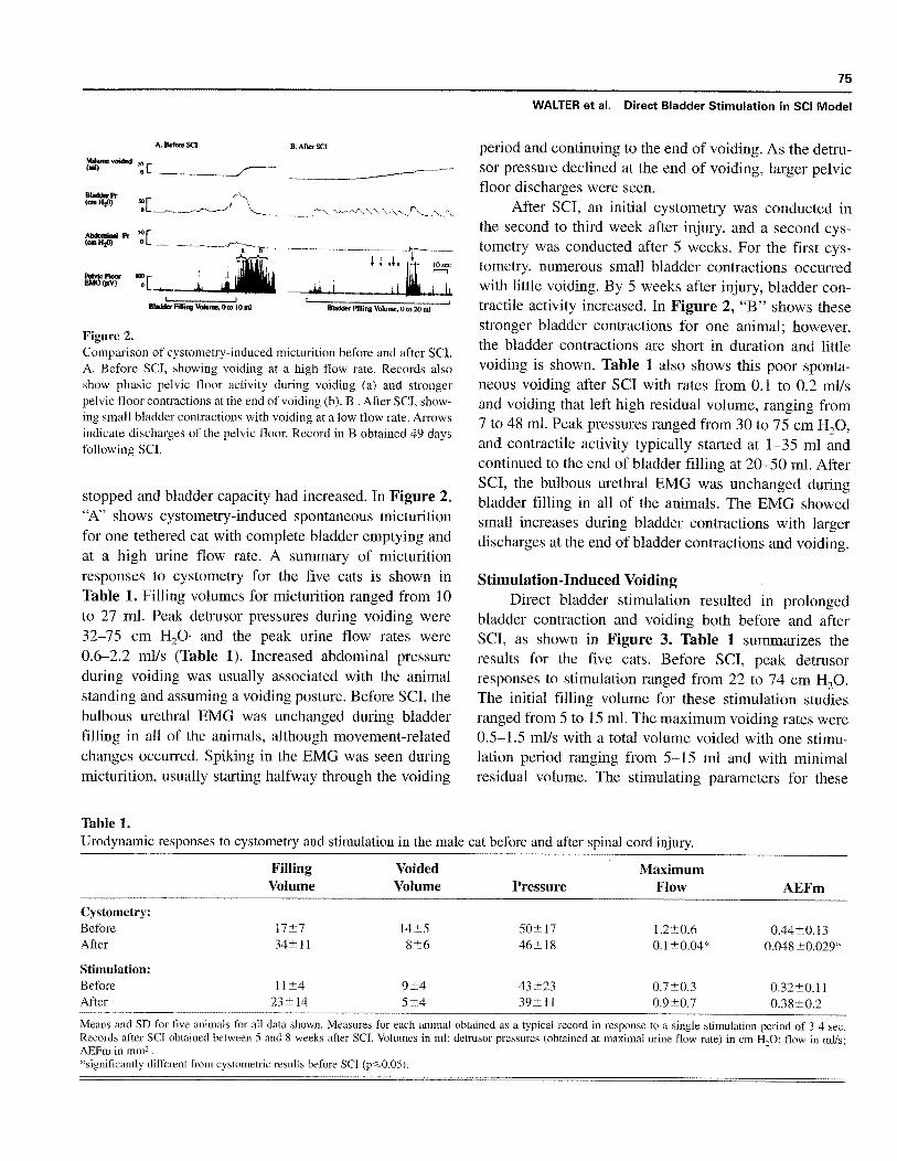
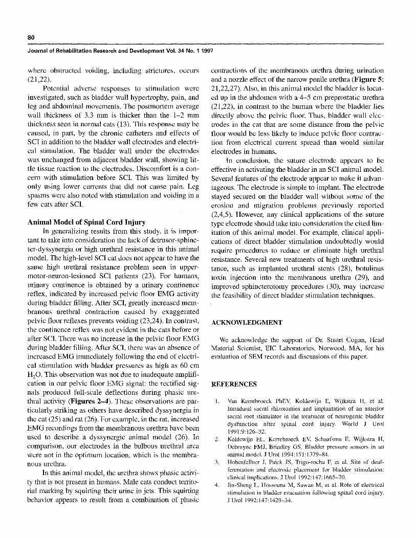
Figure 3.Comparison of stimulation-induced voiding in one cat before and afterSCI. A, Prolonged bladder contraction with 8 ml voided in response to3 s of stimulation before SCI . Phasic pelvic floor contractions areshown during voiding (a) and stronger pelvic floor contractions areseen at the end of voiding (b) . B . Responses to stimulation after SCIwith 7 ml voided . Also shown is phasic activity percent (a), and strongpelvic floor contractions after voiding (b) . Spinal animal 34 dayspostinjury. Note, similar urine flow rates in both records indicated bythe slope of the volume voided record . * EMG not recorded duringelectrical stimulation.
voiding responses were 40 pps, 1 ms pulse duration, 3 strain with a current ranging from 7 .5-40 mA . Before SCI,stimulation that induced strong bladder contractions inthree of the five animals also caused discomfort,However, effective voiding was obtained at lower cur-rents without noticeable discomfort to the animals as isshown in Table 1.
After SCI, three of the cats voided with stimulationin the first 2 weeks, and two of the cats did not respondto stimulation with voiding until the the third week.Average urodynamic responses are again summarized inTable 1 . Maximal responses and stimulation parameterswere similar to before SCI, again the current varied from7.5-40 mA. Filling volumes ranged from 6 40 ml . Peakdetrusor responses were 22 to 74 cm H 2O. The maximumvoiding rates in response to stimulation 5-8 weeks afterSCI ranged from 0.1 to 1 .8 ml/s (Table 1) . After 4-20repeated stimulations, 3 animals completely emptied theirbladders, but 2 animals retained a residual of 6 and 25 ml.These poorer voiding responses at smaller filling vol-umes may have been due to the shorter duration bladder
contractions also seen at these smaller initial volumes(see fluoroscopic observations below) . Another possibleside effect of stimulation is leg movement, and this wasseen in two cats during stimulation, and in two other catsat the end of voiding. An example of this is shown inFigure 3 "B", where a strong discharge in the leg EMGis seen at the ending of voiding.
There was no increase in the EMG record for thefive cats immediately following stimulation either beforeor after SCI (see A and B of Figure 3) . Detrusor pressureswere as high as 60 cm H2O immediately following stim-ulation without increased urethral resistance indicated bythe EMG signal. Spiking or phasic EMG responses wererecorded during voiding and declining bladder pressurestoward the end of voiding . This phasic activity was moreprominent before than after SCI (see A and B of Figure3). At the end of voiding, as bladder pressure declined,large pelvic floor contractions were seen both before andafter SCI.
A urodynamic factor estimating urethral cross-sec-tional area, AEFm, showed that stimulation had littleeffect on urethral resistance (Table 1) . At maximal flow,values for AEFm were not significantly different betweencystometric-induced spontaneous voiding before SCI andstimulation-induced voiding both before and after SCI.Poor spontaneous voiding during cystometry after SCI isreflected in the small AEFm and low urine flow rates . Asshown in Figure 2 "B," the short duration of the bladdercontractions during cystometry after SCI probably con-tributed to this poor voiding response.
Effective direct bladder stimulation techniques weredetermined . The application of negative polarity to theanterior bladder wall electrodes and positive to the poste-rior electrodes was superior in three of five cats bothbefore and after SCI . This was indicated by higher peakdetrusor pressures at lower currents . Stimulation using allfour electrodes resulted in higher peak detrusor pressuresthan any combination of only two electrodes . Also,monopolar electrodes with negative electrodes on thebladder wall and positive electrodes along the backresulted in pain before SCI and in increased abdominalskeletal muscle movement after SCI . Subsequent evalua-tion of stimulating parameters were done using theobserved optimum bipolar electrode arrangement with allof the electrodes on the bladder wall.
The frequency response study was conducted with a1 ms pulse duration, 3-4 s stimulation periods, and at acurrent from 13-25 mA that was not varied in an individ-ual animal . Maximum responses occurred at 40-60 pps in
Volumevoided(ml)
B . After SCI -
a
b
77
WALTER et al . Direct Bladder Stimulation in SCI Model
four animals and 20 pps in the fifth animal ; similarresponses were obtained before and after SCI . For thepulse duration study only three cats were evaluated and300 Rs and 1 ms durations were compared . The 300 pispulse duration induced similar peak detrusor pressures as
s. However, the shorter pulse duration required30-100 percent higher currents . Before SCI, there was noapparent difference in udiycomfort threshold for the ani-mal between the two pulse durations at similar peakguknuorrcopoumem.
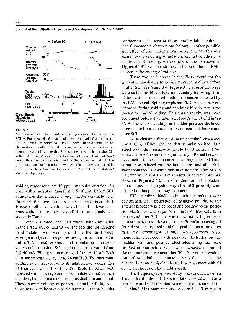
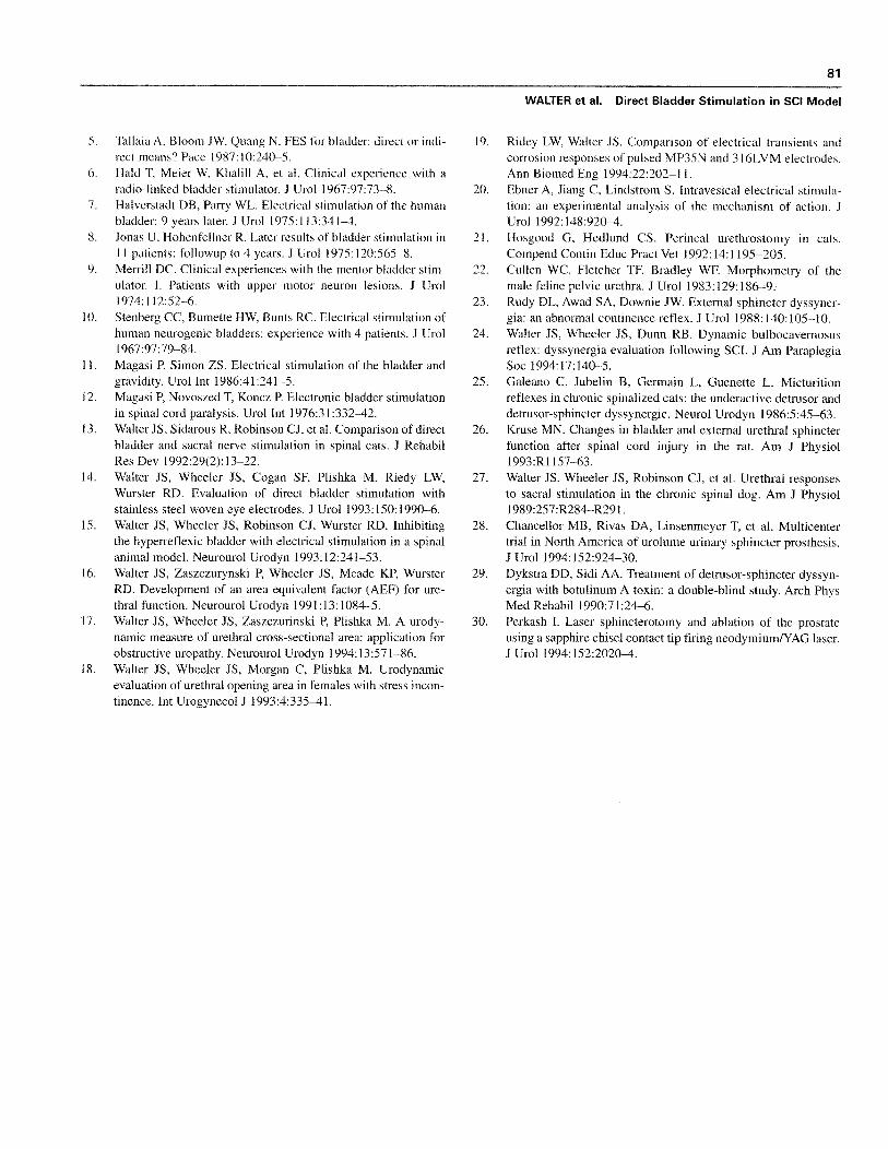
Longer stimulation periods of 20-40 s also inducedbladder contractions with voiding during stimulation inthree of the five animals tested (Figure 4) . For these threeanimals, voiding was induced during stimulation bothbefore and after SCI, at pressures from 40-70 cm H 20,Voiding was induced at 10 and 40 pps and was compara-ble to the 40 ppw, 3-4 s stimulation periods, Responses inthe two remaining animals were more varied . In one ani-mal before SCI, the highest current used was 20 mAwhen the animal indicated slight discomfort, There wasslight abdominal contraction with no bladder contractionand further studies were not conducted. In this same ani-mal, 41 days after SCI the 20 s stimulation period at 25mA induced a bladder contraction of 50 cm H20 with anabdominal pressure of 15 cm H20. The initial volume inthe bladder was approximately, 20 ml and the maximumvolume voided with the single stimulation period was 6ml. The maximal urine flow rate was 0,38 ml/s . A secondcat voided in response to the 20-40 s stimulation with alow flow rate before SCI . After SCI, voiding during this
200
20
40
60
80
10(
Frequency (pps)
Figure 4.Urodynamic responses to 20 s of continuous stimulation showingvoiding during stimulation . A . Before SCI. B . After SCI. * Uw8MGwas turned off during stimulation .
long-term stimulation was not observed in spite of pres-sures as high as 60 cm H20 . Unfortunately, this cat wasnot viewed with fluoroscopy to further explain thisresponse.
Fluoroscopic ObservationsFluoroscopic observations of the urethra during
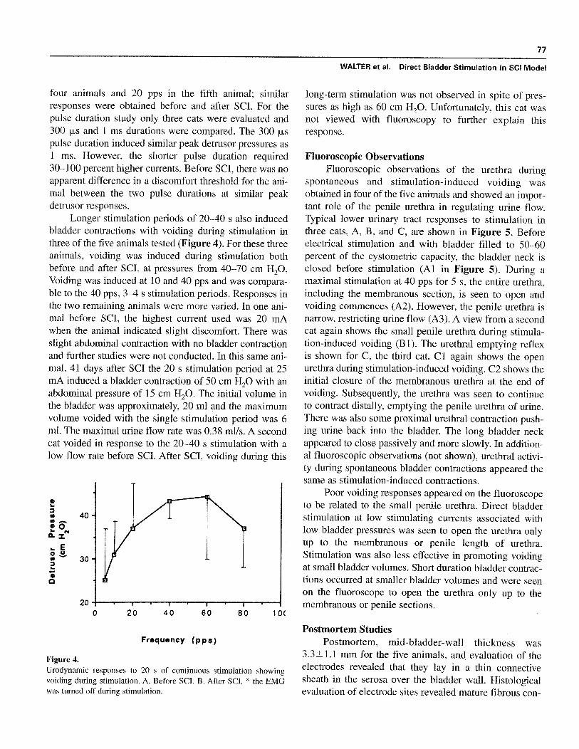
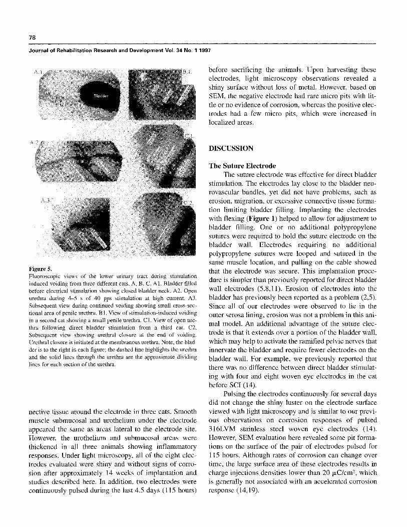
spontaneous and stimulation-induced voiding wasobtained in four of the five animals and showed an impor-tant role of the penile urethra in regulating urine flow.Typical lower urinary tract responses to stimulation inthree cats, A, B, and C, are shown in Figure 5 . BeforecJeutiuol stimulation and with bladder filled to 50-60percent of the cystometric capacity, the bladder neck isclosed before stimulation (Al in Figure 5) . During amaximal stimulation at 40 pps for 5 s, the entire urethra,including the membranous section, is seen to open andvoiding commences (A2) . However, the penile urethra isnarrow, restricting urine flow (A3) . A view from a secondcat again shows the small penile urethra during ubmola-bno-ioduuedvoidiug (Bl) . The urethral emptying reflexis shown for C, the third cat . Cl again shows the openurethra during stimulation-induced voiding. C2 shows theinitial closure of the membranous urethra at the end ofvoiding. Subsequently, the urethra was seen to continueto contract distally, emptying the penile urethra of urine.There was also some proximal urethral contra ption push-ing urine back into the bladder. The long bladder neckappeared to close passively and more slowly. In addition-al fluoroscopic observations (not shown), urethral oukvi-tydnring spontaneous bladder contractions appeared thesame as stimulation-induced contractions.
Poor voiding responses appeared on the fluoroscopeto be related to the small penile urethra . Direct bladderstimulation at low stimulating currents associated withlow bladder pressures was seen to open the urethra onlyup to the membranous or penile length of urethra.Stimulation was also less effective in promoting voidingat small bladder volumes . Short duration bladder contrac-tions occurred at smaller bladder volumes and were seenon the fluoroscope to open the urethra only up to themembranous or penile sections.
Postmortem StudiesPostmortem, mid-bladder-wall thickness was
3 .3 ±! .l mm for the five animals, and evaluation of theelectrodes revealed that they lay in a thin connectivesheath in the serosa over the bladder wall . Histologicalevaluation of electrode sites revealed mature fibrous con-
a
78
Journal of Rehabilitation Research and Development Vol . 34 No. 1 1997
Figure 5.Fluoroscopic views of the lower urinary tract during stimulationinduced voiding from three different cats, A, B, C . Al . Bladder filledbefore electrical stimulation showing closed bladder neck . A2 . Openurethra during 4–5 s of 40 pps stimulation at high current . A3.Subsequent view during continued voiding showing small cross-sec-tional area of penile urethra . B1 . View of stimulation-induced voidingin a second cat showing a small penile urethra. Cl . View of open ure-thra following direct bladder stimulation from a third cat. C2.Subsequent view showing urethral closure at the end of voiding.Urethral closure is initiated at the membranous urethra . Note, the blad-der is to the right in each figure ; the dashed line highlights the urethraand the solid lines through the urethra are the approximate dividinglines for each section of the urethra.
nective tissue around the electrode in three cats . Smoothmuscle submucosal and urothelium under the electrodeappeared the same as areas lateral to the electrode site.However, the urothelium and submucosal areas werethickened in all three animals showing inflammatoryresponses . Under light microscopy, all of the eight elec-trodes evaluated were shiny and without signs of corro-sion after approximately 14 weeks of implantation andstudies described here . In addition, two electrodes werecontinuously pulsed during the last 4.5 days (115 hours)
before sacrificing the animals . Upon harvesting theseelectrodes, light microscopy observations revealed ashiny surface without loss of metal . However, based onSEM, the negative electrode had rare micro pits with lit-tle or no evidence of corrosion, whereas the positive elec-trodes had a few micro pits, which were increased inlocalized areas.
DISCUSSION
The Suture ElectrodeThe suture electrode was effective for direct bladder
stimulation . The electrodes lay close to the bladder neu-rovascular bundles, yet did not have problems, such aserosion, migration, or excessive connective tissue forma-tion limiting bladder filling . Implanting the electrodeswith flexing (Figure 1) helped to allow for adjustment tobladder filling . One or no additional polypropylenesutures were required to hold the suture electrode on thebladder wall . Electrodes requiring no additionalpolypropylene sutures were looped and sutured in thesame muscle location, and pulling on the cable showedthat the electrode was secure . This implantation proce-dure is simpler than previously reported for direct bladderwall electrodes (5,8,11) . Erosion of electrodes into thebladder has previously been reported as a problem (2,5).Since all of our electrodes were observed to lie in theouter serosa lining, erosion was not a problem in this ani-mal model . An additional advantage of the suture elec-trode is that it extends over a portion of the bladder wall,which may help to activate the ramified pelvic nerves thatinnervate the bladder and require fewer electrodes on thebladder wall . For example, we previously reported thatthere was no difference between direct bladder stimulat-ing with four and eight woven eye electrodes in the catbefore SCI (14).
Pulsing the electrodes continuously for several daysdid not change the shiny luster on the electrode surfaceviewed with light microscopy and is similar to our previ-ous observations on corrosion responses of pulsed316LVM stainless steel woven eye electrodes (14).However, SEM evaluation here revealed some pit forma-tions on the surface of the pair of electrodes pulsed for115 hours . Although rates of corrosion can change overtime, the large surface area of these electrodes results incharge injections densities lower than 20 µC/cm 2, whichis generally not associated with an accelerated corrosionresponse (14,19) .
79
WALTER et al . Direct Bladder Stimulation in SCI Model
Effective Stimulation ParametersCurrent results are similar to our previous report
using the woven eye electrode before SCI (14) . Forexample, we confirm that bipolar stimulation with bothelectrodes on the bladder wall is more effective thanmonopolar stimulation with a positive distant electrodeunder the skin, because with monopolar stimulation,abdominal contraction and pain were seen before SCI,and muscle contraction associated with current spread tothe distant electrode after SCI . Bipolar electrodes withpositive and negative electrodes on the bladder wall didnot appear to have these problems . Optimal bipolararrangement in most of the cats had the negative elec-trode anterior, adjacent to the major neural enervation tothe bladder, agreeing with work published by others(3,7,9) . An effective stimulation protocol was capacitor-coupled monophasic pulses using the bipolar electrodeson the bladder wall and stimulating parameters of 40 pps,
300 µs to 1 ms pulse duration and 10–40 mA applied for3 1 s resulting in post-stimulus voiding . Using the opti-mum stimulating parameters, the bladder was emptiedwith a single stimulation period before SCI and, in amajority of the cats, with repeated stimulations after SCI.Bladder reflexes are probably important in all stimulation-induced contractions. The sustained bladder contractionsafter the end of stimulation (Figure 3), both before andafter SCI, indicate a strong reflex component to inducedresponses . Ebner et al . (20) recently showed that directbladder stimulation with intravesical electrodes was medi-ated through sensory nerves and activation of micturitionreflexes . One new result is related to short pulse durations.Previously, we reported that 250 p.s before SCI in the catresulted in pain at currents insufficient to induce peakbladder contractions obtained with longer pulse durations(14) . Since it is advantageous to use short pulse durationsas it reduces the total charge used for stimulation (20), weconducted the pulse duration study here comparing 300p s and 1 ms . Although only three of the five animals wereevaluated, results before SCI showed equally effectivebladder contractions without pain for the two pulse dura-tions. Between 30 and 100 percent higher currents wererequired for the 300 is pulse duration to induce similarpeak pressures as the 1 ms pulse duration . After SCI, therewere similar responses.
Urodynamic Responses of StimulationVoiding in response to direct bladder stimulation was
seen in this study, and we concluded that stimulationbefore and after SCI did not increase urethral resistance
based on four urodynamic measures : 1) the pelvic floorEMG immediately following a 3–4 s stimulation periodwas not increased ; 2) voiding was observed during stimu-lation (Figure 4) ; 3) the urodynamic factor, AEFm,showed no difference in uretheral resistance betweenspontaneous voiding before SCI and stimulation-inducedvoiding before or after SCI (Table 1) ; 4) fluoroscopy ofthe urethra during stimulation-induced voiding showed anopen membranous urethra, and urethral mechanicsappeared the same with spontaneous and stimulation-induced voiding . However, stimulation did not alwaysinduce voiding in spite of high bladder pressures . Atsmaller bladder volumes, stimulation-induced voidingwas reduced or absent. This was seen in the relativelylarge number of stimulations required to empty the blad-der after SCI and also, in some cats, residual urine afterrepeated stimulations . This poor voiding response atsmaller filling volumes was probably due to short durationbladder contractions . For example, after SCI, cystometric-induced spontaneous voiding was poor, reflected in lowAEFm values (Table 1), and this poor voiding was asso-ciated with short duration bladder contractions (Figure 2,`B"). Fluoroscopy of the urethra also revealed that thesmall penile urethra or membranous urethra did not openin some animals during the short bladder contractions atsmall filling volumes . A second observation of poor void-ing was seen in some animals for the first few weeks fol-lowing SCI. Voiding was not seen during stimulation orspontaneous bladder contractions during this early period.This time-dependent effect may have been a result ofincreased intrinsic urethral resistance in the early spinalcats . For example, it is difficult to empty the bladder of thespinal cats using suprapubic pressure early but not lateafter SCI (unpublished observations) . A third observationof poor voiding responses was with the 20–40 s of contin-uous stimulation in one of the cats before and another afterSCI. For the animal before SCI, stimulation induced lowbladder pressures (<30 cm H 2O) probably insufficient toovercome urethral resistance . For the animal after SCI,fluoroscopic observations were not conducted in this ani-mal to provide any further explanation.
In this animal model, we observed that the smallpenile urethra was a primary factor regulating urine flow.The small penile urethra as observed with fluoroscopy(Figure 5) would contribute to the high voiding pressuresthat were recorded in this study. Both stimulation-drivenand spontaneous voiding before and after SCI had highbladder pressures ranging from 22–74 cm H2 O (Table 1).The penile urethra in the male cat is well known as a site
80
Journal of Rehabilitation Research and Development Vol . 34 No . 1 1997
Animal Model of Spinal Cord InjuryIn generalizing results from this study, it is impor-
tant to take into consideration the lack of detrusor-sphinc-ter-dyssynergia or high urethral resistance in this animalmodel . The high-level SCI cat does not appear to have thesame high urethral resistance problem seen in upper-motor-neuron-lesioned SCI patients (23) . For humans,urinary continence is obtained by a urinary continencereflex, indicated by increased pelvic floor EMG activityduring bladder filling . After SCI, greatly increased mem-branous urethral contraction caused by exaggeratedpelvic floor reflexes prevents voiding (23,24) . In contrast,the continence reflex was not evident in the cats before orafter SCI . There was no increase in the pelvic floor EMG
We acknowledge the support of Dr. Stuart Cogan, Headduring bladder filling . After SCI, there was an absence of
Material Scientist, EIC Laboratories, Norwood, MA, for hisincreased EMG immediately following the end of electri-
evaluation of SEM records and discussions of this paper.cal stimulation with bladder pressures as high as 60 cmH2 O. This observation was not due to inadequate amplifi-cation in our pelvic floor EMG signal : the rectified sig-nals produced full-scale deflections during phasic ure-thral activity (Figures 2–4) . These observations are par-ticularly striking as others have described dyssynergia inthe cat (25) and rat (26) . For example, in the rat, increasedEMG recordings from the membranous urethra have beenused to describe a dyssynergic animal model (26) . Incomparison, our electrodes in the bulbous urethral areawere not in the optimum location, which is the membra-nous urethra.
In this animal model, the urethra shows phasic activi-ty that is not present in humans . Male cats conduct territo-rial marking by squirting their urine in jets . This squirtingbehavior appears to result from a combination of phasic
where obstructed voiding, including strictures, occurs(21,22).
Potential adverse responses to stimulation wereinvestigated, such as bladder wall hypertrophy, pain, andleg and abdominal movements . The postmortem averagewall thickness of 3 .3 mm is thicker than the 1–2 mmthickness seen in normal cats (13) . This response may becaused, in part, by the chronic catheters and effects ofSCI in addition to the bladder wall electrodes and electri-cal stimulation . The bladder wall under the electrodeswas unchanged from adjacent bladder wall, showing lit-tle tissue reaction to the electrodes . Discomfort is a con-cern with stimulation before SCI . This was limited byonly using lower currents that did not cause pain . Legspasms were also noted with stimulation and voiding in afew cats after SCI .
contractions of the membranous urethra during urinationand a nozzle effect of the narrow penile urethra (Figure 5;21,22,27) . Also, in this animal model the bladder is locat-ed up in the abdomen with a 4–5 cm preprostatic urethra(21,22), in contrast to the human where the bladder liesdirectly above the pelvic floor. Thus, bladder wall elec-trodes in the cat that are some distance from the pelvicfloor would be less likely to induce pelvic floor contrac-tion from electrical current spread than would similarelectrodes in humans.
In conclusion, the suture electrode appears to beeffective in activating the bladder in an SCI animal model.Several features of the electrode appear to make it advan-tageous . The electrode is simple to implant . The electrodestayed secured on the bladder wall without some of theerosion and migration problems previously reported(2,4,5) . However, any clinical applications of the suturetype electrode should take into consideration the cited lim-itation of this animal model . For example, clinical appli-cations of direct bladder stimulation undoubtedly wouldrequire procedures to reduce or eliminate high urethralresistance. Several new treatments of high urethral resis-tance, such as implanted urethral stents (28), botulinustoxin injection into the membranous urethra (29), andimproved sphincterotomy procedures (30), may increasethe feasibility of direct bladder stimulation techniques.
ACKNOWLEDGMENT
REFERENCES
1. Van Kerrebroeck PhEV, Koldewijn E, Wijkstra H, et al.Intradural sacral rhizotomies and implantation of an anteriorsacral root stimulator in the treatment of neurogenic bladderdysfunction after spinal cord injury. World J Urol1991 :9 :126-32.
2. Koldewijn EL, Kerrebroeck EV, Schaafsma E, Wijkstra H,Debruyne FMJ, Brindley GS . Bladder pressure sensors in ananimal model . J Urol 1994 :151 :1379-84.
3. Hohenfellner J, Paick JS, Trigo-rocha F, et al . Site of deaf-ferentation and electrode placement for bladder stimulation:clinical implications . J Urol 1992:147 :1665-70.
4. Jin-Sheng L, Hossouna M, Sawan M, et al . Role of electricalstimulation in bladder evacuation following spinal cord injury.J Urol 1992 :147 :1429-34 .
81
WALTER et al . Direct Bladder Stimulation in SCI Model
5. Tallaia A, Bloom JW, Quang N . FES for bladder : direct or indi-rect means? Pace 1987 :10 :240-5.
6.
Hald T, Meier W, Khalill A, et al . Clinical experience with aradio-linked bladder stimulator. J Urol 1967 :97 :73-8.
7. Halverstadt DB, Parry WL. Electrical stimulation of the humanbladder: 9 years later. J Urn] 1975 :113 :341-4.
8.
Jonas U, Hohenfellner R . Later results of bladder stimulation in11 patients : followup to 4 years . J Urol 1975 :120 :565-8.
9. Merrill DC. Clinical experiences with the mentor bladder stim-ulator. I . Patients with upper motor neuron lesions . J Urol1974 :112 :52-6.
10. Stenberg CC, Bumette HW, Bunts RC . Electrical stimulation ofhuman neurogenic bladders : experience with 4 patients . J Urol1967 :97 :79-84.
11. Magasi P, Simon ZS . Electrical stimulation of the bladder andgravidity . Urol Int 1986 :41 :241-5.
12. Magasi P, Novoszed T, Koncz P. Electronic bladder stimulationin spinal cord paralysis . Urol Int 1976 :31 :332-42.
13. Walter JS, Sidarous R, Robinson CJ, et al. Comparison of directbladder and sacral nerve stimulation in spinal cats . J RehabilRes Dev 1992 :29(2) :13-22.
14. Walter JS, Wheeler JS, Cogan SF, Plishka M, Riedy LW,Wurster RD . Evaluation of direct bladder stimulation withstainless steel woven eye electrodes . J Urol 1993 :150:1990-6.
15. Walter IS, Wheeler JS, Robinson CJ, Wurster RD . Inhibitingthe hyperreflexic bladder with electrical stimulation in a spinalanimal model . Neurourol Urodyn 1993 :12 :241-53.
16. Walter JS, Zaszczurynski P, Wheeler JS, Meade KP, WursterRD . Development of an area equivalent factor (AEF) for ure-thral function . Neurourol Urodyn 1991 :13 :1084-5.
17. Walter JS, Wheeler JS, Zaszczurinski P, Plishka M . A urody-namic measure of urethral cross-sectional area : application forobstructive uropathy . Neurourol Urodyn 1994 :13 :571-86.
18. Walter JS, Wheeler JS, Morgan C, Plishka M. Urodynamicevaluation of urethral opening area in females with stress incon-tinence . Int Urogynecol J 1993 :4 :335-41 .
19. Ridey LW, Walter JS. Comparison of electrical transients andcorrosion responses of pulsed MP35N and 316LVM electrodes.Ann Biomed Eng1994 :22 :202-11.
20. Ebner A, Jiang C, Lindstrom S . Intravesical electrical stimula-tion : an experimental analysis of the mechanism of action . JUrol 1992 :148 :920-4.
21. Hosgood G, Hedlund CS. Perineal urethrostomy in cats.Compend Contin Educ Pract Vet 1992 :14 :1195-205.
22. Cullen WC, Fletcher TF, Bradley WF. Morphometry of themale feline pelvic urethra . J Urol 1983:129:186-9.
23. Rudy DL, Awad SA, Downie JW. External sphincter dyssyner-gia : an abnormal continence reflex . J Urol 1988:140 :105-10.
24. Walter JS, Wheeler JS, Dunn RB . Dynamic bulbocavernosusreflex: dyssynergia evaluation following SCI . J Am ParaplegiaSoc 1994 :17 :140-5.
25. Galeano C, Jubelin B, Germain L, Guenette L . Micturitionreflexes in chronic spinalized cats : the underactive detrusor anddetrusor-sphincter dyssynergic . Neurol Urodyn 1986 :5 :45-63.
26. Kruse MN. Changes in bladder and external urethral sphincterfunction after spinal cord injury in the rat . Am J Physiol1993 :R1157-63.
27. Walter JS, Wheeler JS, Robinson CJ, et al . Urethral responsesto sacral stimulation in the chronic spinal dog . Am J Physiol1989:257 :R284-R291.
28. Chancellor MB, Rivas DA, Linsenmeyer T, et al . Multicentertrial in North America of urolume urinary sphincter prosthesis.J Urol 1994 :152 :924-30.
29. Dykstra DD, Sidi AA . Treatment of detrusor-sphincter dyssyn-ergia with botulinum A toxin : a double-blind study. Arch PhysMed Rehabil 1990 :71 :24-6.
30. Perkash I. Laser sphincterotomy and ablation of the prostateusing a sapphire chisel contact tip firing neodymiumlYAG laser.J Urol 1994 :152 :2020-4 .