Embed Size (px)
Citation preview
Discover the Bone Healing Power of P-15 Osteogenic Cell Binding Peptide
A Powerful Cell Attachment Factor Backed by Level 1 Human Clinical Data
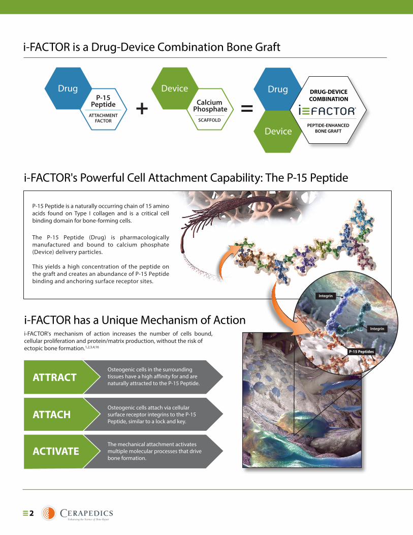
i-FACTOR's Powerful Cell Attachment Capability: The P-15 Peptide
i-FACTOR has a Unique Mechanism of Action
P-15 Peptide is a naturally occurring chain of 15 amino acids found on Type I collagen and is a critical cell binding domain for bone-forming cells.
The P-15 Peptide (Drug) is pharmacologically manufactured and bound to calcium phosphate (Device) delivery particles.
This yields a high concentration of the peptide on the graft and creates an abundance of P-15 Peptide binding and anchoring surface receptor sites.
2
i-FACTOR is a Drug-Device Combination Bone Graft
ATTRACT
ACTIVATE
Osteogenic cells in the surrounding tissues have a high affinity for and are naturally attracted to the P-15 Peptide.
Osteogenic cells attach via cellular surface receptor integrins to the P-15 Peptide, similar to a lock and key.
The mechanical attachment activates multiple molecular processes that drive bone formation.
ATTACH
Drug DeviceP-15
Peptide
ATTACHMENT FACTOR
Device
Drug
+ =DRUG-DEVICE COMBINATION
PEPTIDE-ENHANCED BONE GRAFT
Calcium Phosphate
SCAFFOLD
i-FACTOR's mechanism of action increases the number of cells bound, cellular proliferation and protein/matrix production, without the risk ofectopic bone formation.1,2,3,4,16
Integrin
P-15 Peptides
Integrin

Only 2 FDA Class III PMA Approved Spinal Bone Grafts
There are three FDA regulatory pathways for orthobiologics: Class III, Class II 510k, and HCT/P (tissue based products). Class III devices have the most rigorous pathway requiring a PMA Approved Level 1 Investigational Device Exemption (IDE) human clinical trial to bring the products to market.
i-FACTOR has met the most robust FDA study requirements and is only the second Class III Drug-Device Combination bone graft approved for the US Spine market. The only other spinal bone graft in this category is Medtronic's InfuseTM (BMP-2). The majority of bone grafts on the market are cleared via the 510k pathway.
400+
2
>10
CLASS III2 PRODUCTS
CLASS II400+ PRODUCTS
HCT/Ps>10 PRODUCTS
IDE - LARGE HUMAN CLINICAL STUDYPMA APPROVAL - DEMONSTRATE SAFETY & EFFICACY IN HUMANS
510k - ANIMAL DATASUBSTANTIALLY EQUIVALENT TO PREDICATE DEVICE
NO FDA REVIEWNO ANIMAL OR HUMAN TESTING REQUIRED
3
Physicians are encouraged to find the highest level of evidence to support the safe and effective use of a product in a clinical setting. Evidence-based research studies can range anywhere between Level 1 to Level 6. i-FACTOR has published Level 1 human clinical evidence whereas the majority of 510k bone grafts on the market have lower level studies.
i-FACTOR has Level 1 Human Clinical Evidence
Level 1 Randomized controlled human clinical trial
Level 2 Prospective cohort study
Level 3 Retrospective cohort study
Level 4 Case studies
Level 5 Mechanism-based reasoning
Level 6 Animal studies, in vitro studies
Number of Studies
Fewer High Level Studies (Increased Cost and Quality)
Many Low Level Studies (Lower Cost and Quality)
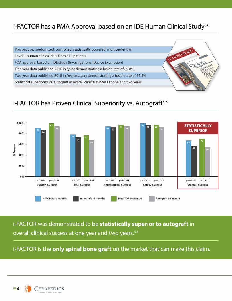
i-FACTOR has a PMA Approval based on an IDE Human Clinical Study5,6
Prospective, randomized, controlled, statistically powered, multicenter trial
Level 1 human clinical data from 319 patients
FDA approval based on IDE study (Investigational Device Exemption)
One year data published 2016 in Spine demonstrating a fusion rate of 89.0%
Two year data published 2018 in Neurosurgery demonstrating a fusion rate of 97.3%
Statistical superiority vs. autograft in overall clinical success at one and two years
i-FACTOR was demonstrated to be statistically superior to autograft in overall clinical success at one year and two years.5,6 i-FACTOR is the only spinal bone graft on the market that can make this claim.
i-FACTOR has Proven Clinical Superiority vs. Autograft5,6
100%
p= 0.4220
Fusion Success
i-FACTOR 12 months Autograft 12 months i-FACTOR 24 months Autograft 24 months
NDI Success Neurological Success Safety Success Overall Success
p= 0.2907 p= 0.8123 p= 0.3085 p= 0.0382p= 0.2195 p= 0.1804 p= 0.6944 p= 0.1379 p= 0.0302
80%
60%
40%
20%
0%
89
79
949897
77
95 95
86
74
9395
94
69 6970
94 91
57 56
STATISTICALLY SUPERIOR
4
% S
ucce
ss
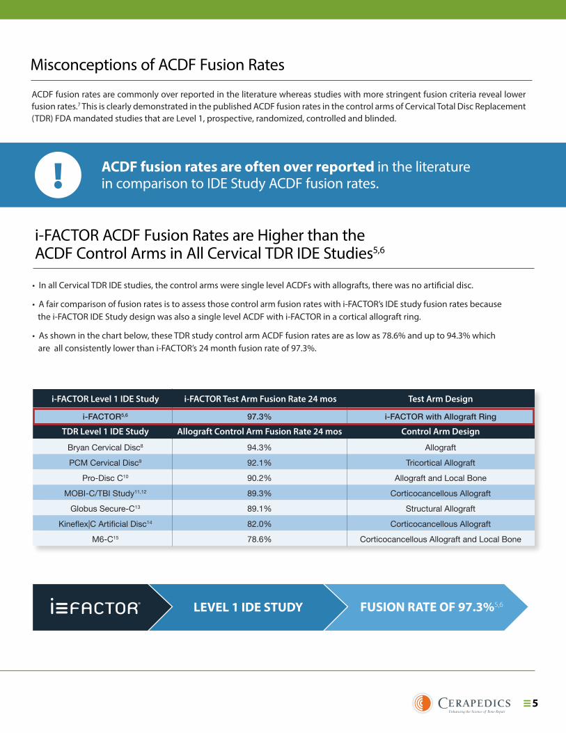
Misconceptions of ACDF Fusion Rates
5
i-FACTOR Level 1 IDE Study i-FACTOR Test Arm Fusion Rate 24 mos Test Arm Design
i-FACTOR5,6 97.3% i-FACTOR with Allograft Ring
TDR Level 1 IDE Study Allograft Control Arm Fusion Rate 24 mos Control Arm Design
Bryan Cervical Disc8 94.3% Allograft
PCM Cervical Disc9 92.1% Tricortical Allograft
Pro-Disc C10 90.2% Allograft and Local Bone
MOBI-C/TBI Study11,12 89.3% Corticocancellous Allograft
Globus Secure-C13 89.1% Structural Allograft
Kineflex|C Artificial Disc14 82.0% Corticocancellous Allograft
M6-C15 78.6% Corticocancellous Allograft and Local Bone
ACDF fusion rates are commonly over reported in the literature whereas studies with more stringent fusion criteria reveal lower fusion rates.7 This is clearly demonstrated in the published ACDF fusion rates in the control arms of Cervical Total Disc Replacement (TDR) FDA mandated studies that are Level 1, prospective, randomized, controlled and blinded.
ACDF fusion rates are often over reported in the literature in comparison to IDE Study ACDF fusion rates.!
LEVEL 1 IDE STUDY FUSION RATE OF 97.3%5,6
i-FACTOR ACDF Fusion Rates are Higher than the ACDF Control Arms in All Cervical TDR IDE Studies5,6
• In all Cervical TDR IDE studies, the control arms were single level ACDFs with allografts, there was no artificial disc.
• A fair comparison of fusion rates is to assess those control arm fusion rates with i-FACTOR’s IDE study fusion rates because the i-FACTOR IDE Study design was also a single level ACDF with i-FACTOR in a cortical allograft ring.
• As shown in the chart below, these TDR study control arm ACDF fusion rates are as low as 78.6% and up to 94.3% which are all consistently lower than i-FACTOR’s 24 month fusion rate of 97.3%.
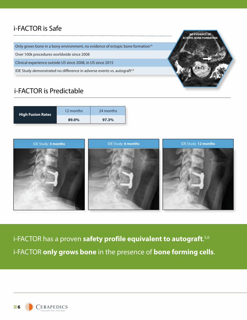
Only grows bone in a bony environment, no evidence of ectopic bone formation16
Over 100k procedures worldwide since 2008
Clinical experience outside US since 2008, in US since 2015
IDE Study demonstrated no difference in adverse events vs. autograft5,6
i-FACTOR is Safe
i-FACTOR is Predictable
i-FACTOR has a proven safety profile equivalent to autograft.5,6
i-FACTOR only grows bone in the presence of bone forming cells.
6
IDE Study: 3 months IDE Study: 6 months IDE Study: 12 months
High Fusion Rates
12 months 24 months
89.0% 97.3%
NO EVIDENCE OFECTOPIC BONE FORMATION
NO EVIDENCE OFECTOPIC BONE FORMATION
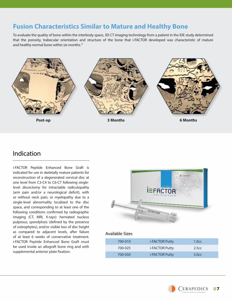
Fusion Characteristics Similar to Mature and Healthy Bone
Post-op 3 Months 6 Months
To evaluate the quality of bone within the interbody space, 3D CT imaging technology from a patient in the IDE study determined that the porosity, trabecular orientation and structure of the bone that i-FACTOR developed was characteristic of mature and healthy normal bone within six months.17
i-FACTOR Peptide Enhanced Bone Graft is indicated for use in skeletally mature patients for reconstruction of a degenerated cervical disc at one level from C3-C4 to C6-C7 following single-level discectomy for intractable radiculopathy (arm pain and/or a neurological deficit), with or without neck pain, or myelopathy due to a single-level abnormality localized to the disc space, and corresponding to at least one of the following conditions confirmed by radiographic imaging (CT, MRI, X-rays): herniated nucleus pulposus, spondylosis (defined by the presence of osteophytes), and/or visible loss of disc height as compared to adjacent levels, after failure of at least 6 weeks of conservative treatment. i-FACTOR Peptide Enhanced Bone Graft must be used inside an allograft bone ring and with supplemental anterior plate fixation.
Available Sizes
700-010 i-FACTOR Putty 1.0cc
700-025 i-FACTOR Putty 2.5cc
700-050 i-FACTOR Putty 5.0cc
Indication
7
Corporate Headquarters11025 Dover Street, Suite 1600 Westminster, CO 80021 USA P: (303) 974-6275 E: [email protected]
www.cerapedics.com
Europe, Middle East, Africa, Asia Pacific and CanadaLondon, England E: [email protected]
2797 Internationally available
References
1. Hennessy KM, Pollot BE, Clem WC, Phipps MC, Sawyer AA, Culpepper BK, Bellis SL. The effect of collagen 1 mimetic peptides on mesenchymal stem cells adhesion and differentiation, and on bone formation at hydroxyapatite surfaces. Biomaterials. 2009 Apr; 30(10): 1898-909.
2. Nguyen H, Qian JJ, Bhatnagar RS, Li S. Enhanced cell attachment and osteoblastic activity by P-15 peptide-coated matrix in hydrogels. Biochem Biophys Res Commun. 2003 Nov 7;311(1):179-86.3. Yang XB, Bhatnagar RS, Li S, Oreffo RO. Biomimetic collagen scaffolds for human bone cell growth and differentiation. Tissue Eng. 2004 Jul-Aug;10(7-8):1148-59.4. Qian JJ, Bhatnagar RS. Enhanced cell attachment to anorganic bone mineral in the presence of a synthetic peptide related to collagen. J Biomed Mater Res. 1996 Aug;31(4):545-54.5. Arnold PM, Sasso RC, Janssen ME, Fehlings MG, Smucker JD, Vaccaro AR, Heary RF, Patel AL, Goulet B, Kalfas IH, Kopjar B. Efficacy of i-Factor™ Bone Graft versus Autograft in Anterior Cervical Disectomy and Fusion.
Results of the Prospective Randomized Single-blinded Food and Drug Administration Investigational Device Exemption Study. Spine. 2016; 41(13): 1075-1083.6. Arnold PM, Sasso RC, Janssen ME, Fehlings MG, Heary RF, Vaccaro AR, Kopjar B. i-FACTOR Bone Graft vs Autograft in Anterior Cervical Discectomy and Fusion: 2-Year Follow-up of the Randomized Single-Blinded Food
and Drug Administration Investigational Device Exemption Study. (2018) Neurosurgery; Vol-83(3): pages 377-384.7. Sethi N1, Devney J, Steiner HL, Riew KD. Diagnosing cervical fusion: a comprehensive literature review. Asian Spine Journal. 2008 Dec;2(2):127-43.8. Heller JG, et al. Comparison of BRYAN cervical disc arthroplasty with anterior cervical decompression and fusion: clinical and radiographic results of a randomized, controlled, clinical trial. Spine. 2009 Jan 15; 34(2): 101-7. 9. Phillips FM, et al. A Prospective, Randomized, Controlled Clinical Investigation Comparing PCM Cervical Disc Arthroplasty with Anterior Cervical Discectomy and Fusion: 2-Year Results from the US FDA IDE Clinical Trial.
Spine. 2013 Jul; 38(15): E907-18. 10. Murrey D, et al. Results of the prospective, randomized, controlled multicenter Food and Drug Administration investigational device exemption study of the ProDisc-C total disc replacement versus anterior discectomy
and fusion for the treatment of 1-level symptomatic cervical disc disease. Spine J. 2009 Apr; 9(4): 275-86. 11. Hisey MS, Bae HW, Davis R, Gaede S, Hoffman G, Kim K, et al. Multi-center, prospective, randomized, controlled investigational device exemption clinical trial comparing Mobi-C Cervical Artificial Disc to anterior
discectomy and fusion in the treatment of symptomatic degenerative disc disease in the cervical spine. Int J Spine Surg. 2014; 8. 12. Zigler JE, Rogers RW, Ohnmeiss DD, Comparison of 1-Level Versus 2-Level Anterior Cervical Discectomy and Fusion: Clinical and Radiographic Follow-Up at 60 Months. Spine. 2016 March; 41(6): 463-9. 13. Vaccaro A, et al. Clinical outcomes with selectively constrained SECURE-C cervical disc arthroplasty: two-year results from a prospective, randomized, controlled, multicenter investigational device exemption study.
Spine. 2013 Dec 15; 38(26): 2227-39. 14. Coric D, Nunley PD, Guyer RD, Musante D, Carmody CN, Gordon CR, et al. Prospective, randomized, multicenter study of cervical arthroplasty: 269 patients from the Kineflex|C artificial disc investigational device
exemption study with a minimum 2-year follow-up. Journal of Neurosurgery: Spine. 2011 Oct; 15(4): 348-58. 15. PMA P170036: FDA Summary of Safety and Effectiveness Data www.accessdata.fda.gov/cdrh_docs/pdf17/P170036B.pdf. 16. Internal Data Report. 17. Kesteloot G, Parish AJB, Johnson S, McNally DS. Three Dimensional remodelling of i-FACTOR Peptide Enhanced Bone Graft substitute in cervical fusions. Accepted as poster, Annual Scientific Meeting of the Belgian
Society of Neurosurgery, March 2016.
Cerapedics is an advanced orthobiologics company focused on developing and commercializing its proprietary small peptide (P-15) technology platform. i-FACTOR® Peptide Enhanced Bone Graft is the only biologic bone graft in orthopaedics that incorporates P-15 osteogenic cell binding peptide to stimulate the natural bone healing process. This novel mechanism of action is designed to support safer and more predictable bone formation compared to commercially available bone growth factors.
© 2020 Cerapedics, Inc. All rights reserved.
ML-0594 06/20