Embed Size (px)
Citation preview
Discovery of a Novel Broad-spectrum Antifungal Agent, Derived
from Albaconazole
Rémi Guillon,† Fabrice Pagniez,*
,‡ Carine Picot,
‡ Damien Hedou,
§ Alain Tonnerre,
† Muriel
Duflos,† Elizabeth Chosson,
§ Thierry Besson,
§ Cédric Logé,*
,† and Patrice Le Pape
‡
† Université de Nantes, Nantes Atlantique Universités, Laboratoire de Chimie Thérapeutique, Cibles et
Médicaments des Infections et du Cancer, IICIMED-EA 1155, UFR Sciences Pharmaceutiques, 1 rue Gaston
Veil, Nantes F-44035 Cedex 1, France ‡ Université de Nantes, Nantes Atlantique Universités, Laboratoire de Parasitologie et Mycologie Médicale,
Cibles et Médicaments des Infections et du Cancer, IICIMED-EA 1155, UFR Sciences Pharmaceutiques, 1 rue
Gaston Veil, Nantes F-44035 Cedex 1, France § Université de Rouen, Laboratoire C.O.B.R.A., CNRS UMR 6014 & FR 3038, Institut de Recherche en Chimie
Organique Fine (I.R.C.O.F.), rue Tesnière, 76130 Mont Saint-Aignan, France
Experimental Procedures
Chemistry
General methods
Melting points were determined using an Electrothermal IA9300 digital melting point
apparatus and reported uncorrected. 1H and 13C NMR spectra were recorded on a Bruker
Bruker Avance 400 spectrometer (400 MHz). Chemical shifts are expressed as δ values (ppm)
relative to tetramethylsilane as internal standard (in NMR description, s = singlet, d = doublet,
t = triplet, q = quadruplet, sext = sextuplet, m = multiplet and b = broad). Coupling constants
J are given in Hertz. IR spectra were obtained in KBr pellets using a Perkin-Elmer Paragon
FTIR 1000 PC spectrometer. Only the most significant absorption bands have been reported.
Mass spectra analysis was performed by the Mass Spectrometry Laboratory of the University
of Rouen. Mass spectra (EI) were recorded with a Waters LCP 1er
XR spectrometer. The
optical rotations were recorded with a polarimeter Schmidt-Haensch Polartronic NH8. All
reactions were monitored by thin-layer chromatography (TLC) using 0.2 mm-thick silica gel
plates 60F-254 (5735 Merck). Column chromatography was carried out using silica gel 60
(70-230 Mesh, ASTM, Merck). Chemicals and solvents used were commercially available.
Optically oxirane (2R,3S) 15 and albaconazole were synthesized according to protocols
described in Ref. 17 and 3, respectively (for compound 15: [α]20
D = -9.0 (c = 1.0 in MeOH)
(lit: [α]20
D = -8.3), for albaconazole: [α]20
D = -8.0 (c = 1.0 in CHCl3) (lit: [α]20
D = -8.3)).
Focused microwave irradiations were carried out with a CEM DiscoverTM
focused microwave
reactor (300W, 2455 MHz, monomode system).
7-bromoquinazolin-4(3H)-one (2)
4-Bromo-2-nitrobenzoic acid (1g, 4.06 mmol, 1), formamide (6.44 mL, 162.14 mmol) and
indium chloride (900 mg, 4.06 mmol) were introduced under argon into a 50 mL round-
bottomed flask equipped with a condenser and a magnetic stirring bar. The flask was placed
in the microwave cavity and exposed to microwave irradiation under argon for 40 min at
150°C using irradiation power of 80 W. After cooling, mixture was diluted with water and
product was extracted with ethyl acetate. Organic layers were dried over anhydrous Na2SO4
and concentrated in vacuo. The residue was purified on silica gel column chromatography
(dichloromethane/ethanol, 99:1) and compound 2 was obtained in a 81% yield as a white
solid. Mp 259-260 °C; 1H NMR (400 MHz, DMSO-d6): δ 7.69 (dd, 1H,
3J = 8.4 Hz,
4J = 1.6
Hz), 7.88 (d, 1H, 4J = 1.6 Hz), 8.03 (d, 1H,
3J = 8.4 Hz), 8,13 (s, 1H), 12.41 (bs, 1H); IR (KBr
cm-1
): 858, 1239, 1609, 1668, 3050, 3466; HRMS calcd for C8H679
BrN2O [M+H]+ 224.9663
found 224.9665, for C8H681
BrN2O [M+H]+
226.9643 found 226.9651.
7-bromo-6-nitroquinazolin-4(3H)-one (3)
To a stirred solution of 2 (1.47 g, 6.53 mmol) in concentrated sulfuric acid (23 mL) was added
fuming nitric acid (0.552 mL, 13.06 mmol) and the solution was stirred at 100°C for 1 h.
After cooling, mixture was poured into ice water and neutralized with ammonia. Product was
extracted with ethyl acetate, dried over anhydrous Na2SO4 and concentrated in vacuo. The
residue was purified on silica gel column chromatography (dichloromethane/ethanol, 99:1)
and compound 3 was obtained in a 76% yield as a pale yellow solid. Mp 313-314 °C; 1H
NMR (400 MHz, DMSO-d6): δ 8.17 (s, 1H), 8.30 (s, 1H), 8.62 (s, 1H), 12.78 (bs, 1H); IR
(KBr cm-1
): 810, 1249, 1332, 1518, 1605, 1656, 1695, 3082, 3504; HRMS calcd for
C8H379
BrN3O3 [M-H]- 267.9358 found 267.9355, for C8H3
81BrN3O3 [M-H]
- 269.9337 found
269.9327.
3-benzyl-7-bromo-6-nitroquinazolin-4(3H)-one (4)
Compound 3 (1.335 g, 4.94 mmol), DMF (7.4 mL), sodium hydride (60% dispersion in
mineral oil) (237 mg, 5.93 mmol) and benzyl bromide (0.588 mL, 4.94 mmol) were
successively introduced into a 50 mL round-bottomed flask equipped with a condenser and a
magnetic stirring bar. The flask was placed in the microwave cavity and exposed to
microwave irradiation for 30 min at 80°C using irradiation power of 40 W. The solvent was
removed under reduced pressure, and the residue was hydrolyzed with water and extracted
with ethyl acetate. Organic layers were dried over anhydrous Na2SO4 and concentrated in
vacuo. The residue was purified on silica gel column chromatography (dichloromethane) and
compound 4 was obtained in a 84% yield as a white solid. Mp 169-170 °C; 1H NMR (400
MHz, DMSO-d6): δ 5.25 (s, 2H), 7.33-7.43 (m, 5H), 8.25 (s, 1H), 8.70 (s, 1H), 8.83 (s, 1H);
IR (KBr cm-1
): 813, 1216, 1350, 1532, 1602, 1679, 3082; HRMS calcd for C15H1179
BrN3O3
[M+H]+ 359.9984 found 359.9983.
6-amino-3-benzyl-7-bromoquinazolin-4(3H)-one (5)
To a stirred solution of 4 (200 mg, 0.55 mmol) in ethanol (1.5 mL) and acetic acid (1.5 mL)
was added iron powder (124 mg, 13.06 mmol) and the solution was stirred under reflux for 1
h. After cooling, mixture was poured into ice water and neutralized with NaOH (5M). Product
was extracted with ethyl acetate, dried over anhydrous Na2SO4 and concentrated in vacuo.
The residue was purified on silica gel column chromatography (dichloromethane/ethanol,
99.5:0.5) and compound 5 was obtained in a 93% yield as a white solid. Mp 166-167 °C; 1H
NMR (400 MHz, DMSO-d6): δ 5.18 (s, 2H), 5.91 (bs, 2H), 7.32-7.39 (m, 5H), 7.49 (s, 1H),
7.81 (s, 1H), 8,31 (s, 1H); IR (KBr cm-1
): 836, 1213, 1483, 1595, 1621, 1669, 3020, 3332,
3453; HRMS calcd for C15H1379
BrN3O [M+H]+ 330.0242 found 330.0239 (93%), for
C15H1381
BrN3O [M+H]+
332.0222 found 332.0222.
3-benzyl-7-bromo-6-[(4-chloro-5H-1,2,3-dithiazol-5-ylidene)amino]quinazolin-4(3H)-one (6)
To a stirred solution of 5 (2.035 g, 6.16 mmol) in anhydrous dichloromethane (39 mL) was
added under argon Appel’s salt (1.542 g, 7.40 mmol) and the solution was stirred at room
temperature for 2 h. Pyridine (1.097 mL, 13.56 mmol) was added and the solution was stirred
at room temperature for 1 h. Solvent were removed under reduced pressure and the residue
was purified on silica gel column chromatography (dichloromethane). Compound 6 was
obtained in a 58% yield as a yellow solid. Mp 214-215 °C; 1H NMR (400 MHz, DMSO-d6): δ
5.23 (s, 2H), 7.34-7.43 (m, 5H), 8.03 (s, 1H), 8.19 (s, 1H), 8.65 (s, 1H); IR (KBr cm-1
): 868,
1366, 1451, 1586, 1677; HRMS calcd for C17H1179
Br35
ClN4OS2 [M+H]+ 464.9246 found
464.9254.
7-bromo-6-[(4-chloro-5H-1,2,3-dithiazol-5-ylidene)amino]quinazolin-4(3H)-one (7)
To a stirred solution of aluminium chloride (2.64 g, 19.80 mmol) in toluene (33 mL) was
added under argon 6 (1.64 g, 3.54 mmol) and the solution was stirred at 65°C for 1 h. Solvent
was removed under reduced pressure and mixture was poured into ice water. Product was
extracted with ethyl acetate and organic layers were washed with saturated sodium
bicarbonate, dried over anhydrous Na2SO4 and concentrated in vacuo. The residue was
purified on silica gel column chromatography (dichloromethane/ethanol, 98:2 and 96:4) and
compound 7 was obtained in a 90% yield as an orange solid. Mp >350 °C; 1H NMR (400
MHz, DMSO-d6): δ 8.01 (s, 1H), 8.15 (s, 1H), 8.16 (d, 1H, 3J = 3.5 Hz), 12.49 (bs, 1H); IR
(KBr cm-1
): 861, 1273, 1447, 1596, 1671, 3030, 3451; HRMS calcd for C10H579
Br35
ClN4OS2
[M+H]+ 374.8777 found 374.8786.
8-oxo-7,8-dihydro-1,3-thiazolo[4,5-g]quinazoline-2-carbonitrile (8)
Compound 7 (600 mg, 1.60 mmol), pyridine (24 mL) and copper iodide (608 mg, 3.19 mmol)
were successively introduced under argon into a 100 mL round-bottomed flask equipped with
a condenser and a magnetic stirring bar. The flask was placed in the microwave cavity and
exposed to microwave irradiation under argon for 15 min at 115°C using irradiation power of
60 W. After cooling, the mixture was dissolved in ethyl acetate, and washed with saturated
sodium thiosulfate solution. Organic layers were dried over anhydrous Na2SO4 and
concentrated in vacuo. The residue was purified on silica gel column chromatography
(dichloromethane/ethanol, 98:2 and 95:5) and compound 8 was obtained in a 86% yield as a
light brown solid. Mp >350 °C; 1H NMR (400 MHz, DMSO-d6): δ 8.24 (d, 1H,
3J = 3.2 Hz),
8.69 (s, 1H), 8.91 (s, 1H), 12.52 (bs, 1H); IR (KBr cm-1
): 1278, 1468, 1619, 1676, 2227,
3054, 3468; HRMS calcd for C10H3N4OS [M-H]- 227.0028 found 227.0039.
1,3-thiazolo[4,5-g]quinazolin-8(7H)-one (9)
Compound 8 (300 mg, 1.32 mmol) and HBr (48%) (33 mL) were introduced into a 100 mL
round-bottomed flask equipped with a condenser and a magnetic stirring bar. The flask was
placed in the microwave cavity and exposed to microwave irradiation for 30 min at 115°C
using irradiation power of 60 W. The solution was neutralized with NaOH (5M) and product
was extracted with ethyl acetate. Organic layers were dried over anhydrous Na2SO4 and
concentrated in vacuo. The residue was purified on silica gel column chromatography
(dichloromethane/ethanol, 95:5) and compound 9 was obtained in a 93% yield as a white
solid. Mp >350 °C; 1H NMR (400 MHz, DMSO-d6): δ 8.16 (d, 1H,
3J = 3.5 Hz), 8.56 (s, 1H),
8.77 (s, 1H), 9.58 (s, 1H), 12.32 (bs, 1H); IR (KBr cm-1
): 845, 1278, 1445, 1615, 1670, 3038,
3455; HRMS calcd for C9H6N3OS [M+H]+ 204.0232 found 204.0220.
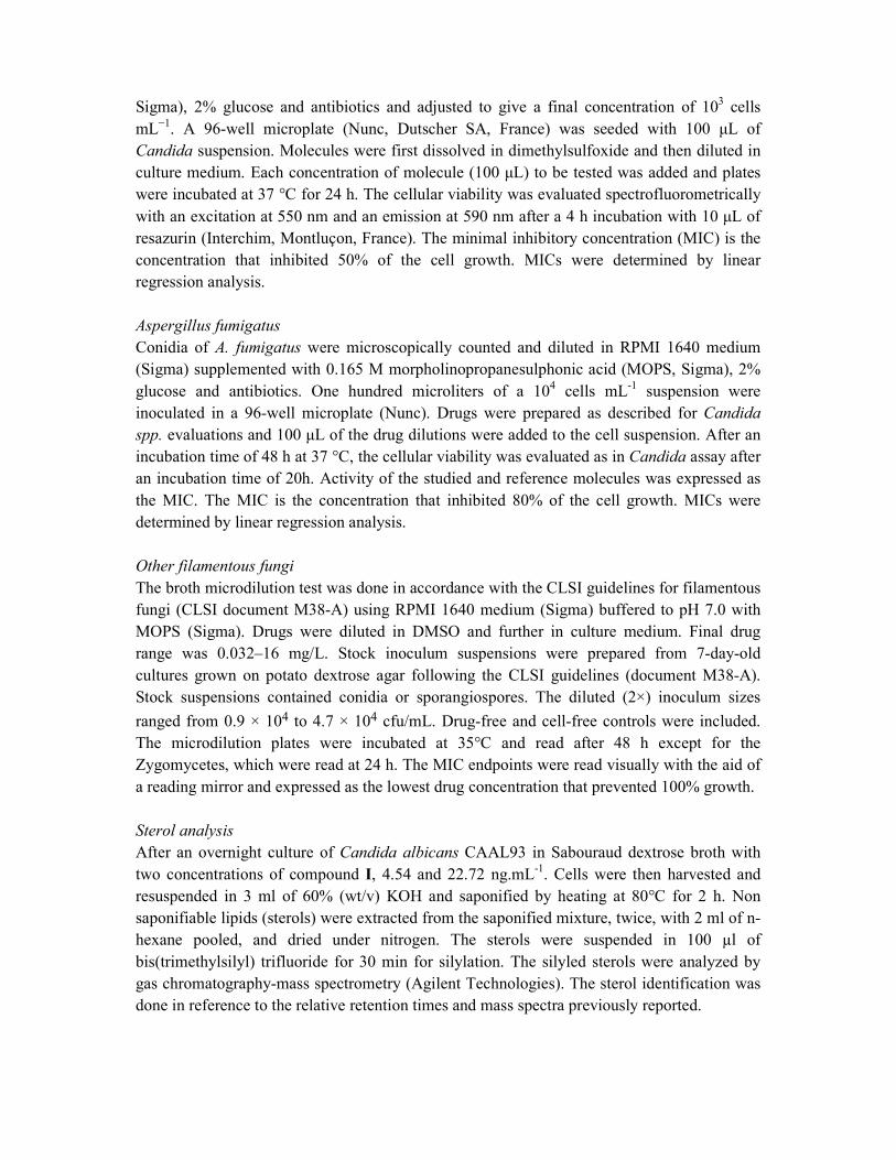
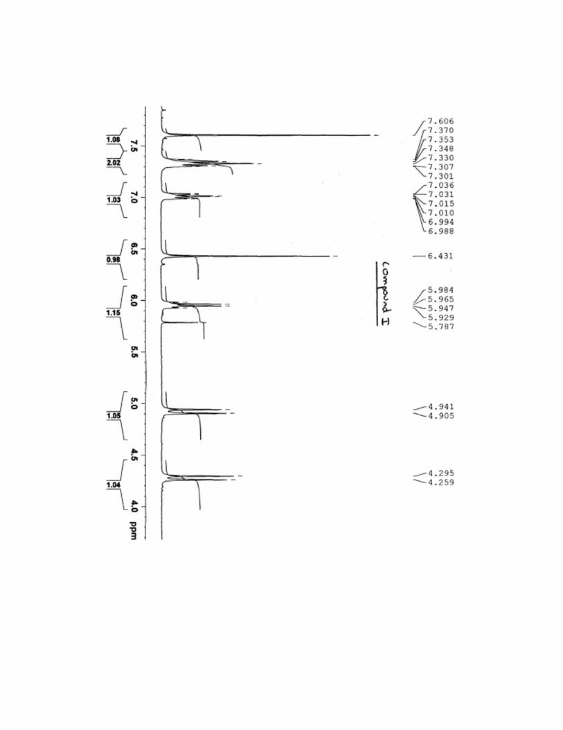
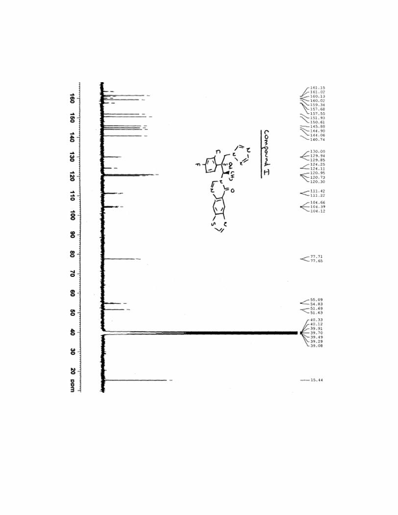
7-[(1R,2R)-2-(2,4-difluorophenyl)-2-hydroxy-1-methyl-3-(1H-1,2,4-triazol-1-
yl)propyl]thiazolo[4,5-g]quinazolin-8(7H)-one (I)
To a stirred solution of compound 15 (111 mg, 0.44 mmol) in N-methyl-2-pyrrolidone (1 mL)
was added compound 9 (90 mg, 0.44 mmol) and potassium carbonate (92 mg, 0.65 mmol).
The mixture was stirred at 80°C for 3 days. After cooling, mixture was diluted with water and
product was extracted with ethyl acetate. Organic layers were dried over anhydrous Na2SO4
and concentrated in vacuo. The residue was purified on silica gel column chromatography
(dichloromethane/ethanol, 99:1) and compound I was obtained in a 47% yield as a white
solid. Mp 140-141 °C; 1H NMR (400 MHz, DMSO-d6): δ 1.27 (d, 3H,
3J = 7.2 Hz), 4.27 (d,
1H, 2J = 14.4 Hz), 4.92 (d, 1H,
2J = 14.4 Hz), 5.93 (q, 1H,
3J = 7.2 Hz), 6.43 (s, 1H), 7.01
(ddd, 1H, 3JH-F =
3JH-H = 8.4 Hz,
4JH-H = 2.2 Hz), 7.25-7.38 (m, 2H), 7.60 (s, 1H), 8.24 (s, 1H),
8.48 (s, 1H), 8.63 (s, 1H), 8.88 (s, 1H), 9.60 (s, 1H); 13
C NMR (400 MHz, DMSO-d6): δ 15.4,
51.6, 54.9, 77.6, 104.4, 111.3, 120.3, 120.7, 121.0, 124.2, 129.9, 140.7, 144.1, 144.9, 145.9,
150.6, 151.9, 158.9, 159.3, 161.5, 162,4; IR (KBr cm-1
): 848, 1140, 1259, 1442, 1501, 1609,
1674, 3418; HRMS calcd for C21H17F2N6O2S [M+H]+ 455.1102 found 455.1101; [α]
20D = -
10.0 (c = 0.1 in CHCl3).
8-[(1R,2R)-2-(2,4-difluorophenyl)-2-hydroxy-1-methyl-3-(1H-1,2,4-triazol-1-
yl)propyl]thiazolo[5,4-f]quinazolin-9(8H)-one (II)
Using the synthetic procedure used for compound I starting from 15 (149 mg, 0.59 mmol) and
compound 14 (120 mg, 0.59 mmol) to obtain compound II in a 45% yield as a white solid.
Mp 238-229 °C; 1H NMR (400 MHz, DMSO-d6): δ 1.31 (d, 3H,
3J = 6.8 Hz), 4.27 (d, 1H,
2J
= 14.1 Hz), 4.92 (d, 1H, 2J = 14.1 Hz), 6.00 (q, 1H,
3J = 6.8 Hz), 6.46 (s, 1H), 7.02 (ddd, 1H,
3JH-F =
3JH-H = 8.4 Hz,
4JH-H = 2.4 Hz), 7.35-7.38 (m, 2H), 7.61 (s, 1H), 7.96 (d, 1H,
3J = 8.8
Hz), 8.23 (s, 1H), 8.60 (d, 1H, 3J = 8.8 Hz), 8.63 (s, 1H), 9.64 (s, 1H);
13C NMR (400 MHz,
DMSO-d6): δ 15.5, 52.2, 54.8, 77.6, 104.4, 111.3, 115.9, 124.0, 126.0, 129.4, 129.9, 130.6,
144.9, 146.3, 146.4, 150.5, 152.2, 158.8, 159.4, 160.2, 162.4; IR (KBr cm-1
): 857, 1158, 1273,
1456, 1504, 1587, 1657, 3450; HRMS calcd for C21H17F2N6O2S [M+H]+ 455.1102 found
455.1089; [α]20
D = -30.0 (c = 0.1 in CHCl3).
In vitro antifungal activity
Candida spp.
Candida spp. suspensions were prepared in RPMI 1640 medium (Sigma, Saint Quentin
Fallavier, France) supplemented with 0.165 M morpholinopropanesulphonic acid (MOPS,
Sigma), 2% glucose and antibiotics and adjusted to give a final concentration of 103 cells
mL−1
. A 96-well microplate (Nunc, Dutscher SA, France) was seeded with 100 µL of
Candida suspension. Molecules were first dissolved in dimethylsulfoxide and then diluted in
culture medium. Each concentration of molecule (100 µL) to be tested was added and plates
were incubated at 37 °C for 24 h. The cellular viability was evaluated spectrofluorometrically
with an excitation at 550 nm and an emission at 590 nm after a 4 h incubation with 10 µL of
resazurin (Interchim, Montluçon, France). The minimal inhibitory concentration (MIC) is the
concentration that inhibited 50% of the cell growth. MICs were determined by linear
regression analysis.
Aspergillus fumigatus
Conidia of A. fumigatus were microscopically counted and diluted in RPMI 1640 medium
(Sigma) supplemented with 0.165 M morpholinopropanesulphonic acid (MOPS, Sigma), 2%
glucose and antibiotics. One hundred microliters of a 104 cells mL
-1 suspension were
inoculated in a 96-well microplate (Nunc). Drugs were prepared as described for Candida
spp. evaluations and 100 µL of the drug dilutions were added to the cell suspension. After an
incubation time of 48 h at 37 °C, the cellular viability was evaluated as in Candida assay after
an incubation time of 20h. Activity of the studied and reference molecules was expressed as
the MIC. The MIC is the concentration that inhibited 80% of the cell growth. MICs were
determined by linear regression analysis.
Other filamentous fungi
The broth microdilution test was done in accordance with the CLSI guidelines for filamentous
fungi (CLSI document M38-A) using RPMI 1640 medium (Sigma) buffered to pH 7.0 with
MOPS (Sigma). Drugs were diluted in DMSO and further in culture medium. Final drug
range was 0.032–16 mg/L. Stock inoculum suspensions were prepared from 7-day-old
cultures grown on potato dextrose agar following the CLSI guidelines (document M38-A).
Stock suspensions contained conidia or sporangiospores. The diluted (2×) inoculum sizes
ranged from 0.9 × 10⁴ to 4.7 × 10⁴ cfu/mL. Drug-free and cell-free controls were included.
The microdilution plates were incubated at 35°C and read after 48 h except for the
Zygomycetes, which were read at 24 h. The MIC endpoints were read visually with the aid of
a reading mirror and expressed as the lowest drug concentration that prevented 100% growth.
Sterol analysis
After an overnight culture of Candida albicans CAAL93 in Sabouraud dextrose broth with
two concentrations of compound I, 4.54 and 22.72 ng.mL-1
. Cells were then harvested and
resuspended in 3 ml of 60% (wt/v) KOH and saponified by heating at 80°C for 2 h. Non
saponifiable lipids (sterols) were extracted from the saponified mixture, twice, with 2 ml of n-
hexane pooled, and dried under nitrogen. The sterols were suspended in 100 µl of
bis(trimethylsilyl) trifluoride for 30 min for silylation. The silyled sterols were analyzed by
gas chromatography-mass spectrometry (Agilent Technologies). The sterol identification was
done in reference to the relative retention times and mass spectra previously reported.

1H and
13C NMR spectra for compound I

![Huge Avenues of Opportunities (With Some Potholes ...therapies [30]. On the other hand, silver nanoparticles display potent and broad-spectrum antibacterial [7,31], antifungal [32]](https://img.pdfslide.net/doc/110x75/60da1dddb324712dfa3a7139/huge-avenues-of-opportunities-with-some-potholes-therapies-30-on-the-other.jpg)