Embed Size (px)
Citation preview

ChemicalScience
EDGE ARTICLE
Ope
n A
cces
s A
rtic
le. P
ublis
hed
on 2
6 Fe
brua
ry 2
018.
Dow
nloa
ded
on 1
/22/
2022
10:
36:1
4 A
M.
Thi
s ar
ticle
is li
cens
ed u
nder
a C
reat
ive
Com
mon
s A
ttrib
utio
n 3.
0 U
npor
ted
Lic
ence
.
View Article OnlineView Journal | View Issue
Discovery of the
aMax-Planck-Institute of Molecular Physio
Otto-Hahn-Str. 11, 44227 Dortmund, GermabFaculty of Chemistry and Chemical Biology,
4a, 44227 Dortmund, Germany. E-mail: hercRIKEN-Max Planck Joint Research Divisio
CSRS, 2-1, Hirosawa, Wako, Saitama 351-0dChemical Biology Research Group, RIKEN C
0198, JapaneChemical Genomics Centre of the Max-P
Dortmund, GermanyfBio-Active Compounds Discovery Research U
Saitama 351-0198, Japan
† Electronic supplementary informa10.1039/c7sc05040b
‡ Present address: Department of ChemiKemitorvet Building 207, Room 124, [email protected].
Cite this: Chem. Sci., 2018, 9, 3014
Received 24th November 2017Accepted 20th February 2018
DOI: 10.1039/c7sc05040b
rsc.li/chemical-science
3014 | Chem. Sci., 2018, 9, 3014–3022
novel autophagy inhibitor aumitinthat targets mitochondrial complex I†
Lucas Robke,abc Yushi Futamura,d Georgios Konstantinidis,e Julian Wilke,ab
Harumi Aono,d Zhwan Mahmoud,b Nobumoto Watanabe,cf Yao-Wen Wu, e
Hiroyuki Osada,cd Luca Laraia ‡*a and Herbert Waldmann *ab
Macroautophagy is a conserved eukaryotic process for degradation of cellular components in response to
lack of nutrients. It is involved in the development of diseases, notably cancer and neurological disorders
including Parkinson's disease. Small molecule autophagy modulators have proven to be valuable tools to
dissect and interrogate this crucial metabolic pathway and are in high demand. Phenotypic screening for
autophagy inhibitors led to the discovery of the novel autophagy inhibitor aumitin. Target identification
and confirmation revealed that aumitin inhibits mitochondrial respiration by targeting complex I. We
show that inhibition of autophagy by impairment of mitochondrial respiration is general for several
mitochondrial inhibitors that target different mitochondrial complexes. Our findings highlight the
importance of mitochondrial respiration for autophagy regulation.
Macroautophagy (henceforth autophagy) is a conserved,eukaryotic process essential for cellular development, homeo-stasis and survival.1,2 It plays a role in the prevention of cancerby removing damaged organelles including dysfunctionalmitochondria, in the cell.3 It also promotes the survival ofcancer cells under stress conditions including nutrient depri-vation.1 During autophagy, cellular components are seques-tered, engulfed by the phagophore, the precursor to theautophagosome, and subsequently eliminated through auto-phagosome–lysosome fusion.4 Autophagy is tightly connectedto metabolism,5 nutrient uptake and cellular energy supply bythe mitochondria.6 Mitochondria regulate autophagy by gener-ation of ATP and production of reactive oxygen species (ROS).7
Conversely autophagy controls mitochondrial homeostasis bymeans of mitophagy.8 Inhibition of autophagy has been linked
logy, Department of Chemical Biology,
ny
TU Dortmund University, Otto-Hahn-Str.
n for Systems Chemical Biology, RIKEN
198, Japan
SRS, 2-1, Hirosawa, Wako, Saitama 351-
lanck-Society, Otto-Hahn-Str. 15, 44227
nit, RIKEN CSRS, 2-1, Hirosawa, Wako,
tion (ESI) available. See DOI:
stry, Technical University of Denmark,800 Kgs. Lyngby, Denmark. E-mail:
to the onset of Parkinson's disease due to impaired mitochon-drial turnover.9 Due to this interplay, small molecules thatmodulate autophagy through modulation of mitochondrialfunction are invaluable tools for the study of the biologicalprocesses involved10–15 and may inspire new drug discoveryprograms.16 Here we describe the discovery of aumitin, a noveldiaminopyrimidine-based autophagy inhibitor which targetsmitochondrial complex I. More generally we show that inhibi-tion of mitochondrial respiration, irrespective of the targetedcomplex, inhibits autophagy.
To identify novel autophagy inhibitors, we employed a high-content screening approach using MCF-7 cells stably expressingthe autophagosome marker, eGFP-LC3 (MCF7-LC3).17 Dia-minopyrimidine based compounds were identied as verypotent autophagy inhibitors, as exemplied by the most potenthit (1), which we termed aumitin (Fig. 1). Aumitin andanalogues thereof inhibited starvation- and rapamycin inducedautophagy dose dependently (Fig. 1A–D), which suggests thatthey might target the pathway downstream of mammaliantarget of rapamycin (mTOR).
Aumitin (for a synthesis see ESI Fig. 1), was chosen for indepth-characterization, as it was the most potent diaminopyr-imidine inhibitor. Upon autophagy induction, the cytosolicprotein microtubule-associated protein light chain 3 (LC3-I) isconjugated to phosphatidylethanolamine (PE) to become themembrane-bound form LC3-II. Aumitin inhibited LC3 lip-idation in a dose-dependent manner in starved and rapamycintreated MCF7-LC3 cells (Fig. 1F and ESI Fig. 2). To examine theimpact of aumitin on autophagic ux, the levels of the auto-phagy substrate p62 were investigated.18 p62 targets proteins fordegradation by the autophagic machinery, where it is degraded
This journal is © The Royal Society of Chemistry 2018
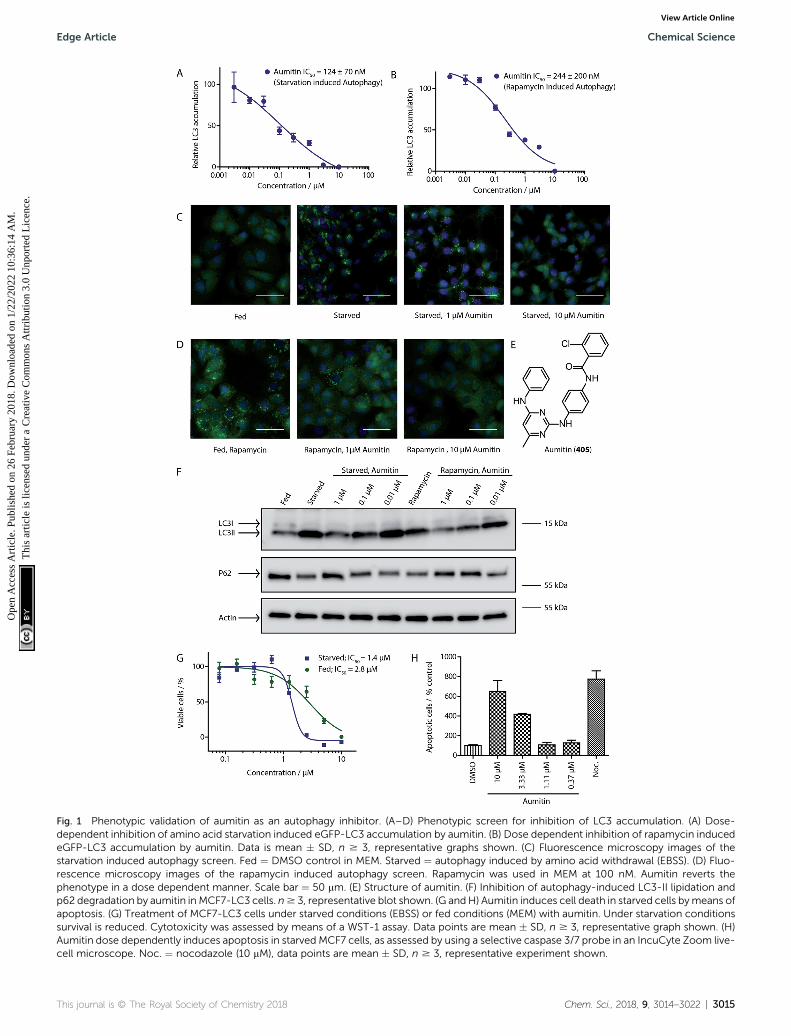
Fig. 1 Phenotypic validation of aumitin as an autophagy inhibitor. (A–D) Phenotypic screen for inhibition of LC3 accumulation. (A) Dose-dependent inhibition of amino acid starvation induced eGFP-LC3 accumulation by aumitin. (B) Dose dependent inhibition of rapamycin inducedeGFP-LC3 accumulation by aumitin. Data is mean � SD, n $ 3, representative graphs shown. (C) Fluorescence microscopy images of thestarvation induced autophagy screen. Fed ¼ DMSO control in MEM. Starved ¼ autophagy induced by amino acid withdrawal (EBSS). (D) Fluo-rescence microscopy images of the rapamycin induced autophagy screen. Rapamycin was used in MEM at 100 nM. Aumitin reverts thephenotype in a dose dependent manner. Scale bar ¼ 50 mm. (E) Structure of aumitin. (F) Inhibition of autophagy-induced LC3-II lipidation andp62 degradation by aumitin in MCF7-LC3 cells. n$ 3, representative blot shown. (G andH) Aumitin induces cell death in starved cells bymeans ofapoptosis. (G) Treatment of MCF7-LC3 cells under starved conditions (EBSS) or fed conditions (MEM) with aumitin. Under starvation conditionssurvival is reduced. Cytotoxicity was assessed by means of a WST-1 assay. Data points are mean � SD, n $ 3, representative graph shown. (H)Aumitin dose dependently induces apoptosis in starved MCF7 cells, as assessed by using a selective caspase 3/7 probe in an IncuCyte Zoom live-cell microscope. Noc. ¼ nocodazole (10 mM), data points are mean � SD, n $ 3, representative experiment shown.
This journal is © The Royal Society of Chemistry 2018 Chem. Sci., 2018, 9, 3014–3022 | 3015
Edge Article Chemical Science
Ope
n A
cces
s A
rtic
le. P
ublis
hed
on 2
6 Fe
brua
ry 2
018.
Dow
nloa
ded
on 1
/22/
2022
10:
36:1
4 A
M.
Thi
s ar
ticle
is li
cens
ed u
nder
a C
reat
ive
Com
mon
s A
ttrib
utio
n 3.
0 U
npor
ted
Lic
ence
.View Article Online
Chemical Science Edge Article
Ope
n A
cces
s A
rtic
le. P
ublis
hed
on 2
6 Fe
brua
ry 2
018.
Dow
nloa
ded
on 1
/22/
2022
10:
36:1
4 A
M.
Thi
s ar
ticle
is li
cens
ed u
nder
a C
reat
ive
Com
mon
s A
ttrib
utio
n 3.
0 U
npor
ted
Lic
ence
.View Article Online
together with its cargo. Aumitin inhibited p62 degradation bystarvation- as well as rapamycin-induced autophagy dose-dependently in MCF7-LC3 cells, suggesting inhibition of auto-phagic ux (Fig. 1F and ESI Fig. 2†). Moreover, inhibition ofautophagic ux was conrmed by an independent cellular assayusing mCherry-eGFP-LC3 cells.19 Because eGFP uorescence isquenched in acidic conditions while mCherry uorescence isstable, the autophagosome and autolysosome can be distin-guished by yellow puncta (eGFP + mCherry) and red puncta(mCherry only), respectively (ESI Fig. 3†). As expected for anautophagy inhibitor, aumitin enhanced death of amino acid-starved cells as compared to fed cells (Fig. 1G).20 Aumitininduced cell death through apoptosis, as assessed using a cas-pase 3/7 probe that stains apoptotic cells (Fig. 1H).
Due to aumitin's structural similarity to known kinaseinhibitors,21 we screened for kinases that can be inhibited byaumitin at a concentration of 1 mM. Out of 419 tested kinasesonly the two phosphatidylinositol kinases PI4KB and PI3KC2Gwere weakly inhibited (ESI Table 2†). However, highly potentand selective inhibitors of these kinases did not lead to auto-phagy inhibition (ESI Table 3†). This suggests that PI4KB andPI3KC2G are not responsible for aumitin's effect on autophagy.
Given the intimate interplay between autophagy and cellularmetabolic demand, we investigated whether aumitin interfereswith key metabolic processes including glucose uptake andoxidative phosphorylation. However, aumitin did not inhibitglucose uptake22 in HCT116 cells (residual activity > 75% at30 mM, n ¼ 4). Mitochondria have been shown to play a role inautophagy regulation.7,8 To test whether aumitin interfered withmitochondrial respiration, it was tested in a cell-based assaymonitoring cellular oxygen consumption using the SeahorseXFe96 Analyzer (Fig. 2 and ESI Fig. 5†). This contains two u-orophores embedded in a polymer in close proximity to theseeded cells. Quenching of one uorophore by oxygen allowsthe kinetic determination of the oxygen consumption rate(OCR), which is representative for respiration. The other uo-rophore is pH sensitive and indicates the extracellular acidi-cation rate (ECAR) whichmirrors anaerobic glycolysis leading tolactate excretion.
OCR and ECAR are regulated by inhibitors of mitochondrialrespiration (Fig. 2). While oxygen consumption declines,extracellular acidication increases in the presence of Aumitin(Fig. 2, time point (a)). Addition of mitochondrial complex Vinhibitor oligomycin at timepoint (b) leads to decrease of OCRlevel (Fig. 2, black line), suggesting ATP-driven respiration.Oligomycin can also be used to determine the extent of protonleak respiration, i.e. the inux of protons into mitochondriaindependent of an ATPase, through the difference in the OCRbetween 0 mM and 10 mM aumitin aer timepoint (b). Additionof protonophore FCCP (Fig. 2, timepoint (c)) reveals themaximal respiratory capacity, as it extinguishes the electro-chemical gradient, such that respiration is fastest. In order todetermine non-mitochondrial respiration, i.e. the lowestpossible OCR level, at timepoint (d) complex I inhibitor rote-none and complex III inhibitor antimycin are added together.-Whereas the recently reported VPS34 inhibitor autophinib17 didnot modulate mitochondrial respiration (ESI Fig. 4†), aumitin
3016 | Chem. Sci., 2018, 9, 3014–3022
potently and dose dependently inhibited this process asassessed by a reduction in OCR and an increase in ECAR inMCF7 cells (Fig. 2) and HeLa cells (ESI Fig. 5†).
Analysis of the structure activity relationship (SAR) indicatedthat inhibition of mitochondrial respiration in MCF7 and HeLacells correlates strongly with inhibition of autophagy by aumitinand analogues (Table 1). Compounds with medium potency inthe cell-based autophagy assay (Table 1, entries 2–5) alsoinhibited mitochondrial respiration less potently than aumitin.A structurally related but inactive compound in the autophagyassay did not inhibit mitochondrial respiration (Table 1, entry6). This indicates a causal connection between the effect onmitochondrial respiration and autophagy inhibition. Due to therapid effect of this compound class on mitochondrial respira-tion (Fig. 2), we speculate that the effect of mitochondrialrespiration inhibition may be the cause of autophagy inhibitionand not vice versa.
The inuence of aumitin on mitochondrial respiration wasinvestigated in greater detail by means of the previously re-ported semi-intact assay.23 The detergent digitonin is employedto permeabilize the plasma membrane, and inhibition of thedifferent mitochondrial complexes can then be investigated byaddition of their distinct substrates. In case of inhibition ofa particular mitochondrial complex, only the substrates of thedownstream complexes will allow continued mitochondrialrespiration, as assessed by oxygen consumption. In this assayaumitin exclusively inhibited mitochondrial complex I (Fig. 2Cand ESI Fig. 7†). The same result was observed for rotenone,a known inhibitor of mitochondrial complex I (ESI Fig. 6 and7†).13,14
Inhibition of the pyruvate/malate-driven respiratory activityby aumitin may occur by direct binding to and inhibition ofcomplex I, or by interception of NADH supply, which is requiredfor the enzymatic activity of complex I. Evaluation of aumitin inan in vitro NADH-CoQ reductase assay using isolated mito-chondria, revealed that aumitin inhibited the NADH-CoQreductase activity, similar to rotenone (Fig. 2D). Thus, aumitinis a direct inhibitor of mitochondrial complex I.
Since aumitin and rotenone both inhibit complex I, we ex-pected that rotenone would also potently inhibit autophagy, asit had previously been reported to do at very low concentrations(10 nM) and varying time points (2 and 24 h) in primary ratcortical neuronal cells.13,14 Interestingly, Giordano et al. showedthat increased colocalisation of mitochondria with lysosomeswas observed at 24 h, which is indicative of mitophagy andsomewhat contradicts the fact that LC3 lipidation and p62degradation were inhibited. However, rotenone has also beenreported to decrease effective lysosomal degradation of auto-phagosomes,11 which would lead to an accumulation of auto-phagosomes. It has also been shown to induce autophagy inU87, HEK293 and SH-SY5Y cells; however, this has only beenreported for very high concentrations (>2.5 mM) and with longerincubation times (typically >16 h).15,24 None-the-less, thesereports may contradict our nding that aumitin inhibits auto-phagic ux, but also appear to show that rotenone may havedifferent effects on autophagy depending on the time pointmonitored and dose used.
This journal is © The Royal Society of Chemistry 2018
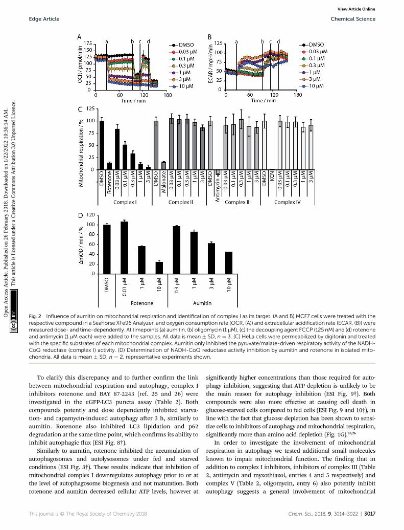
Fig. 2 Influence of aumitin on mitochondrial respiration and identification of complex I as its target. (A and B) MCF7 cells were treated with therespective compound in a Seahorse XFe96 Analyzer, and oxygen consumption rate (OCR, (A)) and extracellular acidification rate (ECAR, (B)) weremeasured dose- and time-dependently. At timepoints (a) aumitin, (b) oligomycin (1 mM), (c) the decoupling agent FCCP (125 nM) and (d) rotenoneand antimycin (1 mM each) were added to the samples. All data is mean � SD, n ¼ 3. (C) HeLa cells were permeabilized by digitonin and treatedwith the specific substrates of each mitochondrial complex. Aumitin only inhibited the pyruvate/malate-driven respiratory activity of the NADH-CoQ reductase (complex I) activity. (D) Determination of NADH-CoQ reductase activity inhibition by aumitin and rotenone in isolated mito-chondria. All data is mean � SD, n ¼ 2, representative experiments shown.
Edge Article Chemical Science
Ope
n A
cces
s A
rtic
le. P
ublis
hed
on 2
6 Fe
brua
ry 2
018.
Dow
nloa
ded
on 1
/22/
2022
10:
36:1
4 A
M.
Thi
s ar
ticle
is li
cens
ed u
nder
a C
reat
ive
Com
mon
s A
ttrib
utio
n 3.
0 U
npor
ted
Lic
ence
.View Article Online
To clarify this discrepancy and to further conrm the linkbetween mitochondrial respiration and autophagy, complex Iinhibitors rotenone and BAY 87-2243 (ref. 25 and 26) wereinvestigated in the eGFP-LC3 puncta assay (Table 2). Bothcompounds potently and dose dependently inhibited starva-tion- and rapamycin-induced autophagy aer 3 h, similarly toaumitin. Rotenone also inhibited LC3 lipidation and p62degradation at the same time point, which conrms its ability toinhibit autophagic ux (ESI Fig. 8†).
Similarly to aumitin, rotenone inhibited the accumulation ofautophagosomes and autolysosomes under fed and starvedconditions (ESI Fig. 3†). These results indicate that inhibition ofmitochondrial complex I downregulates autophagy prior to or atthe level of autophagosome biogenesis and not maturation. Bothrotenone and aumitin decreased cellular ATP levels, however at
This journal is © The Royal Society of Chemistry 2018
signicantly higher concentrations than those required for auto-phagy inhibition, suggesting that ATP depletion is unlikely to bethe main reason for autophagy inhibition (ESI Fig. 9†). Bothcompounds were also more effective at causing cell death inglucose-starved cells compared to fed cells (ESI Fig. 9 and 10†), inline with the fact that glucose depletion has been shown to sensi-tize cells to inhibitors of autophagy and mitochondrial respiration,signicantly more than amino acid depletion (Fig. 1G).25,26
In order to investigate the involvement of mitochondrialrespiration in autophagy we tested additional small moleculesknown to impair mitochondrial function. The nding that inaddition to complex I inhibitors, inhibitors of complex III (Table2, antimycin and myxothiazol, entries 4 and 5 respectively) andcomplex V (Table 2, oligomycin, entry 6) also potently inhibitautophagy suggests a general involvement of mitochondrial
Chem. Sci., 2018, 9, 3014–3022 | 3017

Table 1 Correlation of the SAR between mitochondrial respiration and autophagy. Inhibition of mitochondrial respiration was tested in a Sea-horse XFe96 Analyzer by means of OCR in HeLa cells and MCF7 cells. Data is mean� SD; n¼ 3. Inactive¼ no inhibition at 10 mM. Data is mean�SD; n $ 3
Entry Number R1 R2
Mitochondrial respirationinhibition Autophagy inhibition
HeLa cells(IC50 [mM])
MCF7 cells(IC50 [mM])
Starvation(IC50 [mM])
Rapamycin(IC50 [mM])
1 Aumitin (1) 0.11 � 0.21 0.44 � 0.11 0.12 � 0.07 0.24 � 0.20
2 2 0.92 � 0.15 1.78 � 0.15 0.80 � 0.30 0.81 � 0.68
3 3 0.38 � 0.13 0.65 � 0.09 0.81 � 0.16 0.29 � 0.18
4 4 0.66 � 0.08 1.32 � 0.06 0.97 � 0.47 0.16 � 0.08
5 5 1.23 � 0.16 8.21 � 0.05 1.6 � 0.60 4.1 � 3.0
6 6 Inactive Inactive Inactive Inactive
Chemical Science Edge Article
Ope
n A
cces
s A
rtic
le. P
ublis
hed
on 2
6 Fe
brua
ry 2
018.
Dow
nloa
ded
on 1
/22/
2022
10:
36:1
4 A
M.
Thi
s ar
ticle
is li
cens
ed u
nder
a C
reat
ive
Com
mon
s A
ttrib
utio
n 3.
0 U
npor
ted
Lic
ence
.View Article Online
respiration in autophagy. The ADP/ATP translocase inhibitoratractyloside (Table 2, entry 8) that does not affect the mito-chondrial proton gradient27 did not inhibit autophagy, suggestingthat the latter may play a critical role in autophagy modulation, inline with previous reports.12 This notion was supported by the factthat the uncoupler FCCP (Table 2, entry 7) inhibits autophagy.FCCP shuttles protons across membranes from low to high pHand perturbs the formation of the proton gradient, which isnecessary for proper function of ATP synthase. However, FCCPalso impairs acidication of (auto)lysosomes.28 Thereby matura-tion and clearance of autophagosomal structures are impaired sothat FCCP may also function as an inhibitor of autophagosomematuration. Indeed, FCCP inhibits autophagosome formation atlow doses but leads to accumulation of autophagosomes at highdoses as assessed by the eGFP-LC3 puncta assay and immuno-blotting for LC3 lipidation (ESI Fig. 11 and 12†).
3018 | Chem. Sci., 2018, 9, 3014–3022
Mitochondrial respiration inhibitors have been reported toinduce reactive oxygen species (ROS)29 which regulate auto-phagy by various mechanisms, including the redox inactivationof the protease ATG4 via oxidation at the catalytic cysteine.Thus, we investigated whether application of aumitin androtenone increases ROS levels (ESI Fig. 13†). Aumitin androtenone induced ROS in HeLa cells dose dependently, howeveronly at approximately 200-fold higher concentrations thanrequired to inhibit autophagy (ESI Fig. 13†). This ndingsuggests that the induction of ROS by aumitin might not beresponsible for autophagy inhibition. Additionally, the knownROS inducers L-buthionine-sulfoximine (BSO) and chlorodini-trobenzene (CDNB) as well as >600 ROS inducers identied inan in-house screen (unpublished data, ESI Fig. 14 and 15†) didnot inhibit autophagy, suggesting that ROS induction per se isunlikely to lead to autophagy inhibition. This nding is alsosupported by a previous report from Graef and Nunnari where
This journal is © The Royal Society of Chemistry 2018

Table 2 Examination of inhibitors of mitochondrial respiration on starvation- and rapamycin-induced autophagy. Inactive ¼ no inhibition at>10 mM. Data is mean � SD, n $ 3
EntryCompound name(number) Structure Protein target
Autophagy Inhibition
Starvation IC50 [nM]RapamycinIC50 [nM]
1 Aumitin (1) Complex I 124 � 70 244 � 200
2 Rotenone (7) Complex I 3 � 1 9 � 2
3 BAY 87-2243 (8) Complex I <3 nd
4 Antimycin A (9) Complex III <3 6 � 0.7
This journal is © The Royal Society of Chemistry 2018 Chem. Sci., 2018, 9, 3014–3022 | 3019
Edge Article Chemical Science
Ope
n A
cces
s A
rtic
le. P
ublis
hed
on 2
6 Fe
brua
ry 2
018.
Dow
nloa
ded
on 1
/22/
2022
10:
36:1
4 A
M.
Thi
s ar
ticle
is li
cens
ed u
nder
a C
reat
ive
Com
mon
s A
ttrib
utio
n 3.
0 U
npor
ted
Lic
ence
.View Article Online
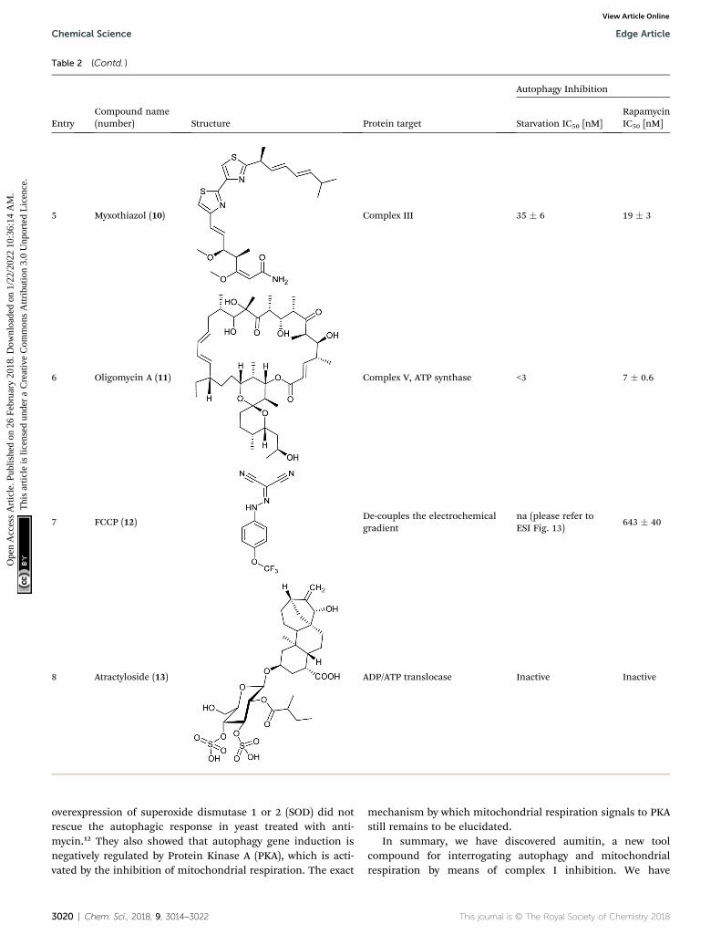
Table 2 (Contd. )
EntryCompound name(number) Structure Protein target
Autophagy Inhibition
Starvation IC50 [nM]RapamycinIC50 [nM]
5 Myxothiazol (10) Complex III 35 � 6 19 � 3
6 Oligomycin A (11) Complex V, ATP synthase <3 7 � 0.6
7 FCCP (12)De-couples the electrochemicalgradient
na (please refer toESI Fig. 13)
643 � 40
8 Atractyloside (13) ADP/ATP translocase Inactive Inactive
Chemical Science Edge Article
Ope
n A
cces
s A
rtic
le. P
ublis
hed
on 2
6 Fe
brua
ry 2
018.
Dow
nloa
ded
on 1
/22/
2022
10:
36:1
4 A
M.
Thi
s ar
ticle
is li
cens
ed u
nder
a C
reat
ive
Com
mon
s A
ttrib
utio
n 3.
0 U
npor
ted
Lic
ence
.View Article Online
overexpression of superoxide dismutase 1 or 2 (SOD) did notrescue the autophagic response in yeast treated with anti-mycin.12 They also showed that autophagy gene induction isnegatively regulated by Protein Kinase A (PKA), which is acti-vated by the inhibition of mitochondrial respiration. The exact
3020 | Chem. Sci., 2018, 9, 3014–3022
mechanism by which mitochondrial respiration signals to PKAstill remains to be elucidated.
In summary, we have discovered aumitin, a new toolcompound for interrogating autophagy and mitochondrialrespiration by means of complex I inhibition. We have
This journal is © The Royal Society of Chemistry 2018
Edge Article Chemical Science
Ope
n A
cces
s A
rtic
le. P
ublis
hed
on 2
6 Fe
brua
ry 2
018.
Dow
nloa
ded
on 1
/22/
2022
10:
36:1
4 A
M.
Thi
s ar
ticle
is li
cens
ed u
nder
a C
reat
ive
Com
mon
s A
ttrib
utio
n 3.
0 U
npor
ted
Lic
ence
.View Article Online
investigated diverse inhibitors of mitochondrial respiration andshown that, irrespective of their mode of action and mito-chondrial target complex, they appear to be potent autophagyinhibitors. Aumitin is a useful alternative to rotenone, anddenes a novel chemotype for complex I inhibition. Rotenonehas been used extensively as a chemical tool compound for thestudy of various pathways and for investigation of Parkinson'sdisease. However, rotenone also appears to have activity inde-pendent of mitochondrial complex I inhibition.30 Thus aumitinwill be useful as a tool compound to further study the role ofmitochondrial respiration and complex I in (patho)physiolog-ical processes.
Statement of contributions
L. R. carried out the synthesis, the SAR analysis and autophagyvalidation experiments. Y. F. and H. A. carried out the mito-chondrial respiration studies. G. K. carried out the confocalmicroscopy experiments. J. W. carried out the ROS experiments.Z. W. and L. L. carried out autophagy validationexperiments. N. W. and H. O. supervised the mitochondrialwork and analysed data. Y. W. supervised autophagyexperiments. L. L. and H. W. coordinated and supervised theproject. L. R., L. L. and H. W. wrote the paper, with suggestionsfrom all co-authors.
Conflicts of interest
There are no conicts to declare.
Acknowledgements
This research was supported by the Max-Planck-Gesellscha. L.R. is grateful to the Boehringer Ingelheim Fonds for a fellow-ship. L. L. is grateful to the Alexander von Humboldt Founda-tion for a fellowship. Y.-W. W. acknowledges funding fromEuropean Research Council (ChemBioAP) and Behrens-Weise-Stiung. We thank Sharon Tooze for the gi of WIPI2 cells.We are grateful to Konstanze Winklhofer for valuable advice.
References
1 D. C. Rubinsztein, P. Codogno and B. Levine, Nat. Rev. DrugDiscovery, 2012, 11, 709.
2 B. Levine and G. Kroemer, Cell, 2008, 132, 27.3 (a) K. Degenhardt, R. Mathew, B. Beaudoin, K. Bray,D. Anderson, G. Chen, C. Mukherjee, Y. Shi, C. Gelinas,Y. Fan, D. A. Nelson, S. Jin and E. White, Cancer Cell, 2006,10, 51; (b) R. Mathew, C. M. Karp, B. Beaudoin, N. Vuong,G. Chen, H.-Y. Chen, K. Bray, A. Reddy, G. Bhanot,C. Gelinas, R. S. Dipaola, V. Karantza-Wadsworth andE. White, Cell, 2009, 137, 1062.
4 P. Boya, F. Reggiori and P. Codogno, Nat. Cell Biol., 2013, 15,1017.
5 D. J. Klionsky, Science, 2000, 290, 1717.6 (a) L. Galluzzi, F. Pietrocola, B. Levine and G. Kroemer, Cell,2014, 159, 1263; (b) J. D. Rabinowitz and E. White, Science,
This journal is © The Royal Society of Chemistry 2018
2010, 330, 1344; (c) R. C. Russell, H.-X. Yuan andK.-L. Guan, Cell Res., 2014, 24, 42.
7 J. Lee, S. Giordano and J. Zhang, Biochem. J., 2012, 441, 523.8 K. Okamoto and N. Kondo-Okamoto, Biochim. Biophys. Acta,2012, 1820, 595.
9 (a) A. M. Pickrell and R. J. Youle, Neuron, 2015, 85, 257; (b)A. J. Whitworth and L. J. Pallanck, Curr. Opin. Genet. Dev.,2017, 44, 47.
10 (a) X. Ma, M. Jin, Y. Cai, H. Xia, K. Long, J. Liu, Q. Yu andJ. Yuan, Chem. Biol., 2011, 18, 1474; (b) C.-J. Lin, C.-C. Lee,Y.-L. Shih, C.-H. Lin, S.-H. Wang, T.-H. Chen andC.-M. Shih, PLoS One, 2012, 7, e38706.
11 B. J. Mader, V. N. Pivtoraiko, H. M. Flippo, B. J. Klocke,K. A. Roth, L. R. Mangieri and J. J. Shacka, ACS Chem.Neurosci., 2012, 3, 1063.
12 M. Graef and J. Nunnari, EMBO J., 2011, 30, 2101.13 S. Giordano, M. Dodson, S. Ravi, M. Redmann, X. Ouyang,
V. M. Darley Usmar and J. Zhang, J. Neurochem., 2014, 131,625.
14 R. K. Dagda, T. Das Banerjee and E. Janda, Int. J. Mol. Sci.,2013, 14, 22163.
15 Y. Chen, E. McMillan-Ward, J. Kong, S. J. Israels andS. B. Gibson, J. Cell Sci., 2007, 120, 4155.
16 (a) N. D. Georgakopoulos, G. Wells and M. Campanella, Nat.Chem. Biol., 2017, 13, 136; (b) L. Galluzzi, J. M. Bravo-SanPedro, B. Levine, D. R. Green and G. Kroemer, Nat. Rev.Drug Discovery, 2017, 16, 487–511.
17 L. Robke, L. Laraia, M. A. Carnero Corrales,G. Konstantinidis, M. Muroi, A. Richters, M. Winzker,T. Engbring, S. Tomassi, N. Watanabe, H. Osada, D. Rauh,H. Waldmann, Y.-W. Wu and J. Engel, Angew. Chem., Int.Ed., 2017, 56, 8153.
18 D. J. Klionsky, et al., Autophagy, 2016, 12, 1.19 S. Kimura, T. Noda and T. Yoshimori, Autophagy, 2014, 3,
452.20 (a) J. Liu, H. Xia, M. Kim, L. Xu, Y. Li, L. Zhang, Y. Cai,
H. V. Norberg, T. Zhang, T. Furuya, M. Jin, Z. Zhu,H. Wang, J. Yu, Y. Li, Y. Hao, A. Choi, H. Ke, D. Ma andJ. Yuan, Cell, 2011, 147, 223; (b) L. Laraia, K. Ohsawa,G. Konstantinidis, L. Robke, Y.-W. Wu, K. Kumar andH. Waldmann, Angew. Chem., Int. Ed., 2017, 56, 2145.
21 (a) Z. Huang, Q. Zhang, L. Yan, G. Zhong, L. Zhang, X. Tanand Y. Wang, Bioorg. Med. Chem. Lett., 2016, 26, 1954; (b)H. R. Lawrence, M. P. Martin, Y. Luo, R. Pireddu, H. Yang,H. Gevariya, S. Ozcan, J.-Y. Zhu, R. Kendig, M. Rodriguez,R. Elias, J. Q. Cheng, S. M. Sebti, E. Schonbrunn andN. J. Lawrence, J. Med. Chem., 2012, 55, 7392.
22 N. Yamamoto, T. Sato, K. Kawasaki, S. Murosaki andY. Yamamoto, Anal. Biochem., 2006, 351, 139.
23 J. K. Salabei, A. A. Gibb and B. G. Hill, Nat. Protoc., 2014, 9,421.
24 N. Xiong, J. Xiong, M. Jia, L. Liu, X. Zhang, Z. Chen, J. Huang,Z. Zhang, L. Hou, Z. Luo, D. Ghoorah, Z. Lin and T. Wang,Behav. Brain Funct., 2013, 9, 13.
25 L. Schockel, A. Glasauer, F. Basit, K. Bitschar, H. Truong,G. Erdmann, C. Algire, A. Hagebarth, P. H. Willems,
Chem. Sci., 2018, 9, 3014–3022 | 3021
Chemical Science Edge Article
Ope
n A
cces
s A
rtic
le. P
ublis
hed
on 2
6 Fe
brua
ry 2
018.
Dow
nloa
ded
on 1
/22/
2022
10:
36:1
4 A
M.
Thi
s ar
ticle
is li
cens
ed u
nder
a C
reat
ive
Com
mon
s A
ttrib
utio
n 3.
0 U
npor
ted
Lic
ence
.View Article Online
C. Kopitz, W. J. Koopman and M. Heroult, Cancer Metab.,2015, 3, 11.
26 P. Ellinghaus, I. Heisler, K. Unterschemmann, M. Haerter,H. Beck, S. Greschat, A. Ehrmann, H. Summer, I. Flamme,F. Oehme, K. Thierauch, M. Michels, H. Hess-Stumpp andK. Ziegelbauer, Cancer Med., 2013, 2, 611.
27 M. Kalbacova, M. Vrbacky, Z. Drahota and Z. Melkova,Cytometry, Part A, 2003, 52, 110.
28 (a) B. E. Steinberg, K. K. Huynh, A. Brodovitch, S. Jabs,T. Stauber, T. J. Jentsch and S. Grinstein, J. Cell Biol., 2010,
3022 | Chem. Sci., 2018, 9, 3014–3022
189, 1171; (b) A. V. Berezhnov, M. P. M. Soutar,E. I. Fedotova, M. S. Frolova, H. Plun-Favreau,V. P. Zinchenko and A. Y. Abramov, J. Biol. Chem., 2016,291, 8701.
29 Y. Chen, E. McMillan-Ward, J. Kong, S. J. Israels andS. B. Gibson, J. Cell Sci., 2007, 120, 4155.
30 (a) B. R. Brinkley, S. S. Barham, S. C. Barranco andG. M. Fuller, Exp. Cell Res., 1974, 85, 41; (b) S. Gree,J. A. L. Wagenaars, R. Jansen, P. H. G. M. Willems andW. J. H. Koopman, Biochim. Biophys. Acta, 2015, 1853, 1606.
This journal is © The Royal Society of Chemistry 2018


![Original Article Puerarin modulates autophagy to …signaling pathway with JNK inhibitor SP600125 can produce protective effects against CIRI [18]. Thrombolysis, antiplatelet agents,](https://img.pdfslide.net/doc/110x75/5e39305f90bc3d6d0c3b72a7/original-article-puerarin-modulates-autophagy-to-signaling-pathway-with-jnk-inhibitor.jpg)