Embed Size (px)
Citation preview
This thesis has been submitted in fulfilment of the requirements for a postgraduate degree
(e.g. PhD, MPhil, DClinPsychol) at the University of Edinburgh. Please note the following
terms and conditions of use:
This work is protected by copyright and other intellectual property rights, which are
retained by the thesis author, unless otherwise stated.
A copy can be downloaded for personal non-commercial research or study, without
prior permission or charge.
This thesis cannot be reproduced or quoted extensively from without first obtaining
permission in writing from the author.
The content must not be changed in any way or sold commercially in any format or
medium without the formal permission of the author.
When referring to this work, full bibliographic details including the author, title,
awarding institution and date of the thesis must be given.

1
Diseases of wild birds of the orders Passeriformes and
Columbiformes - a review of conditions reported from the
United Kingdom and an analysis of results from wild bird
disease surveillance in Scotland 1994-2013
Thomas William Pennycott BVM&S Cert PMP MRCVS
References and Appendices
References……………………………………………...…………………………3-40
Appendix I: Latin names of birds found in Britain and discussed in thesis (including
appendices)………………………………………………………………………41-45
Appendix II: Diagnostic criteria used for carcases of the Orders Passeriformes and
Columbiformes……………………………………………………………….….46-50
Appendix III: Presumptive identification of internal parasites from wild birds of the
Orders Passeriformes and Columbiformes………………………………………51-70
Appendix IV: Antimicrobial susceptibility tests…………………………….…..71-79
Appendix V: Isolation of Salmonella enterica from wild birds 1994-2013……..80-81
Appendix VI: Isolation of potentially pathogenic E. albertii from wild birds 1994-
2013………………………………………………………………………...……82-84
Appendix VII: Isolates of potentially pathogenic E. albertii from different bird
species – results of O86:K61 slide agglutination tests………………………………85
Appendix VIII: Summary of outcomes from wild bird disease surveillance at Ayr
Disease Surveillance Centre 1994-2013 (Orders Passeriformes and Columbiformes)
………………………………………………………………...…………………86-97
Appendix IX: Necropsy forms…………………………………………………98-111
Appendix X: List of images of pathological lesions, parasites and parasite eggs from
wild birds of the Orders Passeriformes and Columbiformes………….………112-151
Appendix XI: Location of carcases…………………………………...………152-157
2
Images of pathological lesions, parasites and parasite eggs from wild birds of the
orders Passeriformes and Columbiformes are provided on the CD in the pocket on
the inside back cover of the references/appendices volume, and on-line at
http://datashare.is.ed.ac.uk/
References
3
References
ABBOTT, S. L., O'CONNOR, J., ROBIN, T., ZIMMER, B. L. & JANDA, J. M.
(2003) Biochemical properties of a newly described Escherichia species, Escherichia
albertii. Journal of Clinical Microbiology 41, 4852-4854
AEBISCHER, N. (2002a) European Turtle Dove. In The Migration Atlas:
Movements of the Birds of Britain and Ireland. Eds C. WERNHAM, M. P. TOMS, J.
H. MARCHANT, J. A. CLARK, G. M. SIRIWARDENA, S. R. BAILLIE. London,
T. & A.D. Poyser. pp. 420-422
AEBISCHER, N. (2002b) Stock Pigeon (Stock Dove). In The Migration Atlas:
Movements of the Birds of Britain and Ireland. Eds C. WERNHAM, M. P. TOMS, J.
H. MARCHANT, J. A. CLARK, G. M. SIRIWARDENA, S. R. BAILLIE. London,
T. & A.D. Poyser. pp. 412-413
ALDOUS, E., FULLER, C., MYNN, J. & ALEXANDER, D. (2004) A molecular
epidemiological investigation of isolates of the variant avian paramyxovirus type 1
virus (PPMV-1) responsible for the 1978 to present panzootic in pigeons. Avian
Pathology 33, 258-269
ALEXANDER, D., PARSONS, G. & MARSHALL, R. (1984) Infection of fowls
with Newcastle disease virus by food contaminated with pigeon faeces. Veterinary
Record 115, 601-602
ALEXANDER, D., WILSON, G., RUSSELL, P., LISTER, S. & PARSONS, G.
(1985) Newcastle disease outbreaks in fowl in Great Britain during 1984. Veterinary
Record 117, 429-434
ALEXANDER, D. J., MANVELL, R. J., IRVINE, R., LONDT, B. Z., COX, B.,
CEERAZ, V., BANKS, J. & BROWN, I. H. (2010) Overview of incursions of Asian
H5N1 subtype highly pathogenic avian influenza virus into Great Britain, 2005-
2008. Avian Diseases 54, 194-200
AMASS, S., CLARK, L., VAN ALSTINE, W., BOWERSOCK, T., MURPHY, D.,
KNOX, K. & ALBREGTS, S. (1994) Interaction of Mycoplasma hyopneumoniae
and Pasteurella multocida infections in swine. Journal of the American Veterinary
Medical Association 204, 102-107
ANDERSON, E., WARD, L. R., DE SAXE, M. J., OLD, D., BARKER, R. &
DUGUID, J. (1978) Correlation of phage type, biotype and source in strains of
Salmonella typhimurium. Journal of Hygiene 81, 203-217
References
4
ANDERSON, N. L., GRAHN, R. A., VAN HOOSEAR, K. & BONDURANT, R.
H. (2009) Studies of trichomonad protozoa in free ranging songbirds: prevalence of
Trichomonas gallinae in house finches (Carpodacus mexicanus) and corvids and a
novel trichomonad in mockingbirds (Mimus polyglottos). Veterinary Parasitology
161, 178-186
ANDERSON, R. C. (1992) Nematode Parasites of Vertebrates - Their Development
and Transmission. London, CAB International. pp. 609-615
ANDERSON, R. M. & MAY, R. M. (1978) Regulation and stability of host-parasite
population interactions: I. Regulatory processes. Journal of Animal Ecology 47, 219-
247
ANDERSON, R. M. & MAY, R. M. (1991) Infectious disease of humans: dynamics
and control. Oxford, Oxford University Press
ANON (1999) TGE confirmed in pigs on Humberside. Veterinary Record 144, 218
ANON (2003) VLA Surveillance Report January 2003 - Wildlife. Veterinary Record
152, 318
ANON (2008a) Trichomonosis in garden birds in Scotland, 2005-2008. Veterinary
Record 163, 234
ANON (2008b) Mass mortality of starlings - coastal Lancashire. In GB Wildlife
Disease Surveillance Quarterly Report January to March 2008. p. 5
ANON (2010a) Metabolic bone disease (MBD) in rehabilitated woodpigeons. In GB
Wildlife Disease Surveillance, Quarterly Report January to March 2010. p. 8
ANON (2010b) Trichomonosis in blackbirds. In GB Wildlife Disease Surveillance,
Quarterly Report July to September 2010. p. 7
ANON (2010c) Tick-related syndrome. In GB Wildlife Disease Surveillance,
Quarterly Report July to September 2010. pp. 8-9
ANON (2010d) Wild bird reports from Scotland. In GB Wildlife Disease
Surveillance, Quarterly Report October to December 2010. p. 12
ANON (2011a) Great Britain AI Wild Bird Surveillance (AIWBS). In GB Wildlife
Disease Surveillance Quarterly Report January to March 2011. p. 3
ANON (2011b) Pigeon paramyxovirus 1 (PPMV-1) infection in collared doves. In
GB Wildlife Disease Surveillance, Quarterly Report January to March 2011. pp. 6-7
ANON (2011c) Mass mortalities of passerine birds due to trauma. In GB Wildlife
Disease Surveillance, Quarterly Report January to March 2011. p. 10
ANON (2011d) PPMV-1: mass mortality of feral pigeons and woodpigeons in
Midlands. In GB Wildlife Disease Surveillance, Quarterly Report July to September
2011. p. 10
References
5
ANON (2012a) Scaly legs in chaffinch. In GB Wildlife Disease Surveillance
Quarterly Report April - June 2012. p. 7
ANON (2012b) Mass mortality of passerines off the east coast of England. In GB
Wildlife Disease Surveillance Quarterly Report July to September 2012. p. 13
ANON (2012c) Notifiable Avian Disease Control Strategy for Great Britain.
http://www.gov.scot/Resource/0039/00398513.pdf Accessed 24/08/15
ANON (2013a) Corvid respiratory disease. In GB Wildlife Disease Surveillance,
Quarterly Report July - September 2013. pp. 6-7
ANON (2013b) Avian haemoparasites in British garden birds. In GB Wildlife
Disease Surveillance Quarterly Report July to September 2013. p. 7
ANON (2013c) Mass mortality of woodpigeons. In GB Wildlife Disease
Surveillance, Quarterly Report October - December 2013. p. 5
ANON (2013d) Trichomonosis in feral pigeons. In GB Wildlife Disease
Surveillance, Quarterly Report October - December 2013. p. 6
ANON (2015) Operation turtle dove. http://operationturtledove.org/turtle-dove-
habitat-advice Accessed 31/12/15
ANWAR, M. (1966) Isospora lacazei (Labbé, 1893) and I. chloridis sp.
nov.(Protozoa: Eimeriidae) from the English Sparrow (Passer domesticus),
Greenfinch (Chloris chloris) and Chaffinch (Fringilla coelebs). Journal of
Eukaryotic Microbiology 13, 84-90
ASH, J. (1957) Post-mortem examinations of birds found dead during the cold spells
of 1954 and 1956. Bird Study 4, 159-166
ASH, J. & SHARPE, G. (1964) Post-mortem and pesticide examinations of birds in
the cold spell of 1963. Bird Study 11, 227-239
ATKINSON, C. T. (2008) Haemoproteus. In Parasitic diseases of wild birds. Eds C.
T. ATKINSON, N. J. THOMAS, D. B. HUNTER. Ames, Iowa, USA, Wiley-
Blackwell. pp. 13-34
AYLING, R. D. & NICHOLAS, R. A. J. (2007) Mycoplasma respiratory infections.
In Diseases of sheep. 4th edn. Ed I. D. AITKEN. Oxford, UK, Blackwell Publishing
Ltd. pp. 231-235
BAKER, J. (1977) The results of post-mortem examination of 132 wild birds. British
Veterinary Journal 133, 327-333
BAKER, J. R. (1982) Parasitic protozoa in British wild animals. Cambridge, Institute
of Terrestial Ecology, Natural Environment Research Council. pp. 3-12
BALMER, D. E., GILLINGS, S., CAFFREY, B. J., SWANN, R. L., DOWNIE, I. S.
& FULLER, R. J. Eds (2013) Bird Atlas 2007-11: the breeding and wintering birds
of Britain and Ireland. Thetford, BTO Books. Appendix 2, pp. 666-686
References
6
BARLOW, A. & SPARKES, A. (2014) Mass mortality of starlings in Somerset.
Veterinary Record 174, 202-203
BARNES, H. J. & GROSS, W. B. (1997) Colibacillosis. In Diseases of Poultry. 10th
edn. Eds B. W. CALNEK, H. J. BARNES, C. W. BEARD, L. R. MCDOUGALD, Y.
M. SAIF. Ames, IA, Iowa State University Press. pp. 131-141
BARRS, V. & BEATTY, J. (2013) Infectious diseases. In BSAVA Manual of Feline
Practice. Eds A. HARVEY, S. TASKER. Quedgeley, Gloucestershire, British Small
Animal Veterinary Association. pp. 440-451
BARTLETT, C. M. (2008) Filarioid nematodes. In Parasitic diseases of wild birds.
Eds C. T. ATKINSON, N. J. THOMAS, D. B. HUNTER. Ames, Iowa, USA, Wiley-
Blackwell. pp. 439-462
BARUS, V. & SERGEJEVA, T. P. (1989) Capillariids parasitic in birds in the
Palearctic region (2). Genera Eucoleus and Echinocoleus. Acta Scientiarum
Naturalium Academiae Scientiarum Bohemoslovacae, Brno. 23, 1-47
BEARD, P., DANIELS, M., HENDERSON, D., PIRIE, A., RUDGE, K., BUXTON,
D., RHIND, S., GREIG, A., HUTCHINGS, M. & MCKENDRICK, I. (2001)
Paratuberculosis infection of nonruminant wildlife in Scotland. Journal of Clinical
Microbiology 39, 1517-1521
BECKER, D. J., STREICKER, D. G. & ALTIZER, S. (2015) Linking anthropogenic
resources to wildlife–pathogen dynamics: a review and meta‐analysis. Ecology
Letters 18, 483-495
BECKMANN, K., BOREL, N., POCKNELL, A., DAGLEISH, M., SACHSE, K.,
JOHN, S., POSPISCHIL, A., CUNNINGHAM, A. & LAWSON, B. (2014a)
Chlamydiosis in British garden birds (2005–2011): retrospective diagnosis and
Chlamydia psittaci genotype determination. EcoHealth, doi:10.1007/s10393-014-
0951-x
BECKMANN, K. M., HARRIS, E., POCKNELL, A. M., JOHN, S. K.,
MACGREGOR, S. K., CUNNINGHAM, A. A. & LAWSON, B. (2014b) Aprocta
cylindrica (Nematoda) infection in a European Robin (Erithacus rubecula) in
Britain. Journal of Wildlife Diseases 50, 986-989
BELL, J. A., WECKSTEIN, J. D., FECCHIO, A. & TKACH, V. V. (2015) A new
real-time PCR protocol for detection of avian haemosporidians. Parasites & Vectors
8:383. doi:10.1186/s13071-015-0993-0
BENNETT, G., PEIRCE, M. & ASHFORD, R. (1993) Avian haematozoa: mortality
and pathogenicity. Journal of Natural History 27, 993-1001
BENNETT, G. F., PEIRCE, M. A. & EARLÉ, R. A. (1994) An annotated checklist
of the valid avian species of Haemoproteus, Leucocytozoon (Apicomplexa:
Haemosporida) and Hepatozoon (Apicomplexa: Haemogregarinidae). Systematic
Parasitology 29, 61-73
References
7
BERG, M., JOHANSSON, M., MONTELL, H. & BERG, A.-L. (2001) Wild birds
as a possible natural reservoir of Borna disease virus. Epidemiology and Infection
127, 173-178
BEST, D. (2003) Small birds. In BSAVA Manual of Wildlife Casualties. Eds E.
MULLINEAUX, D. BEST, J. E. COOPER. Gloucester, UK, British Small Animal
Veterinary Association. p. 266
BIANCHINI, V., BORELLA, L., BENEDETTI, V., PARISI, A., MICCOLUPO, A.,
SANTORO, E., RECORDATI, C. & LUINI, M. (2014a) Prevalence in bulk tank
milk and epidemiology of Campylobacter jejuni in dairy herds in Northern Italy.
Applied and Environmental Microbiology 80, 1832-1837
BIANCHINI, V., LUINI, M., BORELLA, L., PARISI, A., JONAS, R., KITTL, S. &
KUHNERT, P. (2014b) Genotypes and antibiotic resistances of Campylobacter
jejuni isolates from cattle and pigeons in dairy farms. International Journal of
Environmental Research and Public Health 11, 7154-7162
BIGNAL, E., BIGNAL, S. & STILL, E. (1987) Gapeworm infection in Choughs.
Ringing & Migration 8, 56-58
BISHOP, M. A. & BENNETT, G. F. (1990) The haemoproteids of the avian
families Corvidae (crows and jays) and Sturnidae (starlings and mynas)
(Passeriformes). Canadian Journal of Zoology 68, 2251-2256
BLACKMORE, D. & KEYMER, I. (1969) Cutaneous diseases of wild birds in
Britain. British Birds 62, 316-331
BOCK, D. & JANSSEN, O. (1987) The life-cycle of Plagiorchis maculosus
(Rudolphi, 1802) Braun, 1902 (Trematoda: Plagiorchiidae), a parasite of swallows
(Hirundinidae). Systematic Parasitology 9, 203-212
BONFANTE, F., TERREGINO, C., HEIDARI, A., MONNE, I., SALVIATO, A.,
TADDEI, R., RAFFINI, E. & CAPUA, I. (2012) Identification of APMV-1
associated with high mortality of collared doves (Streptoelia decaocto) in Italy.
Veterinary Record 171, 327
BRADBURY, J. M., DARE, C. M., YAVARI, C. A. & FORRESTER, A. (2000)
Evidence of Mycoplasma gallisepticum in British wild birds. In 13th International
Congress of the International Organisation for Mycoplasmology. Fukuoka, Japan. p.
253
BRANDAL, L. T., TUNSJØ, H. S., RANHEIM, T. E., LØBERSLI, I., LANGE, H.
& WESTER, A. L. (2015) Shiga Toxin 2a in Escherichia albertii. Journal of
Clinical Microbiology 53, 1454-1455
BRANT, S., BOCHTE, C. & LOKER, E. (2011) New intermediate host records for
the avian schistosomes Dendritobilharzia pulverulenta, Gigantobilharzia huronensis,
and Trichobilharzia querquedulae from North America. Journal of Parasitology 97,
946-949
References
8
BRENCHLEY, A. (2002) Rook. In The Migration Atlas: Movements of the Birds of
Britain and Ireland. Eds C. WERNHAM, M. P. TOMS, J. H. MARCHANT, J. A.
CLARK, G. M. SIRIWARDENA, S. R. BAILLIE. London, T. & A.D. Poyser. pp.
621-622
BROWN, J. D., STALLKNECHT, D. E., BERGHAUS, R. D. & SWAYNE, D. E.
(2009) Infectious and lethal doses of H5N1 highly pathogenic avian influenza virus
for house sparrows (Passer domesticus) and rock pigeons (Columbia livia). Journal
of Veterinary Diagnostic Investigation 21, 437-445
BROWN, T., JORDAN, F. T. W. & WOOD, A. M. (2008) Fungal diseases. In
Poultry diseases. 6th edn. Eds M. PATTISON, P. F. MCMULLIN, J. M.
BRADBURY, D. J. ALEXANDER. London, Saunders Elsevier. pp. 428-431
BSAC (2013). British Society for Antimicrobial Chemotherapy. Methods for
Antimicrobial Susceptibility Testing. http://bsac.org.uk/ Accessed 16/03/14.
BTO (2013a). Siskin. http://www.bto.org/volunteer-surveys/gbw/gardens-
wildlife/garden-birds/a-z-garden-birds/siskin Accessed 11/10/14
BTO (2013b) Lesser redpolls: our new garden finch. http://www.bto.org/volunteer-
surveys/gbw/about/news/latest/2013/redpoll-rise Accessed 11/10/14
BUCKLEY, A., DAWSON, A., MOSS, S. R., HINSLEY, S. A., BELLAMY, P. E.
& GOULD, E. A. (2003) Serological evidence of West Nile virus, Usutu virus and
Sindbis virus infection of birds in the UK. Journal of General Virology 84, 2807-
2817
BULLOCK, I. D., DREWITT, D. R. & MICKLEBURGH, S. P. (1983) The Chough
in Britain and Ireland. British Birds 76, 377 – 401
BUNBURY, N., BELL, D., JONES, C., GREENWOOD, A. & HUNTER, P. (2005)
Comparison of the InPouch TF culture system and wet-mount microscopy for
diagnosis of Trichomonas gallinae infections in the pink pigeon Columba mayeri.
Journal of Clinical Microbiology 43, 1005-1006
BUNBURY, N., STIDWORTHY, M. F., GREENWOOD, A. G., JONES, C. G.,
SAWMY, S., COLE, R. E., EDMUNDS, K. & BELL, D. J. (2008) Causes of
mortality in free-living Mauritian pink pigeons Columba mayeri, 2002–2006.
Endangered Species Research 9, 213-220
BUTTKE, D. E., DECKER, D. J. & WILD, M. A. (2015) The role of one health in
wildlife conservation: a challenge and opportunity. Journal of Wildlife Diseases 51,
1-8
CAMPBELL, D., BARKER, D. & WOBESER, G. (2004) Reovirus in crows - an
emerging disease? Canadian Co-operative Wildlife Health Centre Newsletter 10, 8
CAMPBELL, J. W. (1935) The gapeworm (Syngamus) in wild birds. Journal of
Animal Ecology 4, 208-215
References
9
CANNON, A. (2002) Black-billed Magpie. In The Migration Atlas: Movements of
the Birds of Britain and Ireland. Eds C. WERNHAM, M. P. TOMS, J. H.
MARCHANT, J. A. CLARK, G. M. SIRIWARDENA, S. R. BAILLIE. London, T.
& A.D. Poyser. pp. 614-616
CARLSON, J. C., FRANKLIN, A. B., HYATT, D. R., PETTIT, S. E. & LINZ, G.
M. (2011) The role of starlings in the spread of Salmonella within concentrated
animal feeding operations. Journal of Applied Ecology 48, 479-486
CARNAGHAN, R. & BLAXLAND, J. (1957) The toxic effect of certain seed-
dressings on wild and game birds. Veterinary Record 69, 324-325
CASWELL, J. L. & WILLIAMS, K. J. (2007) Respiratory system. In Jubb,
Kennedy and Palmer's Pathology of Domestic Animals. Volume 2. Ed M. G.
MAXIE. Philadelphia, PA., Elsevier Saunders. pp. 601-615
CHAMBERLAIN, D. & MAIN, I. (2002) Common blackbird. In The Migration
Atlas: Movements of the Birds of Britain and Ireland. Eds C. WERNHAM, M. P.
TOMS, J. H. MARCHANT, J. A. CLARK, G. M. SIRIWARDENA, S. R. BAILLIE.
London, T&AD Poyser. pp. 521-526
CHAMBERLAIN, D. E., VICKERY, J. A., GLUE, D. E., ROBINSON, R. A.,
CONWAY, G. J., WOODBURN, R. J. & CANNON, A. R. (2005) Annual and
seasonal trends in the use of garden feeders by birds in winter. Ibis 147, 563-575
CHAMBERLAIN, D., CANNON, A., TOMS, M., LEECH, D., HATCHWELL, B.
& GASTON, K. (2009) Avian productivity in urban landscapes: a review and meta‐analysis. Ibis 151, 1-18
CHI, J. F., LAWSON, B., DURRANT, C., BECKMANN, K., JOHN, S.,
ALREFAEI, A. F., KIRKBRIDE, K., BELL, D. J., CUNNINGHAM, A. A. &
TYLER, K. M. (2013) The finch epidemic strain of Trichomonas gallinae is
predominant in British non-passerines. Parasitology 140, 1234-1245
CHIN, R., DAFT, B., METEYER, C. & YAMAMOTO, R. (1991)
Meningoencephalitis in commercial meat turkeys associated with Mycoplasma
gallisepticum. Avian Diseases 35, 986-993
CHITTY, J. R. (2003) Pigeons and doves. In BSAVA Manual of Wildlife Casualties.
Eds E. MULLINEAUX, D. BEST, J. E. COOPER. Gloucester, UK, British Small
Animal Veterinary Association. pp. 255-259
CHRISTENSEN, J. P., BOJESEN, A. M. & BISGAARD, M. (2008) Fowl Cholera.
In Poultry Diseases. 6th edn. Eds M. PATTISON, P. F. MCMULLIN, J. M.
BRADBURY, D. J. ALEXANDER. London, Elsevier. pp. 149-153
CIVIL AVIATION AUTHORITY (2012) CAA confirmed birdstrike statistics.
http://www.caa.co.uk/docs/2008/srg_as_ukbirdstrikestoptenspectotal_2012.pdf
Accessed 23/09/15
References
10
CLAPHAM, P. A. (1940) On wild birds as transmitters of helminth parasites to
domestic stock. Journal of Helminthology 18, 39-44
CLAPHAM, P. (1953) Pseudotuberculosis among stock-doves in Hampshire. Nature
172, 353
CLAPHAM, P. A. (1957) Helminth parasites in some wild birds. Bird Study 4, 193-
196
CLARKE, C. & LITTLE, D. (1996) The pathology of ovine paratuberculosis: gross
and histological changes in the intestine and other tissues. Journal of Comparative
Pathology 114, 419-437
COGLIATI, M. (2013) Global molecular epidemiology of Cryptococcus neoformans
and Cryptococcus gattii: an atlas of the molecular types. Scientifica 2013.
http://dx.doi.org/10.1155/2013/675213 Accessed 11/02/16.
COLLES, F., MCCARTHY, N., HOWE, J., DEVEREUX, C., GOSLER, A. &
MAIDEN, M. (2009) Dynamics of Campylobacter colonisation of a natural host,
Sturnus vulgaris (European starling). Environmental Microbiology 11, 258-267
COLLINS, P., MCDIARMID, A., THOMAS, L. & MATTHEWS, P. (1985)
Comparison of the pathogenicity of Mycobacterium paratuberculosis and
Mycobacterium spp. isolated from the wood pigeon (Columba palumbus-L). Journal
of Comparative Pathology 95, 591-597
COLVILE, K., LAWSON, B., POCKNELL, A., DAGLEISH, M., JOHN, S. &
CUNNINGHAM, A. (2012) Chlamydiosis in British songbirds. Veterinary Record
171, 177-180
COOMBS, C. (1960) Ectoparasites and nest fauna of rooks and jackdaws in
Cornwall. Ibis 102, 326-328
COOMBS, F. (1978) The Crows: A Study of the Corvids of Europe. London, B.T.
Batsford Ltd
COOPER, J., GSCHMEISSNER, S. & GREENWOOD, A. (1989) Atoxoplasma in
greenfinches (Carduelis chloris) as a possible cause of “going light”. Veterinary
Record 124, 343-344
COOPER, J. E. & ANWAR, M. (2001) Blood parasites of birds: a plea for more
cautious terminology. Ibis 143, 149-150
CORN, J. L., MANNING, E. J., SREEVATSAN, S. & FISCHER, J. R. (2005)
Isolation of Mycobacterium avium subsp. paratuberculosis from free-ranging birds
and mammals on livestock premises. Applied and Environmental Microbiology 71,
6963-6967
COUSQUER, G. (2003) Trichomoniasis in wild birds presented to a southwest
wildlife hospital 1998-2002. In British Veterinary Zoological Proceedings November
2003. Edinburgh. pp. 81-88

References
11
COUSQUER, G. (2005) Ingluvitis and oesophagitis in wild finches. Veterinary
Record 157, 455
COUSQUER, G., DANKOSKI, E. & PATTERSON-KANE, J. (2007) Metabolic
bone disease in wild collared doves (Streptopelia decaocto). Veterinary Record 160,
78-84
CROMIE, R. L., LEE, R. & HUGHES, B. (2006) Avian influenza: A short review
of the disease in wild birds, and of European wild bird surveillance during winter
2005/06. Wildfowl 56, 197-202
CROSS, A. (2002) Common Raven. In The Migration Atlas: Movements of the
Birds of Britain and Ireland. Eds C. WERNHAM, M. P. TOMS, J. H. MARCHANT,
J. A. CLARK, G. M. SIRIWARDENA, S. R. BAILLIE. London, T. & A.D. Poyser.
pp. 626-628
CUNNINGHAM, A. A., LAWSON, B., BENNETT, M., CHANTREY, J.,
KIRKWOOD, J. K., PENNYCOTT, T. W. & SIMPSON, V. (2005) Garden bird
health. Veterinary Record 156, 656
CUNNINGHAM, A. A., DOBSON, A. P. & HUDSON, P. J. (2012) Disease
invasion: impacts on biodiversity and human health. Philosophical Transactions of
the Royal Society B: Biological Sciences 367, 2804-2806
DABERT, J., MIHALCA, A. D. & SÁNDOR, A. D. (2011) The first report of
Knemidocoptes intermedius Fain et Macfarlane, 1967 (Acari: Astigmata) in naturally
infected European birds. Parasitology Research 109, 237-240
DABERT, J., DABERT, M., GAL, A. F., MICLĂUŞ, V., MIHALCA, A. D. &
SÁNDOR, A. D. (2013) Multidisciplinary analysis of Knemidocoptes jamaicensis
parasitising the Common Chaffinch, Fringilla coelebs: proofs for a multispecies
complex? Parasitology Research 112, 2373-2380
DAHL, C., PERMIN, A., CHRISTENSEN, J., BISGAARD, M., MUHAIRWA, A.,
PETERSEN, K., POULSEN, J. & JENSEN, A. (2002) The effect of concurrent
infections with Pasteurella multocida and Ascaridia galli on free range chickens.
Veterinary Microbiology 86, 313-324
DARWICH, L., CABEZÓN, O., ECHEVERRIA, I., PABÓN, M., MARCO, I.,
MOLINA-LÓPEZ, R., ALARCIA-ALEJOS, O., LÓPEZ-GATIUS, F., LAVÍN, S. &
ALMERÍA, S. (2012) Presence of Toxoplasma gondii and Neospora caninum DNA
in the brain of wild birds. Veterinary Parasitology 183, 377-381
DAVIES, R. (2001) Salmonella typhimurium DT104: has it had its day? In Practice
23, 342-351
DAVIES, Z. G., FULLER, R. A., LORAM, A., IRVINE, K. N., SIMS, V. &
GASTON, K. J. (2009) A national scale inventory of resource provision for
biodiversity within domestic gardens. Biological Conservation 142, 761-771
References
12
DE GRUCHY, P. (1983) Chlamydiosis in collared doves. Veterinary Record 113,
327
DEFRA (2015) Avian influenza (bird ‘flu) – detailed guidance.
https://www.gov.uk/guidance/avian-influenza-bird-flu Accessed 08/10/15
DI FRANCESCO, A., DONATI, M., LAROUCAU, K., BALBONI, A., GALUPPI,
R., MERIALDI, G., SALVATORE, D. & RENZI, M. (2015) Chlamydiae in
corvids. Veterinary Record 177, 466. doi: 10.1136/vr.103218
DOBINSON, H. & RICHARDS, A. (1964) The effects of the severe winter of
1962/63 on birds in Britain. British Birds 57, 373-434
DOUGALL, T. (2002a) Meadow Pipit. In The Migration Atlas: Movements of the
Birds of Britain and Ireland. Eds C. WERNHAM, M. P. TOMS, J. H. MARCHANT,
J. A. CLARK, G. M. SIRIWARDENA, S. R. BAILLIE. London, T. & A.D. Poyser.
pp. 470-473
DOUGALL, T. (2002b) Pied Wagtail. In The Migration Atlas: Movements of the
Birds of Britain and Ireland. Eds C. WERNHAM, M. P. TOMS, J. H. MARCHANT,
J. A. CLARK, G. M. SIRIWARDENA, S. R. BAILLIE. London, T. & A.D. Poyser.
pp. 483-486
DREWE, J., HOINVILLE, L., COOK, A., FLOYD, T. & STÄRK, K. (2012)
Evaluation of animal and public health surveillance systems: a systematic review.
Epidemiology and Infection 140, 575-590
DUBEY, J., FELIX, T. & KWOK, O. (2010) Serological and parasitological
prevalence of Toxoplasma gondii in wild birds from Colorado. Journal of
Parasitology 96, 937-939
DUFF, J. P. (2002) Wildlife diseases in the UK 2002. In Annual Report to Defra and
OIE. Ed J. P. DUFF. p. 12
DUFF, J. (2013) Mass mortality of starlings roosting by a roadside. Veterinary
Record 173, 613-614
DUFF, J., HOLMES, J. & BARLOW, A. (2010) Surveillance turns to wildlife.
Veterinary Record 167, 154-156
DUFF, J., HOLMES, J. & STREETE, P. (2012) Suspected ethanol toxicity in
juvenile blackbirds and redwings. Veterinary Record 171, 453
DUNCAN, R. (2002) Bohemian Waxwing. In The Migration Atlas: Movements of
the Birds of Britain and Ireland. Eds C. WERNHAM, M. P. TOMS, J. H.
MARCHANT, J. A. CLARK, G. M. SIRIWARDENA, S. R. BAILLIE. London, T.
& A.D. Poyser. pp. 487-489
EARLE, R., BASTIANELLO, S. S., BENNETT, G. & KRECEK, R. (1993)
Histopathology and morphology of the tissue stages of Haemoproteus columbae
causing mortality in Columbiformes. Avian Pathology 22, 67-80

References
13
ECDC (2014). Annual epidemiological report 2014 – emerging and vector-borne
diseases. pp. 45-50.
http://www.ecdc.europa.eu/en/publications/Publications/emerging-vector-borne-
diseases_annual-epidemiological-report-2014.pdf#page=51 Accessed 16/01/15.
ELTON, C. & BUCKLAND, F. (1928) The gape-worm (Syngamus trachea
Montagu) in Rooks (Corvus frugilegus L.). Parasitology 20, 448-450
ENNE, V. I., LIVERMORE, D. M., STEPHENS, P. & HALL, L. M. (2001)
Persistence of sulphonamide resistance in Escherichia coli in the UK despite national
prescribing restriction. The Lancet 357, 1325-1328
ERDELYI, K. (2012) Chaffinch papilloma. In Infectious diseases of wild mammals
and birds in Europe. Eds D. GAVIER-WIDEN, J. P. DUFF, A. L. MEREDITH.
Chichester, West Sussex, Wiley-Blackwell. pp. 230-233
ERWIN, K. G., KLOSS, C., LYLES, J., FELDERHOFF, J., FEDYNICH, A. M.,
HENKE, S. E. & ROBERSON, J. A. (2000) Survival of Trichomonas gallinae in
white-winged dove carcasses. Journal of Wildlife Diseases 36, 551-554
EVERAERT, J. & STIENEN, E. W. (2007) Impact of wind turbines on birds in
Zeebrugge (Belgium). Biodiversity and Conservation 16, 3345-3359
FARRANT, W., PHILLIPS, A. & ROGERS, S. (1964) Salmonella typhimurium in
London pigeons. Monthly Bulletin of the Ministry of Health and the Public Health
Laboratory Service 23, 231-232
FEARE, C. (1984) The Starling. Oxford, United Kingdom, Oxford University Press.
pp. 172-197
FEARE, C. (2002) Common starling. In The Migration Atlas: Movements of the
Birds of Britain and Ireland. Eds C. WERNHAM, M. P. TOMS, J. H. MARCHANT,
J. A. CLARK, G. M. SIRIWARDENA, S. R. BAILLIE. London, T&AD Poyser. pp.
629-632
FELTON, C., BROWN, P., FLETCHER, M., STANLEY, P., QUICK, M. &
MACHIN, A. (1981) Bird poisoning following the use of warble fly treatments
containing famphur. Veterinary Record 108, 440
FENLON, D. (1983) Wild birds as campylobacter vectors in the agricultural
environment. In Second International Workshop on Campylobacter Infections. Eds
A. D. PEARSON, M. SKIRROW, B. ROWE, J. R. DAVIES, D. M. JONES. PHLS,
London
FENNESSY, G. & HARPER, D. (2002) European robin. In The Migration Atlas:
Movements of the Birds of Britain and Ireland. Eds C. WERNHAM, M. P. TOMS, J.
H. MARCHANT, J. A. CLARK, G. M. SIRIWARDENA, S. R. BAILLIE. London,
T&AD Poyser. pp. 498-501
References
14
FERNANDO, M. A. & BARTA, J. R. (2008) Tracheal Worms. In Parasitic diseases
of wild birds. Eds C. T. ATKINSON, N. J. THOMAS, D. B. HUNTER. Ames, Iowa,
Wiley-Blackwell. pp. 343-354
FISCHER, J. R., STALLKNECHT, D. E., LUTTRELL, P., DHONDT, A. A. &
CONVERSE, K. A. (1997) Mycoplasmal conjunctivitis in wild songbirds: the spread
of a new contagious disease in a mobile host population. Emerging Infectious
Diseases 3, 69-72
FISCHER, J., SCHMOGER, S., JAHN, S., HELMUTH, R. & GUERRA, B. (2013)
NDM-1 carbapenemase-producing Salmonella enterica subsp. enterica serovar
Corvallis isolated from a wild bird in Germany. Journal of Antimicrobial
Chemotherapy 68, 2954-2956
FITZGERALD, S., SULLIVAN, J. & EVERSON, R. (1990) Suspected ethanol
toxicosis in two wild cedar waxwings. Avian Diseases 34, 488-490
FLETCHER, M. (1994) Pesticide poisoning of wildfowl in England and Wales.
Wildfowl 45, 255-259
FORRESTER, D. J. & GREINER, E. C. (2008) Leucocytozoonosis. In Parasitic
diseases of wild birds. Eds C. T. ATKINSON, N. J. THOMAS, D. B. HUNTER.
Ames, Iowa, USA, Wiley-Blackwell. pp. 54-107
FORZAN, M. J., VANDERSTICHEL, R., MELEKHOVETS, Y. F. &
MCBURNEY, S. (2010) Trichomoniasis in finches from the Canadian Maritime
provinces - an emerging disease. Canadian Veterinary Journal 51, 391
FOSTER, G., ROSS, H., PENNYCOTT, T., HOPKINS, G. & MCLAREN, I. (1998)
Isolation of Escherichia coli O86: K61 producing cyto-lethal distending toxin from
wild birds of the finch family. Letters in Applied Microbiology 26, 395-398
FOSTER, G., HOPKINS, G., GUNN, G., TERNENT, H., THOMSON‐CARTER, F.,
KNIGHT, H., GRAHAM, D., EDGE, V. & SYNGE, B. (2003) A comparison of
two pre‐enrichment media prior to immunomagnetic separation for the isolation of E. coli O157 from bovine faeces. Journal of Applied Microbiology 95, 155-159
FOSTER, G., MALNICK, H., LAWSON, P. A., KIRKWOOD, J., MACGREGOR,
S. K. & COLLINS, M. D. (2005) Suttonella ornithocola sp. nov., from birds of the
tit families, and emended description of the genus Suttonella. International Journal
of Systematic and Evolutionary Microbiology 55, 2269-2272
FOSTER, G., EVANS, J., KNIGHT, H. I., SMITH, A. W., GUNN, G. J., ALLISON,
L. J., SYNGE, B. A. & PENNYCOTT, T. W. (2006) Analysis of feces samples
collected from a wild-bird garden feeding station in Scotland for the presence of
verocytotoxin-producing Escherichia coli O157. Applied and Environmental
Microbiology 72, 2265-2267
FRASCA JR, S., HINCKLEY, L., FORSYTH, M. H., GORTON, T. S., GEARY, S.
J. & VAN KRUININGEN, H. J. (1997) Mycoplasmal conjunctivitis in a European
starling. Journal of Wildlife Diseases 33, 336-339
References
15
FRASER, S. J., ALLAN, S. J. R., ROWORTH, M., SMITH, H. V. & HOLME, S.
A. (2008) Cercarial dermatitis in the UK. Clinical and Experimental Dermatology
34, 344-346
FRIEND, M. & TRAINER, D. O. (1969) Aspergillosis in captive herring gulls.
Journal of Wildlife Diseases 5, 271-275
FRY, C., FERGUSON‐LEES, I. & ASH, J. (1969) Mite lesions in sedge warblers
and bee-eaters in Africa. Ibis 111, 611-612
FULLER, R. A., WARREN, P. H., ARMSWORTH, P. R., BARBOSA, O. &
GASTON, K. J. (2008) Garden bird feeding predicts the structure of urban avian
assemblages. Diversity and Distributions 14, 131-137
FURNESS, R., GREENWOOD, J. & JARVIS, P. (1993) Can birds be used to
monitor the environment? In Birds as monitors of environmental change, Springer.
pp. 1-41
GALBRAITH, J. A., BEGGS, J. R., JONES, D. N. & STANLEY, M. C. (2015)
Supplementary feeding restructures urban bird communities. Proceedings of the
National Academy of Sciences 112, E2648-E2657
GANAS, P., JASKULSKA, B., LAWSON, B., ZADRAVEC, M., HESS, M. &
BILIC, I. (2014) Multi-locus sequence typing confirms the clonality of Trichomonas
gallinae isolates circulating in European finches. Parasitology 141, 652-661
GARCÍA-ÁLVAREZ, L., HOLDEN, M. T., LINDSAY, H., WEBB, C. R.,
BROWN, D. F., CURRAN, M. D., WALPOLE, E., BROOKS, K., PICKARD, D. J.
& TEALE, C. (2011) Meticillin-resistant Staphylococcus aureus with a novel mecA
homologue in human and bovine populations in the UK and Denmark: a descriptive
study. Lancet Infectious Diseases 11, 595-603
GAUKLER, S. M., LINZ, G. M., SHERWOOD, J. S., DYER, N. W., BLEIER, W.
J., WANNEMUEHLER, Y. M., NOLAN, L. K. & LOGUE, C. M. (2009)
Escherichia coli, Salmonella, and Mycobacterium avium subsp. paratuberculosis in
wild European starlings at a Kansas cattle feedlot. Avian Diseases 53, 544-551
GEOHIVE REGIONAL POPULATIONS (2015).
http://www.geohive.com/earth/his_history1.aspx Accessed 04/10/15
GEORGE, R. (1989) Large populations of fleas (Siphonaptera) in birds’ nests and a
species new to England. Entomologist’s Gazette 40, 337-343
GERLACH, H. (2001) Megabacteriosis. In Seminars in Avian and Exotic Pet
Medicine, Elsevier. pp. 12-19
GILLINGS, S. & DOUGALL, T. (2002) Skylark. In The Migration Atlas:
Movements of the Birds of Britain and Ireland. Eds C. WERNHAM, M. P. TOMS, J.
H. MARCHANT, J. A. CLARK, G. M. SIRIWARDENA, S. R. BAILLIE. London,
T. & A.D. Poyser. pp. 455-457
References
16
GILLINGS, S. & BALMER, D. E. (2013) Interpretation of species accounts. In Bird
Atlas 2007-2011. The breeding and wintering birds of Britain and Ireland. Eds D. E.
BALMER, S. GILLINGS, B. J. CAFFREY, R. SWANN, I. S. DOWNIE, R. J.
FULLER. Thetford, BTO Books. pp. 596-597
GILLIVER, M. A., BENNETT, M., BEGON, M., HAZEL, S. M. & HART, C. A.
(1999) Enterobacteria: antibiotic resistance found in wild rodents. Nature 401, 233-
234
GIRDWOOD, R., FRICKER, C., MUNRO, D., SHEDDEN, C. & MONAGHAN, P.
(1985) The incidence and significance of salmonella carriage by gulls (Larus spp.) in
Scotland. Journal of Hygiene 95, 229-241
GLUE, D. E. (1982) The Garden Bird Book. London and Basingstoke, Macmillan
London, in association with The British Trust for Ornithology. ISBN 0-333-33151-6
GONDIM, L. S., ABE-SANDES, K., UZÊDA, R. S., SILVA, M. S., SANTOS, S.
L., MOTA, R. A., VILELA, S. M. & GONDIM, L. F. (2010) Toxoplasma gondii
and Neospora caninum in sparrows ( Passer domesticus) in the northeast of Brazil.
Veterinary Parasitology 168, 121-124
GOODCHILD, W. & TUCKER, J. (1968) Salmonellae in British wild birds and
their transfer to domestic fowl. British Veterinary Journal 124, 95-101
GOSLER, A. G. (2002) Blue Tit. In The Migration Atlas: Movements of the Birds of
Britain and Ireland. Eds C. WERNHAM, M. P. TOMS, J. H. MARCHANT, J. A.
CLARK, G. M. SIRIWARDENA, S. R. BAILLIE. London, T. & A.D. Poyser. pp.
599-601
GOUGH, R. E. & BEVAN, B. J. (1983) Isolation and identification of Chlamydia
psittaci from collared doves (Streptopelia decaocto). Veterinary Record 112, 552
GOUGH, R. E. & MCNULTY, M. S. (2008) Picornaviridae. In Poultry diseases. 6th
edn. Eds M. PATTISON, P. F. MCMULLIN, J. M. BRADBURY, D. J.
ALEXANDER. London, Saunders Elsevier. pp. 350-358
GOURLAY, P., DECORS, A., JOUET, D., TREILLES, M., LEMBERGER, K.,
FAURE, E., MOINET, M., CHI, J., TYLER, K., CUNNINGHAM, A. A. &
LAWSON, B. (2011) Finch trichomonosis spreads to France. Bulletin of the Wildlife
Disease Association (European Section) 2, 9-10
GRANT, D., TODD, P. A. & PENNYCOTT, T. (2007) Monitoring wild greenfinch
(Carduelis chloris) for Salmonella enterica typhimurium. Ecological Research 22,
571-574
GROGAN, A. & KELLY, A. (2013) A review of RSPCA research into wildlife
rehabilitation. Veterinary Record 172, 211-214
GRYSEELS, B., POLMAN, K., CLERINX, J. & KESTENS, L. (2006) Human
schistosomiasis. The Lancet 368, 1106-1118
References
17
GUENTHER, S., EWERS, C. & WIELER, L. H. (2011) Extended-spectrum beta-
lactamases producing E. coli in wildlife, yet another form of environmental
pollution? Frontiers in Microbiology 2, 1-13
GUSTAFSSON, K., BOOK, M., DUBEY, J. & UGGLA, A. (1997) Meningo-
encephalitis in capercaillie (Tetrao urogallus L.) caused by a Sarcocystis-like
organism. Journal of Zoo and Wildlife Medicine 28, 280-284
HAFEEZ, M. A., STASIAK, I., DELNATTE, P., EL-SHERRY, S., SMITH, D. A. &
BARTA, J. R. (2014) Description of two new Isospora species causing visceral
coccidiosis in captive superb glossy starlings, Lamprotornis superbus (Aves:
Sturnidae). Parasitology Research 113, 3287-3297
HAMOUDI, H., RUDNICK, J. C., PRAUSE, J. U., TAUSCHER, K.,
BREITHAUPT, A., TEIFKE, J. P. & HEEGAARD, S. (2013) Anterior segment
dysgenesis (Peters' anomaly) in two snow leopard (Panthera uncia) cubs. Veterinary
Ophthalmology 16, 130-134
HARBOURNE, J. (1955) The isolation of Salmonella gallinarum in wild birds.
Journal of Comparative Pathology and Therapeutics 65, 250-254
HARPER, F. (1991) Hexamita species present in some avian species in South Wales.
Veterinary Record 128, 130
HARPER, F. D. W. (1996) Pigeons - poor performance and weight loss. In Manual
of Raptors, Pigeons and Waterfowl. Eds N. A. FORBES, N. H. HARCOURT-
BROWN. Cheltenham, Gloucestershire, British Small Animal Veterinary
Association. pp. 272-277
HARRISON, T. J., SMITH, J. A., MARTIN, G. R., CHAMBERLAIN, D. E.,
BEARHOP, S., ROBB, G. N. & REYNOLDS, S. J. (2010) Does food
supplementation really enhance productivity of breeding birds? Oecologia 164, 311-
320
HARROP, A. H., COLLINSON, J. M., DUDLEY, S. P. & KEHOE, C. (2013) The
British list: a checklist of birds of Britain. Ibis 155, 635-676
HARTLEY, I. R. (2002) Hedge Accentor (Dunnock). In The Migration Atlas:
Movements of the Birds of Britain and Ireland. Eds C. WERNHAM, M. P. TOMS, J.
H. MARCHANT, J. A. CLARK, G. M. SIRIWARDENA, S. R. BAILLIE. London,
T. & A.D. Poyser. pp. 496-497
HARTLEY, M. & GILL, E. (2010) Assessment and mitigation processes for disease
risks associated with wildlife management and conservation interventions.
Veterinary Record 166, 487-490
HARTUP, B. K., MOHAMMED, H. O., KOLLIAS, G. V. & DHONDT, A. A.
(1998) Risk factors associated with mycoplasmal conjunctivitis in house finches.
Journal of Wildlife Diseases 34, 281-288
References
18
HAYHOW, D. B., CONWAY, G., EATON, M. A., GRICE, P. V., HALL, C.,
HOLT, C. A., KUEPFER, A., NOBLE, D. G., OPPEL, S., RISELY, K., STRINGER,
C., STROUD, D. A., WILKINSON, N. & WOTTON, S. (2014) The state of the
UK's birds 2014. RSBP, WWT, JNCC, NE, NIEA, NRW AND SNH. Sandy,
Bedfordshire. pp. 1-52
HAYHOW, D. B., BOND, A. L., EATON, M. A., GRICE, P. V., HALL, C., HALL,
J., HARRIS, S. J., HEARN, R. D., HOLT, C. A., NOBLE, D. G., STROUD, D. A. &
WOTTON, S. (2015) The state of the UK's birds 2015. RSPB, BTO, WWT, JNCC,
NE, NIEA, NRW and SNH. Sandy, Bedfordshire. pp. 28-29
HENDERSON, I. (2002) Eurasian Jackdaw. In The Migration Atlas: Movements of
the Birds of Britain and Ireland. Eds C. WERNHAM, M. P. TOMS, J. H.
MARCHANT, J. A. CLARK, G. M. SIRIWARDENA, S. R. BAILLIE. London, T.
& A.D. Poyser. pp. 619-620
HIGGINS, R. & RANDALL, C. (1981) Pasteurella multocida meningoencephalitis
in a pheasant (Phasianus colchicus). Veterinary Record 108, 360
HIGNETT, S. & MACKENZIE, D. (1940) The occurrence of tuberculosis in the
starling (Sturnus v. vulgaris L.). Veterinary Record 52, 585-587
HOINVILLE, L., ALBAN, L., DREWE, J., GIBBENS, J., GUSTAFSON, L.,
HÄSLER, B., SAEGERMAN, C., SALMAN, M. & STÄRK, K. (2013) Proposed
terms and concepts for describing and evaluating animal-health surveillance systems.
Preventive Veterinary Medicine 112, 1-12
HOLE, D. G., WHITTINGHAM, M. J., BRADBURY, R. B., ANDERSON, G. Q.,
LEE, P. L., WILSON, J. D. & KREBS, J. R. (2002) Agriculture: widespread local
house-sparrow extinctions. Nature 418, 931-932
HOLMES, P. & DUFF, J. (2005) Ingluvitis and oesophagitis in wild finches.
Veterinary Record 157, 455
HORÁK, P., MIKEŠ, L., LICHTENBERGOVÁ, L., SKÁLA, V., SOLDÁNOVÁ,
M. & BRANT, S. V. (2015) Avian schistosomes and outbreaks of cercarial
dermatitis. Clinical Microbiology Reviews 28, 165-190
HORTON, D., LAWSON, B., EGBETADE, A., JEFFRIES, C., JOHNSON, N.,
CUNNINGHAM, A. & FOOKS, A. (2012) Targeted surveillance for Usutu virus in
British birds (2005–2011). Veterinary Record 172:17 doi:10.1136/vr.101275
HORTON, R., WU, G., SPEED, K., KIDD, S., DAVIES, R., COLDHAM, N. &
DUFF, J. (2013) Wild birds carry similar Salmonella enterica serovar Typhimurium
strains to those found in domestic animals and livestock. Research in Veterinary
Science 95, 45-48
HUDSON, S., LIGHTFOOT, N., COULSON, J., RUSSELL, K., SISSON, P. &
SOBO, A. (1991) Jackdaws and magpies as vectors of milkborne human
Campylobacter infection. Epidemiology and Infection 107, 363-372
References
19
HUFFMAN, J. E. & FRIED, B. (2008) Schistosomes. In Parasitic diseases of wild
birds. Eds C. T. ATKINSON, N. J. THOMAS, D. B. HUNTER. Ames, Iowa, Wiley-
Blackwell. pp. 246-260
HUGHES, J., SHARP, E., TAYLOR, M., MELTON, L. & HARTLEY, G. (2013)
Monitoring agricultural rodenticide use and secondary exposure of raptors in
Scotland. Ecotoxicology 22, 974-984
HUGHES, L. A., SHOPLAND, S., WIGLEY, P., BRADON, H.,
LEATHERBARROW, A. H., WILLIAMS, N. J., BENNETT, M., DE PINNA, E.,
LAWSON, B. & CUNNINGHAM, A. A. (2008) Characterisation of Salmonella
enterica serotype Typhimurium isolates from wild birds in northern England from
2005–2006. BMC Veterinary Research 4:4. doi:10.1186/1746-6148-4-4
HUGHES, L. A., BENNETT, M., COFFEY, P., ELLIOTT, J., JONES, T. R.,
JONES, R. C., LAHUERTA-MARIN, A., LEATHERBARROW, A. H., MCNIFFE,
K. & NORMAN, D. (2009) Molecular epidemiology and characterization of
Campylobacter spp. isolated from wild bird populations in northern England.
Applied and Environmental Microbiology 75, 3007-3015
HUGHES, L.A., WIGLEY, P., BENNETT, M., CHANTREY, J. & WILLIAMS, N.
(2010) Multi‐locus sequence typing of Salmonella enterica serovar Typhimurium
isolates from wild birds in northern England suggests host‐adapted strain. Letters in Applied Microbiology 51, 477-479
HUHTAMO, E., UZCÁTEGUI, N. Y., MANNI, T., MUNSTERHJELM, R.,
BRUMMER-KORVENKONTIO, M., VAHERI, A. & VAPALAHTI, O. (2007)
Novel orthoreovirus from diseased crow, Finland. Emerging Infectious Diseases 13,
1967-1969
HUMAIR, P.-F., TURRIAN, N., AESCHLIMANN, A. & GERN, L. (1993) Ixodes
ricinus immatures on birds in a focus of Lyme borreliosis. Folia Parasitologica 40,
237-242
HUTSON, A. M. (1984) Keds, flat-flies and bat-flies. London, Royal Entomological
Society. pp. 8-12
HUYS, G., CNOCKAERT, M., JANDA, J. M. & SWINGS, J. (2003) Escherichia
albertii sp. nov., a diarrhoeagenic species isolated from stool specimens of
Bangladeshi children. International Journal of Systematic and Evolutionary
Microbiology 53, 807-810
ICBP (1989) Disease and Threatened Birds. Ed J. E. COOPER. Cambridge, England,
International Council for Bird Preservation. pp. 151-153
JAMES, M. C., FURNESS, R. W., BOWMAN, A. S., FORBES, K. J. & GILBERT,
L. (2011) The importance of passerine birds as tick hosts and in the transmission of
Borrelia burgdorferi, the agent of Lyme disease: a case study from Scotland. Ibis
153, 293-302
References
20
JANSSON, D. S. (2012) Aspergillosis. In Infectious diseases of wild mammals and
birds in Europe. Eds D. GAVIER-WIDEN, J. P. DUFF, A. L. MEREDITH.
Chichester, West Sussex, Wiley-Blackwell. pp. 455-461
JENKINS, E. J., SIMON, A., BACHAND, N. & STEPHEN, C. (2015) Wildlife
parasites in a One Health world. Trends in Parasitology 31, 174-180
JENNINGS, A. (1955) Diseases in wild birds. Bird Study 2, 69-72
JENNINGS, A. (1959) Diseases of wild birds, fifth report. Bird Study 6, 19-22
JENNINGS, A. (1961) An analysis of 1,000 deaths in wild birds. Bird Study 8, 25-31
JENNINGS, A. R. & SOULSBY, E. J. L. (1956) Diseases in wild birds: third report.
Bird Study 3, 270-272
JENNINGS, A. R. & SOULSBY, E. J. L. (1957) Diseases of Wild Birds, fourth
report. Bird Study 4, 216-220
JNCC (2012). The UK Biodiversity Action Plan.
http://jncc.defra.gov.uk/default.aspx?page=5155 Accessed 01/10/15
JOHNE, R., FERNÁNDEZ-DE-LUCO, D., HÖFLE, U. & MÜLLER, H. (2006)
Genome of a novel circovirus of starlings, amplified by multiply primed rolling-
circle amplification. Journal of General Virology 87, 1189-1195
JOHNSON, I. (1996) Pesticide poisoning of wildlife in Britain. British Wildlife 7,
273-278
JOHNSTON, W., MACLACHLAN, G. & HOPKINS, G. (1979) The possible
involvement of seagulls (Larus sp) in the transmission of salmonella in dairy cattle.
Veterinary Record 105, 526-527
JONES, K. E., PATEL, N. G., LEVY, M. A., STOREYGARD, A., BALK, D.,
GITTLEMAN, J. L. & DASZAK, P. (2008) Global trends in emerging infectious
diseases. Nature 451, 990-993
JONES, P., COLLINS, P., BROWN, G. & AITKEN, M. (1983) Salmonella saint-
paul infection in two dairy herds. Journal of Hygiene 91, 243-257
JORDAN, F., HOWSE, J., ADAMS, M. & FATUNMBI, O. (1981) The isolation of
Mycoplasma columbinum and M. columborale from feral pigeons. Veterinary Record
109, 450
JOYS, A., CLARK, J., CLARK, N. & ROBINSON, R. (2003) Research Report 324:
An investigation of the effectiveness of rehabilitation of birds as shown by ringing
recoveries. Thetford, Norfolk, BTO. pp. 1-49.
http://www.bto.org/sites/default/files/shared_documents/publications/research-
reports/2003/rr324.pdf Accessed 21/10/15
References
21
KALETA, E. (2012) Avian paramyxovirus infections. In Infectious diseases of wild
mammals and birds in Europe. Eds D. GAVIER-WIDEN, J. P. DUFF, A. L.
MEREDITH. Oxford, Blackwell Publishing Ltd. pp. 59-66
KEYMER, I. F. (1958) A survey and review of the causes of mortality in British
birds and the significance of wild birds as disseminators of disease. Veterinary
Record 70, 713-720, 736-740
KEYMER, I. (1961) Newcastle disease in the jackdaw (Corvus monedula).
Veterinary Record 73, 119-122
KEYMER, I., ROSE, J., BEESLEY, W. & DAVIES, S. (1962) A survey and review
of parasitic diseases of wild and game birds in Great Britain. Veterinary Record 74,
887-894
KEYMER, I. & BLACKMORE, D. (1964) Diseases of the skin and soft parts of
wild birds. British Birds 57, 175-179
KEYMER, I. F., SMITH, G. R., ROBERTS, T. A., HEANEY, S. I. & HIBBERD,
D. J. (1972) Botulism as a factor in waterfowl mortality at St. James's Park, London.
Veterinary Record 90, 111-114
KINDE, H., FOATE, E., BEELER, E., UZAL, F., MOORE, J. & POPPENGA, R.
(2012) Strong circumstantial evidence for ethanol toxicosis in Cedar Waxwings
(Bombycilla cedrorum). Journal of Ornithology 153, 995-998
KIRKWOOD, J. (1993) Interventions for wildlife health, conservation and welfare.
Veterinary Record 132, 235-238
KIRKWOOD, J. (1998) Population density and infectious disease at bird tables.
Veterinary Record 142, 468
KIRKWOOD, J. K. (2003) Introduction: wildlife casualties and the veterinary
surgeon. In BSAVA Manual of Wildlife Casualties. Eds E. MULLINEAUX, D.
BEST, J. E. COOPER. Gloucester, UK, British Small Animal Veterinary
Association. pp. 1-5
KIRKWOOD, J., HOLMES, J. & MACGREGOR, S. (1995) Garden bird
mortalities. Veterinary Record 136, 372
KIRKWOOD, J. & SAINSBURY, A. (1996) Ethics of interventions for the welfare
of free-living wild animals. Animal Welfare 5, 235-244
KIRKWOOD, J. & BEST, R. (1998) Treatment and rehabilitation of wildlife
casualties: legal and ethical aspects. In Practice 20, 214-216
KIRKWOOD, J., MACGREGOR, S., MALNICK, H. & FOSTER, G. (2006)
Unusual mortality incidents in tit species (family Paridae) associated with the novel
bacterium Suttonella ornithocola. Veterinary Record 158, 203-205
References
22
KLOPFLEISCH, R., WERNER, O., MUNDT, E., HARDER, T. & TEIFKE, J.
(2006) Neurotropism of highly pathogenic avian influenza virus
A/chicken/Indonesia/2003 (H5N1) in experimentally infected pigeons (Columba
livia f. domestica). Veterinary Pathology Online 43, 463-470
KOLÁŘOVÁ, L., HORÁK, P., SKÍRNISSON, K., MAREČKOVÁ, H. &
DOENHOFF, M. (2013) Cercarial dermatitis, a neglected allergic disease. Clinical
Reviews in Allergy & Immunology 45, 63-74
KUNZE, Z., PORTAELS, F. & MCFADDEN, J. (1992) Biologically distinct
subtypes of Mycobacterium avium differ in possession of insertion sequence IS901.
Journal of Clinical Microbiology 30, 2366-2372
LA RAGIONE, R., MCLAREN, I., FOSTER, G., COOLEY, W. & WOODWARD,
M. (2002) Phenotypic and genotypic characterization of avian Escherichia coli O86:
K61 isolates possessing a gamma-like intimin. Applied and Environmental
Microbiology 68, 4932-4942
LACHISH, S., KNOWLES, S. C., ALVES, R., WOOD, M. J. & SHELDON, B. C.
(2011) Fitness effects of endemic malaria infections in a wild bird population: the
importance of ecological structure. Journal of Animal Ecology 80, 1196-1206
LACHISH, S., BONSALL, M. B., LAWSON, B., CUNNINGHAM, A. A. &
SHELDON, B. C. (2012) Individual and population-level impacts of an emerging
poxvirus disease in a wild population of great tits. PLoS ONE 7, e48545
LAING, P. (1990) Salmonella typhimurium in various species. Veterinary Record
126, 173
LALLO, M. A., CALÁBRIA, P. & MILANELO, L. (2012) Encephalitozoon and
Enterocytozoon (Microsporidia) spores in stool from pigeons and exotic birds:
microsporidia spores in birds. Veterinary Parasitology 190, 418-422
LAWSON, B., CUNNINGHAM, A., CHANTREY, J., HUGHES, L., KIRKWOOD,
J., PENNYCOTT, T. & SIMPSON, V. (2006a) Epidemic finch mortality. Veterinary
Record 159, 367
LAWSON, B., MACDONALD, S., HOWARD, T., MACGREGOR, S. &
CUNNINGHAM, A. (2006b) Exposure of garden birds to aflatoxins in Britain.
Science of the Total Environment 361, 124-131
LAWSON, B., HOWARD, T., KIRKWOOD, J., MACGREGOR, S., PERKINS, M.,
ROBINSON, R., WARD, L. & CUNNINGHAM, A. A. (2010) Epidemiology of
salmonellosis in garden birds in England and Wales, 1993 to 2003. EcoHealth 7,
294-306
LAWSON, B., CUNNINGHAM, A. A., CHANTREY, J., HUGHES, L. A., JOHN,
S. K., BUNBURY, N., BELL, D. J. & TYLER, K. M. (2011a) A clonal strain of
Trichomonas gallinae is the aetiologic agent of an emerging avian epidemic disease.
Infection, Genetics and Evolution 11, 1638-1645
References
23
LAWSON, B., MALNICK, H., PENNYCOTT, T. W., MACGREGOR, S. K.,
JOHN, S. K., DUNCAN, G., HUGHES, L. A., CHANTREY, J. &
CUNNINGHAM, A. A. (2011b) Acute necrotising pneumonitis associated with
Suttonella ornithocola infection in tits (Paridae). Veterinary Journal 188, 96-100
LAWSON, B., ROBINSON, R. A., NEIMANIS, A., HANDELAND, K.,
ISOMURSU, M., AGREN, E. O., HAMNES, I. S., TYLER, K. M., CHANTREY, J.
& HUGHES, L. A. (2011c) Evidence of spread of the emerging infectious disease,
finch trichomonosis, by migrating birds. EcoHealth 8, 143-153
LAWSON, B., LACHISH, S., COLVILE, K. M., DURRANT, C., PECK, K. M.,
TOMS, M. P., SHELDON, B. C. & CUNNINGHAM, A. A. (2012a) Emergence of
a novel avian pox disease in British tit species. PLoS ONE 7, e40176
LAWSON, B., ROBINSON, R. A., COLVILE, K. M., PECK, K. M., CHANTREY,
J., PENNYCOTT, T. W., SIMPSON, V. R., TOMS, M. P. & CUNNINGHAM, A.
A. (2012b) The emergence and spread of finch trichomonosis in the British Isles.
Philosophical Transactions of the Royal Society B: Biological Sciences 367, 2852-
2863
LAWSON, B., DE PINNA, E., HORTON, R. A., MACGREGOR, S. K., JOHN, S.
K., CHANTREY, J., DUFF, J. P., KIRKWOOD, J. K., SIMPSON, V. R. &
ROBINSON, R. A. (2014) Epidemiological evidence that garden birds are a source
of human salmonellosis in England and Wales. PLoS ONE 9, e88968
LAWSON, B., DASTJERDI, A., SHAH, S., EVEREST, D., NÚÑEZ, A.,
POCKNELL, A., HICKS, D., HORTON, D. L., CUNNINGHAM, A. A. & IRVINE,
R. M. (2015a) Mortality associated with avian reovirus infection in a free-living
magpie (Pica pica) in Great Britain. BMC Veterinary Research 11, 20. DOI
10.1186/s12917-015-0329-5
LAWSON, B., DUFF, J. P., BECKMANN, K. M., CHANTREY, J., PECK, K. M.,
IRVINE, R. M., ROBINSON, R. A. & CUNNINGHAM, A. A. (2015b) Drowning is
an apparent and unexpected recurrent cause of mass mortality of common starlings
(Sturnus vulgaris). Scientific Reports 5, 17020. Doi:10.1038/srep17020.
http://www.nature.com/articles/srep17020 Accessed 25/11/15.
LAWSON, B., PETROVAN, & CUNNINGHAM, A.A (2015c). Citizen Science and
Wildlife Disease Surveillance. EcoHealth, pp1-10.
http://link.springer.com/article/10.1007/s10393-015-1054-z/fulltext.html. Accessed
02/10/15
LAWTON, S. P., LIM, R. M., DUKES, J. P., COOK, R. T., WALKER, A. J. &
KIRK, R. S. (2014) Identification of a major causative agent of human cercarial
dermatitis, Trichobilharzia franki (Müller and Kimmig 1994), in southern England
and its evolutionary relationships with other European populations. Parasites &
Vectors 7, 277
References
24
LEHIKOINEN, A., LEHIKOINEN, E., VALKAMA, J., VÄISÄNEN, R. A. &
ISOMURSU, M. (2013) Impacts of trichomonosis epidemics on Greenfinch Chloris
chloris and Chaffinch Fringilla coelebs populations in Finland. Ibis 155, 357-366
LENNON, R. J., DUNN, J. C., STOCKDALE, J. E., GOODMAN, S. J., MORRIS,
A. J. & HAMER, K. C. (2013) Trichomonad parasite infection in four species of
Columbidae in the UK. Parasitology 140, 1368-1376
LINDSEY, R. L., FEDORKA-CRAY, P. J., ABLEY, M., TURPIN, J. B. &
MEINERSMANN, R. J. (2015) Evaluating the occurrence of Escherichia albertii in
chicken carcass rinses by PCR, Vitek analysis, and sequencing of the rpoB gene.
Applied and Environmental Microbiology 81, 1727-1734
LISTER, S., ALEXANDER, D. & HOGG, R. (1986) Evidence for the presence of
avian paramyxovirus type 1 in feral pigeons in England and Wales. Veterinary
Record 118, 476-479
LISTER, S. A. & BARROW, P. (2008) Enterobacteriaceae. In Poultry Diseases. 6th
edn. Eds M. PATTISON, P. F. MCMULLIN, J. M. BRADBURY, D. J.
ALEXANDER. London, Elsevier. pp. 130-133
LITERÁK, I., PINOWSKI, J., ANGER, M., JUŘICOVÁ, Z., KYU‐HWANG, H. &
ROMANOWSKI, J. (1997a) Toxoplasma gondii antibodies in house sparrows
(Passer domesticus) and tree sparrows (P. montanus). Avian Pathology 26, 823-827
LITERÁK, I. & SITKO, J. (1997b) Prevalence of the trematode Collyriclum faba in
robins (Erithacus rubecula) in Slovakia. Veterinary Record 141, 273-274
LITERÁK, I., SMID, B., DUSBABEK, F., HALOUZKA, R. & NOVOTNY, L.
(2005) Co-infection with papillomavirus and Knemidokoptes jamaicensis (Acari:
Knemidokoptidae) in a chaffinch (Fringilla coelebs) and a case of beak
papillomatosis in another chaffinch. Veterinarni Medicina 50, 276-280
LITERÁK, I., KULICH, P., ROBESOVA, B., ADAMIK, P. & ROUBALOVA, E.
(2010) Avipoxvirus in great tits (Parus major). European Journal of Wildlife
Research 56, 529-534
LIVERMORE, D. M., WARNER, M., HALL, L., ENNE, V. I., PROJAN, S. J.,
DUNMAN, P. M., WOOSTER, S. L. & HARRISON, G. (2001) Antibiotic
resistance in bacteria from magpies (Pica pica) and rabbits (Oryctolagus cuniculus)
from west Wales. Environmental Microbiology 3, 658-661
LLOYD, S., IRVINE, K., EVES, S. & GIBSON, J. (2005) Fluid absorption in the
small intestine of healthy game birds and those infected with Spironucleus spp. Avian
Pathology 34, 252-257
MACDONALD, J. (1962a) Chaffinch with cnemidocoptic mange. British Birds 55,
421
MACDONALD, J. W. (1962b) Mortality in wild birds with some observations on
weights. Bird Study 9, 147-167

References
25
MACDONALD, J., EVERETT, M. & MAULE, M. (1968) Blackbirds with
salmonellosis. British Birds 61, 85-87
MACDONALD, J. & CORNELIUS, L. (1969) Salmonellosis in wild birds. British
Birds 62, 28-30
MACDONALD, J. & GUSH, G. (1975) Knemidokoptic mange in chaffinches.
British Birds 68, 103-107
MACDONALD, J. & BELL, J. (1980) Salmonellosis in horses and wild birds.
Veterinary Record 107, 46-47
MACDONALD, J., OWEN, D., SPENCER, K. & CURTIS, P. (1981) Pasteurellosis
in wild birds. Veterinary Record 109, 58
MACDONALD, J. & GUSH, G. (1983) Finches with knemidocoptic mange.
Ringing & Migration 4, 191-192
MAGNINO, S., HAAG-WACKERNAGEL, D., GEIGENFEIND, I., HELMECKE,
S., DOVČ, A., PRUKNER-RADOVČIĆ, E., RESIDBEGOVIĆ, E., ILIESKI, V.,
LAROUCAU, K. & DONATI, M. (2009) Chlamydial infections in feral pigeons in
Europe: review of data and focus on public health implications. Veterinary
Microbiology 135, 54-67
MAIN, I. (2002) European Greenfinch. In The Migration Atlas: Movements of the
Birds of Britain and Ireland. Eds C. WERNHAM, M. P. TOMS, J. H. MARCHANT,
J. A. CLARK, G. M. SIRIWARDENA, S. R. BAILLIE. London, T. & A.D. Poyser.
pp. 644-647
MAIR, N. (1973) Yersiniosis in wildlife and its public health implications. Journal
of Wildlife Diseases 9, 64-71
MARCHANT, J., WERNHAM, C. & TOMS, M. P. (2002) Movement patterns of
British and Irish birds: main and minor species accounts. In The Migration Atlas:
Movements of the Birds of Britain and Ireland. Eds C. WERNHAM, M. P. TOMS, J.
H. MARCHANT, J. A. CLARK, G. M. SIRIWARDENA, S. R. BAILLIE. London,
T. & A.D. Poyser. pp. 103-729
MARSOT, M., HENRY, P.-Y., VOURC’H, G., GASQUI, P., FERQUEL, E.,
LAIGNEL, J., GRYSAN, M. & CHAPUIS, J.-L. (2012) Which forest bird species
are the main hosts of the tick, Ixodes ricinus, the vector of Borrelia burgdorferi
sensu lato, during the breeding season? International Journal for Parasitology 42,
781-788
MARZAL, A., DE LOPE, F., NAVARRO, C. & MØLLER, A. P. (2005) Malarial
parasites decrease reproductive success: an experimental study in a passerine bird.
Oecologia 142, 541-545
MASON, R. & FAIN, A. (1988) Knemidocoptes intermedius identified in forest
ravens (Corvus tasmanicus). Australian Veterinary Journal 65, 260
References
26
MATTHEWS, P., MCDIARMID, A., COLLINS, P. & BROWN, A. (1977) The
dependence of some strains of Mycobacterium avium on mycobactin for initial and
subsequent growth. Journal of Medical Microbiology 11, 53-57
MATTHEWS, P. & MCDIARMID, A. (1979) The production in bovine calves of a
disease resembling paratuberculosis with a Mycobacterium sp. isolated from a
woodpigeon (Columba palumbus L). Veterinary Record 104, 286
MAZGAJSKI, T. & KEDRA, A. (1998) Endoparasite Isospora sp. (Coccidia,
Eimeriidae) affects the growth of starling Sturnus vulgaris nestling. Acta
Parasitologica 43, 214-216
MCDIARMID, A. (1948) The occurence of tuberculosis in the wild wood-pigeon.
Journal of Comparative Pathology and Therapeutics 58, 128-133
MCDIARMID, A. (1955) Aspergillosis in free-living wild birds. Journal of
Comparative Pathology and Therapeutics 65, 246-249
MCDIARMID, A. (1956) Some diseases of free-living wild birds in Britain. Bulletin
of the British Ornithologists' Club 76, 145-150
MCGILL, I., FELTRER, Y., JEFFS, C., SAYERS, G., MARSHALL, R., PEIRCE,
M., STIDWORTHY, M., POCKNELL, A. & SAINSBURY, A. (2010) Isosporoid
coccidiosis in translocated cirl buntings (Emberiza cirlus). Veterinary Record 167,
656-660
MCKINNEY, M. L. (2002) Urbanization, Biodiversity, and Conservation.
BioScience 52, 883-890
MCNALLY, A., CHEASTY, T., FEARNLEY, C., DALZIEL, R., PAIBA, G.,
MANNING, G. & NEWELL, D. (2004) Comparison of the biotypes of Yersinia
enterocolitica isolated from pigs, cattle and sheep at slaughter and from humans with
yersiniosis in Great Britain during 1999–2000. Letters in Applied Microbiology 39,
103-108
MCNAMEE, P., PENNYCOTT, T. & MCCONNELL, S. (1995) Clinical and
pathological changes associated with Atoxoplasma in a captive bullfinch (Pyrrhula
pyrrhula). Veterinary Record 136, 221-222
MEAD, C. (1984) Robins. London, UK, Whittet Books Ltd. pp. 39-48
MEAD, C. (1996) Wildlife Reports. British Wildlife 7, 251-252
MEGRAUD, F. (1987) Isolation of Campylobacter spp. from pigeon feces by a
combined enrichment-filtration technique. Applied and Environmental Microbiology
53, 1394-1395
MEREDITH, A. L. (2014) Essentials of wildlife triage. In Proceedings from the
British Wildlife Rehabilitation Council Symposium 2014. Dumfries. pp. 7-9
http://bwrc.org.uk/#/newsletters/4588041608 Accessed 16/01/15
References
27
METEYER, C., DOCHERTY, D., IP, H., RAMSEY, N., SAITO, E. & OAKS, L.
(2009) Reovirus-associated necrotizing enteritis in American crows.
http://www.nwhc.usgs.gov/outreach/wda_2009_conf_abstracts.jsp. Accessed
16/07/15
MEYER, R. M. & SIMPSON, V. R. (1988) Gapeworm infection in Choughs
Pyrrhocorax pyrrhocorax: further evidence. Bird Study 35, 223-225
MIDDLETON, A. & JULIAN, R. (1983) Lymphoproliferative disease in the
American goldfinch, Carduelis tristis. Journal of Wildlife Diseases 19, 280-285
MITCHELL, T. & RIDGWELL, T. (1971) The frequency of salmonellae in wild
ducks. Journal of Medical Microbiology 4, 359-361
MOLONY, S., BAKER, P., GARLAND, L., CUTHILL, I. & HARRIS, S. (2007)
Factors that can be used to predict release rates for wildlife casualties. Animal
Welfare 16, 361
MONAGHAN, P., SHEDDEN, C., ENSOR, K., FRICKER, C. & GIRDWOOD, R.
(1985) Salmonella carriage by herring gulls in the Clyde area of Scotland in relation
to their feeding ecology. Journal of Applied Ecology 22, 669-679
MONKS, D., FISHER, M. & FORBES, N. (2006) Ixodes frontalis and avian tick‐related syndrome in the United Kingdom. Journal of Small Animal Practice 47, 451-
455
MORISHITA, T. Y., LEY, E. C. & HARR, B. S. (1999) Survey of pathogens and
blood parasites in free-living passerines. Avian Diseases 43, 549-552
MOSS, S. (2003) The garden bird handbook: how to attract, identify and watch the
birds in your garden. London, New Holland, in association with The Wildlife Trusts
MOSS, M. T., MALIK, Z. P., TIZARD, M. L., GREEN, E. P., SANDERSON, J. D.
& HERMON-TAYLOR, J. (1992) IS902, an insertion element of the chronic-
enteritis-causing Mycobacterium avium subsp. silvaticum. Journal of General
Microbiology 138, 139-145
MUHAIRWA, A., CHRISTENSEN, J. & BISGAARD, M. (2000) Investigations on
the carrier rate of Pasteurella multocida in healthy commercial poultry flocks and
flocks affected by fowl cholera. Avian Pathology 29, 133-142
MURTON, R. K. (1965) The Wood-pigeon. London, Collins. pp. 172-189
MUSGROVE, A., AEBISCHER, N., EATON, M., HEARN, R., NEWSON, S. E.,
NOBLE, D. G., PARSONS, M., RISELY, K. & STROUD, D. A. (2013) Population
estimates of birds in Great Britain and the United Kingdom. British Birds 106, 64-
100
NATIONAL MUSEUMS OF NORTHERN IRELAND (2010) Acute or lateritic
bladder snail. http://www.habitas.org.uk/molluscireland/species.asp?ID=58
Accessed 11/12/15.

References
28
NEIMANIS, A. S., HANDELAND, K., ISOMURSU, M., ÅGREN, E.,
MATTSSON, R., HAMNES, I. S., BERGSJØ, B. & HIRVELÄ-KOSKI, V. (2010)
First report of epizootic trichomoniasis in wild finches (Family Fringillidae) in
southern Fennoscandia. Avian Diseases 54, 136-141
NEWTH, J., CROMIE, R., BROWN, M., DELAHAY, R., MEHARG, A.,
DEACON, C., NORTON, G., O’BRIEN, M. & PAIN, D. (2013) Poisoning from
lead gunshot: still a threat to wild waterbirds in Britain. European Journal of Wildlife
Research 59, 195-204
NEWTON, I. (1967) The adaptive radiation and feeding ecology of some British
finches. Ibis 109, 33-96
NEWTON, I. (1972) Finches. London, Collins
NICHOLAS, R. (2011) Bovine mycoplasmosis: silent and deadly. Veterinary Record
168, 459-462
NORMAN, D. (2002) Chaffinch. In The Migration Atlas: Movements of the Birds of
Britain and Ireland. Eds C. WERNHAM, M. P. TOMS, J. H. MARCHANT, J. A.
CLARK, G. M. SIRIWARDENA, S. R. BAILLIE. London, T. & A.D. Poyser. pp.
637-640
O' BRIEN, V. A., METEYER, C. U., IP, H. S., LONG, R. R. & BROWN, C. R.
(2010) Pathology and virus detection in tissues of nestling house sparrows naturally
infected with Buggy Creek virus (Togaviridae). Journal of Wildlife Diseases 46, 23-
32
O' DONOGHUE, P. D. (2002) Carrion Crow. In The Migration Atlas: Movements of
the Birds of Britain and Ireland. Eds C. WERNHAM, M. P. TOMS, J. H.
MARCHANT, J. A. CLARK, G. M. SIRIWARDENA, S. R. BAILLIE. London, T.
& A.D. Poyser. pp. 623-625
OAKS, J. L., BESSER, T. E., WALK, S. T., GORDON, D. M., BECKMEN, K. B.,
BUREK, K. A., HALDORSON, G. J., BRADWAY, D. S., OUELLETTE, L. &
RURANGIRWA, F. R. (2010) Escherichia albertii in wild and domestic birds.
Emerging Infectious Diseases 16, 638
OH, J.-Y., KANG, M.-S., HWANG, H.-T., AN, B.-K., KWON, J.-H. & KWON, Y.-
K. (2011) Epidemiological investigation of eaeA-positive Escherichia coli and
Escherichia albertii strains isolated from healthy wild birds. Journal of Microbiology
49, 747-752
OIE (2011) 2nd OIE Training Workshop for Focal Points for Wildlife. OIE. pp 1-79
OLIAS, P., GRUBER, A. D., HEYDORN, A., KOHLS, A., MEHLHORN, H.,
HAFEZ, H. M. & LIERZ, M. (2009) A novel Sarcocystis-associated encephalitis
and myositis in racing pigeons. Avian Pathology 38, 121-128
References
29
OLSÉN, B., JAENSON, T. & BERGSTRÖM, S. (1995) Prevalence of Borrelia
burgdorferi sensu lato-infected ticks on migrating birds. Applied and Environmental
Microbiology 61, 3082-3087
OLSEN, B., PERSSON, K. & BROHOLM, K. A. (1998) PCR detection of
Chlamydia psittaci in faecal samples from passerine birds in Sweden. Epidemiology
and Infection 121, 481-483
OOKA, T., SETO, K., KAWANO, K., KOBAYASHI, H., ETOH, Y., ICHIHARA,
S., KANEKO, A., ISOBE, J., YAMAGUCHI, K. & HORIKAWA, K. (2012)
Clinical significance of Escherichia albertii. Emerging Infectious Diseases 18, 488-
492
OOKA, T., TOKUOKA, E., FURUKAWA, M., NAGAMURA, T., OGURA, Y.,
ARISAWA, K., HARADA, S. & HAYASHI, T. (2013) Human gastroenteritis
outbreak associated with Escherichia albertii, Japan. Emerging Infectious Diseases
19, 144-146
ORAVCOVA, V., ZUREK, L., TOWNSEND, A., CLARK, A. B., ELLIS, J. C.,
CIZEK, A. & LITERAK, I. (2014) American crows as carriers of vancomycin‐resistant enterococci with vanA gene. Environmental Microbiology 16, 939-949
ORO, D., GENOVART, M., TAVECCHIA, G., FOWLER, M. S. & MARTÍNEZ‐ABRAÍN, A. (2013) Ecological and evolutionary implications of food subsidies from
humans. Ecology Letters 16, 1501-1514
ORROS, M. E. & FELLOWES, M. D. (2014) Supplementary feeding of the
reintroduced Red Kite Milvus milvus in UK gardens. Bird Study 61, 260-263
OSBORN, D., NUTTALL, P., HALL, A. & HARWOOD, J. (1990) Wildlife
Disease Investigation Review. Swindon, Department of the Environment and Natural
Environment Research Council.
OWEN, R. W. & PEMBERTON, R. (1962) Helminth infection of the starling
(Sturnus vulgaris L.) in northern England. In Proceedings of the Zoological Society
of London, Wiley Online Library. pp. 557-587
OZEKI, H., SHIRAI, S., NOZAKI, M., SAKURAI, E., MIZUNO, S., ASHIKARI,
M., MATSUNAGA, N. & OGURA, Y. (2000) Ocular and systemic features of
Peters’ anomaly. Graefe's Archive for Clinical and Experimental Ophthalmology
238, 833-839
PATERSON, G. K., LARSEN, A., ROBB, A., EDWARDS, G., PENNYCOTT, T.,
FOSTER, G., MOT, D., HERMANS, K., BAERT, K. & PEACOCK, S. (2012) The
newly described mecA homologue, mecALGA251, is present in methicillin-resistant
Staphylococcus aureus isolates from a diverse range of host species. Journal of
Antimicrobial Chemotherapy 67, 2809-2813
PEACH, W., FOWLER, J. & HAY, J. (1989) Incidence of Toxoplasma infection in
a population of European starlings Sturnus vulgaris from central England. Annals of
Tropical Medicine and Parasitology 83, 173-177
References
30
PEACH, W., FEU, C. D. & MCMEEKING, J. (1995) Site tenacity and survival
rates of wrens Troglodytes troglodytes and treecreepers Certhia familiaris in a
Nottinghamshire wood. Ibis 137, 497-507
PEES, M. (2008) Pigeons: gastrointestinal tract diseases. In BSAVA Manual of
Raptors, Pigeons and Passerine Birds. Eds J. CHITTY, M. LEIRZ. Gloucester,
British Small Animal Veterinary Association. pp. 328-333
PEIRCE, M. & BEVAN, B. (1977) Blood parasites of imported psittacine birds.
Veterinary Record 100, 282-283
PEIRCE, M., GREENWOOD, A. & SWINNERTON, K. (1997) Pathogenicity of
Leucocytozoon marchouxi in the pink pigeon (Columba mayeri) in Mauritius.
Veterinary Record 140, 155-156
PEMBERTON, R. T. (1959) Life-cycle of Cyathostoma lari, Blanchard 1849
(Nematoda, Strongyloidea). Nature 184, 1423
PEMBERTON, R. T. (1963) Helminth parasites of three species of British gulls,
Larus argentatus, L. fuscus and L. ridibundus. Journal of Helminthology 37, 57-88
PENICHE, G., VAUGHAN-HIGGINS, R., CARTER, I., POCKNELL, A.,
SIMPSON, D. & SAINSBURY, A. (2011) Long-term health effects of harness-
mounted radio transmitters in red kites (Milvus milvus) in England. Veterinary
Record 169, 311
PENNYCOTT, T. W. (1996a) Pigeons - diarrhoea. In Manual of Raptors, Pigeons
and Waterfowl. Eds N. A. FORBES, N. H. HARCOURT-BROWN. Cheltenham,
Gloucestershire, British Small Animal Veterinary Association. pp. 278-283
PENNYCOTT, T. W. (1996b) Pigeons - nervous conditions. In Manual of Raptors,
Pigeons and Waterfowl. Eds N. A. FORBES, N. H. HARCOURT-BROWN.
Cheltenham, Gloucestershire, British Small Animal Veterinary Association. pp. 267-
271
PENNYCOTT, T. (2003) Scaly leg, papillomas and pox in wild birds. Veterinary
Record 152, 444
PENNYCOTT, T. (2008) Pigeons: infectious diseases. In BSAVA Manual of
Raptors, Pigeons and Passerine Birds. Eds J. CHITTY, M. LEIRZ. Gloucester,
British Small Animal Veterinary Association. pp. 311-319
PENNYCOTT, T. & MIDDLETON, J. (1997) Suspected PTFE toxicity in wild
birds. Veterinary Record 141, 255
PENNYCOTT, T., ROSS, H., MCLAREN, I., PARK, A., HOPKINS, G. &
FOSTER, G. (1998) Causes of death of wild birds of the family Fringillidae in
Britain. Veterinary Record 143, 155-158
References
31
PENNYCOTT, T., CINDEREY, R., PARK, A., MATHER, H. & FOSTER, G.
(2002a) Salmonella enterica subspecies enterica serotype Typhimurium and
Escherichia coli O86 in wild birds at two garden sites in south-west Scotland.
Veterinary Record 151, 563-567
PENNYCOTT, T., GOUGH, R., WOOD, A. & REID, H. (2002b) Encephalitis of
unknown aetiology in young starlings (Sturnus vulgaris) and house sparrows (Passer
domesticus). Veterinary Record 151, 213-214
PENNYCOTT, T., DUNCAN, G. & VENUGOPAL, K. (2003) Marek's disease,
candidiasis and megabacteriosis in a flock of chickens (Gallus gallus domesticus)
and Japanese quail (Coturnix japonica). Veterinary Record 153, 293-297
PENNYCOTT, T., CINDEREY, R., PARK, A., MATHER, H., FOSTER, G. &
GRANT, D. (2005a) Further monitoring for Salmonella species and Escherichia coli
O86 at a bird table in south-west Scotland. Veterinary Record 157, 477-480
PENNYCOTT, T., DARE, C., YAVARI, C. & BRADBURY, J. (2005b)
Mycoplasma sturni and Mycoplasma gallisepticum in wild birds in Scotland.
Veterinary Record 156, 513-515
PENNYCOTT, T., LAWSON, B., CUNNINGHAM, A., SIMPSON, V. &
CHANTREY, J. (2005c) Necrotic ingluvitis in wild finches. Veterinary Record 157,
360
PENNYCOTT, T., PARK, A. & MATHER, H. (2006) Isolation of different serovars
of Salmonella enterica from wild birds in Great Britain between 1995 and 2003.
Veterinary Record 158, 817-820
PENNYCOTT, T. W., DAGLEISH, M. P., WOOD, A. M. & GARCIA, C. (2009)
Chlamydophila psittaci in wild birds in the UK. Veterinary Record 164, 157-158
PENNYCOTT, T., MATHER, H., BENNETT, G. & FOSTER, G. (2010)
Salmonellosis in garden birds in Scotland, 1995 to 2008: geographic region,
Salmonella enterica phage type and bird species. Veterinary Record 166, 419-421
PÉREZ-TRIS, J., WILLIAMS, R., ABEL-FERNÁNDEZ, E., BARREIRO, J.,
CONESA, J., FIGUEROLA, J., MARTINEZ-MARTÍNEZ, M., RAMÍREZ, A. &
BENITEZ, L. (2011) A multiplex PCR for detection of poxvirus and papillomavirus
in cutaneous warts from live birds and museum skins. Avian Diseases 55, 545-553
PHAM, J. N., BELL, S. M., MARTIN, L. & CARNIEL, E. (2000) The β-lactamases
and β-lactam antibiotic susceptibility of Yersinia enterocolitica. Journal of
Antimicrobial Chemotherapy 46, 951-957
PHILBEY, A., MATHER, H., TAYLOR, D. & COIA, J. (2008) Isolation of avian
strains of Salmonella enterica serovar Typhimurium from cats with enteric disease in
the United Kingdom. Veterinary Record 162, 120-122

References
32
PHILBEY, A., MATHER, H., GIBBONS, J., THOMPSON, H., TAYLOR, D. &
COIA, J. (2014) Serovars, bacteriophage types and antimicrobial sensitivities
associated with salmonellosis in dogs in the UK (1954–2012). Veterinary Record
174, doi:10.1136/vr.101864
PHIPPS, L., DUFF, J., HOLMES, J., GOUGH, R., MCCRACKEN, F.,
MCELHINNEY, L., JOHNSON, N., HUGHES, L., CHANTREY, J. &
PENNYCOTT, T. (2008) Surveillance for West Nile virus in British birds (2001 to
2006). Veterinary Record 162, 413-415
PIERSMA, T. & VAN DER VELDE, M. (2012) Dutch house martins Delichon
urbicum gain blood parasite infections over their lifetime, but do not seem to suffer.
Journal of Ornithology 153, 907-912
PIETZSCH, M., MITCHELL, R., JAMESON, L., MORGAN, C., MEDLOCK, J.,
COLLINS, D., CHAMBERLAIN, J., GOULD, E., HEWSON, R. & TAYLOR, M.
(2008) Preliminary evaluation of exotic tick species and exotic pathogens imported
on migratory birds into the British Isles. Veterinary Parasitology 155, 328-332
PLANT, C. (1978) Salmonellosis in wild birds feeding at sewage treatment works.
Journal of Hygiene 81, 43-48
PLUMMER, K. E., BEARHOP, S., LEECH, D. I., CHAMBERLAIN, D. E. &
BLOUNT, J. D. (2013) Fat provisioning in winter impairs egg production during the
following spring: a landscape‐scale study of blue tits. Journal of Animal Ecology 82,
673-682
POMEROY, D. (1962) Birds with abnormal bills. British Birds 55, 49-72
POULDING, R. H. (1960) Fowl pox in a carrion crow. British Birds 53, 174-175
RABSCH, W., ANDREWS, H. L., KINGSLEY, R. A., PRAGER, R., TSCHÄPE,
H., ADAMS, L. G. & BÄUMLER, A. J. (2002) Salmonella enterica serotype
Typhimurium and its host-adapted variants. Infection and Immunity 70, 2249-2255
RAPPOLE, J. H., DERRICKSON, S. R. & HUBÁLEK, Z. (2000) Migratory birds
and spread of West Nile virus in the Western Hemisphere. Emerging Infectious
Diseases 6, 319-327
REFSUM, T., VIKØREN, T., HANDELAND, K., KAPPERUD, G. & HOLSTAD,
G. (2003) Epidemiologic and pathologic aspects of Salmonella typhimurium
infection in passerine birds in Norway. Journal of Wildlife Diseases 39, 64-72
REHN, M., RINGBERG, H., RUNEHAGEN, A., HERRMANN, B., OLSEN, B.,
PETERSSON, A., HJERTQVIST, M., KUHLMANN-BERENZON, S. &
WALLENSTEN, A. (2013) Unusual increase of psittacosis in southern Sweden
linked to wild bird exposure, January to April 2013. Eurosurveillance
18(19):pii=20478
References
33
REID, J. M., BIGNAL, E. M., BIGNAL, S., BOGDANOVA, M. I., MONAGHAN,
P. & MCCRACKEN, D. I. (2011) Diagnosing the timing of demographic
bottlenecks: sub-adult survival in red-billed choughs. Journal of Applied Ecology 48,
797-805
REISEN, W. K., CHILES, R. E., GREEN, E. N., FANG, Y., MAHMOOD, F.,
MARTINEZ, V. M. & LAVER, T. (2003) Effects of immunosuppression on
encephalitis virus infection in the House Finch, Carpodacus mexicanus. Journal of
Medical Entomology 40, 206-214
REPERANT, L. A., OSTERHAUS, A. D. M. E. & KUIKEN, T. (2012) Influenza
virus infections. In Infectious diseases of wild mammals and birds in Europe. Eds D.
GAVIER-WIDEN, J. P. DUFF, A. L. MEREDITH. Oxford, Blackwell Publishing
Ltd. pp. 37-58
RHS/WT (2007) Birds in Your Garden. The Royal Horticultural Society and The
Wildlife Trusts. Pan Mcmillan Publishing, London and Basingstoke. ISBN 978-1-
84525-044-7
RICHARDSON, D. J. & NICKOL, B. B. (2008) Acanthocephala. In Parasitic
diseases of wild birds. Eds C. T. ATKINSON, N. J. THOMAS, D. B. HUNTER.
Ames, Iowa, Wiley-Blackwell. pp. 277-288
RIMLER, R. B. & GLISSON, J. R. (1997) Fowl Cholera. In Diseases of Poultry.
10th edn. Eds B. W. CALNEK, H. J. BARNES, C. W. BEARD, L. R.
MCDOUGALD, Y. M. SAIF. Ames, IA, Iowa State University Press. pp. 143-159
ROBB, A., PENNYCOTT, T., DUNCAN, G. & FOSTER, G. (2013)
Staphylococcus aureus carrying divergent mecA homologue (mecALGA251) isolated
from a free-ranging wild bird. Veterinary Microbiology 162, 300-301
ROBB, G. N., MCDONALD, R. A., CHAMBERLAIN, D. E., REYNOLDS, S. J.,
HARRISON, T. J. & BEARHOP, S. (2008) Winter feeding of birds increases
productivity in the subsequent breeding season. Biology Letters 4, 220-223
ROBERTS, R. J. (2014) Mass mortality of starlings. Veterinary Record 174, 101-102
ROBINSON, R. A., LAWSON, B., TOMS, M. P., PECK, K. M., KIRKWOOD, J.
K., CHANTREY, J., CLATWORTHY, I. R., EVANS, A. D., HUGHES, L. A.,
HUTCHINSON, O. C., JOHN, S. K., PENNYCOTT, T. W., PERKINS, M. W.,
ROWLEY, P. S., SIMPSON, V. R., TYLER, K. M. & CUNNINGHAM, A. A.
(2010) Emerging infectious disease leads to rapid population declines of common
British birds. PLoS ONE 5, e12215
ROCHE, B., DOBSON, A. P., GUÉGAN, J.-F. & ROHANI, P. (2012) Linking
community and disease ecology: the impact of biodiversity on pathogen
transmission. Philosophical Transactions of the Royal Society B: Biological Sciences
367, 2807-2813
ROSE, J. & KEYMER, I. (1958) An outbreak of ornithostrongylosis in domestic
pigeons. Veterinary Record 70, 932-933

References
34
ROSEN, M. N. (1964) Aspergillosis in wild and domestic fowl. Avian Diseases 8, 1-
6
ROSNER, B., STARK, K., HÖHLE, M. & WERBER, D. (2012) Risk factors for
sporadic Yersinia enterocolitica infections, Germany 2009–2010. Epidemiology and
Infection 140, 1738-1747
ROUTH, A. & SLEEMAN, J. (1995) Greenfinch mortalities. Veterinary Record
136, 500
RSPB (2009) When to feed wild birds. 3rd
November 2009.
http://www.rspb.org.uk/makeahomeforwildlife/advice/helpingbirds/feeding/whentofe
ed.aspx. Accessed 01/05/15
RSPB (2010) Farming and crofting for wildlife: wild bird cover.
https://www.rspb.org.uk/Images/wildbirdcover_ni_tcm9-230710.pdf Accessed
24/11/15.
RSPCA (2010) Establishment standards for wildlife rehabilition.
http://science.rspca.org.uk/sciencegroup/wildlife/reportsandresources/rehabilitation/s
tandards Accessed 06/01/16.
RSPCA (2015) Back to the wild: wildlife treatment and rehabilitation.
http://www.rspca.org.uk/whatwedo/rehabilitation/wildliferehabilitation/centres
Accessed 06/01/16.
RUDOLFOVÁ, J., SITKO, J. & HORÁK, P. (2006) Unusual finding of
Trichobilharzia sp. in Motacilla alba in the Czech Republic. Journal of
Helminthology 80, 83-85
SAIDUR, R., RAHIM, N., ISLAM, M. & SOLANGI, K. (2011) Environmental
impact of wind energy. Renewable and Sustainable Energy Reviews 15, 2423-2430
SAINSBURY, A., BENNETT, P. & KIRKWOOD, J. (1995) The welfare of free-
living wild animals in Europe: harm caused by human activities. Animal Welfare 4,
183-206
SANTANIELLO, A., GARGIULO, A., BORRELLI, L., DIPINETO, L., CUOMO,
A., SENSALE, M., FONTANELLA, M., CALABRIA, M., MUSELLA, V. &
MENNA, L. F. (2010) Survey of Shiga toxin-producing Escherichia coli O157: H7
in urban pigeons (Columba livia) in the city of Napoli, Italy. Italian Journal of
Animal Science 6, 313-316
SCHRENZEL, M. D., MAALOUF, G. A., GAFFNEY, P. M., TOKARZ, D.,
KEENER, L. L., MCCLURE, D., GRIFFEY, S., MCALOOSE, D. & RIDEOUT, B.
A. (2005) Molecular characterization of isosporoid coccidia (Isospora and
Atoxoplasma spp.) in passerine birds. Journal of Parasitology 91, 635-647
SCOTTISH GOVERNMENT (2005) Analysis of responses to the consultation
document “Proposals to revise existing animal welfare legislation”
http://www.gov.scot/Publications/2005/02/20767/53711 Accessed 06/01/16.
References
35
SELLIN, M., PALMGREN, H., BROMAN, T., BERGSTRÖM, S. & OLSEN, B.
(2000) Involving ornithologists in the surveillance of vancomycin-resistant
enterococci. Emerging Infectious Diseases 6, 87-88
SHARPLES, E. & BAINES, S. (2009) Prevalence of Chlamydophila psittaci-
positive cloacal PCR tests in wild avian casualties in the UK. Veterinary Record 164,
16-17
SHEPPARD, S. K., DALLAS, J. F., STRACHAN, N. J., MACRAE, M.,
MCCARTHY, N. D., WILSON, D. J., GORMLEY, F. J., FALUSH, D., OGDEN, I.
D. & MAIDEN, M. C. (2009) Campylobacter genotyping to determine the source of
human infection. Clinical Infectious Diseases 48, 1072-1078
SIMPSON, V. (2002) Wild animals as reservoirs of infectious diseases in the UK.
Veterinary Journal 163, 128-146
SIMPSON, V. R. & BEVAN, R. (1989) Chlamydia psittaci infection in robins.
Veterinary Record 125, 537
SIMPSON, V., EUDEN, P., PRYS-JONES, R. & MEAD, C. (1991) Birds, milk,
and campylobacter. The Lancet 337, 975
SIMPSON, V. & MOLENAAR, F. (2006) Increase in trichomonosis in finches.
Veterinary Record 159, 606
SIMPSON, V. R., HAAS, M. & DAVISON, N. (2014) Eufilaria delicata infection
associated with cardiomyopathy in a blackbird (Turdus merula). In British
Veterinary Zoological Society Spring Meeting. Marwell Zoo, Hampshire. p. 37
SINGER, N., WEISSMAN, Y., YOM-TOV, Y. & MARDER, U. (1977) Isolation of
Salmonella hessarek from starlings (Sturnus vulgaris). Avian Diseases 21, 117-119
SIRONI, G. & GALLAZZI, D. (1992) Papillomavirus infection in greenfinches
(Carduelis chloris). Journal of Veterinary Medicine 39, 454-458
SKUBALLA, J., TARASCHEWSKI, H., PETNEY, T. N., PFÄFFLE, M. &
SMALES, L. R. (2009) The avian acanthocephalan Plagiorhynchus cylindraceus
(Palaeacanthocephala) parasitizing the European hedgehog (Erinaceus europaeus)
in Europe and New Zealand. Parasitology Research 106, 431-437
SLOAN, D. J. & PARRIS, V. (2014) Cryptococcal meningitis: epidemiology and
therapeutic options. Clinical Epidemiology 6, 169-182
SMYTH, J. A. & MCNAMEE, P. T. (2008) Staphylococci, streptococci and
enterococci. In Poultry diseases. 6th edn. Eds M. PATTISON, P. F. MCMULLIN, J.
M. BRADBURY, D. J. ALEXANDER. London, Saunders Elsevier. pp. 191-199
SOLTYS, M. & WISE, D. (1967) Atypical mycobacterium in tuberculosis‐like
lesions in wood pigeons. Journal of Pathology and Bacteriology 93, 351-352
SOPER, E. A. & HOSKING, E. (1961) Fungus disease affecting robins and other
species. British Birds 54, 289-290
References
36
SOUTHERN, J., SMITH, R. & PALMER, S. (1990) Bird attack on milk bottles:
possible mode of transmission of Campylobacter jejuni to man. The Lancet 336,
1425-1427
SPALDING, M. G., YOWELL, C. A., LINDSAY, D. S., GREINER, E. C. &
DAME, J. B. (2002) Sarcocystis meningoencephalitis in a northern gannet (Morus
bassanus). Journal of Wildlife Diseases 38, 432-437
SPENCER, R. & GUSH, G. (1973) Siskins feeding in gardens. British Birds 66, 91-
99
SRUC (2015) The presence of antimicrobial resistance (AMR) in wild bird
populations. SRUC Rural Policy Research Briefing 2015/11. April 2015.
http://www.sruc.ac.uk/downloads/file/2417/2015_the_presence_of_antimicrobial_res
istance_amr_in_wild_bird_populations Accessed 04/01/16.
STEELE, K., LINN, M., SCHOEPP, R., KOMAR, N., GEISBERT, T., MANDUCA,
R., CALLE, P., RAPHAEL, B., CLIPPINGER, T. & LARSEN, T. (2000) Pathology
of fatal West Nile virus infections in native and exotic birds during the 1999
outbreak in New York City, New York. Veterinary Pathology Online 37, 208-224
STERNER, M. C. & COLE, R. A. (2008) Diplotriaena, Serratospiculum and
Serratospiculoides. In Parasitic diseases of wild birds. Eds C. T. ATKINSON, N. J.
THOMAS, D. B. HUNTER. Ames, Iowa, USA, Wiley-Blackwell. pp. 434-438
STEWART, G. (1971) Naturally occurring clinical Newcastle disease in the racing
pigeon (Columba livia). Veterinary Record 89, 225-226
STOCKDALE, J. E., DUNN, J. C., GOODMAN, S. J., MORRIS, A. J., SHEEHAN,
D. K., GRICE, P. V. & HAMER, K. C. (2015) The protozoan parasite Trichomonas
gallinae causes adult and nestling mortality in a declining population of European
Turtle Doves, Streptopelia turtur. Parasitology 142, 490-498
STOCKER, L. (2000) Practical wildlife care for veterinary nurses, animal care
students and rehabilitators. Oxford, Blackwell Science Ltd. pp. 116-126
STROHM, B., BLANKESPOOR, H. & MEIER, P. (1981) Natural infections of the
dermatitis-producing schistosome Gigantobilharzia huronensis Najim, 1950 in
passerines in southeastern Michigan. Proceedings of Helminthological Society of
Washington 48, 80-82
STRUGNELL, B. W., DAGLEISH, M., BAYNE, C., BROWN, M., AINSWORTH,
H., NICHOLAS, R. A., WOOD, A. & HODGSON, J. (2011) Investigations into an
outbreak of corvid respiratory disease associated with Pasteurella multocida. Avian
Pathology 40, 329-336
SUMMERS-SMITH, J. D. & THOMAS, D. K. (2002) House Sparrow. In The
Migration Atlas: Movements of the Birds of Britain and Ireland. Eds C.
WERNHAM, M. P. TOMS, J. H. MARCHANT, J. A. CLARK, G. M.
SIRIWARDENA, S. R. BAILLIE. London, T. & A.D. Poyser. pp. 633-634
References
37
SWARBRICK, O. (1990) Hexamitiasis and an emaciation syndrome in pheasant
poults: clinical aspects and differential diagnosis. Veterinary Record 126, 265-267
SWAYNE, D. E., GETZY, D., SLEMONS, R. D., BOCETTI, C. & KRAMER, L.
(1991) Coccidiosis as a cause of transmural lymphocytic enteritis and mortality in
captive Nashville warblers (Vermivora ruficapilla). Journal of Wildlife Diseases 27,
615-620
SWINTON, J., WOOLHOUSE, M. E. J., BEGON, M. E., DOBSON, A. P.,
FERROGLIO, E., GRENFELL, B. T., GUBERTI, V., HAILS, R. S.,
HEESTERBEEK, J. A. P., LAVAZZA, A., ROBERTS, M. G., WHITE, P. J. &
WILSON, K. (2001) Microparasite transmission and persistence. In The Ecology of
Wildlife Diseases. Eds P. J. HUDSON, A. RIZZOLI, B. T. GRENFELL, H.
HEESTERBEEK, A. P. DOBSON. Oxford, Oxford University Press. pp. 83-101
TAYLOR, M. A., COOP, R. L. & WALL, R. L. (2007) Veterinary Parasitology 3rd
edition. Oxford, UK., Blackwell Publishing Ltd. pp. 505-506
TELFER, B. L., MOBERLEY, S. A., HORT, K. P., BRANLEY, J. M., DWYER, D.
E., MUSCATELLO, D. J., CORRELL, P. K., ENGLAND, J. & MCANULTY, J. M.
(2005) Probable psittacosis outbreak linked to wild birds. Emerging Infectious
Diseases 11, 391-397
THOEN, C. O. (1997) Tuberculosis. In Diseases of Poultry. 10th edn. Eds B. W.
CALNEK, H. J. BARNES, C. W. BEARD, L. R. MCDOUGALD, Y. M. SAIF.
Ames, IA, Iowa State University Press. pp. 167-178
THOMSON, D. I. (2002) Song thrush. In The Migration Atlas: Movements of the
Birds of Britain and Ireland. Eds C. WERNHAM, M. P. TOMS, J. H. MARCHANT,
J. A. CLARK, G. M. SIRIWARDENA, S. R. BAILLIE. London, T&AD Poyser. pp.
530-533
THOREL, M.-F., KRICHEVSKY, M. & LÉVY-FRÉBAULT, V. V. (1990)
Numerical taxonomy of mycobactin-dependent mycobacteria, emended description
of Mycobacterium avium, and description of Mycobacterium avium subsp. avium
subsp. nov., Mycobacterium avium subsp. paratuberculosis subsp. nov., and
Mycobacterium avium subsp. silvaticum subsp. nov. International Journal of
Systematic Bacteriology 40, 254-260
THORNE, C. J. R. (1971) Grotty Leg. In Report of Wicken Fen Group. pp. 17-18
TODD, D. & MCNULTY, M. S. (2008) Circoviridae. In Poultry diseases. 6th edn.
Eds M. PATTISON, P. F. MCMULLIN, J. M. BRADBURY, D. J. ALEXANDER.
London, Saunders Elsevier. pp. 398-404
TOMASZEWSKI, E. K., LOGAN, K. S., SNOWDEN, K. F., KURTZMAN, C. P. &
PHALEN, D. N. (2003) Phylogenetic analysis identifies the ‘megabacterium’of birds
as a novel anamorphic ascomycetous yeast, Macrorhabdus ornithogaster gen. nov.,
sp. nov. International Journal of Systematic and Evolutionary Microbiology 53,
1201-1205
References
38
TOMPKINS, D. M., CARVER, S., JONES, M. E., KRKOSEK, M. & SKERRATT,
L. F. (2015) Emerging infectious diseases of wildlife: a critical perspective. Trends
in Parasitology 31, 149-159
TOMS, M. P. & STERRY, P. (2008) Garden Birds and Wildlife. Basingstoke,
Hampshire, AA Publishing, in association with The British Trust for Ornithology
TOMS, M. P. (2011) Going for gold: goldfinch. Bird Table Autumn 2011, 18-19.
http://www.bto.org/sites/default/files/shared_documents/gbw/associated_files/bird-
table-67-goldfinch-article.pdf Accessed 01/05/15.
TRABOULSI, E. I. & MAUMENEE, I. H. (1992) Peters' anomaly and associated
congenital malformations. Archives of Ophthalmology 110, 1739-1742
TRASK, A. E., BIGNAL, E. M., MCCRACKEN, D. I., MONAGHAN, P.,
PIERTNEY, S. B. & REID, J. M. (2016) Evidence of the phenotypic expression of a
lethal recessive allele under inbreeding in a wild population of conservation concern.
Journal of Animal Ecology 2016. doi:10.1111/1365-2656.12503
TREES, A. J. (2008) Parasitic diseases. In Poultry Diseases. 6th edn. Eds M.
PATTISON, P. F. MCMULLIN, J. M. BRADBURY, D. J. ALEXANDER. London,
Elsevier. pp. 444-467
VAN FRANEKER, J. A., BLAIZE, C., DANIELSEN, J., FAIRCLOUGH, K.,
GOLLAN, J., GUSE, N., HANSEN, P.-L., HEUBECK, M., JENSEN, J.-K., LE
GUILLOU, G., OLSEN, B., OLSEN, K.-O., PEDERSEN, J., STIENEN, E. W. M. &
TURNER, D. M. (2011) Monitoring plastic ingestion by the northern fulmar
Fulmarus glacialis in the North Sea. Environmental Pollution 159, 2609-2615
VÁZQUEZ, B., ESPERÓN, F., NEVES, E., LÓPEZ, J., BALLESTEROS, C. &
MUÑOZ, M. J. (2010) Research screening for several potential pathogens in feral
pigeons (Columba livia) in Madrid. Acta Veterinaria Scandinavica 52, 45.
http://www.biomedcentral.com/content/pdf/1751-0147-52-45.pdf Accessed
19/08/15.
VELARDE, R., PORRERO, M. C., SERRANO, E., MARCO, I., GARCÍA, M.,
TÉLLEZ, S., DOMÍNGUEZ, L., AYMÍ, R. & LAVÍN, S. (2012) Septicaemic
salmonellosis caused by Salmonella Hessarek in wintering and migrating song
thrushes (Turdus philomelos) in Spain. Journal of Wildlife Diseases 48, 113-121
WALDENSTRÖM, J., BROMAN, T., CARLSSON, I., HASSELQUIST, D.,
ACHTERBERG, R. P., WAGENAAR, J. A. & OLSEN, B. (2002) Prevalence of
Campylobacter jejuni, Campylobacter lari, and Campylobacter coli in different
ecological guilds and taxa of migrating birds. Applied and Environmental
Microbiology 68, 5911-5917
WALKER, L. A., SHORE, R. F., TURK, A., PEREIRA, M. G. & BEST, J. (2008)
The predatory bird monitoring scheme: identifying chemical risks to top predators in
Britain. AMBIO: A Journal of the Human Environment 37, 466-471
References
39
WATSON, W., HUNTER, D. & BELLHOUSE, R. (1967) Studies on vibrionic
infection of sheep and carrion crows. Veterinary Record 81, 220-225
WEISSENBOCK, H., KOLODZIEJEK, J., URL, A., LUSSY, H., REBEL-
BAUDER, B. & NOWOTNY, N. (2002) Emergence of Usutu virus, an African
mosquito-borne flavivirus of the Japanese encephalitis virus group, central Europe.
Emerging Infectious Diseases 8, 652-656
WENZEL, M. A., WEBSTER, L. M. I., BLANCO, G., BURGESS, M. D.,
KERBIRIOU, C., SEGELBACHER, G., PIERTNEY, S. B. & REID, J. M. (2012)
Pronounced genetic structure and low genetic diversity in European red-billed
chough (Pyrrhocorax pyrrhocorax) populations. Conservation Genetics 13, 1213-
1230
WHARTON, D. (1979) The structure of the egg-shell of Porrocaecum ensicaudatum
(Nematoda: Ascaridida). International Journal for Parasitology 9, 127-131
WIIS (1998-2004) Wildlife Incident Investigation Scheme Reports 1998-2004
http://www.pesticides.gov.uk/guidance/industries/pesticides/topics/ reducing-
environmental-impact/wildlife/annual-report-pesticide-poisoning-of-animals.htm
Accessed 17/01/15
WILDLIFE CONSERVATION SOCIETY (2004). One world, one health.
http://www.wcs.org/conservation-challenges/wildlife-health/wildlife-humans-and-
livestock/one-world-one-health.aspx Accessed 04/10/15
WILLIAMS, E. S. & THORNE, E. T. (1996) Exertional myopathy (capture
myopathy). In Non-infectious diseases of wildlife. 2nd edn. Eds A. FAIRBROTHER,
L. N. LOCKE, G. L. HOFF. London, Manson Publishing / Veterinary Press. pp 181-
193
WILSON, A. (2002) Eurasian Jay. In The Migration Atlas: Movements of the Birds
of Britain and Ireland. Eds C. WERNHAM, M. P. TOMS, J. H. MARCHANT, J. A.
CLARK, G. M. SIRIWARDENA, S. R. BAILLIE. London, T. & A.D. Poyser. pp.
612-613
WILSON, J. E. (1960) Avian tuberculosis: an account of the disease in poultry,
captive birds and wild birds. British Veterinary Journal 116, 380-393
WILSON, J. & SLAVIN, D. (1955) Hexamitiasis of turkeys. Veterinary Record 67,
236-242
WILSON, J. & MACDONALD, J. (1967) Salmonella infection in wild birds. British
Veterinary Journal 123, 212-218
WILSON, K., BJORNSTAD, O. N., DOBSON, A. P., MERLER, S., POGLAYEN,
G., RANDOLPH, S. E., READ, A. F. & SKORPING, A. (2001) Heterogeneities in
macroparasite infections: patterns and processes. In The Ecology of Wildlife
Diseases. Eds P. J. HUDSON, A. RIZZOLI, B. T. GRENFELL, H.
HEESTERBEEK, A. P. DOBSON. Oxford, Oxford University Press. pp. 6-44

References
40
WOOLLIAMS, J. (2012) Influence of genetics and inbreeding on disease. In
Practice 34, 196-203
WORLD POPULATION LIVE (2015)
http://www.theworldcounts.com/counters/shocking_environmental_facts_and_statisti
cs/world_population_clock_live Accessed 04/10/15.
WÜNSCHMANN, A., ARMIEN, A., REED, L., GRUBER, A. & OLIAS, P. (2011)
Sarcocystis calchasi-associated neurologic disease in a domestic pigeon in North
America. Transboundary and Emerging Diseases 58, 526-530
WWF (2015)
http://wwf.panda.org/about_our_earth/top_5_environmental_questions/top_faq_ques
tions_about_species/ Accessed 04/10/15
WWF and ZSL (2014) LIVING PLANET REPORT.
http://wwf.panda.org/about_our_earth/all_publications/living_planet_report/
Accessed 04/10/15
XIE, S. & NEVIS, J. (2012) Coccidiosis as a cause of death in orphaned American
Robins: a pilot study in assessing the effectiveness of Ponazuril as a means of
reducing mortality rate. Journal of Wildlife Rehabilitation 32, 7-10
YABSLEY, M. J. (2008) Capillarid nematodes. In Parasitic diseases of wild birds.
Eds C. T. ATKINSON, N. J. THOMAS, D. B. HUNTER. Ames, Iowa, Wiley-
Blackwell. pp. 463-497
YAKIMENKO, V., BOGDANOV, I. & TAGIL'TSEV, A. (1990) Arthropods in
rook colonies in the forest-steppe and steppe zones of West Siberia. Parazitologiya
24, 466-473
ZADRAVEC, M., MARHOLD, C., SLAVEC, B., ROJS, O. Z. & RĂCNIK, J.
(2012) Trichomonosis in finches in Slovenia. Veterinary Record 171, 253-254
ZUREK, L., DENNING, S. S., SCHAL, C. & WATSON, D. W. (2001) Vector
competence of Musca domestica (Diptera:Muscidae) for Yersinia
pseudotuberculosis. Journal of Medical Entomology 38, 333-335
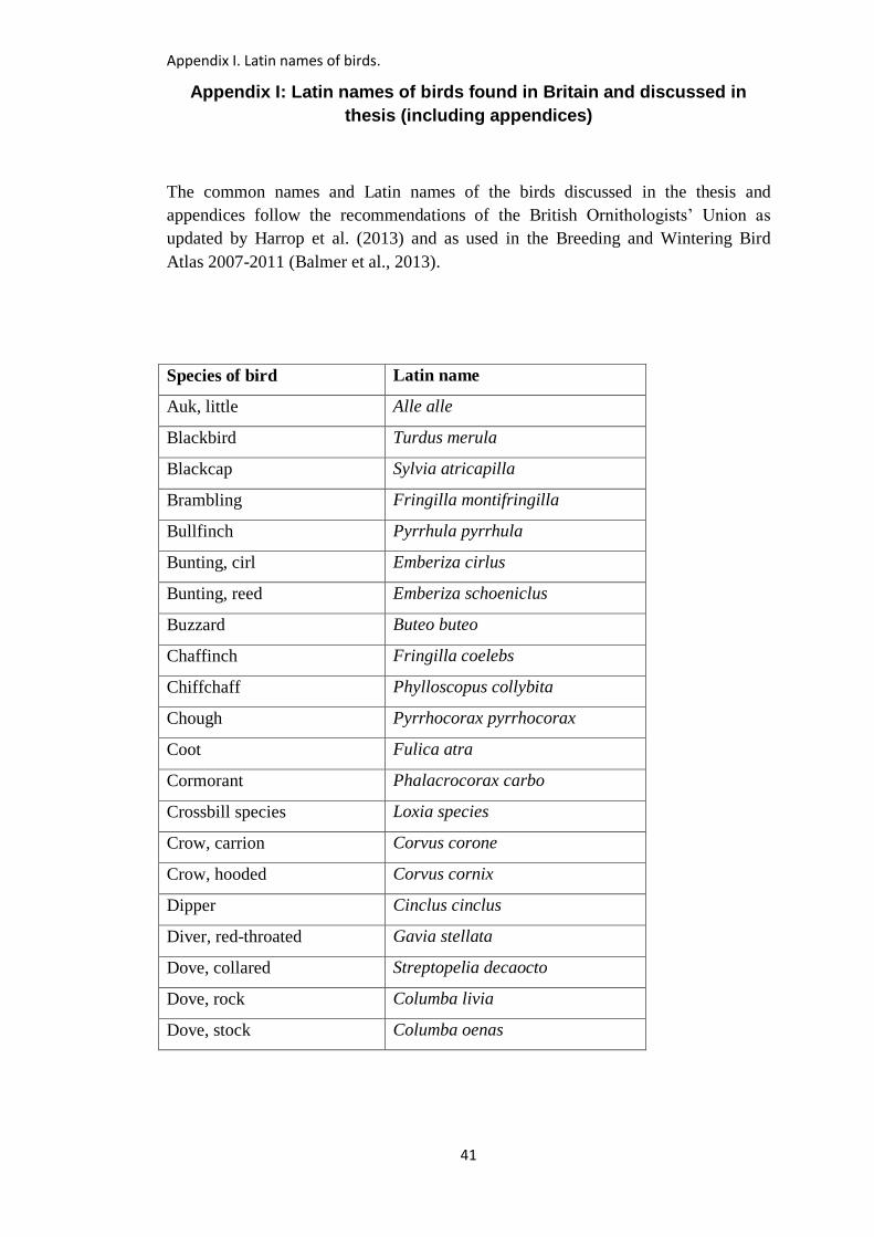
Appendix I. Latin names of birds.
41
Appendix I: Latin names of birds found in Britain and discussed in
thesis (including appendices)
The common names and Latin names of the birds discussed in the thesis and
appendices follow the recommendations of the British Ornithologists’ Union as
updated by Harrop et al. (2013) and as used in the Breeding and Wintering Bird
Atlas 2007-2011 (Balmer et al., 2013).
Species of bird Latin name
Auk, little Alle alle
Blackbird Turdus merula
Blackcap Sylvia atricapilla
Brambling Fringilla montifringilla
Bullfinch Pyrrhula pyrrhula
Bunting, cirl Emberiza cirlus
Bunting, reed Emberiza schoeniclus
Buzzard Buteo buteo
Chaffinch Fringilla coelebs
Chiffchaff Phylloscopus collybita
Chough Pyrrhocorax pyrrhocorax
Coot Fulica atra
Cormorant Phalacrocorax carbo
Crossbill species Loxia species
Crow, carrion Corvus corone
Crow, hooded Corvus cornix
Dipper Cinclus cinclus
Diver, red-throated Gavia stellata
Dove, collared Streptopelia decaocto
Dove, rock Columba livia
Dove, stock Columba oenas
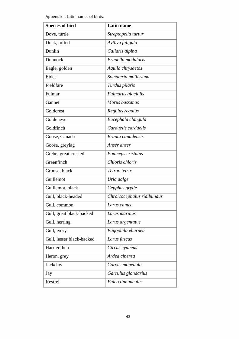
Appendix I. Latin names of birds.
42
Species of bird Latin name
Dove, turtle Streptopelia turtur
Duck, tufted Aythya fuligula
Dunlin Calidris alpina
Dunnock Prunella modularis
Eagle, golden Aquila chrysaetos
Eider Somateria mollissima
Fieldfare Turdus pilaris
Fulmar Fulmarus glacialis
Gannet Morus bassanus
Goldcrest Regulus regulus
Goldeneye Bucephala clangula
Goldfinch Carduelis carduelis
Goose, Canada Branta canadensis
Goose, greylag Anser anser
Grebe, great crested Podiceps cristatus
Greenfinch Chloris chloris
Grouse, black Tetrao tetrix
Guillemot Uria aalge
Guillemot, black Cepphus grylle
Gull, black-headed Chroicocephalus ridibundus
Gull, common Larus canus
Gull, great black-backed Larus marinus
Gull, herring Larus argentatus
Gull, ivory Pagophila eburnea
Gull, lesser black-backed Larus fuscus
Harrier, hen Circus cyaneus
Heron, grey Ardea cinerea
Jackdaw Corvus monedula
Jay Garrulus glandarius
Kestrel Falco tinnunculus
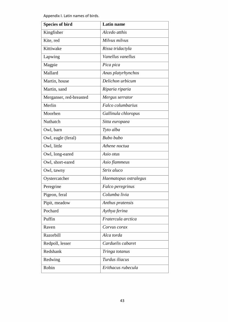
Appendix I. Latin names of birds.
43
Species of bird Latin name
Kingfisher Alcedo atthis
Kite, red Milvus milvus
Kittiwake Rissa tridactyla
Lapwing Vanellus vanellus
Magpie Pica pica
Mallard Anas platyrhynchos
Martin, house Delichon urbicum
Martin, sand Riparia riparia
Merganser, red-breasted Mergus serrator
Merlin Falco columbarius
Moorhen Gallinula chloropus
Nuthatch Sitta europaea
Owl, barn Tyto alba
Owl, eagle (feral) Bubo bubo
Owl, little Athene noctua
Owl, long-eared Asio otus
Owl, short-eared Asio flammeus
Owl, tawny Strix aluco
Oystercatcher Haematopus ostralegus
Peregrine Falco peregrinus
Pigeon, feral Columba livia
Pipit, meadow Anthus pratensis
Pochard Aythya ferina
Puffin Fratercula arctica
Raven Corvus corax
Razorbill Alca torda
Redpoll, lesser Carduelis cabaret
Redshank Tringa totanus
Redwing Turdus iliacus
Robin Erithacus rubecula
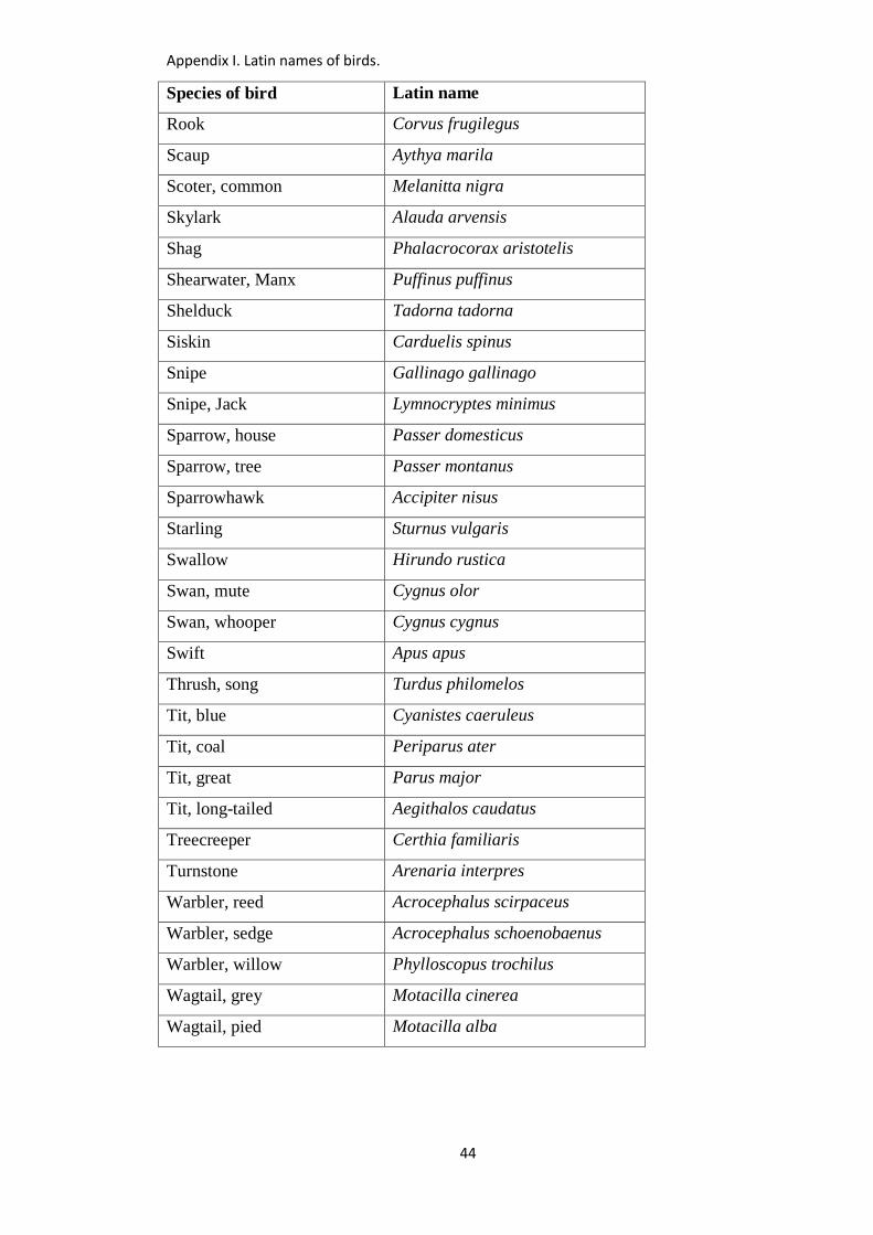
Appendix I. Latin names of birds.
44
Species of bird Latin name
Rook Corvus frugilegus
Scaup Aythya marila
Scoter, common Melanitta nigra
Skylark Alauda arvensis
Shag Phalacrocorax aristotelis
Shearwater, Manx Puffinus puffinus
Shelduck Tadorna tadorna
Siskin Carduelis spinus
Snipe Gallinago gallinago
Snipe, Jack Lymnocryptes minimus
Sparrow, house Passer domesticus
Sparrow, tree Passer montanus
Sparrowhawk Accipiter nisus
Starling Sturnus vulgaris
Swallow Hirundo rustica
Swan, mute Cygnus olor
Swan, whooper Cygnus cygnus
Swift Apus apus
Thrush, song Turdus philomelos
Tit, blue Cyanistes caeruleus
Tit, coal Periparus ater
Tit, great Parus major
Tit, long-tailed Aegithalos caudatus
Treecreeper Certhia familiaris
Turnstone Arenaria interpres
Warbler, reed Acrocephalus scirpaceus
Warbler, sedge Acrocephalus schoenobaenus
Warbler, willow Phylloscopus trochilus
Wagtail, grey Motacilla cinerea
Wagtail, pied Motacilla alba
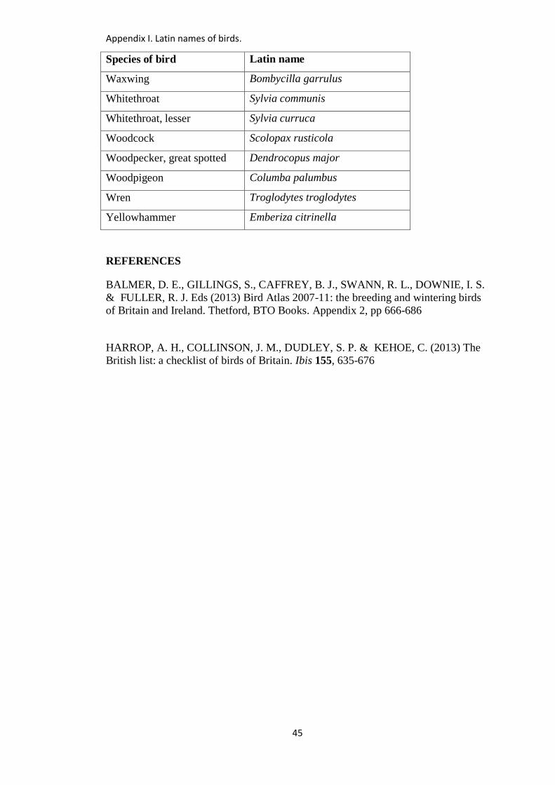
Appendix I. Latin names of birds.
45
Species of bird Latin name
Waxwing Bombycilla garrulus
Whitethroat Sylvia communis
Whitethroat, lesser Sylvia curruca
Woodcock Scolopax rusticola
Woodpecker, great spotted Dendrocopus major
Woodpigeon Columba palumbus
Wren Troglodytes troglodytes
Yellowhammer Emberiza citrinella
REFERENCES
BALMER, D. E., GILLINGS, S., CAFFREY, B. J., SWANN, R. L., DOWNIE, I. S.
& FULLER, R. J. Eds (2013) Bird Atlas 2007-11: the breeding and wintering birds
of Britain and Ireland. Thetford, BTO Books. Appendix 2, pp 666-686
HARROP, A. H., COLLINSON, J. M., DUDLEY, S. P. & KEHOE, C. (2013) The
British list: a checklist of birds of Britain. Ibis 155, 635-676
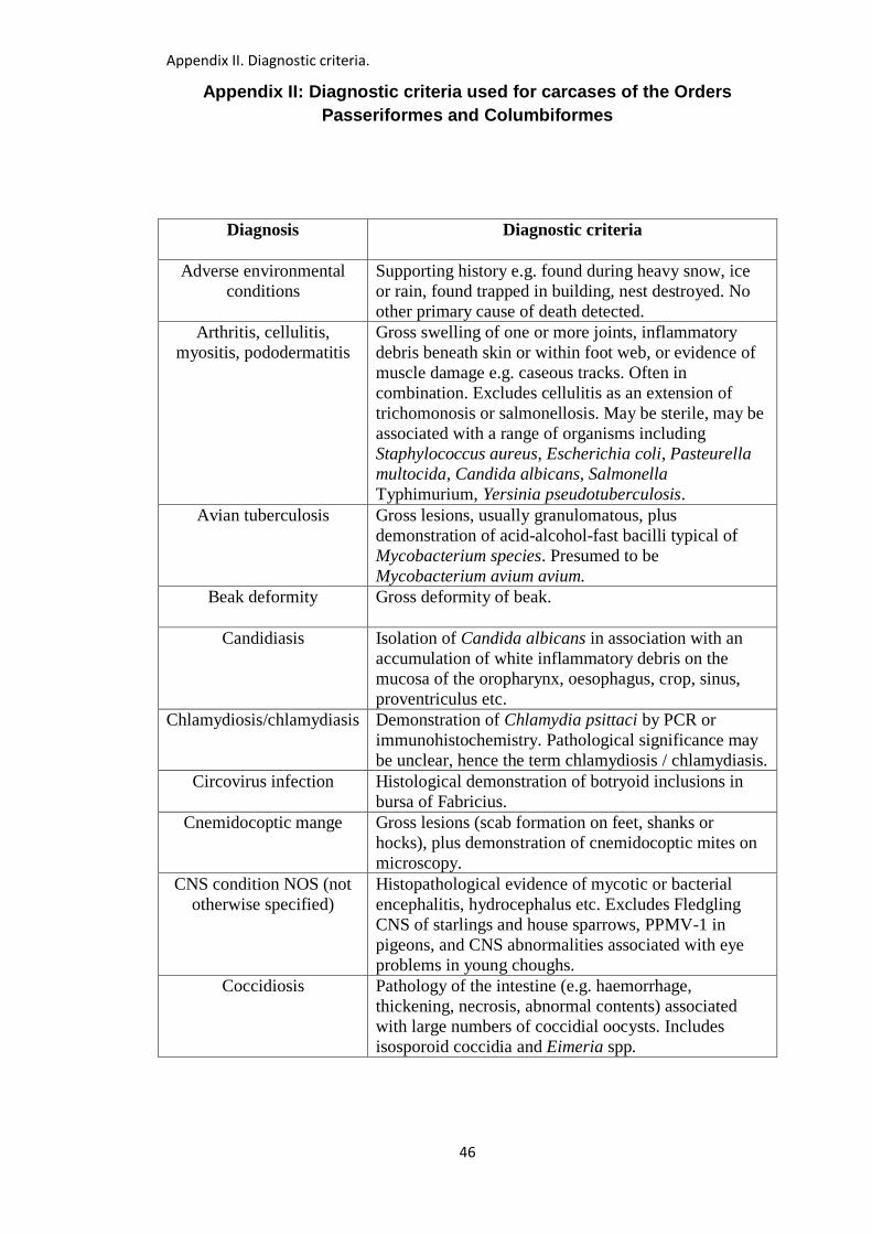
Appendix II. Diagnostic criteria.
46
Appendix II: Diagnostic criteria used for carcases of the Orders
Passeriformes and Columbiformes
Diagnosis
Diagnostic criteria
Adverse environmental
conditions
Supporting history e.g. found during heavy snow, ice
or rain, found trapped in building, nest destroyed. No
other primary cause of death detected.
Arthritis, cellulitis,
myositis, pododermatitis
Gross swelling of one or more joints, inflammatory
debris beneath skin or within foot web, or evidence of
muscle damage e.g. caseous tracks. Often in
combination. Excludes cellulitis as an extension of
trichomonosis or salmonellosis. May be sterile, may be
associated with a range of organisms including
Staphylococcus aureus, Escherichia coli, Pasteurella
multocida, Candida albicans, Salmonella
Typhimurium, Yersinia pseudotuberculosis.
Avian tuberculosis Gross lesions, usually granulomatous, plus
demonstration of acid-alcohol-fast bacilli typical of
Mycobacterium species. Presumed to be
Mycobacterium avium avium.
Beak deformity Gross deformity of beak.
Candidiasis Isolation of Candida albicans in association with an
accumulation of white inflammatory debris on the
mucosa of the oropharynx, oesophagus, crop, sinus,
proventriculus etc.
Chlamydiosis/chlamydiasis Demonstration of Chlamydia psittaci by PCR or
immunohistochemistry. Pathological significance may
be unclear, hence the term chlamydiosis / chlamydiasis.
Circovirus infection Histological demonstration of botryoid inclusions in
bursa of Fabricius.
Cnemidocoptic mange Gross lesions (scab formation on feet, shanks or
hocks), plus demonstration of cnemidocoptic mites on
microscopy.
CNS condition NOS (not
otherwise specified)
Histopathological evidence of mycotic or bacterial
encephalitis, hydrocephalus etc. Excludes Fledgling
CNS of starlings and house sparrows, PPMV-1 in
pigeons, and CNS abnormalities associated with eye
problems in young choughs.
Coccidiosis Pathology of the intestine (e.g. haemorrhage,
thickening, necrosis, abnormal contents) associated
with large numbers of coccidial oocysts. Includes
isosporoid coccidia and Eimeria spp.
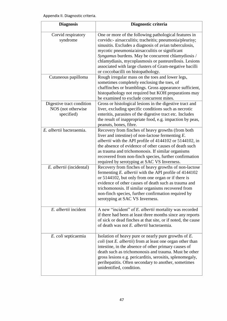
Appendix II. Diagnostic criteria.
47
Diagnosis
Diagnostic criteria
Corvid respiratory
syndrome
One or more of the following pathological features in
corvids:- airsacculitis; tracheitis; pneumonia/pleurisy;
sinusitis. Excludes a diagnosis of avian tuberculosis,
mycotic pneumonia/airsacculitis or significant
Syngamus burdens. May be concurrent chlamydiosis /
chlamydiasis, mycoplasmosis or pasteurellosis. Lesions
associated with large clusters of Gram-negative bacilli
or coccobacilli on histopathology.
Cutaneous papilloma Rough irregular mass on the toes and lower legs,
sometimes completely enclosing the toes, of
chaffinches or bramblings. Gross appearance sufficient,
histopathology not required but KOH preparations may
be examined to exclude concurrent mites.
Digestive tract condition
NOS (not otherwise
specified)
Gross or histological lesions in the digestive tract and
liver, excluding specific conditions such as necrotic
enteritis, parasites of the digestive tract etc. Includes
the result of inappropriate food, e.g. impaction by peas,
peanuts, bones, fibre.
E. albertii bacteraemia.
Recovery from finches of heavy growths (from both
liver and intestine) of non-lactose fermenting E.
albertii with the API profile of 4144102 or 5144102, in
the absence of evidence of other causes of death such
as trauma and trichomonosis. If similar organisms
recovered from non-finch species, further confirmation
required by serotyping at SAC VS Inverness.
E. albertii (incidental) Recovery from finches of heavy growths of non-lactose
fermenting E. albertii with the API profile of 4144102
or 5144102, but only from one organ or if there is
evidence of other causes of death such as trauma and
trichomonosis. If similar organisms recovered from
non-finch species, further confirmation required by
serotyping at SAC VS Inverness.
E. albertii incident A new “incident” of E. albertii mortality was recorded
if there had been at least three months since any reports
of sick or dead finches at that site, or if noted, the cause
of death was not E. albertii bacteraemia.
E. coli septicaemia Isolation of heavy pure or nearly pure growths of E.
coli (not E. albertii) from at least one organ other than
intestine, in the absence of other primary causes of
death such as trichomonosis and trauma. Must be other
gross lesions e.g. pericarditis, serositis, splenomegaly,
perihepatitis. Often secondary to another, sometimes
unidentified, condition.

Appendix II. Diagnostic criteria.
48
Diagnosis
Diagnostic criteria
Eye problems Includes corneal opacity and associated problems in
young choughs, developmental abnormalities in other
species, and hypopyon.
Feather/skin abnormality
NOS (not otherwise
specified)
Abnormalities of the feathers or skin, including foot
lesions but excluding abnormalities caused by trauma,
oiling, pox or external parasites.
Fledgling CNS History of central nervous signs such as circling and
torticollis in fledgling starlings and house sparrows. If
histopathology carried out, a non-suppurative
encephalitis is present. Aetiology uncertain.
Foreign body Presence of a foreign body in the digestive tract that is
causing a significant problem, excluding inappropriate
food.
Inclusion body hepatitis Histological demonstration of basophilic or
eosinophilic intranuclear inclusion bodies in
hepatocytes.
Kidney disorder Pale kidneys and/or urate deposits on heart, liver,
peritoneum, subcutaneous tissues or joints.
Malnourished nestling /
fledgling corvids
Immature corvids in poor body condition, often with
poor feathering and soft bones/leg deformities. May be
concurrent internal or external parasites, terminal
trauma etc. Usually because corvids hatch in an
asynchronous fashion, and the last bird to hatch may
receive less food, especially in years of limited food
supply.
Megabacteriosis Demonstration of avian gastric yeasts Macrorhabdus
ornithogaster (“megabacteria”) in wet preparations or
histopathology sections from the proventriculus.
Pathological significance often unclear.
Metabolic bone disease Immature birds. Bones of legs soft and pliable.
Sometimes also bones of skull, ribs etc. Excludes
malnourished nestling/fledgling corvids.
Mycotic pneumonia /
airsacculitis
Gross lesions (e.g. granulomata in lungs, plaques on
airsacs), and culture of pathogenic fungi or
demonstration of fungal elements on histopathology.
Necrotic enteritis Focal or diffuse diphtheritic necrosis of the small
intestine associated with Clostridium perfringens.
Neoplasia Gross lesions and histopathology.
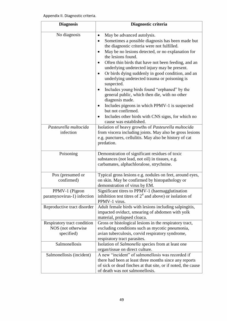
Appendix II. Diagnostic criteria.
49
Diagnosis
Diagnostic criteria
No diagnosis May be advanced autolysis.
Sometimes a possible diagnosis has been made but the diagnostic criteria were not fulfilled.
May be no lesions detected, or no explanation for the lesions found.
Often thin birds that have not been feeding, and an
underlying undetected injury may be present.
Or birds dying suddenly in good condition, and an underlying undetected trauma or poisoning is
suspected.
Includes young birds found “orphaned” by the general public, which then die, with no other
diagnosis made.
Includes pigeons in which PPMV-1 is suspected
but not confirmed.
Includes other birds with CNS signs, for which no cause was established.
Pasteurella multocida
infection
Isolation of heavy growths of Pasteurella multocida
from viscera including joints. May also be gross lesions
e.g. punctures, cellulitis. May also be history of cat
predation.
Poisoning Demonstration of significant residues of toxic
substances (not lead, not oil) in tissues, e.g.
carbamates, alphachloralose, strychnine.
Pox (presumed or
confirmed)
Typical gross lesions e.g. nodules on feet, around eyes,
on skin. May be confirmed by histopathology or
demonstration of virus by EM.
PPMV-1 (Pigeon
paramyxovirus-1) infection
Significant titres to PPMV-1 (haemagglutination
inhibition test titres of 24 and above) or isolation of
PPMV-1 virus.
Reproductive tract disorder Adult female birds with lesions including salpingitis,
impacted oviduct, smearing of abdomen with yolk
material, prolapsed cloaca.
Respiratory tract condition
NOS (not otherwise
specified)
Gross or histological lesions in the respiratory tract,
excluding conditions such as mycotic pneumonia,
avian tuberculosis, corvid respiratory syndrome,
respiratory tract parasites.
Salmonellosis Isolation of Salmonella species from at least one
organ/tissue on direct culture.
Salmonellosis (incident) A new “incident” of salmonellosis was recorded if
there had been at least three months since any reports
of sick or dead finches at that site, or if noted, the cause
of death was not salmonellosis.
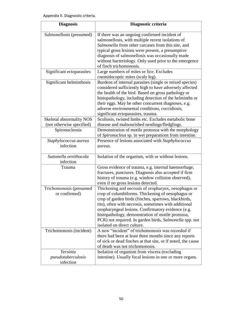
Appendix II. Diagnostic criteria.
50
Diagnosis
Diagnostic criteria
Salmonellosis (presumed) If there was an ongoing confirmed incident of
salmonellosis, with multiple recent isolations of
Salmonella from other carcases from this site, and
typical gross lesions were present, a presumptive
diagnosis of salmonellosis was occasionally made
without bacteriology. Only used prior to the emergence
of finch trichomonosis.
Significant ectoparasites Large numbers of mites or lice. Excludes
cnemidocoptic mites (scaly leg).
Significant helminthosis Burdens of internal parasites (single or mixed species)
considered sufficiently high to have adversely affected
the health of the bird. Based on gross pathology or
histopathology, including detection of the helminths or
their eggs. May be other concurrent diagnoses, e.g.
adverse environmental conditions, coccidiosis,
significant ectoparasites, trauma.
Skeletal abnormality NOS
(not otherwise specified)
Scoliosis, twisted limbs etc. Excludes metabolic bone
disease and malnourished nestlings/fledglings.
Spironucleosis Demonstration of motile protozoa with the morphology
of Spironucleus sp. in wet preparations from intestine.
Staphylococcus aureus
infection
Presence of lesions associated with Staphylococcus
aureus.
Suttonella ornithocola
infection
Isolation of the organism, with or without lesions.
Trauma Gross evidence of trauma, e.g. internal haemorrhage,
fractures, punctures. Diagnosis also accepted if firm
history of trauma (e.g. window collision observed),
even if no gross lesions detected.
Trichomonosis (presumed
or confirmed)
Thickening and necrosis of oropharynx, oesophagus or
crop of columbiforms. Thickening of oesophagus or
crop of garden birds (finches, sparrows, blackbirds,
tits), often with necrosis, sometimes with additional
oropharyngeal lesions. Confirmatory evidence (e.g.
histopathology, demonstration of motile protozoa,
PCR) not required. In garden birds, Salmonella spp. not
isolated on direct culture.
Trichomonosis (incident) A new “incident” of trichomonosis was recorded if
there had been at least three months since any reports
of sick or dead finches at that site, or if noted, the cause
of death was not trichomonosis.
Yersinia
pseudotuberculosis
infection
Isolation of organism from viscera (excluding
intestine). Usually focal lesions in one or more organs.
Appendix III. Presumptive identification of internal parasites.
51
Appendix III: Presumptive identification of internal parasites from wild
birds of the Orders Passeriformes and Columbiformes
Some of the internal parasites and/or their eggs that were encountered during this study
are described in this appendix. Where appropriate, presumptive identification of the
parasites is given. However, this is not a detailed description of the parasites, but rather a
basic introductory guide to help non-parasitologists identify the commonest internal
parasites to group or genus level. Measurements below are expressed as mean ± 2
standard deviations. Images, where indicated, are in Appendix X. It is hoped that the
descriptions in this appendix can be expanded in future years.
Descriptive terms used
Spherical – all dimensions equal or nearly equal.
Sub-spherical – nearly spherical.
Ellipsoid – like a stretched-out circle.
Elongate ellipsoid – ellipsoid where length is more than three times the width.
Ovoid – blunt end and sharp end readily recognised.
Pyriform – pear-shaped, with blunt end considerably larger than sharp end.

Appendix III. Presumptive identification of internal parasites.
52
Chapter 3 – finches, sparrows, buntings, dunnocks and tits
Syngamid worms from trachea of house sparrows
White or reddish worms, circular in cross-section, large female and small male
joined together to form a Y-shape. 1 mm in diameter, around 6-8 mm long.
Fresh adult females may have a “barber-pole” appearance caused by the red
digestive tract and white undulating reproductive tract.
Presumptive identification as Syngamus sp., most likely Syngamus trachea (see
3.2.10 in thesis).
Eggs ellipsoid, clear/colourless with a dark morula of 8-16 blastomeres and a thick
smooth shell. Operculum at both poles. Images 15a-16b.
35 eggs from two house sparrows:
o length 91 µm ± 7 µm
o width 45 µm ± 3 µm
Hairworms from digestive tract of dunnocks
Long thin worms barely visible to the naked eye, usually seen on microscopy of
smears from oesophagus or intestine.
Typical hairworm eggs (see below) seen in adult female worms.
Presumptive identification of worms in the upper digestive tract as Eucoleus sp., and
worms in lower digestive tract as Capillaria / Baruscapillaria sp., but some debate
about nomenclature and described generically in thesis as “hairworms”.
Eggs brown or golden in colour, ellipsoid, with distinctive bipolar plugs that
sometimes protrude. Images 38a-38c.
20 hairworm eggs from two dunnocks measured:
o length 60 µm ± 10 µm
o width 26 µm ± 3 µm
Appendix III. Presumptive identification of internal parasites.
53
Schistosome-like eggs from intestinal contents of dunnocks and house sparrow
Unidentified eggs, described in the thesis as schistosome-like eggs, were detected in
the intestinal mucosa and/or contents of dunnocks, and the intestinal contents of a
house sparrow. Also detected in intestinal contents of blackbirds.
Their identity is discussed more fully in sections 3.3.9, 8.3.9 and 8.4.3 of the thesis.
Eggs spherical, sub-spherical or ellipsoid, with thin smooth outer membrane. Dark
granular contents that may fill the egg, or may be retracted to produce a spherical or
ellipsoid mass with a clear space between the contents and the outer membrane.
Images 23-32 (dunnocks) and images 33-36 (house sparrow).
May be found within the mucosa of the intestine, sometimes in large numbers. Image
22.
Variable in size. The figures below are expressed as mean and range, rather than
mean ± 2 standard deviations, because the dimensions did not form a normal
distribution. Instead, there appeared to be two sub-populations. For the purposes of
the images in Appendix X, those 75 µm and above are described as “larger form”,
and those less than 75 µm are described as “smaller form”.
o Mean length 75 µm (range 54-105) and width 68 µm (range 53-97) (110 eggs
from three dunnocks).
o Mean length 75 µm (range 61-110) and width 68 µm (range 52-99) (38 eggs
from one house sparrow).
Coccidial oocysts from intestine of finches and sparrows
Unsporulated oocysts are spherical/sub-spherical, thick walled, sometimes
containing a contracted spherical sporont. Images 17-19.
o 20 oocysts from one house sparrow: spherical/sub-spherical oocyst, length 26 µm
± 3 µm and width 24 µm ± 3 µm.
o 27 oocysts (smaller form) from two chaffinches: spherical/sub-spherical oocyst,
length 23 µm ± 3 µm and width 21 µm ± 4 µm. Images 17a-17c.
Appendix III. Presumptive identification of internal parasites.
54
o 33 oocysts (larger form) from two chaffinches: spherical/sub-spherical oocyst,
length 27 µm ± 2 µm and width 25 µm ± 3 µm. Images 18a-18c.
On the few occasions that sporulation was carried out, two sporocysts were present.
Image 20.
Described as isosporoid coccidia (see 3.2.9 in thesis). Probably several different
species, possibly even within the same host.
Trichomonas sp. in upper digestive tract of finches
Motile protozoa that are difficult to detect microscopically unless they are actively
moving. The greater the time interval between the death or euthanasia of the bird
and microscopic examination of the material, the greater the likelihood that the
organisms will stop moving and not be detected.
Round, pyriform or crescent-shaped. Measure approximately 5-20 µm by 2-10 µm
(about the same size as an avian red blood cell).
Have a prominent undulating membrane, protruding axostyle, but no ventral sucker,
and have several anterior flagella.
Move in a relatively slow, rotational fashion.
Presumptive identification as Trichomonas gallinae (see 6.2 in thesis).
Appendix III. Presumptive identification of internal parasites.
55
Chapter 8 – blackbirds, thrushes, robins and starlings
Large roundworms from intestine of blackbirds/thrushes and starlings
Large whitish worms, circular in cross-section, pointed ends. 1-2 mm in diameter.
Mostly 15-45 mm long. Images 142, 143, 162.
Presumptive identification as Porrocaecum sp., most likely Porrocaecum
ensicaudatum (see 8.2.11 in thesis).
Eggs ellipsoid, thick scalloped shell, opercula sometimes visible at one or both poles.
Dark contents, often retracted into circular or ellipsoid solid mass with space
between shell and contents.
45 eggs from three blackbirds (images 144-148):
o length 103 µm ± 15 µm
o width 75 µm ± 12 µm
24 eggs from one starling (images 149-152):
o length 97 µm ± 7 µm
o width 67 µm ± 5 µm
Syngamid worms from trachea of blackbirds/thrushes and starlings
White or reddish worms, circular in cross-section, large female and small male
joined together to form a Y-shape. 1 mm in diameter, around 8-12 mm long.
Fresh adult females may have a “barber-pole” appearance caused by the red
digestive tract and white undulating reproductive tract.
Presumptive identification as Syngamus sp., most likely Syngamus trachea (see
8.2.11 in thesis).
Eggs ellipsoid, clear/colourless, with a dark morula of 8-16 blastomeres and a thick
smooth shell. Operculum at both poles. Images 156-161.
Appendix III. Presumptive identification of internal parasites.
56
7 eggs from one blackbird:
o length 91 µm ± 6 µm
o width 49 µm ± 8 µm
19 eggs from one starling:
o length 85 µm ± 7 µm
o width 50 µm ± 7 µm
Hairworms from digestive tract of blackbirds/thrushes, robins and starlings
Long thin worms barely visible to the naked eye, usually seen on microscopy of
smears from oesophagus or intestine.
Typical hairworm eggs (see below) seen in adult female worms.
Presumptive identification of worms in the upper digestive tract as Eucoleus sp., and
worms in lower digestive tract as Capillaria / Baruscapillaria sp., but some debate
about nomenclature and described generically in thesis as “hairworms”.
Eggs brown or golden in colour, barrel-shaped, with distinctive bipolar plugs that
sometimes protrude. Images 139-141.
Thick-shelled, with granular contents that fill the egg.
17 hairworm eggs from one blackbird measured:
o length 61 µm ± 10 µm
o width 29 µm ± 5 µm
Tapeworms and eggs from intestine of blackbirds/thrushes, robins and starlings
Usually found as fragments up to 4mm in width, cream coloured. Flattened.
Fragments often tapered in size to one extremity. Image 162.
Identification not attempted in thesis. The commonest tapeworms found in
blackbirds/thrushes and starlings are Hymenolepis spp. and Dilepis spp. (see 8.2.11
in thesis).
Appendix III. Presumptive identification of internal parasites.
57
Eggs recovered from intestinal contents of blackbirds/thrushes vary in appearance,
with a hexacanth larva with six hooks enclosed in one or more envelopes.
Type 1. Six-hooked hexacanth larva enclosed in two well-defined envelopes. Images
171-174.
o Spherical/subspherical hexacanth, length 48 µm ± 4 µm and width 46 µm ± 4 µm
(10 eggs from one blackbird).
o Inner envelope spherical/subspherical, length 57 µm ± 4 µm and width 54 µm ±
3 µm (10 eggs from one blackbird).
o Outer envelope ellipsoid, length 99 µm ± 15 µm and width 75 µm ± 13 µm (10
eggs from one blackbird).
Type 2. Small six-hooked hexacanth larva enclosed in one well-defined envelope
and a thin outer envelope. Images 175-178.
o Spherical hexacanth, length 23 µm ± 2 µm and width 23 µm ± 2 µm (7 eggs from
one redwing).
o Inner envelope spherical, length 28 µm ± 4 µm and width 28 µm ± 4 µm (7 eggs
from one redwing).
o Outer envelope thin, ellipsoid, sometimes distorted, length 62 µm ± 12 µm and
width 54 µm ± 14 µm (7 eggs from one redwing).
Type 3. Six-hooked hexacanth larva enclosed in a single well-defined envelope.
Dimensions variable. Example 1 (no images):
o Spherical/subspherical hexacanth, length 52 µm ± 8 µm and width 50 µm ± 7 µm
(14 eggs from two blackbirds).
o Spherical/subspherical envelope, length 69 µm ± 9 µm and width 66 µm ± 7 µm
(14 eggs from two blackbirds).
Type 3. Six-hooked hexacanth larva enclosed in a single well-defined envelope.
Dimensions variable. Example 2. Images 165-168:
o Spherical/subspherical hexacanth, length 39 µm ± 6 µm and width 36 µm ± 5 µm
(18 eggs from one blackbird and one redwing).
o Spherical/subspherical envelope, length 60 µm ± 11 µm and width 50 µm ± 8 µm
(18 eggs from one blackbird and one redwing).
Appendix III. Presumptive identification of internal parasites.
58
Type 3. Six-hooked hexacanth larva enclosed in a single well-defined envelope.
Dimensions variable. Example 3. Images 163-164:
o Spherical/subspherical hexacanth, length 43 µm ± 4 µm and width 40 µm ± 4 µm
(5 eggs from one blackbird).
o Spherical/subspherical envelope, length 87 µm ± 8 µm and width 83 µm ± 7 µm
(5 eggs from one blackbird).
Flukes and fluke eggs from digestive tract of blackbirds/thrushes, robins and starlings
Flukes usually not seen grossly, but flukes or fluke eggs seen on microscopy of wet
preparations from the intestine. Fluke eggs found in intestine may originate from
flukes elsewhere in the body.
Identification not attempted in thesis. See 8.2.11 in thesis for possible species.
Eggs ovoid or ellipsoid, brown in colour, with contents filling the shell. May be
asymmetric, with one side wall longer than the other side. Operculum may be visible
at one pole. Images 191-198.
13 eggs from one redwing:
o length 32 µm ± 4 µm
o width 19 µm ± 2 µm
15 eggs from one robin:
o length 34 µm ± 5 µm
o width 23 µm ± 2 µm
Thorny-headed worms from digestive tract of blackbirds/thrushes and starlings
White, brown or grey when in situ in intestine. Attached to mucosa.
Approximately 3-10 mm long, 1 mm wide. After storage in alcohol or formol saline,
look white and less wrinkled. Images 179-181.
May be smooth and turgid and circular on cross-section, or corrugated and flaccid.
Thin proboscis may be apparent grossly, protruding from one end.
Appendix III. Presumptive identification of internal parasites.
59
Cylindrical proboscis with many hooks visible on microscopy. Images 182-183.
Presumptive identification as Plagiorhynchus sp., most likely Plagiorhynchus
(Prosthorhynchus) cylindraceus (see 8.2.11 in thesis). Specimens from one starling
confirmed as Plagiorhynchus (Prosthorhynchus) cylindraceus by Mrs Eileen Harris
at the Natural History Museum, London.
Alternatively, the proboscis may have a spherical tip with large spines, and small
spines on the posterior proboscis – possibly Sphaerirostris sp. Image 184.
Eggs seldom seen in intestinal smears, but when obtained from worms a variety of
forms may be seen:
o Granular embryo within a thick-shelled membrane. Most likely Plagiorhynchus
sp. Images 185-186.
The embryo forms an extended ellipse, length 35 µm ± 7 µm and width 11
µm ± 2 µm (16 eggs from two blackbirds).
The thick outer membrane is ellipsoid, length 46 µm ± 4 µm and width 18 µm
± 2 µm (23 eggs from two blackbirds).
o Larger form with three or more different layers. Possibly Sphaerirostris sp.
Images 187-190.
Outer layer is ellipsoid, length 73 µm ± 12 µm and width 33 µm ± 6 µm (24
eggs from two blackbirds).
Coccidial oocysts from intestine of blackbirds/thrushes, robins and starlings
Unsporulated oocysts are spherical/subspherical, thick walled, sometimes containing
a contracted spherical sporont. Images 111-118.
Spherical sporont, length 20 µm ± 1 µm and width 19 µm ± 2 µm. Contained
within spherical/subspherical oocyst wall, length 26 µm ± 2 µm and width 24 µm
± 2 µm (11 oocysts from one blackbird).
Spherical/subspherical oocyst wall, length 22 µm ± 4 µm and width 21 µm ± 4
µm (15 unsporulated oocysts from one starling).
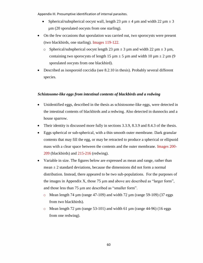
Appendix III. Presumptive identification of internal parasites.
60
Spherical/subspherical oocyst wall, length 23 µm ± 4 µm and width 22 µm ± 3
µm (20 sporulated oocysts from one starling).
On the few occasions that sporulation was carried out, two sporocysts were present
(two blackbirds, one starling). Images 119-122.
o Spherical/subspherical oocyst length 23 µm ± 3 µm and width 22 µm ± 3 µm,
containing two sporocysts of length 15 µm ± 5 µm and width 10 µm ± 2 µm (9
sporulated oocysts from one blackbird).
Described as isosporoid coccidia (see 8.2.10 in thesis). Probably several different
species.
Schistosome-like eggs from intestinal contents of blackbirds and a redwing
Unidentified eggs, described in the thesis as schistosome-like eggs, were detected in
the intestinal contents of blackbirds and a redwing. Also detected in dunnocks and a
house sparrow.
Their identity is discussed more fully in sections 3.3.9, 8.3.9 and 8.4.3 of the thesis.
Eggs spherical or sub-spherical, with a thin smooth outer membrane. Dark granular
contents that may fill the egg, or may be retracted to produce a spherical or ellipsoid
mass with a clear space between the contents and the outer membrane. Images 200-
209 (blackbirds) and 215-216 (redwing).
Variable in size. The figures below are expressed as mean and range, rather than
mean ± 2 standard deviations, because the dimensions did not form a normal
distribution. Instead, there appeared to be two sub-populations. For the purposes of
the images in Appendix X, those 75 µm and above are described as “larger form”,
and those less than 75 µm are described as “smaller form”.
o Mean length 74 µm (range 47-109) and width 72 µm (range 59-109) (37 eggs
from two blackbirds).
o Mean length 72 µm (range 53-101) and width 61 µm (range 44-96) (16 eggs
from one redwing).
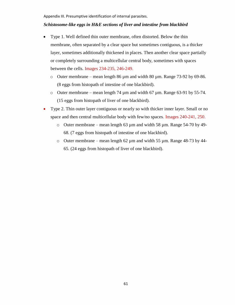
Appendix III. Presumptive identification of internal parasites.
61
Schistosome-like eggs in H&E sections of liver and intestine from blackbird
Type 1. Well defined thin outer membrane, often distorted. Below the thin
membrane, often separated by a clear space but sometimes contiguous, is a thicker
layer, sometimes additionally thickened in places. Then another clear space partially
or completely surrounding a multicellular central body, sometimes with spaces
between the cells. Images 234-235, 246-249.
o Outer membrane – mean length 86 µm and width 80 µm. Range 73-92 by 69-86.
(8 eggs from histopath of intestine of one blackbird).
o Outer membrane – mean length 74 µm and width 67 µm. Range 63-91 by 55-74.
(15 eggs from histopath of liver of one blackbird).
Type 2. Thin outer layer contiguous or nearly so with thicker inner layer. Small or no
space and then central multicellular body with few/no spaces. Images 240-241, 250.
o Outer membrane – mean length 63 µm and width 58 µm. Range 54-70 by 49-
68. (7 eggs from histopath of intestine of one blackbird).
o Outer membrane – mean length 62 µm and width 55 µm. Range 48-73 by 44-
65. (24 eggs from histopath of liver of one blackbird).
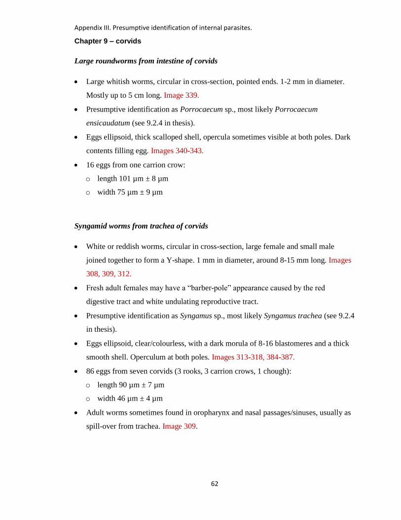
Appendix III. Presumptive identification of internal parasites.
62
Chapter 9 – corvids
Large roundworms from intestine of corvids
Large whitish worms, circular in cross-section, pointed ends. 1-2 mm in diameter.
Mostly up to 5 cm long. Image 339.
Presumptive identification as Porrocaecum sp., most likely Porrocaecum
ensicaudatum (see 9.2.4 in thesis).
Eggs ellipsoid, thick scalloped shell, opercula sometimes visible at both poles. Dark
contents filling egg. Images 340-343.
16 eggs from one carrion crow:
o length 101 µm ± 8 µm
o width 75 µm ± 9 µm
Syngamid worms from trachea of corvids
White or reddish worms, circular in cross-section, large female and small male
joined together to form a Y-shape. 1 mm in diameter, around 8-15 mm long. Images
308, 309, 312.
Fresh adult females may have a “barber-pole” appearance caused by the red
digestive tract and white undulating reproductive tract.
Presumptive identification as Syngamus sp., most likely Syngamus trachea (see 9.2.4
in thesis).
Eggs ellipsoid, clear/colourless, with a dark morula of 8-16 blastomeres and a thick
smooth shell. Operculum at both poles. Images 313-318, 384-387.
86 eggs from seven corvids (3 rooks, 3 carrion crows, 1 chough):
o length 90 µm ± 7 µm
o width 46 µm ± 4 µm
Adult worms sometimes found in oropharynx and nasal passages/sinuses, usually as
spill-over from trachea. Image 309.
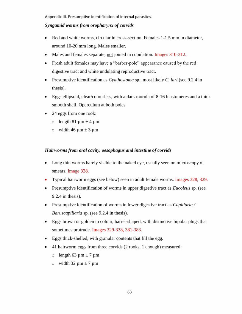
Appendix III. Presumptive identification of internal parasites.
63
Syngamid worms from oropharynx of corvids
Red and white worms, circular in cross-section. Females 1-1.5 mm in diameter,
around 10-20 mm long. Males smaller.
Males and females separate, not joined in copulation. Images 310-312.
Fresh adult females may have a “barber-pole” appearance caused by the red
digestive tract and white undulating reproductive tract.
Presumptive identification as Cyathostoma sp., most likely C. lari (see 9.2.4 in
thesis).
Eggs ellipsoid, clear/colourless, with a dark morula of 8-16 blastomeres and a thick
smooth shell. Operculum at both poles.
24 eggs from one rook:
o length 81 µm ± 4 µm
o width 46 µm ± 3 µm
Hairworms from oral cavity, oesophagus and intestine of corvids
Long thin worms barely visible to the naked eye, usually seen on microscopy of
smears. Image 328.
Typical hairworm eggs (see below) seen in adult female worms. Images 328, 329.
Presumptive identification of worms in upper digestive tract as Eucoleus sp. (see
9.2.4 in thesis).
Presumptive identification of worms in lower digestive tract as Capillaria /
Baruscapillaria sp. (see 9.2.4 in thesis).
Eggs brown or golden in colour, barrel-shaped, with distinctive bipolar plugs that
sometimes protrude. Images 329-338, 381-383.
Eggs thick-shelled, with granular contents that fill the egg.
41 hairworm eggs from three corvids (2 rooks, 1 chough) measured:
o length 63 µm ± 7 µm
o width 32 µm ± 7 µm
Appendix III. Presumptive identification of internal parasites.
64
Spirurid worms from gizzard of choughs
White worms up to 16 mm long and 1 mm wide, embedded in underside of koilin
layer of gizzard. Images 388-393.
Microscopy – cervical papillae at anterior end but no obvious cordons. Images 394-
398.
Microscopy – spicules, caudal alae and papillae at posterior end of male. Images
399-402.
Microscopy – large numbers of eggs in female. Images 403-404.
Eggs ellipsoid, clear/colourless, thick smooth shell, no obvious operculum, usually
with a well-formed larva visible. Images 405, 406, 408.
30 eggs from one chough:
o length 40 µm ± 3 µm
o width 21 µm ± 2 µm
Described as nematodes of the Order Spirurida (spirurid worms). Not species of
Streptocara, Echinuria, Dyspharynx, Cosmocephalus or Tetrameres, but identity
unknown. See 9.3.5 of thesis.
Tapeworms and eggs from intestine of corvids
Usually found as fragments up to 4 mm in width, cream coloured. Flattened.
Fragments often tapered in size to one extremity. Images 346-348, 356.
Identification not attempted. The commonest tapeworms found in corvids are
Hymenolepis spp. and Dilepis spp. (see 9.2.4 in thesis).
Eggs recovered from intestinal contents of corvids. Images 349-355.
o Spherical/subspherical six-hooked hexacanth larva, length 44 µm ± 7 µm and
width 40 µm ± 7 µm (31 eggs from five corvids [4 rooks, 1 carrion crow]).
o Enclosed in single well-defined spherical/subspherical envelope, length 64 µm ±
14 µm and width 58 µm ± 13 µm (31 eggs from five corvids [4 rooks, 1 carrion
crow]).
Appendix III. Presumptive identification of internal parasites.
65
Thorny-headed worms from digestive tract of corvids
White/grey when in situ in intestine. Attached to mucosa. Images 356-359, 409-416.
Approximately 3-10 mm long, 1 mm wide. After storage in alcohol or formol saline,
look white and less wrinkled.
May be smooth and turgid and circular on cross-section, or corrugated and flaccid.
Thin proboscis may be apparent grossly, protruding from one end. Cylindrical
proboscis with many hooks visible on microscopy. Images 360-361, 417-418.
The commonest species reported from corvids is Plagiorhynchus (Prosthorhynchus)
cylindraceus (see 9.2.4 in thesis). Specimens from two choughs confirmed as
Plagiorhynchus (Prosthorhynchus) cylindraceus by Mrs Eileen Harris at the Natural
History Museum, London. But possibly other species also found in corvids.
Eggs seldom seen in intestinal smears but when obtained from worms a variety of
forms may be seen. Unclear if these represent different stages of development or
different species.
o a) Granular embryo within a thick-shelled membrane. Images 364-369.
Embryo forms an extended ellipse, length 46 µm ± 8 µm and width 16 µm ±
5 µm (28 eggs from one rook).
Enclosed in a thicker membrane which is ellipsoid, length 53 µm ± 10 µm
and width 23 µm ± 7 µm (28 eggs from one rook).
o b) Ellipsoid form with two (occasionally three) different layers. Images 362-363.
Inner layer is ellipsoid, length 44 µm ± 3 µm and width 21 µm ± 3 µm (5
eggs from 1 carrion crow).
Outer layer is ellipsoid, length 54 µm ± 8 µm and width 27 µm ± 4 µm (5
eggs from 1 carrion crow).
o c) Larger ellipsoid form filled with granular material. Images 419-422.
Length 60 µm ± 6 µm and width 20 µm ± 4 µm (22 eggs from 1 chough).
o d) Larger ellipsoid form with multiple layers and indistinct contents. Images 370-
373.
Outer Length 70 µm ± 7 µm and width 30 µm ± 4 µm (27 eggs from 1 rook).
Appendix III. Presumptive identification of internal parasites.
66
Coccidial oocysts from intestine of corvids
Unsporulated oocysts are ellipsoid, thick-walled, often containing a contracted
spherical sporont. Images 344 (a-c)
o Ellipsoid oocyst wall - length 19 µm ± 3 µm and width 18 µm ± 3 µm (119
oocysts from five corvids [3 rooks, 1 jackdaw, 1 carrion crow]).
o Spherical sporont, length 15 µm ± 3 µm and width 14 µm ± 3 µm. (39
unsporulated oocysts from two rooks).
When sporulation was carried out, two sporocysts were present (one rook, one
jackdaw, one carrion crow). Images 345 (a-c).
o Ellipsoid oocyst length 21 µm ± 4 µm and width 18 µm ± 3 µm (25 sporulated
oocysts from three corvids), containing two sporocysts of length 12 µm ± 3 µm
and width 9 µm ± 2 µm (32 sporocysts from one jackdaw and one carrion crow).
Described as isosporoid coccidia (see 9.2.4 in thesis). Probably several different
species, possibly even within the same host.
Coccidial oocysts from faeces of choughs
Spherical / subspherical, thick-walled, unsporulated with granular contents filling the
oocyst. Images 344 (d-f).
Length 21 µm ± 3 µm and width 20 µm ± 3 µm (38 oocysts from multiple faeces)
Described as isosporoid coccidia (see 9.2.4 in thesis).
Appendix III. Presumptive identification of internal parasites.
67
Chapter 10 – other passerines
Flukes and fluke eggs from digestive tract of house martins and swallows
Flukes usually not seen grossly, but flukes or fluke eggs seen on microscopy of wet
preparations from the intestine. Images 576-577.
Eggs ovoid or ellipsoid, brown or gold in colour, with contents filling the shell.
Operculum may be visible at one pole. Images 578-579.
33 eggs from two house martins:
o length 33 µm ± 4 µm
o width 22 µm ± 2 µm
Identification not attempted in thesis. However, Plagiorchis maculosus is a well-
recognised intestinal trematode of swallows and house martins in Europe (see 10.4
of thesis), and produces eggs of this order of magnitude.
Nematodes from airsacs of blackcap
Long, thin, white worms, measuring up to 45mm. Images 574-575.
Identified by Mrs Eileen Harris at the Natural History Museum, London, as
Diplotriaena tridens.
No images or measurements of eggs from the current study.
Coccidial oocysts from intestine of pied wagtail
Spherical / subspherical, thick-walled, unsporulated with granular contents filling the
oocyst. No images or measurements.
Presumed to be isosporoid coccidia (see 10.3.5 in thesis).
Appendix III. Presumptive identification of internal parasites.
68
Chapter 11 – pigeons and doves
Coccidial oocysts from pigeons/doves
Unsporulated oocysts are spherical/sub-spherical, thick-walled, granular contents
usually filling oocyst. Images 651-655.
o 41 oocysts from one feral pigeon: mostly spherical/sub-spherical, length 20 µm ±
4 µm and width 19 µm ± 4 µm.
o 25 oocysts from one woodpigeon: mostly spherical/sub-spherical, length 20 µm ±
3 µm and width 19 µm ± 3 µm.
o 30 oocysts from one woodpigeon: mostly ovoid, length 19 µm ± 3 µm and width
16 µm ± 1 µm.
Likely to be Eimeria spp., e.g. E. labbeana and E. columbarum (see 11.2.9 in thesis).
Possibly several species, therefore described as Eimeria spp.
Hairworms from intestine of pigeons/doves
Long thin worms barely visible to the naked eye, usually seen on microscopy of
smears.
Presumptive identification of worms in lower digestive tract as Aonchotheca
caudinflata or Baruscapillaria obsignata (see 11.2.9 in thesis).
Typical hairworm eggs (see below) seen in adult female worms. Images 656-657.
Eggs brown or golden in colour, ellipsoid, with distinctive bipolar plugs that
sometimes protrude. Images 658-662.
Eggs thick-shelled, with granular contents that fill the egg.
35 hairworm eggs from one feral pigeon measured:
o length 55 µm ± 6 µm
o width 23 µm ± 3 µm
Appendix III. Presumptive identification of internal parasites.
69
Small thin roundworms from intestine of pigeons/doves
Small white worms, up to 1 cm long but thin, most likely to be seen on microscopy.
Eggs ellipsoid, thin shell, granular contents almost filling the egg. Image 668.
Inflated cuticle at anterior end of worm. Images 663-665.
Male with bursal rays and spicules. Images 666-667.
Presumptive identification as Ornithostrongylus quadriradiatus (see 11.3.10 in
thesis).
36 eggs from one woodpigeon and one stock dove:
o length 75 µm ± 8 µm
o width 45 µm ± 4 µm
Tapeworms and eggs from intestine of pigeons/doves
Usually found as fragments up to 4 mm in width, cream coloured. Flattened.
Fragments often tapered in size to one extremity.
Identification not attempted. The commonest tapeworms found in woodpigeons are
Hymenolepis spp. and Raillietina spp. (see 11.2.9 in thesis).
No images of tapeworms or tapeworm eggs from pigeons in the current study. See
descriptions for tapeworms and their eggs in blackbirds and corvids above.
Flukes from intestine of pigeons/doves
Flukes usually not seen grossly, but flukes or their eggs seen on microscopy of wet
preparations from the intestine. Fluke eggs found in intestine may originate from
flukes elsewhere in the body.
Identification not attempted in thesis. Intestinal flukes of the genera Echinostoma
and Echinoparyphium have been found in racing pigeons (Pennycott 1996a).
No images or measurements are available from the current study.
Appendix III. Presumptive identification of internal parasites.
70
Spironucleus (Hexamita) sp. in intestinal contents of pigeons/doves
Motile protozoa that are difficult to detect microscopically unless they are actively
moving. The greater the time interval between the death or euthanasia of the bird and
microscopic examination of the material, the greater the likelihood that the
organisms will stop moving and not be detected.
Pyriform. Measure approximately 5-12 µm by 2-7 µm (approximately half the size
of an avian red blood cell).
No undulating membrane, axostyle or ventral sucker, but several flagella directed
anteriorly or posteriorly are present.
Move in a rapid darting fashion.
Described in thesis as Spironucleus sp. (see 11.4.2).
Trichomonas sp. in upper digestive tract of pigeons/doves
Motile protozoa that are difficult to detect microscopically unless they are actively
moving. The greater the time interval between the death or euthanasia of the bird and
microscopic examination of the material, the greater the likelihood that the
organisms will stop moving and not be detected.
Round, pyriform or crescent-shaped. Measure approximately 5-20 µm by 2-10 µm
(about the same size as an avian red blood cell).
Have a prominent undulating membrane, protruding axostyle, but no ventral sucker,
and have several anterior flagella.
Move in a relatively slow, rotational fashion.
Presumptive identification as Trichomonas gallinae (see 11.2.5 in thesis).
Appendix IV. Antimicrobial susceptibility tests.
71
Appendix IV: Antimicrobial Susceptibility Tests - Methodology and Results
Antimicrobial susceptibility tests - methodology
When considered appropriate, antimicrobial susceptibility tests were carried out by the
disc diffusion method (BSAC 2013) using ISO Sensitest agar (Oxoid) and commercially
available antimicrobial impregnated discs (Mast Group Ltd, UK). The antimicrobials
selected varied during the period of the study, but reflected those used in the poultry
industry or human medicine. In the years 1994 to February 2006, an organism was
considered to be resistant to the antimicrobial in question if the zone of inhibition was
less than or equal to 13mm. From March 2006, the zones of inhibition were assessed
against a database of clinical breakpoints for different organisms, based on data
published by the British Society for Antimicrobial Chemotherapy (BSAC 2013) and
categorised as susceptible, resistant or intermediate. In September 2007, the amoxicillin
disc was replaced by a cefpodoxime disc, to include testing for potential extended-
spectrum beta-lactamase (ESBL)-producers. Further details of the discs used and the
zone diameter breakpoints are shown in Tables 1-3.
References
BSAC (2013). British Society for Antimicrobial Chemotherapy. Methods for
Antimicrobial Susceptibility Testing. http://bsac.org.uk/ Accessed 16/03/14.

Appendix IV. Antimicrobial susceptibility tests.
72
Table 1: Antimicrobials included in testing panels 1994-2013.
Period Antimicrobials included in testing panel
January 1994 – December
1999
Ampicillin; apramycin; clavulanic acid potentiated
amoxicillin; enrofloxacin; furazolidone; neomycin;
tetracycline; trimethoprim potentiated sulphonamide.
No breakpoints.
January 2000-February 2006 Amoxicillin; ampicillin; apramycin; cephalexin;
clavulanic acid potentiated amoxicillin; enrofloxacin;
florfenicol (some); framycetin; neomycin; penicillin;
sulphisoxazole; tetracycline; tilmicosin; trimethoprim
potentiated sulphonamide; tylosin. No breakpoints.
March 2006 – August 2007 Amoxicillin; ampicillin; apramycin; clavulanic acid
potentiated amoxicillin; enrofloxacin; lincomycin;
neomycin; spectinomycin; tiamulin; tetracycline;
trimethoprim potentiated sulphonamide; tylosin.
Breakpoints based on BSAC data.
September 2007 - December
2013
Ampicillin; apramycin; cefpodoxime; clavulanic acid
potentiated amoxicillin; enrofloxacin; lincomycin;
neomycin; spectinomycin; tetracycline; tiamulin;
trimethoprim potentiated sulphonamide; tylosin.
Breakpoints based on BSAC data.
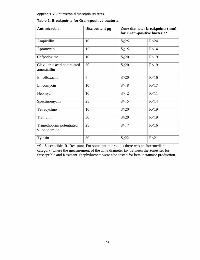
Appendix IV. Antimicrobial susceptibility tests.
73
Table 2: Breakpoints for Gram-positive bacteria.
Antimicrobial Disc content µg Zone diameter breakpoints (mm)
for Gram-positive bacteria*
Ampicillin 10 S≥25 R<24
Apramycin 15 S≥15 R<14
Cefpodoxime 10 S≥20 R<19
Clavulanic acid potentiated
amoxicillin
30 S≥20 R<19
Enrofloxacin 5 S≥20 R<16
Lincomycin 10 S≥18 R<17
Neomycin 10 S≥12 R<11
Spectinomycin 25 S≥15 R<14
Tetracycline 10 S≥20 R<19
Tiamulin 30 S≥20 R<19
Trimethoprim potentiated
sulphonamide
25 S≥17 R<16
Tylosin 30 S≥22 R<21
*S - Susceptible. R- Resistant. For some antimicrobials there was an Intermediate
category, where the measurement of the zone diameter lay between the zones set for
Susceptible and Resistant. Staphylococci were also tested for beta lactamase production.
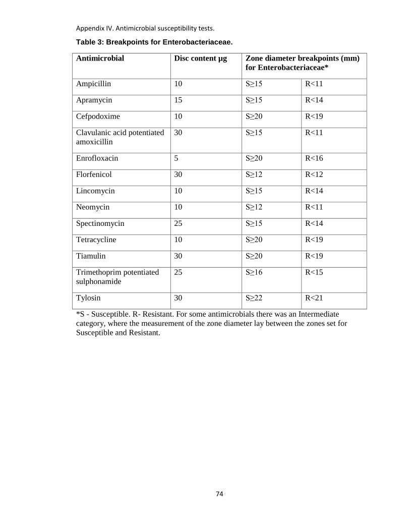
Appendix IV. Antimicrobial susceptibility tests.
74
Table 3: Breakpoints for Enterobacteriaceae.
Antimicrobial Disc content µg Zone diameter breakpoints (mm)
for Enterobacteriaceae*
Ampicillin 10 S≥15 R<11
Apramycin 15 S≥15 R<14
Cefpodoxime 10 S≥20 R<19
Clavulanic acid potentiated
amoxicillin
30 S≥15 R<11
Enrofloxacin 5 S≥20 R<16
Florfenicol 30 S≥12 R<12
Lincomycin 10 S≥15 R<14
Neomycin 10 S≥12 R<11
Spectinomycin 25 S≥15 R<14
Tetracycline 10 S≥20 R<19
Tiamulin 30 S≥20 R<19
Trimethoprim potentiated
sulphonamide
25 S≥16 R<15
Tylosin 30 S≥22 R<21
*S - Susceptible. R- Resistant. For some antimicrobials there was an Intermediate
category, where the measurement of the zone diameter lay between the zones set for
Susceptible and Resistant.
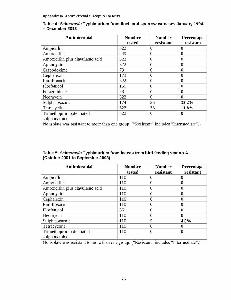
Appendix IV. Antimicrobial susceptibility tests.
75
Table 4: Salmonella Typhimurium from finch and sparrow carcases January 1994 – December 2013
Antimicrobial Number
tested
Number
resistant
Percentage
resistant
Ampicillin 322 0 0
Amoxicillin 249 0 0
Amoxicillin plus clavulanic acid 322 0 0
Apramycin 322 0 0
Cefpodoxime 73 0 0
Cephalexin 173 0 0
Enrofloxacin 322 0 0
Florfenicol 160 0 0
Furazolidone 28 0 0
Neomycin 322 0 0
Sulphisoxazole 174 56 32.2%
Tetracycline 322 38 11.8%
Trimethoprim potentiated
sulphonamide
322 0 0
No isolate was resistant to more than one group. (“Resistant” includes “Intermediate”.)
Table 5: Salmonella Typhimurium from faeces from bird feeding station A (October 2001 to September 2003)
Antimicrobial Number
tested
Number
resistant
Percentage
resistant
Ampicillin 110 0 0
Amoxicillin 110 0 0
Amoxicillin plus clavulanic acid 110 0 0
Apramycin 110 0 0
Cephalexin 110 0 0
Enrofloxacin 110 0 0
Florfenicol 86 0 0
Neomycin 110 0 0
Sulphisoxazole 110 5 4.5%
Tetracycline 110 0 0
Trimethoprim potentiated
sulphonamide
110 0 0
No isolate was resistant to more than one group. (“Resistant” includes “Intermediate”.)

Appendix IV. Antimicrobial susceptibility tests.
76
Table 6: Pathogenic strains of Escherichia albertii from finch carcases January 1994 – December 2013
Antimicrobial Number
tested
Number
resistant
Percentage
resistant
Ampicillin 125 1 0.8%
Amoxicillin 87 1 1.1%
Amoxicillin plus clavulanic acid 125 2 1.6%
Apramycin 125 1 0.8%
Cefpodoxime 38 0 0
Cephalexin 29 0 0
Enrofloxacin 125 4 3.2%
Florfenicol 15 0 0
Furazolidone 7 0 0
Neomycin 125 1 1.6%
Sulphisoxazole 29 1 3.4%
Tetracycline 125 1 0.8%
Trimethoprim potentiated
sulphonamide
125 0 0
One isolate (from a siskin) was resistant to two groups of antimicrobial (β lactams and
fluoroquinolones). No isolate was resistant to four or more different antimicrobial
groups. (“Resistant” includes “Intermediate”.)
Table 7: Escherichia fergusonii from finch and sparrow carcases January 1994 – December 2013
Antimicrobial Number
tested
Number
resistant
Percentage
resistant
Ampicillin 6 0 0
Amoxicillin 1 0 0
Amoxicillin plus clavulanic acid 6 0 0
Apramycin 6 0 0
Cefpodoxime 5 0 0
Cephalexin 1 0 0
Enrofloxacin 6 0 0
Neomycin 6 0 0
Sulphisoxazole 1 0 0
Tetracycline 6 0 0
Trimethoprim potentiated
sulphonamide
6 0 0
(“Resistant” includes “Intermediate”.)
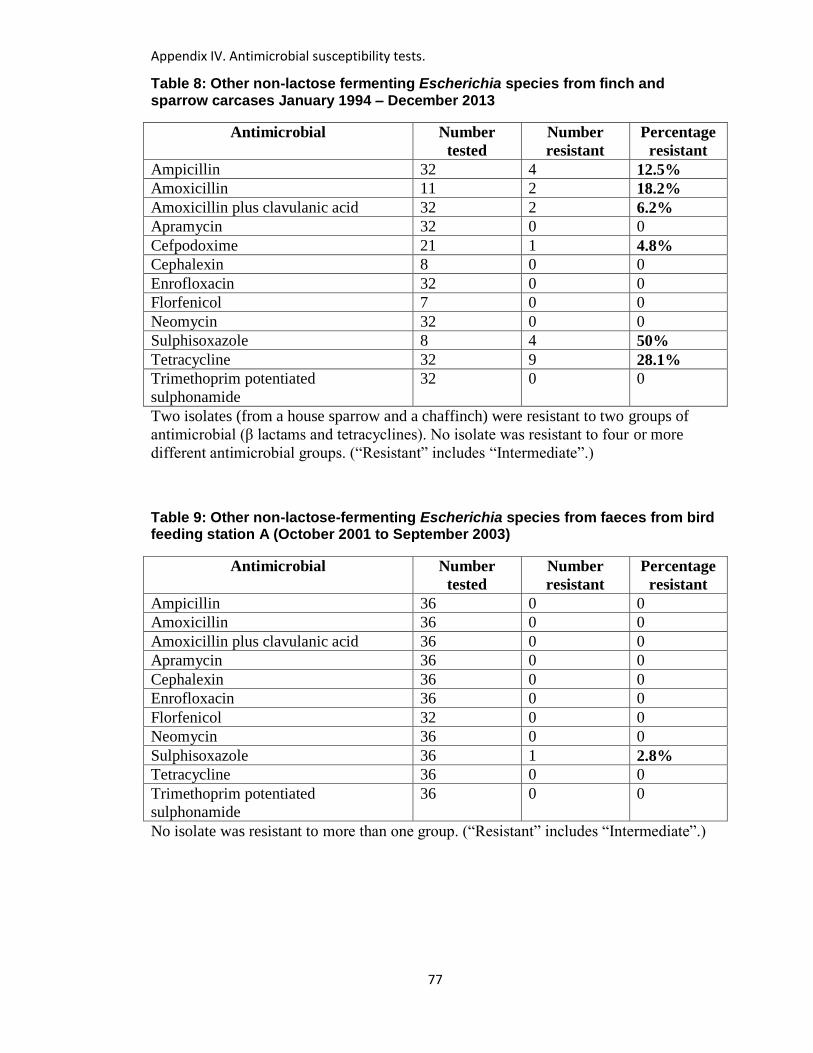
Appendix IV. Antimicrobial susceptibility tests.
77
Table 8: Other non-lactose fermenting Escherichia species from finch and sparrow carcases January 1994 – December 2013
Antimicrobial Number
tested
Number
resistant
Percentage
resistant
Ampicillin 32 4 12.5%
Amoxicillin 11 2 18.2%
Amoxicillin plus clavulanic acid 32 2 6.2%
Apramycin 32 0 0
Cefpodoxime 21 1 4.8%
Cephalexin 8 0 0
Enrofloxacin 32 0 0
Florfenicol 7 0 0
Neomycin 32 0 0
Sulphisoxazole 8 4 50%
Tetracycline 32 9 28.1%
Trimethoprim potentiated
sulphonamide
32 0 0
Two isolates (from a house sparrow and a chaffinch) were resistant to two groups of
antimicrobial (β lactams and tetracyclines). No isolate was resistant to four or more
different antimicrobial groups. (“Resistant” includes “Intermediate”.)
Table 9: Other non-lactose-fermenting Escherichia species from faeces from bird feeding station A (October 2001 to September 2003)
Antimicrobial Number
tested
Number
resistant
Percentage
resistant
Ampicillin 36 0 0
Amoxicillin 36 0 0
Amoxicillin plus clavulanic acid 36 0 0
Apramycin 36 0 0
Cephalexin 36 0 0
Enrofloxacin 36 0 0
Florfenicol 32 0 0
Neomycin 36 0 0
Sulphisoxazole 36 1 2.8%
Tetracycline 36 0 0
Trimethoprim potentiated
sulphonamide
36 0 0
No isolate was resistant to more than one group. (“Resistant” includes “Intermediate”.)
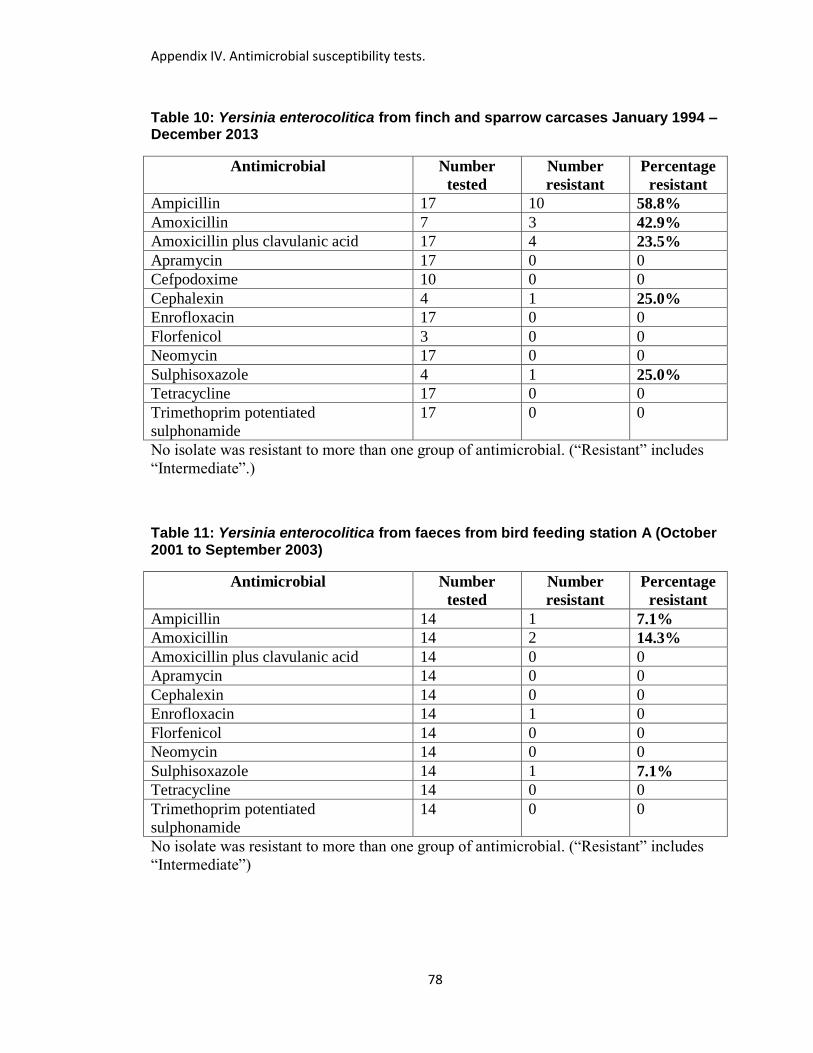
Appendix IV. Antimicrobial susceptibility tests.
78
Table 10: Yersinia enterocolitica from finch and sparrow carcases January 1994 – December 2013
Antimicrobial Number
tested
Number
resistant
Percentage
resistant
Ampicillin 17 10 58.8%
Amoxicillin 7 3 42.9%
Amoxicillin plus clavulanic acid 17 4 23.5%
Apramycin 17 0 0
Cefpodoxime 10 0 0
Cephalexin 4 1 25.0%
Enrofloxacin 17 0 0
Florfenicol 3 0 0
Neomycin 17 0 0
Sulphisoxazole 4 1 25.0%
Tetracycline 17 0 0
Trimethoprim potentiated
sulphonamide
17 0 0
No isolate was resistant to more than one group of antimicrobial. (“Resistant” includes
“Intermediate”.)
Table 11: Yersinia enterocolitica from faeces from bird feeding station A (October 2001 to September 2003)
Antimicrobial Number
tested
Number
resistant
Percentage
resistant
Ampicillin 14 1 7.1%
Amoxicillin 14 2 14.3%
Amoxicillin plus clavulanic acid 14 0 0
Apramycin 14 0 0
Cephalexin 14 0 0
Enrofloxacin 14 1 0
Florfenicol 14 0 0
Neomycin 14 0 0
Sulphisoxazole 14 1 7.1%
Tetracycline 14 0 0
Trimethoprim potentiated
sulphonamide
14 0 0
No isolate was resistant to more than one group of antimicrobial. (“Resistant” includes
“Intermediate”)
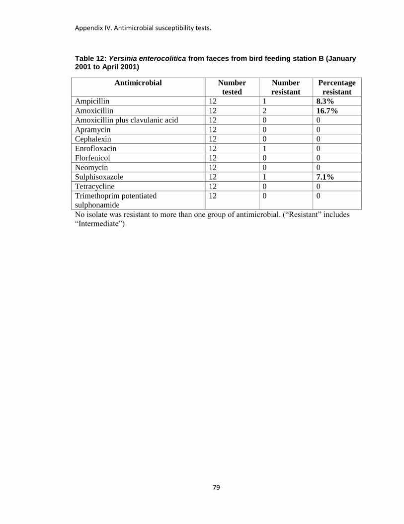
Appendix IV. Antimicrobial susceptibility tests.
79
Table 12: Yersinia enterocolitica from faeces from bird feeding station B (January 2001 to April 2001)
Antimicrobial Number
tested
Number
resistant
Percentage
resistant
Ampicillin 12 1 8.3%
Amoxicillin 12 2 16.7%
Amoxicillin plus clavulanic acid 12 0 0
Apramycin 12 0 0
Cephalexin 12 0 0
Enrofloxacin 12 1 0
Florfenicol 12 0 0
Neomycin 12 0 0
Sulphisoxazole 12 1 7.1%
Tetracycline 12 0 0
Trimethoprim potentiated
sulphonamide
12 0 0
No isolate was resistant to more than one group of antimicrobial. (“Resistant” includes
“Intermediate”)
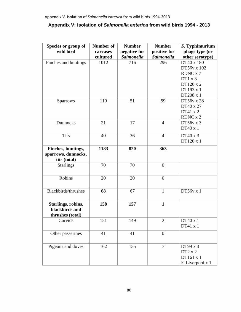
Appendix V. Isolation of Salmonella enterica from wild birds 1994-2013
80
Appendix V: Isolation of Salmonella enterica from wild birds 1994 - 2013
Species or group of
wild bird
Number of
carcases
cultured
Number
negative for
Salmonella
Number
positive for
Salmonella
S. Typhimurium
phage type (or
other serotype)
Finches and buntings 1012 716 296 DT40 x 180
DT56v x 102
RDNC x 7
DT1 x 3
DT120 x 2
DT193 x 1
DT208 x 1
Sparrows 110 51 59 DT56v x 28
DT40 x 27
DT41 x 2 RDNC x 2
Dunnocks 21 17 4 DT56v x 3
DT40 x 1
Tits 40 36 4 DT40 x 3
DT120 x 1
Finches, buntings,
sparrows, dunnocks,
tits (total)
1183 820 363
Starlings 70 70 0
Robins 20 20 0
Blackbirds/thrushes 68 67 1 DT56v x 1
Starlings, robins,
blackbirds and
thrushes (total)
158 157 1
Corvids 151 149 2 DT40 x 1
DT41 x 1
Other passerines 41 41 0
Pigeons and doves 162 155 7 DT99 x 3
DT2 x 2
DT161 x 1
S. Liverpool x 1
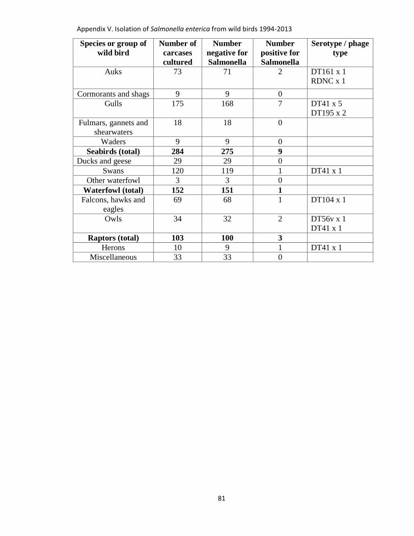
Appendix V. Isolation of Salmonella enterica from wild birds 1994-2013
81
Species or group of
wild bird
Number of
carcases
cultured
Number
negative for
Salmonella
Number
positive for
Salmonella
Serotype / phage
type
Auks 73 71 2 DT161 x 1
RDNC x 1
Cormorants and shags 9 9 0
Gulls 175 168 7 DT41 x 5
DT195 x 2
Fulmars, gannets and
shearwaters
18 18 0
Waders 9 9 0
Seabirds (total) 284 275 9
Ducks and geese 29 29 0
Swans 120 119 1 DT41 x 1
Other waterfowl 3 3 0
Waterfowl (total) 152 151 1
Falcons, hawks and
eagles
69 68 1 DT104 x 1
Owls 34 32 2 DT56v x 1
DT41 x 1
Raptors (total) 103 100 3
Herons 10 9 1 DT41 x 1
Miscellaneous 33 33 0
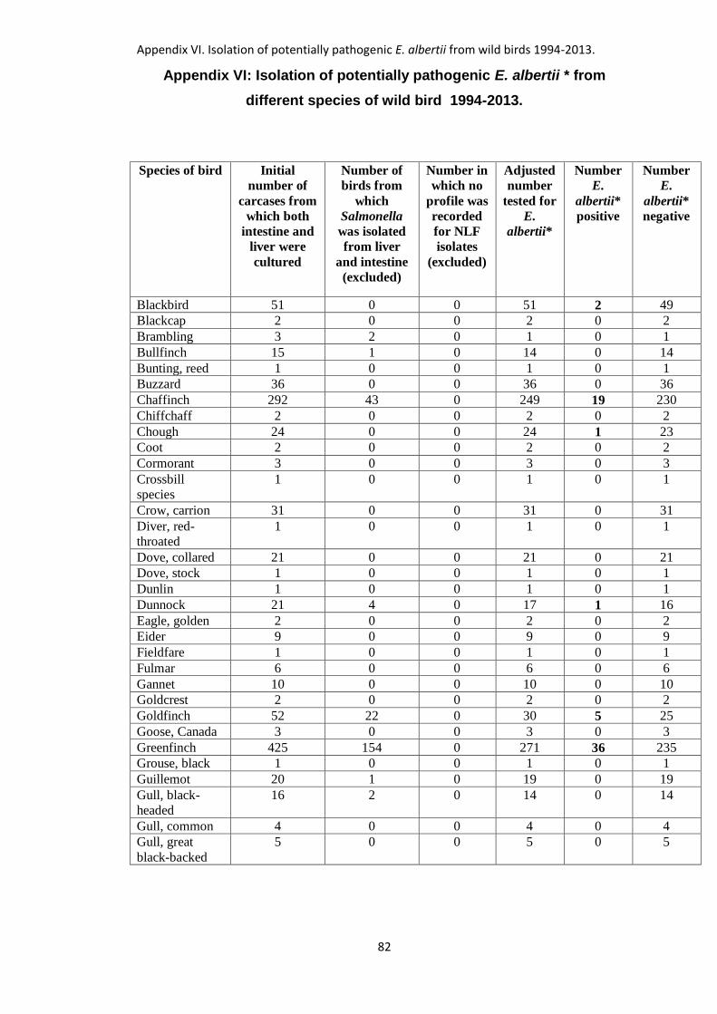
Appendix VI. Isolation of potentially pathogenic E. albertii from wild birds 1994-2013.
82
Appendix VI: Isolation of potentially pathogenic E. albertii * from
different species of wild bird 1994-2013.
Species of bird Initial
number of
carcases from
which both
intestine and
liver were
cultured
Number of
birds from
which
Salmonella
was isolated
from liver
and intestine
(excluded)
Number in
which no
profile was
recorded
for NLF
isolates
(excluded)
Adjusted
number
tested for
E.
albertii*
Number
E.
albertii*
positive
Number
E.
albertii*
negative
Blackbird 51 0 0 51 2 49
Blackcap 2 0 0 2 0 2
Brambling 3 2 0 1 0 1
Bullfinch 15 1 0 14 0 14
Bunting, reed 1 0 0 1 0 1
Buzzard 36 0 0 36 0 36
Chaffinch 292 43 0 249 19 230
Chiffchaff 2 0 0 2 0 2
Chough 24 0 0 24 1 23
Coot 2 0 0 2 0 2
Cormorant 3 0 0 3 0 3
Crossbill
species
1 0 0 1 0 1
Crow, carrion 31 0 0 31 0 31
Diver, red-
throated
1 0 0 1 0 1
Dove, collared 21 0 0 21 0 21
Dove, stock 1 0 0 1 0 1
Dunlin 1 0 0 1 0 1
Dunnock 21 4 0 17 1 16
Eagle, golden 2 0 0 2 0 2
Eider 9 0 0 9 0 9
Fieldfare 1 0 0 1 0 1
Fulmar 6 0 0 6 0 6
Gannet 10 0 0 10 0 10
Goldcrest 2 0 0 2 0 2
Goldfinch 52 22 0 30 5 25
Goose, Canada 3 0 0 3 0 3
Greenfinch 425 154 0 271 36 235
Grouse, black 1 0 0 1 0 1
Guillemot 20 1 0 19 0 19
Gull, black-
headed
16 2 0 14 0 14
Gull, common 4 0 0 4 0 4
Gull, great
black-backed
5 0 0 5 0 5
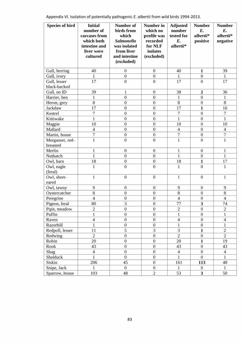
Appendix VI. Isolation of potentially pathogenic E. albertii from wild birds 1994-2013.
83
Species of bird Initial
number of
carcases from
which both
intestine and
liver were
cultured
Number of
birds from
which
Salmonella
was isolated
from liver
and intestine
(excluded)
Number in
which no
profile was
recorded
for NLF
isolates
(excluded)
Adjusted
number
tested for
E.
albertii*
Number
E.
albertii*
positive
Number
E.
albertii*
negative
Gull, herring 40 0 0 40 1 39
Gull, ivory 1 0 0 1 0 1
Gull, lesser
black-backed
17 0 0 17 0 17
Gull, no ID 39 1 0 38 2 36
Harrier, hen 1 0 0 1 0 1
Heron, grey 8 0 0 8 0 8
Jackdaw 17 0 0 17 1 16
Kestrel 7 0 0 7 0 7
Kittiwake 1 0 0 1 0 1
Magpie 10 0 0 10 0 10
Mallard 4 0 0 4 0 4
Martin, house 7 0 0 7 0 7
Merganser, red-
breasted
1 0 0 1 0 1
Merlin 1 0 0 1 0 1
Nuthatch 1 0 0 1 0 1
Owl, barn 18 0 0 18 1 17
Owl, eagle
(feral)
1 0 0 1 0 1
Owl, short-
eared
1 0 0 1 0 1
Owl, tawny 9 0 0 9 0 9
Oystercatcher 8 0 0 8 0 8
Peregrine 4 0 0 4 0 4
Pigeon, feral 80 3 0 77 3 74
Pipit, meadow 2 0 0 2 0 2
Puffin 1 0 0 1 0 1
Raven 4 0 0 4 0 4
Razorbill 1 0 0 1 0 1
Redpoll, lesser 11 5 3 3 1 2
Redwing 2 0 0 2 0 2
Robin 20 0 0 20 1 19
Rook 43 0 0 43 0 43
Shag 4 0 0 4 0 4
Shelduck 1 0 0 1 0 1
Siskin 206 45 0 161 113 48
Snipe, Jack 1 0 0 1 0 1
Sparrow, house 103 48 2 53 3 50
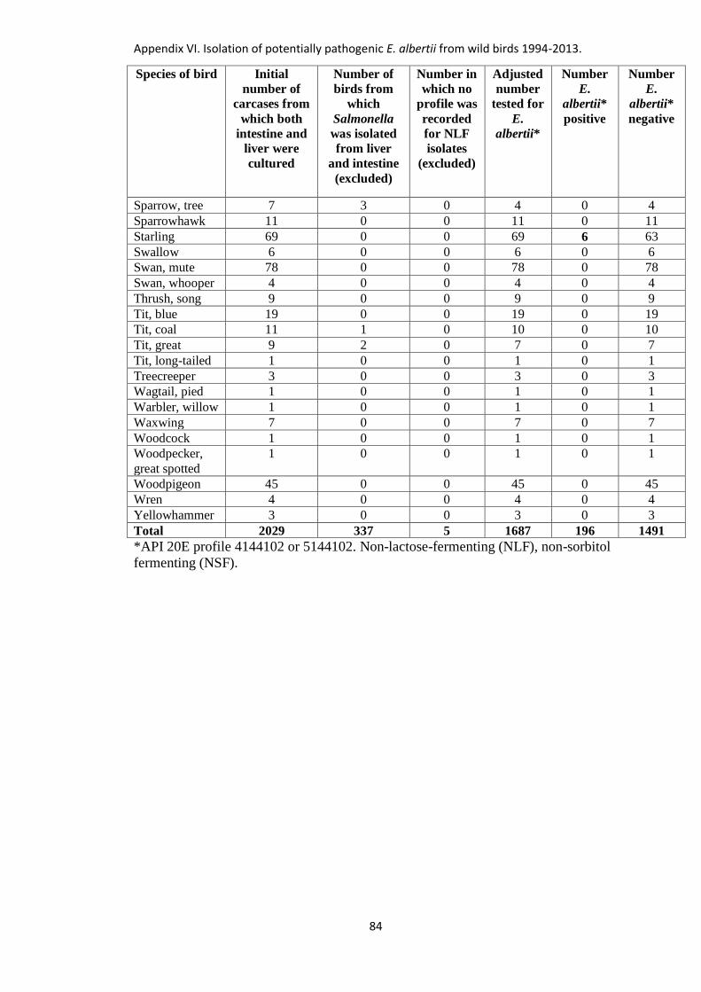
Appendix VI. Isolation of potentially pathogenic E. albertii from wild birds 1994-2013.
84
Species of bird Initial
number of
carcases from
which both
intestine and
liver were
cultured
Number of
birds from
which
Salmonella
was isolated
from liver
and intestine
(excluded)
Number in
which no
profile was
recorded
for NLF
isolates
(excluded)
Adjusted
number
tested for
E.
albertii*
Number
E.
albertii*
positive
Number
E.
albertii*
negative
Sparrow, tree 7 3 0 4 0 4
Sparrowhawk 11 0 0 11 0 11
Starling 69 0 0 69 6 63
Swallow 6 0 0 6 0 6
Swan, mute 78 0 0 78 0 78
Swan, whooper 4 0 0 4 0 4
Thrush, song 9 0 0 9 0 9
Tit, blue 19 0 0 19 0 19
Tit, coal 11 1 0 10 0 10
Tit, great 9 2 0 7 0 7
Tit, long-tailed 1 0 0 1 0 1
Treecreeper 3 0 0 3 0 3
Wagtail, pied 1 0 0 1 0 1
Warbler, willow 1 0 0 1 0 1
Waxwing 7 0 0 7 0 7
Woodcock 1 0 0 1 0 1
Woodpecker,
great spotted
1 0 0 1 0 1
Woodpigeon 45 0 0 45 0 45
Wren 4 0 0 4 0 4
Yellowhammer 3 0 0 3 0 3
Total 2029 337 5 1687 196 1491
*API 20E profile 4144102 or 5144102. Non-lactose-fermenting (NLF), non-sorbitol
fermenting (NSF).
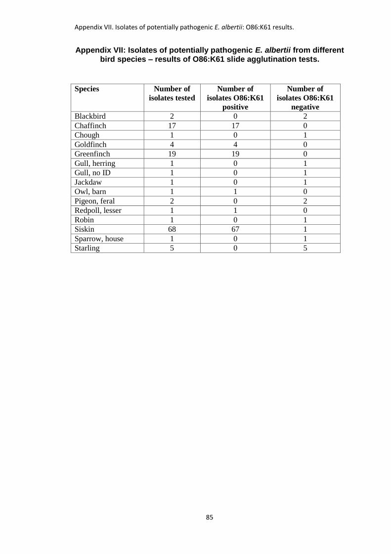
Appendix VII. Isolates of potentially pathogenic E. albertii: O86:K61 results.
85
Appendix VII: Isolates of potentially pathogenic E. albertii from different
bird species – results of O86:K61 slide agglutination tests.
Species Number of
isolates tested
Number of
isolates O86:K61
positive
Number of
isolates O86:K61
negative
Blackbird 2 0 2
Chaffinch 17 17 0
Chough 1 0 1
Goldfinch 4 4 0
Greenfinch 19 19 0
Gull, herring 1 0 1
Gull, no ID 1 0 1
Jackdaw 1 0 1
Owl, barn 1 1 0
Pigeon, feral 2 0 2
Redpoll, lesser 1 1 0
Robin 1 0 1
Siskin 68 67 1
Sparrow, house 1 0 1
Starling 5 0 5
Appendix VIII. Summary of outcomes of wild bird disease surveillance.
86
Appendix VIII: Summary of outcomes from wild bird disease
surveillance at Ayr Disease Surveillance Centre 1994 – 2013
(Orders Passeriformes and Columbiformes)
The outcomes of surveillance in different species or groups of wild birds of the
Orders Passeriformes and Columbiformes are listed in Table 1. The population of
interest was defined as wild birds of the Orders Passeriformes and Columbiformes
found dead or sick in Scotland between 1994 and 2013, and the different categories
of outcome are described below. “NOS” refers to conditions “not otherwise
specified”.
Outcome category 1.
Hazard-specific (targeted) surveillance for specified pathogens or diseases
(Salmonella spp., Escherichia albertii, Mycoplasma spp., avian influenza viruses and
West Nile virus).
Outcome category 2.
A condition or infectious agent, known to be present in the population of interest
(endemic disease), or one that was sufficiently common in other wild bird groups in
the UK for it to be expected to occur in the population of interest. No additional
significant information was obtained in the current study but general awareness was
raised.
Outcome category 3.
As for Category 2 but further significant information was obtained in addition to
raising general awareness.
Outcome category 4.
A re-emerging disease i.e. one that was previously present in the population of
interest but which had declined to a low level or disappeared, but which re-appeared
or significantly increased in prevalence during the study period.
Appendix VIII. Summary of outcomes of wild bird disease surveillance.
87
Outcome category 5.
A disease or agent, previously known in wild birds of a particular group in a country
other than the UK, which had never been described in that group of wild birds in the
UK (exotic disease).
Outcome category 6.
A new disease or syndrome, previously unidentified in that group of wild birds in the
UK or elsewhere, for which the aetiology or identity was not established.
Outcome category 7.
A new disease, syndrome or organism, previously unidentified in that group of wild
birds in the UK or elsewhere, for which the aetiology has been established. May
result from changes in existing pathogens, or may be caused by a previously
unrecognised pathogen.
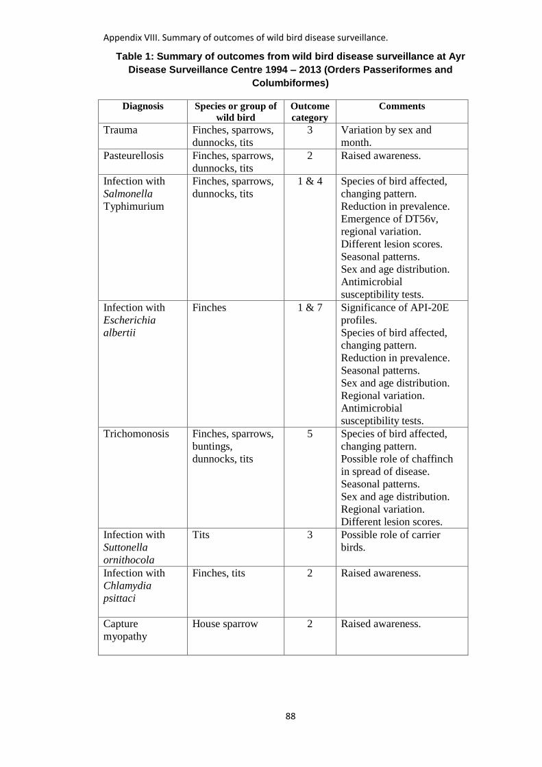
Appendix VIII. Summary of outcomes of wild bird disease surveillance.
88
Table 1: Summary of outcomes from wild bird disease surveillance at Ayr
Disease Surveillance Centre 1994 – 2013 (Orders Passeriformes and
Columbiformes)
Diagnosis Species or group of
wild bird
Outcome
category
Comments
Trauma Finches, sparrows,
dunnocks, tits
3 Variation by sex and
month.
Pasteurellosis
Finches, sparrows,
dunnocks, tits
2 Raised awareness.
Infection with
Salmonella
Typhimurium
Finches, sparrows,
dunnocks, tits
1 & 4 Species of bird affected,
changing pattern.
Reduction in prevalence.
Emergence of DT56v,
regional variation.
Different lesion scores.
Seasonal patterns.
Sex and age distribution.
Antimicrobial
susceptibility tests.
Infection with
Escherichia
albertii
Finches 1 & 7 Significance of API-20E
profiles.
Species of bird affected,
changing pattern.
Reduction in prevalence.
Seasonal patterns.
Sex and age distribution.
Regional variation.
Antimicrobial
susceptibility tests.
Trichomonosis Finches, sparrows,
buntings,
dunnocks, tits
5 Species of bird affected,
changing pattern.
Possible role of chaffinch
in spread of disease.
Seasonal patterns.
Sex and age distribution.
Regional variation.
Different lesion scores.
Infection with
Suttonella
ornithocola
Tits 3 Possible role of carrier
birds.
Infection with
Chlamydia
psittaci
Finches, tits 2 Raised awareness.
Capture
myopathy
House sparrow 2 Raised awareness.
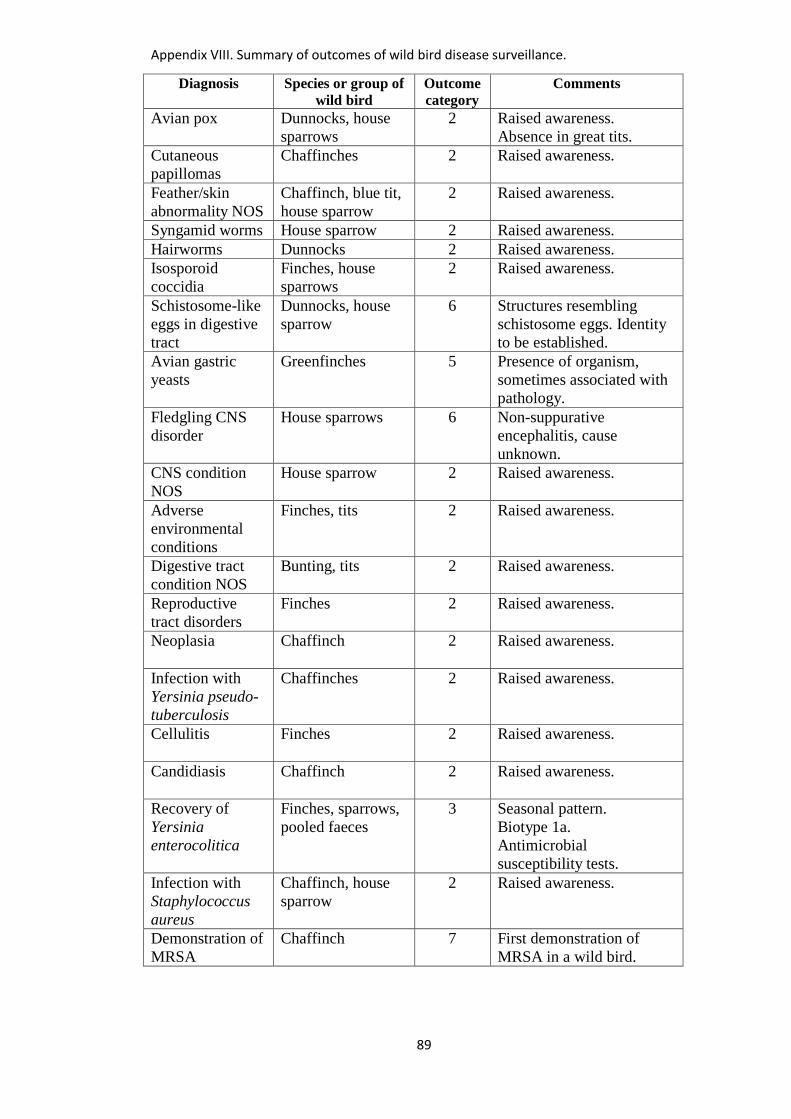
Appendix VIII. Summary of outcomes of wild bird disease surveillance.
89
Diagnosis Species or group of
wild bird
Outcome
category
Comments
Avian pox
Dunnocks, house
sparrows
2 Raised awareness.
Absence in great tits.
Cutaneous
papillomas
Chaffinches 2 Raised awareness.
Feather/skin
abnormality NOS
Chaffinch, blue tit,
house sparrow
2 Raised awareness.
Syngamid worms House sparrow 2 Raised awareness.
Hairworms Dunnocks 2 Raised awareness.
Isosporoid
coccidia
Finches, house
sparrows
2 Raised awareness.
Schistosome-like
eggs in digestive
tract
Dunnocks, house
sparrow
6 Structures resembling
schistosome eggs. Identity
to be established.
Avian gastric
yeasts
Greenfinches 5 Presence of organism,
sometimes associated with
pathology.
Fledgling CNS
disorder
House sparrows 6 Non-suppurative
encephalitis, cause
unknown.
CNS condition
NOS
House sparrow 2 Raised awareness.
Adverse
environmental
conditions
Finches, tits 2 Raised awareness.
Digestive tract
condition NOS
Bunting, tits 2 Raised awareness.
Reproductive
tract disorders
Finches 2 Raised awareness.
Neoplasia
Chaffinch 2 Raised awareness.
Infection with
Yersinia pseudo-
tuberculosis
Chaffinches 2 Raised awareness.
Cellulitis
Finches 2 Raised awareness.
Candidiasis
Chaffinch 2 Raised awareness.
Recovery of
Yersinia
enterocolitica
Finches, sparrows,
pooled faeces
3 Seasonal pattern.
Biotype 1a.
Antimicrobial
susceptibility tests.
Infection with
Staphylococcus
aureus
Chaffinch, house
sparrow
2 Raised awareness.
Demonstration of
MRSA
Chaffinch 7 First demonstration of
MRSA in a wild bird.
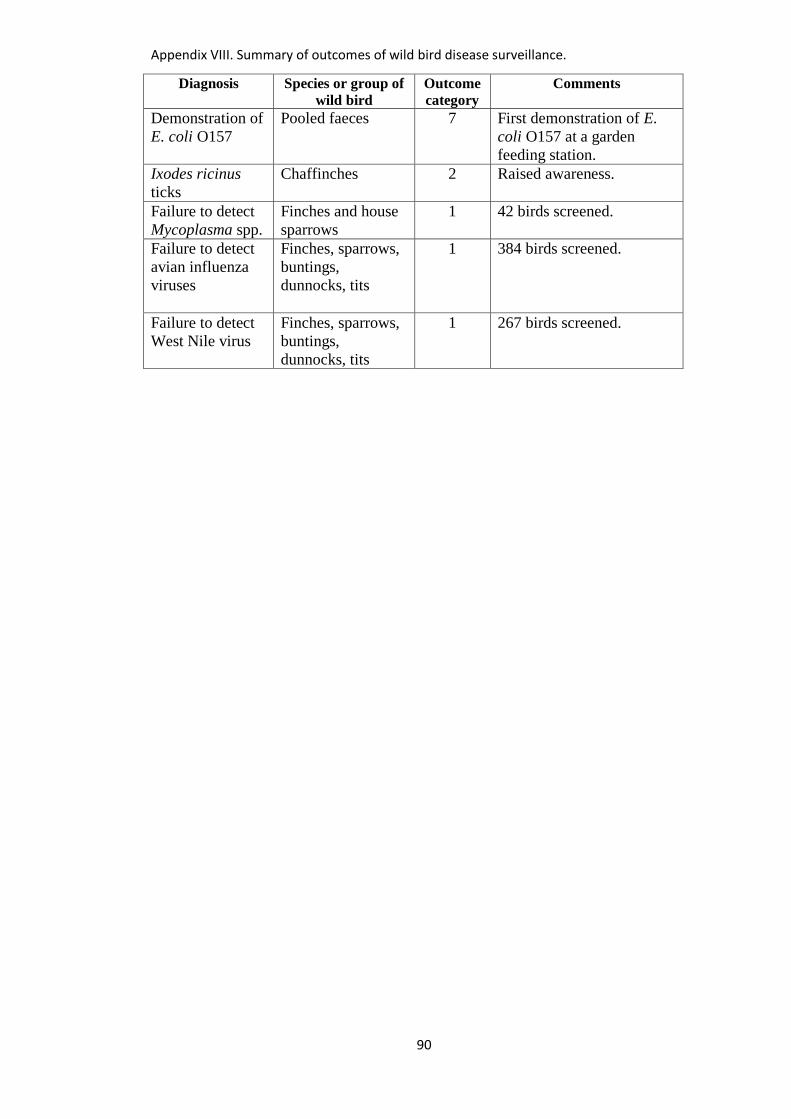
Appendix VIII. Summary of outcomes of wild bird disease surveillance.
90
Diagnosis Species or group of
wild bird
Outcome
category
Comments
Demonstration of
E. coli O157
Pooled faeces 7 First demonstration of E.
coli O157 at a garden
feeding station.
Ixodes ricinus
ticks
Chaffinches 2 Raised awareness.
Failure to detect
Mycoplasma spp.
Finches and house
sparrows
1 42 birds screened.
Failure to detect
avian influenza
viruses
Finches, sparrows,
buntings,
dunnocks, tits
1 384 birds screened.
Failure to detect
West Nile virus
Finches, sparrows,
buntings,
dunnocks, tits
1 267 birds screened.
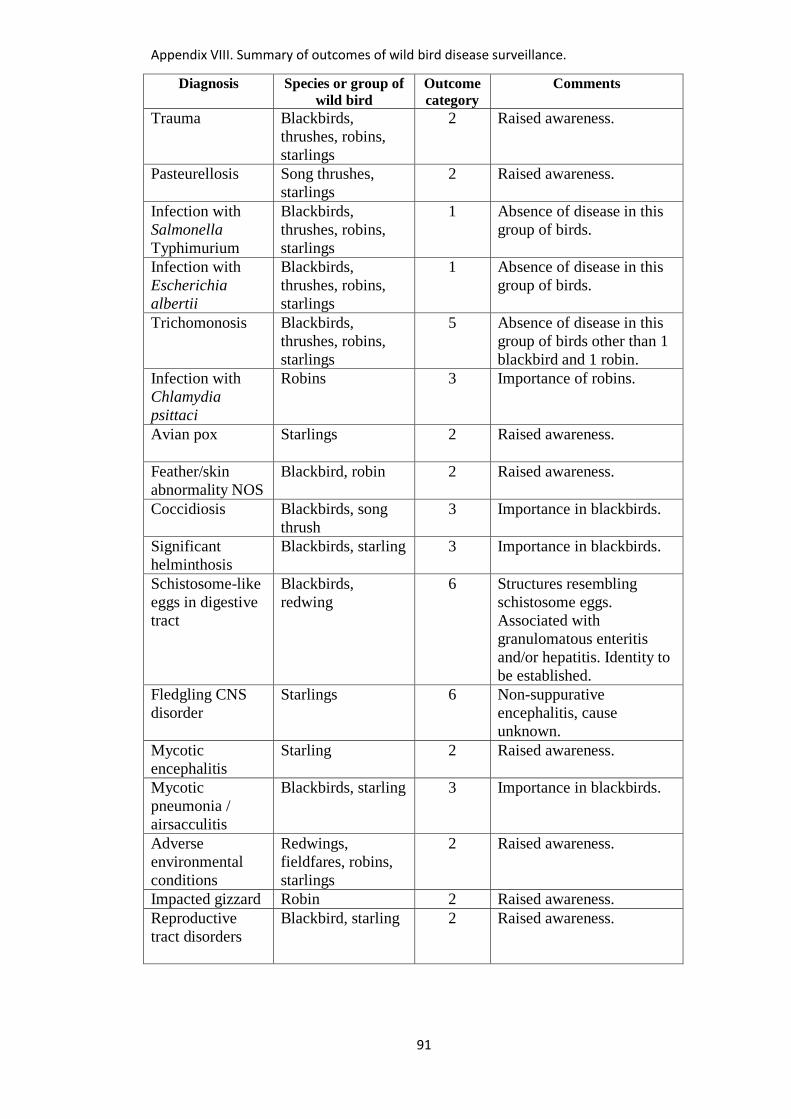
Appendix VIII. Summary of outcomes of wild bird disease surveillance.
91
Diagnosis Species or group of
wild bird
Outcome
category
Comments
Trauma Blackbirds,
thrushes, robins,
starlings
2 Raised awareness.
Pasteurellosis
Song thrushes,
starlings
2 Raised awareness.
Infection with
Salmonella
Typhimurium
Blackbirds,
thrushes, robins,
starlings
1 Absence of disease in this
group of birds.
Infection with
Escherichia
albertii
Blackbirds,
thrushes, robins,
starlings
1 Absence of disease in this
group of birds.
Trichomonosis Blackbirds,
thrushes, robins,
starlings
5 Absence of disease in this
group of birds other than 1
blackbird and 1 robin.
Infection with
Chlamydia
psittaci
Robins 3 Importance of robins.
Avian pox
Starlings 2 Raised awareness.
Feather/skin
abnormality NOS
Blackbird, robin 2 Raised awareness.
Coccidiosis Blackbirds, song
thrush
3 Importance in blackbirds.
Significant
helminthosis
Blackbirds, starling 3 Importance in blackbirds.
Schistosome-like
eggs in digestive
tract
Blackbirds,
redwing
6 Structures resembling
schistosome eggs.
Associated with
granulomatous enteritis
and/or hepatitis. Identity to
be established.
Fledgling CNS
disorder
Starlings 6 Non-suppurative
encephalitis, cause
unknown.
Mycotic
encephalitis
Starling 2 Raised awareness.
Mycotic
pneumonia /
airsacculitis
Blackbirds, starling 3 Importance in blackbirds.
Adverse
environmental
conditions
Redwings,
fieldfares, robins,
starlings
2 Raised awareness.
Impacted gizzard Robin 2 Raised awareness.
Reproductive
tract disorders
Blackbird, starling 2 Raised awareness.
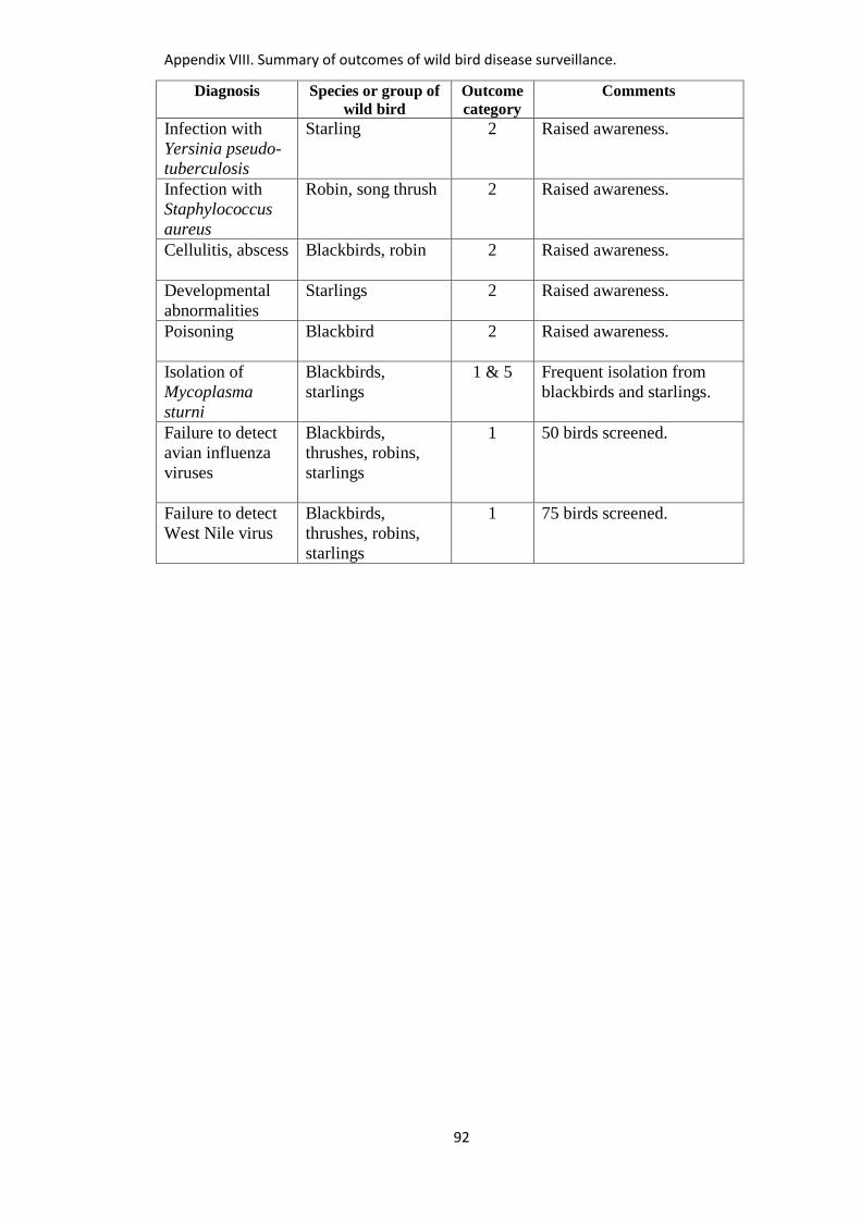
Appendix VIII. Summary of outcomes of wild bird disease surveillance.
92
Diagnosis Species or group of
wild bird
Outcome
category
Comments
Infection with
Yersinia pseudo-
tuberculosis
Starling 2 Raised awareness.
Infection with
Staphylococcus
aureus
Robin, song thrush 2 Raised awareness.
Cellulitis, abscess
Blackbirds, robin 2 Raised awareness.
Developmental
abnormalities
Starlings 2 Raised awareness.
Poisoning
Blackbird 2 Raised awareness.
Isolation of
Mycoplasma
sturni
Blackbirds,
starlings
1 & 5 Frequent isolation from
blackbirds and starlings.
Failure to detect
avian influenza
viruses
Blackbirds,
thrushes, robins,
starlings
1 50 birds screened.
Failure to detect
West Nile virus
Blackbirds,
thrushes, robins,
starlings
1 75 birds screened.

Appendix VIII. Summary of outcomes of wild bird disease surveillance.
93
Diagnosis Species or group of
wild bird
Outcome
category
Comments
Trauma
Corvids, including
choughs
2 Raised awareness.
Malnourished
fledglings
Corvids 2 Raised awareness.
Mycotic
pneumonia /
airsacculitis
Corvids 3 Disease confined to
jackdaws.
Avian
tuberculosis
Corvids 3 Disease confined to
jackdaws.
Significant
helminthosis
(general)
Corvids 3 Differences between
species of corvid.
Significant
helminthosis
(choughs)
Choughs 7 Importance of gastro-
intestinal parasites in
choughs. First description
of gizzard worms and
thorny-headed worms in
UK choughs.
Isosporoid
coccidia
Corvids, including
choughs
2 Raised awareness.
Corvid
respiratory
syndrome
Corvids 3 Respiratory syndrome of
corvids associated with
Mycoplasma gallisepticum-
like organisms and
Pasteurella multocida.
Respiratory tract
condition NOS
Corvids 2 Raised awareness.
Isolation of
Mycoplasma
sturni
Corvids 1 & 5 Frequent isolation from
corvids.
Detection of
Mycoplasma
gallisepticum
Corvids, including
choughs
1 & 7 Detection by PCR in
choughs, carrion crows,
rooks. First isolation from
corvids (choughs).
Infection with
Chlamydia
psittaci
Corvids 3 Importance in corvids.
Pasteurellosis
Corvids 2 Raised awareness.
Avian pox Corvids 3 Differences between
species of corvid.
Feather/skin
abnormality NOS
Corvids 2 Raised awareness.
Cnemidocoptic
mange
Corvids 2 Raised awareness.
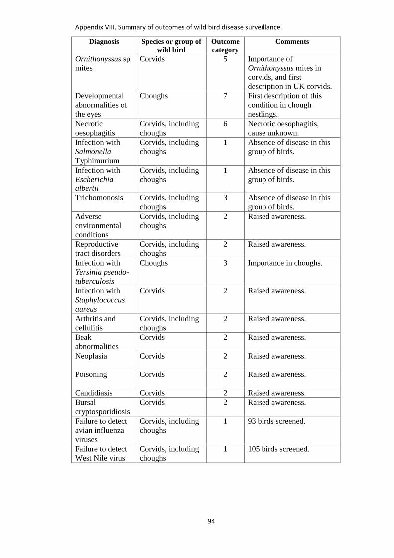
Appendix VIII. Summary of outcomes of wild bird disease surveillance.
94
Diagnosis Species or group of
wild bird
Outcome
category
Comments
Ornithonyssus sp.
mites
Corvids 5 Importance of
Ornithonyssus mites in
corvids, and first
description in UK corvids.
Developmental
abnormalities of
the eyes
Choughs 7 First description of this
condition in chough
nestlings.
Necrotic
oesophagitis
Corvids, including
choughs
6 Necrotic oesophagitis,
cause unknown.
Infection with
Salmonella
Typhimurium
Corvids, including
choughs
1 Absence of disease in this
group of birds.
Infection with
Escherichia
albertii
Corvids, including
choughs
1 Absence of disease in this
group of birds.
Trichomonosis
Corvids, including
choughs
3 Absence of disease in this
group of birds.
Adverse
environmental
conditions
Corvids, including
choughs
2 Raised awareness.
Reproductive
tract disorders
Corvids, including
choughs
2 Raised awareness.
Infection with
Yersinia pseudo-
tuberculosis
Choughs 3 Importance in choughs.
Infection with
Staphylococcus
aureus
Corvids 2 Raised awareness.
Arthritis and
cellulitis
Corvids, including
choughs
2 Raised awareness.
Beak
abnormalities
Corvids 2 Raised awareness.
Neoplasia
Corvids 2 Raised awareness.
Poisoning
Corvids 2 Raised awareness.
Candidiasis Corvids 2 Raised awareness.
Bursal
cryptosporidiosis
Corvids 2 Raised awareness.
Failure to detect
avian influenza
viruses
Corvids, including
choughs
1 93 birds screened.
Failure to detect
West Nile virus
Corvids, including
choughs
1 105 birds screened.
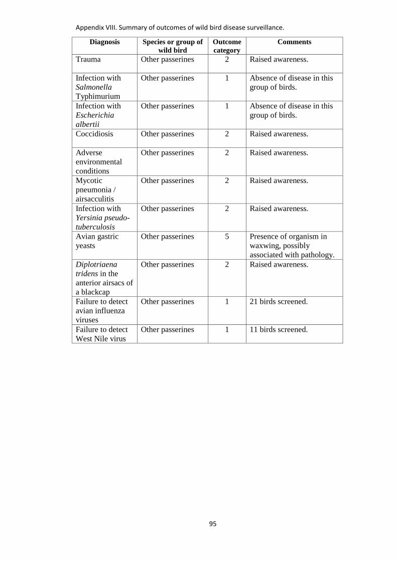
Appendix VIII. Summary of outcomes of wild bird disease surveillance.
95
Diagnosis Species or group of
wild bird
Outcome
category
Comments
Trauma
Other passerines 2 Raised awareness.
Infection with
Salmonella
Typhimurium
Other passerines 1 Absence of disease in this
group of birds.
Infection with
Escherichia
albertii
Other passerines 1 Absence of disease in this
group of birds.
Coccidiosis
Other passerines 2 Raised awareness.
Adverse
environmental
conditions
Other passerines 2 Raised awareness.
Mycotic
pneumonia /
airsacculitis
Other passerines 2 Raised awareness.
Infection with
Yersinia pseudo-
tuberculosis
Other passerines 2 Raised awareness.
Avian gastric
yeasts
Other passerines 5 Presence of organism in
waxwing, possibly
associated with pathology.
Diplotriaena
tridens in the
anterior airsacs of
a blackcap
Other passerines 2 Raised awareness.
Failure to detect
avian influenza
viruses
Other passerines 1 21 birds screened.
Failure to detect
West Nile virus
Other passerines 1 11 birds screened.
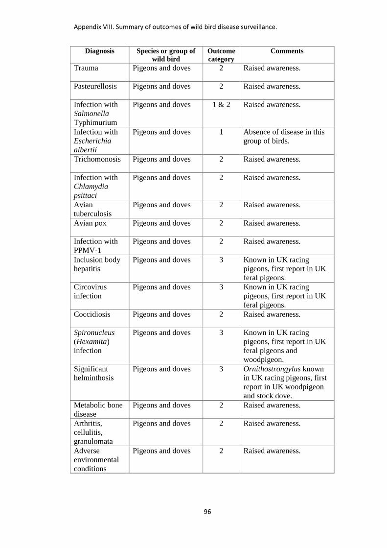
Appendix VIII. Summary of outcomes of wild bird disease surveillance.
96
Diagnosis Species or group of
wild bird
Outcome
category
Comments
Trauma
Pigeons and doves 2 Raised awareness.
Pasteurellosis
Pigeons and doves 2 Raised awareness.
Infection with
Salmonella
Typhimurium
Pigeons and doves 1 & 2 Raised awareness.
Infection with
Escherichia
albertii
Pigeons and doves 1 Absence of disease in this
group of birds.
Trichomonosis
Pigeons and doves 2 Raised awareness.
Infection with
Chlamydia
psittaci
Pigeons and doves 2 Raised awareness.
Avian
tuberculosis
Pigeons and doves 2 Raised awareness.
Avian pox Pigeons and doves 2
Raised awareness.
Infection with
PPMV-1
Pigeons and doves 2 Raised awareness.
Inclusion body
hepatitis
Pigeons and doves 3 Known in UK racing
pigeons, first report in UK
feral pigeons.
Circovirus
infection
Pigeons and doves 3 Known in UK racing
pigeons, first report in UK
feral pigeons.
Coccidiosis
Pigeons and doves 2 Raised awareness.
Spironucleus
(Hexamita)
infection
Pigeons and doves 3 Known in UK racing
pigeons, first report in UK
feral pigeons and
woodpigeon.
Significant helminthosis
Pigeons and doves 3 Ornithostrongylus known in UK racing pigeons, first
report in UK woodpigeon
and stock dove.
Metabolic bone
disease
Pigeons and doves 2 Raised awareness.
Arthritis,
cellulitis,
granulomata
Pigeons and doves 2 Raised awareness.
Adverse
environmental
conditions
Pigeons and doves 2 Raised awareness.
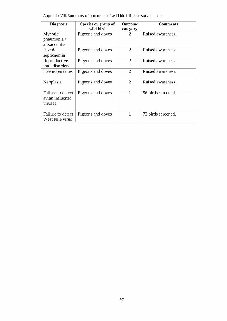
Appendix VIII. Summary of outcomes of wild bird disease surveillance.
97
Diagnosis Species or group of
wild bird
Outcome
category
Comments
Mycotic
pneumonia /
airsacculitis
Pigeons and doves 2 Raised awareness.
E. coli
septicaemia
Pigeons and doves 2 Raised awareness.
Reproductive
tract disorders
Pigeons and doves 2 Raised awareness.
Haemoparasites
Pigeons and doves 2 Raised awareness.
Neoplasia
Pigeons and doves 2 Raised awareness.
Failure to detect
avian influenza
viruses
Pigeons and doves 1 56 birds screened.
Failure to detect
West Nile virus
Pigeons and doves 1 72 birds screened.
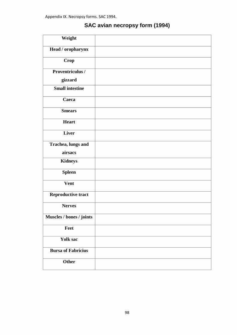
Appendix IX. Necropsy forms. SAC 1994.
98
SAC avian necropsy form (1994)
Weight
Head / oropharynx
Crop
Proventriculus /
gizzard
Small intestine
Caeca
Smears
Heart
Liver
Trachea, lungs and
airsacs
Kidneys
Spleen
Vent
Reproductive tract
Nerves
Muscles / bones / joints
Feet
Yolk sac
Bursa of Fabricius
Other
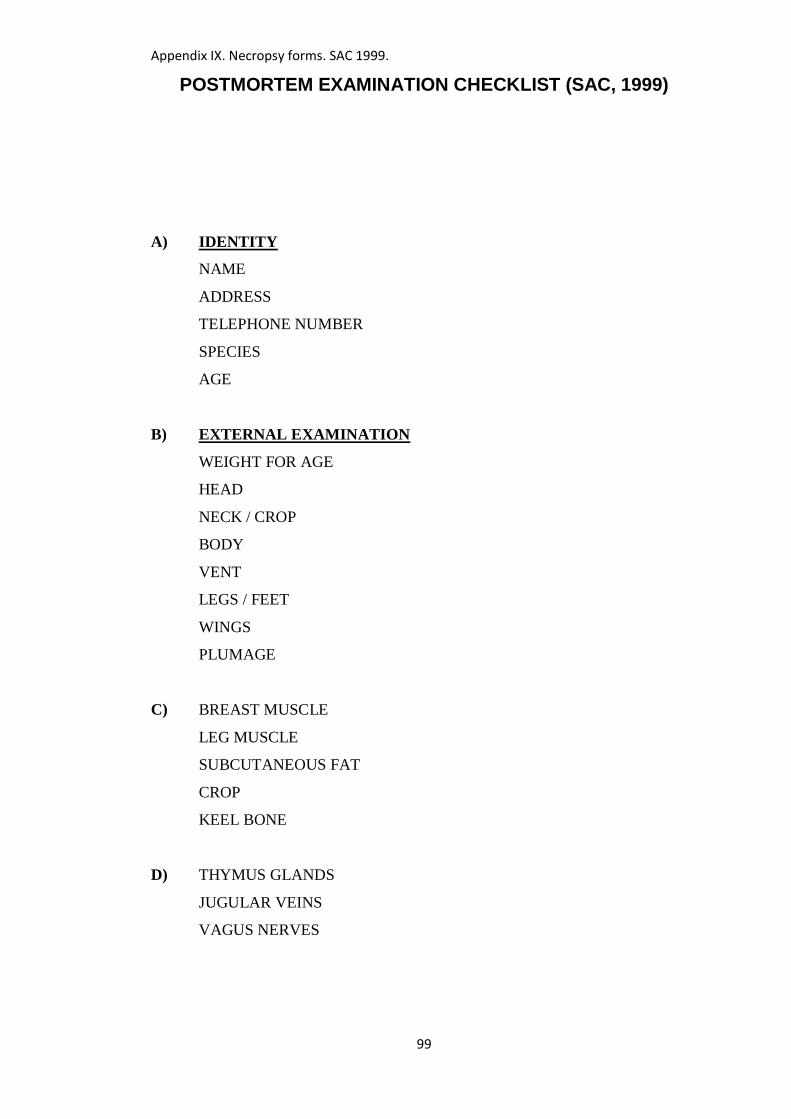
Appendix IX. Necropsy forms. SAC 1999.
99
POSTMORTEM EXAMINATION CHECKLIST (SAC, 1999)
A) IDENTITY
NAME
ADDRESS
TELEPHONE NUMBER
SPECIES
AGE
B) EXTERNAL EXAMINATION
WEIGHT FOR AGE
HEAD
NECK / CROP
BODY
VENT
LEGS / FEET
WINGS
PLUMAGE
C) BREAST MUSCLE
LEG MUSCLE
SUBCUTANEOUS FAT
CROP
KEEL BONE
D) THYMUS GLANDS
JUGULAR VEINS
VAGUS NERVES
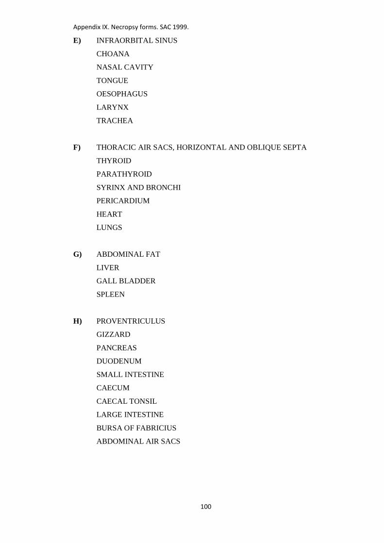
Appendix IX. Necropsy forms. SAC 1999.
100
E) INFRAORBITAL SINUS
CHOANA
NASAL CAVITY
TONGUE
OESOPHAGUS
LARYNX
TRACHEA
F) THORACIC AIR SACS, HORIZONTAL AND OBLIQUE SEPTA
THYROID
PARATHYROID
SYRINX AND BRONCHI
PERICARDIUM
HEART
LUNGS
G) ABDOMINAL FAT
LIVER
GALL BLADDER
SPLEEN
H) PROVENTRICULUS
GIZZARD
PANCREAS
DUODENUM
SMALL INTESTINE
CAECUM
CAECAL TONSIL
LARGE INTESTINE
BURSA OF FABRICIUS
ABDOMINAL AIR SACS
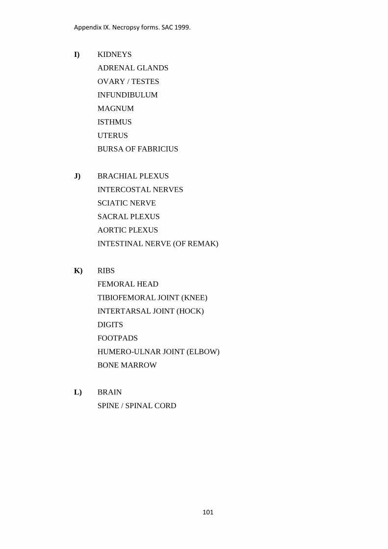
Appendix IX. Necropsy forms. SAC 1999.
101
I) KIDNEYS
ADRENAL GLANDS
OVARY / TESTES
INFUNDIBULUM
MAGNUM
ISTHMUS
UTERUS
BURSA OF FABRICIUS
J) BRACHIAL PLEXUS
INTERCOSTAL NERVES
SCIATIC NERVE
SACRAL PLEXUS
AORTIC PLEXUS
INTESTINAL NERVE (OF REMAK)
K) RIBS
FEMORAL HEAD
TIBIOFEMORAL JOINT (KNEE)
INTERTARSAL JOINT (HOCK)
DIGITS
FOOTPADS
HUMERO-ULNAR JOINT (ELBOW)
BONE MARROW
L) BRAIN
SPINE / SPINAL CORD
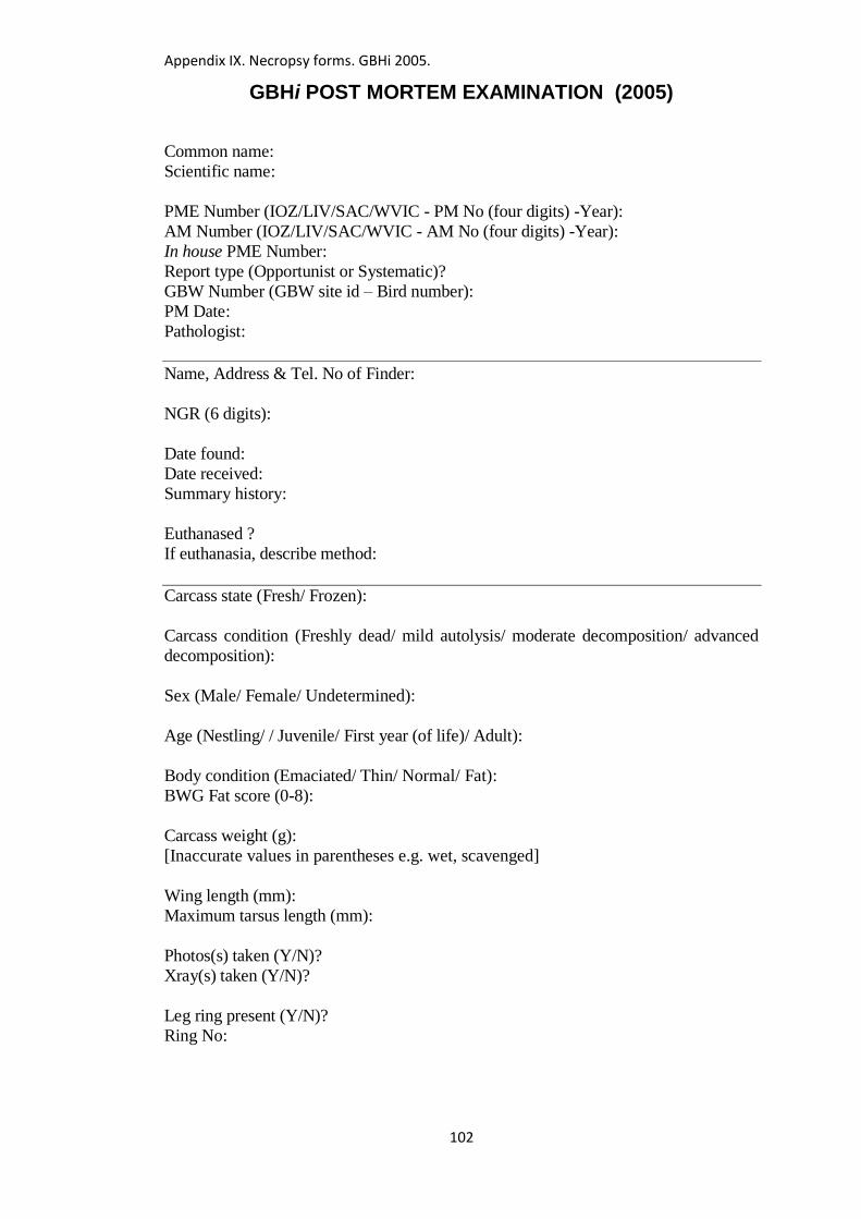
Appendix IX. Necropsy forms. GBHi 2005.
102
GBHi POST MORTEM EXAMINATION (2005)
Common name:
Scientific name:
PME Number (IOZ/LIV/SAC/WVIC - PM No (four digits) -Year):
AM Number (IOZ/LIV/SAC/WVIC - AM No (four digits) -Year):
In house PME Number:
Report type (Opportunist or Systematic)?
GBW Number (GBW site id – Bird number):
PM Date:
Pathologist:
Name, Address & Tel. No of Finder:
NGR (6 digits):
Date found:
Date received:
Summary history:
Euthanased ?
If euthanasia, describe method:
Carcass state (Fresh/ Frozen):
Carcass condition (Freshly dead/ mild autolysis/ moderate decomposition/ advanced
decomposition):
Sex (Male/ Female/ Undetermined):
Age (Nestling/ / Juvenile/ First year (of life)/ Adult):
Body condition (Emaciated/ Thin/ Normal/ Fat):
BWG Fat score (0-8):
Carcass weight (g):
[Inaccurate values in parentheses e.g. wet, scavenged]
Wing length (mm):
Maximum tarsus length (mm):
Photos(s) taken (Y/N)?
Xray(s) taken (Y/N)?
Leg ring present (Y/N)?
Ring No:
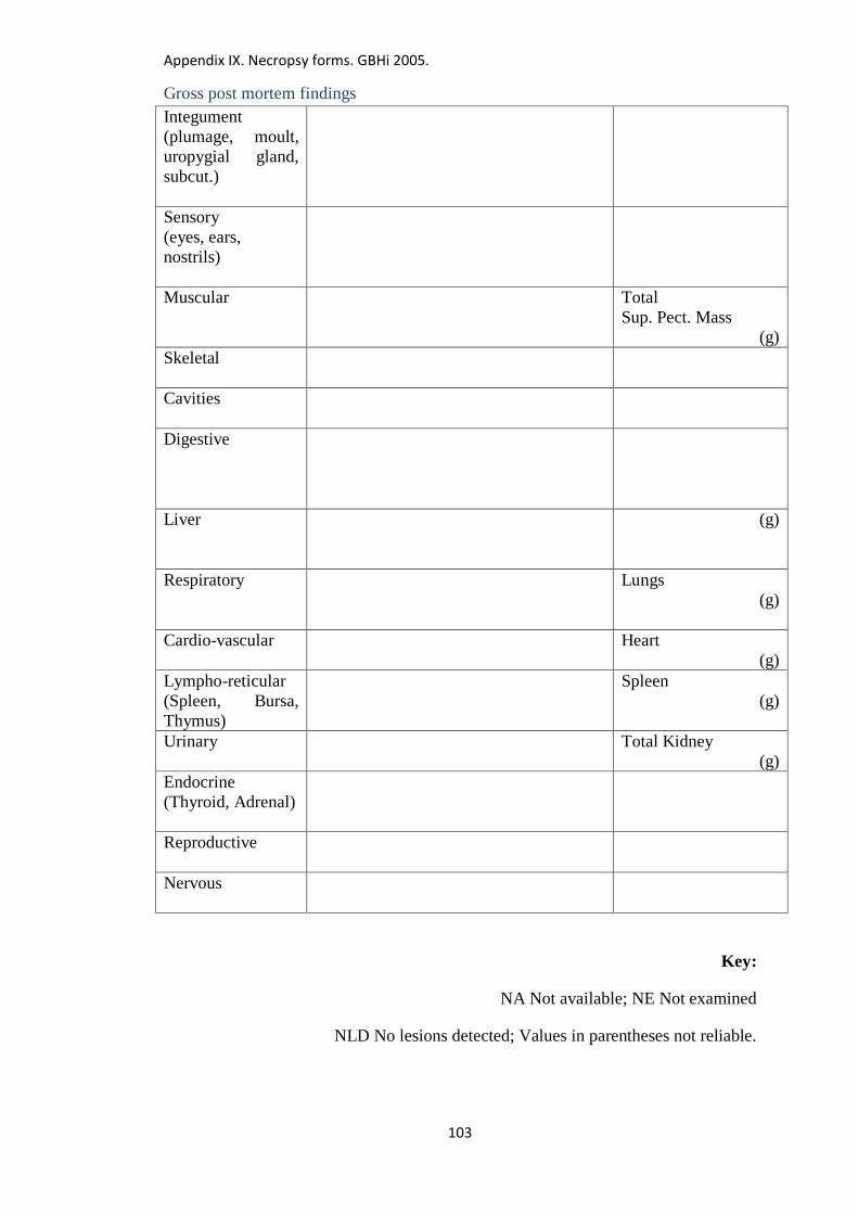
Appendix IX. Necropsy forms. GBHi 2005.
103
Gross post mortem findings
Integument
(plumage, moult,
uropygial gland,
subcut.)
Sensory
(eyes, ears,
nostrils)
Muscular
Total
Sup. Pect. Mass
(g)
Skeletal
Cavities
Digestive
Liver
(g)
Respiratory
Lungs
(g)
Cardio-vascular
Heart
(g)
Lympho-reticular
(Spleen, Bursa,
Thymus)
Spleen
(g)
Urinary
Total Kidney
(g)
Endocrine
(Thyroid, Adrenal)
Reproductive
Nervous
Key:
NA Not available; NE Not examined
NLD No lesions detected; Values in parentheses not reliable.
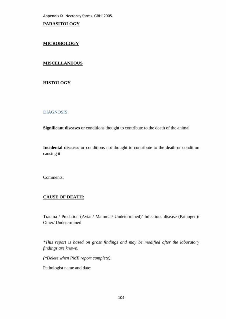
Appendix IX. Necropsy forms. GBHi 2005.
104
PARASITOLOGY
MICROBOLOGY
MISCELLANEOUS
HISTOLOGY
DIAGNOSIS
Significant diseases or conditions thought to contribute to the death of the animal
Incidental diseases or conditions not thought to contribute to the death or condition
causing it
Comments:
CAUSE OF DEATH:
Trauma / Predation (Avian/ Mammal/ Undetermined)/ Infectious disease (Pathogen)/
Other/ Undetermined
*This report is based on gross findings and may be modified after the laboratory
findings are known.
(*Delete when PME report complete).
Pathologist name and date:
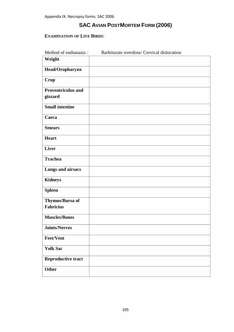
Appendix IX. Necropsy forms. SAC 2006.
105
SAC AVIAN POSTMORTEM FORM (2006)
EXAMINATION OF LIVE BIRDS:
Method of euthanasia : Barbiturate overdose/ Cervical dislocation
Weight
Head/Oropharynx
Crop
Proventriculus and
gizzard
Small intestine
Caeca
Smears
Heart
Liver
Trachea
Lungs and airsacs
Kidneys
Spleen
Thymus/Bursa of
Fabricius
Muscles/Bones
Joints/Nerves
Feet/Vent
Yolk Sac
Reproductive tract
Other
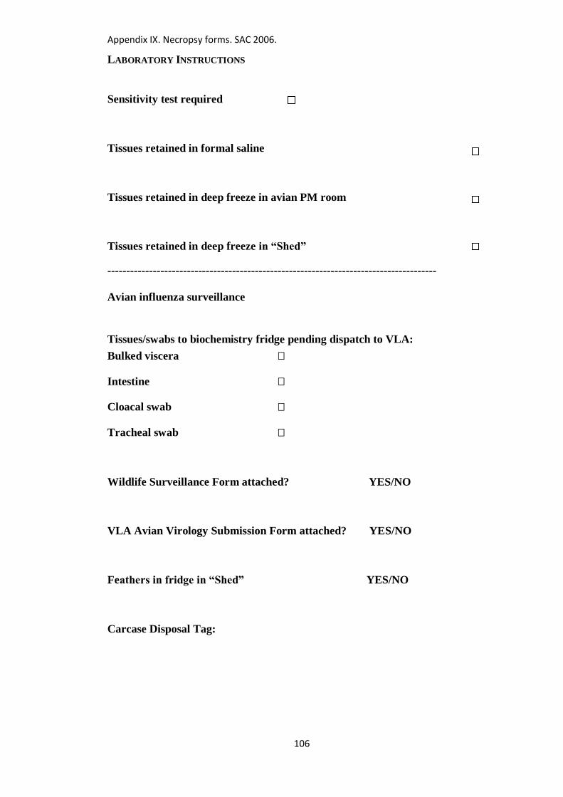
Appendix IX. Necropsy forms. SAC 2006.
106
LABORATORY INSTRUCTIONS
Sensitivity test required
Tissues retained in formal saline
Tissues retained in deep freeze in avian PM room
Tissues retained in deep freeze in “Shed”
---------------------------------------------------------------------------------------
Avian influenza surveillance
Tissues/swabs to biochemistry fridge pending dispatch to VLA:
Bulked viscera
Intestine
Cloacal swab
Tracheal swab
Wildlife Surveillance Form attached? YES/NO
VLA Avian Virology Submission Form attached? YES/NO
Feathers in fridge in “Shed” YES/NO
Carcase Disposal Tag:
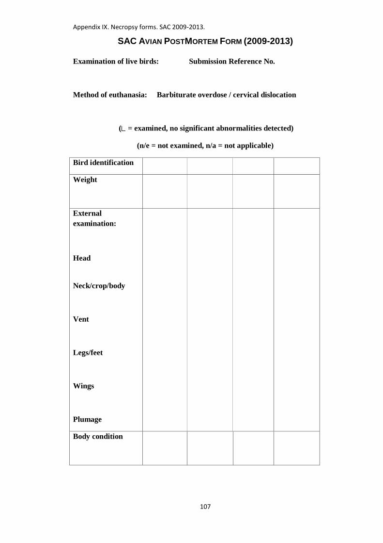
Appendix IX. Necropsy forms. SAC 2009-2013.
107
SAC AVIAN POSTMORTEM FORM (2009-2013)
Examination of live birds: Submission Reference No.
Method of euthanasia: Barbiturate overdose / cervical dislocation
( = examined, no significant abnormalities detected)
(n/e = not examined, n/a = not applicable)
Bird identification
Weight
External
examination:
Head
Neck/crop/body
Vent
Legs/feet
Wings
Plumage
Body condition
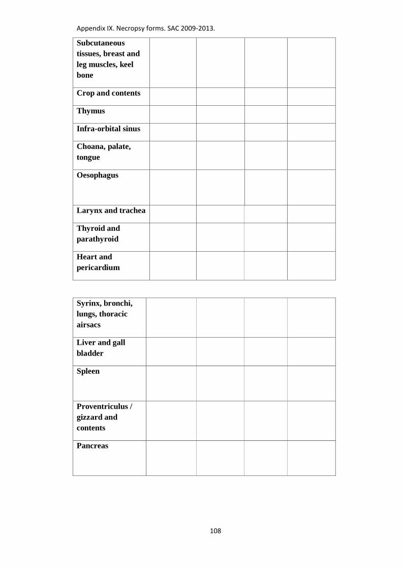
Appendix IX. Necropsy forms. SAC 2009-2013.
108
Subcutaneous
tissues, breast and
leg muscles, keel
bone
Crop and contents
Thymus
Infra-orbital sinus
Choana, palate,
tongue
Oesophagus
Larynx and trachea
Thyroid and
parathyroid
Heart and
pericardium
Syrinx, bronchi,
lungs, thoracic
airsacs
Liver and gall
bladder
Spleen
Proventriculus /
gizzard and
contents
Pancreas
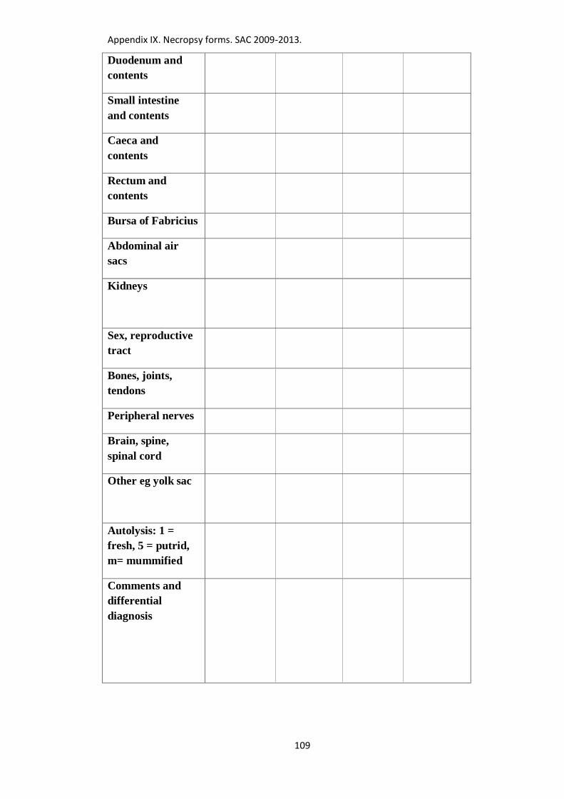
Appendix IX. Necropsy forms. SAC 2009-2013.
109
Duodenum and
contents
Small intestine
and contents
Caeca and
contents
Rectum and
contents
Bursa of Fabricius
Abdominal air
sacs
Kidneys
Sex, reproductive
tract
Bones, joints,
tendons
Peripheral nerves
Brain, spine,
spinal cord
Other eg yolk sac
Autolysis: 1 =
fresh, 5 = putrid,
m= mummified
Comments and
differential
diagnosis
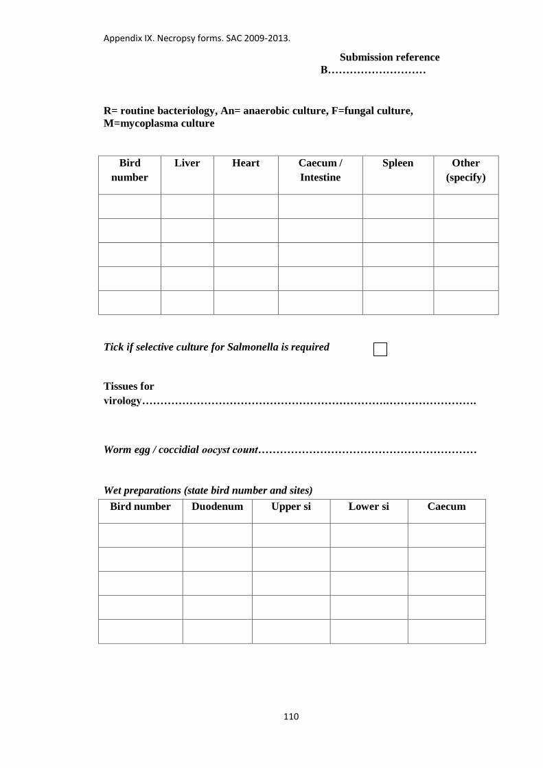
Appendix IX. Necropsy forms. SAC 2009-2013.
110
Submission reference
B………………………
R= routine bacteriology, An= anaerobic culture, F=fungal culture,
M=mycoplasma culture
Bird
number
Liver Heart Caecum /
Intestine
Spleen Other
(specify)
Tick if selective culture for Salmonella is required
Tissues for
virology………………………………………………………….…………………….
Worm egg / coccidial oocyst count……………………………………………………
Wet preparations (state bird number and sites)
Bird number Duodenum Upper si Lower si Caecum
Operator
ign/Date
Appendix IX. Necropsy forms. SAC 2009-2013.
111
Histopathology
Send on………………Brain/lung/liver/kidney/spleen/intestine/caecum/heart/skeletal
muscle/bursa of Fabricius/other tissue
Retain (2/12 months) Brain/lung/liver/kidney/spleen/intestine/caecum/heart/skeletal
muscle/bursa of Fabricius/other tissue
Tissues retained in deep freeze
(Specify which tissues, which freezer, discard date)
Toxicology/rodenticide screen/Serology/ Other
Avian influenza surveillance and West Nile virus surveillance (Avian Virology,
VLA)
Bulked viscera Brain
Intestine Kidney
Cloacal swab
Tracheal swab
Carcase Disposal Tag:
Veterinary signature………………………………………………
Date of necropsy……………………..
Appendix X. List of images.
112
Appendix X: List of images of pathological lesions, parasites and
parasite eggs from wild birds of the Orders Passeriformes and
Columbiformes.
Images are attached separately in removable electronic storage medium at the inside
back cover of this volume, and on-line at http://datashare.is.ed.ac.uk/
Chapter 3, part 1. Images of pox, papillomas and other skin conditions in finches,
sparrows, buntings, dunnocks and tits. ……………………………...Images 1-14.
Chapter 3, part 2. Images of parasite eggs, coccidial oocysts, “megabacteria”, and
other miscellaneous findings in finches, sparrows, buntings, dunnocks and tits
………………………………………………………….……….. Images 15a-38c.
Chapter 4. Images of salmonellosis in finches, sparrows, buntings, dunnocks and
tits. ………………………………………………………………….Images 39-70.
Chapters 5 and 6. Images of E. albertii bacteraemia and trichomonosis in finches
and buntings. ……………………………………………………….Images 71-96.
Chapter 8, part 1. Images of trichomonosis, chlamydiosis, pox, and other skin
conditions in blackbirds, thrushes, robins and starlings. ………….Images 97-110.
Chapter 8, part 2. Images of coccidiosis in blackbirds, thrushes, robins and
starlings. …………………………………………………………Images 111-138.
Chapter 8, part 3. Images of helminths and helminth eggs in blackbirds, thrushes,
robins and starlings. ……………………………………………..Images 139-198.
Chapter 8, part 4. Images of schistosome-like eggs in blackbirds, and other eggs
for comparison. ………………………………………………….Images 199-271.
Chapter 8, part 5. Images of fledgling CNS disorder in starlings, and aspergillosis
in blackbirds. …………………………………………………….Images 272-285.
Chapter 8, part 6. Images of miscellaneous conditions in starlings and blackbirds.
……………………………………………………………………Images 286-293.
Chapter 9, part 1. Images of trauma, malnourished fledglings, mycotic
pneumonia / airsacculitis and avian tuberculosis in corvids. ……Images 294-307.
Appendix X. List of images.
113
Chapter 9, part 2. Images of syngamid worms, hairworms, roundworms and
coccidia in corvids. ……………………………………………..Images 308-345c.
Chapter 9, part 3. Images of tapeworms and thorny-headed worms in corvids.
……………………………………………………………………Images 346-378.
Chapter 9, part 4. Images of hairworms, gapeworms, gizzard worms and thorny-
headed worms in choughs. ………………………………………Images 379-422.
Chapter 9, part 5. Images of corvid respiratory syndrome (CRS) and respiratory
tract conditions not otherwise specified in corvids. ……………..Images 423-469.
Chapter 9, part 6. Images of pox, feather loss, Cnemidocoptes, Ornithonyssus,
lice and feather mites in corvids. ………………………………..Images 470-504.
Chapter 9, part 7. Images of eye/brain abnormalities in young choughs.
……………………………………………………………………Images 505-528.
Chapter 9, part 8. Images of necrotic oesophagitis and other digestive tract
conditions not otherwise specified in corvids. …………………..Images 529-548.
Chapter 9, part 9. Images of adverse environmental conditions, Yersinia
pseudotuberculosis, cellulitis, neoplasia and poisoning in corvids.
……………………………………………………………………Images 549-565.
Chapter 10. Images of miscellaneous conditions in “other Passeriformes”.
……………………………………………………………………Images 566-579.
Chapter 11, part 1. Images of avian tuberculosis in pigeons and doves.
……………………………………………………………………Images 580-615.
Chapter 11, part 2. Images of pox in pigeons and doves.
……………………………………………………………………Images 616-632.
Chapter 11, part 3. Images of trichomonosis, PPMV-1, salmonellosis, inclusion
body hepatitis and circovirus infection in pigeons and doves.
……………………………………………………………………Images 633-650.
Chapter 11, part 4. Images of internal parasites, arthritis, granuloma, neoplasia
and crop milk in pigeons and doves. …………………………….Images 651-678.
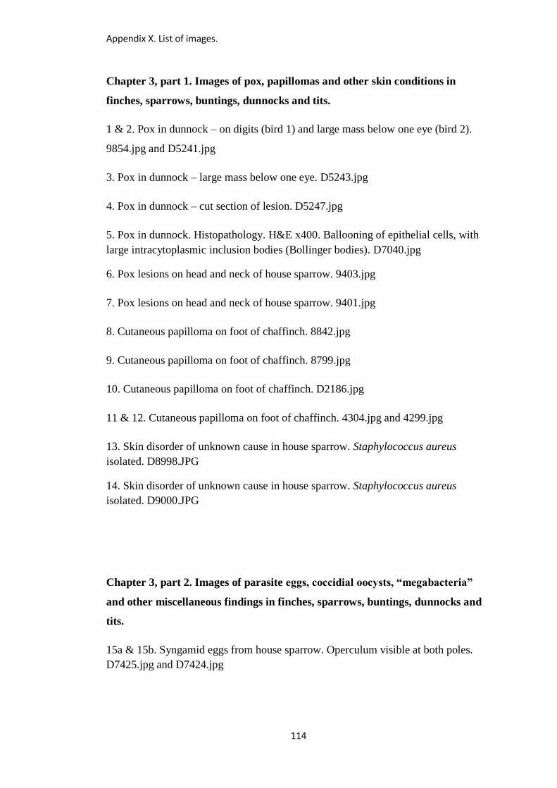
Appendix X. List of images.
114
Chapter 3, part 1. Images of pox, papillomas and other skin conditions in
finches, sparrows, buntings, dunnocks and tits.
1 & 2. Pox in dunnock – on digits (bird 1) and large mass below one eye (bird 2).
9854.jpg and D5241.jpg
3. Pox in dunnock – large mass below one eye. D5243.jpg
4. Pox in dunnock – cut section of lesion. D5247.jpg
5. Pox in dunnock. Histopathology. H&E x400. Ballooning of epithelial cells, with
large intracytoplasmic inclusion bodies (Bollinger bodies). D7040.jpg
6. Pox lesions on head and neck of house sparrow. 9403.jpg
7. Pox lesions on head and neck of house sparrow. 9401.jpg
8. Cutaneous papilloma on foot of chaffinch. 8842.jpg
9. Cutaneous papilloma on foot of chaffinch. 8799.jpg
10. Cutaneous papilloma on foot of chaffinch. D2186.jpg
11 & 12. Cutaneous papilloma on foot of chaffinch. 4304.jpg and 4299.jpg
13. Skin disorder of unknown cause in house sparrow. Staphylococcus aureus
isolated. D8998.JPG
14. Skin disorder of unknown cause in house sparrow. Staphylococcus aureus
isolated. D9000.JPG
Chapter 3, part 2. Images of parasite eggs, coccidial oocysts, “megabacteria”
and other miscellaneous findings in finches, sparrows, buntings, dunnocks and
tits.
15a & 15b. Syngamid eggs from house sparrow. Operculum visible at both poles.
D7425.jpg and D7424.jpg
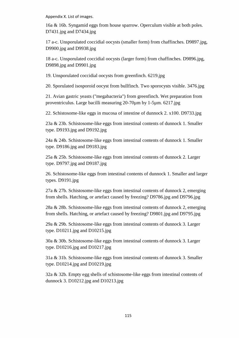
Appendix X. List of images.
115
16a & 16b. Syngamid eggs from house sparrow. Operculum visible at both poles.
D7431.jpg and D7434.jpg
17 a-c. Unsporulated coccidial oocysts (smaller form) from chaffinches. D9897.jpg,
D9900.jpg and D9938.jpg
18 a-c. Unsporulated coccidial oocysts (larger form) from chaffinches. D9896.jpg,
D9898.jpg and D9901.jpg
19. Unsporulated coccidial oocysts from greenfinch. 6219.jpg
20. Sporulated isosporoid oocyst from bullfinch. Two sporocysts visible. 3476.jpg
21. Avian gastric yeasts ("megabacteria") from greenfinch. Wet preparation from
proventriculus. Large bacilli measuring 20-70µm by 1-5µm. 6217.jpg
22. Schistosome-like eggs in mucosa of intestine of dunnock 2. x100. D9733.jpg
23a & 23b. Schistosome-like eggs from intestinal contents of dunnock 1. Smaller
type. D9193.jpg and D9192.jpg
24a & 24b. Schistosome-like eggs from intestinal contents of dunnock 1. Smaller
type. D9186.jpg and D9183.jpg
25a & 25b. Schistosome-like eggs from intestinal contents of dunnock 2. Larger
type. D9797.jpg and D9187.jpg
26. Schistosome-like eggs from intestinal contents of dunnock 1. Smaller and larger
types. D9191.jpg
27a & 27b. Schistosome-like eggs from intestinal contents of dunnock 2, emerging
from shells. Hatching, or artefact caused by freezing? D9786.jpg and D9796.jpg
28a & 28b. Schistosome-like eggs from intestinal contents of dunnock 2, emerging
from shells. Hatching, or artefact caused by freezing? D9801.jpg and D9795.jpg
29a & 29b. Schistosome-like eggs from intestinal contents of dunnock 3. Larger
type. D10211.jpg and D10215.jpg
30a & 30b. Schistosome-like eggs from intestinal contents of dunnock 3. Larger
type. D10216.jpg and D10217.jpg
31a & 31b. Schistosome-like eggs from intestinal contents of dunnock 3. Smaller
type. D10214.jpg and D10219.jpg
32a & 32b. Empty egg shells of schistosome-like eggs from intestinal contents of
dunnock 3. D10212.jpg and D10213.jpg

Appendix X. List of images.
116
33a & 33b. Schistosome-like eggs from intestinal contents of house sparrow. Smaller
type. D9848.jpg and D9850.jpg
34a & 34b. Schistosome-like eggs from intestinal contents of house sparrow. Smaller
type. D9854.jpg and D9856.jpg
35a & 35b. Schistosome-like eggs from intestinal contents of house sparrow. Larger
type. D9851.jpg and D9849.jpg
36a & 36b. Schistosome-like eggs from intestinal contents of house sparrow. Larger
type. D9852.jpg and D9845.jpg
37. For comparison: egg of the schistosome Trichobilharzia filiformis from a swan.
Image courtesy of Dr Jana Bulantova, Charles University, Prague. Image5.tif
38 a-c. Hairworm eggs from intestinal contents of dunnock. D9957.jpg, D9958.jpg
and D9959.jpg
Chapter 4. Images of salmonellosis in finches, sparrows, buntings, dunnocks
and tits.
39. Salmonellosis in greenfinch. Nodules in oesophagus and crop (opened). 5989.jpg
40. Salmonellosis in greenfinch. Nodules in liver. 5997.jpg
41. Salmonellosis in greenfinch. Enlarged spleen with nodules. 5986.jpg
42. Salmonellosis in greenfinch. Enlarged spleen with nodules. 5998.jpg
43. Salmonellosis in greenfinch. Nodules in oesophagus. D9124.JPG
44. Salmonellosis in greenfinch. Nodules in oesophagus. D9127.JPG
45. Salmonellosis in greenfinch. Distended oesophagus (unopened). D9128.JPG
46. Salmonellosis in greenfinch. Oesophagus opened - necrotic mucosa. D9129.JPG
47. Salmonellosis in greenfinch. Nodules in liver. D9126.JPG
48. Salmonellosis in chaffinch. Nodules in liver and spleen. 8035.jpg
49. Salmonellosis in house sparrow. Nodules in oesophagus. 7721.jpg
50. Salmonellosis in house sparrow. Nodules at caeca. 7726.jpg
51 & 52. Salmonellosis in house sparrow and tree sparrow. Thickened crop
(unopened). 8027.jpg and 8050.jpg
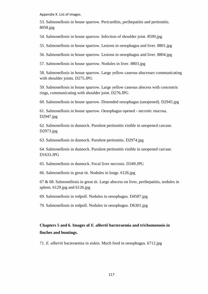
Appendix X. List of images.
117
53. Salmonellosis in house sparrow. Pericarditis, perihepatitis and peritonitis.
8058.jpg
54. Salmonellosis in house sparrow. Infection of shoulder joint. 8599.jpg
55. Salmonellosis in house sparrow. Lesions in oesophagus and liver. 8801.jpg
56. Salmonellosis in house sparrow. Lesions in oesophagus and liver. 8804.jpg
57. Salmonellosis in house sparrow. Nodules in liver. 8803.jpg
58. Salmonellosis in house sparrow. Large yellow caseous abscesses communicating
with shoulder joints. D275.JPG
59. Salmonellosis in house sparrow. Large yellow caseous abscess with concentric
rings, communicating with shoulder joint. D276.JPG
60. Salmonellosis in house sparrow. Distended oesophagus (unopened). D2945.jpg
61. Salmonellosis in house sparrow. Oesophagus opened - necrotic mucosa.
D2947.jpg
62. Salmonellosis in dunnock. Purulent peritonitis visible in unopened carcase.
D2973.jpg
63. Salmonellosis in dunnock. Purulent peritonitis. D2974.jpg
64. Salmonellosis in dunnock. Purulent peritonitis visible in unopened carcase.
D1633.JPG
65. Salmonellosis in dunnock. Focal liver necrosis. D349.JPG
66. Salmonellosis in great tit. Nodules in lungs. 6126.jpg
67 & 68. Salmonellosis in great tit. Large abscess on liver, perihepatitis, nodules in
spleen. 6129.jpg and 6126.jpg
69. Salmonellosis in redpoll. Nodules in oesophagus. D4587.jpg
70. Salmonellosis in redpoll. Nodules in oesophagus. D6301.jpg
Chapters 5 and 6. Images of E. albertii bacteraemia and trichomonosis in
finches and buntings.
71. E. albertii bacteraemia in siskin. Much food in oesophagus. 6712.jpg
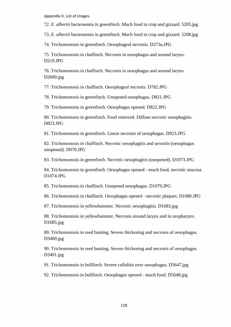
Appendix X. List of images.
118
72. E. albertii bacteraemia in greenfinch. Much food in crop and gizzard. 5205.jpg
73. E. albertii bacteraemia in greenfinch. Much food in crop and gizzard. 5208.jpg
74. Trichomonosis in greenfinch. Oesophageal necrosis. D273a.JPG
75. Trichomonosis in chaffinch. Necrosis in oesophagus and around larynx.
D219.JPG
76. Trichomonosis in chaffinch. Necrosis in oesophagus and around larynx.
D2600.jpg
77. Trichomonosis in chaffinch. Oesophageal necrosis. D782.JPG
78. Trichomonosis in greenfinch. Unopened oesophagus. D821.JPG
79. Trichomonosis in greenfinch. Oesophagus opened. D822.JPG
80. Trichomonosis in greenfinch. Food removed. Diffuse necrotic oesophagitis.
D823.JPG
81. Trichomonosis in greenfinch. Linear necrosis of oesophagus. D923.JPG
82. Trichomonosis in chaffinch. Necrotic oesophagitis and serositis (oesophagus
unopened). D970.JPG
83. Trichomonosis in greenfinch. Necrotic oesophagitis (unopened). D1073.JPG
84. Trichomonosis in greenfinch. Oesophagus opened - much food, necrotic mucosa.
D1074.JPG
85. Trichomonosis in chaffinch. Unopened oesophagus. D1079.JPG
86. Trichomonosis in chaffinch. Oesophagus opened - necrotic plaques. D1080.JPG
87. Trichomonosis in yellowhammer. Necrotic oesophagitis. D1683.jpg
88. Trichomonosis in yellowhammer. Necrosis around larynx and in oropharynx.
D1685.jpg
89. Trichomonosis in reed bunting. Severe thickening and necrosis of oesophagus.
D3400.jpg
90. Trichomonosis in reed bunting. Severe thickening and necrosis of oesophagus.
D3401.jpg
91. Trichomonosis in bullfinch. Severe cellulitis over oesophagus. D5647.jpg
92. Trichomonosis in bullfinch. Oesophagus opened - much food. D5648.jpg
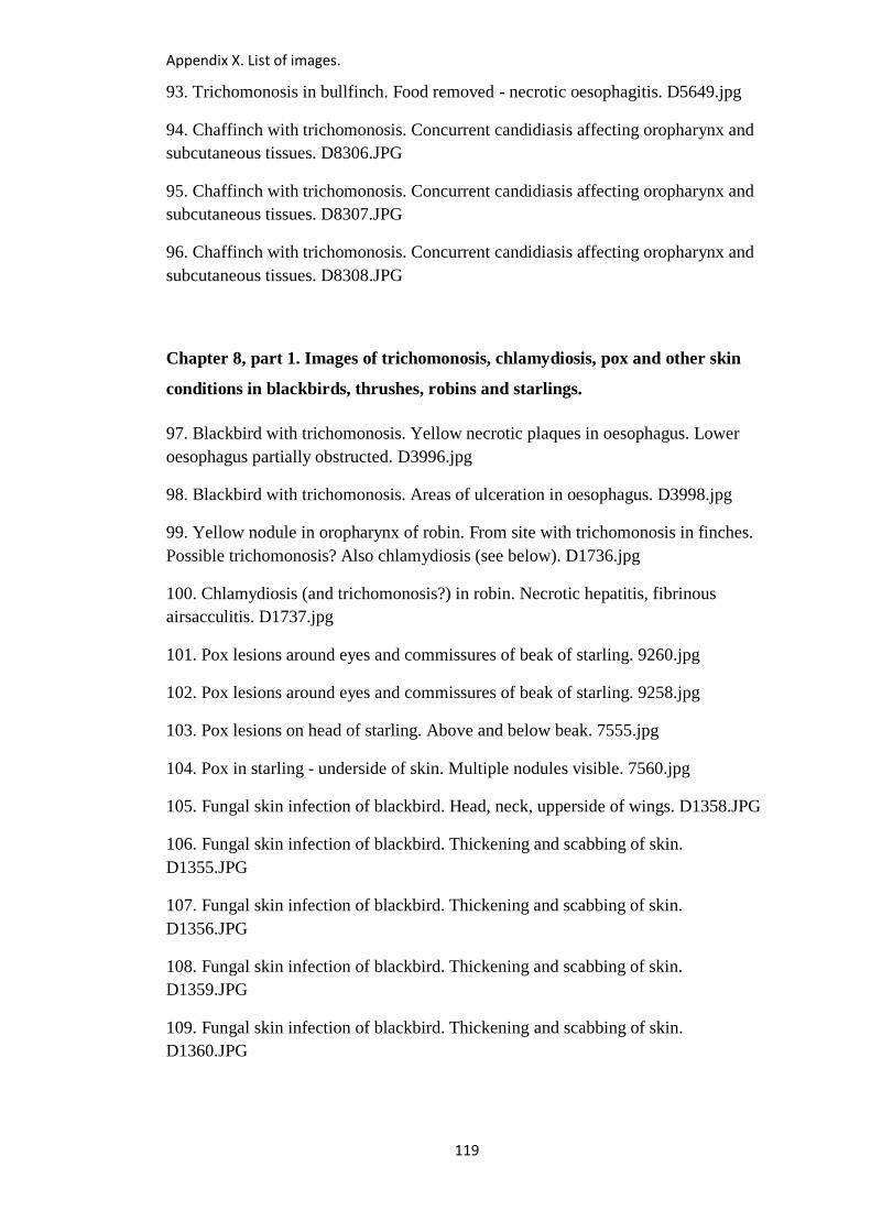
Appendix X. List of images.
119
93. Trichomonosis in bullfinch. Food removed - necrotic oesophagitis. D5649.jpg
94. Chaffinch with trichomonosis. Concurrent candidiasis affecting oropharynx and
subcutaneous tissues. D8306.JPG
95. Chaffinch with trichomonosis. Concurrent candidiasis affecting oropharynx and
subcutaneous tissues. D8307.JPG
96. Chaffinch with trichomonosis. Concurrent candidiasis affecting oropharynx and
subcutaneous tissues. D8308.JPG
Chapter 8, part 1. Images of trichomonosis, chlamydiosis, pox and other skin
conditions in blackbirds, thrushes, robins and starlings.
97. Blackbird with trichomonosis. Yellow necrotic plaques in oesophagus. Lower
oesophagus partially obstructed. D3996.jpg
98. Blackbird with trichomonosis. Areas of ulceration in oesophagus. D3998.jpg
99. Yellow nodule in oropharynx of robin. From site with trichomonosis in finches.
Possible trichomonosis? Also chlamydiosis (see below). D1736.jpg
100. Chlamydiosis (and trichomonosis?) in robin. Necrotic hepatitis, fibrinous
airsacculitis. D1737.jpg
101. Pox lesions around eyes and commissures of beak of starling. 9260.jpg
102. Pox lesions around eyes and commissures of beak of starling. 9258.jpg
103. Pox lesions on head of starling. Above and below beak. 7555.jpg
104. Pox in starling - underside of skin. Multiple nodules visible. 7560.jpg
105. Fungal skin infection of blackbird. Head, neck, upperside of wings. D1358.JPG
106. Fungal skin infection of blackbird. Thickening and scabbing of skin.
D1355.JPG
107. Fungal skin infection of blackbird. Thickening and scabbing of skin.
D1356.JPG
108. Fungal skin infection of blackbird. Thickening and scabbing of skin.
D1359.JPG
109. Fungal skin infection of blackbird. Thickening and scabbing of skin.
D1360.JPG
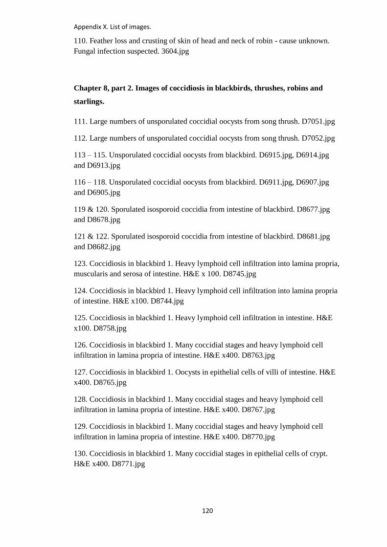
Appendix X. List of images.
120
110. Feather loss and crusting of skin of head and neck of robin - cause unknown.
Fungal infection suspected. 3604.jpg
Chapter 8, part 2. Images of coccidiosis in blackbirds, thrushes, robins and
starlings.
111. Large numbers of unsporulated coccidial oocysts from song thrush. D7051.jpg
112. Large numbers of unsporulated coccidial oocysts from song thrush. D7052.jpg
113 – 115. Unsporulated coccidial oocysts from blackbird. D6915.jpg, D6914.jpg
and D6913.jpg
116 – 118. Unsporulated coccidial oocysts from blackbird. D6911.jpg, D6907.jpg
and D6905.jpg
119 & 120. Sporulated isosporoid coccidia from intestine of blackbird. D8677.jpg
and D8678.jpg
121 & 122. Sporulated isosporoid coccidia from intestine of blackbird. D8681.jpg
and D8682.jpg
123. Coccidiosis in blackbird 1. Heavy lymphoid cell infiltration into lamina propria,
muscularis and serosa of intestine. H&E x 100. D8745.jpg
124. Coccidiosis in blackbird 1. Heavy lymphoid cell infiltration into lamina propria
of intestine. H&E x100. D8744.jpg
125. Coccidiosis in blackbird 1. Heavy lymphoid cell infiltration in intestine. H&E
x100. D8758.jpg
126. Coccidiosis in blackbird 1. Many coccidial stages and heavy lymphoid cell
infiltration in lamina propria of intestine. H&E x400. D8763.jpg
127. Coccidiosis in blackbird 1. Oocysts in epithelial cells of villi of intestine. H&E
x400. D8765.jpg
128. Coccidiosis in blackbird 1. Many coccidial stages and heavy lymphoid cell
infiltration in lamina propria of intestine. H&E x400. D8767.jpg
129. Coccidiosis in blackbird 1. Many coccidial stages and heavy lymphoid cell
infiltration in lamina propria of intestine. H&E x400. D8770.jpg
130. Coccidiosis in blackbird 1. Many coccidial stages in epithelial cells of crypt.
H&E x400. D8771.jpg
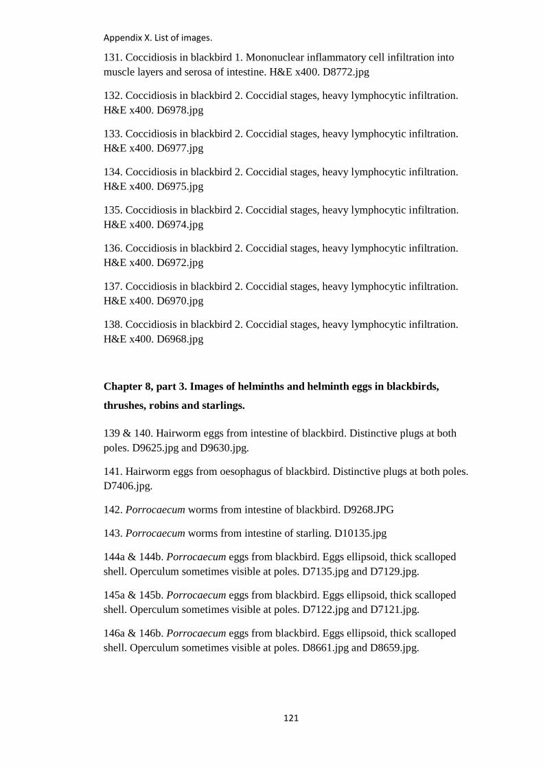
Appendix X. List of images.
121
131. Coccidiosis in blackbird 1. Mononuclear inflammatory cell infiltration into
muscle layers and serosa of intestine. H&E x400. D8772.jpg
132. Coccidiosis in blackbird 2. Coccidial stages, heavy lymphocytic infiltration.
H&E x400. D6978.jpg
133. Coccidiosis in blackbird 2. Coccidial stages, heavy lymphocytic infiltration.
H&E x400. D6977.jpg
134. Coccidiosis in blackbird 2. Coccidial stages, heavy lymphocytic infiltration.
H&E x400. D6975.jpg
135. Coccidiosis in blackbird 2. Coccidial stages, heavy lymphocytic infiltration.
H&E x400. D6974.jpg
136. Coccidiosis in blackbird 2. Coccidial stages, heavy lymphocytic infiltration.
H&E x400. D6972.jpg
137. Coccidiosis in blackbird 2. Coccidial stages, heavy lymphocytic infiltration.
H&E x400. D6970.jpg
138. Coccidiosis in blackbird 2. Coccidial stages, heavy lymphocytic infiltration.
H&E x400. D6968.jpg
Chapter 8, part 3. Images of helminths and helminth eggs in blackbirds,
thrushes, robins and starlings.
139 & 140. Hairworm eggs from intestine of blackbird. Distinctive plugs at both
poles. D9625.jpg and D9630.jpg.
141. Hairworm eggs from oesophagus of blackbird. Distinctive plugs at both poles.
D7406.jpg.
142. Porrocaecum worms from intestine of blackbird. D9268.JPG
143. Porrocaecum worms from intestine of starling. D10135.jpg
144a & 144b. Porrocaecum eggs from blackbird. Eggs ellipsoid, thick scalloped
shell. Operculum sometimes visible at poles. D7135.jpg and D7129.jpg.
145a & 145b. Porrocaecum eggs from blackbird. Eggs ellipsoid, thick scalloped
shell. Operculum sometimes visible at poles. D7122.jpg and D7121.jpg.
146a & 146b. Porrocaecum eggs from blackbird. Eggs ellipsoid, thick scalloped
shell. Operculum sometimes visible at poles. D8661.jpg and D8659.jpg.
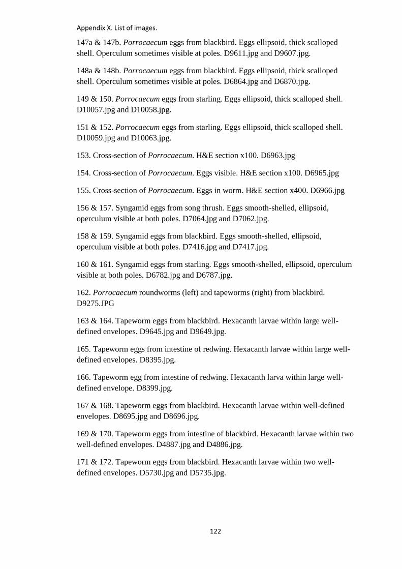
Appendix X. List of images.
122
147a & 147b. Porrocaecum eggs from blackbird. Eggs ellipsoid, thick scalloped
shell. Operculum sometimes visible at poles. D9611.jpg and D9607.jpg.
148a & 148b. Porrocaecum eggs from blackbird. Eggs ellipsoid, thick scalloped
shell. Operculum sometimes visible at poles. D6864.jpg and D6870.jpg.
149 & 150. Porrocaecum eggs from starling. Eggs ellipsoid, thick scalloped shell.
D10057.jpg and D10058.jpg.
151 & 152. Porrocaecum eggs from starling. Eggs ellipsoid, thick scalloped shell.
D10059.jpg and D10063.jpg.
153. Cross-section of Porrocaecum. H&E section x100. D6963.jpg
154. Cross-section of Porrocaecum. Eggs visible. H&E section x100. D6965.jpg
155. Cross-section of Porrocaecum. Eggs in worm. H&E section x400. D6966.jpg
156 & 157. Syngamid eggs from song thrush. Eggs smooth-shelled, ellipsoid,
operculum visible at both poles. D7064.jpg and D7062.jpg.
158 & 159. Syngamid eggs from blackbird. Eggs smooth-shelled, ellipsoid,
operculum visible at both poles. D7416.jpg and D7417.jpg.
160 & 161. Syngamid eggs from starling. Eggs smooth-shelled, ellipsoid, operculum
visible at both poles. D6782.jpg and D6787.jpg.
162. Porrocaecum roundworms (left) and tapeworms (right) from blackbird.
D9275.JPG
163 & 164. Tapeworm eggs from blackbird. Hexacanth larvae within large well-
defined envelopes. D9645.jpg and D9649.jpg.
165. Tapeworm eggs from intestine of redwing. Hexacanth larvae within large well-
defined envelopes. D8395.jpg.
166. Tapeworm egg from intestine of redwing. Hexacanth larva within large well-
defined envelope. D8399.jpg.
167 & 168. Tapeworm eggs from blackbird. Hexacanth larvae within well-defined
envelopes. D8695.jpg and D8696.jpg.
169 & 170. Tapeworm eggs from intestine of blackbird. Hexacanth larvae within two
well-defined envelopes. D4887.jpg and D4886.jpg.
171 & 172. Tapeworm eggs from blackbird. Hexacanth larvae within two well-
defined envelopes. D5730.jpg and D5735.jpg.
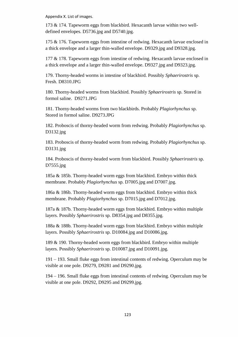
Appendix X. List of images.
123
173 & 174. Tapeworm eggs from blackbird. Hexacanth larvae within two well-
defined envelopes. D5736.jpg and D5740.jpg.
175 & 176. Tapeworm eggs from intestine of redwing. Hexacanth larvae enclosed in
a thick envelope and a larger thin-walled envelope. D9329.jpg and D9328.jpg.
177 & 178. Tapeworm eggs from intestine of redwing. Hexacanth larvae enclosed in
a thick envelope and a larger thin-walled envelope. D9327.jpg and D9323.jpg.
179. Thorny-headed worms in intestine of blackbird. Possibly Sphaerirostris sp.
Fresh. D8310.JPG
180. Thorny-headed worms from blackbird. Possibly Sphaerirostris sp. Stored in
formol saline. D9271.JPG
181. Thorny-headed worms from two blackbirds. Probably Plagiorhynchus sp.
Stored in formol saline. D9273.JPG
182. Proboscis of thorny-headed worm from redwing. Probably Plagiorhynchus sp.
D3132.jpg
183. Proboscis of thorny-headed worm from redwing. Probably Plagiorhynchus sp.
D3131.jpg
184. Proboscis of thorny-headed worm from blackbird. Possibly Sphaerirostris sp.
D7555.jpg
185a & 185b. Thorny-headed worm eggs from blackbird. Embryo within thick
membrane. Probably Plagiorhynchus sp. D7005.jpg and D7007.jpg.
186a & 186b. Thorny-headed worm eggs from blackbird. Embryo within thick
membrane. Probably Plagiorhynchus sp. D7015.jpg and D7012.jpg.
187a & 187b. Thorny-headed worm eggs from blackbird. Embryo within multiple
layers. Possibly Sphaerirostris sp. D8354.jpg and D8355.jpg.
188a & 188b. Thorny-headed worm eggs from blackbird. Embryo within multiple
layers. Possibly Sphaerirostris sp. D10084.jpg and D10086.jpg.
189 & 190. Thorny-headed worm eggs from blackbird. Embryo within multiple
layers. Possibly Sphaerirostris sp. D10087.jpg and D10091.jpg.
191 – 193. Small fluke eggs from intestinal contents of redwing. Operculum may be
visible at one pole. D9279, D9281 and D9290.jpg.
194 – 196. Small fluke eggs from intestinal contents of redwing. Operculum may be
visible at one pole. D9292, D9295 and D9299.jpg.
Appendix X. List of images.
124
197 & 198. Small fluke eggs from intestinal contents of fledgling robin. Operculum
may be visible at one pole. 4392.jpg and 4391.jpg
Chapter 8, part 4. Images of schistosome-like eggs in blackbirds, and other eggs
for comparison.
199. Necrotic hepatitis in blackbird 1. Schistosome-like eggs found in liver and
intestine. D2845.jpg
200. Schistosome-like eggs in intestinal contents of blackbird 1. x40. D4860.jpg
201. Schistosome-like eggs in intestinal mucosa and contents of blackbird 1, plus
tapeworm. x40. D4868.jpg
202. Schistosome-like eggs from intestinal contents of blackbird 1, plus tapeworm.
x100. D4869.jpg
203. Schistosome-like eggs from intestinal contents of blackbird 1. x100. D4866.jpg
204. For comparison. Schistosome-like eggs in mucosa of intestine of dunnock.
x100. D9733.jpg
205a & 205b. Schistosome-like eggs from intestinal contents of blackbird 3. Smaller
type. D9657.jpg and D9666.jpg
206a & 206b. Schistosome-like eggs from intestinal contents of blackbird 3. Smaller
type. D9651.jpg and D9659.jpg
207a & 207b. Schistosome-like eggs from intestinal contents of blackbird 1. Larger
type. D6829.jpg and D6844.jpg
208a & 208b. Schistosome-like eggs from intestinal contents of blackbird 1. Larger
type. D6827.jpg and D6832.jpg
209a & 209b. Schistosome-like eggs from intestinal contents of blackbird 1. Larger
type. D6856.jpg and D6859.jpeg
210. For comparison: Schistosome-like eggs from intestinal contents of dunnock.
D9191.jpg
211a & 211b. For comparison: Schistosome-like eggs from intestinal contents of
dunnock. D9188.jpg and D9187.jpg
Appendix X. List of images.
125
212a & 212b. For comparison: Schistosome-like eggs from intestinal contents of
house sparrow. Smaller type. D9848.jpg and D9850.jpg
213a & 213b. For comparison: Schistosome-like eggs from intestinal contents of
house sparrow. Larger type. D9852.jpg and D9845.jpg
214a & 214b. For comparison: Schistosome-like eggs from intestinal contents of
house sparrow. Larger type. D9851.jpg and D9849.jpg
215a & 215b. For comparison: Schistosome-like eggs from intestinal contents of
redwing. Larger type. D10167.jpg and D10173.jpg
216a & 216b. For comparison: Schistosome-like eggs from intestinal contents of
redwing. Smaller type. D10170.jpg and D10168.jpg
217 & 218. For comparison: Schistosoma japonicum eggs in unstained wet mount
of human faeces. Eggs measure 70-100µm by 55-64µm. DPDx - Laboratory
Identification of Parasitic Diseases of Public Health Concern.
http://www.cdc.gov/dpdx/schistosomiasis/gallery.html#xsection
219 & 220. For comparison: Schistosoma japonicum eggs in unstained wet mount
of human faeces. Eggs measure 70-100µm by 55-64µm. DPDx - Laboratory
Identification of Parasitic Diseases of Public Health Concern.
http://www.cdc.gov/dpdx/schistosomiasis/gallery.html#xsection
221a & 221b. For comparison: Schistosome-like eggs from intestinal contents of
tawny owl. True parasites or pseudoparasites from prey? D8428.jpg and D8427.jpg
222a & 222b. For comparison: Schistosome-like eggs from intestinal contents of
sparrowhawk. True parasites or pseudoparasites from prey? D5060.jpg and
D5061.jpg
223a & 223b. For comparison: Schistosome-like egg from blackbird (left) and
tapeworm egg from same bird (right). D4875.jpg and D4887.jpg
224 & 225. For comparison: Schistosome-like egg from blackbird (left) and
tapeworm egg from same bird (right). D4884.jpg and D4886.jpg
226. Schistosome-like eggs in blackbird 1 liver. H&E x100. D5124.jpg
227. Schistosome-like eggs in blackbird 1 liver. H&E x100. D5125.jpg
228. Schistosome-like eggs in blackbird 1 liver. H&E x100. D5128.jpg
229. Schistosome-like eggs in blackbird 1 liver. H&E x100. D5129.jpg
230. Schistosome-like eggs in blackbird 1 liver. H&E x100. D5132.jpg
Appendix X. List of images.
126
231. Schistosome-like eggs in blackbird 1 liver. H&E x100. D5140.jpg
232. For comparison: Schistosoma japonicum eggs in hepatic portal tract.
https://en.wikipedia.org/wiki/Schistosomiasis
233. For comparison: Schistosoma haematobium infection of the bladder.
https://www.studyblue.com/switch/cpt.html
234. Schistosome-like eggs in blackbird 1 liver. H&E x400. D5142.jpg
235. Schistosome-like eggs in blackbird 1 liver. H&E x400. D5143.jpg
236. For comparison: Heterobilharzia americana in liver.
https://sites.google.com/site/vetpathforum/infectious-organisms
237. For comparison: Heterobilharzia americana in small intestine.
https://sites.google.com/site/vetpathforum/infectious-organisms
238. For comparison: intestinal granuloma associated with Schistosoma mansoni.
http://www.eurosurveillance.org/ViewArticle.aspx?ArticleId=21151
239. For comparison: Schistosoma mansoni egg in gall bladder.
http://www.ijcasereportsandimages.com/archive/2014/006-2014-ijcri/CR-10395-06-
2014-manes/ijcri-1039506201495-manes-full-text.php
240. Schistosome-like eggs in blackbird 1 liver. H&E x400. D5146.jpg
241. Schistosome-like eggs in blackbird 1 liver. H&E x400. D5154.jpg
242. Schistosome-like eggs in blackbird 1 intestine. H&E x40. D5156.jpg
243. Schistosome-like eggs in blackbird 1 intestine. H&E x40. D5159.jpg
244. Schistosome-like eggs in blackbird 1 intestine. H&E x100. D5163.jpg
245. Schistosome-like eggs in blackbird 1 intestine. H&E x100. D5165.jpg
246. Schistosome-like eggs in blackbird 1 intestine. H&E x400. D5167.jpg
247. Schistosome-like eggs in blackbird 1 intestine. H&E x400. D5168.jpg
248. Schistosome-like eggs in blackbird 1 intestine. H&E x400. D5169.jpg
249. Schistosome-like eggs in blackbird 1 intestine. H&E x400. D5177.jpg
250. Schistosome-like eggs in blackbird 1 intestine. H&E x400. D5171.jpg
Appendix X. List of images.
127
251. For comparison: eggs of Schistosoma mansoni in colon.
http://www.ucdmc.ucdavis.edu/pathology/education/residency_program/caseofthemo
nth/201103/final.html
252. For comparison: egg of Schistosoma mansoni in granuloma.
http://www.askjpc.org/wsco/wsc/wsc96/96wsc03.htm
253. Necrotic hepatitis in blackbird 2. Schistosome-like eggs seen in liver on
histopathology. D2359.jpg
254a & 254b. Schistosome-like eggs in liver of blackbird 2. H&E x400. D9434.jpg
and D9435.jpg
255a & 255b. Schistosome-like eggs in liver of blackbird 2. H&E x400. D9436.jpg
and D9437.jpg
256a & 256b. Schistosome-like eggs in liver of blackbird 2. H&E x400. D9438.jpg
and D9439.jpg
257a & 257b. Schistosome-like eggs in liver of blackbird 2. H&E x400. D9440.jpg
and D9441.jpg
258a & 258b. Schistosome-like eggs in liver of blackbird 2. H&E x400. D9442.jpg
and D9443.jpg
259. For comparison: eggs of Schistosoma haematobium in bladder.
http://emedicine.medscape.com/article/2055346-overview
260a. For comparison: Schistosome eggs in mucosa of intestine of mute swan. H&E
x400. D4621.jpg.
260b. For comparison: Schistosome eggs in mucosa of intestine of mute swan. H&E
x400. D4622. jpg.
261a. For comparison: Schistosome eggs in mucosa of intestine of mute swan. H&E
x400. D8551. jpg.
261b. For comparison: Schistosome eggs in mucosa of intestine of mute swan. H&E
x100. D8552. jpg.
262a. For comparison: Schistosome eggs in mucosa of intestine of mute swan. H&E
x100. D5697. jpg.
262b. For comparison: Schistosome eggs in mucosa of intestine of mute swan. H&E
x100. D5698. jpg.
263a. For comparison: Porrocaecum eggs from female worm in intestine of
blackbird. H&E x400. D6966. jpg.
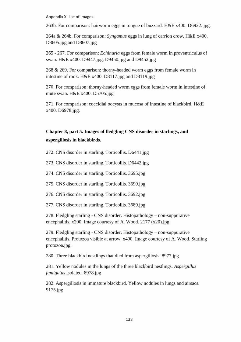
Appendix X. List of images.
128
263b. For comparison: hairworm eggs in tongue of buzzard. H&E x400. D6922. jpg.
264a & 264b. For comparison: Syngamus eggs in lung of carrion crow. H&E x400.
D8605.jpg and D8607.jpg
265 - 267. For comparison: Echinuria eggs from female worm in proventriculus of
swan. H&E x400. D9447.jpg, D9450.jpg and D9452.jpg
268 & 269. For comparison: thorny-headed worm eggs from female worm in
intestine of rook. H&E x400. D8117.jpg and D8119.jpg
270. For comparison: thorny-headed worm eggs from female worm in intestine of
mute swan. H&E x400. D5705.jpg
271. For comparison: coccidial oocysts in mucosa of intestine of blackbird. H&E
x400. D6978.jpg.
Chapter 8, part 5. Images of fledgling CNS disorder in starlings, and
aspergillosis in blackbirds.
272. CNS disorder in starling. Torticollis. D6441.jpg
273. CNS disorder in starling. Torticollis. D6442.jpg
274. CNS disorder in starling. Torticollis. 3695.jpg
275. CNS disorder in starling. Torticollis. 3690.jpg
276. CNS disorder in starling. Torticollis. 3692.jpg
277. CNS disorder in starling. Torticollis. 3689.jpg
278. Fledgling starling - CNS disorder. Histopathology – non-suppurative
encephalitis. x200. Image courtesy of A. Wood. 2177 (x20).jpg
279. Fledgling starling - CNS disorder. Histopathology – non-suppurative
encephalitis. Protozoa visible at arrow. x400. Image courtesy of A. Wood. Starling
protozoa.jpg.
280. Three blackbird nestlings that died from aspergillosis. 8977.jpg
281. Yellow nodules in the lungs of the three blackbird nestlings. Aspergillus
fumigatus isolated. 8978.jpg
282. Aspergillosis in immature blackbird. Yellow nodules in lungs and airsacs.
9175.jpg
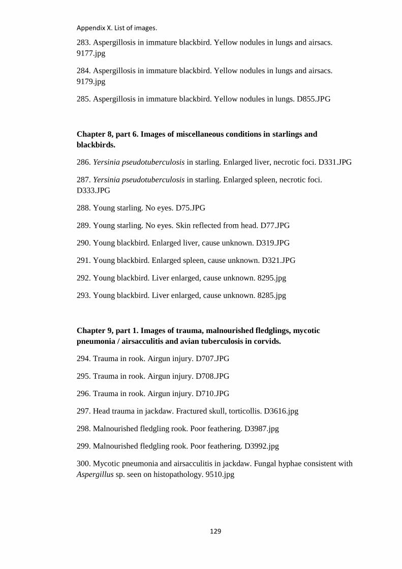
Appendix X. List of images.
129
283. Aspergillosis in immature blackbird. Yellow nodules in lungs and airsacs.
9177.jpg
284. Aspergillosis in immature blackbird. Yellow nodules in lungs and airsacs.
9179.jpg
285. Aspergillosis in immature blackbird. Yellow nodules in lungs. D855.JPG
Chapter 8, part 6. Images of miscellaneous conditions in starlings and
blackbirds.
286. Yersinia pseudotuberculosis in starling. Enlarged liver, necrotic foci. D331.JPG
287. Yersinia pseudotuberculosis in starling. Enlarged spleen, necrotic foci.
D333.JPG
288. Young starling. No eyes. D75.JPG
289. Young starling. No eyes. Skin reflected from head. D77.JPG
290. Young blackbird. Enlarged liver, cause unknown. D319.JPG
291. Young blackbird. Enlarged spleen, cause unknown. D321.JPG
292. Young blackbird. Liver enlarged, cause unknown. 8295.jpg
293. Young blackbird. Liver enlarged, cause unknown. 8285.jpg
Chapter 9, part 1. Images of trauma, malnourished fledglings, mycotic
pneumonia / airsacculitis and avian tuberculosis in corvids.
294. Trauma in rook. Airgun injury. D707.JPG
295. Trauma in rook. Airgun injury. D708.JPG
296. Trauma in rook. Airgun injury. D710.JPG
297. Head trauma in jackdaw. Fractured skull, torticollis. D3616.jpg
298. Malnourished fledgling rook. Poor feathering. D3987.jpg
299. Malnourished fledgling rook. Poor feathering. D3992.jpg
300. Mycotic pneumonia and airsacculitis in jackdaw. Fungal hyphae consistent with
Aspergillus sp. seen on histopathology. 9510.jpg
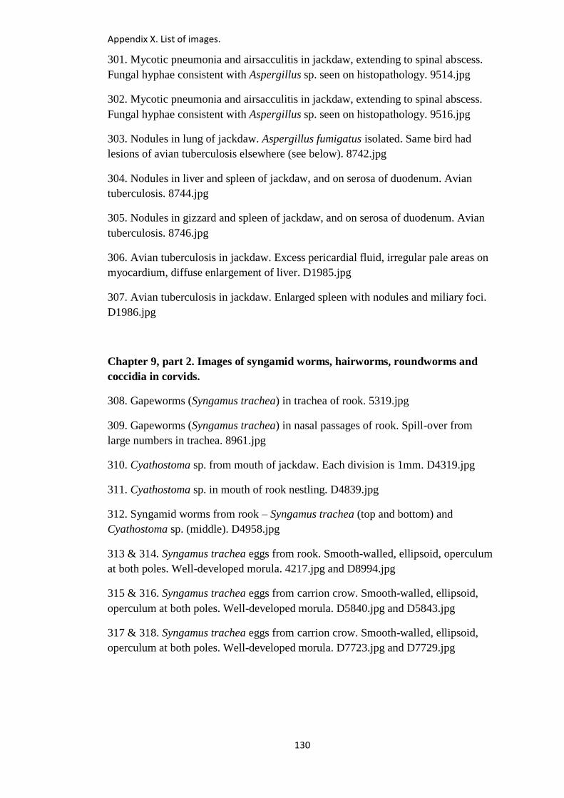
Appendix X. List of images.
130
301. Mycotic pneumonia and airsacculitis in jackdaw, extending to spinal abscess.
Fungal hyphae consistent with Aspergillus sp. seen on histopathology. 9514.jpg
302. Mycotic pneumonia and airsacculitis in jackdaw, extending to spinal abscess.
Fungal hyphae consistent with Aspergillus sp. seen on histopathology. 9516.jpg
303. Nodules in lung of jackdaw. Aspergillus fumigatus isolated. Same bird had
lesions of avian tuberculosis elsewhere (see below). 8742.jpg
304. Nodules in liver and spleen of jackdaw, and on serosa of duodenum. Avian
tuberculosis. 8744.jpg
305. Nodules in gizzard and spleen of jackdaw, and on serosa of duodenum. Avian
tuberculosis. 8746.jpg
306. Avian tuberculosis in jackdaw. Excess pericardial fluid, irregular pale areas on
myocardium, diffuse enlargement of liver. D1985.jpg
307. Avian tuberculosis in jackdaw. Enlarged spleen with nodules and miliary foci.
D1986.jpg
Chapter 9, part 2. Images of syngamid worms, hairworms, roundworms and
coccidia in corvids.
308. Gapeworms (Syngamus trachea) in trachea of rook. 5319.jpg
309. Gapeworms (Syngamus trachea) in nasal passages of rook. Spill-over from
large numbers in trachea. 8961.jpg
310. Cyathostoma sp. from mouth of jackdaw. Each division is 1mm. D4319.jpg
311. Cyathostoma sp. in mouth of rook nestling. D4839.jpg
312. Syngamid worms from rook – Syngamus trachea (top and bottom) and
Cyathostoma sp. (middle). D4958.jpg
313 & 314. Syngamus trachea eggs from rook. Smooth-walled, ellipsoid, operculum
at both poles. Well-developed morula. 4217.jpg and D8994.jpg
315 & 316. Syngamus trachea eggs from carrion crow. Smooth-walled, ellipsoid,
operculum at both poles. Well-developed morula. D5840.jpg and D5843.jpg
317 & 318. Syngamus trachea eggs from carrion crow. Smooth-walled, ellipsoid,
operculum at both poles. Well-developed morula. D7723.jpg and D7729.jpg
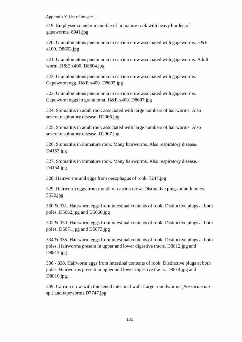
Appendix X. List of images.
131
319. Emphysema under mandible of immature rook with heavy burden of
gapeworms. 8941.jpg
320. Granulomatous pneumonia in carrion crow associated with gapeworms. H&E
x100. D8603.jpg
321. Granulomatous pneumonia in carrion crow associated with gapeworms. Adult
worm. H&E x400. D8604.jpg
322. Granulomatous pneumonia in carrion crow associated with gapeworms.
Gapeworm egg. H&E x400. D8605.jpg
323. Granulomatous pneumonia in carrion crow associated with gapeworms.
Gapeworm eggs in granuloma. H&E x400. D8607.jpg
324. Stomatitis in adult rook associated with large numbers of hairworms. Also
severe respiratory disease. D2966.jpg
325. Stomatitis in adult rook associated with large numbers of hairworms. Also
severe respiratory disease. D2967.jpg
326. Stomatitis in immature rook. Many hairworms. Also respiratory disease.
D4153.jpg
327. Stomatitis in immature rook. Many hairworms. Also respiratory disease.
D4154.jpg
328. Hairworms and eggs from oesophagus of rook. 7247.jpg
329. Hairworm eggs from mouth of carrion crow. Distinctive plugs at both poles.
5532.jpg
330 & 331. Hairworm eggs from intestinal contents of rook. Distinctive plugs at both
poles. D5662.jpg and D5666.jpg
332 & 333. Hairworm eggs from intestinal contents of rook. Distinctive plugs at both
poles. D5671.jpg and D5673.jpg
334 & 335. Hairworm eggs from intestinal contents of rook. Distinctive plugs at both
poles. Hairworms present in upper and lower digestive tracts. D8812.jpg and
D8813.jpg.
336 - 338. Hairworm eggs from intestinal contents of rook. Distinctive plugs at both
poles. Hairworms present in upper and lower digestive tracts. D8814.jpg and
D8816.jpg.
339. Carrion crow with thickened intestinal wall. Large roundworms (Porrocaecum
sp.) and tapeworms.D7747.jpg
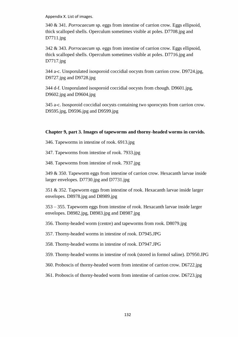
Appendix X. List of images.
132
340 & 341. Porrocaecum sp. eggs from intestine of carrion crow. Eggs ellipsoid,
thick scalloped shells. Operculum sometimes visible at poles. D7708.jpg and
D7711.jpg
342 & 343. Porrocaecum sp. eggs from intestine of carrion crow. Eggs ellipsoid,
thick scalloped shells. Operculum sometimes visible at poles. D7716.jpg and
D7717.jpg
344 a-c. Unsporulated isosporoid coccidial oocysts from carrion crow. D9724.jpg,
D9727.jpg and D9728.jpg
344 d-f. Unsporulated isosporoid coccidial oocysts from chough. D9601.jpg,
D9602.jpg and D9604.jpg
345 a-c. Isosporoid coccidial oocysts containing two sporocysts from carrion crow.
D9595.jpg, D9596.jpg and D9599.jpg
Chapter 9, part 3. Images of tapeworms and thorny-headed worms in corvids.
346. Tapeworms in intestine of rook. 6913.jpg
347. Tapeworms from intestine of rook. 7933.jpg
348. Tapeworms from intestine of rook. 7937.jpg
349 & 350. Tapeworm eggs from intestine of carrion crow. Hexacanth larvae inside
larger envelopes. D7730.jpg and D7731.jpg
351 & 352. Tapeworm eggs from intestine of rook. Hexacanth larvae inside larger
envelopes. D8978.jpg and D8989.jpg
353 – 355. Tapeworm eggs from intestine of rook. Hexacanth larvae inside larger
envelopes. D8982.jpg, D8983.jpg and D8987.jpg
356. Thorny-headed worm (centre) and tapeworms from rook. D8079.jpg
357. Thorny-headed worms in intestine of rook. D7945.JPG
358. Thorny-headed worms in intestine of rook. D7947.JPG
359. Thorny-headed worms in intestine of rook (stored in formol saline). D7950.JPG
360. Proboscis of thorny-headed worm from intestine of carrion crow. D6722.jpg
361. Proboscis of thorny-headed worm from intestine of carrion crow. D6723.jpg
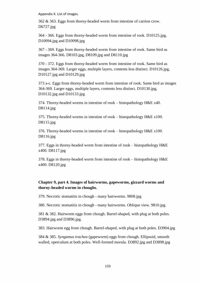
Appendix X. List of images.
133
362 & 363. Eggs from thorny-headed worm from intestine of carrion crow.
D6727.jpg
364 - 366. Eggs from thorny-headed worm from intestine of rook. D10125.jpg,
D10094.jpg and D10098.jpg
367 - 369. Eggs from thorny-headed worm from intestine of rook. Same bird as
images 364-366. D8103.jpg, D8109.jpg and D8110.jpg
370 - 372. Eggs from thorny-headed worm from intestine of rook. Same bird as
images 364-369. Larger eggs, multiple layers, contents less distinct. D10126.jpg,
D10127.jpg and D10129.jpg
373 a-c. Eggs from thorny-headed worm from intestine of rook. Same bird as images
364-369. Larger eggs, multiple layers, contents less distinct. D10130.jpg,
D10132.jpg and D10133.jpg
374. Thorny-headed worms in intestine of rook – histopathology H&E x40.
D8114.jpg
375. Thorny-headed worms in intestine of rook – histopathology H&E x100.
D8115.jpg
376. Thorny-headed worms in intestine of rook – histopathology H&E x100.
D8116.jpg
377. Eggs in thorny-headed worm from intestine of rook – histopathology H&E
x400. D8117.jpg
378. Eggs in thorny-headed worm from intestine of rook – histopathology H&E
x400. D8120.jpg
Chapter 9, part 4. Images of hairworms, gapeworms, gizzard worms and
thorny-headed worms in choughs.
379. Necrotic stomatitis in chough - many hairworms. 9808.jpg
380. Necrotic stomatitis in chough - many hairworms. Oblique view. 9810.jpg
381 & 382. Hairworm eggs from chough. Barrel-shaped, with plug at both poles.
D3894.jpg and D3896.jpg.
383. Hairworm egg from chough. Barrel-shaped, with plug at both poles. D3904.jpg
384 & 385. Syngamus trachea (gapeworm) eggs from chough. Ellipsoid, smooth
walled, operculum at both poles. Well-formed morula. D3892.jpg and D3898.jpg
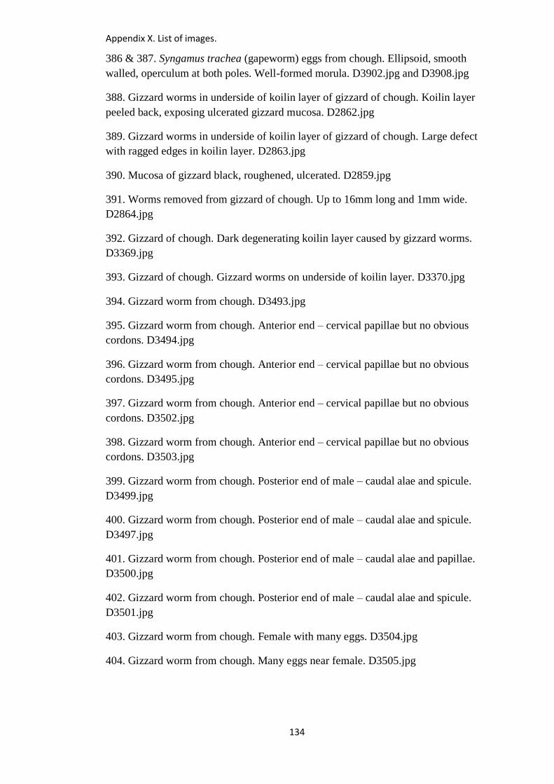
Appendix X. List of images.
134
386 & 387. Syngamus trachea (gapeworm) eggs from chough. Ellipsoid, smooth
walled, operculum at both poles. Well-formed morula. D3902.jpg and D3908.jpg
388. Gizzard worms in underside of koilin layer of gizzard of chough. Koilin layer
peeled back, exposing ulcerated gizzard mucosa. D2862.jpg
389. Gizzard worms in underside of koilin layer of gizzard of chough. Large defect
with ragged edges in koilin layer. D2863.jpg
390. Mucosa of gizzard black, roughened, ulcerated. D2859.jpg
391. Worms removed from gizzard of chough. Up to 16mm long and 1mm wide.
D2864.jpg
392. Gizzard of chough. Dark degenerating koilin layer caused by gizzard worms.
D3369.jpg
393. Gizzard of chough. Gizzard worms on underside of koilin layer. D3370.jpg
394. Gizzard worm from chough. D3493.jpg
395. Gizzard worm from chough. Anterior end – cervical papillae but no obvious
cordons. D3494.jpg
396. Gizzard worm from chough. Anterior end – cervical papillae but no obvious
cordons. D3495.jpg
397. Gizzard worm from chough. Anterior end – cervical papillae but no obvious
cordons. D3502.jpg
398. Gizzard worm from chough. Anterior end – cervical papillae but no obvious
cordons. D3503.jpg
399. Gizzard worm from chough. Posterior end of male – caudal alae and spicule.
D3499.jpg
400. Gizzard worm from chough. Posterior end of male – caudal alae and spicule.
D3497.jpg
401. Gizzard worm from chough. Posterior end of male – caudal alae and papillae.
D3500.jpg
402. Gizzard worm from chough. Posterior end of male – caudal alae and spicule.
D3501.jpg
403. Gizzard worm from chough. Female with many eggs. D3504.jpg
404. Gizzard worm from chough. Many eggs near female. D3505.jpg

Appendix X. List of images.
135
405 & 406. Gizzard worm from chough. Eggs containing larvae. D3958.jpg and
D3960.jpg
407 & 408. Left – syngamid egg from chough. Right – gizzard worm eggs from
chough. D3966.jpg and D3962.jpg
409. Thorny-headed worms and tapeworms in intestine of chough. D3490.jpg
410. Thorny-headed worms and tapeworms in intestine of chough. D3491.jpg
411. Thorny-headed worms from chough (from alcohol). D3437.jpg
412. Thorny-headed worms in chough. Wall of intestine thickened, much sand in
contents of upper intestine. D5227.jpg
413. Thorny-headed worms in lower intestine of chough. Wall of intestine thickened.
D5231.jpg
414. Thorny-headed worms in lower intestine of chough. Contents haemorrhagic.
D5232.jpg
415. Thorny-headed worms removed from intestine of chough. D5236.jpg
416. Thorny-headed worms removed from intestine of chough (stored in alcohol).
Each division is 1mm. D5239.jpg
417. Proboscis of thorny-headed worm from chough. D5551.jpg
418. Proboscis of thorny-headed worm from chough. D5552.jpg
419 & 420. Eggs of thorny-headed worm from chough. D5555.jpg and D5560.jpg
421 & 422. Eggs of thorny-headed worm from chough. D5565.jpg and D5568.jpg
Chapter 9, part 5. Images of corvid respiratory syndrome (CRS) and
respiratory tract conditions not otherwise specified in corvids.
423. Corvid respiratory syndrome. Bird 5. Rook. Mild pericarditis, extensive
pneumonia. PCR for Mycoplasma gallisepticum positive. 8693.JPG
424. Corvid respiratory syndrome. Bird 5. Rook. Dorsal aspect of lungs. Extensive
grey-coloured pneumonia and pleurisy, and multiple small yellow nodules in lungs.
PCR for Mycoplasma gallisepticum positive. 8695.JPG
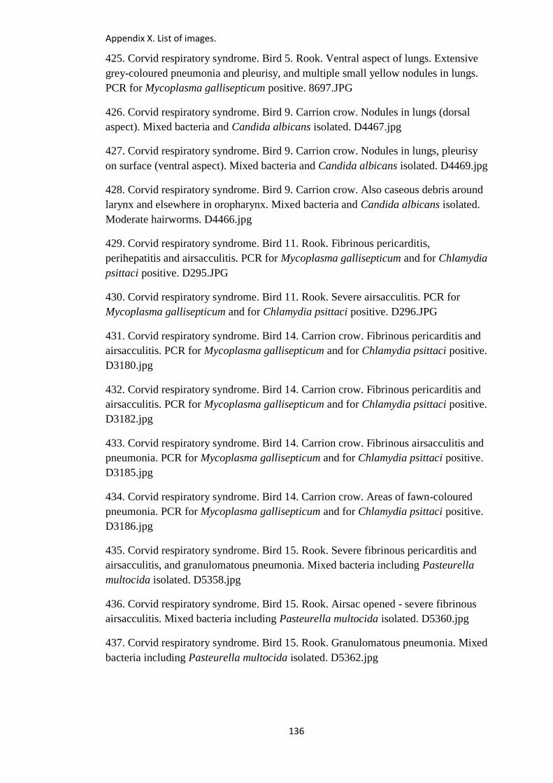
Appendix X. List of images.
136
425. Corvid respiratory syndrome. Bird 5. Rook. Ventral aspect of lungs. Extensive
grey-coloured pneumonia and pleurisy, and multiple small yellow nodules in lungs.
PCR for Mycoplasma gallisepticum positive. 8697.JPG
426. Corvid respiratory syndrome. Bird 9. Carrion crow. Nodules in lungs (dorsal
aspect). Mixed bacteria and Candida albicans isolated. D4467.jpg
427. Corvid respiratory syndrome. Bird 9. Carrion crow. Nodules in lungs, pleurisy
on surface (ventral aspect). Mixed bacteria and Candida albicans isolated. D4469.jpg
428. Corvid respiratory syndrome. Bird 9. Carrion crow. Also caseous debris around
larynx and elsewhere in oropharynx. Mixed bacteria and Candida albicans isolated.
Moderate hairworms. D4466.jpg
429. Corvid respiratory syndrome. Bird 11. Rook. Fibrinous pericarditis,
perihepatitis and airsacculitis. PCR for Mycoplasma gallisepticum and for Chlamydia
psittaci positive. D295.JPG
430. Corvid respiratory syndrome. Bird 11. Rook. Severe airsacculitis. PCR for
Mycoplasma gallisepticum and for Chlamydia psittaci positive. D296.JPG
431. Corvid respiratory syndrome. Bird 14. Carrion crow. Fibrinous pericarditis and
airsacculitis. PCR for Mycoplasma gallisepticum and for Chlamydia psittaci positive.
D3180.jpg
432. Corvid respiratory syndrome. Bird 14. Carrion crow. Fibrinous pericarditis and
airsacculitis. PCR for Mycoplasma gallisepticum and for Chlamydia psittaci positive.
D3182.jpg
433. Corvid respiratory syndrome. Bird 14. Carrion crow. Fibrinous airsacculitis and
pneumonia. PCR for Mycoplasma gallisepticum and for Chlamydia psittaci positive.
D3185.jpg
434. Corvid respiratory syndrome. Bird 14. Carrion crow. Areas of fawn-coloured
pneumonia. PCR for Mycoplasma gallisepticum and for Chlamydia psittaci positive.
D3186.jpg
435. Corvid respiratory syndrome. Bird 15. Rook. Severe fibrinous pericarditis and
airsacculitis, and granulomatous pneumonia. Mixed bacteria including Pasteurella
multocida isolated. D5358.jpg
436. Corvid respiratory syndrome. Bird 15. Rook. Airsac opened - severe fibrinous
airsacculitis. Mixed bacteria including Pasteurella multocida isolated. D5360.jpg
437. Corvid respiratory syndrome. Bird 15. Rook. Granulomatous pneumonia. Mixed
bacteria including Pasteurella multocida isolated. D5362.jpg
Appendix X. List of images.
137
438. Corvid respiratory syndrome. Bird 15. Rook. Ventral aspect of lungs -
granulomatous pneumonia. Mixed bacteria including Pasteurella multocida isolated.
D5363.jpg
439. Corvid respiratory syndrome. Bird 16. Rook. Severe fibrinous pericarditis and
airsacculitis. PCR for Mycoplasma gallisepticum positive. D2969.jpg
440. Corvid respiratory syndrome. Bird 16. Rook. Severe fibrinous pericarditis and
airsacculitis. Granulomas in lungs. PCR for Mycoplasma gallisepticum positive.
D2970.jpg
441. Corvid respiratory syndrome. Bird 16. Rook. Granulomas in lungs. PCR for
Mycoplasma gallisepticum positive. D2971.jpg
442. Corvid respiratory syndrome. Bird 16. Rook. Granulomas in lungs – cut
surfaces. PCR for Mycoplasma gallisepticum positive. D2972.jpg
443. Corvid respiratory syndrome. Bird 17. Rook. Pericarditis, airsacculitis,
pneumonia. Enlarged liver. PCR for Mycoplasma gallisepticum positive. D4301.jpg
444. Corvid respiratory syndrome. Bird 17. Rook. Fibrinous pericarditis. PCR for
Mycoplasma gallisepticum positive. D4303.jpg
445. Corvid respiratory syndrome. Bird 17. Rook. Red exudative pneumonia plus
pale nodules. PCR for Mycoplasma gallisepticum positive. D4305.jpg
446. Corvid respiratory syndrome. Bird 18. Rook. Thoracic and abdominal
airsacculitis. PCR for Mycoplasma gallisepticum and for Chlamydia psittaci positive.
D4156.jpg
447. Corvid respiratory syndrome. Bird 18. Rook. Purulent debris on underside of
sternum. PCR for Mycoplasma gallisepticum and for Chlamydia psittaci positive.
D4158.jpg
448. Corvid respiratory syndrome. Bird 18. Rook. Thoracic and abdominal
airsacculitis. PCR for Mycoplasma gallisepticum and for Chlamydia psittaci positive.
D4160.jpg
449. Corvid respiratory syndrome. Bird 18. Rook. Red lung consolidation with grey
granulomas. PCR for Mycoplasma gallisepticum and for Chlamydia psittaci positive.
D4166.jpg
450. Corvid respiratory syndrome. Bird 18. Rook. Red lung consolidation with grey
granulomas (ventral surface). PCR for Mycoplasma gallisepticum and for Chlamydia
psittaci positive. D4167.jpg
Appendix X. List of images.
138
451. Corvid respiratory syndrome. Bird 19. Rook. Enlarged liver, extensive
airsacculitis. D4642.jpg
452. Corvid respiratory syndrome. Bird 19. Rook. Airsac reflected. D4645.jpg
453. Corvid respiratory syndrome. Bird 19. Rook. Small foci of pneumonia.
D4647.jpg
454. Corvid respiratory syndrome. Bird 19. Rook. Excess mucus, green nodule
caused by sinusitis extending into palate. D4641.jpg
455. Corvid respiratory syndrome. Bird 20. Jackdaw. Ocular discharge and peri-
orbital loss of feathers. Sinusitis and pneumonia. D4133.jpg
456. Corvid respiratory syndrome. Bird 20. Jackdaw. Purulent sinusitis. Also
pneumonia. D4135.jpg
457. Respiratory tract condition NOS. Bird 22. Lung from hooded crow. Focal
pneumonia and pleurisy. Mixed bacteria isolated. 4804.jpg
458. Respiratory tract condition NOS. Bird 29. Lung from carrion crow. Cream-
coloured area of pneumonia. Heavy growth of Pasteurella haemolytica – like
bacteria isolated. 5953.jpg
459. Respiratory tract condition NOS. Bird 29. Heart from carrion crow. Purulent
pericarditis. Heavy growth of Pasteurella haemolytica – like bacteria isolated.
5955.jpg
460 & 461. Respiratory tract condition NOS. Bird 29. Carrion crow. Left: Caseous
debris on either side of tongue and laryngeal mound. Right: Caseous debris around
underside of tongue (removed). Heavy growth of Candida albicans and Pasteurella
haemolytica – like bacteria isolated. 5959.jpg and 5957.jpg
462. Respiratory tract condition NOS. Bird 31. Adult rook. Abdomen filled with
yellow fluid. 7734.jpg
463. Respiratory tract condition NOS. Bird 31. Adult rook. Enlarged liver with tiny
necrotic foci. Fibrinous pericarditis, perihepatitis, airsacculitis, pneumonia. Heavy
growths of E. coli isolated. 7738.jpg
464. Respiratory tract condition NOS. Bird 36. Carrion crow with loss of feathers
from around eye. Lice and mites present. Airsacculitis and pneumonia. Mixed
bacteria isolated. 9054.jpg
465. Respiratory tract condition NOS. Bird 39. Immature rook. Caseous airsacculitis
visible through abdominal wall. D936.JPG
Appendix X. List of images.
139
466. Respiratory tract condition NOS. Bird 39. Immature rook. Heart and liver
partially covered by extensive caseous pericarditis, perihepatitis and airsacculitis.
Mixed bacteria isolated. D938.JPG
467. Respiratory tract condition NOS. Bird 39. Immature rook. Heart and liver
partially covered by extensive caseous pericarditis, perihepatitis and airsacculitis.
Portion of airsac removed. Mixed bacteria isolated. D940.JPG
468. Respiratory tract condition NOS. Bird 40. Carrion crow. Euthanased in early
stages. Pericarditis, perihepatitis, enlarged liver. Mixed bacteria and Candida
albicans isolated from lung and sinus. D4487.jpg
469. Respiratory tract condition NOS. Bird 40. Carrion crow. Euthanased in early
stages. Cross-section through beak – purulent sinusitis. Mixed bacteria and Candida
albicans isolated from lung and sinus. D4489.jpg
Chapter 9, part 6. Images of pox, feather loss, Cnemidocoptes, Ornithonyssus,
lice and feather mites in corvids.
470. Pox nodules in jackdaw. 7462.jpg
471. Pox nodules in jackdaw. Ventral aspect. 7466.jpg
472. Pox nodules on toes of magpie. 5861.jpg
473. Pox nodules on toes of magpie. 5865.jpg
474 & 475. Feather loss in immature jackdaw. Cause unknown. 4331.jpg and
4333.jpg
476. Immature magpie. Feather loss from inner and outer aspects of both legs. Died
from necrotic enteritis. D4028.jpg
477. Cnemidocoptic mange (scaly leg) in rook. 7163.jpg
478. Cnemidocoptic mange (scaly leg) in rook. 7165.jpg
479. Early cnemidocoptic mange (scaly leg) in rook. 7167.jpg
480. Early cnemidocoptic mange (scaly leg) in rook. 9326.jpg
481. Cnemidocoptic mange (scaly leg) in carrion crow. 4097.jpg
482 - 484. Cnemidocoptes sp. mites from rook. Adult female (left), adult male
(middle), larva (right). 7256.jpg, 7264.jpg, and 7254.jpg
Appendix X. List of images.
140
485. Large numbers of northern fowl mite (Ornithonyssus sp.) on rook. 8263.jpg
486. Large numbers of northern fowl mite (Ornithonyssus sp.) on rook. 8268.jpg
487. Large numbers of northern fowl mite (Ornithonyssus sp.) on rook (different
bird). 3608.jpg
488. Northern fowl mite (Ornithonyssus sp.) from rook. 3940.jpg
489 - 490. Left: anal plate from northern fowl mite (Ornithonyssus sp.) from rook –
ovoid, with anterior anus. Right: anal plate from red mite (Dermanyssus sp.) from
chicken – cup-shaped, with posterior anus. Mites partially cleared in KOH. 3945.jpg
and 3966.jpg
491. Lice and mites on rook. D1653.jpg
492. Lice and mites on rook. D1652.jpg
493. Lice and mites on rook. D1654.jpg
494. Lice from rook. D2960.jpg
495. Lice from rook (closer). D2960.jpg
496. Feather loss in immature rook – many lice. Also heavy burden of mixed internal
parasites. D5101.jpg
497. Feather loss in immature rook – many lice. Also heavy burden of mixed internal
parasites. D5108.jpg
498. Feather loss in immature rook – many louse eggs around eye and corner of
beak. Also heavy burden of mixed internal parasites. D5103.jpg
499. Feather loss in immature rook – many louse eggs under mandible. Also heavy
burden of mixed internal parasites. D5105.jpg
500. Feather loss in immature rook – many lice. Also heavy burden of mixed internal
parasites. D5107.jpg
501. Feather mite from chough. Male. D5001.jpg
502. Feather mite from chough. Male. D5003.jpg
503. Feather mite from chough. Female. D5000.jpg
504. Feather mite from chough. Female. D5002.jpg
Appendix X. List of images.
141
Chapter 9, part 7. Images of eye/brain abnormalities in young choughs.
505. Immature chough. Corneal opacity. D92.JPG
506. Immature chough. Corneal opacity. D93.JPG
507. Immature chough. Corneal opacity. D95.JPG
508. Immature chough. Corneal opacity. D3518.jpg
509. Immature chough. Corneal opacity. D3520.jpg
510. Immature chough. Corneal opacity. D3525.jpg
511. Eye abnormality in chough fledgling. Corneal opacity of left eye. D4924.jpg
512. Eye abnormality in chough fledgling. Green discoloration of cornea of right eye.
D4925.jpg
513. Eye abnormality in chough fledgling. Disparity in size of globes – left eye
larger. D4926.jpg
514. Eye abnormality in chough fledgling. Disparity in size of globes – left eye
(larger). D4928.jpg
515. Eye abnormality in chough fledgling. Disparity in size of globes – right eye
(smaller). D4929.jpg
516. Eye abnormality in chough fledgling. Disparity in size of globes – left eye
larger. D4932.jpg
517. Eye abnormality in chough fledgling. Disparity in size of globes. Right eye
larger. D98.JPG
518. Eye abnormality in chough fledgling. Disparity in size of globes. Right eye
larger. D99.JPG
519. Corneal opacity in chough - histopathology. H&E x 400. Vacuolation of corneal
epithelium. D4843.jpg
520. Corneal opacity in chough - histopathology. H&E x 400. Vacuolation of corneal
epithelium. D4845.jpg
521. Corneal opacity in chough - histopathology. H&E x 400. Vacuolation of corneal
epithelium. D4858.jpg
522. Corneal opacity in chough - histopathology. H&E x 400. Vacuolation of corneal
epithelium. D4859.jpg
Appendix X. List of images.
142
523. Brain abnormality (hydrocephalus) in chough nestling aged 4 weeks (left). No
gross brain abnormalities in bird on right. D4814.jpg
524. Brain abnormality (hydrocephalus) in chough nestling aged 4 weeks (above).
No gross brain abnormalities in bird below. D4816.jpg
525. Brain abnormality (hydrocephalus) in chough nestling aged 4 weeks. D4821.jpg
526. Chough nestling aged 4 weeks. No obvious hydrocephalus. D4824.jpg
527. Brain abnormality (hydrocephalus) in chough nestling aged 4 weeks (lower). No
gross brain abnormalities in bird above. D4825.jpg
528. Brain abnormality (hydrocephalus) in chough nestling aged 4 weeks (left). No
gross brain abnormalities in bird on right. D4827.jpg
Chapter 9, part 8. Images of necrotic oesophagitis and other digestive tract
conditions not otherwise specified in corvids.
529. Necrotic oesophagitis in immature chough (Bird 1). Food in upper oesophagus.
D6551.jpg
530. Necrotic oesophagitis in immature chough (Bird 1). Yellow serositis over
oesophagus and trachea. D6552.jpg
531. Necrotic oesophagitis in immature chough (Bird 1). Food particles attached to
necrotic mucosa. No food in gizzard. D6555.jpg
532. Necrotic oesophagitis in immature chough (Bird 1). Necrotic mucosa (food
particles removed). D6559.jpg
533. Necrotic oesophagitis in immature chough (Bird 2). Food in upper oesophagus.
D6560.jpg
534. Necrotic oesophagitis in immature chough (Bird 2). Yellow serositis over
oesophagus and trachea. D6562.jpg
535. Necrotic oesophagitis in immature chough (Bird 2). Mucosa of oesophagus
yellow/orange and necrotic. D6564.jpg
536. Necrotic oesophagitis in immature chough (Bird 3). Yellow serositis over
oesophagus and trachea. D6565.jpg
537. Necrotic oesophagitis in immature chough (Bird 3). Thickening of oesophagus
(unopened). D6566.jpg
Appendix X. List of images.
143
538. Necrotic oesophagitis in immature chough (Bird 3). Mucosa of oesophagus
yellow/orange and necrotic. D6569.jpg
539. Necrotic oesophagitis in immature chough (Bird 4). Thickening of oesophagus
(unopened). D6591.jpg
540. Necrotic oesophagitis in immature chough (Bird 4). Mucosa of oesophagus
yellow/orange and necrotic. D6594.jpg
541. Necrotic oesophagitis in immature chough (Bird 4). Mucosa of oesophagus
yellow/orange and necrotic. D6597.jpg
542. Fledgling jackdaw found under garden feeder. Impacted gizzard. D964.JPG
543. Fledgling jackdaw found under garden feeder. Impacted intestine. D963.JPG
544. Fledgling jackdaw found under garden feeder. Impacted intestine. D962.JPG
545. Liver abnormality in carrion crow. Cause not established. D2181.jpg
556. Liver abnormality in carrion crow. Cause not established. D2182.jpg
547. Carrion crow. Grey debris in oropharynx, small nodules in oesophagus.
Moderate hairworm eggs in oropharynx. Mixed bacteria isolated. Histopathology –
heterophilic granulomas in oesophagus. D3171.jpg
548. Carrion crow. Grey debris in oropharynx, small nodules in oesophagus.
Moderate hairworm eggs in oropharynx. Mixed bacteria isolated. Histopathology –
heterophilic granulomas in oesophagus. D3173.jpg
Chapter 9, part 9. Images of adverse environmental conditions, Yersinia
pseudotuberculosis, cellulitis, neoplasia and poisoning in corvids.
549. Rook nestlings - nests destroyed in high winds. D4831.jpg
550. Yersinia pseudotuberculosis in chough. Infected elbow joint. D4963.jpg
551. Yersinia pseudotuberculosis in chough. Fine focal liver necrosis. D4966.jpg
552. Yersinia pseudotuberculosis in chough. Fine focal necrosis in spleen. D4968.jpg
553. Cellulitis in adult rook. No significant bacteria isolated. D1519.jpg
554. Cellulitis in adult rook. No significant bacteria isolated. D8075.jpg
555. Cellulitis in adult rook. No significant bacteria isolated. D8077.jpg
Appendix X. List of images.
144
556. Cellulitis in adult rook. No significant bacteria isolated. D8078.jpg
557. Cellulitis in neck of adult magpie. Mixed bacteria isolated. D3081.jpg
558. Abscess below eye of rook. Staphylococcus sp. isolated. D6518.jpg
559. Abscess below eye of rook. Staphylococcus sp. isolated. D6520.jpg
560. Tumour in rook - squamous cell carcinoma. Nodules on mucosa of oesophagus
(opened). D1655.jpg
561. Tumour in rook - squamous cell carcinoma. Nodules on mucosa of oesophagus
(opened). D1656.jpg
562. Tumour in rook - squamous cell carcinoma. Diffuse neoplastic infiltration
around larynx. D1657.jpg
563. Accidental diazinon poisoning in raven. Stomach contents - lamb scrotum and
rubber ring, and lamb tail. D3612.jpg
564. Accidental diazinon poisoning in raven. Stomach contents - lamb scrotum
(opened) and rubber ring. D3615.jpg
565. Accidental diazinon poisoning in raven. Many unhatched fly eggs on carcase.
D3609.jpg
Chapter 10. Images of miscellaneous conditions in “Other Passeriformes”.
566. Trauma in waxwing. Enlarged pale liver. Blood around heart and in abdomen.
D1878.JPG
567. Trauma in waxwing. Many berries in oesophagus. D1877.JPG
568. Yersinia pseudotuberculosis infection in house martin. Pale foci and dark
haemorrhages on liver. D1085.jpg
569. Yersinia pseudotuberculosis infection in house martin. Pale foci and dark
haemorrhages on liver. Pale nodules in enlarged spleen. D1086.jpg
570. Yersinia pseudotuberculosis infection in house martin. Pale area of necrosis on
palate. D1087jpg.
571. Yersinia pseudotuberculosis infection in house martin. Large cream-coloured
granuloma on palate (different bird). D279.jpg.
Appendix X. List of images.
145
572. Mycotic pneumonia in waxwing. Most of one lung replaced by cream-coloured
mass. Aspergillus fumigatus isolated. 8186.JPG
573. Hippoboscid flies and pupal cases from house martin nest. 9118.JPG
574. Nematodes in airsacs of blackcap. Diplotriaena sp. D326.JPG
575. Nematodes removed from airsacs of blackcap. Diplotriaena sp. D343.JPG
576. Intestinal fluke from house martin. Many eggs visible. 4244.JPG
577. Intestinal flukes from house martin. Many eggs visible. 4411.JPG
578. Eggs from intestinal fluke from house martin. 4246.JPG
579. Eggs from intestinal fluke from house martin. 4248.JPG
Chapter 11, part 1. Images of avian tuberculosis in pigeons and doves.
580. Avian tuberculosis in woodpigeon. Large cream raised nodules throughout liver.
5196.jpg
581. Avian tuberculosis in woodpigeon. Large fawn-coloured raised nodule in lung.
5197.jpg
582. Avian tuberculosis in woodpigeon. Spleen replaced by large cream mass.
5199.jpg
583. Avian tuberculosis in woodpigeon. Pale nodules in wall of intestine. 5201.jpg
584. Avian tuberculosis in woodpigeon. Marked atrophy of breast muscles. 5193.jpg
585. Avian tuberculosis in woodpigeon. Much gelatinous abdominal fluid. Multiple
pale foci in liver. D6013.jpg
586. Avian tuberculosis in woodpigeon. Multiple pale foci in liver. D6015.jpg
587. Avian tuberculosis in woodpigeon. Large fawn nodule in lung. D6021.jpg
588. Avian tuberculosis in woodpigeon. Spleen replaced by large cream mass.
D6019.jpg
589. Avian tuberculosis in woodpigeon. Cut section of spleen. D6020.jpg
590. Avian tuberculosis in woodpigeon. Pale nodules in wall of intestine. Fluid
intestinal contents. D6024.jpg
Appendix X. List of images.
146
591. Avian tuberculosis in woodpigeon. Nodule on head. 8060.jpg
592. Avian tuberculosis in woodpigeon. Nodule on head – skin removed. 8062.jpg
593. Avian tuberculosis in woodpigeon. Nodule on head – opened. 8064.jpg
594. Avian tuberculosis in woodpigeon. Mucosa of intestine thickened, orange-
coloured, fissured. 8070.jpg
595. Avian tuberculosis in woodpigeon. Mucosa of oesophagus thickened and
orange-coloured. 8110.jpg
596. Avian tuberculosis in woodpigeon. Enlarged liver with multiple small pale foci.
8108.jpg
597. Avian tuberculosis in woodpigeon. Enlarged liver with a few pale foci.
Pericarditis. 8194.jpg
598. Avian tuberculosis in woodpigeon. Greatly enlarged pale spleen (top), most of
lung replaced by fawn mass (lower left), pale plaques and nodules on dark liver
(lower right). 9773.jpg
599. Avian tuberculosis in woodpigeon. Mucosa of intestine thickened, granular,
fissured. 9775.jpg
600. Avian tuberculosis in woodpigeon. Adhesions between heart, duodenum and
liver. 8914.jpg
601. Avian tuberculosis in woodpigeon. Adhesions between heart, duodenum and
liver. 8916.jpg
602. Avian tuberculosis in woodpigeon. Heart covered in pale plaques and nodules.
8918.jpg
603. Avian tuberculosis in woodpigeon. Pale nodules on cut surface of spleen.
8922.jpg
604. Cutaneous form of avian tuberculosis in collared dove. 4277.jpg
605. Cutaneous form of avian tuberculosis in collared dove. 4279.jpg
606. Cutaneous form of avian tuberculosis in collared dove. Cut section. 4283.JPG
607. Cutaneous form of avian tuberculosis in woodpigeon. Dorsal aspect. 4269.jpg
608. Cutaneous form of avian tuberculosis in woodpigeon. Ventral aspect. 4271.jpg
609. Avian tuberculosis in feral pigeon. Large mass between crop and skin.
D874.JPG
Appendix X. List of images.
147
610. Avian tuberculosis in feral pigeon. Large mass (opened) between crop and skin.
D877.JPG
611. Urates in joints and under the skin of the foot of woodpigeon with avian
tuberculosis. 8192.jpg
612. Urates in joints (hips, knees and hocks) of woodpigeon with avian tuberculosis.
8207.jpg
613. Urates in joints (tarsometatarsal) of woodpigeon with avian tuberculosis.
8205.jpg
614. Avian tuberculosis in woodpigeon. Ziehl-Neelsen stained smear from liver,
showing acid-alcohol-fast bacilli. ATB2li.jpg
615. Avian tuberculosis in woodpigeon. Ziehl-Neelsen stained smear from liver,
showing acid-alcohol-fast bacilli. ATB4li.jpg
Chapter 11, part 2. Images of pox in pigeons and doves.
616. Pox in woodpigeon (confirmed). Nodules around eye. D283.JPG
617. Pox in woodpigeon (confirmed). Nodules on digits. D284.JPG
618. Pox in woodpigeon (confirmed). Nodules on digits. D285.JPG
619. Pox in woodpigeon (confirmed). Large nodule around upper mandible.
D350.JPG
620. Pox in woodpigeon (confirmed). Large nodule around upper mandible.
D351.JPG
621. Pox in woodpigeon (confirmed). Large nodule around upper mandible, yellow
debris on palate. D352.JPG
622. Pox in woodpigeon (confirmed). Nodules on digits. D612.JPG
623. Pox in woodpigeon (confirmed). Nodules on lower beak and around eye.
D4913.JPG
624. Pox in woodpigeon (confirmed). Nodules on lower beak and around eye.
D4914.JPG
625. Pox in woodpigeon (confirmed). Nodules on digits. D5798.JPG
626. Pox in woodpigeon (confirmed). Dry masses on wing. 8117.JPG
Appendix X. List of images.
148
627. Pox in woodpigeon (confirmed). Dry masses on wing. 8121.JPG
628. Pox in woodpigeon (confirmed). Dry masses on wing. 8125.JPG
629. Pox in woodpigeon - histopathology. H&E x 400. Many “Bollinger bodies”
(intracytoplasmic inclusions). pox3.jpg
630. Enlarged liver in woodpigeon with confirmed pox. Leucocytozoon detected on
liver histopathology. 8127.JPG
631. Enlarged liver in woodpigeon with confirmed pox. Leucocytozoon detected in
blood cells on liver histopathology. H&E x 400. Image courtesy of Alisdair Wood.
wpliver40.jpg
632. Enlarged liver in woodpigeon with confirmed pox. Leucocytozoon detected on
liver histopathology. Eosinophilic round structures compressing host blood cell
nuclei into narrow crescents. H&E x 1000. Image courtesy of Alisdair Wood.
wpliver100.jpg
Chapter 11, part 3. Images of trichomonosis, PPMV-1, salmonellosis, inclusion
body hepatitis and circovirus infection in pigeons and doves.
633. Presumed trichomonosis in woodpigeon. Yellow caseous lesions in oropharynx
and oesophagus. D39.JPG
634. Presumed trichomonosis in woodpigeon. Yellow caseous debris on palate.
D41.JPG
635. Presumed trichomonosis in woodpigeon. Yellow caseous lesions in oropharynx
and oesophagus. D4240.JPG
636. Presumed trichomonosis in woodpigeon. Yellow caseous lesions in oropharynx
and oesophagus. D4241.JPG
637. Confirmed trichomonosis in woodpigeon. Yellow caseous lesions in oropharynx
and oesophagus. D1232.JPG
638. Presumed trichomonosis in collared dove. Yellow caseous lesions in
oropharynx and oesophagus. 144.JPG
639. Trichomonosis in collared dove (confirmed). Yellow caseous lesions in
oropharynx. D368. JPG
640. Trichomonosis (confirmed) in stock dove. Swollen face with crusted exudate.
D8320. JPG
Appendix X. List of images.
149
641. Trichomonosis (confirmed) in stock dove. Yellow caseous exudate under skin
of face, extending from infra-orbital sinus. D8321. JPG
642. Trichomonosis (confirmed) in stock dove. Caseous debris on palate and in infra-
orbital sinus. D8322.JPG
643. Feral pigeon. Pigeon paramyxovirus-1 (PPMV-1) infection. Bird ataxic.
5624.JPG
644. Feral pigeon. Pigeon paramyxovirus-1 (PPMV-1) infection. Bird ataxic.
5622.JPG
645. Nodules on ventral aspect of liver of feral pigeon. Salmonella Typhimurium
isolated. 4441.JPG
646. Nodules on ventral aspect of liver of feral pigeon. Cut surface. Salmonella
Typhimurium isolated. 4443.JPG
647. Salmonellosis in feral pigeon. Pneumonia and pleurisy. 8703.JPG
648. Inclusion body hepatitis in feral pigeon. Pericarditis, perihepatitis, enlarged
liver. 4599.JPG
649a. Inclusion body hepatitis in feral pigeon. Basophilic intra-nuclear inclusion
bodies. H&E x 100. D7777.jpg
649b. Inclusion body hepatitis in feral pigeon. Basophilic intra-nuclear inclusion
bodies. H&E x 400. D7779.jpg
650. Circovirus and secondary bacterial infection of bursa of feral pigeon. Yellow
nodular granuloma in lumen of bursa of Fabricius. D4951.jpg
Chapter 11, part 4. Images of internal parasites, arthritis, granuloma, neoplasia
and crop milk in pigeons and doves.
651. Coccidial oocysts from woodpigeon. Different sizes. woodpigeon6.jpg
652a & 652b. Coccidial oocysts from woodpigeon. Different sizes. Woodpigeon2.jpg
and Woodpigeon5.jp
653a-c. Coccidial oocysts from feral pigeon. D9575.jpg, D9576.jpg and D9578.jpg
654 a-c. Coccidial oocysts from collared dove. cdove1.jpg, cdove2.jpg, cdove3.jpg
655 a-c. Coccidial oocysts from collared dove. cdove8.jpg, cdove9.jpg, cdove10.jpg
Appendix X. List of images.
150
656 & 657. Hairworm eggs (ellipsoid, plug at both poles) inside female worm. From
intestine of feral pigeon. D9562.jpg and D9563.jpg
658 a-c. Hairworm eggs from intestine of feral pigeon. D9571.jpg, D9580.jpg and
D9583.jpg
659a & 659b. Hairworm eggs and coccidial oocysts from intestine of feral pigeon.
D9581.jpg and D9582.jpg.
660. Hairworm egg (ellipsoid, plug at both poles) and coccidial oocyst from intestine
of woodpigeon. woodpigeon7.jpg
661 & 662. Hairworm eggs (ellipsoid, plug at both poles) and coccidial oocysts from
intestine of woodpigeon. woodpigeon8.jpg and woodpigeon9.jpg
663. Ornithostrongylus quadriradiatus in intestine of stock dove. Inflated cuticle at
anterior end of worm. D8367.jpg
664. Ornithostrongylus quadriradiatus in intestine of stock dove. Inflated cuticle at
anterior end of worm. D8373.jpg
665. Ornithostrongylus quadriradiatus in intestine of stock dove. Inflated cuticle at
anterior end of worm. D8374.jpg
666. Ornithostrongylus quadriradiatus in intestine of stock dove. Male worm with
bursal rays. D8378.jpg
667. Ornithostrongylus quadriradiatus in intestine of woodpigeon. Male worm with
bursal rays and spicules. 4469.jpg
668. Ornithostrongylus quadriradiatus in intestine of stock dove. Eggs inside female
worm. Eggs ellipsoid, thin-walled, granular contents. D8387.jpg
669. Infected elbow joint. Collared dove. No significant bacteria isolated. 6390.JPG
670. Infected elbow joint (opened). Collared dove. No significant bacteria isolated.
6392.JPG
671. Granuloma in feral pigeon, extending into crop. Initial damage due to trauma?
D3440.jpg
672. Granuloma in feral pigeon. Feathers and scabs removed. Granuloma extending
into crop. Initial damage due to trauma? D3444.jpg
673. Pedunculated chondrosarcoma on wing of feral pigeon. D4096.jpg
674. Pedunculated chondrosarcoma on wing of feral pigeon. D4098.jpg
Appendix X. List of images.
151
675. Pedunculated chondrosarcoma on wing of feral pigeon. Cut-section. D4100.jpg
676. Adult woodpigeon. Shot. Thickened crop due to production of “crop milk”.
D5203.jpg
677. Adult woodpigeon. Shot. Thickened crop due to production of “crop milk”.
Unopened crop. D5205.jpg
678. Adult woodpigeon. Shot. Thickened crop due to production of “crop milk”.
Opened crop. D5206.jpg
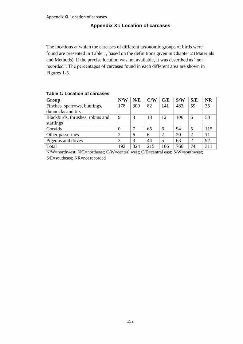
Appendix XI. Location of carcases
152
Appendix XI: Location of carcases
The locations at which the carcases of different taxonomic groups of birds were
found are presented in Table 1, based on the definitions given in Chapter 2 (Materials
and Methods). If the precise location was not available, it was described as “not
recorded”. The percentages of carcases found in each different area are shown in
Figures 1-5.
Table 1: Location of carcases
Group N/W N/E C/W C/E S/W S/E NR
Finches, sparrows, buntings,
dunnocks and tits
178 300 82 141 483 59 35
Blackbirds, thrushes, robins and
starlings
9 8 18 12 106 6 58
Corvids 0 7 65 6 94 5 115
Other passerines 2 6 6 2 20 2 11
Pigeons and doves 3 3 44 5 63 2 92
Total 192 324 215 166 766 74 311 N/W=northwest; N/E=northeast; C/W=central west; C/E=central east; S/W=southwest;
S/E=southeast; NR=not recorded
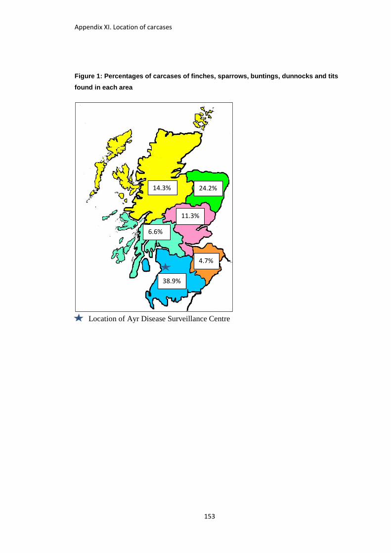
Appendix XI. Location of carcases
153
Figure 1: Percentages of carcases of finches, sparrows, buntings, dunnocks and tits
found in each area
Location of Ayr Disease Surveillance Centre
11.3%
6.6%
24.2% 14.3%
4.7%
38.9%
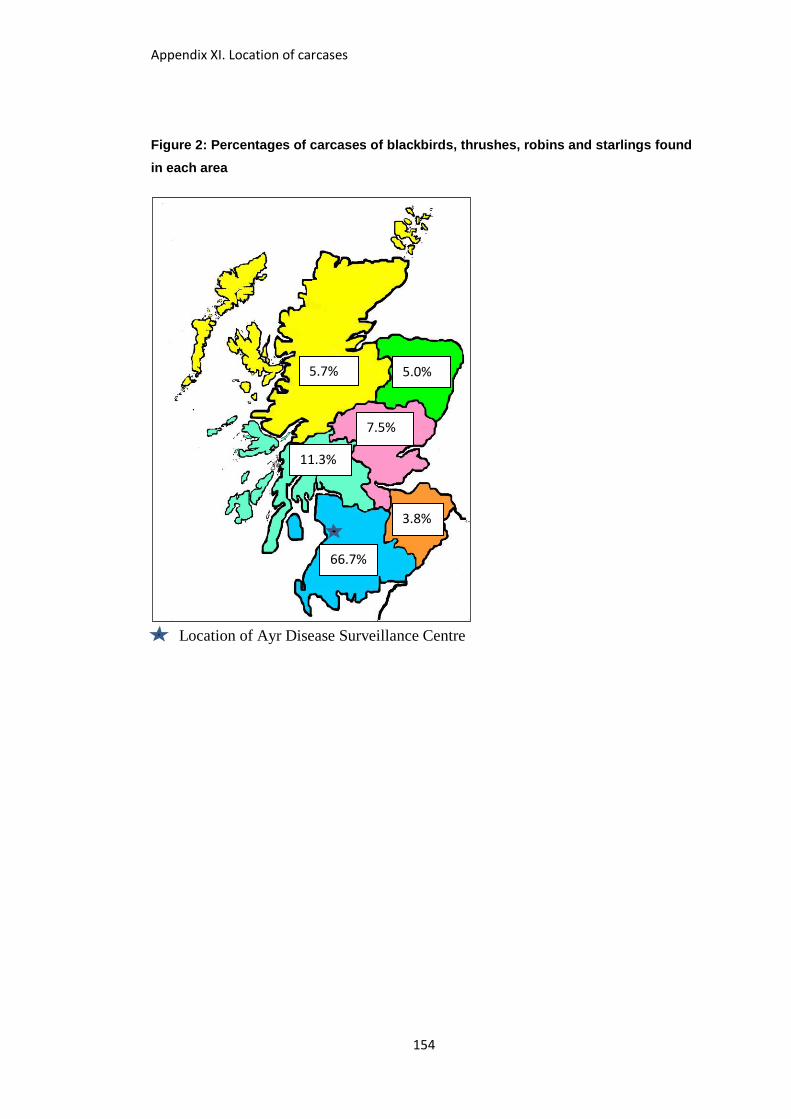
Appendix XI. Location of carcases
154
Figure 2: Percentages of carcases of blackbirds, thrushes, robins and starlings found
in each area
Location of Ayr Disease Surveillance Centre
7.5%
11.3%
5.0% 5.7%
3.8%
66.7%
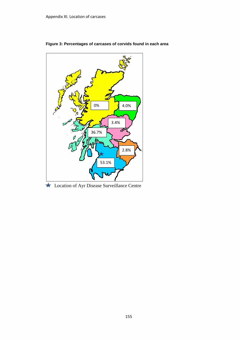
Appendix XI. Location of carcases
155
Figure 3: Percentages of carcases of corvids found in each area
Location of Ayr Disease Surveillance Centre
3.4%
36.7%
4.0% 0%
2.8%
53.1%

Appendix XI. Location of carcases
156
Figure 4: Percentages of carcases of “other passerines” found in each area
Location of Ayr Disease Surveillance Centre
5.3%
15.8%
15.8% 5.3%
5.3%
52.5%

Appendix XI. Location of carcases
157
Figure 5: Percentages of carcases of pigeons and doves found in each area
Location of Ayr Disease Surveillance Centre
4.2%
36.7%
2.5% 2.5%
1.7%
52.4%