Embed Size (px)
Citation preview

RESEARCH Open Access
Displacement and force distribution ofsplinted and tilted mandibular anteriorteeth under occlusal loads: an in silico 3Dfinite element analysisAllahyar Gerami1, Sepideh Dadgar 2, Vahid Rakhshan3,4, Puya Jannati5 and Farhad Sobouti2*
Abstract
Background: Fixed orthodontic retainers have numerous advantages, but it is not known whether they can exertpathological forces on supporting tissues around the splinted teeth. The purpose of this study was to investigatehow the inclination of the lower anterior teeth can affect dental displacement and also change the direction ofocclusal loads exerted to dental and its supporting tissues.
Methods: Four three-dimensional finite element models of the anterior part of the mandible were designed. Allthe models contained the incisors and canines, their periodontal ligament layers (PDLs), the supporting bone (bothspongy and cortical), and a pentaflex splinting wire placed in the lingual side of the teeth. Teeth inclination wasconsidered to be 80° (model 1), 90° (model 2), 100° (model 3), and 110° (model 4) to the horizontal plane. Thelower incisors were loaded with a 187-N vertical force. Their displacement patterns and the stress in their PDLswere evaluated.
Results: In incisors with 80° of inclination, less than a 0.1-mm lingual displacement was seen on the incisal edgeand a similar distance of displacement towards the labial was seen on their root apices. However, in models with90°–110° of inclination, the incisal edge displaced labially between about 0.01 and 0.45 mm, while root apicesdisplaced lingually instead. By increasing the angle of the teeth, the strain in the periodontal ligament increasedfrom about 37 to 58 mJ. The von Mises stresses around the cervical and apical areas differed for each tooth andeach model, without a similar pattern. Increasing the angle of the teeth resulted in much higher cervical stresses inthe incisors, but not in the canines. In the lateral incisor, cervical stress increased until 100° of inclination butreduced to about half by increasing the angle to 110°. Apical stress increased rather consistently in the incisorand lateral incisors, by increasing the inclination. However, in the canines, apical stress reduced to about half,from the first to fourth models.
Conclusions: Increasing the labial inclination can mostly harm the central incisors, followed by the lateralincisors. This finding warns against long durations of splinting in patients with higher and/or patients withreduced labial bone thickness.
Keywords: Lower anterior teeth, Tooth inclination, Retention, Orthodontic treatment, Splinting, Finite element method
* Correspondence: [email protected] of Orthodontics, Dental Faculty, Mazandaran University ofMedical Sciences, PO Box: 19551-624, Sari, IranFull list of author information is available at the end of the article
© 2016 Gerami et al. Open Access This article is distributed under the terms of the Creative Commons Attribution 4.0International License (http://creativecommons.org/licenses/by/4.0/), which permits unrestricted use, distribution, andreproduction in any medium, provided you give appropriate credit to the original author(s) and the source, provide a link tothe Creative Commons license, and indicate if changes were made.
Gerami et al. Progress in Orthodontics (2016) 17:16 DOI 10.1186/s40510-016-0129-x
BackgroundThe stability of orthodontic treatment outcome is amajor clinical concern, since many cases especiallymandibular anterior teeth relapse after aligning [1, 2].Permanent or long-term retention seems to be theonly way to provide a proper post-treatment align-ment [2, 3]. A proper method for this purpose is touse fixed retainers that remain permanently in themouth and are invisible, compliance-free, and welltolerated [2, 4]. Fixed retainers were commonly madeof stainless steel round wires and later thinner coaxialor braided round wires; among various fixed retainersof different metals, diameters, and designs, the flex-ible spiral wire (twisted steel wire) is very popularbetween orthodontists for providing acceptable long-term retention [1, 2, 4–8].Advantages of fixed retainers in relapse control are well
documented in the literature. Despite their popularity,their adverse effects remain unclear. Their negative effectsare a matter of controversy for many years, and they areregarded as a rather unpleasant strategy from a periodon-tist’s perspective because of plaque accumulation and hy-giene control problems [8–15]. The question remaining tobe answered is whether fixed retainers have biomechanical
disadvantages, because their biomechanical aspects havenever been evaluated numerically [8]. The extent of toothdisplacement and also the distribution of occlusal forceexerted on periodontal tissues of the retained teeth arenot known. This is crucial especially when assuming thatthe post-treatment inclination of the mandibular teethvaries depending on the protocol of treatment. Patientswith extraction treatment plans might have more uprightteeth, while those with non-extraction treatments mighthave mandibular teeth tilted labially. Since masticatoryforces are exerted in a vertical direction, inclination ofteeth might make the masticatory forces more hazardousfor teeth inclined labially than for those positioned moreupright and parallel to the force direction. Therefore, thepurpose of this study was to quantify the degree ofmandibular teeth’s movement and changes in stressdistribution around supporting tissues of mandibularanterior teeth splinted by a pentaflex wire with fourdifferent labiolingual inclinations (incisal mandibularplane angle (IMPA) = 80°, 90°, 100°, and 110°).
MethodsFour 3D finite element models were designed of amandibular anterior segment. It included six anterior
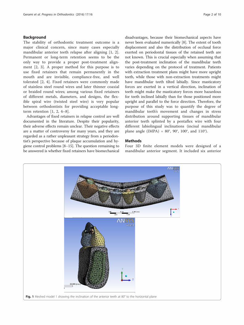
Fig. 1 Meshed model 1 showing the inclination of the anterior teeth at 80° to the horizontal plane
Gerami et al. Progress in Orthodontics (2016) 17:16 Page 2 of 10

teeth with the average dimensions and supportingstructures [8, 16]. Each model consisted of a cancel-lous bone surrounded by a 1-mm-thick cortical layer.A simplified 0.25-mm-thick periodontal ligament layer(PDL) was modeled based on the root-form geometryof the teeth [8, 17]. All models had a bonded fixed re-tainer in the lingual surface of the anterior teeth. Themodels were similar except for the angle of the lowerincisors to the horizontal plane. The inclination of thelower incisors to the horizontal plane was 80° inmodel 1, 90° in model 2, 100° in model 3, and 110° inmodel 4 (Figs. 1, 2, 3, and 4, respectively).SolidWorks 2014 (300 Baker Ave. Concord, MA
01742, USA) was selected for the modeling phase.The models were then transferred to the ANSYSWorkbench Ver. 11.0 (ANSYS Inc., Southpointe, 275Technology drive, Canonsburg PA 15317, USA) forcalculation [8, 17]. All the vital tissues were pre-sumed elastic, homogeneous, and isotropic. The cor-responding elastic properties such as Young’smodulus and Poisson’s ratio were applied (Table 1).The relationship between the teeth, their PDL, the
Fig. 3 Meshed model 3 showing the inclination of the anterior teeth at 100° to the horizontal plane
Fig. 2 Meshed model 2 showing the inclination of the anterior teethat 90° to the horizontal plane
Gerami et al. Progress in Orthodontics (2016) 17:16 Page 3 of 10

spongy and cortical bones, and the multi-strand wirewith composite and the teeth was provided by con-tact elements (Figs. 1, 2, 3, and 4). All rigid bodymotions were prevented. A vertical force of 187 N(as an average occlusal force usually exerted on thelower incisors) was applied at each incisal edge ofthe central incisors [8, 18, 19]. Tooth displacementsin labial and gingival direction, the energy increasein the PDLs of the anterior teeth, and the von Mises
stress in the cervical and apical parts of the PDLswere assessed.
ResultsTooth displacementThe incisor displacement was −0.0725 mm (towards lingual)in the incisal edge and 0.0800 mm (towards labial) in theapical area, in model 1. The incisal edge movement turnedto labial in models 2–4 (between 0.00939 and 0.4538 mm inthe incisal edge) and between −0.00477 and −0.3119 mm inthe apical area (Figs. 5, 6, 7, and 8; Table 2). Almost thesame pattern is followed by the lateral incisor. The patternof lingual displacement of the cusp tip (−0.0725 mm) andlabial displacement in the apex (=0.008 mm) in model 1was observed in the canine (Table 2).
The von Mises stress in cervical and apical areasIn all models, the stresses are higher in the apical areacompared to the cervical part. The numeric findings arepresented in Table 3 and Figs. 9 and 10. The canine find-ings are noticeable.
Fig. 4 Meshed model 4 showing the inclination of the anterior teeth at 110° to the horizontal plane
Table 1 Mechanical properties of the materials used inmodeling
Young’s modulus (MPa) Poisson’s ratio
Tooth [8] 20,300 0.26
PDL [8] 0.667 0.49
Composite [8] 16,600 0.24
Spongy bone [8, 17] 13,400 0.38
Cortical bone [8, 17] 34,000 0.26
Pentaflex wire [8] 90,000 0.3
Gerami et al. Progress in Orthodontics (2016) 17:16 Page 4 of 10
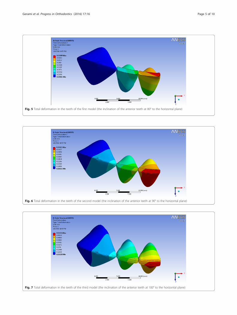
Fig. 5 Total deformation in the teeth of the first model (the inclination of the anterior teeth at 80° to the horizontal plane)
Fig. 6 Total deformation in the teeth of the second model (the inclination of the anterior teeth at 90° to the horizontal plane)
Fig. 7 Total deformation in the teeth of the third model (the inclination of the anterior teeth at 100° to the horizontal plane)
Gerami et al. Progress in Orthodontics (2016) 17:16 Page 5 of 10

Fig. 8 Total deformation in the teeth of the fourth model (the inclination of the anterior teeth at 110° to the horizontal plane)
Table 2 The incisal and apical displacements of the anterior teeth in various models
Model 1 (80°) Model 2 (90°) Model 3 (100°) Model 4 (110°)
Central incisor Incisal −0.07250 0.09390 0.28871 0.45381
Apical 0.08000 −0.04770 −0.19580 −0.31195
Lateral incisor Incisal −0.02040 0.08130 0.20380 0.33305
Apical 0.05000 −0.03560 −0.13740 −0.23122
Canine Incisal −0.07250 0.02740 0.08260 0.07580
Apical 0.08000 −0.01090 −0.06520 −0.01430
Table 3 The von Mises stress (MPa) in the PDL of the anterior teeth
Model 1 (80°) Model 2 (90°) Model 3 (100°) Model 4 (110°)
Cervical Central incisor 0.97731 0.95656 1.5886 1.4942
Lateral incisor 0.93226 0.72064 1.2836 0.71427
Canine 0.5551 0.38212 0.48572 0.76753
Apical Central incisor 1.2173 1.4611 1.3266 2.2735
Lateral incisor 1.0151 1.00949 0.90826 1.6879
Canine 1.0218 0.59451 0.67781 0.48768
Fig. 9 The von Mises stress in the cervical area of the PDL
Gerami et al. Progress in Orthodontics (2016) 17:16 Page 6 of 10

Fig. 10 The von Mises stress in the apical area of the PDL
Fig. 11 The strain energy of the lower right central, lateral, and canine PDLs
Fig. 12 Max./Min. of strain energy in the lower right incisors and the canine
Gerami et al. Progress in Orthodontics (2016) 17:16 Page 7 of 10

Strain energyThe strain energy of the anterior teeth PDL was 36.734 mJin model 1 and increased to 37.874 mJ in model 2. Thisincrease was noticed in model 3 (45.28) and model 4(57.502, Figs. 11, 12, and 13).
DiscussionThe viscoelastic nature of periodontal tissues plus ad-aptations in the anatomic characteristics like the bonemass and level and the width of the periodontal liga-ment space are the key to the physiologic tooth mobil-ity [13, 20]. The wire in a fixed retainer can undergoelastic deflection by being mechanically deformedunder masticatory loads [13, 20]. In an average malepatient, the bite force can increase up to 113 N, whichmight cause mechanical deformation of the retainer[21]. It is desirable for the teeth not to be fixed in toorigid positions during the orthodontic retention period[4, 5].In this study, in the incisors with 80° of inclination,
less than a 0.1-mm lingual displacement was seen on theincisal edge and a similar distance of displacement to-wards the labial was seen on the root apices. However,in models 2 to 4 (with 90° to 110° of inclination), the in-cisal edge displaced labially between about 0.01 and0.45 mm, while root apices displaced lingually instead.These small extends of displacement have clinical impli-cations. It is shown that about a 0.2-mm displacementmight exert a vertical force about 1 N together with ahorizontal force about 1.5 N [13, 20]. By increasing theinclination of teeth, the strain in the periodontal liga-ment increased from about 37 to 58 mJ. The vonMises stresses around the cervical and apical areasdiffered for each tooth and each model, without asimilar pattern. Increasing the angle of the teeth re-sulted in much higher cervical stresses in the centralincisors, but not in the canines. In the lateral incisor,cervical stress increased until 100° of inclination butreduced to about half by increasing the angle to 110°.
Apical stress increased rather consistently in the inci-sor and lateral incisors, by increasing the inclination.However, in the canines, it reduces to about half,from the first to fourth models. It was previouslyshown that the act of splinting itself can change thedisplacement pattern. The reason can be the lack of atelescopic movement in the connection of wire with acomposite. Additionally, the pattern of displacementdepends on the coordinates of the applied force in re-lation to the center of resistance of the tooth [8].This study showed that in patients with upright an-terior teeth, the displacement can be lingual, whereasin a patient with an increased IMPA, incisal displace-ments will be labial while root apices will move to-wards the lingual direction. Our results warn againstlong durations of splinting in patients with greater la-bial inclination of mandibular teeth and/or patientswith reduced labial bone thickness, because in suchpatients the loads might be more of pathologic natureand cause periodontal damage and pathologic toothmobility [8, 22, 23].It is not known if bonded lingual retainers have a
negative effect on the periodontal tissues [13, 24].Gingival damage and recession can be caused by nu-merous factors, among which mechanical trauma andbacterial periodontal disease are the most importantones [13, 25–28]. Besides increasing plaque, these ap-pliances are also criticized for changing the mode offunctional loads exerted on the anterior teeth, andcompromising the health of periodontium [13, 29–31].However; the studies regarding the consequences ofsplinting on the status of periodontium are limited, andno results exist regarding force distributions [13, 29–31].Many studies have shown no significant evidence re-garding any damage caused to periodontium or soft tis-sues adjacent to teeth, after using fixed lingual retainerseven for long durations [9–14, 24]. This level of safetymight not change depending on the wire used in thefixed retainer, even in durations as long as 10 years
Fig. 13 Strain energy in the anterior teeth PDL
Gerami et al. Progress in Orthodontics (2016) 17:16 Page 8 of 10
[10]. Nevertheless, using wire diameters that allow forphysiologic tooth movement, especially in patients athigher risk for developing periodontal diseases, is rec-ommended, as an ideal bonded retainer should be pas-sive and semi-rigid to maintain physiologic toothmobility after splinting [5, 13, 32]. Even plaque accu-mulation following the application of lingual-fixed re-tainers is questionable [24]. There were also reports ofno significant displacement after using fixed retainers[13]. However, a negative effect of bonded retainers ontooth mobility was observed by Watted et al. [15]. An-other study as well showed negative effects of long-term fixed retention on periodontal health, althoughthe changes were mostly mild [14]. Also increased gin-gival recession, increased plaque retention, and bleed-ing upon probing have been reported in another study[32]. In that study, gingival recession was more ad-vanced in patients with past histories of orthodontictreatment, which might be attributed to previous ortho-dontic movements and tooth rotations which mighthave stretched collagen fibers within periodontal andgingival tissues [13, 32–41].This study was limited by some factors. In vitro studies
cannot reproduce the highly dynamic nature of oral en-vironment with occlusal loads rapidly changing in extentand direction. However, there is no alternative to thismethod, as in vivo studies need to either be on radio-graphic images (which cannot show the extent of boneloss accurately) or be in animals, which again are irrele-vant to human; and none of other options can show thedistribution of forces [13, 14, 17]. Moreover, utilizationof radiographic and computerized tomography tech-niques only for the sake of research and without anytreatment needs would expose patients to unnecessarydoses of carcinogen X-ray and hence are not easily justi-fiable ethically [42].
ConclusionsIncreasing the labial inclination can mostly harm thecentral incisors, followed by the lateral incisors. Thisfinding warns against long durations of splinting in pa-tients with increased inclination of the mandibular inci-sors (i.e., increased IMPA) and/or patients with reducedlabial bone thickness.
Competing interestsThe authors declare that they have no competing interests.
Authors’ contributionsAG designed the study, created the FEM models and figures, performed theFEM analyses, drafted the methods and results, interpreted the FEM analyses,and critically reviewed the article. SD searched the literature and designedthe study. VR interpreted the FEM analyses, drafted the introduction anddiscussion, participated in drafting of the other parts, and revised the paper.PJ searched the literature. FS searched the literature, came up with the
research idea, designed the study, interpreted the FEM analyses, and draftedthe methods and results. All authors read and approved the final manuscript.
Author details1Department of Orthodontics, Dental Faculty, Tehran University of MedicalUniversity, Tehran, Iran. 2Department of Orthodontics, Dental Faculty,Mazandaran University of Medical Sciences, PO Box: 19551-624, Sari, Iran.3Department of Dental Anatomy and Morphology, Dental School, AzadUniversity, Tehran, Iran. 4Iranian Tissue Engineering and Research Center,Tehran University, Tehran, Iran. 5Student Research Committee, Faculty ofDentistry, Mazandaran University of Medical Sciences, Sari, Iran.
Received: 26 February 2016 Accepted: 6 May 2016
References1. Littlewood SJ, Millett DT, Doubleday B, Bearn DR, Worthington HV.
Retention procedures for stabilising tooth position after treatment withorthodontic braces. Cochrane Database Syst Rev. 2006;1:CD002283.
2. Salehi P, Zarif Najafi H, Roeinpeikar SM. Comparison of survival timebetween two types of orthodontic fixed retainer: a prospective randomizedclinical trial. Prog Orthod. 2013;14:25.
3. Stormann I, Ehmer U. A prospective randomized study of different retainertypes. J Orofac Orthop. 2002;63:42–50.
4. Zachrisson BU. Clinical experience with direct-bonded orthodontic retainers.Am J Orthod. 1977;71:440–8.
5. Zachrisson BU. The bonded lingual retainer and multiple spacing of anteriorteeth. Swed Dent J Suppl. 1982;15:247–55.
6. Bearn DR. Bonded orthodontic retainers: a review. Am J Orthod DentofacialOrthop. 1995;108:207–13.
7. Bearn DR, McCabe JF, Gordon PH, Aird JC. Bonded orthodonticretainers: the wire-composite interface. Am J Orthod DentofacialOrthop. 1997;111:67–74.
8. Geramy A, Retrouvey JM, Sobuti F, Salehi H. Anterior teeth splinting afterorthodontic treatment: 3D analysis using finite element method. J Dent(Tehran). 2012;Spring; 9(2):90–8.
9. Artun J, Spadafora AT, Shapiro PA. A 3-year follow-up study of various typesof orthodontic canine-to-canine retainers. Eur J Orthod. 1997;19:501–9.
10. Artun J. Caries and periodontal reactions associated with long-term use ofdifferent types of bonded lingual retainers. Am J Orthod. 1984;86:112–8.
11. Artun J, Spadafora AT, Shapiro PA, McNeill RW, Chapko MK. Hygiene statusassociated with different types of bonded, orthodontic canine-to-canineretainers. A clinical trial. J Clin Periodontol. 1987;14:89–94.
12. Gorelick L, Geiger AM, Gwinnett AJ. Incidence of white spot formation afterbonding and banding. Am J Orthod. 1982;81:93–8.
13. Oshagh M, Heidary S, Dehghani Nazhvani A, Koohpeima F, Koohi HosseinabadiO. Evaluation of histological impacts of three types of orthodontic fixedretainers on periodontium of rabbits. J Dent (Shiraz). 2014;15:104–11.
14. Pandis N, Vlahopoulos K, Madianos P, Eliades T. Long-term periodontalstatus of patients with mandibular lingual fixed retention. Eur J Orthod.2007;29:471–6.
15. Watted N, Wieber M, Teuscher T, Schmitz N. Comparison of incisor mobilityafter insertion of canine-to-canine lingual retainers bonded to two or to sixteeth. A clinical study. J Orofac Orthop. 2001;62:387–96.
16. Mackinejad SA, Kaviani R, Rakhshan V, Khabir F. Assessment of the cut-offpoint of mesiodistal and buccolingual widths of permanent teeth fordetermination of sex. Dent J (Isfahan). 2015;11(2):153-62.
17. Vafaei F, Khoshhal M, Bayat-Movahed S, Ahangary AH, Firooz F, Izady A,Rakhshan V. Comparative stress distribution of implant-retained mandibularball-supported and bar-supported overlay dentures: a finite elementanalysis. J Oral Implantol. 2011;37(4):421–9. doi:10.1563/AAID-JOI-D-10-00057. Epub 2010 Aug 16.
18. Hsu ML, Chen FC, Kao HC, Cheng CK. Influence of off-axis loading of ananterior maxillary implant: a 3-dimensional finite element analysis. Int J OralMaxillofac Implants. 2007;22(2):301–9.
19. Clelland NL, Lee JK, Bimbenet OC, Brantley WA. A three-dimensional finiteelement stress analysis of angled abutments for an implant placed in theanterior maxilla. J Prosthodont. 1995;4(2):95–100.
20. Sifakakis I, Pandis N, Eliades T, Makou M, Katsaros C, Bourauel C. In-vitroassessment of the forces generated by lingual fixed retainers. Am J OrthodDentofacial Orthop. 2011;139:44–8.
Gerami et al. Progress in Orthodontics (2016) 17:16 Page 9 of 10

21. Kiliaridis S, Johansson A, Haraldson T, Omar R, Carlsson GE. Craniofacialmorphology, occlusal traits, and bite force in persons with advancedocclusal tooth wear. Am J Orthod Dentofacial Orthop. 1995;107:286–92.
22. Reinhardt RA, Killeen AC. Do mobility and occlusal trauma impactperiodontal longevity? Dent Clin N Am. 2015;59(4):873–83.
23. De Boever J, De Boever A. Occlusion and periodontal health. Functionalocclusion in restorative dentistry and prosthodontics. 2015;2:189.
24. Heier EE, De Smit AA, Wijgaerts IA, Adriaens PA. Periodontal implications ofbonded versus removable retainers. Am J Orthod Dentofacial Orthop. 1997;112:607–16.
25. Kassab MM, Cohen RE. The etiology and prevalence of gingival recession.J Am Dent Assoc. 2003;134:220–5.
26. Rawal SY, Claman LJ, Kalmar JR, Tatakis DN. Traumatic lesions of the gingiva:a case series. J Periodontol. 2004;75:762–9.
27. Litonjua LA, Andreana S, Bush PJ, Cohen RE. Toothbrushing and gingivalrecession. Int Dent J. 2003;53:67–72.
28. Levin L, Zadik Y, Becker T. Oral and dental complications of intra-oralpiercing. Dent Traumatol. 2005;21:341–3.
29. Booth FA, Edelman JM, Proffit WR. Twenty-year follow-up of patients withpermanently bonded mandibular canine-to-canine retainers. Am J OrthodDentofacial Orthop. 2008;133:70–6.
30. Gher ME. Changing concepts. The effects of occlusion on periodontitis.Dent Clin North Am. 1998;42:285–99.
31. Baruch H, Ehrlich J, Yaffe A. Splinting—a review of the literature. RefuatHapeh Vehashinayim. 2001;18:29–40. 76.
32. Levin L, Samorodnitzky-Naveh GR, Machtei EE. The association of orthodontictreatment and fixed retainers with gingival health. J Periodontol. 2008;79:2087–92.
33. Blake M, Bibby K. Retention and stability: a review of the literature. Am JOrthod Dentofacial Orthop. 1998;114:299–306.
34. Lombardo L, Scuzzo G, Arreghini A, Gorgun O, Ortan YO, Siciliani G. 3D FEMcomparison of lingual and labial orthodontics in en masse retraction. ProgOrthod. 2014;15(1):38. doi:10.1186/s40510-014-0038-9.
35. MacGinnis M, Chu H, Youssef G, Wu KW, Machado AW, Moon W. The effectsof micro-implant assisted rapid palatal expansion (MARPE) on thenasomaxillary complex—a finite element method (FEM) analysis. ProgOrthod. 2014;15(1):52. doi:10.1186/s40510-014-0052-y.
36. Moon W, Wu KW, MacGinnis M, Sung J, Chu H, Youssef G, Machado A. Theefficacy of maxillary protraction protocols with the micro-implant-assistedrapid palatal expander (MARPE) and the novel N2 mini-implant-a finiteelement study. Prog Orthod. 2015;16:16. doi:10.1186/s40510-015-0083-z.
37. Elsaka SE, Hammad SM, Ibrahim NF. Evaluation of stresses developed in differentbracket-cement-enamel systems using finite element analysis with in vitro bondstrength tests. Prog Orthod. 2014;15(1):33. doi:10.1186/s40510-014-0033-1.
38. Sivamurthy G, Sundari S. Stress distribution patterns at mini-implant siteduring retraction and intrusion-a three-dimensional finite element study.Prog Orthod. 2016;17(1):4. doi:10.1186/s40510-016-0117-1.
39. Tanaka OM, Saga AY, Pithon MM, Argenta MA. Stresses in the midpalatalsuture in the maxillary protraction therapy: a 3D finite element analysis.Prog Orthod. 2016;17(1):8. doi:10.1186/s40510-016-0121-5.
40. Aziz T, Ansari K, Lagravere MO, Major MP, Flores-Mir C. Effect of non-surgicalmaxillary expansion on the nasal septum deviation: a systematic review.Prog Orthod. 2015;16:15. doi:10.1186/s40510-015-0084-y.
41. Maspero C, Galbiati G, Giannini L, Farronato G. Sagittal and vertical effects oftransverse sagittal maxillary expander (TSME) in three different malocclusiongroups. Prog Orthod. 2015;16:6. doi:10.1186/s40510-015-0075-z.
42. Rakhshan V. Meta-analysis and systematic review of factors biasing the observedprevalence of congenitally missing teeth in permanent dentition excluding thirdmolars. Prog Orthod. 2013;14:33. doi:10.1186/2196-1042-14-33. Submit your manuscript to a
journal and benefi t from:
7 Convenient online submission
7 Rigorous peer review
7 Immediate publication on acceptance
7 Open access: articles freely available online
7 High visibility within the fi eld
7 Retaining the copyright to your article
Submit your next manuscript at 7 springeropen.com
Gerami et al. Progress in Orthodontics (2016) 17:16 Page 10 of 10