Embed Size (px)
Citation preview

Citrate anticoagulation using the Prismaflex® system
Renal Intensive CareSelf-learning module
With integrated calcium management
Gambro Lundia ABPO Box 10101SE-22010 LundSwedenPhone +46 46 16 90 [email protected]
This learning module has been developed as a Prismaflex® system training aid while using citrate anticoagulation.
Prior to and during the operation of the Prismaflex® machine, consult the Operator’s Manual and the Instructions for Use enclosed with the Prismaflex® disposable set.
Prior to using Gambro solutions, read and refer to the Summary of Product Characteristics (SPC) or any other prescribing information provided by your local Gambro representative.
Prior to using any other medicinal products mentioned in this learning module, read and refer to the SPC or any other prescribing information attached to the products.
The Gambro solutions and medicinal products mentioned in this learning module may not be registered in your country. Always consult your country’s local regulations and the SPC or any other prescribing information prior to prescription.
Gambro, Prismaflex, Prismocitrate, Prism0cal, PrismaSate, PrismaSol and Prisma are trademarks belonging to the Gambro group.
HC
EN
5454
_3 ©
201
1.11
. Gam
bro
Lund
ia A
B


3
Table of contents
1 Introduction 8
2 Learning objectives 9
3 What is citrate? 103.1 Chemical properties 103.2 Chemical properties of trisodium citrate 113.3 Use of citrates in the medical field 123.4 Citrate as an anticoagulant in CRRT 123.5 Quiz 13
4 How important is calcium? 144.1 Regulation of calcium in the human body 144.2 Function of calcium in the human body 14
4.2.1 General 144.2.2 The role of calcium in the clotting cascade 15
4.3 Calcium plasma distribution 154.3.1 Calcium plasma distribution in the patient 154.3.2 Calcium plasma distribution in the extracorporeal circuit 16
4.4 Calcium loss during CRRT 174.5 Quiz 17
5 How does citrate work? 185.1 Chelation of calcium (and magnesium) 185.2 Calcium-citrate losses in effluent 195.3 Use of dialysate solutions 205.4 Calcium level in the extracorporeal circuit 205.5 Use of post-dilution replacement solutions 205.6 Metabolism of citrate 205.7 Calcium reinfusion 215.8 Quiz 22

4
6 When should citrate be used as an anticoagulant in CRRT and 23 when not? 6.1 Indications 23
6.1.1 Increased bleeding risk 236.1.2 Heparin-induced thrombocytopenia (and thrombosis) 24
6.2 Contraindications 266.2.1 Severe liver failure 266.2.2 Citrate intolerance 26
6.3 Citrate as a standard anticoagulation method? 276.4 Quiz 28
7 Which solutions are required during citrate anticoagulation? 297.1 Citrate anticoagulation solutions 29
7.1.1 Trisodium citrate 297.1.2 ACD-A 307.1.3 Prismocitrate™ 10/2 from Gambro 317.1.4 Prismocitrate 18/0 from Gambro 327.1.5 PrismoCit 4K from Gambro 32
7.2 Dialysate solutions 337.2.1 Calcium-free dialysate solutions 347.2.2 PrismOcal™ from Gambro 347.2.3 Prism0cal B22 from Gambro 34
7.3 Replacement solutions 347.3.1 Calcium-free relacement solutions 347.3.2 Solutions with calcium content 357.3.3 Solutions with calcium content 35
7.4 Calcium infusion 7.4.1 Calcium gluconate 10% 357.4.2 Calcium chloride 10% 35
7.5 Priming solutions 367.6 Quiz 36
8 Which CRRT therapy options with the Prismaflex® system 38 allow citrate anticoagulation? 8.1 SCUF 38
8.1.1 Clinical practice 38
Table of contents

5
8.1.2 Description 388.2 CVVH 40
8.2.1 Clinical practice 408.2.2 Description 40
8.3 CVVHD 418.3.1 Clinical practice 418.3.2 Description 42
8.4 CVVHDF 438.4.1 Clinical practice 438.4.2 Description 43
8.5 Quiz 45
9 How does the Prismaflex system manage regional 46 citrate anticoagulation methods?9.1 Selection of citrate method 469.2 Selection of citrate anticoagulation solutions 469.3 Software updated settings on the Prismaflex monitor 47
9.3.1 Blood flow rate 479.3.2 PBP citrate flow rate 489.3.3 Citrate dose 48
9.4 Calcium management during citrate anticoagulation with the 49 Prismaflex system
9.4.1 Using anticoagulation method ‘Citrate - Calcium via the 49 syringe pump of the Prismaflex system’ (Citrate/Calcium)9.4.2 Using anticoagulation method ‘Citrate - Calcium via External 50 Infusion Pump’ (Citrate)
9.5 Software updated flow rates during treatment 51 (related to citrate anticoagulation)9.6 Safety system 52
9.6.1 Alarm specific to both regional citrate anticoagulation methods 529.6.2 Alarms specific to ‘Citrate – Calcium via the syringe pump of 52 the Prismaflex system‘ anticoagulation method
9.7 Prescription Indicator calculations in the Prismaflex system 539.7.1 Indicator specific to both regional citrate anticoagulation methods 539.7.2 Indicator specific to ‘Citrate - Calcium via External Infusion Pump’ 53 anticoagulation method

6
9.8 Quiz 54
10 Which monitoring is required during citrate anticoagulation? 5610.1 Patient monitoring 56
10.1.1 Ionized calcium 5610.1.2 Total calcium 5610.1.3 Ratio Catot / Caion 5710.1.4 pH 5710.1.5 Bicarbonate and base excess 5810.1.6 Magnesium 5810.1.7 Other 58
10.2 Effective anticoagulation 5810.2.1 Post-filter ionized calcium 5910.2.2 Hemofilter life 59
10.3 Quiz 60
11 What are possible complications and how can we 61 avoid/manage them? 11.1 Metabolic alkalosis 6111.2 Metabolic acidosis 6111.3 Hypocalcemia 6111.4 Hypercalcemia 6211.5 Hypernatremia 6211.6 Hyponatremia 6211.7 Hypomagnesemia 6211.8 Citrate accumulation/toxicity 6311.9 Quiz 64
12 Treatment dose and citrate anticoagulation 6512.1 Quiz 66
13 How to implement citrate anticoagulation in the ICU 6713.1 Expertise and skills in CRRT 6713.2 Organization issues (lab storage) 6713.3 Protocol/Physician’s orders/Procedure/Documentation 68
Table of contents

7
13.4 Education/Training 6913.5 Quiz 69
14 Summary 70
15 Answers to quiz 71
16 References 73

8
1 Introduction
During continuous renal replacement therapy (CRRT), anticoagulation of the extracorporeal circuit is generally required to prevent clotting of the circuit, preserve filter performance, optimize circuit survival, and prevent blood loss due to circuit clotting.[1]
Heparin is the most frequently used anticoagulant but is associated with a risk of bleeding that is seen in 4-30% of patients.[2, 3, 4] Moreover, heparin may not provide ideal anticoagulation in patients that have intrinsic clotting system activation, antithrombin III deficiency, or evidence of intravascular coagulation. In addition, despite the low incidence of heparin-induced thrombocytopenia (1-3%)[5, 6], the use of heparin may be impossible in some heparin-induced cases.
Various alternative methods have been developed to ensure anticoagulation in the extracorporeal circuit. These include low-molecular-weight heparin, heparinoids, hirudin, prostacyclin, serineprotease inhibitors, direct thrombin inhibitors, and activated protein C. Regional anticoagulation with heparin and its antagonist protamine is an option alternatively used in some facilities. [7] None of these systems has been widely established[8] due to different factors: no existing antagonist, difficult monitoring, lack of studies, and various side-effects like hypotension, anaphylaxis, prolonged half-life, systemic bleeding and increased intracranial pressure. Other alternatives are saline flushes, the use of non-thrombogenic membrane surfaces, and treatments with increased pre-dilution delivery of replacement fluids. Even CRRT treatments without anticoagulant have been described.[9] In fact, in the recent American dose/outcome study (ATN Trial),[10] nearly 55% of CRRT treatments were provided with no anticoagulation. However, it was not reported what effect this approach had on average extracorporeal set life.
The use of regional citrate anticoagulation (RCA) has been described in various studies in the setting of CRRT and is gaining acceptance. Citrate seems to be an effective method of anticoagulation for CRRT in patients with contraindications to heparin. However, this method increases the complexity of CRRT by requiring special replacement and/or dialysate solutions to minimize metabolic complications in some cases and intravenous calcium reinfusion to prevent systemic hypocalcemia.[11]

9
2 Learning objectives
This module will allow the student to acquire an understanding and knowledge of:
•What citrate is and how it works as an anticoagulant•The indications and contraindications of the use of citrate•Different solutions required during citrate anticoagulation•How citrate can be used during different therapy modalities•How the Prismaflex system controls citrate anticoagulation •The required monitoring during citrate anticoagulation •Possible complications when using citrate as an anticoagulant•How citrate anticoagulation must be implemented in an ICU

10
3 What is citrate?
The dictionary definition of citrate is: ‘A salt or ester of citric acid’. Citrates are a family of compounds of which trisodium citrate and citric acid are the most relevant with respect to RCA for CRRT.
3.1 Chemical properties of citric acidThe chemical formula for citric acid, which is a weak organic acid, is C6H8O7. It is a natural preservative and is also used to add an acidic, or sour taste to foods and soft drinks. In biochemistry, it is important in the metabolism of almost all living things.
Citric acid is a citrate molecule with 3 H+ ions (protons). The citrate component of this molecule forms complexes (chelates) with ionized calcium, magnesium and other divalent cations (e.g. Mn2+, Fe2+), but affinity to calcium and magnesium is highest.
C
C
C
C
C
C
O
O
O
O- H+
O- H+
O- H+
H
OH
H
H
HCitric acid

11
3.2 Chemical properties of trisodium citrate
The chemical formula for trisodium citrate is Na3C6H5O7. It is a conjugate base of a weak acid and therefore organic neutral. Trisodium citrate is a salt of citric acid, sometimes referred to simply as sodium citrate, although sodium citrate can refer to any of the three sodium salts of citric acid. Sodium citrate is used as a preservative, flavoring agent and buffering agent in the food industry. In the medical field, it is used in different ways, which will be described in the next section. Sodium citrate is metabolized in the liver to bicarbonate.
Trisodium citrate is a citrate molecule with 3 Na+ ions. The citrate component of this molecule also forms complexes (chelates) with ionized calcium, magnesium and other divalent cations (e.g. Mn2+, Fe2+), but affinity to calcium and magnesium is highest.
C
C
C
C
C
C
O
O
O
O- Na+
O- Na+
O- Na+
H
OH
H
H
HTrisodium citrate

12
3.3 Use of citrates in the medical fieldCitric acid and its salts (citrates) inhibit coagulation by binding (chelating) calcium and thus removing a critical component of the coagulation cascade (see Chapter 4.1). For this reason, Albert Hustin and Luis Agote used sodium citrate as an anticoagulant in blood transfusions in 1914.[12] It continues to be used today in blood collection tubes and for the preservation of blood in blood banks. The use of citrate as an anticoagulant in renal replacement therapies has become a common alternative to systemic heparinization.
Other examples of citrate use are:
• Potassium citrate is widely used to treat urinary calculi (kidney stones), and is often used by patients with cystinuria (a specific disorder causing kidney stones)[13]
• Magnesium citrate is a chemical agent used as a saline laxative and to empty the bowel prior to a surgery or colonoscopy[14]
• Sildenafil-citrate is the active pharmaceutical used in oral drugs for male impotence, also known as erectile dysfunction (ED)[15]
3.4 Citrate used as an anticoagulant in CRRTCitrate as an anticoagulant in hemodialysis was first reported in the 1960s by Morita and colleagues[16] and applied in 1990 by Mehta and colleagues as RCA in patients undergoing CRRT. Especially in chronic dialysis, citrate anticoagulation is already a standard procedure in many clinics.
A patient with a high risk of bleeding is a primary indication for the use of citrate (see Chapter 5). As mentioned in more recent studies, heparin-induced thrombocytopenia (HIT) and recurring filter clotting are now accepted as additional indications.[1] More details will be provided in chapter 4.

13
3.5 Quiz
1. What is the most frequently used anticoagulant during CRRT?
a. Citrate
b. Unfractioned heparin
c. Low-molecular-weight heparin
2. Which of the following statements is false?
a. Trisodium citrate is a natural preservative
b. Trisodium citrate and citric acid are both citrates
c. Trisodium citrate is organic neutral
3. Three possible uses of citrate, related to anticoagulation, in the medical field are:
a. Anticoagulation in blood transfusion, blood collection tubes and Viagra® medicine
b. Anticoagulant in blood transfusions, blood collection tubes and preservation of blood in blood banks
c. Anticoagulation in blood transfusions, blood collection tubes and saline laxatives
4. When was citrate first used as an anticoagulant during CRRT and by whom?
a. 1914 by Hustin and Agote
b. 1960s by Morita
c. 1990 by Metha
14
4 How important is calcium?
A good understanding of citrate anticoagulation requires knowledge about calcium. Calcium is one of the coagulation factors (Factor IV) and participates in the clotting cascade. The interaction between the trisodium citrate molecule and the calcium ion is responsible for the anticoagulation process discussed in this module.
4.1 Regulation of calcium in the human bodyCalcium plays an essential role in many cellular processes. The human body must closely regulate calcium levels within a narrow physiological range. Even small changes in blood calcium levels can have dramatic effects, such as muscle and brain dysfunction, heart failure and even death. Despite the essential role of calcium, 99% of this electrolyte is found in the bones. Bones therefore are an important reserve of calcium for use throughout the body.
Calcium homeostasis is controlled by three hormones: calcitonin, parathyroid hormone (PTH) and vitamin D. Each of these hormones can manipulate bone cells and other tissues to raise or lower the concentration of calcium in the blood. When the calcium level in the blood rises, the hormone calcitonin acts to reduce blood calcium. When calcium levels are too low, PTH and vitamin D act synergistically to increase blood calcium.
4.2 Function of calcium in the human body
4.2.1 General
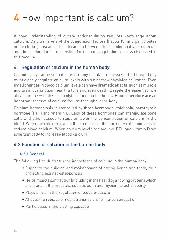
The following list illustrates the importance of calcium in the human body:
•Supports the building and maintenance of strong bones and teeth, thus protecting against osteoporosis
•Helps muscle contraction (including in the heart) by allowing proteins which are found in the muscles, such as actin and myosin, to act properly
•Plays a role in the regulation of blood pressure
•Affects the release of neurotransmitters for nerve conduction
•Participates in the clotting cascade

15
4.3 Calcium plasma distribution
4.3.1 Calcium plasma distribution in the patient
Calcium is present in blood plasma in three fractions which are in equilibriumwith one another, i.e., the ionized and complex bound calcium which togethercomprise the diffusible and ultrafiltrable fraction, and the non-diffusible calciumwhich is bound to the plasma proteins. The ionized calcium is considered to bethe physiologically active fraction[17] and must be maintained between 1.0 and1.3 mmol/l in the patients blood plasma.
4.2.2 The role of calcium in the clotting cascade
Calcium, which is coagulation factor IV, plays an important role in the wholeanticoagulation cascade. Its ionized form participates in each of the threepathways: intrinsic, extrinsic and common.
•Intrinsic pathway: Factor IXa, together with factor VIII, phospholipid andionized calcium convert factor X to its activated form, factor Xa.
•Extrinsic pathway: The combination of tissue factor with factor VIIa andionized calcium convert factor X to its activated form, factor Xa.
•Common pathway: The final common sequence involves the combinationof factors Xa and V, phospholipid and ionized calcium into a complex thatconverts prothrombin in thrombin.
Injured blood vessel Contact activation
Injured tissue
XII
XIIa
XI XIa
IX IXa
INTRINSICPATHWAY
EXTRINSICPATHWAY Tissue factor
VASCULAR SPASM
PLATELETAGGREGATION
PLATELET PLUG
CLOTProthrombin
(II)Thrombin
(IIa)
VIIa VII
COMMONPATHWAY
X Xa
Fibrinogen FibrinF XIIIaF XIIIaF
PF3PF3PF
Collagen
F VIIIaF VIIIaFPF3PF3PFCa++
IXa
Thrombin
XaF VaF VaFPF3PF3PFCa++
Xa
Ca++
Tissue factor
VIIVII
16
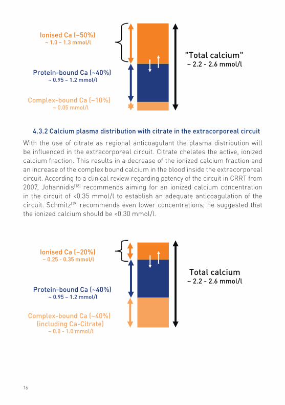
4.3.2 Calcium plasma distribution with citrate in the extracorporeal circuit
With the use of citrate as regional anticoagulant the plasma distribution will be influenced in the extracorporeal circuit. Citrate chelates the active, ionized calcium fraction. This results in a decrease of the ionized calcium fraction and an increase of the complex bound calcium in the blood inside the extracorporeal circuit. According to a clinical review regarding patency of the circuit in CRRT from 2007, Johannidis[18] recommends aiming for an ionized calcium concentration in the circuit of <0.35 mmol/l to establish an adequate anticoagulation of the circuit. Schmitz[19] recommends even lower concentrations; he suggested that the ionized calcium should be <0.30 mmol/l.
Total calcium ~ 2.2 - 2.6 mmol/l
Ionised Ca (~20%)~ 0.25 - 0.35 mmol/l
Protein-bound Ca (~40%)~ 0.95 – 1.2 mmol/l
Complex-bound Ca (~40%)(including Ca-Citrate)
~ 0.8 - 1.0 mmol/l
"Total calcium"~ 2.2 - 2.6 mmol/l
Ionised Ca (~50%)~ 1.0 – 1.3 mmol/l
Protein-bound Ca (~40%)~ 0.95 – 1.2 mmol/l
Complex-bound Ca (~10%)~ 0.05 mmol/l

17
4.4 Calcium loss during CRRTWhen using citrate as an anticoagulation method, a certain amount of ionized, complex bound but also some protein bound calcium will be cleared through the filter and lost in the effluent. This amount must be compensated to avoid hypocalcemia. Calcium and citrate-calcium complexes are small molecules. Therefore, this part of the calcium loss in the effluent is proportional to the removal of small molecules. The amount of calcium which is cleared by the filter and lost in the effluent depends on calcium concentration in the blood, blood flow rate, fluid flow rates and filter size.
4.5 Quiz
1. Which percentage of calcium in the human body can be found in the bones?
a. 1%
b. 50%
c. 99%
2. Which fraction of calcium plays an important role in the clotting cascade?
a. Ionized calcium
b. Protein bound calcium
c. Complex bound calcium
3. The ionized calcium level in the patient blood should be:
a. <35 mmol/l
b. 1.0–1.3 mmol/l
c. 2.2–2.6 mmol/l

18
5 How does citrate work?
The following schematic provides an overview of citrate anticoagulation duringCRRT. The numbers correspond to the text below, which contains a detaileddescription of each step in the process.
5.1 Chelation of calcium (and magnesium)A trisodium citrate solution is infused into the arterial access line of theextracorporeal set. The concentration of citrate in the patient’s blood depends onon the blood flow and citrate infusion flow rate. This concentration is expressedin “mmol/l blood”.
In the blood, calcium circulates primarily either in a free form or in a formbound to protein. Of note, the standard laboratory measurement for calcium(i.e. total calcium concentration) accounts for both the bound and free formsof calcium. The free form, termed ionized calcium, is the calcium componentwhich participates in the coagulation cascade. Citrate binds and forms a complex
76
1
2
3
45

19
(chelate) with ionized calcium of the patient’s blood. This binding process results in a decreased concentration of ionized calcium in the extracorporeal circuit. The ionized calcium, which is coagulation factor IV, loses its influence in the clotting cascade and coagulation within the set is interrupted. It should be mentioned that citrate non-specifically binds positively charged ions with double valency (i.e. double positive charge). Therefore, in addition to binding calcium, it also chelates magnesium. Therefore, a decrease in the magnesium concentration of the patient’s serum is to be expected.
5.2 Calcium-citrate losses in effluentDepending on the treatment mode and flow rates, a certain percentage of the calcium-citrate complex in the blood is cleared by the filter and lost in the effluent. Consequently, only a portion of the infused citrate is delivered to the patient. Based on the citrate infusion rate and the rate of citrate loss in the effluent, the amount of citrate delivered to the patient can be calculated. This is called the ‘patient citrate load’.
The amount of calcium removed in the effluent as part of the citrate-calcium complex must be compensated to avoid hypocalcemia in the patient. The approximate amount can be calculated, based on patient data, flow rates and assumptions.
Ca2+
Na3C6H5O7
Ca3(C6H5O7)2
Na+Na+
Na+Na+Na+Na+
Na3C6H5O7
Ca2+Ca2+
+ +

20
5.3 Use of dialysate solutionIf dialysate solutions are used (for CVVHD or CVVHDF), calcium-free solutions are recommended to avoid antagonism of the anticoagulation effect. More details will follow in chapter 6.
5.4 Calcium level in the extracorporeal circuitTo assess the anticoagulation status of blood in the set, monitoring of post-filter ionized calcium is recommended. The targeted ionized calcium level of the extracorporeal circuit is 0.25 – 0.35 mmol/l. More details will follow in chapter 10.
5.5 Use of post-dilution replacement solutionIf replacement solutions are used in pre-dilution (CVVH or CVVHDF), calcium-free solutions are recommended to avoid antagonism of the anticoagulation effect.
If replacement solutions are used in post-dilution, calcium-free solutions are also recommended. However, one may consider using a calcium-containing solution as long as the amount of calcium delivered into the circuit does not increase either the circulating ionized calcium or the post-filter ionized calcium level to a concentration beyond the target value of each. More details will follow in chapter 6.
5.6 Metabolism of citrateThe calcium-citrate in the blood leaving the filter via the venous bloodline is delivered to the patient. In the body of the patient the complex is metabolized by the liver mainly, with additional contribution by the kidneys and skeletal muscles. Citrate is converted to bicarbonate: 1 mmol of citrate delivers 3 mmol of bicarbonate. Therefore, the metabolism of the calcium-citrate complex results in a net addition of alkali (i.e., bicarbonate) to the patient, thus influencing the patient’s acid/base status. When ionized calcium is released, it becomes available as a coagulation factor in the patient’s blood. Systemic anticoagulation does not occur if the serum ionized calcium concentration is maintained at a physiological level (1.0–1.3 mmol/l).

21
5.7 Calcium reinfusionThe amount of calcium, bound in the calcium-citrate complex, which is cleared through the filter and lost in the effluent, must be compensated to avoid hypocalcemia in the patient. A separate central venous catheter or peripheral venous catheter must be used for the reinfusion of calcium. The serum calcium level must be monitored to define the amount of calcium needed for reinfusion. The samples can either be arterial or venous. The targeted serum calcium level of the patient’s systemic blood is 1.0–1.3 mmol/l. More details will follow in chapter 9.
calcium-citrate complex
Bicarbonate
Calcium
The calcium-citrate complex is metabolized by the patient’s liver. This process converts citrate to bicarbonate and releases the ionized calcium.
1 citrate = 3 bicarbonate

22
5.8 Quiz1. Which reaction has consequences for the anticoagulation process?
a. Chelation of protein bound calcium and citrate
b. Chelation of ionized calcium and citrate
c. Chelation of magnesium and citrate
2. The concentration of citrate in the patient’s blood, before entering the filter, depends on:
a. Blood flow and citrate infusion flow
b. Blood flow, dialysate flow and replacement flow
c. Blood flow, citrate infusion flow, dialysate flow and replacement flow
3. What is patient citrate load?
a. The amount of citrate which will be delivered to the extra-corporeal circuit
b. The amount of citrate which will be effectively delivered to the patient
c. The amount of citrate which will be cleared by the filter and lost in the effluent
4. Where in the patient’s body is the calcium-citrate complex metabolized?
a. Liver, renal cortex and spleen
b. Liver, intestine and skeleton muscles
c. Liver, renal cortex and skeleton muscles
5. Calcium-free replacement solutions are recommended during citrate anticoagulation to:
a. Avoid hypercalcaemia
b. Avoid antagonism of the anticoagulation effect
c. Lower the costs of the treatment

23
6 When should citrate be used as an anticoagulant in CRRT and when not?
6.1 IndicationsMany critically ill patients cannot tolerate conventional anticoagulation with systemic heparin during CRRT due to the risk of the precipitation or exacerbation of bleeding, severe coagulopathy, or heparin-induced thrombocytopenia. In these cases, CRRT with RCA should be considered.
6.1.1 Increased bleeding risk
RCA, as an alternative to heparin, achieves anticoagulation of the extracorporeal circuit while minimizing or excluding the risk of bleeding complications which can occur with any form of systemic anticoagulation. Bleeding during systemic anticoagulation is reported in 10-25% of the patients[20] and may increase morbidity and mortality.[21]
At risk are those patients with active bleeding, recent bleeding or a high risk for bleeding in case of:
•Recent surgery or trauma
- especially heart surgery
•Hemorrhagic diathesis
- for example hemophilia, von Willebrand disease, leukemia
•Mucosal lesions
•Intracranial lesions
- stroke, tumor
•Uremic pericarditis
- as a result of acute or chronic renal failure
•Severe diabetic retinopathy
- ocular manifestation of diabetes mellitus

24
•Malignant hypertension
- elevated blood pressure, and organ damage in the eyes, brain, lung and/or kidneys
•Uncontrollable coagulopathy
- defect in the body’s mechanism for blood clotting
- frequently due to sepsis in acute kidney injury as part of the clinical picture of consumptive coagulapathy/disseminated intravascular coagulation.
6.1.2 Heparin-induced thrombocytopenia (and thrombosis)
Heparin-induced thrombocytopenia (HIT) is the most frequent drug-induced thrombocytopenia and is divided into two types.
One type is non-immunogenic, causes mild thrombocytopenia in the first few days after starting heparin and requires no treatment. Heparin can be continued.
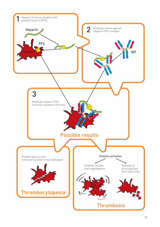
The second type (formerly called type 2) is an immune-mediated disease in which antibodies (mainly IgG) are formed against the complex of heparin with platelet factor 4 (PF4). These antibodies bind to the platelet cell membrane resulting in platelet aggregation and destruction. This leads to a paradoxical situation in which both a bleeding tendency (due to a low platelet count) and a clotting tendency occur simultaneously.
25
Heparin forms a complex with platelet factor 4 (PF4)
Heparin
IgGPlatelet
PF4
Antibody reacts against Heparin-PF4 complex
Antibody-heparin-PF4 immune complex is formed
Possible results
Platelet destruction (removal by splenic macrophages)
Platelet activation
Platelet release then aggregation
Release of procoagulant microparticles
Thrombocytopenia
Thrombosis
12
3

26
When HIT occurs, heparin needs to be discontinued immediately. Anticoagulation of the extracorporeal circuit during CRRT with citrate is a good alternative in this case.
6.2 Contraindications
6.2.1 Severe liver failure
Under physiologic circumstances, citrate is rapidly metabolized by the liver and to a lesser extent in the skeletal muscle and kidneys. In critically ill patients with acute liver failure, impaired citrate metabolism has been described. It is unknown whether critically ill cirrhotic patients are at risk of citrate accumulation.[22] More frequent monitoring for potential signs of citrate accumulation is required. (See Section 6.2.2 for clinical manifestations of citrate accumulation/intolerance.)
Patients with septic shock and lactic acidosis, in whom a limited metabolism of citrate might be expected due to disturbed liver and muscle perfusion, can generally receive CRRT with citrate without signs of citrate accumulation.[21] Nevertheless, frequent monitoring for potential signs of citrate accumulation is required. (See Section 6.2.2 for clinical manifestations of citrate accumulation/intolerance.)
6.2.2 Citrate intolerance
Clinical evidence of citrate intolerance or intoxication (due to citrate accumulation) also contraindicates the use of RCA. The initial clinical manifestations of citrate intolerance are primarily neuromuscular, including paresthesias (numbness or tingling) of the extremities and mouth along with muscle cramps. More serious complications are hypotension and arrhythmia. These complications are related directly to systemic hypocalcemia, the origin of which is discussed below.
A patient’s inability to metabolize the citrate-calcium complex and the resultant accumulation of citrate has three major clinical consequences: 1) decreased ionized calcium concentration in the blood because free calcium is not released from the citrate-calcium complex; 2) increased total calcium concentration in the blood because the accumulating citrate-calcium complex still contributes to total calcium concentration. This results in an abnormally high “calcium gap”, defined as the difference between the total calcium concentration (the standard laboratory measurement) and the ionized calcium concentration in the blood. The low ionized calcium concentration may prompt the prescription of a higher calcium reinfusion rate, which only worsens the hypercalcemia (based on the total serum calcium); 3) metabolic acidosis due to the net bicarbonate deficiency created by insufficient citrate breakdown. Because citrate is an “unmeasured anion”, this results in an “anion gap” metabolic acidosis.

27
6.3 Citrate as a standard anticoagulation method?The use of citrate anticoagulation is not uniform and probably currently restricted to some areas. It has become increasingly popular in North America and Europe, for the simple fact that it provides the opportunity to deliver CRRT without the need for systemic anticoagulation.[22]
Despite this fact, anticoagulation with citrate has complex metabolic consequences. It also requires frequent and close monitoring of the patient’s blood, extensive training, and should be guided by a strict protocol. Finally, treatment dosing as recommended by Ronco in 2000[23] or Bellomo in 2009[24]
is not always easy to achieve with a fixed citrate protocol. Every aspect will be explained in more detail subsequently in this module.

28
6.4 Quiz
1. Regional anticoagulation means:
a. Anticoagulation occurs in the patient’s body and the extracorporeal circuit
b. Anticoagulation is restricted to the patient’s body
c. Anticoagulation is restricted to the extracorporeal circuit
2. Platelet activation during the heparin-induced thrombocytopenia leads to:
a. Thrombocytopenia
b. Thrombosis
c. Thrombocytopenia and thrombosis
3. In which patients would you expect a limited metabolism of citrate?
a. Liver failure, septic shock and lactic acidosis
b. Liver failure, HIT and lactic acidosis
c. Liver failure, HIT and septic shock

29
7 Which solutions are required during citrate anticoagulation?
7.1 Citrate anticoagulation solutionsCitrate anticoagulation using the Prismaflex system requires a citrate solution on the PBP (pre-blood pump) scale. The goal is to inhibit coagulation as soon as the blood enters the extracorporeal set, achieved by using the PBP infusion site located nearest the patient access site. Table 1 below shows the composition of the three citrate solutions most commonly used for citrate anticoagulation during CRRT:
Table 1:
Trisodium citrate mmol/l
Citric acid mmol/l
Na mmol/l
Glucose mmol/l
Potassium mmol/l
4% Tri-Na citrate 136 408 0 0
ACD-A 74.8 38.1 224 124 0
Prismocitrate® 10/2 10 2 136 0 0
Prismocitrate® 18/0 18 0 140 0 0
PrismoCit® 4K 10 2 140 0 4
7.1.1 Trisodium citrate
Several published citrate protocols employ different trisodium citrate solutions. As the basis for these, customized and commercially available 4% trisodium citrate solutions commonly used include:
•4% trisodium citrate (TSC), usually delivered in 250 or 500 ml-containers
•4% sodium citrate, usually delivered in 1000 ml
The citrate solution Mehta[25] used in 1990 for continuous hemodiafiltration was undiluted 4% TSC solution. However, because this type of solution is hypertonic with respect to sodium and alkali equivalent, hypernatremia and metabolic alkalosis are frequent complications. Mehta and colleagues employed a dialysate solution with hypotonic concentrations of sodium and alkali equivalent to compensate for the unphysiologic nature of the citrate anticoagulant. To

30
lower the risk of these metabolic disturbances further, Tolwani[11] diluted the 4 percent solution to 2 percent TSC in 1999, which consisted of 1500 ml of 5 percent dextrose and 1500 ml of 4 percent citrate. This solution was also used in a continuous hemodiafiltration modality.
In 2006 Tolwani[26] compared the use of a 0.67 percent TSC solution with a 0.5 percent TSC solution in CVVHDF. The 0.5 percent solution represents a citrate content of 18 mmol/l and the 0.67 percent TSC a citrate content of 23 mmol/l. The differences in anticoagulation efficacy were not significant but metabolic alkalosis and hypernatremia occurred more frequently in the 0.67 percent citrate group. This study confirmed the relatively high risk of metabolic disorders (hypernatremia and metabolic alkalosis) associated with the use of concentrated citrate solutions. Additionally, as noted above, many of these concentrated solutions require the use of dialysate or replacement fluids with unphysiologically low concentrations of sodium and bicarbonate to “compensate” for the citrate solutions’ supra-normal concentrations of these constituents.
7.1.2 ACD-A
Anticoagulant citrate dextrose form A (ACD-A) is also commonly used for RCA in CRRT. The ACD-A anticoagulant solutions are commercially available and usually delivered in 500 and/or 1000 ml containers. The ACD-A solution has the following content:
•Citrate 112.9 mmol/l
- Trisodium citrate: 74.8 mmol/l
- Citric acid: 38.1 mmol/l
•Sodium: 224 mmol/l
•Glucose: 124 mg/dl
The pH of these solutions is low (approximately 5).
In 2003 Mitchell and colleagues[8] used ACD-A solution in their study of RCA in CVVHD.
In 2004, Cointault[4] et al published their experience with an ACD-A solution containing 112.9 mmol/l citrate (3.22%), 114.2 mmol/l hydrogen ion, 224.4 mmol/l sodium and 123.6 mg/dl glucose in the CVVHDF mode.

31
Since ACD-A solutions have an intermediate concentration of trisodium citrate and citric acid, they are less likely to cause citrate toxicity and other complications compared to trisodium citrate, but the risk is not averted totally. Even with the use of these lower concentrated citrate solutions, non-physiologic dialysate and/or replacement solutions, as mentioned above, are required. The high concentration of glucose in this solution is also considered a disadvantage.
7.1.3 Prismocitrate® 10/2[27] solution from Gambro
Prismocitrate 10/2 sterile solution for anticoagulation has been offered by Gambro since 2005 in 5-liter bags. Prismocitrate 10/2 solution is not available in the US and a few other countries (e.g. Canada). The solution is ready for use and has the following composition:
•Citrate: 10 mmol/l
•Citric acid: 2 mmol/l
•Sodium: 136 mmol/l
•Chloride: 106 mmol/l
A liter of Prismocitrate 10/2 solution contains 10 mmol of trisodium citrate. This concentration results in a physiologic equivalent of bicarbonate, which is 30 mmol after metabolism in liver, skeletal muscles and kidneys. The addition of 2 mmol citric acid to the 10 mmol of trisodium citrate augments the anticoagulation effect. However, the citric acid is not metabolized to bicarbonate but directly to CO2 and H2O. The net effect is a reduced risk of metabolic alkalosis. Standard commercially available physiologic solutions can be used as dialysate when using Prismocitrate 10/2 solution in the CVVHDF mode. In addition, the physiologic concentration of sodium essentially eliminates the risk of hypernatremia.
Prismocitrate 10/2 solution can be used in a variety of CRRT modalities, in conjunction with commercially available calcium-free dialysate and replacement fluids having standard solute concentrations.
Since Prismocitrate 10/2 solution is a diluted citrate solution, it requires a higher infusion rate to achieve the given anticoagulant effect, relative to more concentrated citrate solutions. This creates the opportunity to prescribe relatively high convective doses, as recommended by Ronco[23], or an efficient prescribed dose of 25 ml/kg/h in CVVHDF, as recommended by Bellomo[24] after the results of the RENAL trial, rather than to focus on citrate dosing only.

32
7.1.4 Prismocitrate® 18/0[28] solution from Gambro
Prismocitrate 18/0 sterile solution for anticoagulation has been offered by Gambro since 2011 in 5 liter bags.
Prismocitrate 18/0 solution is a CE-marked medical device commercially available in Europe.
The solution is ready for use and has the following composition:
Citrate: 18 mmol/L Sodium: 140 mmol/L
Chloride: 86 mmol/L
A liter of Prismocitrate 18/0 solution contains 18 mmol of trisodium citrate. This concentration results in a equivalent of 54 mmol bicarbonate after metabolism in liver, skeletal muscles and kidneys. The net effect is a reduced risk of metabolic alkalosis, when using dialysate and replacement solutions with a bicarbonate content of 25 mmol/L, ideally with Gambro’s Prism0cal B22 dialysate solution, which has 22 mmol/L bicarbonate and 3 mmol/L lactate. The combined use of the Prismocitrate 18/0 and Prism0cal B22 solution is in line with the Tolwani[26] ‘Alabama Protocol’ of 2006 when she used TSC 0.5% (18 mmol/) and a 25-bicarbonate solution.
Prismocitrate 18/0 solution can be used in a variety of CRRT modalities, in conjunction with commercially
Available calcium-free dialysate and replacement solutions having bicarbonate concentration of approximately 25 mmol/ L.
Since Prismocitrate 18/0 solution is a relatively diluted citrate solution, it requires a higher infusion rate to achieve the given anticoagulant effect, relative to more concentrated citrate solutions. This creates the opportunity to prescribe relatively high convective doses, as recommended by Ronco[23] or an efficient prescribed dose of 25 ml/kg/h in CVVHDF, as recommended by Bellomo[24]
after the results of the RENAL trial, rather then to focus on citrate dose only.
7.1.5 PrismoCit® 4K[29] solution from Gambro
PrismoCit 4K sterile solution for anticoagulation has been offered by Gambro since 2011in 5 liter bags.
PrismoCit 4K solution is a CE-marked medical device commercially available in Europe. The solution is ready for use and has the following composition:

33
Citrate: 10 mmol/L
Citric Acid: 2 mmol/L
Sodium: 140 mmol/L
Chloride: 114 mmol/L
Potassium: 4 mmol/L
A liter of PrismoCit 4K solution contains 10 mmol of trisodium citrate. This concentration results in a physiological equivalent of bicarbonate, which is 30 mmol after metabolism in liver, skeletal muscles and kidneys. The addition of 2 mmol citric acid to the 10 mmol of trisodium citrate augments the anticoagulation effect. However, the citric acid is not metabolized to bicarbonate but directly to CO2 and H2O. The net effect is a reduced risk of metabolic alkalosis.
PrismoCit 4K solution contains a slightly higher concentration of Sodium (140 mmol/L) to prevent Hyponatremia. The solution contains as well 4 mmol/ L of Potassium for stable treatments.
PrismoCit 4K solution can be used in a variety of CRRT modalities, in conjunction with commercially
Available calcium-free dialysate and replacement solutions having standard solute concentrations
Since PrismoCit 4K solution is a diluted citrate solution, it requires a higher infusion rate to achieve the given anticoagulant effect, relative to more concentrated citrate solutions. This creates the opportunity to prescribe relatively high convective doses, as recommended by Ronco[23] or an efficient prescribed dose of 25 ml/kg/h in CVVHDF, as recommended by Bellomo[24]
after the results of the RENAL trial, rather then to focus on citrate dose only.
7.2 Dialysate solutionsWhen CVVHD or CVVHDF has been chosen as the therapy modality, a solution is required on the dialysate scale. Ideally a calcium-free solution should be used to prevent the possibility of calcium counteracting the effect of citrate anticoagulation. However, the literature suggests calcium-containing solutions are also feasible, as demonstrated by Mitchell and colleagues.[8]
As mentioned previously, dialysate solutions with unphysiologically low concentrations of sodium and bicarbonate are typically required when used in conjunction with highly concentrated citrate solutions.

34
7.2.1 Calcium-free dialysate solutions
The binding of calcium to citrate inhibits coagulation in the extracorporeal circuit. Therefore, the use of calcium-free solutions is usually required as a dialysate when CVVHDF or CVVHD is prescribed. Otherwise, calcium counteracts the anticoagulant effect of citrate and clotting of the hemofilter/system may occur.
7.2.2 Prism0cal®[30] from Gambro
The Prism0cal solution is bicarbonate buffered dialysate solution without calcium content and is offered by Gambro in 5-liter bags. The Prism0cal solution is not available in the US. The solution contains bicarbonate at the level of 32 mmol/l fluid. The Prism0cal solution should only be used as a dialysate solution. In Canada and Singapore, the Prism0cal solution can be used as dialysate and replacement solution.
7.2.3 Prism0cal B22[31] from Gambro
The Prism0cal B22 solution is a sterile bicarbonate buffered dialysate solution without calcium and is offered by Gambro since 2011 in 5 liter bags. Prism0cal B22 solution is a CE-marked medical device commercially available in Europe.
The solution contains bicarbonate at the level of 22 mmol/L fluid and lactate at the level of 3 mmol/L fluid. This gives an equivalent of 25 mmol bicarbonate after lactate is metabolized in the liver.
The Prism0cal B22 can ideally be used in conjunction with Prismocitrate 18/0, following the Tolwani[26] ‘Alabama protocol. Prism0cal B22 should only be used as a dialysate solution.
7.3 Replacement solutionsWhen CVVH or CVVHDF has been chosen as the therapy modality, a solution is required on the replacement scale. Calcium-free solution ideally should be used to avoid any counteracting effects in the citrate anticoagulation process.
7.3.1 Calcium-free replacement solutions
At this time, the availability of commercially available calcium-free replacement fluids is limited. As calcium in the replacement fluid counteracts the anti-coagulant effect of citrate in the return line of the extracorporeal circuit, coagulation may occur at this site.

35
7.3.2 Solutions with calcium content
When a calcium-containing solution is used as a replacement fluid, delivery in post-dilution is recommended. However, it is necessary to ensure that the amount of calcium delivered into the circuit does not increase either the cir-culating ionized calcium or the post-filter ionized calcium level to a concentration greater than the target value of each.
7.4 Calcium infusionTo keep the ionized calcium serum level of the patient’s blood between 1.0 and 1.3 mmol/l, the calcium loss in the effluent must be compensated. Usually, one of two different calcium solutions is used: calcium gluconate 10% or calcium chloride 10%.
The calcium solution must be infused through a separate central venous catheter, thus avoiding the use of the CRRT access site. If the calcium is infused via a peripheral catheter, irritation of the vein might occur or, in case of extravasation from a displaced peripheral catheter, necrosis and calcification of the tissue.
The calcium solution must be slowly infused. Overly rapid infusion can be associated with cardiotoxicity, hypotension, local thrombophlebitis, tingling sensation, calcium taste, flushing, nausea, vomiting, or sweating. Consult your physician and pharmacist to understand any possible contraindications of calcium infusion related to interactions with other drugs used in the ICU.
7.4.1 Calcium gluconate 10%[32]
Calcium gluconate 10% is available in 10 ml ampules. Each 10 ml ampule of calcium gluconate 10% contains 1g calcium gluconate. The effective calcium content is 2.23 mmol/10 ml or 4.6 mEq/10 ml.
7.4.2 Calcium chloride 10%[33]
Each 10 ml ampule of calcium chloride 10% contains 1 g calcium chloride dihydrate or 1 g calcium chloride hexahydrate.
The effective calcium content when using calcium chloride dihydrate is 6.803 mmol/10ml or 13.605 mEq/10ml.
The effective calcium content when using calcium chloride hexahydrate is 4.56 mmol/10ml or 9.12 mEq/10ml.

36
7.5 Priming solutionsThe use of priming solutions needs special attention when CRRT with citrate anticoagulation has been chosen. The choice of the solution is not different but there is no need to add heparin. In all cases, follow the instructions for use (IFU) provided for the Prismaflex disposable set chosen. The use of heparin during priming when treating HIT patients must be avoided.
7.6 Quiz
1. Highly concentrated citrate solutions are more likely to cause:
a. Hyponatremia, metabolic alkalosis and citrate toxicity
b. Hypernatremia, metabolic alkalosis and citrate toxicity
c. Hypernatremia, metabolic acidosis and citrate toxicity
2. The use of highly concentrated citrate solutions requires non-physiological dialysate- and/or replacement solutions. What are the characteristics of these solutions?
a. Higher content of bicarbonate and lower content of sodium
b. Lower content of bicarbonate and higher content of sodium
c. Lower content of bicarbonate and lower content of sodium
3. Why is 2 mmol of citric acid added per liter of Prismocitrate® 10/2 solution?
a. To reduce the risk of metabolic alkalosis
b. To reduce the risk of metabolic acidosis
c. To reduce the risk of precipitation
4. Which of the following statements about the Prism0cal® solution from Gambro is true?
a. The Prism0cal solution is not available in the US and can only be used as a dialysate
b. The Prism0cal solution is not available in the US and can be used either as dialysate or replacement
c. The Prism0cal solution is available worldwide and can be used either as dialysate or replacement

37
5. Why should calcium preferentially be infused through a (separate) central venous catheter rather than through a peripheral catheter?
a. To avoid cardiotoxicity
b. To avoid a tingling sensation
c. To avoid the risk of irritation to the vein or, in worse cases, necrosis of the tissue

38
8 Which CRRT therapy options with the Prismaflex® system allow citrate anticoagulation?In this chapter the descriptions of citrate anticoagulation are restricted to CRRT treatments using the Prismaflex system[34] with a software version 5.00 or higher, although the experiences shown in the literature are mainly based on the use of the previous generation of CRRT machines.
The Prismaflex system uses a philosophy which makes choice of treatment modality the primary consideration while the choice of the anticoagulation method is secondary. Therefore, a variety of different CRRT modalities using the Prismaflex system are possible when using citrate as an anticoagulant.
8.1 SCUFSlow continuous ultrafiltration is a therapy for fluid removal, typically used in patients with diuretic-resistant volume overload. No dialysate or replacement fluids are used since correction of azotemia and metabolic disturbances is not the primary goal.
8.1.1 Clinical practice
The use of citrate during CRRT in a SCUF modality is not described in the literature, mainly due to the fact that with previous generation CRRT machines, this modality has not been combined with citrate anticoagulation.
8.1.2 Description
Citrate anticoagulation can be delivered with the Prismaflex system in a SCUF modality by the pre-blood pump (PBP). However, an important consideration for SCUF is that the ability to influence acid/base and electrolyte balance by manipulating dialysate and/or replacement fluid flow rates does not exist because these fluids are not used in SCUF. Therefore, if “Citrate” is chosen as the anticoagulation method for the Prismaflex system, the citrate load delivered to the patient must be limited. Specifically, PBP citrate flow rate in a SCUF modality is limited to 1000 ml/hr, which in turn automatically sets the blood flow rate in a restricted range. For example, when using Prismocitrate 10/2 solution the following limitations are set by the Prismaflex system:

39
•Target citrate dose of 2 mmol/l blood: maximum blood flow rate =100 ml/min (with PBP citrate flow rate of 1000 mL/hr)
•Target citrate dose of 3 mmol/l blood: maximum blood flow rate =60 ml/min (with PBP citrate flow rate of 900 ml/hr)
•Target citrate dose of 5 mmol/l blood: maximum blood flow rate =40 ml/min (with PBP citrate flow rate of 1000 ml/hr)
WARNING:The use of citrate solutions, especially when highly concentrated,in a SCUF modality requires advanced knowledge of citrate pharmaco-kinetics and metabolism in critically ill patients. Clinical experience withRCA for this modality has not been reported in the literature.
❤❤❤❤❤❤❤Scale Scale
Effluent bag PBP citrate bag
SCUF
CVVH
CVVHD
CVVHDF

40
8.2 CVVHIn CVVH, convection and ultrafiltration are used to remove waste products and plasmatic water. Convection is the movement of solutes under pressure through a membrane along with the movement of water.
8.2.1 Clinical practice
Several systems for citrate anticoagulation in a CVVH modality are described in the literature and are used worldwide in clinical practice.
In 1999 Palsson and Niles showed a simplified system for delivering citrate anticoagulation during CVVH.[35] They used a dilute citrate-based (13.3 mmol/l) replacement fluid which was delivered in pre-dilution. None of the patients developed metabolic alkalosis or hypernatremia. The target citrate dose during this study was 2 mmol/l blood.
Oudemans-van Straaten describes in the ‘Review and Guidelines for Regional Anticoagulation with Citrate in Continuous Hemofiltration’ the use of citrate in both pre- and post-dilution.[36] The review includes also different circuit options for citrate anticoagulation during CVVH for either pre- or post-dilution. Oudemans-van Straaten explains in particular a post-dilution CVVH modality with a highly concentrated (15%) trisodium citrate administrated pre-filter and a post-dilution replacement solution which is ideally a combination of a buffer-free low sodium and a bicarbonate-buffered saline.
8.2.2 Description
During treatment with a Prismaflex machine in a CVVH modality, the PBP is available for infusion of the citrate solution. If solutions are completely or just partially delivered pre-dilution, the fact must be taken into account that solutions containing calcium counteract the anticoagulant effect of citrate. In post-dilution, the delivery of calcium-containing solutions is possible but there is potential risk of clotting in the venous return line. The use of a calcium-free solution is preferable. The use of bicarbonate-buffered solutions as replacement is also recommended. The replacement solution should contain bicarbonate in the range of 30-35 mmol/L and sodium at physiological level when using Prismocitrate 10/2 or PrismoCit 4K solution as an anticoagulant. When using Prismocitrate 18/0, the replacement solution should contain approximately 25 mmol/L of bicarbonate. The use of more concentrated citrate solutions generally requires replacement solutions with lower concentrations of bicarbonate and sodium.

41
8.3 CVVHDIn continuous venovenous hemodialysis, diffusion and ultrafiltration are usedto remove waste products and plasmatic water. The fluids used are known asdialysate fluids. Dialysate is infused against the current of the blood flow, in theoutside compartment of the hemofilter to provide diffusive clearance primarilyof small-molecular-weight substances (< 500 Dalton).
8.3.1 Clinical practice
The use of citrate anticoagulation during CVVHD is well described in theliterature, even though the limited solute clearance capabilities render it arelatively uncommon choice for patients with acute kidney injury.
Tolwani et al described in 2001[11] a citrate protocol instituted for CVVHD using2 percent trisodium citrate as a regional anticoagulant and normal saline withsupplemental potassium and magnesium as the dialysate. This strategy changedto CVVHDF in 2006 and is explained in detail later in this chapter.
Swartz et al implemented citrate anticoagulation in CVVHD, which is reportedin 2004.[37] They used ACD-A as an anticoagulant and a calcium-free dialysatesolution with minimum flow rates of 2000 ml/hr.
The Prismaflex system allows the use of calcium-containing replacement solutions, but restricts thedelivery to post-dilution only.
❤❤❤❤
SCUF
CVVHDF
CVVHD
CVVH
Scale Scale Scale Scale
Effluent bag PBP citrate bag Ca-free replac. bag Ca-free replac. bag

42
Mitchell also described a CVVHD protocol in 2003[8], where ACD-A was usedas an anticoagulant and a variable bicarbonate-buffered solution with calciumcontent as a dialysate.
8.3.2 Description
The application of citrate in a CVVHD modality with the Prismaflex system canbe achieved by using the PBP. As is the case with other modalities, the citrateinfusion rate needs to be titrated to blood flow rate according to the desired bloodcitrate concentration. Therefore, for a given target blood citrate concentrationand blood flow rate, the required PBP citrate infusion rate increases as citratesolution concentration decreases
In CVVHD, the dialysate solution should not contain calcium as this counteractsthe anticoagulant effect of citrate. The use of bicarbonate-buffered solutionsas a dialysate is also recommended. The dialysate solution should containbicarbonate in the range of 30-35 mmol/L and sodium at phisiological level whenusing Prismocitrate 10/2 or PrismoCit 4K solution as an anticoagulant. Whenusing Prismocitrate 18/0, the dialysate solution should contain approximately 25mmol/L of bicarbonate. The use of more concentrated citrate solutions generallyrequires dialysate solutions with lower bicarbonate and sodium concentrations.
❤❤❤❤
SCUF
CVVHDF
CVVHD
CVVH
Scale Scale Scale
Effluent bag PBP citrate bag Ca-free dialyzate bag

43
8.4 CVVHDFIn continuous venovenous hemodiafiltration, diffusion, convection, and ultrafiltration are used to remove waste and plasmatic water. In this method, dialysate and replacement fluid are used simultaneously in various combinations of rates. The goal is to offer both a convective therapy for clearance of middle and large molecular-weight substances, and a diffusive therapy for the removal of smaller substances.
8.4.1 Clinical practice
Several studies have reported the use of citrate anticoagulation for CVVHDF. In patients treated with this modality, Tolwani[26] et al compared a 0.67 percent (23 mmol/l) and 0.5 percent (18 mmol/l) trisodium citrate solution. Both citrate solutions contained a physiologic sodium concentration (140 mmol/l) and were given in pre-dilution with the Prisma® pre-pump pre-dilution set from Gambro. A bicarbonate-buffered (25 mmol/l) solution with a sodium concentration (140 mmol/l) equivalent to that of the citrate solution was used as dialysate.
Cointault also reported an RCA protocol for CVVHDF in 2004.[4] An ACD-A solution was infused into the access line with an external pump. Both the replacement solution and dialysate were a physiologic bicarbonate-buffered solution containing calcium with the replacement fluid applied in pre-dilution. In some cases, bicarbonate was withheld from the dialysate to reduce the risk of metabolic alkalosis and hypernatremia.
8.4.2 Description
As in the other CRRT modalities with the Prismaflex system, during CVVHDF the PBP is available to deliver a citrate solution to the access line of the extracorporeal circuit. A calcium-free solution is recommended as a dialysate. The replacement solution should be calcium-free if delivered in pre-dilution; otherwise it would counteract the anticoagulant effect of citrate. In post-dilution, the delivery of calcium-containing solutions is possible but there is a potential risk of clotting in the return line. Therefore, the use of a calcium-free solution in post-dilution is preferable. Bicarbonate-buffered solutions are recommended as replacement and dialysate solutions. These solutions should contain bicarbonate in the range of 30-35 mmol/L and sodium at physiological level when using Prismocitrate 10/2 or PrismoCit 4K solution as an anticoagulant. When using Prismocitrate 18/0, the replacement and dialysate solutions should contain approximately 25 mmol/L of bicarbonate. The use of more highly concentrated citrate solutions generally requires replacement and dialysate solutions with lower concentrations of bicarbonate and sodium.

44
❤❤❤❤
SCUF
CVVHDF
CVVHD
CVVH
Scale Scale Scale Scale
Effluent bag PBP citrate bag Ca-free dialyzate bag Ca-free replac. bag
The Prismaflex system allows the use of calcium-containing replacement solutions, but restricts thedelivery to post-dilution only.

45
8.5 Quiz
1. Which study shows a simplified citrate anticoagulation protocol in CVVH?
a. Tolwani (2006)
b. Palsson and Niles (1999)
c. Cointault (2004)
2. Which citrate solution was used by Tolwani during her study in 2001 (CVVHD)?
a. Trisodium citrate 2%
b. ACD-A
c. Prismocitrate®
3. Is it possible to use replacement solutions with calcium if given in post-dilution during CVVHDF?
a. No, this is not recommended
b. Yes, this is recommended
c. Yes, but there is a potential risk of clotting in the return line; the use of a calcium-free solution in post-dilution would be preferable

46
The software version 5.00 and higher of the Prismaflex system allows the delivery of citrate anticoagulation in a safe manner. This chapter explains how the software of the Prismaflex system supports the user during setup and treatment for CRRT modalities in particular.
9.1 Selection of citrate method During the setup the user can select 2 different citrate anticoagulation methods:
•Citrate – Calcium via the syringe pump of the Prismaflex system (Citrate/Calcium)
•Citrate – Calcium via External Infusion Pump (Citrate)
The following must be considered with these two options:
•The ‘Citrate/Calcium’ and ‘Citrate’ options must be enabled in Service mode.
•When ‘Citrate – Calcium via the syringe pump of the Prismaflex system’ has been chosen the syringe pump of the Prismaflex system allows an automatic replacement of the lost calcium
•When ‘Citrate – Calcium via External Infusion Pump’ has been chosen, the syringe pump of the Prismaflex system is disabled for the entire treatment. An external syringe/infusion pump must be used to replace the lost calcium.
9.2 Selection of citrate anticoagulation solutions The selection of an anticoagulation method is followed by a review of the chosen therapy and anticoagulation method. The screen displays also:
•For both ‘Citrate/Calcium’ and ‘Citrate’ methods: The pre-selected citrate solution to be used on the PBP citrate scale with the following information:
- Name of the solution, as written on the label - Concentration of citrate per liter solution (mmol/l) - Concentration of citric acid per liter solution (mmol/l) - Volume of the citrate solution bag
9 How does the Prismaflex® system[34] manage regionalcitrate anticoagulation methods?

47
•For the ‘Citrate/Calcium’ method only: The pre-selected calcium solution to be used on the syringe of the Prismaflex system with the following information:
- Name of the solution, as written on the label
- Concentration of calcium per liter (mmol/l)
The label on the solution bag, container or ampoule to be used must be verified and correspond with the information on the screen for the selected solution(s).
A broad range of citrate and calcium solutions can be used within the system. Allowed concentration ranges are dependent on therapy. The clinic’s choice of solutions can be preset in Service mode by a service technician.
The operator selects which solution to use for the treatment according to the prescription and changes default values for Anticoagulation Settings parameters in Custom mode.
When using calcium containing replacement solutions in post-dilution only, the operator must define the calcium concentration of the bag in Custom mode. When a calcium concentration of >0 mmol/L has been selected, it is not possible to deliver replacement in pre-dilution for CVVH and CVVHDF therapies.
9.3 Software updated settings on the Prismaflex monitorIn citrate anticoagulation the Prismaflex system software links the blood flow rate, PBP citrate rate and citrate dose. Blood flow rate and citrate dose can be set by the user; the PBP citrate rate depends on the other two settings.
9.3.1 Blood flow rate
The blood flow rate affects the PBP citrate flow rate. A change of the blood flow rate automatically results in a:
•Change of the PBP citrate flow rate
•Change of effluent and UFR dose (ml/kg/hr)
•Change of the estimated patient citrate load
•Change of calcium syringe flow rate (Citrate/Calcium method only)
•Change of estimated change of calcium loss in effluent (Citrate method only)
This synchronization of the blood flow rate and PBP citrate flow rate is necessary to maintain the selected citrate dose (mmol/l blood) at the desired level.

48
9.3.2 PBP citrate flow rate
PBP citrate = Citrate solution, used on the PBP scale
PBP citrate flow rate cannot be selected directly. The PBP citrate flow rate is a result of the selected blood flow rate and targeted citrate dose (mmol/l blood).
PBP citrate flow rate is affected by the blood flow rate. A change of the blood flow rate automatically results in a:
•Change of the PBP citrate flow rate
•Change of Effluent and UFR dose (ml/kg/hr)
•Change of the estimated patient citrate load
•Change of calcium syringe flow rate (Citrate/Calcium method only)
•Change of estimated change of calcium loss in effluent (Citrate method only)
This synchronization of blood flow rate and PBP citrate flow rate is controlled by the selected citrate dose (mmol/l blood).
PBP citrate flow is affected by the citrate dose (mmol/l blood). A change of the citrate dose automatically results in a change of the PBP citrate flow rate (see table above).
9.3.3 Citrate dose
Citrate dose = concentration of citrate per liter of blood treated (mmol/l blood)
Citrate dose is defined as the amount of citrate infused per liter of patient’s blood treated, expressed in mmol/l blood (targeted citrate dose: 2-5 mmol/l blood). To obtain the selected citrate dose, the control unit of the Prismaflex
Changing factors in citrate anticoagulant
RESULT
CHANGE
Blood flow Citrate dose PBP citrate Citrate load
Blood flow increase unchanged increase increase
Blood flow decrease unchanged decrease decrease
Citrate dose increase unchanged increase increase
Citrate dose decrease unchanged decrease decrease
The table below illustrates this:

49
system adjusts the PBP citrate flow rate within the right proportion to blood flow rate. To achieve anticoagulation of the blood in set, the citrate dose must be appropriate. Monitoring of post-filter ionized calcium (blue sample port) is required to assess the anticoagulation status of the blood in set and modify the citrate dose accordingly.
Citrate dose affects the PBP citrate flow rate. A change of citrate dose automatically results in a:
•Change of PBP citrate flow rate•Change of Effluent and UFR dose (ml/kg/hr)•Change of estimated patient citrate load•Change of calcium syringe flow rate (Citrate/Calcium method only)•Change of estimated change of calcium loss in effluent (Citrate method only)
The synchronization of citrate dose and PBP citrate flow rate allows for an adaptation of the citrate infusion rate (higher or lower) the newly selected citrate dose (mmol/l blood; see table above).
9.4 Calcium management during citrate anticoagulation with the Prismaflex systemThe calcium loss into the effluent must be compensated to avoid hypocalcemia in the patient’s blood. The Prismaflex system offers the user an integrated calcium management system where the calcium will be delivered by the syringe pump of the Prismaflex system. However, the user can also decide to manage the calcium infusion with an external syringe/infusion pump. Both possibilities are described below.
9.4.1 Using anticoagulation method ‘Citrate - Calcium via the syringe pump of the Prismaflex system’ (Citrate/Calcium)
When the ‘Citrate – Calcium via the syringe pump of the Prismaflex system’ anticoagulation method has been chosen, the syringe pump of the Prismaflex system will be used for calcium infusion. This method requires the dedicated Prismaflex calcium infusion line, connected to the 50 ml syringe.
Using the syringe pump of the Prismaflex system will facilitate maintenance of normocalcemia in the patient. During treatment, the syringe pump of the Prismaflex system, used for calcium infusion, is synchronized with the PBP citrate pump. Whenever the citrate infusion stops, the calcium infusion stops as well. The syringe pump of the Prismaflex system for calcium infusion is also synchronized with the calcium loss in the effluent. This synchronization keeps the syringe flow rate proportional to the estimated calcium loss rate in

50
the effluent. In fact, the calculated calcium loss in the effluent is automaticallycompensated by the syringe pump of the Prismaflex system.
This compensation can be controlled by the user and is called ‘CalciumCompensation’. Calcium Compensation is defined as the relative dosage ofcalcium infusion to compensate for the estimated calcium loss in the effluent,expressed in percentage. A complete compensation of the calcium lost in theeffluent corresponds with a calcium compensation of 100%, but this assumesno calcium content in both dialysate and replacement fluids.
Calcium Compensation can be set between 5% and 200%. Any change of oneor more flow rates or anticoagulation settings affects the calcium syringe flowrate, as the consequence of estimated calcium clearance change.
Calcium must be infused with a separate venous catheter and this should be acentral venous (rather than peripheral) catheter. Calcium infusion before thedearation chamber could result in coagulation in the venous return line. Theuse of stop valves, connected to the return line, is not safe. In fact, in case ofinversion of bloodlines without changing the calcium infusion site, e.g. to centralvenous catheter, calcium enters the access line. The calcium concentration in thedisposable set will increase and lead to coagulation in the filter.
Using anticoagulation method‘Citrate—Calcium via the syringepump of the Prismaflex system’(Citrate/Calcium)
9.4.2 Using anticoagulation method ‘Citrate – Calcium via External Infusion Pump’ (Citrate)
When the ‘Citrate – Calcium via External Infusion Pump’ anticoagulation methodhas been chosen, the syringe pump of the Prismaflex system is disabled forthe entire treatment. An external syringe or infusion pump must be used for

51
infusion of the lost calcium. The calcium infusion must be adjusted or stoppedaccording to the physician’s prescription and/or facility protocol.
The Prismaflex system supports the user regarding the adjustment of thecalcium re-infusion by providing a calcium loss indicator. This calcium lossindicator is called ‘Estimated Change of Calcium Loss Rate in Effluent’ andrepresents the relative variation of calcium losses in effluent due to change(s)in prescription settings.
Calcium must be infused with a separate venous catheter and this should be acentral venous (rather than peripheral) catheter. Calcium infusion before thedearation chamber could result in coagulation in the venous return line. Theuse of stop valves, connected to the return line, is not safe. In fact, in case ofinversion of bloodlines without changing the calcium infusion site, e.g. to centralvenous catheter, calcium enters the access line. The calcium concentration inthe disposable set will increase and lead to coagulation in the filter.
9.5 Software updated flow rates during treatment (related to citrate anticoagulation)During treatment flow settings and/or anticoagulation settings can be changed,but it needs to be considered that these changes have consequences. Not onlywill the PBP citrate flow rate change, but the ‘Effluent dose’, ‘UFR dose’ and‘Patient citrate load’ will also change as a result of more or less fluid deliveryfrom the PBP citrate pump.
Using anticoagulation method‘Citrate – Calcium via ExternalInfusion Pump’(Citrate)

52
9.6 Safety SystemThe Prismaflex system provides additional alarms when citrate anticoagulation is performed. The type of alarm depends on which citrate anticoagulation method has been chosen.
9.6.1 Alarm specific to both regional citrate anticoagulation methods
ADVISORY: Anticoagulation checkpoints. The alarm reminds the user about the need for additional monitoring of patient parameters. The occurrence of the alarm can be selected in ‘System Tools’ or in Custom mode.
9.6.2 Alarms specific to ‘Citrate – Calcium via the syringe pump of the Prismaflex system‘ anticoagulation method
WARNING: Unsuitable Calcium Solution. This alarm occurs after ‘Confirm Set Loaded’ screen if there is not a valid set of initial flow settings with reasonable operating ranges available for the selected calcium solution.
ADVISORY: Calcium Line Not Connected. This alarm occurs when the calcium infusion line is not connected after syringe change or installation.
WARNING: Calcium Line Not Connected. This alarm is the equivalent of ‘Advisory: Calcium Line Not Connected’ for Setup mode (see above).
CAUTION: Calcium Infusion Stopped. This alarm occurs when the calcium syringe pump has been stopped for more than six minutes over the last two hours of treatment. Under this condition treatment and anticoagulation infusion are interrupted until calcium syringe infusion is resumed.
9.6.3 Alarm specific to ‘Citrate – Calcium via External Infusion’ anticoagulation method.
ADVISORY: Fluid Pumps Stopped. This alarm occurs only when the fluid pumps
Regarding calcium management, the changes are related to the calcium loss in the effluent and depend on the selected citrate anticoagulation method:
•When using the ‘Citrate – Calcium via the syringe pump of the Prismaflex system’ method, the calcium syringe rate changes to keep the chosen calcium compensation in the same proportions to the calcium loss in the effluent.
•When using the ‘Citrate – Calcium via External Infusion’ method, the ‘Estimated change calcium loss in effluent’ will change’, which helps the user adapt the external calcium infusion to the patient.
The software of the Prismaflex system calculates these changes and displays them in the screen ‘View Prescription Changes’.

53
have stopped due to an alarm for more than 10 minutes during treatment. Under this condition treatment and anticoagulation are interrupted while calcium delivery by the external syringe/infusion pump continues. This alarm also alerts the user about the need for additional monitoring of patient parameters.
9.7 Prescription indicator calculations in the Prismaflex® systemThe software of the Prismaflex system computes two indicators of the current citrate anticoagulation according to the disposable set in use, the flow rates and the citrate anticoagulation settings. The indicators depend on which citrate anticoagulation method has been chosen.
9.7.1 Indicator specific to both regional citrate anticoagulation methods
Estimated patient citrate load (in mmol/hr):
This represents the amount of citrate effectively delivered to the patient. This information is important in the clinical evaluation of a patient’s acid/base status, particularly with respect to the potential development of metabolic alkalosis.
The calculation of the citrate load is based on the following two factors:
•Rate of citrate infusion, based on blood flow rate and citrate dose
•Estimation of citrate clearance, as a function of blood flow, PBP citrate, replacement and dialysate and patient fluid removal rates, as well as the disposable set in use.
9.7.2 Indicator specific to ‘Citrate – Calcium via External Infusion’ anticoagulation method
Estimated change of calcium loss rate in effluent (in %):
This represents the relative variation of calcium losses in the effluent as a consequence of change(s) in prescription settings. The calculation is based on the estimation of calcium clearance as a function of blood flow, PBP citrate, replacement and dialysate and patient fluid removal rates, as well as the disposable set in use. The relative change in calcium clearance provides an estimation of the relative change of calcium loss rate in the effluent.
The change in calcium loss is expressed as a negative percentage (less loss) or a positive percentage (more loss). This percentage can assist the operator in adjusting the external calcium infusion to the patient but does not eliminate the need to monitor patient parameters (Ca2+, Mg2+, Na+, HCO3
-, pH).

54
9.8 Quiz
1. Which statement about the selection of a citrate anticoagulation method is not true?
a. Citrate methods must be enabled in Service mode
b. The selection of ‘Citrate – Calcium via External Infusion’ disables the syringe pump of the Prismaflex system for the entire treatment
c. The selection of a citrate method can be changed to Standard (with heparin) during treatment
2. The user can select (but not modify) other citrate solutions in:
a. Custom mode
b. Service mode
c. Setup mode
3. Which parameter is important to assess the anticoagulation status in the extracorporeal circuit and to choose an appropriate citrate dose?
a. Ionized calcium
b. Total calcium
c. Post-filter ionized calcium
4. During CRRT with citrate anticoagulation a change in blood flow will automatically result in change of:
a. PBP citrate flow rate, citrate dose and treatment dose
b. PBP citrate flow rate, treatment dose and patient citrate load
c. Dialysate flow rate, treatment dose and patient citrate load
5. During CRRT with citrate anticoagulation a change in citrate dose will automatically result in a change of:
a. Blood flow rate, PBP citrate flow rate and treatment dose
b. Blood flow rate, treatment dose and patient citrate load
c. PBP citrate flow rate, treatment dose and patient citrate load

55
6. Calcium Compensation, when using the ‘Citrate – Calcium via the syringepump of the Prismaflex system’ method, is expressed in:
a. mmol/l
b. ml/hr
c. %
7. Which screen provides an overview of the software updated flow rates andprescription indicators if a flow rate must been changed during treatment?
a. View prescription changes
b. Review prescription
c. Enter flow rates
8. Where can you modify the occurrence of the ‘ADVISORY: AnticoagulationCheckpoints’ alarm during treatment?
a. Service mode
b. Custom mode
c. System tools

56
A CRRT treatment with citrate anticoagulation requires additional monitoring of the patient’s blood chemistries. An assessment of the effectiveness of anticoagulation is also required.
10.1 Patient monitoringCitrate anticoagulation is often perceived as time-consuming and costly due to additional monitoring of a patient’s blood chemistries compared to a standard anticoagulation method such as heparin. Measurement of ionized serum calcium concentration is one of the main parameters used to adjust the calcium re-infusion rate. The drawbacks of this extra work and cost are justified in light of the potential benefits of increased hemofilter life and decreased bleeding risk.
10.1.1 Ionized calcium
Adequate monitoring of ionized serum calcium concentrations is mandatory since the calcium level in the patient’s blood decreases due to the loss of calcium in the effluent. In addition, when the metabolism of the calcium-citrate-complex is impaired, such as in patients with severe liver failure, the ionized calcium concentration may be low.
The serum ionized calcium concentration should be measured at least every six hours or more often if indicated.[25] Tolwani describes in the article of 2006[25] a measurement of ionized calcium one hour after initiation of CRRT and then every six hours thereafter. The monitoring of one ionized calcium can be done quickly by using an ABG device (arterial blood-gas analysis), which is installed in the ICU. Venous samples also can be used to monitor the ionized calcium.
10.1.2 Total calcium
Total calcium levels should be measured daily[11] in patients receiving CRRT with citrate anticoagulation to detect citrate accumulation when the metabolism of the calcium-citrate complex is impaired. The ionized serum calcium concentrations can be normal even when calcium reinfusion is constantly increased and an increasing calcium reinfusion rate resulting in a total calcium concentration > 3 mmol/l should be regarded as suspicious for accumulation of the calcium-citrate complex. In this situation, it is the rise in the bound component that
10 Which monitoring is required during citrate anticoagulation?

57
causes the increase in the total calcium concentration. The measurement of total calcium must be done in the laboratory.
A patient’s inability to metabolize the citrate-calcium complex and the resultant accumulation of citrate has three major clinical consequences: 1) decreased ionized calcium concentration in the blood because free calcium is not released from the citrate-calcium complex); 2) increased total calcium concentration in the blood because the accumulating citrate-calcium complex still contributes to total calcium concentration. This results in an abnormally high “calcium gap”, defined as the difference between the total calcium concentration (the standard laboratory measurement) and the ionized calcium concentration in the blood. The low ionized calcium concentration may prompt the prescription of a higher calcium reinfusion rate, which only worsens the hypercalcemia (based on the total serum calcium); 3) metabolic acidosis due to the net bicarbonate deficiency created by insufficient citrate breakdown. Because citrate is an “unmeasured anion”, this results in an “anion gap” metabolic acidosis.
10.1.3 Ratio Catot / Caion
This is another way in which the possibility of citrate accumulation can be assessed. The total calcium can be compared with the ionized calcium and be expressed in a ratio. Normally the Catot / Caion ratio in the systemic circulation is 2. A ratio of 2.5 or higher suggests calcium-citrate complex accumulation, with the risk of citrate toxicity.[38] The Catot / Caion ratio is a calculation based on the total calcium and ionized calcium concentrations of the patient’s blood.
10.1.4 pH
Although the pH is the first parameter to detect acidosis or alkalosis in the patient’s blood, it is not enough to judge the nature of the disturbance. The deviation in the pH from normal may be due to respiratory disturbances or due to metabolic disturbances.
•If the cause has a respiratory nature, a change in pCO2 is observed
•If the cause has a metabolic nature, a change in the serum bicarbonate concentration is observed
The use of citrate can cause changes in the pH in both directions. A normal pH value lies between 7.35–7.45. pH is included in an ABG analysis and must be measured at least once daily during CRRT with citrate anticoagulation.[26] The monitoring should ideally follow the same frequency as the calcium monitoring (every 6 hours).[36]

58
10.1.5 Bicarbonate and base excess
Bicarbonate (HCO3-) is a buffer in the patient’s blood. The normal standard
bicarbonate concentration lies between 22 and 26 mmol/l. Metabolic acidosis is associated with a low standard bicarbonate concentration while metabolic alkalosis is associated with a high standard bicarbonate concentration.
Standard bicarbonate is related to base excess (BE). The BE is negative in metabolic acidosis and positive in metabolic alkalosis. Standard bicarbonate and BE are included in an ABG analysis and must be measured at least once daily during CRRT with citrate anticoagulation.[25] The monitoring should ideally follow the same frequency as the calcium and pH monitoring (every six hours).[36]
10.1.6 Magnesium
Because citrate non-specifically chelates divalent cations (i.e., positive ions with a double-positive charge), it also binds magnesium. Therefore, close monitoring of the patient’s serum magnesium concentration is necessary to avoid hypomagnesemia. Because magnesium is normally not part of the ABG measurement, the sample typically must be sent to the laboratory. Magnesium levels must be monitored at least once a day during CRRT with citrate anticoagulation.[36]
Hypomagnesemia may occur due to increased loss of magnesium, bound to citrate, in the effluent. This complication is seldom seen since plasma magnesium is corrected by a shift from the intracellular compartment.[19] The magnesium control can be achieved using a dialysate containing magnesium or with direct infusion.
10.1.7 Other
The electrolytes sodium, potassium and chloride should be monitored every six hours.[26]
A complete blood count, phosphate, blood urea nitrogen and creatinine should be measured at least daily.[35] An assessment of liver function must be performed since the use of citrate requires metabolism by this important organ.
10.2 Effective anticoagulationAs with other methods of anticoagulation the use of citrate needs an assessment regarding effectiveness. Although ACT (activated clotting time) can be used[34], the most efficient and clinically validated measurement is the estimation of post-filter ionized calcium. Adequate anticoagulation is necessary to increase filter life, which provides for the effectiveness of the therapy.

59
10.2.1 Post-filter ionized calcium
The monitoring of post-filter ionized calcium concentration is an effective way to assess the anticoagulation of the extracorporeal circuit. Tolwani[26] measured those levels one hour after the initiation of CRRT and then every six hours thereafter. Ionized calcium concentrations between 0.25–0.35 mmol/l are consistent with adequate regional anticoagulation. The measurement is very simple—a blood sample is drawn from the blue sample port of the Prismaflex set and analyzed by an ABG device or laboratory.
An ionized calcium level below 0.25 mmol/l requires a reduction in citrate dose while a level above 0.35 mmol/l requires an increase in citrate dose.
10.2.2 Hemofilter life
Frequent hemofilter clotting reduces effective CRRT treatment time and may result in blood loss. This has consequences for the effectiveness of clearance and dose and may increase blood transfusion requirements. The use of citrate anticoagulation is associated with increased hemofilter life but it is not possible to make a definitive statement about hemofilter survival time. There are many patient-related factors, such as activation of coagulation, platelet count and hematocrit, that also influence filter life. In addition, catheter-related factors, such as length, diameter and positioning, must be taken into account. The hemofilter itself is another variable which may influence filter survival. Finally, treatment-related factors, such as modality, blood flow, filtration fraction, and operator troubleshooting response time, need also to be considered.
All these factors should be taken into account in any clinical study to clearly identify whether citrate anticoagulation increases hemofilter survival time or not. Failure to include these criteria into a study design makes it difficult to assess the study’s results.[39, 40]

60
10.3 Quiz
1. Is the measurement of total calcium relevant when ionized calcium is measured regularly?
a. Yes, the ionized serum calcium concentrations can be normal under conditions when calcium reinfusion is constantly increased. An increase of total calcium could detect citrate accumulation
b. No, only ionized calcium is relevant. A decrease of ionized calcium would detect citrate accumulation
c. No, only ionized calcium is relevant. An increase of ionized calcium would detect citrate accumulation
2. A low standard bicarbonate is associated with:
a. Metabolic alkalosis
b. Respiratory alkalosis
c. Metabolic acidosis
3. Which post-filter Ionized calcium level would be chosen to achieve a good regional anticoagulation?
a. 0 mmol/l
b. 0.30 mmol/l
c. 0.60 mmol/l
4. Important parameters like ionized calcium, standard bicarbonate and pH, but also sodium and potassium should be (according to the literature) monitored at least:
a. Every hour
b. Every three hours
c. Every six hours

61
11.1 Metabolic alkalosisMetabolic alkalosis is one of the main complications in patients receiving CRRT with citrate anticoagulation.[11] Citrate influences the acid-base status of the patient since one millimole of citrate is converted to 3 millimoles of bicarbonate, primarily in the liver. Metabolic alkalosis has been reported as a common complication of RCA with the use of highly concentrated citrate solutions. This is frequently accompanied by hypernatremia, due to the associated high concentration of sodium in the highly concentrated citrate solutions. These complications can be avoided by using dilute citrate solutions with physiologic sodium concentrations.
11.2 Metabolic acidosisMetabolic acidosis in the context of citrate anticoagulation can be a sign of citrate accumulation. Meier-Kriesche describes in an article from 2001[38] the correlation between an elevated calcium ratio and a low pH (acidosis) with an increased anion gap. Since metabolic acidosis is often seen in critically ill patients and the nature of the disturbance can be difficult to detect, it may be hard to define a clear case of citrate accumulation. To diagnose citrate accumulation, the ionized calcium and total calcium levels of the patient’s serum should be measured. If the Catot / Caion ratio is > 2.5, citrate accumulation is strongly suggested. In this case, citrate anticoagulation must be stopped. The most common cause of citrate accumulation is impaired citrate metabolism to bicarbonate due to severe liver failure.
11.3 HypocalcemiaHypocalcemia may occur if calcium is not compensated properly after a certain amount is lost in the effluent. Systemic ionized hypocalcemia may also occur if calcium-citrate complexes are accumulating. Hypocalcemia can cause tetany, confusion and arrhythmia in severe situations. Frequent monitoring of ionized calcium is necessary to assess the calcium level of the patient’s serum and to make appropriate adjustments in the calcium reinfusion rate. It is recommended to assess the total calcium concentration once a day to detect possible calcium-citrate complex accumulation.
11 What are possible complications and how can we avoid/manage them?

62
11.4 HypercalcemiaHypercalcemia may occur if calcium compensation is too aggressive or reinfusion of calcium continues after the CRRT treatment and citrate anticoagulation have been stopped. In this case, the calcium infusion to the patient must be stopped. Hypercalcemia can cause hallucinations, coma or arrhythmia. Frequent monitoring of ionized calcium is necessary to assess the calcium level of the patient’s serum and to make appropriate adjustments in the calcium reinfusion rate.
11.5 HypernatremiaHypernatremia may occur if the citrate flow is high relative to blood flow. Use of highly concentrated citrate solutions is particularly likely to result in this complication. Hypernatremia is usually accompanied by metabolic alkalosis (see above). Hypernatremia can cause shrinking of brain cells, confusion and coma. The use of dilute citrate solutions, combined with dialysate or replacement solutions with physiologic concentrations, can avoid this complication.
11.6 HyponatremiaHyponatremia may occur with the use of replacement or dialysate solutions having unphysiologically low concentrations of sodium. These solutions are typically used to counteract the metabolic effects of highly concentrated citrate solutions
Hyponatremia can cause swelling of brain cells, stupor and coma. The use of dilute citrate solutions with physiologic sodium concentrations, combined with appropriate dialysate or replacement solutions, can avoid this complication.
11.7 HypomagnesemiaHypomagnesemia may occur due to increased loss of magnesium, bound to citrate, in the effluent. This complication is seldom seen, since plasma magnesium is corrected by a shift from the intracellular compartment.[35] The magnesium control can be achieved using dialysate and replacement solutions containing magnesium or with direct infusion.

63
11.8 Citrate accumulation/toxicityCitrate accumulates in the blood if the liver and muscles fail to metabolize citric acid. This may happen in decompensated liver cirrhosis or severe shock. The acid accumulates, resulting in an increase in the anion gap, reflecting the increase in ionized citrate. A metabolic acidosis can be observed. In this way the citrate toxicity is developed with the following symptoms:
•Tetany
•Arrhythmia
•Hypotension
•Hypokalemia
•Acid-base disturbances
In these conditions citrate anticoagulation must be discontinued.

64
11.9 Quiz
1. When can we conclude a citrate accumulation?
a. When the Catot /Caion ratio is >2.5
b. When the Catot /Caion ratio is <2.5
c. When the pH is <7.35
2. Tetany, confusion and arrhythmia are symptoms of:
a. Hypercalcemia
b. Hypocalcemia
c. Hypernatremia
3. Hyponatremia can cause:
a. Swelling of brain cells
b. Shrinking of brain cells
c. Arrhythmia
4. If the following symptoms are observed what could be suspected?
- Tetany
- Arrhytmia
- Hypotension
- Hypokalemia
- Acid-base disturbances
a. Liver cirrhosis
b. Citrate toxicity
c. Septic shock

65
Since 2000 the awareness of the importance of the delivery of an adequate treatment dose to patients receiving CRRT has been growing. Ronco provided the medical field with direction toward dosing with his landmark study which established 35 ml/kg/hr (based on effluent flow rate) as an appropriate dose for patients receiving CRRT.[23] How can this dose be delivered when using citrate anticoagulation?
Tolwani’s article from 2006[26] gives an indication how a CRRT protocol can include citrate anticoagulation without limitations regarding high solute clearance. The main advantages of this protocol are listed as following:
•It provides high solute clearance and solution rates can be adjusted to maintain an effluent of 35 ml/kg/hr
•It uses standardized solutions that require no additional modifications
•The risk of metabolic catastrophe is minimized with the use of diluted citrate solutions and dialysate or replacement solutions with electrolytes at physiologic levels
12 Treatment dose and citrate anticoagulation

66
12.1 Quiz
1. Which aspect of the Tolwani protocol from 2006 finds alignment with Ronco’s landmark study from 2000?
a. It uses standardized solutions that require no additional modifications
b. The risk of metabolic catastrophe is minimized with the use of diluted citrate solutions and dialysate or replacement solutions with electrolytes at physiological levels
c. It provides high solute clearance and solution rates can be adjusted to maintain an effluent of 35 ml/kg/hr

67
13 How to implement citrate anticoagulation in the ICU
“Anticoagulation with citrate is advised in critically ill patients with active bleeding, an increased risk of bleeding or with heparin-induced thrombocytopenia (HIT), if and when training has been adequate and the method is performed guided by a strict protocol”.[36]
Citrate anticoagulation during CRRT is a complex procedure which needs time to be considered, planned and trained. The complexity of the principles, mechanisms and list of eventual complications mandate a careful approach to implement this anticoagulation method.
13.1 Expertise and skills in CRRTImplementing citrate anticoagulation without knowledge of CRRT in general is possible if the use of this anticoagulation method has been chosen as the standard anticoagulation approach. In these cases, the use of citrate must be completely integrated into an extended educational program when CRRT is introduced at an institution.
Under normal circumstances the choice of another approach is recommended. Expertise and skills in CRRT, especially with the new generation of machines should be collected for at least a year before citrate anticoagulation is introduced as an anticoagulation alternative. This approach allows for an adequate understanding of the machine and the necessary skills in troubleshooting of different alarms before the introduction of citrate anticoagulation. Advanced knowledge of the CRRT therapies, skills on equipment handling and citrate anticoagulation expertise are mandatory while implementing this anti-coagulation method.
13.2 Organization issues The use of citrate anticoagulation requires an appropriate infrastructure in the ICU and the facility. Different solutions must be stored without the risk of interchanging them. Due to the frequent need to measure blood chemistries during CRRT with RCA, the laboratory needs to be able to handle blood samples 24/7.

68
13.3 Protocol/Physicians’ orders/Procedure/Documentation Before the first treatment with citrate anticoagulation starts, a protocol should be written and be accessible for all team members of the ICU who are involved in patient care. The protocol should be written by the intensivist or nephrologist who is in charge of patients receiving CRRT. A protocol should guide the medical staff in the understanding of the principles of the anticoagulation method, how to prepare the machine with the right products, what and how often to monitor, what can be expected, etc.
These are the items to be considered as primary guidelines:
•Treatment modality
•Treatment dose
•Anticoagulation method
•Hemofilter to be used
•Monitoring and monogram
Flow sheets can be adapted to follow the process of the treatment.
Special documentation sheets regarding citrate anticoagulation are recom-mended, since a large amount of data is required to assess the patient’s situation under a treatment with this method. A documentation sheet helps the medical staff to structure the workload which is part of citrate anticoagulation follow-up.
13.4 Education/Training Training is an essential part in the preparatory phase, which is mandatory before initiating a treatment with citrate anticoagulation. The ICU medical and staff member should receive training on all aspects mentioned in this module on citrate anticoagulation, including the handling of the equipment. A periodic recertification program should also be part of general practice in ICU.

69
13.5 Quiz1. A citrate anticoagulation protocol must be written by:
a. Gambro clinical employees
b. ICU nurses
c. Intensivists or nephrologists
2. How should an ideal training on citrate anticoagulation be implemented?
a. Mandatory training before initiating treatments
b. Mandatory training before initiating treatments, followed by a periodic recertification program
c. Mandatory training after some experience with treatments, followed by a periodic recertification program

70
Regional anticoagulation with citrate for CRRT represents an alternative method of anticoagulation in critically ill patients with active bleeding or at high risk of bleeding and in patients with heparin-induced thrombocytopenia.
Different citrate solutions are available and described in the literature. However, it should be considered that the majority of the described methods are established with the previous generation of CRRT machines and customized citrate solutions.
Citrate anticoagulation can now be applied in all CRRT modalities when using the Prismaflex system. Management of the citrate anticoagulation method can be performed using Gambro commercially available dilute citrate solutions and physiologic bicarbonate-buffered, calcium-free dialysate and/or replacement solutions.
Close metabolic monitoring is mandatory and is obtained if the treatments are guided by a standard protocol and avoided in patients who cannot metabolize citrate adequately, especially those with severe liver failure.[36]
Training of the medical team on the use of citrate anticoagulation within all treatment modalities can maximize the chance of safe and effective CRRT treatment delivery and follow-up.
14 Summary

71
15 Answers to quiz
3.5 What is citrate?
1. b2. a3. b4. c
4.5 How important is calcium?
1. c2. a3. b
5.8 How does citrate work?
1. b2. a3. b4. c5. b
6.4 When should citrate be used as an anticoagulant in CRRT and when not?
1. c2. b3. a
7.6 Which solutions are required during citrate anticoagulation?
1. b2. c3. a4. a5. c
8.5 Which CRRT options allow citrate anticoagulation?
1. b2. a3. c

72
9.8 How does the Prismaflex® system control citrate anticoagulation?
1. c2. a3. c4. b5. c6. c7. a8. c
10.3 Which monitoring is required during citrate anticoagulation?
1. a2. c3. b4. c
11.9 What are possible complications and how can we avoid/manage them?
1. a2. b3. a4. b
12.1 Treatment dose and citrate anticoagulation.
1. c
13.5 How to implement citrate anticoagulation in the ICU.
1. c2. b

73
16 References1. H.M. Oudemans-van Straaten, J.P.J. Wester, A.C.J.M. de Pont, M.R.C. Schets. Anticoagulation strategies
in continuous renal replacement therapy: can the choice be evidence based? Intensive Care Med 2006; 32: 188-202
2. J. van de Wetering, R.G. Westendorp, J.G. van der Hoeven et al. Heparin use in coninuous renal replacement procedures: the struggle between filter coagulation and patient hemorrhage. J Am Soc Nephrol 1996; 7: 145-150
3. D.M. Ward, R.L. Metha. Extracorporeal management of acute renal failure patients at high risk of bleeding. Kidney Int Suppl 1993; 41: S237-S244
4. O. Cointault, N. Kamar, P. Bories et al. Regional citrate anticoagulation in continuous venovenous haemodiafiltration using commercial solutions. Nephrol Dial Transplant 2004; 19: 171-178
5. J.O. Ballard. Anticoagulant induced thrombosis. JAMA 1999; 282: 310-312
6. T.E. Warkentin, M.N. Levine, J. Hirsch et al. Heparin induced thrombocytopenia in patients treated with low-molecular-weight heparin or unfractionated heparin. N Engl J Med 1995; 332: 1330-1335
7. S. Abramson, J.L. Niles. Anticoagulation in continuous renal replacement therapy. Current Opinion in Nephrology and Hypertension 1999; 8:701-707
8. A. Mitchell, A.E. Daul, M. Beiderlinden et al. A new system for citrate anticoagulation in continuous veno-venous hemodialysis. Clinical Nephrology 2003; 2: 106-114
9. H.K. Tan, I. Baldwin, R. Bellomo. Continuous veno-venous hemofiltration without anticoagulation in high-risk patients. Intensive Care Med 2000; 26: 1652-1657
10. The VA/NIH Acute Renal Failure Trial Network: Intensity of Renal Support in Critically Ill Patients with Acute Kidney Injury. NEJM 2008;359:7-20.
11. A.J. Tolwani, R.C. Campbell, M.B. Schenk et al. Simplified citrate anticoagulation for continuous renal replacement therapy. Kidney Int 2001; 60: 370-374
12. http://en.wikipedia.org/wiki/Trisodium_citrate
13. http://www.drugs.com/mtm/potassium-citrate.html
14. http://www.drugs.com/mtm/magnesium-citrate.html
15. http://www.drugs.com/pdr/sildenafil-citrate.html
16. Y. Morita et al. Regional anticoagulation during hemodialysis using citrate. Am J Med Sci 1961; 242: 32-42
17. M.R. Wills, M.R. Lewin. Plasma calcium fractions and the protein-binding of calcium in normal subjects and in patients with hypercalcaemia and hypocalcaemia. J. clin. Path., 1971, 24, 856-866.
18. M. Johannidis, H.M. Oudemans-van Straaten. Clinical review: Patency of the circuit in continuous renal replacement therapy. Critical Care, 2007, 11:218
19. M. Schmitz, G. Taskaya, et al. Continuous venovenous haemofiltration using a citrate buffered substitution fluid. Anaesth. Intensive Care 2007, 35:529-535
20. R.L. Metha. Anticoagulation strategies for continuous renal replacement therapies: What works? Am J Kidney Dis 1996; 28(Suppl 3): S8-14
21. H. Favre, Y. Martin, C. Spoermann. Anticoagulation in continuous extracorporeal renal replacement therapy. Seminars in Dialysis 1996; 9: 112-8
22. L. Kramer, E. Bauer, C. Joukhadar et al. Citrate pharmacokinetics and metabolism in cirrhotic and noncirrhotic critically ill patients. Crit Care Med 2003; 31:2450-2455
23. C. Ronco, R. Bellomo, P. Homel et al. Effects on different doses in continuous veno-venous hemofiltration on outcomes of acute renal failure: A prospective randomized trial. Lancet 2000; 355: 26-30
24. R. Bellomo, A. Cass, L. Cole, et al. Intensity of Continuous Renal-Replacement Therapy in Critically Ill Patients (RENAL Trial). NEJM 2009; 1627-1638

74
25. R. Metha, B. McDonald, M. Aguilax, D. Ward. Regional citrate anticoagulation for continuous arteriovenuous hemodialysis in critically ill patients. Kidney Int 1990:
26. A.J. Tolwani, M.B. Prendergast et al. A Practical Citrate Anticoagulation Continuous Venovenous Hemodiafiltration Protocol for Metabolic Control and High Solute Clearance. Clin J Am Soc Nephrol 2006; 1: 79-87
27. Instructions for use Prismocitrate 10/2, Gambro Dasco S.p.A. Sondalo Plant, Via Stelvio 94, IT-23035 Sondalo (SO), ITALY
28. Instructions for use Prismocitrate 18/0, Gambro Dasco S.p.A. Sondalo Plant, Via Stelvio 94, IT-23035 Sondalo (SO), ITALY
29. Instructions for use PrismoCit 4K, Gambro Dasco S.p.A. Sondalo Plant, Via Stelvio 94, IT-23035 Sondalo (SO), ITALY
30. Patient information leaflet Prism0cal, Gambro Dasco S.p.A. Sondalo Plant, Via Stelvio 94, IT-23035 Sondalo (SO), ITALY
31. Instructions for use Prism0cal B22, Gambro Dasco S.p.A. Sondalo Plant, Via Stelvio 94, IT-23035 Sondalo (SO), ITALY
32. Product description Calcium Gluconate 10%, IN2710, Rev. 2/00, MG#14274. American Regent Laboratories. Inc.
33. Product description Calcium Chloride 10%, IN3910, Rev. 2/00, MG#14061. American Regent Laboratories. Inc.
34. Prismaflex® Operator’s Manual SW 4.00, Gambro Lundia AB, 2008, SW 6.0 Lundia AB, 2011
35. R. Palsson, J.L. Niles. Regional citrate anticoagulation in continuous venovenous hemofiltration in critically ill patients with a high risk of bleeding. Kidney International 1999; Vol 5:1991-1997
36. H.M. Oudemans-van Straaten Review and Guidelines for regional anticoagulation with citrate in continuous hemofiltration. Department of Intensive Care, Onze Lieve Vrouwe Gasthuis, Amsterdam. 2007
37. R. Swartz et al. Improving the delivery of continuous renal replacement therapy using regional citrate anticoagulation. Clinical Nephrology, Vol 61 – No2/2004 (134-143).
38. H.U. Meier-Kriesche, J. Gitomer et al. Increased total to ionized calcium ratio during continous venovenous hemodialysis with regional citrate anticoagulation. Critical Care Medicine 2001; Vol 29, No 4
39. Gabutti L, Marone C. et al. Citrate anticoagulation in continous venovenous hemodiafiltration: a metabolic challenge. Intensive Care Medicine 2002; 28:1419-1425
40. G. Dehua, J. Daxi, et al. Trisodium citrate combined with low dosage of low molecular weight heparin anticoagulation in continous venovenous hemofiltration. Blood Purification 2002; 20:305-323

75

76

7567
13 How to implement citrate anticoagulation in the ICU
“Anticoagulation with citrate is advised in critically ill patients with active bleeding, an increased risk of bleeding or with heparin-induced thrombocytopenia (HIT), if and when training has been adequate and the method is performed guided by a strict protocol”.[36]
Citrate anticoagulation during CRRT is a complex procedure which needs time to be considered, planned and trained. The complexity of the principles, mechanisms and list of eventual complications mandate a careful approach to implement this anticoagulation method.
13.1 Expertise and skills in CRRTImplementing citrate anticoagulation without knowledge of CRRT in general is possible if the use of this anticoagulation method has been chosen as the standard anticoagulation approach. In these cases, the use of citrate must be completely integrated into an extended educational program when CRRT is introduced at an institution.
Under normal circumstances the choice of another approach is recommended. Expertise and skills in CRRT, especially with the new generation of machines should be collected for at least a year before citrate anticoagulation is introduced as an anticoagulation alternative. This approach allows for an adequate understanding of the machine and the necessary skills in troubleshooting of different alarms before the introduction of citrate anticoagulation. Advanced knowledge of the CRRT therapies, skills on equipment handling and citrate anticoagulation expertise are mandatory while implementing this anti-coagulation method.
13.2 Organization issues The use of citrate anticoagulation requires an appropriate infrastructure in the ICU and the facility. Different solutions must be stored without the risk of interchanging them. Due to the frequent need to measure blood chemistries during CRRT with RCA, the laboratory needs to be able to handle blood samples 24/7.

7668
13.3 Protocol/Physicians’ orders/Procedure/Documentation Before the first treatment with citrate anticoagulation starts, a protocol should be written and be accessible for all team members of the ICU who are involved in patient care. The protocol should be written by the intensivist or nephrologist who is in charge of patients receiving CRRT. A protocol should guide the medical staff in the understanding of the principles of the anticoagulation method, how to prepare the machine with the right products, what and how often to monitor, what can be expected, etc.
These are the items to be considered as primary guidelines:
•Treatment modality
•Treatment dose
•Anticoagulation method
•Hemofilter to be used
•Monitoring and monogram
Flow sheets can be adapted to follow the process of the treatment.
Special documentation sheets regarding citrate anticoagulation are recom-mended, since a large amount of data is required to assess the patient’s situation under a treatment with this method. A documentation sheet helps the medical staff to structure the workload which is part of citrate anticoagulation follow-up.
13.4 Education/Training Training is an essential part in the preparatory phase, which is mandatory before initiating a treatment with citrate anticoagulation. The ICU medical and staff member should receive training on all aspects mentioned in this module on citrate anticoagulation, including the handling of the equipment. A periodic recertification program should also be part of general practice in ICU.


Citrate anticoagulation using the Prismaflex® system
Renal Intensive CareSelf-learning module
With integrated calcium management
Gambro Lundia ABPO Box 10101SE-22010 LundSwedenPhone +46 46 16 90 [email protected]
This learning module has been developed as a Prismaflex® system training aid while using citrate anticoagulation.
Prior to and during the operation of the Prismaflex® machine, consult the Operator’s Manual and the Instructions for Use enclosed with the Prismaflex® disposable set.
Prior to using Gambro solutions, read and refer to the Summary of Product Characteristics (SPC) or any other prescribing information provided by your local Gambro representative.
Prior to using any other medicinal products mentioned in this learning module, read and refer to the SPC or any other prescribing information attached to the products.
The Gambro solutions and medicinal products mentioned in this learning module may not be registered in your country. Always consult your country’s local regulations and the SPC or any other prescribing information prior to prescription.
Gambro, Prismaflex, Prismocitrate, Prism0cal, PrismaSate, PrismaSol and Prisma are trademarks belonging to the Gambro group.
HC
EN
5454
_3 ©
201
1.11
. Gam
bro
Lund
ia A
B