Embed Size (px)
Citation preview

Disruption of the WFS1 gene in micecauses progressive b-cell loss and impairedstimulus–secretion coupling in insulin secretion
Hisamitsu Ishihara1, Satoshi Takeda4, Akira Tamura1, Rui Takahashi1, Suguru Yamaguchi1,
Daisuke Takei1, Takahiro Yamada1, Hiroshi Inoue5, Hiroyuki Soga2, Hideki Katagiri3,
Yukio Tanizawa6 and Yoshitomo Oka1,*
1Division of Molecular Metabolism and Diabetes, 2Division of Immunology and Embryology, and 3Division of Advanced
Therapeutics for Metabolic Diseases, Tohoku University Graduate School of Medicine, Sendai, Japan, 4Otsuka GEN
Research Institute, Otsuka Pharmaceutical Co., Tokushima, Japan, 5Division of Diabetes and Endocrinology,
Department of Medicine, Kawasaki Medical School, Kurashiki, Japan and 6Division of Molecular Analysis of Human
Disorders, Department of Bio-Signal Analysis, Yamaguchi University Graduate School of Medicine, Ube, Japan
Received February 8, 2004; Revised and Accepted March 26, 2004
Wolfram syndrome, an autosomal recessive disorder characterized by juvenile-onset diabetes mellitusand optic atrophy, is caused by mutations in the WFS1 gene. In order to gain insight into the pathophysiologyof this disease, we disrupted the wfs1 gene in mice. The mutant mice developed glucose intolerance or overtdiabetes due to insufficient insulin secretion in vivo. Islets isolated from mutant mice exhibited a decrease ininsulin secretion in response to glucose. The defective insulin secretion was accompanied by reduced cel-lular calcium responses to the secretagogue. Immunohistochemical analyses with morphometry andmeasurement of whole-pancreas insulin content demonstrated progressive b-cell loss in mutant mice,while the a-cell, which barely expresses WFS1 protein, was preserved. Furthermore, isolated islets frommutant mice exhibited increased apoptosis, as assessed by DNA fragment formation, at high concentrationof glucose or with exposure to endoplasmic reticulum-stress inducers. These results strongly suggest thatWFS1 protein plays an important role in both stimulus–secretion coupling for insulin exocytosis and main-tenance of b-cell mass, deterioration of which leads to impaired glucose homeostasis. These WFS1 mutantmice provide a valuable tool for understanding better the pathophysiology of Wolfram syndrome as well asWFS1 function.
INTRODUCTION
Wolfram syndrome (OMIM 222300) is a rare autosomal reces-sive disorder characterized by juvenile-onset non-autoimmunediabetes mellitus, optic atrophy, sensorineural deafness anddiabetes insipidus (1). In addition, psychiatric illnesses suchas depression and impulsive behavior are frequently observedin affected individuals (2). The nuclear gene responsible forthis syndrome was identified by us (3) and others (4), and desig-nated WFS1 (3). More than 100 mutations of the WFS1 genehave been identified to date in Wolfram syndrome patients.Most are inactivating mutations, suggesting loss of func-tion to be responsible for the disease phenotype (5). WFS1
mutations underlie not only autosomal recessive Wolframsyndrome but also autosomal dominant low-frequency sensori-neural hearing loss (LFSNHL). Heterozygous, non-inactivatingWFS1 mutations were recently found in families with LFSNHLlinked to chromosome 4p16 (DFNA6/14/38) (OMIM 600965)(6,7). The observation that the first-degree relatives of Wolframsyndrome patients have increased frequencies of diabetesmellitus and certain psychiatric disorders suggests sequencevariants of the WFS1 gene predispose these individuals tosuch conditions (2,8). Indeed, several WFS1 sequence variantshave been shown to be significantly associated with morecommon forms of diabetes mellitus (9,10) as well as withsuicidal and impulsive behavior (11).
Human Molecular Genetics, Vol. 13, No. 11 # Oxford University Press 2004; all rights reserved
*To whom correspondence should be addressed at: Division of Molecular Metabolism and Diabetes, Tohoku University Graduate School of Medicine,2-1 Seiryo-machi, Aoba-ku, Sendai 980-8575, Japan. Tel: þ81 227177173; Fax: þ81 227177179; Email: [email protected]
Human Molecular Genetics, 2004, Vol. 13, No. 11 1159–1170DOI: 10.1093/hmg/ddh125Advance Access published on March 31, 2004
Downloaded from https://academic.oup.com/hmg/article-abstract/13/11/1159/699067by gueston 19 February 2018

The WFS1 protein, also called wolframin (4), consists of890 amino acids and was predicted to have nine or ten mem-brane spanning domains (3,4). Proteins with sequence simi-larity are now found in public databases of other organisms,Drosophila melanogaster (CG4917), Anopheles gambiae(EBIP3764) and Fugu rubripes (SINFRUP82345), but littleis known about their functions, suggesting WFS1 protein tobelong to a novel family. The WFS1 protein is expressed invarious tissues but at higher levels in the brain, heart, lungand pancreas (3,4). We showed the WFS1 protein to be loca-lized predominantly in the endoplasmic reticulum (ER) andsuggested a possible role of this protein in membrane traffick-ing, protein processing and/or regulation of cellular calciumhomeostasis (12). A recent study showed this protein tocontain nine transmembrane domains and to be embedded inthe ER membrane with the amino-terminus in the cytosoland the carboxy-terminus in the ER lumen (13). ER dysfunc-tion is known to cause apoptosis, which underlies a number ofgenetic disorders (14,15), possibly including a subset of dia-betes (15). Since severe atrophic changes have been reportedin the brain and in pancreatic islets of subjects withWolfram syndrome (16,17), it is reasonable to speculate thatWFS1 protein plays an essential role in the survival of neur-onal cells and islet b-cells.
In this study, to gain insight into the pathophysiology ofWolfram syndrome, we disrupted the wfs1 gene in mice. Themice developed glucose intolerance or overt diabetes, depend-ing on their genetic background. Our results demonstrate thatthe impaired glucose homeostasis in these mice results frominsufficient insulin secretion due to defects in both stimulus–secretion coupling and maintenance of b-cell mass.
RESULTS
Targeted disruption of the WFS1 gene
We first studied wfs1 protein expression in the pancreas, asthis was essential to understand the diabetic phenotype inmice with a disrupted wfs1 gene. Mouse pancreas sectionswere stained using an antibody raised against the 290 aminoacid amino-terminus peptide of murine WFS1 (a-mWFS1-N) and those against islet hormones (Fig. 1A–L). Importantly,the WFS1 protein is strongly expressed in b-cells, and themajority of a, d and F-cells are essentially devoid of wfs1protein immunoreactivity. Double-staining of dispersed isletcells with these antibodies showed .80% of insulin-positivecells to be stained with anti-WFS1 antibody, while few cellsexpress both WFS1 protein and one of the following: gluca-gon, somatostatin or pancreatic polypeptide (Fig. 1M–P).
In order to study the pathophysiology of Wolfram syn-drome, we sought to inactivate the wfs1 gene by inserting aneomycin-resistance gene into the second exon of the wfs1gene which contains the initial ATG codon (Fig. 2A and B).When analyzed using an antibody against a-mWFS1-N,WFS1 protein bands of 95 kDa were abolished in whole-brain lysates from mutant mice (Fig. 2C). In addition, WFS1protein staining was detected in neither pancreatic islets(Fig. 2D and E) nor the hippocampus (Fig. 2F and G)in mutant animals. It was subsequently recognized that ourdisruption strategy resulted in altered splicing transcripts in
mutant animals. Reverse transcription–polymerase chain reac-tion on brain, heart and islet mRNA revealed existence of awfs1 mRNA that lacks exon 2 in mutant animals (data notshown). Such an altered mRNA was not detected in wild-type tissues. The mutant transcript could generate amino-terminus-truncated WFS1 protein resulting from initiation oftranslation from one of the internal methionines. There existmethionine residues at 81, 184, 230 and 299, as well asfurther downstream, in murine WFS1 protein. We constructeda cDNA encoding WFS1 protein lacking the first 80 aminoacids (WFS1-del80) and expressed it in COS7 cells. TheWFS1-del80 protein was recognized by the antibodya-mWFS1-N (data not shown), while no bands were detectedin brain lysates from mutant animals (Fig. 2C), indicating thatWFS1-del80 is not expressed in mutant mice and that mutantproteins, if present, would be WFS1 protein lacking thefirst 183 amino acids or with larger truncations. We speculatethat such truncated WFS1 proteins do not have normal func-tions since human substitution mutations at alanine 126,alanine 133 or glutamate 169 and a deletion mutation thatlacks both lysine 178 and alanine 179 residues causeWolfram syndrome (5). Therefore, we conclude that WFS1function is lost, or at least severely impaired, in mice with adisrupted wfs1 gene.
Mice homozygous for the mutated wfs1 gene constitute theexpected 25% of offspring born to heterozygous mutantparents, and are normal in appearance, growth and fertility.We did not see ataxic posture or gait disturbance. In addition,there were no differences in urine osmolality between wild-type and mutant mice. In the following experiments onlymale mice were used because an earlier study indicatedfemales to have a milder phenotype. Since juvenile-onsetdiabetes mellitus is the most prominent feature of Wolframsyndrome, we have focused on this issue herein. Detailedstudies on other aspects of this syndrome, including opticatrophy, hearing disorders, diabetes insipidus or psychiatricillness, are currently underway.
Impaired glucose homeostasis in mutant mice
Blood glucose levels in these mice were studied in non-fastedstates. Initially, using mice on the [(129Sv � B6) � B6]F2hybrid background, we found that blood glucose levels ofmutant mice started to rise at around 16 weeks of age and.60% of mice (8 out of 13) had overt diabetes by 36 weeks(Fig. 3A). Since the heterogeneous contribution of B6 and129Sv strains in the mixed background mice could cause alarge variance in data, making interpretation difficult, wesought to generate mutant animals on a nearly homogenousgenetic background. For this purpose, male mice with a dis-rupted wfs1 gene were backcrossed for five successive gene-rations with female mice of the B6 strain, which isfrequently used for diabetes and obesity research. On the B6background, no apparent increase in blood glucose levelswas observed even at 36 weeks in mice homozygous fordisrupted wfs1 alleles (Fig. 3B). However, impaired glucosehomeostasis was evident in mice on the B6 backgroundwhen they were subjected to oral glucose tolerance test(Fig. 3C). Blood glucose levels at 15 and 30 min were signifi-cantly higher in mutant than in wild-type mice at 17 weeks of
1160 Human Molecular Genetics, 2004, Vol. 13, No. 11
Downloaded from https://academic.oup.com/hmg/article-abstract/13/11/1159/699067by gueston 19 February 2018

age. These data indicated that disruption of the wfs1 locusinduced impaired glucose homeostasis in mice, as is seen inhuman Wolfram syndrome.
In order to investigate the pathophysiology of impairedglucose homeostasis in mutant mice, plasma immunoreactiveinsulin (IRI) levels in response to a glucose load were evalu-ated. Although plasma insulin levels after a 6 h fast were com-parable between wild-type and mutant animals at 17 weeks ofage (Fig. 3D), hormone responses were markedly blunted inWFS1-deficient mice. We also studied non-fasting plasmainsulin levels in these mice. Plasma insulin levels in mutantmice were similar to that in wild-type mice at 24 weeks buthad decreased to half the wild-type level at 36 weeks(Fig. 3E). Intraperitoneal insulin injection tests did not show
insulin resistance in mutant mice at 14 (data not shown) and19 weeks (Fig. 3F). In fact, WFS1-deficient mice were some-what more insulin sensitive. Taken together, these data indi-cate impaired glucose homeostasis in mice with a disruptedwfs1 gene to be due to insulin secretory defects rather thaninsulin resistance.
Impaired stimulus–secretion coupling in b-cells frommutant mice
Since defects in both stimulus–secretion coupling and insulinproduction could be the cause of insulin secretory defectsin vivo, insulin secretory responses were studied using isolatedislets. When we isolated islets from these mice, we noticed that
Figure 1. b-Cell specific expression of WFS1 protein in the pancreas. (A–L) Paraffin embedded mouse pancreatic sections were immunostained with antibodiesagainst WFS1 protein (green) (A–D) and islet hormones (red): insulin (E), glucagon (F), somatostatin (G), or pancreatic polypeptide (H). A and E are the samesection, and the two are merged in I. Similarly, J, K, L are merged versions of B and F, C and G, D and H, respectively. Bars ¼ 10 mm. Ins, insulin; Glu,glucagon; Sms, somatostatin; PP, pancreatic polypeptide. (M–P) Dispersed islet cells were stained with anti-WFS1 antibody (green) together with thoseagainst islet hormones (red): insulin (M), glucagon (N), somatostatin (O) or pancreatic polypeptide (P). Bars ¼ 10 mm.
Human Molecular Genetics, 2004, Vol. 13, No. 11 1161
Downloaded from https://academic.oup.com/hmg/article-abstract/13/11/1159/699067by gueston 19 February 2018

it was possible to obtain only 100 islets or even less from amutant mouse, while around 200 islets can normally be isolatedfrom a wild-type mouse. Insulin content in the WFS1-deficientislets was slightly (16%) but significantly less than that in isletsof wild-type mice [61.8+ 2.3 ng/islet (n ¼ 10 experiments)versus 73.4+ 3.3 (n ¼ 10 experiments), P ¼ 0.039, mutantand wild-type islets, respectively].We used these islets infectedwith either AdCAGlacZ (as a control) or AdCAGmWFS1(Fig. 4A), because we also wanted to examine effects of WFS1
re-expression in WFS1-deficient islets and of its overexpres-sion in wild-type islets. Glucose (15 mM)-stimulated insulinsecretion, after normalization with insulin content, wasreduced by 23% in islets from mutant mice (Fig. 4B). Carba-chol (1.0 mM)-stimulated insulin secretion, which is thoughtto be evoked by Ca2þ release from the ER and Ca2þ entrythrough the Ca2þ release-activated channel, was also reducedby 26% (Fig. 4C). When WFS1 protein was re-expressed inislets from mutant animals via a recombinant adenovirus,
Figure 2. Targeted disruption of the WFS1 gene. (A) Schematic representation of the mouse wfs1 targeting strategy. Boxes are exons. Neo, neomycin resistancegene; DTA, diphtheria toxin A chain gene. (B) PCR genotyping of mutant mice. A 1200 bp longer band is observed in DNA from the disrupted allele.(C) Western blot analysis using whole-brain lysates from wild-type and mutant animals probed with anti-WFS1 antibody. (D–G) Immunohistochemical analysesusing anti-WFS1 antibody in pancreatic (D, E) and hippocampal (F, G) tissues from 14-week-old wild-type and mutant mice. Bars ¼ 10 mm for pancreaticsections and 50 mm for hippocampal sections.
1162 Human Molecular Genetics, 2004, Vol. 13, No. 11
Downloaded from https://academic.oup.com/hmg/article-abstract/13/11/1159/699067by gueston 19 February 2018
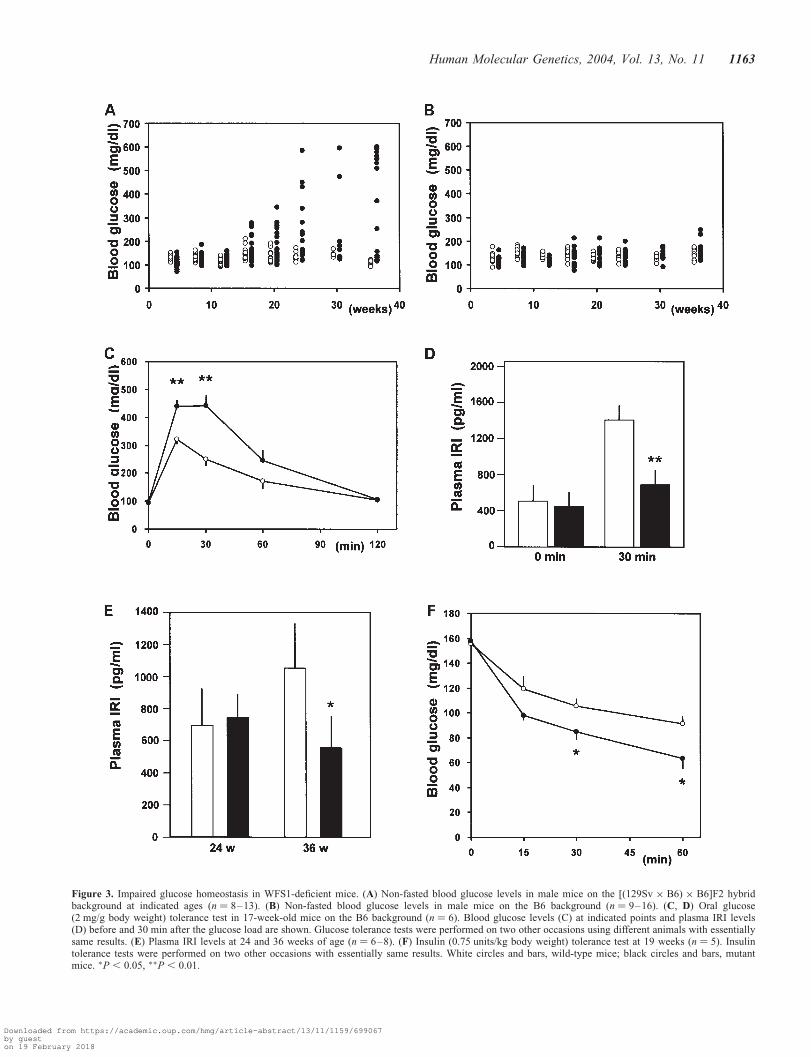
Figure 3. Impaired glucose homeostasis in WFS1-deficient mice. (A) Non-fasted blood glucose levels in male mice on the [(129Sv � B6) � B6]F2 hybridbackground at indicated ages (n ¼ 8–13). (B) Non-fasted blood glucose levels in male mice on the B6 background (n ¼ 9–16). (C, D) Oral glucose(2 mg/g body weight) tolerance test in 17-week-old mice on the B6 background (n ¼ 6). Blood glucose levels (C) at indicated points and plasma IRI levels(D) before and 30 min after the glucose load are shown. Glucose tolerance tests were performed on two other occasions using different animals with essentiallysame results. (E) Plasma IRI levels at 24 and 36 weeks of age (n ¼ 6–8). (F) Insulin (0.75 units/kg body weight) tolerance test at 19 weeks (n ¼ 5). Insulintolerance tests were performed on two other occasions with essentially same results. White circles and bars, wild-type mice; black circles and bars, mutantmice. �P , 0.05, ��P , 0.01.
Human Molecular Genetics, 2004, Vol. 13, No. 11 1163
Downloaded from https://academic.oup.com/hmg/article-abstract/13/11/1159/699067by gueston 19 February 2018

Figure 4. Impaired stimulus–secretion coupling in WFS1-deficient b-cells. (A) Islets from wild-type and mutant mice were infected with either AdCAGlacZ orAdCAGmWFS1. After 36 h, islets were subjected to western blot analyses using anti-WFS1 antibody. Lane 1, wild-type islets infected with AdCAGlacZ; lane 2,mutant islets infected with AdCAGlacZ; lane 3, wild-type islets infected with AdCAGmWFS1; lane 4, mutant islets infected with AdCAGmWFS1. Western blotexperiments were performed twice with similar results and one of them is shown. (B, C) Islets were challenged with 15 mM glucose (B) or 1 mM carbachol in thepresence of 2.5 mM glucose (C) for 1 h. Absolute insulin secretion in response to glucose was 3.11+ 0.34 and 2.03+ 0.26 ng/islet/h, respectively, for wild-typeand mutant islets infected with the control virus (AdCAGlacZ). Data are means+ SEM, n ¼ 5 experiments. White bars, wild-type islets infected with AdCA-lacZ; black bars, mutant islets with AdCAGlacZ; hatched bars, wild-type islets with AdCAGmWFS1; dotted bars, mutant islets with AdCAGmWFS1. (D, E)Intracellular Ca2þ responses to 15 mM glucose in wild-type (gray line and white bar) and WFS1-deficient (black line and black bar) b-cells. Representative tracesout of 21 wild-type and 24 WFS1-deficient b-cells from one experiment were shown in D. Areas under the curve during a 5 min period after the onset of Ca2þ
rises to glucose were summarized in E. Similar significant differences were observed in the other four experiments. �P , 0.05, ��P , 0.01.
1164 Human Molecular Genetics, 2004, Vol. 13, No. 11
Downloaded from https://academic.oup.com/hmg/article-abstract/13/11/1159/699067by gueston 19 February 2018

glucose- and carbachol-stimulated insulin secretion was restored(Fig. 4B and C), indicating reduced insulin secretion in isletsfrom mutant mice to be a direct consequence of absence ofnormal WFS1 function. Interestingly, glucose- and carbachol-stimulated insulin secretion from wild-type islets increased by41 and 53%, respectively, with overexpression of the WFS1protein, suggesting involvement of the WFS1 protein in stimu-lus–secretion coupling for insulin exocytosis (Fig. 4B and C).
To gain insight into the mechanisms of impaired insulinsecretion in islets from mutant mice, intracellular calciumdynamics were then studied in single b-cells challenged withglucose. The glucose-stimulated rise in the cytosolic Ca2þ
response was reduced by 36% in WFS1-deficient b-cellswhen compared with that in wild-type b-cells (Fig. 4D and E).
Progressive b-cell loss in mutant mice
We then focused on aspects of insulin production, deterio-ration of which could be a cause of impaired glucose homeo-stasis in mice with disruption of the wfs1 gene. There were nodifferences in pancreatic weight between wild-type and mutantmice (data not shown). Whole-pancreas insulin content wasalready decreased at 2 weeks, the earliest age studied, anddropped further with age (Fig. 5A). Immunohistochemicalstudies (Fig. 5B–E) showed the number of insulin-positivecells to be reduced at 36 weeks in the mutant mouse pancreas(Fig. 5E). Morphometric analysis demonstrated a markedreduction in the insulin-positive area per pancreatic area inmutant mice when compared with wild-type mice (Fig. 5H),indicating the decrease in insulin content to be due to lossof islet b-cells. These features were more prominent in themutant mouse pancreas on the [(129Sv � B6) � B6]F2 back-ground, which was associated with overt diabetes (Fig. 5F andG). In contrast to the b-cell changes, glucagon-positive cellswere increased and scattered throughout WFS1-deficientislets (Fig. 5E and G). Indeed, the pancreatic glucagoncontent in mutant mice at 36 weeks of age on the B6 back-ground was 2.4-fold higher than that in wild-type mice[12.3+ 1.8 ng/mg (n ¼ 4) versus 5.2+ 0.7 (n ¼ 4),P ¼ 0.0296].
Increased susceptibility of WFS1-deficientislets to apoptosis
To study whether the observed b-cell loss was due toincreased apoptosis, we conducted an extensive search forapoptotic b-cells in pancreatic sections. However, TUNELor activated-caspase 3-positive cells were sparse withinislets in pancreatic sections from both mutant and wild-typeanimals (data not shown). Therefore, we turned to in vitrostudies, and examined whether WFS1-deficient islet cells aremore susceptible to apoptotic insults. For this purpose, apop-totic DNA fragmentation was studied in isolated islets bythe ligation-mediated PCR (LM-PCR) method. When isletswere cultured for 3 days in RPMI media with 5 or 25 mM
glucose, ladder formation was increased at 25 mM glucose inboth wild-type and mutant islets when compared with that at5 mM glucose, indicating that apoptotic cell death may havebeen induced by glucose toxicity (Fig. 6A). Importantly, at25 mM glucose, islets from mutant mice showed more DNA
fragment formation than wild-type islets (1.7+ 0.3-fold,n ¼ 5), while no significant differences were observed at5 mM glucose. Since recent studies have suggested so-calledER-stress to be an important mediator of apoptosis inb-cells (14,15), DNA fragmentation was studied after treat-ment with two different ER-stress inducers (18), tunicamycin(2 mg/ml) and thapsigargin (2 mM). DNA fragmentation at5 mM glucose was significantly increased, by 2.2+ 0.4-foldand 2.4+ 0.4-fold after tunicamycin (Fig. 6B) and thapsigargin(Fig. 6C) treatments, respectively, in WFS1-deficient isletswhen compared with wild-type islets. In contrast, there wereno differences in DNA fragmentation after combined tumornecrosis factor-a and interferon-g treatment (Fig. 6D),which triggers apoptosis through a signaling pathway differentfrom that originating in the ER.
DISCUSSION
We generated mice with a disrupted wfs1 gene. Although thediabetic phenotype was milder than that seen clinically inWolfram syndrome (1), the progressive b-cell loss andimpaired glucose homeostasis observed in these mice areessentially consistent with findings in patients (1,17). Thus,the mutant mice are indeed a model of Wolfram syndrome.The underlying anatomic condition of this syndrome has notbeen studied in great detail in humans, and the cellular basisfor the diabetic phenotype and associated neuro-psychiatricdisorders remains obscure. Creation of an animal model thatreflects aspects of the disease is thus an important first stepin understanding Wolfram syndrome.
The present data demonstrate that the pathophysiologicalbasis of diabetes in Wolfram syndrome is insufficient insulinsecretion due to progressive b-cell loss and impaired stimulus–secretion coupling in b-cells. Progressive b-cell loss has beenexpected from clinical observations of progressive deteriorationof insulin-requiring states in affected patients as well as theirpostmortem findings, i.e. selective b-cell loss with an increasein a-cells and preservation of d-cells (17). In contrast, impairedstimulus–secretion coupling in the b-cell, a quite unexpectedresult, was demonstrated for the first time in this study. Inaddition, we also showed for the first time that WFS1 proteinis expressed selectively in b-cells, but very little in a, d andF-cells, within the endocrine pancreas, suggesting that b-cellloss is a direct consequence of WFS1 deficiency.
The severity of the diabetic phenotype due to wfs1 gene dis-ruption was dependent on the mouse genetic background:.60% of mice on the [(129Sv � B6) � B6]F2 backgrounddeveloped overt diabetes, while mutant mice on the B6 back-ground had impaired glucose tolerance but not overt diabetes.Modifying effects of genetic background on glucose homeo-stasis have been reported previously in a number of mutantmice. An earlier pioneering study established that the B6 back-ground confers more diabetes resistance to db/db and ob/obmice (19). A diabetes-resistant phenotype has also beenreported in insulin receptor substrate (IRS)-2 knockout miceon the B6 background (20), while anti-sense glucokinase-mRNA expressing mice (21) and mice double heterozygousfor deletion of the insulin receptor and IRS-1 (22), on the sameB6 background, were reportedly diabetes prone. Therefore, the
Human Molecular Genetics, 2004, Vol. 13, No. 11 1165
Downloaded from https://academic.oup.com/hmg/article-abstract/13/11/1159/699067by gueston 19 February 2018

Figure 5. Progressive b-cell loss in mutant mice. (A) Insulin content extracted from whole pancreata of wild-type and mutant mice. Data represent percent ofinsulin content in wild-type littermates. Absolute insulin content in wild-type pancreata were 1367+ 103 ng/mg pancreas at 2 weeks, 268+ 18 (8 weeks),329+ 25 (24 weeks) and 372+ 33 (36 weeks), n ¼ 4–7. White bars, wild-type pancreata; black bars, WFS1-deficient pancreata. (B–G) Insulin (green)and glucagon (red) are stained in pancreatic sections from 8-week-old wild-type (B), mutant (C), 36-week-old wild-type (D) and mutant mice (E) on the B6background, and 24-week-old wild-type (F) and mutant (G) mice on the [(129Sv � B6) � B6]F2 background. Bars ¼ 10 mm. (H) Ratios of total insulin-positivearea per whole pancreatic area in pancreas from wild-type and mutant mice on the B6 background. n ¼ 4 animals for each group. �P , 0.05, ��P , 0.01.
1166 Human Molecular Genetics, 2004, Vol. 13, No. 11
Downloaded from https://academic.oup.com/hmg/article-abstract/13/11/1159/699067by gueston 19 February 2018

contribution of genetic background is apparently complex. Inany case, progressive b-cell loss was observed in mutant micein both [(129Sv � B6) � B6]F2 and B6 strains, independentof the mouse genetic background. It is not surprising thatmutant mice on the B6 background did not develop overt dia-betes. Overt diabetes was known to be induced when.90% ofthe pancreas was removed (23), while the insulin content ofmutant mouse pancreas at 36 weeks was decreased by 73%on the B6 background in this study.
The present data provide an intriguing clue that may help toelucidate WFS1 protein function. WFS1-deficient islets exhi-bited impaired insulin secretion in response to glucose andcarbachol, which was restored by re-expression of WFS1 protein.In addition, overexpression of WFS1 protein in wild-type islets
resulted in an increase in glucose- and carbachol-induced insulinsecretion. These data from islets with different WFS1 proteinlevels demonstrated this protein to be involved directly in theregulation of insulin secretion. Furthermore, impaired calciumresponses to glucose suggested that WFS1 protein is involvedin regulation of calcium homeostasis in the b-cell. This notionis supported by the recent report that expression of WFS1protein in Xenopus oocytes confers a novel cation channelactivity (24). The present data also provide insight into themechanism of b-cell loss in mice with a mutant wfs1 gene.Although we rarely detected apoptotic cells in pancreaticsections from mutant mice, apoptosis cannot be excluded as apossible mechanism of b-cell loss, since our failure couldpresumably be due to slow progression of apoptosis in vivo
Figure 6. Increased apoptosis susceptibility in islets from mutant mice. (A) Islets from wild-type and mutant mice were cultured for 3 days in 5 and 25 mM
glucose concentrations and DNA fragmentation was assessed by the LM-PCR method. (B–D) Islets from wild-type and mutant mice were treated with tunica-mycin (Tm; 2 mg/ml) (B), thapsigargin (Tg; 2 mM) (C) for 24 h or with the combination of tumor necrosis factor-a (TNFa; 500 units/ml) and interferon-g(IFNg; 100 units/ml) (D) for 48 h and DNA fragmentation was assessed by the LM-PCR method; n ¼ 4–6 experiments. �P , 0.05.
Human Molecular Genetics, 2004, Vol. 13, No. 11 1167
Downloaded from https://academic.oup.com/hmg/article-abstract/13/11/1159/699067by gueston 19 February 2018

and rapid clearance of cells undergoing apoptosis, as wassuggested recently in another animal model of diabetes (25).Increased apoptosis susceptibility in response to high glucoseand ER-stress inducers, demonstrated in isolated islets frommutant mice, is likely to contribute to b-cell loss. In contrast,the apoptosis induced by exposure to tumor necrosis factor-aand interferon-g, in which the ER-stress response is notinvolved, did not differ between wild-type and WFS1-deficientislets. Although the mechanism whereby high concentrationsof glucose induce apoptosis is not completely understood atpresent (26,27), increased insulin translation in perk 2/2 isletsindicates the ER-stress response or the unfolded protein responseto be operative in islets cultured with high concentrations ofglucose (28). Therefore, it is possible that increased DNA frag-mentation in WFS1-deficient islets at 25 mM glucose could alsobe attributable to increased susceptibility to ER-stress-inducedapoptosis. However, it remains to be clarified how WFS1deficiency renders b-cells more susceptible to apoptosis,especially to ER-stress-induced apoptosis.
Recent studies showing b-cell mass to be decreased inhuman type 2 diabetes, due to increased b-cell apoptosis (29),have attracted considerable attention to this potential patho-genic mechanism of type 2 diabetes development. Therefore,maintaining b-cell mass is an important strategy for preventingdiabetes as well as halting disease progression. Since the WFS1protein is likely to belong to a novel family, elucidating theWFS1 protein function could lead to establishment of newtreatments not only for Wolfram syndrome but also for morecommon forms of diabetes mellitus.
MATERIALS AND METHODS
Targeted disruption of the wfs1 gene
The wfs1 gene was cloned from a 129Sv mouse genomic DNAlibrary using its cDNA probe (3). A targeting vector was con-structed by inserting a neomycin-resistance gene at the SmaIsite in exon 2 of the wfs1 gene. The diphtheria toxin Achain expressing unit was inserted downstream (Fig. 2A).The wfs1 gene targeting vector was microinjected into129Sv embryonic stem cells. Homologous recombinationwas successful in two independent embryonic stem cell lines(lines 133 and 190). Positive chimeric male mice were thencrossed with female C57BL/6J (B6) mice to produce wfs1 het-erozygous mice. Initial analyses demonstrated essentially thesame phenotypes between the two lines, and therefore wehave analyzed line 133 mice. In order to analyze animalswith as homogenous a genetic background as possible, malewfs1 heterozygous mice were backcrossed with female B6mice for five successive generations. We also analyzed wfs1homozygous mice on the [(129Sv � B6) � B6]F2 hybridbackground. The mice were kept in standard, specific patho-gen-free conditions under a constant dark/light cycle. Allanimal experiments were approved by the local ethical com-mittee for animal research at the Tohoku University.
Physiological studies
Control animals were age-matched siblings. Blood glucose levelsin the non-fasting state were measured at 9:00–10:00 a.m. using
a GluTest blood glucose monitor (Sanwa Chemicals, Tokyo,Japan). Serum insulin levels were determined by radioimmuno-assay using a rat insulin RIA kit (Linco Research, St Charles,MO, USA). For oral glucose tolerance tests, animals after a 6 hfast were administered with 20% glucose solution (2 mg/g bodyweight) by gastric tubes. Whole-blood samples were collectedfrom the tail tip at the indicated time points. Insulin tolerancetests were performed after a 6 h fast by an intraperitonialinjection of human regular insulin (0.75 units/kg body weight).
Immunohistochemistry and morphometry
For brain sections, animals were anesthetized by ethylethel,and 4% formalin was perfused from the left ventricle. For pan-creatic sections, the animals were killed by cervical dislo-cation. Dissected pancreas pieces were fixed in 4% formalin.Formalin-fixed paraffin-embedded sections of pancreas werede-paraffinized and re-hydrated. For insulin and glucagonstaining, the sections were then incubated with a guinea piganti-insulin IgG (DAKO Japan, Kyoto, Japan) diluted 1 : 1000and a mouse anti-glucagon IgG (Sigma-Aldrich Japan, Tokyo,Japan) diluted 1 : 2000 for 1 h at room temperature. The anti-insulin and -glucagon primary antibodies were followed by a45 min incubation with a fluorescein isothiocyanate (FITC)-conjugated anti-guinea pig IgG and a Texas Red-conjugatedanti-mouse IgG (Jackson ImmunoResearch, West Grove,PA, USA). The antibody raised against the 290 amino acida-mWFS1-N was described previously (30). Pancreatic sec-tions incubated with the anti-WFS1 antibody were thenstained with an FITC-conjugated anti-rabbit IgG (JacksonImmunoResearch). Immunohistochemical analyses were per-formed, sacrificing at least four different animals for each con-dition. For measurements of b-cell area, more than 10pancreatic tissue sections per animal were randomly selected,stained with anti-insulin IgG and eosin. Pancreatic area andb-cell area were each estimated using the intensity threshold-ing function of the NIH Image software. Four animals wereanalyzed for each group.
Pancreatic insulin and glucagon content
Pancreases were suspended in cold acid ethanol and mincedby scissors, and left at 2208C for 48 h, with sonicationevery 24 h. Insulin content in the acid ethanol supernatantwas determined with a rat insulin RIA Kit (Linco Research).Glucagon content in the same extract was measured by aglucagon RIA kit (Linco Research).
Islet studies
Construction of a recombinant adenovirus expressing murineWFS1 protein. A recombinant adenovirus AdCAGmWFS1,bearing an EcoR I fragment of murine WFS1 cDNA, wasconstructed by the method described previously (31,32).AdCAGlacZ expressing b-galactosidase was used as a controladenovirus. Isolated islets were infected with the recombinantadenoviruses at 1.2 � 105 particles per islet in 1.0 ml mediumfor 60 min.
1168 Human Molecular Genetics, 2004, Vol. 13, No. 11
Downloaded from https://academic.oup.com/hmg/article-abstract/13/11/1159/699067by gueston 19 February 2018

Isolation and static incubation of islets. Islets isolated fromage-matched wild-type and mutant siblings at 14–17 weekswere isolated by retrograde injection of collagenase (Serva,Heidelberg, Germany) into the pancreatic duct according tostandard procedures. For secretion studies, batches of 10islets (triplicates for each condition) were kept in Krebs–Ringer-bicarbonate-HEPES buffer [KRBH; 140 mM NaCl,3.6 mM KCl, 0.5 mM NaH2PO4, 0.5 mM MgSO4, 1.5 mM
CaCl2, 2 mM NaHCO3, 10 mM HEPES (pH 7.4)] containing0.1% BSA and stimulators indicated. Islet insulin contentwas measured following extraction by acid ethanol. Insulinwas detected by radioimmunoassay.
Single cell Ca2þ measurement. Islets isolated from mice at12–16 weeks were dispersed, plated on glass-bottomeddishes and cultured for 3 days before measurement. b-Cellswere identified by adenovirus-mediated expression of greenfluorescent protein driven by the insulin promoter (33). Weperformed experiments without adenovirus-mediatedexpression of green fluorescent protein, identifying b-cellswith immunostaining after perfusion, and observed similarresults (data not shown). Cells were incubated with 1 mM
Fura 2-AM (Dojindo, Kumamoto, Japan) for 30 min, perfusedwith KRBH and excited at 340 and 380 nm. A cooled CCDcamera (Hamamatsu Photonics, Shizuoka, Japan) mounted ona microscope (Leica Microsystems, Heerbrugg, Switzerland)was used to capture fluorescence images. Ca2þ rises werecompared by calculating areas between Ca2þ curves andbaselines for the 300 s after the onset of Ca2þ rises.
LM-PCR amplification of DNA fragments. Groups of 50 isletsisolated from mice at 15–17 weeks of age were culturedfor 3 days in RPMI with different glucose concentrations.In another series of experiments, groups of 50 islets weretreated with 2 mg/ml tunicamycin (Sigma-Aldrich Japan),2 mM thapsigargin (Alamone Labs, Jerusalem, Israel) or acombination of interferon-g (100 units/ml; PeproTech,London, UK) and tumor necrosis factor-a (500 units/ml;PeproTech). Genomic DNA was isolated from treated isletsusing the DNeasy kit (Qiagen-Japan, Tokyo, Japan). ThePicoGreenw dsDNA quantitation kit (Molecular Probes,Eugene, OR, USA) was used to determine the DNA concen-trations. 200 ng of the genomic DNA was ligated with anadaptor, which has been generated by annealing two syntheticoligonucleotides 50-AGCACTCTCGAGCCTCT CACCGCA-30
and 50-TGCGGTGAGAGG-30. A portion of ligation mixture(30%) was used for the PCR amplification with a primer50-AGCACTCTCGAGCCTCTCACCGCA-30. The resultingPCR products were run on 1.2% agarose gels. Intensities ofladders between 500 and 1000 bp were analyzed using theScion Image software. In order to compare data from separategels, band intensity was normalized to the average laddering ofthe control islets at 5 mM glucose.
Statistical analyses
Data are presented as mean+ SE, unless otherwise noted.Differences between wild-type and mutant animals wereassessed by Student’s t-test.
ACKNOWLEDGEMENTS
We thank Professor H. Takeshima, Dr Y. Ohwada and ProfessorT. Itoh, Tohoku University, for their help in Ca2þ imaging andimmunohistochemical analyses. We are also grateful toN. Nishino, T. Wadatsu and N. Miyazawa, Otsuka GENResearch Institute, for their help in generation of WFS1-deficient mice. Y. Takahashi is gratefully acknowledged forher excellent technical assistance. This study was supportedby Grants in Aid for Scientific Research (13204062) to Y.O.from the Ministry of Education, Science, Sports and Cultureof Japan.
REFERENCES
1. Wolfram, D.J. and Wagener, H.P. (1938) Diabetes mellitus and simpleoptic atrophy among siblings: report on four cases. Mayo Clinic Proc.,13, 715–718.
2. Swift, M. and Swift, R.G. (2001) Psychiatric disorders and mutationsat the Wolfram syndrome locus. Biol. Psychiatry, 47, 787–793.
3. Inoue, H., Tanizawa, Y., Wasson, J., Behn, P., Kalidas, K.,Bernal-Mizrachi, E., Mueckler, M., Marshall, H., Donis-Keller, H.,Crock, P. et al. (1998) A gene encoding a transmembrane protein ismutated in patients with diabetes mellitus and optic atrophy(Wolfram syndrome). Nat. Genet., 20, 143–148.
4. Strom, T.M., Hoetnagael, K., Hofmann, S., Gekeler, F., Scharfe, C.,Rabl, W., Gerbitz, K.D. and Meitinger, T. (1998) Diabetes insipidus,diabetes mellitus, optic atrophy and deafness (DIDMOAD) caused bymutations in a novel gene (wilframin) coding for a predictedtransmembrane protein. Hum. Mol. Genet., 7, 2021–2028.
5. Cryns, K., Sivakumaran, T.A., Van den Ouweland, J.M., Pennings, R.J.,Cremers, C.W., Flothmann, K., Young, T.L., Smith, R.J., Lesperance,M.M. and Van Camp, G. (2003) Mutational spectrum of the WFS1gene in Wolfram syndrome, nonsyndromic hearing impairment,diabetes mellitus, and psychiatric disease. Hum. Mut., 22, 275–287.
6. Bespalova, I.N., Van Camp, G., Bom, S.J., Brown, D.J., Cryns, K.,DeWan, A.T., Erson, A.E., Flothmann, K., Kunst, H.P., Kurnool, P. et al.(2001) Mutations in the Wolfram syndrome 1 gene (WFS1) are acommon cause of low frequency sensorineural hearing loss. Hum. Mol.Genet., 15, 2501–2508.
7. Young, T.L., Ives, E., Lynch, E., Person, R., Snook, S., MacLaren, L.,Cater, T., Griffin, A., Fernandez, B., Lee, M.K. et al. (2001)Non-syndromic progressive hearing loss DFNA38 is caused byheterozygous missense mutation in the Wolfram syndrome geneWFS1. Hum. Mol. Genet., 15, 2509–2514.
8. Ohta, T., Koizumi, A., Kayo, T., Shoji, Y., Watanabe, A, Monoh, K.,Higashi, K., Ito, S., Ogawa, O., Wada, Y. et al. (1998) Evidence of anincreased risk of hearing loss in heterozygous carriers in a Wolframsyndrome family. Hum. Genet., 103, 470–474.
9. Minton, J.A., Hattersley, A.T., Owen, K., McCarthy, M.I., Walker, M.,Latif, F., Barrett, T. and Frayling, T.M. (2002) Association studies ofgenetic variation in the WFS1 gene and type 2 diabetes in U.K.populations. Diabetes, 51, 1287–1290.
10. Awata, T., Inoue, K., Kurihara, S., Ohkubo, T., Inoue, I., Abe, T.,Takino, H., Kanazawa, Y. and Katayama, S. (2000) Missense variationsof the gene responsible for Wolfram syndrome (WFS1/wolframin) inJapanese: possible contribution of the Arg456His mutation to type 1diabetes as a nonautoimmune genetic basis. Biochem. Biophys. Res.Commun., 268, 612–616.
11. Sequeira, A. Kim, C., Seguin, M., Lesage, A., Chawky, N., Desautels, A.,Tousignant, M., Vanier, C., Lipp, O., Benkelfat, C. et al. (2003)Wolfram syndrome and suicide: evidence for a role of WFS1 in suicidaland impulsive behavior. Am. J. Mol. Genet., 119B, 108–113.
12. Takeda, K., Inoue, H., Tanizawa, Y., Matsuzaki, Y., Oba, J., Watanabe, Y.,Shinoda, K. and Oka, Y. (2001) WFS1 (Wolfram syndrome 1) geneproduct: predominant subcellular localization to endoplasmicreticulum in cultured cells and neuronal expression in rat brain.Hum. Mol. Genet., 10, 477–484.
13. Hofmann, S., Philbrook, C., Gerbitz, K.D. and Bauer, M.F. (2003) Wolframsyndrome: structural and functional analyses of mutant and wild-typewolframin, the WFS1 gene product. Hum. Mol. Genet., 12, 2003–2012.
Human Molecular Genetics, 2004, Vol. 13, No. 11 1169
Downloaded from https://academic.oup.com/hmg/article-abstract/13/11/1159/699067by gueston 19 February 2018

14. Kaufmann, R. (2002) Orchestrating the unfolded protein response inhealth and disease. J. Clin. Invest., 110, 1389–1398.
15. Harding, H.P. and Ron, D. (2002) Endoplasmic reticulum stress andthe development of diabetes. Diabetes, 51 (Suppl. 3), S455–S461.
16. Rando, T.A., Horton, J.C. and Layzer, R.B. (1992) Wolfram syndrome:evidence of a diffuse neurodegenerative disease by magnetic resonanceimaging. Neurology, 42, 1220–1224.
17. Karasik, A., O’Hara, C., Srikanta, S., Swift, M., Soeldner, J.S., Kahn, C.R.and Herskowitz, R.D. (1989) Genetically programmed selective isletbeta-cell loss in diabetic subjects with Wolfram’s syndrome.Diabetes Care, 12, 135–138.
18. Ferri, K.F. and Koemer, G. (2001) Organelle-specific initiation of celldeath pathways. Nat. Cell Biol., 3, E255–E263.
19. Coleman, D.L. (1982) Diabetes–obesity syndromes in mice. Diabetes, 31
(Suppl. 2), 1–6.
20. Terauchi, Y., Matsui, J., Suzuki, R., Kubota, N., Komeda, K., Aizawa, S.,Eto, K., Kimura, S., Nagai, R., Tobe, K. et al. (2003) Impact ofgenetic background and ablation of insulin receptor substrate (IRS)-3on IRS-2 knock-out mice. J. Biol. Chem., 278, 14284–14290.
21. Ishihara, H., Tashiro, F., Ikuta, K., Asano, T., Katagiri, H., Inukai, K.,Kikuchi, M., Yazaki, Y., Oka, Y. and Miyazaki, J. (1995) Inhibitionof pancreatic beta-cell glucokinase by antisense RNA expression intransgenic mice: mouse strain-dependent alteration of glucosetolerance. FEBS Lett., 371, 329–332.
22. Kulkarni, R.N., Almind, K., Goren, H.J., Winnay, J.N., Ueki, K.,Okada, T. and Kahn, C.R. (2003) Impact of genetic background ondevelopment of hyperinsulinemia and diabetes in insulin receptor/insulinreceptor substrate-1 double heterozygous mice. Diabetes, 52, 1528–1534.
23. Wier, G.C., Bonner-Wier, S. and Leahy, J.L. (1990) Islet mass andfunction in diabetes and transplantation. Diabetes, 39, 401–405.
24. Osman, A.A., Saito, M., Makepeace, C, Permutt, M.A., Schlesinger, P.and Mueckler, M. (2003) Wolframin expression induces novel ionchannel activity in endoplasmic reticulum membranes and increasesintracellular calcium. J. Biol. Chem., 278, 52755–52762.
25. Reddy, S. Bradley, J., Ginn, S., Pathipati, P. and Ross, J.M. (2003)Immunohistochemical study of caspase-3-expressing cells within thepancreas of non-obese diabetic mice during cyclophosphamide-accelerated diabetes. Histochem. Cell Biol., 119, 451–461.
26. Donath, M.Y., Gross, D.J., Cerasi, E. and Kaiser, N. (1999)Hyperglycemia-induced beta-cell apoptosis in pancreatic islets ofPsammomys obesus during development of diabetes. Diabetes,48, 738–744.
27. Maedler, K., Sergeev, P., Ris, F., Oberholzer, J., Joller-Jemelka, H.I.,Spinas, G.A., Kaiser, N., Halban, P.A. and Donath, M.Y. (2002)Glucose-induced beta cell production of IL-1beta contributes toglucotoxicity in human pancreatic islets. J. Clin. Invest., 110, 851–860.
28. Harding, H.P., Zeng, H., Xhang, Y., Jungries, R., Chung, P., Plesken, H.,Sabatini, D.D. and Ron, D. (2001) Diabetes mellitus and exocrinepancreatic dysfunction in Perk 2/2 mice reveals a role for translationalcontrol in secretory cell survival. Mol. Cell, 7, 1153–1163.
29. Butler, A.E., Janson, J., Bonner-Weir, S., Ritzel, R., Rizza, R.A. andButler, P.C. (2003) b-Cell deficit and increased b-cell apoptosis inhumans with type 2 diabetes. Diabetes, 52, 102–110.
30. Cryns, K., Thys, S., van Laer, L., Oka, Y., Pfister, M., van Nassauw, L.,Smith, R.J.H., Timmermans, J.P. and Van Camp, G. (2003) The WFS1gene, responsible for low frequent sensorineural hearing loss andWolfram syndrome, is expressed in a variety of inner ear cells.Histochem. Cell Biol., 119, 247–256.
31. Miyake, S., Makimura, M., Kanegae, Y., Harada, S., Sato, Y.,Takamori, K., Tokuda, C. and Saito, I. (1996) Efficient generation ofrecombinant adenoviruses using adenovirus DNA–terminal proteincomplex and a cosmid bearing the full-length virus genome. Proc.Natl Acad. Sci. USA, 93, 1320–1324.
32. Niwa, H., Yamamura, K. and Miyazaki, J. (1991) Efficient selectionfor high-expression transfectants with a novel eukaryotic vector.Gene, 108, 193–199.
33. Ishihara, H., Maechler, P., Gjinovci, A., Herrera, P.-L. and Wollheim,C.B. (2003) Islet b-cell secretion determines glucagon secretionfrom the neighboring a-cells. Nat. Cell Biol., 5, 330–335.
1170 Human Molecular Genetics, 2004, Vol. 13, No. 11
Downloaded from https://academic.oup.com/hmg/article-abstract/13/11/1159/699067by gueston 19 February 2018