Embed Size (px)
Citation preview
Andreia Filipa Dimas Ferreira
Licenciada em Biologia Celular e Molecular
Dissecting neuronal dysfunction and
microglia/motoneurons cross-talk in ALS: an
immunofluorescence directed study
Dissertação para obtenção do Grau de Mestre em
Genética Molecular e Biomedicina
Orientador: Dora Maria Tuna de Oliveira Brites,
Investigadora Coordenadora e Professora Catedrática Convidada
Faculdade de Farmácia, Universidade de Lisboa
Co-orientador: Ana Rita Mendonça Vaz,
PhD
Faculdade de Farmácia, Universidade de Lisboa
Júri: Presidente: Doutora Margarida Casal Ribeiro Castro-Caldas Braga Arguente: Doutora Paula Pousinha Vogal: Doutora Ana Rita Mendonça Vaz
Dezembro de 2013

ii
iii
Dissecting neuronal dysfunction and microglia/motoneurons cross-talk in ALS: an
immunofluorescence directed study
Copyright Andreia Filipa Dimas Ferreira, FCT/UNL, UNL
A Faculdade de Ciências e Tecnologia e a Universidade Nova de Lisboa têm o direito, perpétuo
e sem limites geográficos, de arquivar e publicar esta dissertação através de exemplares
impressos reproduzidos em papel ou de forma digital, ou por qualquer outro meio conhecido ou
que venha a ser inventado, e de a divulgar através de repositórios científicos e de admitir a sua
cópia e distribuição com objectivos educacionais ou de investigação, não comerciais, desde
que seja dado crédito ao autor e editor.

iv
v
Part of the results discussed in this thesis was presented in the following meetings:
Ferreira A, Barbosa M, Cunha C, Marçal AM, Vaz AR, Brites D. Modulation by Glycoursodeoxycholic
Acid on an organotypic-based model of ALS. 5th iMed.UL Postgraduate Students Meeting, 18 July
2013, Lisbon. [Abstract and Poster – see Annex 1(A)]
Barbosa M, Ferreira A, Vaz AR, Brites D. Role of microglia-motor neurons cross-talk in ALS modelling.
5th iMed.UL Postgraduate Students Meeting, 18 July 2013, Lisbon. [Abstract and Poster - see Annex
1 (B)]
Vaz AR, Barbosa M, Ferreira A, Cunha JC, Brites D. Exploring the role of inflammation to motor
neuron degeneration in ALS. XIII Reunião da Sociedade Portuguesa de Neurociências, 30 May – 1
June 2013, Coimbra. [Abstract, Poster and Fire talk communication]
Vaz AR, Ferreira A, Barbosa M, Cunha C, Brites D. Exploring anti-inflammatory strategies on motor
neuron degeneration in ALS. 13th ESNI Course, Porto, 3-6 July, 2013 [Abstract].
Vaz AR, Barbosa M, Ferreira A, Cunha JC, Brites D. Role of inflammatory modulators in ALS models.
Champalimaud NeuroScience Symposium. Lisboa, 25-28 September, 2013. [Abstract and Poster]
Some of the results described in this Master Thesis were obtained in association with Marta Barbosa,
a Master Student from the same group.
This work was supported by FEDER (COMPETE Programme) and by National funds (Fundação para
a Ciência e a Tecnologia – FCT, Portugal) with the projects PTDC/SAU-FAR/118787/2010 to D.B. and
PEst-OE/SAU/UI4013/2011 and 2012 to iMed.UL.

vi
vii
Aos meus pais…

viii
ix
Agradecimentos
Quero começar por agradecer à Professora Doutora Dora Brites, por me ter aberto as portas do
mundo da investigação, ao receber-me no seu grupo “Neuron Glia Biology in Health and Disease” e
pela dedicação e exigência com que sempre seguiu este projecto. Obrigada por me mostrar que,
também na ciência, o espírito criativo tem uma enorme importância. Obrigada também pelos
“desafios” que me foi colocando ao longo do tempo pois aprendi bastante com eles. Obrigada pela
verdade que emprega em tudo o que faz e pela frontalidade com que sempre nos mostrou o mundo lá
fora. Este ano ganhei um novo modelo de força e dedicação. Espero ter estado à altura das
oportunidades que me ofereceu, pois para mim foi um privilégio integrar esta equipa. Muito obrigada!
À Doutora Ana Rita Vaz, pela enorme capacidade de se dividir entre mil tarefas, sem nunca me
deixar desamparada. Pelo carinho e conforto que sempre incluiu nas suas explicações. Pelos
inesgotáveis “Não faz mal nenhum! Tenho a certeza que este é um erro que nunca mais cometes!”.
Por estar do lado das “suas meninas”, aconteça o que acontecer, além até das suas obrigações
enquanto orientadora. Espero que no futuro, o teu sucesso se torne proporcional à tua garra! Um
muito obrigada!
Ao Doutor José Paulo Sampaio, enquanto Coordenador do Mestrado em Genética Molecular e
Biomedicina, por procurar sempre o melhor para os seus alunos e por tanto se interessar em
melhorar este grande mestrado.
À Doutora Isabel Sá Nogueira, um enorme agradecimento que remonta aos meus tempos de
licenciatura, por se ter tornado um modelo, enquanto Investigadora e enquanto Mulher. Pela garra,
determinação e justiça que emprega em tudo o que faz, um Muito Obrigada!
Gostaria também de agradecer à Doutora Margarida Castro Caldas, por me ter feito descobrir a
minha paixão pela Neurociência. Á Doutora Alexandra Brito pela enorme simpatia com que me
recebeu e pelos bons conselhos que deu ao longo deste trabalho. Ao Professor Rui Silva pela
disponibilidade que sempre mostrou em recordar as matérias de neurobiologia.
À Doutora Júlia Costa e à Doutora Teresa Pais, expresso o meu agradecimento por gentilmente
terem cedido as linhas celulares NSC-34 e N9, respectivamente. Sem esta colaboração, a presente
dissertação não seria possível. Os meus votos de um enorme sucesso profissional.
À Inês Palmela… Desde o início, decidi que tinhas que ter um parágrafo só teu nos meus
agradecimentos! Queria que soubesses que te admiro imenso, pela tua enorme capacidade de
organização e por ainda assim, teres tempo para todas as milhentas coisas a que te propões, sendo
sempre a pessoa sorridente e bem-disposta a que nos habituaste! Sempre que te vejo no laboratório
penso que serias o tipo de pessoa com capacidade para ter qualquer profissão que quisesses e por
isso, agradeço-te que tenhas escolhido esta, dando-me o privilégio de aprender contigo! Obrigada!
À Filipa Cardoso, porque foste tu que me “meteste o bichinho” para vir para o grupo, com as tuas
aulas excelentes! E porque, uma vez no grupo, foste tu a primeira a dar-me a mão! Porque és das
pessoas mais doces de sempre! Desejo-te um futuro muito feliz!
x
À Carolina, a minha querida “Mestrinha” pela simpatia e companheirismo que exalas por todos
os poros! Acho que, sem ti, este grupo perdia muita da sua magia!!! Agradeço-te imenso os
momentos de alegria que trouxeste durante este aninho, assim como a eterna disponibilidade para
ajudar em tudo o que foi necessário! Obrigada por me introduzires ao negro mundo da contagem de
células, hehe…Sem represálias futuras, juro! Espero que a vida tenha um plano super risonho para
ti!!!
À Cátia pela eterna paciência com todas as minhas dúvidas e dilemas. Obrigada por teres tido
sempre um tempinho para me ajudar quando precisei (e por afinal, não teres cobrado €, hihi). Espero
que consigas a tua bolsa, porque tenho a certeza que vais saber aproveitá-la ao máximo,
especialmente se te derem um microscópio para a mão!!! :P
À Marta, por ser a “Ex.mª amiga” que me acompanha desde 1814 (ou quase)! Obrigada pela
entreajuda que tem existido desde então e que tão útil tem sido para o meu crescimento enquanto
“quasi-cientista” e enquanto pessoa. Obrigada pela inicial partilha de bancada (que foi quase um
prolongamento das nossas aulitas na faculdade) e pelas nossas eternas maluqueiras que sempre
alegraram os meus dias, mas também pelos nossos momentos de partilha depressiva (remontando a
LBCM). Obrigada pela companhia nas noites Champalimaud, pelas nossas saídas-relâmpago, por
fazeres de minha mãezinha de vez em quando, pelas nossas eternas peregrinações calóricas e por
me ensinares o conceito de “Experiência independente” (significou muito para mim, muahah). Um
grande beijinho e votos de muito sucesso, pois considero-te uma pessoa muito dedicada e
competente!
Ao Gonçalo Lidónio, com um beijinho muito grande e votos de um sucesso enorme! Obrigada
pela companhia e amizade ao longo destes anitos todos… Embora saibas que te odeio, mas isso não
importa porque preferes sempre os amigos de Santarém :P Acho que mais do que eu, quem tem
muito a agradecer-te é o dono da máquina dos bolos do CPM (ouvi dizer que já comprou uma
penthouse em Miami com o lucro que fez desde que chegaste)… Agora (mais) a sério: És das
pessoas mais aplicadas que conheço e é por isso que desejo sinceramente que tenhas pela frente
um futuro brilhante! Não te esqueças que o mundo é teu, tens apenas que decidir o que fazer com ele
;) Quero estar sempre em condição de aplaudir os teus sucessos e cá estarei também para aquele
abraço de quando o dia corre longe do planeado! E afinal, parece que não te odeio… Acho que até
gosto de ti, “pulhinha”!
À Vera, muito obrigada por teres baptizado o Sr. Amén…e acho que não tenho mais nada a
agradecer-te… Hihi, estou a brincar :P Obrigada por seres a pessoa espectacular que és! Por estares
sempre pronta a ajudar e por me ter identificado contigo desde o início. Espero que tenhas um
espacinho por aí para manter a nossa amizade no matter what :P Obrigada pelos nossos risos, pelos
voos matinais quando as escadas pareciam demasiado enfadonhas, pela patinagem no gelo (para a
próxima quero uma foquinha laranja), pelos projécteis de café e pelos recadinhos nos post-its
sanitários! Para ti, um beijinho gigantesco, daqueles que espalmam bochechas!
xi
À Gisela, por todos os dias me ensinares sobre a extrema necessidade de comer gelado após o
almoço (a OMS devia ouvir os teus conselhos) :P Obrigada por me puxares para um cafezinho
sempre que tenho a cabeça apilhada de trabalho e pela tua facilidade em ajudar a tornar tudo mais
simples! Espero que esta nova etapa da tua vida seja muito sorridente e te traga tudo o que mais
desejares Um grande beijinho*
Ao Andrew Durães, Andreia Nora, Filipe Torres, Gonçalo Silva, Inês Oliveira, João Maurício, Luís
Monteiro, Mariana Pinhão, Ricardo Almeida, Ruben Nunes e Sara Salazar: Quero agradecer a cada
um de vós, o carinho que sempre me deram ao longo destes anos. Cada um contribuiu, à sua
maneira, para que os momentos felizes fossem realmente felizes e para que os momentos tristes
fossem oportunidades para me erguer novamente e lutar com mais dedicação. Assim, a todos vós eu
agradeço por acreditarem em mim e pelo apoio e amizade. Cada um à sua maneira, vocês são
especiais e espero ter-vos para sempre “pertinho” de mim… Um abraço enorme a todos!
A todos os companheiros da Cave, um enorme beijinho por a tornarem num lugar
verdadeiramente agradável (apesar da janela ser tão pequenita). Tenho imenso a agradecer-vos… A
forma como me receberam e me fizeram sentir parte do grande grupo que é o iMed.UL, tudo o que
me ensinaram, o carinho com que sempre acompanharam cada passo, a constante disponibilidade
em ajudar no que quer que seja, a preocupação que revelam quando o dia não corre tão bem como
gostaríamos. Sei que estou no início da minha carreira mas é com muita certeza que vos digo que
gostaria que todos os meus grupos de trabalho ao longo da vida fossem como vocês!
Sei que cheguei ao CPM num ano economicamente péssimo, mas queria deixar aqui a minha
admiração, porque convosco aprendi que é possível fazer imenso com muito pouco. Vocês são
grandes!
À Raríssimas - Associação Nacional de Deficiências Mentais e Raras e em especial à Joana
Neves, muito obrigada pela simpatia e pelo interesse e entusiasmo em esclarecer as minhas dúvidas.
Um grande bem-haja pelo vosso trabalho.
Ao Professor Doutor Mamede Carvalho pela prontidão e simpatia com que esclareceu as minhas
dúvidas.
Às minhas meninas do Mestrado: Graça, Cátia, Clara, Juliana, Cristiana, Sara, Soraia e Diana…
Graças a vocês, o ano 2011/2012 foi espectacular! Muito obrigada por reformarem o ambiente da
FCT, que bem precisava! E muito obrigada por cada momento em que nos encontramos se tornar
numa festa! Espero que sejam todas muito felizes e que, independentemente de onde a vida vos
levar na próxima etapa, não se esqueçam de mim! Gosto muito de vocês (e de Nutella)
Aos meus queridos colegas e amigos do Pavilhão do Conhecimento, muito obrigada por darem
cor aos meus fins-de-semana. Pelo interesse que tiveram desde o ínicio neste trabalho e pelas
sugestões e ofertas de ajuda que recebi. Sem dúvida que o vosso carinho e companheirismo
xii
alegraram muito esta etapa da minha vida e trouxeram bastante ímpeto a esta pesquisa! E agora que
a tese está entregue, prometo estar mais presente nas vossas saídas ;)
Ao avô António, um muito obrigado por sempre me desejares o melhor. À avó Nanda, que eu
sempre ouvi dizer “Não vou estar cá para ver, mas um dia a minha netinha vai ser doutora”, ainda
falta imenso para essa etapa, mas certamente ficarias orgulhosa de saber que me juntei àqueles que
ocupam a sua vida a estudar doenças neurodegenerativas, como aquela que te levou. À avó Tila, um
grande beijinho e obrigada pela dedicação e orgulho com que acompanhas cada uma das minhas
etapas desde o meu primeiro dia!
Ao meu mano Tim Porque apesar de seres tão pequenito, trouxeste muita alegria e boa
disposição no decorrer deste ano trabalhoso. Apesar de perceberes pouco mais do que a palavra
“Ba-Ta-Ta”, fica aqui registada a promessa de que poderás sempre contar com a mana Um grande
beijinho!
Ao Vitor e Ana, um muito obrigada pela preocupação que mostraram por mim e pelo meu
trabalho ao longo deste tempo. Desejo-vos tudo de bom. Beijinho
Aos meus pais: À ti mãe, um obrigado muito sincero, por todos os momentos que partilhamos,
por todo o carinho e amizade, por todas as palavras e dedicação! Por estares sempre pronta a deixar
tudo e vir a correr quando eu preciso (é impossível esquecer certos gestos tão teus)… Por seres a
mãe mais vaidosa e babada de sempre, e agora a dobrar Obrigada por seres parte integrante de
cada uma das minhas vitórias… Por seres simplesmente tu, Mãe!
A ti pai, por partilhares comigo, desde sempre, o teu amor pela ciência. Por teres sido o primeiro
dos meus mestres e aquele que eu mais respeito! Por fazeres crescer em mim a dúvida e a
curiosidade, a vontade de saber mais e mais. Pela tua enorme capacidade de sonhar, que nos leva
aos dois a conversas que duram horas a fio e com as quais eu tanto aprendo. Obrigada por me
ensinares a erguer sempre que a vida me deita abaixo, por teres semeado em mim tanto de ti! Quero
que saibas que sairei vencedora desta vida, se um dia tiver um filho que me admire e tenha por mim
1/10 da estima que eu tenho por ti! Obrigada!
Mãe e Pai, a vocês dedico esta tese, assim como todas as outras “vitórias” da minha vida (nas
quais vocês desempenham sempre um papel crucial). Obrigada por tudo! Amo-vos imenso!
Obrigada a todas as outras pessoas que acompanharam o meu trajecto académico e/ou pessoal
e permitiram, de alguma forma, que este trabalho fosse possível.
xiii
Resumo
A esclerose Lateral Amiotrófica (ELA) é uma doença neurodegenerativa fatal, afectando de
0.4 a 1.8/100,000 habitantes. Caracteriza-se pela degeneração dos neurónios motores (NM), mas
também afeta a microglia. Contudo, a contribuição desta célula na doença não está esclarecida.
Pretendeu-se: (i) explorar os processos de disfunção dos NM na ELA, nomeadamente a
dinâmica mitocondrial (fusão/fissão) e transporte axonal (anterógrado/retrógrado), assim como
mecanismos de morte celular; (ii) avaliar a contribuição da microglia pelo uso de culturas mistas de
NM-microglia; iii) implementar o modelo de culturas organotípicas de medula espinhal (ME) de
ratinhos transgénicos para ELA, para avaliar efeitos neuroprotectores pelo ácido glico-ursodeoxicólico
(AGUDC).
Utilizaram-se: (i) células NSC-34, uma linha celular de NM, transfectada com superóxido
dismutase humana (hSOD1) normal (WT) ou com mutação G93A; (ii) células N9, uma linha celular
microglial, em cultura mista com NSC-34 (hSOD1WT ou hSOD1G93A); (iii) culturas organotípicas de
segmentos lombares de ME de murganhos com 7 dias (SJL-wt) ou transgénicos, contendo a SOD1
humana mutada (TgSOD1-G93A), incubados ou não com AGUDC aos 10 dias-in-vitro (DIV).
Utilizaram-se técnicas de imunocitoquímica, citometria de fluxo e ensaio fluorimétrico/colorimétrico
para o ATP e óxido nítrico (NO), respectivamente.
A viabilidade das células NSC-34/hSOD1G93A e da marcação para a βIII-tubulina diminuiu com
a diferenciação. A apoptose (estádios iniciais) e a libertação de NO (P<0.01) e ATP (P<0.05)
aumentou. Verificou-se disfunção da dinâmica mitocondrial por maior fissão (P<0.05) e menor fusão
(P<0.01), diminuindo o transporte axonal retrógrado aos 7 DIV (P<0.01). Nas culturas mistas, a
microglia aumentou a produção de NO e diminuiu a de ATP (P<0.05). As culturas organotípicas de
ME foram implementadas e os ensaios com AGUDC sugerem recuperação da viabilidade celular sem
alteração nos níveis de NO e ATP.
Uma melhor compreensão da falência celular na ELA e da eficácia do AGUDC podem abrir
novas possibilidades terapêuticas para a doença.
Palavras-chave: Degeneração dos neurónios motores, desregulação da dinâmica mitocondrial,
disfunção do transporte axonal, apoptose, efeitos neuroprotetores do AGUDC.

xiv
Abstract
xv
Abstract
Amyotrophic lateral sclerosis (ALS) is a fatal neurodegenerative disease with an incidence
rate of 0.4-1.8/100,000 habitants. It is characterized by motoneuron (MN) degeneration, but also
affects microglia. However, microglia contribution to ALS is not clarified.
We aimed to: (i) explore the processes leading to MN dysfunction in ALS, namely
mitochondrial dynamics (fusion/fission) and axonal transport (anterograde/retrograde) changes,
together with cell death mechanisms; (ii) evaluate the role of microglia in the disease by using mixed
cultures of mutated MN-microglia; (iii) implement the organotypic culture model from spinal cord (SC)
of ALS-transgenic mice to evaluate if the neuroprotective glycoursodeoxycholic acid (GUDCA) would
have benefits.
We used as ALS models: (i) NSC-34 cells, a hybrid cell line of neuroblastoma and MN
obtained from mouse SC, transfected with human superoxide dismutase 1 (hSOD1) wild type (WT) or
with a G93A mutation; (ii) microglial N9 cell line in mixed culture with NSC-34, either with hSOD1WT
or hSOD1G93A; (iii) lumbar segments of SC from 7-days SJL WT or TgSOD1-G93A (mice), plus or
minus GUDCA at 11 days-in-vitro (DIV). Immunostaining assays, flow cytometry and
fluorimetric/colorimetric assays for ATP and nitric oxide (NO), respectively, were used.
NSC-34/hSOD1G93A cells lose βIII-tubulin and viability along the 7 DIV differentiation,
evidencing early apoptotic features, particularly at 4 DIV, and release of NO (P<0.01) and ATP
(P<0.05) at 7 DIV. Alterations in mitochondrial dynamics involved increased fission (P<0.05) and
decreased fusion (P<0.01), decreasing retrograde axonal transport at 7 DIV (P<0.01). In mixed
cultures, microglia contributed to NO generation while decreasing ATP production (P<0.05). We were
successful in implementing organotypic cultures from lumbar SC of ALS mice and assays with
GUDCA suggest benefits in recovering cell viability without changing NO and ATP.
Better understanding about MN and microglia failure in ALS and GUDCA efficacy may open
new therapeutic strategies to the disease.
Keywords: Motoneuron degeneration, mitochondrial dynamics deregulation, axonal transport
impairment, apoptosis, GUDCA neuroprotection.

xvi
xvii
TABLE OF CONTENTS
Abbreviations .................................................................................................................................... XXIII
I. Introduction ..........................................................................................................................................1
1. Amyotrophic lateral sclerosis (ALS): basic concepts .....................................................................1
1.1 The onset: Several hypotheses and no consensus ...................................................................2
1.2 Genetics and features of the disease ........................................................................................3
1.3 Molecular biology of motoneuron disease .................................................................................4
1.3.1 Mitochondrial dysfunction ..................................................................................................6
1.3.2 Glutamate mediated excitotoxicity .....................................................................................8
1.3.3 Axonal transport dysfunction .............................................................................................9
1.3.4 Oxidative stress .............................................................................................................. 11
1.3.5 Endoplasmic reticulum stress ......................................................................................... 12
1.3.6 Cell Death ....................................................................................................................... 13
1.4 ALS is a non-cell autonomous disease: the role of glial cells ................................................ 15
1.4.1 Oligodendrocytes and Schwann cells ............................................................................. 16
1.4.2 Astrocytes ....................................................................................................................... 17
1.4.3 Microglia .......................................................................................................................... 18
1.5 Neuroinflammation: The Breaking Point ................................................................................. 19
2. Microglia: Neuroprotective or contributors for neurodegeneration in ALS? ................................ 20
2.1 The different phenotypes ..................................................................................................... 20
2.2 The migration to the injured tissue ....................................................................................... 22
2.3 Phagocytosis ........................................................................................................................ 22
2.3 The role of microglia in ALS ................................................................................................. 25
3. Different models for the study of neurodegeneration in ALS ..................................................... 25
3.1 Cell models .......................................................................................................................... 26
3.1.1 NSC-34 cells ................................................................................................................... 26
3.1.2 Organotypic cultures ....................................................................................................... 27
4. Therapeutic strategies: Yesterday, today and tomorrow ........................................................... 28
4.1 Microglia as a therapeutic target in the future ...................................................................... 29
4.2 GUDCA: Beneficial effects in ALS ....................................................................................... 30
5. Aims ........................................................................................................................................... 32
II. Materials and Methods .................................................................................................................... 33
1. Materials ...................................................................................................................................... 33
1.1 Chemicals ................................................................................................................................ 33
1.2 Antibodies used for immunostaining ...................................................................................... 33
1.3 Equipment .............................................................................................................................. 34
2. Methods ...................................................................................................................................... 35
2.1 In vitro studies ........................................................................................................................ 35
2.1.1 NSC-34 cell line .............................................................................................................. 35
2.1.2 N9 cell line ...................................................................................................................... 35
xviii
2.1.3 NSC-34 Pure Cultures .................................................................................................... 35
2.1.4 NSC-34/N9 Mixed Cultures ............................................................................................ 37
2.2 Ex vivo studies ........................................................................................................................ 36
2.2.1 Animals ........................................................................................................................... 36
2.2.2 Organotypic cultures ....................................................................................................... 37
2.3 Evaluations .............................................................................................................................. 38
2.3.1 Immunocytochemistry ..................................................................................................... 38
2.3.2 Immunohistochemistry .................................................................................................... 39
2.3.3 Quantifying the release of Nitric Oxide ........................................................................... 39
2.3.4 Quantification of extracellular ATP ................................................................................. 39
2.3.5 Detection of Apoptosis/necrosis ..................................................................................... 39
2.4 Statistical Analysis .................................................................................................................. 40
III. Results ............................................................................................................................................. 41
1. Isolated NSC-34 cells, a MN-like cell model .............................................................................. 41
1.1 Evaluation of cellular viability of differentiated NSC-34 cells, transfected with mutant SOD1
as a model of motoneuron degeneration in ALS .......................................................................... 41
1.2 Exploring mitochondrial dynamics/dysfunction in NSC-34/hSOD1G93A .................................. 43
1.3 Evaluation of Axonal transport dysfunction ............................................................................ 46
2. Mixed Cultures ........................................................................................................................... 48
2.1 Implementation and characterization of mixed cultures ....................................................... 49
2.2 Evaluation of mitochondrial function in mixed cultures ........................................................ 49
3. Organotypic Cultures ................................................................................................................. 51
3.1 Implementation and characterization of SC organotypic cultures ...................................... 51
3.2 Glycoursodeoxycholic acid is able to prevent cell demise that occurs in TgSOD1-G93A SC
cultures ....................................................................................................................................... 52
4. Supplementary Data .................................................................................................................. 54
4.1 Cell viability of NSC-34 cell line seems to be less committed in the presence of N9 cell line
.................................................................................................................................................... 54
IV. Discussion ...................................................................................................................................... 57
Future Perspectives .................................................................................................................. 63
V. References ....................................................................................................................................... 65
VI. Annexes
xix
INDEX OF FIGURES
I. Introduction ..........................................................................................................................................1
Figure I.1 – Amyotrophic lateral sclerosis (ALS) selectively affects lower motoneurons (MN)
from the ventral horn of the spinal cord and brainstem and upper MN from the motor cortex .....2
Figure I.2 – Cellular pathways that are compromised in motoneurons (MN) in amyotrophic
lateral sclerosis (ALS), leading to neurodegeneration .................................................................6
Figure I.3 – Axonal transport and mitochondrial impairment in amyotrophic lateral sclerosis
(ALS) .......................................................................................................................................... 11
Figure I.4 - Mechanisms of cell death in amyotrophic lateral sclerosis (ALS) ........................... 15
Figure I.5 - Motoneurons in amyotrophic lateral sclerosis (ALS) and the Influence of non-
neuronal neighbors. ................................................................................................................... 16
Figure I.6 - Neuroinflammation in amyotrophic lateral sclerosis (ALS): neuroprotection vs.
neurotoxicity ............................................................................................................................... 20
Figure I.7 - Microglial cells experiment different phenotypes, depending of the surrounding
environment and neuronal injury ................................................................................................ 24
Figure I.8 Glycoursodeoxycholic acid (GUDCA) may play a protective role in NSC-
34/hSOD1G93A cells, by preventing motoneuron degeneration ................................................. 30
II. Materials and Methods .................................................................................................................... 31
Figure II.1 – Experimental procedure used for pure culture cells of NSC-34 cells .................... 36
Figure II.2 – Experimental scheme of mixed culture cells with NSC-34 and N9 cell lines ........ 37
Figure II.3 – Experimental scheme of organotypic cultures of 7-days SJL (Wt) and transgenic
mice carrying a human protein mSOD1 (TgSOD1-G93A mice) ................................................. 38
III. Results ............................................................................................................................................. 41
Figure III.1 – Differentiated NSC-34/hSOD1G93A cells have less βIII-Tubulin content,
suggesting compromised cell viability ........................................................................................ 42
Figure III.2 – Differentiated NSC-34/hSOD1G93A cells have higher cell death levels, especially
apoptosis after 4 and 7 days of differentiation ........................................................................... 43
Figure III.3 – Differentiated NSC-34/hSOD1G93A have mitochondrial dysfunction, which
involves fission and fusion processes ......................................................................................... 44
Figure III.4 – Differentiated NSC-34/hSOD1G93A release increased of nitric oxide (NO) and
Adenosine Triphosphate (ATP), thus reinforcing mitochondrial dysfunction ............................ 45
Figure III.5 – Differentiated NSC-34/hSOD1G93A have axonal transport impairment .............. 46
Figure III.6 – Mixed cultures of NSC-34 cell line and microglial cells from N9 cell line at 4 DIV
were successfully implemented and represent the ratio 3/1 as previously described ............... 48
xx
Figure III.7 – Differentiated NSC-34/hSOD1G93A cells have altered metabolic function,
evidenced by increased production/release of nitric oxide (NO) and Adenosine Triphosphate
(ATP), which are modulated by the presence of microglia in mixed cultures ........................... 49
Figure III.8 – Schematic representation of a transversal section of the lumbar spinal cord ..... 51
Figure III.9 – Preliminary results suggest that GUDCA may recover cell dysfunction in
organotypical spinal cord cultures from TgSOD1-G93A ............................................................ 52
Figure III.10 – TgSOD1-G93A Spinal Cord slices do not differ from Wt ones in terms of NO
release but have a slight decreased in ATP levels, which is suggested to be recovered after
glycoursodeoxycholic acid exposure ......................................................................................... 54
Figure S.1 – Viability of NSC- 34/hSOD1G93A cell line seems to be more compromised in the
presence of N9 cells ................................................................................................................... 55
IV. Discussion ...................................................................................................................................... 57
Figure IV.1 – Schematic representation of the major findings of this Master Thesis and the
potential mechanisms that may be involved. ............................................................................. 62
xxi
INDEX OF TABLES
II. Materials and Methods .................................................................................................................... 33
Table II.1 – Primary antibodies used and respective information ............................................. 33
Table II.2 – Secondary antibodies used and respective information ......................................... 34
III. Results ............................................................................................................................................. 41
Table III.1 - Percentage of viable cells in NSC-34 cultures decreases overtime ....................... 42
Table III.2 – Percentage of Early Apoptosis and Late Apoptosis/Necrosis in NSC-34 cultures
shown a decrease in early apoptosis leads differentiation but almost no alteration in late
apoptosis/necrosis ...................................................................................................................... 43
Table III.3 – Differentiated NSC-34/hSOD1G93A reveal mitochondrial dysfunction, involving
fission and fusion processes ...................................................................................................... 44
Table III.4– Differentiated NSC-34/hSOD1G93A release increased levels of extracelular nitric
oxide (NO) and Adenosine Triphosphate (ATP), thus reinforcing mitochondrial dysfunction .... 46
Table III.5 – Differentiated NSC-34/hSOD1G93A reveal axonal transport impairment ................ 47
Table III.6 – Differentiated NSC-34/hSOD1G93A cells shown altered metabolic function,
evidenced by the increase of production/release of nitric oxide (NO) and Adenosine
Triphosphate (ATP), which are modulated by the presence of microglia in mixed cultures ...... 50
Table III.7 – Preliminary data point that spinal cord cultures from transgenic mice have
decreased viability which is suggested to be recovered by Glicoursodeoxycholic acid (GUDCA)
.................................................................................................................................................... 52
Table III.8– TgSOD1-G93A Spinal Cord slices do not differ from Wt ones in terms of Nitric
Oxide (NO) release but showed slight decreased Adenosine Triphosphate (ATP), which was
recovered after Glycoursdeoxycholioc acid (GUDCA) exposure ............................................... 53
Table S.1 – The presence of N9 cells seems to compromise the viability of NSC-34/hSOD1G93A
cell line ....................................................................................................................................... 55

xxii
xxiii
Abbreviations
ALS – Amyotrophic lateral sclerosis
AMPA - α-Amino-3-hydroxy-5-methyl-4-isoxazolepropionic acid
ATP – Adenosine-5'-triphosphate
BBB – Blood-brain barrier
BSA – Bovine serum albumin
CNS – Central nervous system
CSF – Cerebrospinal fluid
CX3CL1 - Chemokine (C-X3-C motif) ligand 1
CX3CR1 - Chemokine (C-X3-C motif) receptor 1
DAPI – 4',6-diamidino-2-phenylindole
DIV – Days in vitro
DMEM - Dulbecco’s modified Eagle’s medium-Ham’s
Drp1 - Dynamin-related protein 1
EAAT2 - Excitatory amino-acid transporter 2
ER – Endoplasmic reticulum
fALS – familial amyotrophic lateral sclerosis
FBS – Fetal bovine serum
FDA - Food and Drug Administration
Fis1 - Fission 1
FUS – Fused in Sarcoma
GFAP - Glial fibrillary acidic protein
GM-CSF - Granulocyte-macrophage colony-stimulating factor
GUDCA - Glycoursodeoxycholic acid
Iba1 – Ionized calcium-binding adapter molecule 1
iPSC - Induced pluripotent stem cell
LHVS - N-morpholinourea-leucine-homophenylalanine-phenyl-vinylsulfone
LMN – Lower motoneurons
LPS - Lipopolysaccharide-binding protein
M-CSF – Macrophage colony-stimulating factor
Mfn1 – Mitofusin-1
Mfn2 – Mitofusin-2
MN – Motoneurons
mSOD1 – Mutant Superoxide dismutase 1
NADPH oxidase - Nicotinamide adenine dinucleotide phosphate-oxidase
NMDA – N-methyl-D-aspartate
NO – Nitric oxide
NOS - Nitric oxide synthase
NOX - NADPH-oxidase
xxiv
NT - Neurotransmitter
OPA1 - Optic atrophy 1
p38 MAPK - P38 mitogen-activated protein kinases
PAMP - Pathogen-associated molecular pattern
PBS - Phosphate-buffered solution
PDI – Protein disulphide isomerase
PDL - Poly-D-lysine
PNS - Peripheral nervous system
ROS - Reactive oxygen species
RNS - Reactive nitrogen species
sALS – sporadic amyotrophic lateral sclerosis
SC – Spinal cord
SOD1 – Superoxide dismutase 1
TDP-43 - TAR DNA-binding protein 43
TGF - Transforming growth factor
TLR - Toll-like receptor
TNF - Tumor necrosis factor
TARDBP – Transactive Response DNA binding protein
UMN – Upper motoneurons
UPR - Unfolded-protein response
Introduction
1
I. Introduction
1. Amyotrophic lateral sclerosis (ALS): basic concepts
Amyotrophic lateral sclerosis (ALS) was initially described by French Jean-Marie Charcot,
considered “the father of neurology” that in 1896 related the progressive weakness, muscle atrophy,
fasciculation and muscle spasticity with lesions in both white and gray matter of the central nervous
system (CNS) (Goetz, 2000). Etymologically, ALS means stiffening (Sclerosis) that begins in nerve
cells from one specific side (Lateral) due to skeletal muscle atrophy (Amyotrophic) (Gowing et al.,
2008). ALS is described as an adult-onset neurodegenerative progressive disease, which selectively
affects lower motoneurons (MN) from the ventral horn of spinal cord (SC) (Mitchell and Borasio, 2007)
and brainstem, and upper MN from the motor cortex (D'Ambrosi et al., 2009) (Figure I.1). By affecting
MN, this disease causes muscle weakness and fasciculation (twitching muscles) and hyper reflexivity
of facial muscles (bulbar onset) or limbs (spinal onset), but also largely spares cognitive ability,
sensation and autonomic nervous function (Redler and Dokholyan, 2012). In this pathology, injury in
lower MN causes loss of movements in the limbs, neck and body, causing problems of ambulation. On
the other hand, injury in upper MN causes difficulty in chewing, talking, swallowing and other quotidian
actions. Interestingly, the first symptoms usually appear at a focal site and later spread along
contiguous anatomic paths (Redler and Dokholyan, 2012).
In a more advanced state of the disease, the progressive neuromuscular communication
failure may culminate in respiratory failure, leading to death (Ferraiuolo et al., 2011). The average
survival symptom onset is approximately 1 to 3 years after diagnosis (Gowing et al., 2008); however,
there is a small percentage of patients that have a slower disease progression (Wood-Allum and
Shaw, 2010).
ALS is the most common adult-onset MN disorder (Redler and Dokholyan, 2012). The
worldwide incidence of ALS is 1 to 2 per 100,000 individuals (Ferraiuolo et al., 2011) and has no racial
or ethnic prevalence. According to Professor Mamede de Carvalho (a reference in the study of ALS
disease in Portugal, and responsible for consultation in Centro Hospitalar Lisboa-Norte - Hospital de
Santa Maria), despite the absence of epidemiological studies of ALS in Portugal, it is estimated that
Introduction
2
there are 400-500 Portuguese patients with such disease. Worldwide, men seem to be more affected
than women, but this may be simply justified by the lack of attendance of female patients in the
hospital still occurring in many regions, inclusive in statistical studies (Das et al., 2012).
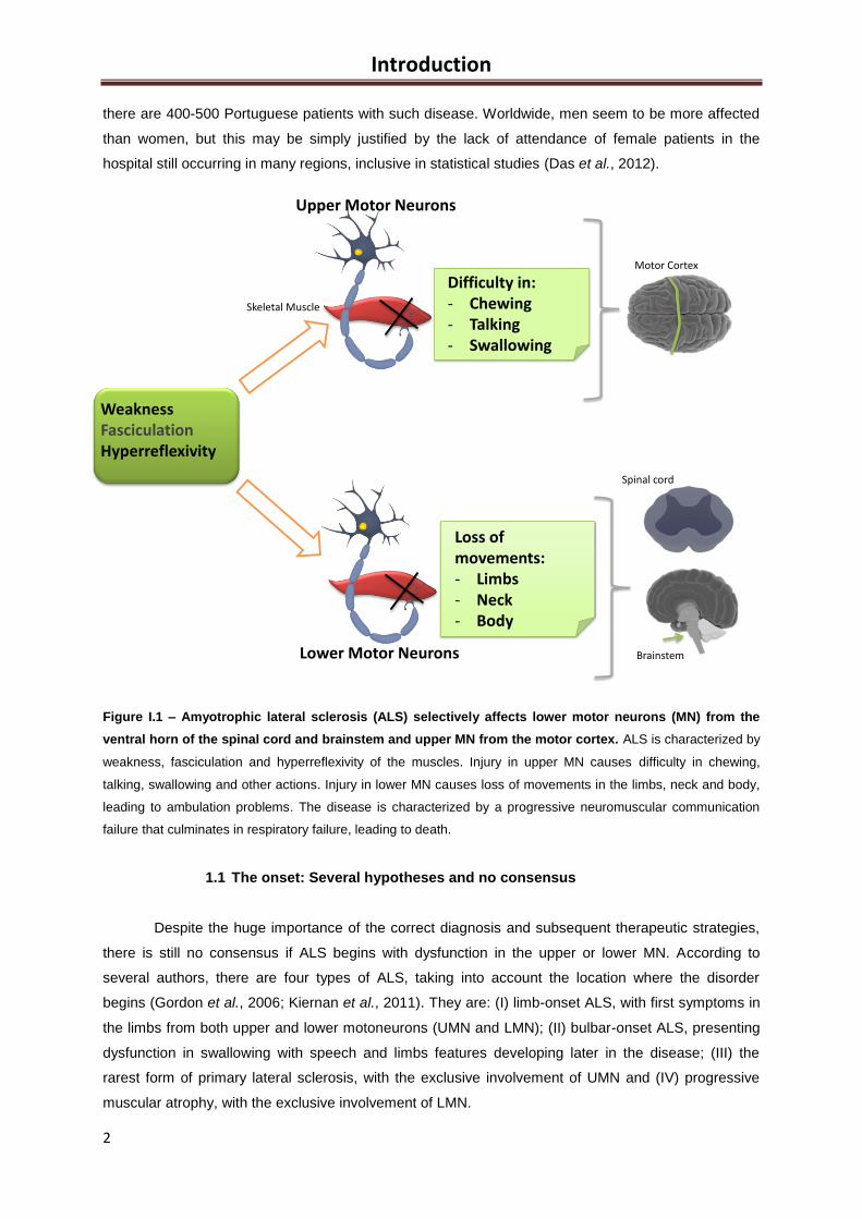
Figure I.1 – Amyotrophic lateral sclerosis (ALS) selectively affects lower motor neurons (MN) from the
ventral horn of the spinal cord and brainstem and upper MN from the motor cortex. ALS is characterized by
weakness, fasciculation and hyperreflexivity of the muscles. Injury in upper MN causes difficulty in chewing,
talking, swallowing and other actions. Injury in lower MN causes loss of movements in the limbs, neck and body,
leading to ambulation problems. The disease is characterized by a progressive neuromuscular communication
failure that culminates in respiratory failure, leading to death.
1.1 The onset: Several hypotheses and no consensus
Despite the huge importance of the correct diagnosis and subsequent therapeutic strategies,
there is still no consensus if ALS begins with dysfunction in the upper or lower MN. According to
several authors, there are four types of ALS, taking into account the location where the disorder
begins (Gordon et al., 2006; Kiernan et al., 2011). They are: (I) limb-onset ALS, with first symptoms in
the limbs from both upper and lower motoneurons (UMN and LMN); (II) bulbar-onset ALS, presenting
dysfunction in swallowing with speech and limbs features developing later in the disease; (III) the
rarest form of primary lateral sclerosis, with the exclusive involvement of UMN and (IV) progressive
muscular atrophy, with the exclusive involvement of LMN.
Loss ofmovements:- Limbs- Neck- Body
Upper Motor Neurons
Difficulty in:- Chewing- Talking- Swallowing
Lower Motor Neurons
Spinal cord
Skeletal Muscle
WeaknessFasciculationHyperreflexivity
Motor Cortex
Brainstem
Introduction
3
More recently, some researchers developed the “dying-forward” and “dying-back” hypothesis.
In the “dying-forward” hypothesis, ALS is seen as a disorder of corticomotor neurons, which connect
with anterior horn cells monosynaptically, mediating anterograde degeneration of anterior horn cells,
via glutamate excitotoxicity (Kiernan et al., 2011). In the “dying-back” hypothesis, ALS starts at level of
neuromuscular junction (NMJ) or within the muscle cells. This last hypothesis proposes that the cause
is a deficiency of a motor neurotrophic hormone normally released by postsynaptic cells and
transported by retrograde transport from the presynaptic axon to the soma where it exerts its effects
(Kiernan et al., 2011).
Furthermore, recent studies show the involvement of spinocerebellar and sensory pathways
and neuronal groups within the substantia nigra and the hippocampal dentate granule layer
(Ferraiuolo et al., 2011).
1.2 Genetics and features of the disease
ALS is referred to as a multifactorial disease, apparently having environmental, occupational and
toxicological components (Das et al., 2012), as well as evidence of a complex interaction between
genetic and molecular pathways. Surprisingly, there are authors suggesting that lifetime of intensive
sport or physical activity seems to be a risk factor for ALS (Kiernan et al., 2011).
It is known that ALS may be sporadic (sALS) in about 90-95% of cases, or genetic/familial (fALS)
in about 5-10% of cases. However, fALS and sALS are clinically and neuropathologically similar
(Gowing et al., 2008) and the only clinical feature that distinguishes fALS from sALS is a lower mean
age of onset in the former (Andersen and Al-Chalabi, 2011).
fALS can occur more commonly by an autosomal dominant (Ince et al., 2011), but also by an
autosomal recessive or X-linked inheritance and is a polygenetic disease with a variable penetrance
(Andersen and Al-Chalabi, 2011).
The most commonly affected gene is SOD1. In 1991, Brown and his group (Massachusetts
General Hospital) found that fALS is sometimes linked to chromosome 21q22 (20% of cases of fALS),
namely due to an autosomal dominant missense mutation in the SOD1 gene (that encodes cytosolic
Cu/Zn superoxide dismutase 1), which is a mitochondrial and cytoplasmic enzyme, essential for the
anti-oxidant defenses of the organism, since it is responsible for the detoxification of free radicals
produced in the mitochondria, namely superoxide anion.
In recent years, there are described more than 160 mutations in the SOD1 gene (Sabatelli et al.,
2013). It is important to mention that the ability of mutant Superoxide Dismutase 1 (mSOD1) to cause
neurodegeneration is not linked to a loss of dismutase function (Redler and Dokholyan, 2012). More
than affecting the activity of the enzyme, mSOD1 seems to induce a gain of toxic function (Yang et al.,
2010) probably related to protein misfolding (Costa et al., 2010), what explains that the knockout
mouse SOD1 does not present symptoms of ALS (Reaume et al., 1996). Additionally, conformational
instability and misfolding of the SOD1 peptide result in formation of intracellular aggregates, that inhibit
normal proteosomic function, disrupting axonal transport systems and vital cellular functions (Kiernan
et al., 2011). Recent studies showed that in fALS patients and in vitro mSOD1, protein instability and
Introduction
4
the increase of aggregation rate are correlated with the decrease of survival time (Byström et al.,
2010; Wang et al., 2008).
Another gene that can be mutated in ALS patients is the TARDBP gene (which encodes TAR
DNA-binding protein 43 protein, known as TDP-43), a major constituent of the ubiquitinated protein
inclusions found in surviving MN in most forms of ALS (Ferraiuolo et al., 2011). TDP-43 is responsible
for 4% of fALS cases and 1.5% of sALS cases (Mackenzie et al., 2011) and, under physiological
conditions, it functions as an RNA/DNA binding protein, being involved in alternative splicing,
transcriptional regulation, mRNA stabilization and microRNA processing (Ince et al., 2011).
Is also described FUS (Fused in Sarcoma), another mutated gene in ALS. FUS is situated in
chromosome 16, and encodes a RNA/DNA-binding protein implicated in transcriptional regulation,
alternative splicing, microRNA processing and mRNA transport (Ferraiuolo et al., 2011). The FUS
gene is mutated in 4% of fALS cases and in less than 1% of the sALS patients (Mackenzie et al.,
2010). The inheritance seems to be autosomal dominant (Vance et al., 2009), but mutations have
been reported in a large family originating from the Cape Verde islands showing autosomal recessive
inheritance of ALS (Kwiatkowski et al., 2009).
1.3 Molecular biology of motoneuron disease
Several cellular pathways have been shown to be dysregulated in tissues of patients and cell
models of ALS, which lead to MN damage and death. The sequence of pathogenic events is unclear
and most of them are intimately correlated (Costa et al., 2010), forming a complex network that
contributes to exacerbate the disease. Atrophy and death of MN, altered RNA processing,
mitochondrial dysfunction, glutamate mediated excitotoxicity, protein aggregate formation,
endoplasmic reticulum stress, axonal transport dysfunction, oxidative stress and neuroinflammation
are some of the pathophysiological phenomena known as biomarkers of ALS (Figure I.2).
Mitochondrial function, axonal transport, glutamate homeostasis, oxidative stress and apoptosis will be
further discussed in more detail in subsequent sections (1.3.1-1.3.6) due to their relevance for the
present thesis. In fact, many of the events can be caused for and consequence of each other and they
create a vicious cycle that results in motor axon disruption of neuronal equilibrium, denervation and
ultimately MN degeneration in ALS (Ferraiuolo et al., 2011).
Once this pathology is known as a MN disease, it is important to understand why these MN are
selectively vulnerable in ALS. First of all, MN are large cells with large axonal compartment and large
terminal arbors, which require an exigent metabolic capacity and a robust cytoskeleton and axonal
transport efficiency (Ferraiuolo et al., 2011). Moreover, MN have are highly dependent from a normal
mitochondrial function, which is the main source of reactive oxygen species (ROS) that can lead to
oxidative stress if dysfunctional. These neurons have particular sensitivity to excitotoxicity and
dysregulation of intracellular calcium homeostasis since they have a high expression of calcium-
permeable α-amino-3-hydroxy-5-methyl-4-isoxazole propionic acid (AMPA) receptors, which lack the
GluR2 subunit (Williams et al., 1997). They also evidence reduced expression of calcium-buffering
Introduction
5
proteins and high dependence of synaptic glutamate re-uptake transport mechanisms. Other reasons
are the reduced capacity for heat shock response and chaperone activity that MN seems to have,
leading to defective correction of protein folding, increasing sensitivity to endoplasmic reticulum stress
(Saxena et al., 2009) and mitochondria features that predispose the cells to oxidative damage and
calcium overload (Panov et al., 2011). Ultimately, this defective protein folding associated with the
high expression of particular proteins (e.g. SOD1), and consequently, a high vulnerability to toxicity of
mutant proteins (Ferraiuolo et al., 2011) contribute to the accumulation of protein aggregates and
death of MN.
In patients carrying mSOD1, it may occurs the upregulation of genes promoting the MN survival
during the disease process, principally those encoding phosphatidylinositol 3-kinase and phosphatase
and tensin homolog-protein kinase B pathway (Kirby et al., 2011). The understanding of the properties
of the neurons that make them more or less resistant to the occurrence of ALS is very important to find
strategies to increase defense mechanisms and promote new therapies.
Introduction
6
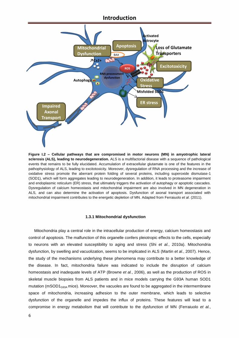
Figure I.2 – Cellular pathways that are compromised in motor neurons (MN) in amyotrophic lateral
sclerosis (ALS), leading to neurodegeneration. ALS is a multifactorial disease with a sequence of pathological
events that remains to be fully elucidated. Accumulation of extracellular glutamate is one of the features in the
pathophysiology of ALS, leading to excitotoxicity. Moreover, dysregulation of RNA processing and the increase of
oxidative stress promote the aberrant protein folding of several proteins, including superoxide dismutase-1
(SOD1), which will form aggregates leading to neurodegeneration. In addition, it leads to proteasome impairment
and endoplasmic reticulum (ER) stress, that ultimately triggers the activation of autophagy or apoptotic cascades.
Dysregulation of calcium homeostasis and mitochondrial impairment are also involved in MN degeneration in
ALS, and can also determine the activation of apoptosis. Dysfunction of axonal transport associated with
mitochondrial impairment contributes to the energetic depletion of MN. Adapted from Ferraiuolo et al. (2011).
1.3.1 Mitochondrial dysfunction
Mitochondria play a central role in the intracellular production of energy, calcium homeostasis and
control of apoptosis. The malfunction of this organelle confers pleiotropic effects to the cells, especially
to neurons with an elevated susceptibility to aging and stress (Shi et al., 2010a). Mitochondria
dysfunction, by swelling and vacuolization, seems to be implicated in ALS (Martin et al., 2007). Hence,
the study of the mechanisms underlying these phenomena may contribute to a better knowledge of
the disease. In fact, mitochondria failure was indicated to include the disruption of calcium
homeostasis and inadequate levels of ATP (Browne et al., 2006), as well as the production of ROS in
skeletal muscle biopsies from ALS patients and in mice models carrying the G93A human SOD1
mutation (mSOD1G93A mice). Moreover, the vacuoles are found to be aggregated in the intermembrane
space of mitochondria, increasing adhesion to the outer membrane, which leads to selective
dysfunction of the organelle and impedes the influx of proteins. These features will lead to a
compromise in energy metabolism that will contribute to the dysfunction of MN (Ferraiuolo et al.,
ActivatedAstrocyte
Loss of GlutamateTransporters
Excitotoxicity
↗Ca2+
BAX
ApoptosisMitochondrialDysfunction
RNA processmentdysfunction
ROS
ER stress
OxidativeStress
Misfolded SOD1
ImpairedAxonal
Transport
Autophagy
Introduction
7
2011), together with the activation of caspases, that can trigger apoptotic cell death. In these mSOD1
mice models, calcium buffering is also deficient in mitochondria and enhances the susceptibility of MN
to the calcium homeostasis deregulation, that can be associated with glutamate-mediated
excitotoxicity and with the activation of pro-oxidant and apoptotic factors such as nitric oxide synthase
(NOS), phospholipases and endonucleases. It is important to notice that calcium-buffering ability is
particularly deleterious to neurons and skeletal muscle, whose operation requires frequent influx of
calcium to generate action potentials (Redler and Dokholyan, 2012).
On the other hand, although most SOD1 is localized in the cytosol, a fraction of mutant SOD1
(mSOD1) is associated with the mitochondria (Vande Velde et al., 2008) and its accumulation seems
to exacerbate mitochondrial damage (Ferraiuolo et al., 2011). However, the mechanism that leads to
this event is still a matter of debate, although there are some theories: (i) mSOD1 allows the release
of cytochrome c, activating the apoptotic cascade and opening the pores of the outer membrane of
mitochondria (Pasinelli and Brown, 2006); (ii) abnormal interaction of misfolded proteins and
oligomers with other mitochondrial proteins can promote mitochondrial damage and apoptosis
following associating with Bcl-2, a pro-survival factor (Redler and Dokholyan, 2012); (iii) aggregation of
mSOD1 in the outer membrane can result in the disruption of translocation machinery, limiting the
input of functional proteins into the organelle (Pasinelli and Brown, 2006); (iv) misfolded and
aggregated mSOD1 also accumulate on the cytoplasmic face of the outer mitochondrial membrane
and bind directly to the voltage-dependent anion channel, depolarizing the membrane and disrupting
the normal functioning of complexes I and IV of the electron transport chain (Costa et al., 2010; Liu et
al., 2009).
Other events that deserve our attention are mitochondrial fusion and fission (Figure I.3 B).
Mitochondria are actively transported and they can have defined subcellular distributions that can
change as necessary. Indeed, this organelle keeps their shape, size, morphology, distribution and
physiological function through fusion and fission processes (Shi et al., 2010a). An imbalance of these
two opposing events results in excessive mitochondrial fragmentation or elongation (Chan, 2012).
Moreover, it is believed that mitochondrial morphology, metabolic function, membrane potential,
axonal transport, fission and fusion are highly inter-dependent (Shi et al., 2010a). The main
constituents of the fusion machinery in mammalian cells are Mitofusin 1 (Mfn1), Mitofusin 2 (Mfn2) and
Optic atrophy 1 (OPA1) (Figure I.3 C). Mfn1 and Mfn2, localized in the mitochondrial outer membrane,
belong to GTPase family and their depletion leads to loss of mitochondria fusion, high fragmentation,
no mitochondrial tubules and decreased cellular respiration (Chen et al., 2005). Moreover, in humans,
mutations in Mfn2 cause Charcot-Marie-Tooth neuropathy type 2A (Zuchner et al., 2004), a disease of
the group of peripheral neuropathies with symptoms such as distal muscle weakness and atrophy,
less severe sensory loss, and depressed tendon reflexes (Ranieri et al., 2013). OPA1 is a dynamin
family GTPase and localizes within the mitochondrial intermembrane space and mutations cause the
most common form of hereditary optic atrophy (Alexander et al., 2000).
Dynamin-related protein 1 and fission 1 (Drp1 and Fis1, respectively) are the components of
mitochondrial fission machinery in mammals. Dominant-negative mutants of Drp1 inhibit mitochondrial
Introduction
8
division and result in highly interconnected mitochondrial tubules (Smirnova et al., 2001).
Overexpression of Fis1 leads to mitochondrial fragmentation, release of cytochrome c and, therefore,
apoptosis (James et al., 2003). It is highly suggestive that mitochondrial fusion and fission may be
influenced in the presence of mSOD1, causing disturbances at the level of mitochondrial dynamics,
which are linked to disorders such as the Alzheimer’s disease (Shi et al., 2010a).
1.3.2 Glutamate mediated excitotoxicity
Glutamate is the major excitatory neurotransmitter (NT) in the CNS and its signal is ended by its
removal from the synaptic cleft by transporters such as EAAT2 (Excitatory amino-acid transporter 2),
which is mainly expressed by astrocytes (Maragakis et al., 2004).
There are three groups of glutamate receptors in postsynaptic neurons essential to the physiological
neurotransmission. These receptors can be divided into metabotropic and ionotropic. Metabotropic
receptors are G protein-coupled and operate through signal transduction cascades. Ionotropic
receptors act as ion channels and are divided into three groups: N-methyl-D-aspartate (NMDA), α-
amino-3-hydroxy-5-methyl-4-isoxazolepropionic acid (AMPA) and Kainate receptors. NMDA receptors
are stimulated by calcium and sodium symport entry, and non-NMDA receptors (generic designation
for AMPA/Kainate receptors) are traditionally seen as mainly permeable to monovalent ions such as
Na+ and K
+ (Agrawal and Fehlings, 1997). The calcium permeability of AMPA receptors is broadly
determined by the GluR2 subunit, responsible to making the receptor impermeable to calcium, which
is extremely important in preventing glutamate excitotoxicity (Ferraiuolo et al., 2011).
Excitotoxicity is a neuronal injury which can then result from the excessive activation of glutamate
receptors, AMPA and NMDA, and may be caused by increased levels of glutamate in the synaptic
cleft or by the increased sensitivity of the postsynaptic neurons to this NT, leading, in both situations,
to an increase in intracellular calcium (Ferraiuolo et al., 2011). Disruption of intracellular calcium
homeostasis, with secondary activation of proteolytic enzyme systems and generation of ROS,
disruption of mitochondrial function, production of ATP, promotion of transcription factors of pro-
apoptotic genes or suppression of anti-apoptotic genes are key components of excitotoxicity
(Ferraiuolo et al., 2011) that leads to neuronal death.
In fALS and sALS patients, as well as in mutant SOD1 mice models, there are decreased levels of
functional EAAT2 protein and increased circulating glutamate in the cerebrospinal fluid (CSF)
(Howland et al., 2002). Although the precise mechanism(s) by which EAAT2 is down-regulated in ALS
are not yet understood, it is known that this gene deletion induces progressive neurodegeneration,
while its overexpression was shown to delay symptom onset in ALS mouse models (Rothstein et al.,
2005). EAAT2 is indirectly affected when other associated processes suffer from some dysfunction,
suggesting that excitotoxicity may be a secondary event in ALS pathogenesis. Indeed, when caspase-
3 is activated, it results in a truncated or inactive version of EAAT2. Oxidative damage to the C-
terminus of EAAT2 diminishes its ability to transport glutamate (Redler and Dokholyan, 2012). In
addition, prolonged hyperstimulation by glutamate induces cell death by allowing persistent calcium
Introduction
9
influx through the AMPA receptors, which are specifically abundant in MN (Van Den Bosch et al.,
2000).
It is noteworthy to mention that blocking the excitotoxic effects of extracellular glutamate is the only
strategy approved by U.S. Food and Drug Administration (FDA) that has shown to be able to slow the
ALS progression. This is the case of riluzole, a benzothiazole derivative, that has several effects,
including the inhibition of the excitotoxic stress in neurons by slowing glutamate release (due to the
inactivation of voltage-dependent Na+ channels on glutamatergic nerve terminals), as well as the
activation of a G-protein dependent signal transduction process. Moreover, riluzole seems to be able
to block some of the postsynaptic effects of glutamate by non-competitive inhibition of NMDA and
AMPA receptors (Van Den Bosch et al., 2006; Vucic et al., 2013), which showed to cause an increase
in patient survival but only for few months.
1.3.3 Axonal transport dysfunction
Axon is a long and slender projection of the neuron that conducts electrical impulses and all the
molecules that need to be transported (Shi et al., 2010b). Since the genetic material and the majority
of the protein synthesis machinery are localized to the cell body, it is necessary to exist a way to
transport all materials (generically known as cargo) from the cell body to the axon terminal, and from
axon terminal to cell body. The microtubules serve as rails, along the entire axon and secretory
vesicles are transported to sites of release through the action of microtubule-based motor proteins. In
neurons, these transport processes are collectively known as axonal transport (Siegel et al., 2006).
Growth and maintenance of neuronal processes requires timely, efficient delivery of material to
axonal and dendritic domains. For this, there are the anterograde and retrograde transports. The first
occurs from cellular body to axon, mediated by kinesin molecular motor protein and the second occurs
from axon to cellular body, mediated by dynein molecular motor protein (Ferraiuolo et al., 2011).
However, sometimes the axonal transport does not work properly. In ALS, this dysfunction is
described mainly due to the formation of neurofilament aggregates, which causes disruption of axonal
transport that combined with mitochondrial dysfunction causes energetic depletion of distal axonal
compartment of MN, thus leading to degeneration (Ikenaka et al., 2012) (Figure I.3 A). The disruption
can occur at anterograde or retrograde level, or simultaneously in both as a consequence of
decreased mobility of motor proteins or decreased binding of cargos to these motor proteins. The
three main cargos indicated as biomarkers of MN degeneration by accumulation in distal axon
compartment are neurofilaments, mitochondria and autophagosomes. The neurofilaments set the
diameter of the axons, and its aggregation (by phosphorylation or stoichiometric imbalance) is
pathological. Kinesin or dynein dysfunction leads to accumulation of neurofilamentous swellings
(spheroids), as in the KIF5A and in the dynactin-1 mutant mice (Ikenaka et al., 2012; King et al.,
2011), which are two models used in the study of axonal transport dysfunction since they have
mutations commonly linked to the dysfunction of anterograde and retrograde transport, respectively.
Introduction
10
The disruption of the anterograde and retrograde transport (Hirokawa et al., 2010), leads to
mitochondrial accumulation in a certain region of the cell, leading to energy depletion elsewhere,
which can result in cell death (Ikenaka et al., 2012).
Finally, it is known that the lysosome-autophagosome pathway is responsible by recycling
intracellular compounds; therefore, its dysfunction may also cause neurodegeneration. Since this
cargo is transported bi-directionally along microtubules, alterations in both types of transport will cause
the accumulation of autophagosomes (Ikenaka et al., 2012).
A deficiency of motor proteins associated with axonal transport can occur due to chronic exposure
to neurotoxins, such as acrylamide, which has been described as being able to directly inhibit the
function of kinesin, therefore the anterograde transport (Sickles et al., 2002). Thus, for example,
mutation with loss of function in KIF5A (kinesin subunit) causes a deficiency in binding of Kinesin I to
microtubules, leading to failure of anterograde transport (Ikenaka et al., 2012).
There is also evidence that mutations in SOD1 such as A4V, G85R and G93A, promotes SOD1
interaction with the complex dynein-dynactin in cell cultures and in affected tissues of ALS mice (Shi et
al., 2010b). The same authors suggest that mSOD1 and dynein interaction play a key role in the
formation of large inclusions containing mSOD1. In the mSOD1 mice model, the impairment of axonal
transport occurs at an early stage of the disease. The mechanisms behind the dysfunction in this
model are still unknown, but appear to derive from an increase in tumor necrosis factors (TNFs), which
is observed in mSOD1 mice, leading to the disruption of kinesin function, by a mechanism that
involves the activation of p38 MAPK pathway, which has been observed in models of ALS(Shi et al.,
2010b).
Introduction
11
a)
MitochondrialDysfuncion
Disruptionof Axonal Transport
Anterogradetransport
Retrograde transport
Neurofilamentaggregates
DRP1
Fusion
Fission
c)
Dynein mutated
Kinesin mutated
mSOD1 in vacuoles
Mfn2OPA1
Energeticdeplection of
the cell
b)
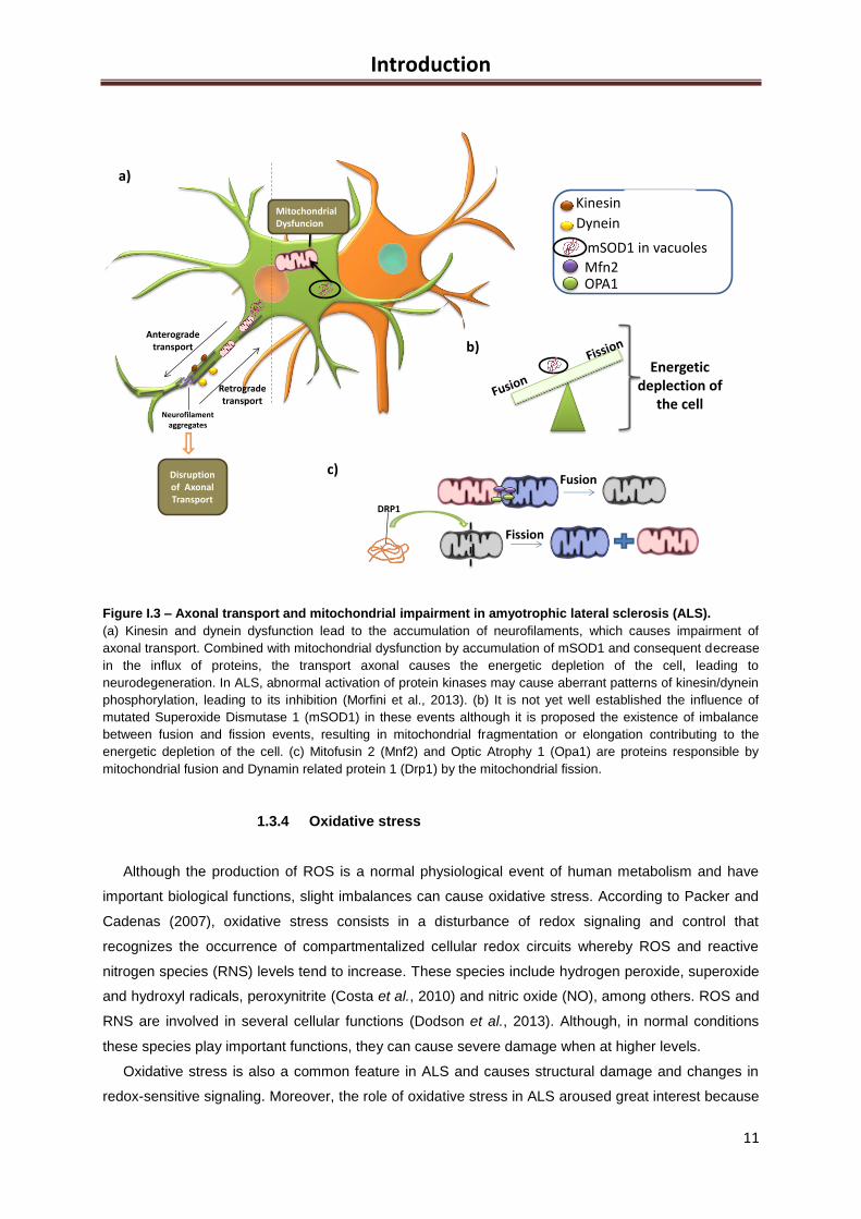
Figure I.3 – Axonal transport and mitochondrial impairment in amyotrophic lateral sclerosis (ALS).
(a) Kinesin and dynein dysfunction lead to the accumulation of neurofilaments, which causes impairment of
axonal transport. Combined with mitochondrial dysfunction by accumulation of mSOD1 and consequent decrease
in the influx of proteins, the transport axonal causes the energetic depletion of the cell, leading to
neurodegeneration. In ALS, abnormal activation of protein kinases may cause aberrant patterns of kinesin/dynein
phosphorylation, leading to its inhibition (Morfini et al., 2013). (b) It is not yet well established the influence of
mutated Superoxide Dismutase 1 (mSOD1) in these events although it is proposed the existence of imbalance
between fusion and fission events, resulting in mitochondrial fragmentation or elongation contributing to the
energetic depletion of the cell. (c) Mitofusin 2 (Mnf2) and Optic Atrophy 1 (Opa1) are proteins responsible by
mitochondrial fusion and Dynamin related protein 1 (Drp1) by the mitochondrial fission.
1.3.4 Oxidative stress
Although the production of ROS is a normal physiological event of human metabolism and have
important biological functions, slight imbalances can cause oxidative stress. According to Packer and
Cadenas (2007), oxidative stress consists in a disturbance of redox signaling and control that
recognizes the occurrence of compartmentalized cellular redox circuits whereby ROS and reactive
nitrogen species (RNS) levels tend to increase. These species include hydrogen peroxide, superoxide
and hydroxyl radicals, peroxynitrite (Costa et al., 2010) and nitric oxide (NO), among others. ROS and
RNS are involved in several cellular functions (Dodson et al., 2013). Although, in normal conditions
these species play important functions, they can cause severe damage when at higher levels.
Oxidative stress is also a common feature in ALS and causes structural damage and changes in
redox-sensitive signaling. Moreover, the role of oxidative stress in ALS aroused great interest because
Introduction
12
mutations in SOD1, which encodes a major antioxidant protein, account for 20% of fALS cases (Silva
et al., 2011). There is also a large body of evidence of oxidative stress in sALS and fALS, as indicated
by the increase of 3-nitrotyrosine levels, considered a marker of oxidative stress resultant from the
elevation of peroxynitrites (Costa et al., 2010).
Several studies showed that oxidative stress interacts with other pathophysiological processes that
contribute to MN disease, including excitotoxicity (Rao and Weiss, 2004), mitochondrial dysfunction
(Duffy et al., 2011), protein aggregation (Wood et al., 2003), stress of ER (Kanekura et al., 2009) and
changes in signaling from microglia and astrocytes (Blackburn et al., 2009; Sargsyan et al., 2005).
Therefore, an effective reduction of oxidative stress may improve some aspects of the
pathophysiology of MN degeneration. However, therapeutics directed to the regulation of the
oxidative stress have not been yet effective in humans, although samples of CSF, serum and urine of
ALS patients evidence markers of free radical damage (Mitsumoto et al., 2008). In addition,
postmortem tissue from sALS and mSOD1-related fALS cases also present elevated levels of
oxidative damage to proteins, lipids and DNA (Ferraiuolo et al., 2011). Some mRNA species appear to
have increased susceptibility to oxidation, such as those involved in the mitochondrial electron
transport chain, protein biosynthesis, folding and degradation pathways, myelination, cytoskeleton
proteins, and the tricarboxylic acid cycle and glycolysis pathways (Chang et al., 2008). Also mSOD1
seems to be particularly susceptible to oxidative translation modification.
In cellular models of mutant TAR DNA-binding protein 43 (TDP-43)-related ALS, the presence of
this mutant protein has shown to induce oxidative stress in MN cell lines (Duan et al., 2010). Finally, in
other nerve cells, namely microglia, mSOD1 seems to increase NADPH oxidase (NOX)-mediated
superoxide production, resulting in prolongation of ROS production (Harraz et al., 2008). It was
observed an increase in NOX2 expression in mSOD1 mice and in CNS of ALS patients. It seems that
in mSOD1 models, as well as in CNS of ALS patients, there is a dysregulation of the erythroid 2-
related factor 2 (NRF-2), which is the main regulator of the antioxidant response (Sarlette et al., 2008).
It is import to note that the CNS is extremely sensitive to oxidative stress, since it has a reduced
expression of antioxidant enzymes, high levels of easily oxidized substrates and high production of
ROS by neurochemical reactions (Carri et al., 2003).
1.3.5 Endoplasmic reticulum stress
Intracellular inclusions related to accumulation of misfolded or unfolded proteins in aggregates are
hallmarks of several neurodegenerative diseases, including ALS (Vijayalakshmi et al., 2011).
These events, together with oxidative stress and loss of calcium homeostasis (Rao et al., 2004a;
Rao et al., 2004b), induce the ER stress. ER is an organelle responsible for maintaining cellular
calcium homeostasis and synthesize/regulate the synthesis and the folding of proteins. For this, ER
has resident chaperones that recognize aberrant proteins and correct their folding. This is crucial,
since non-functional proteins can cause suppression of general translation and ER-associated protein
degradation (Ferraiuolo et al., 2011). Initially, this mechanism is cytoprotective but a prolonged
activation can lead to apoptosis (Yamagishi et al., 2007). According to some studies, the protein
Introduction
13
disulphide isomerase (PDI), an unfolded-protein response (UPR) chaperone existing in ER, is
activated in mSOD1 mice, where it co-localizes with mSOD1 inclusions, and in samples from sALS
patients (Atkin et al., 2006; Atkin et al., 2008). It is suggested that ER stress is involved in the early
stages of MN injury, once PDI and other UPR-induced proteins are up regulated before the disease
onset in mSOD1 rodents (Atkin et al., 2008). Up-regulated markers of ER stress, such as PDI, are
also present in the CSF and SC of postmortem samples of ALS patients (Atkin et al., 2008; Sasaki,
2010).
Interestingly, the exposure of NSC-34 cells, an hybrid cell line produced by fusion of
neuroblastoma with mouse MN-enriched primary SC cells and primary spinal MN, to CSF from ALS
patients led to ER stress, including expression of ER fragmentation, UPR markers and activation of
caspase-12 (Vijayalakshmi et al., 2011). However, it was not possible to identify the CSF constituents
that are responsible for such changes.
UPR activation seems to be cytoprotective, at least in the initial phases of cellular stress.
Nevertheless, an increase in survival lacking a key UPR transcription factor accompanied by
increased activation of ER-associated protein degradation, enhanced autophagy and decreased
mSOD1 aggregation were observed in the mSOD1 mice model (Hetz et al., 2009).
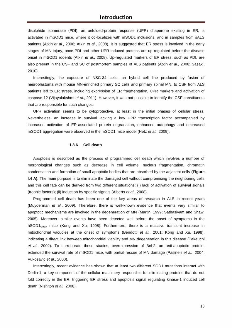
1.3.6 Cell death
Apoptosis is described as the process of programmed cell death which involves a number of
morphological changes such as decrease in cell volume, nucleus fragmentation, chromatin
condensation and formation of small apoptotic bodies that are absorbed by the adjacent cells (Figure
I.4 A). The main purpose is to eliminate the damaged cell without compromising the neighboring cells
and this cell fate can be derived from two different situations: (i) lack of activation of survival signals
(trophic factors); (ii) induction by specific signals (Alberts et al., 2008).
Programmed cell death has been one of the key areas of research in ALS in recent years
(Muyderman et al., 2009). Therefore, there is well-known evidence that events very similar to
apoptotic mechanisms are involved in the degeneration of MN (Martin, 1999; Sathasivam and Shaw,
2005). Moreover, similar events have been detected well before the onset of symptoms in the
hSOD1G93A mice (Kong and Xu, 1998). Furthermore, there is a massive transient increase in
mitochondrial vacuoles at the onset of symptoms (Bendotti et al., 2001; Kong and Xu, 1998),
indicating a direct link between mitochondrial viability and MN degeneration in this disease (Takeuchi
et al., 2002). To corroborate these studies, overexpression of Bcl-2, an anti-apoptotic protein,
extended the survival rate of mSOD1 mice, with partial rescue of MN damage (Pasinelli et al., 2004;
Vukosavic et al., 2000).
Interestingly, recent evidence has shown that at least two different SOD1 mutations interact with
Derlin-1, a key component of the cellular machinery responsible for eliminating proteins that do not
fold correctly in the ER, triggering ER stress and apoptosis signal regulating kinase-1 induced cell
death (Nishitoh et al., 2008).
Introduction
14
Although not a mechanism of death broadly described in ALS, necrosis is also a cell death pathway
(Figure I.4 B). However, unlike apoptosis, is not programmed and results in cellular injury. In this
case, the cells swell to break, releasing intracellular components, what can cause inflammation
(Alberts et al., 2008). The SOD1G93A mice model is the one that has features of both apoptosis and
necrosis, with ‘‘necrotic-like’’ and ‘‘apoptotic-like’’ processes dominating in different cell types and/or
disease stages (Martin et al., 1998; Martin et al., 2007).
Finally, autophagy (Figure I.4 C) is a catabolic fundamental process of degradation for
macromolecules and organelles, crucial for cell and tissue homeostasis (Birgisdottir et al., 2013) and
lysosome-dependent (Klionsky, 2007; Levine and Klionsky, 2004). Thus, this is an essential process
for cell survival (Kim et al., 2013). The impairment of this mechanism is implicated in chronic
neurodegenerative diseases such as Alzheimer’s disease, Huntington’s disease and Parkinson’s
disease (Meredith et al., 2002; Nixon, 2007; Nixon et al., 2005; Ravikumar et al., 2004). Although the
boosting of autophagy seems to retard the progression of the disease by promoting the removal of
misfolded proteins it is still unclear whether autophagy activation would be beneficial or detrimental in
ALS (Song et al., 2012). Mutations that disrupt autophagy (as CHMP2B mutation) are found in some
ALS cases, supporting the hypothesis of autophagy with a protective role in ALS (Cox et al., 2010;
Parkinson et al., 2006). By other hand, post-mortem studies and work in animal models have shown
that the number of autophagosomes is increased in SC affected by ALS (Morimoto et al., 2007;
Sasaki, 2011). Moreover, whether the accumulation of autophagosomes signifies decreased or
increased autophagic flux has yet to be established. Furthermore, reported that treatment with lithium
known to activate autophagy exacerbates ALS progression in mice models (Pizzasegola et al., 2009).
Trials using rapamycin as an autophagy inducer, also revealed disease progression in ALS mice
(Zhang et al., 2011). Controversially, other groups have reported the opposite effects with lithium and
rapamycin in the same model (Fornai et al., 2008; Wang et al., 2012).
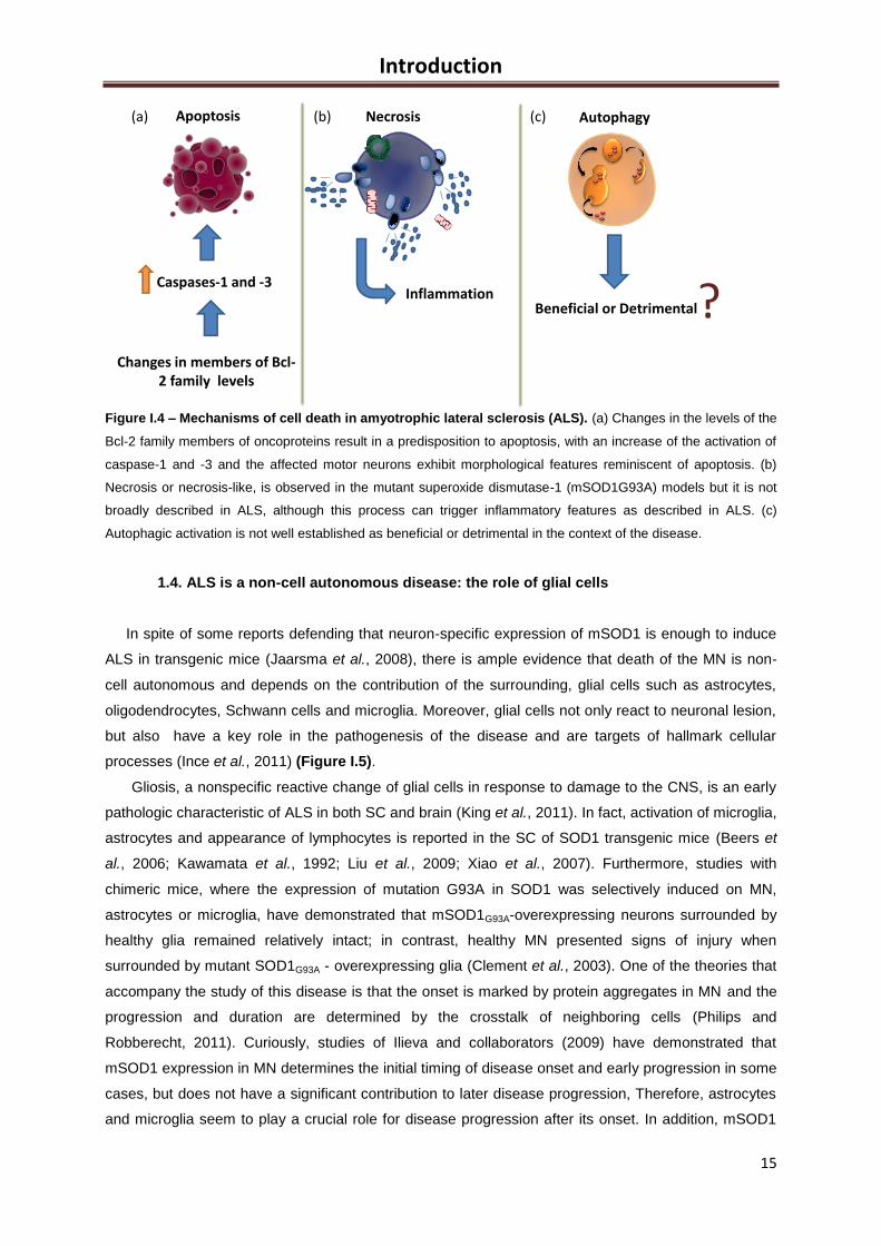
Introduction
15
Figure I.4 – Mechanisms of cell death in amyotrophic lateral sclerosis (ALS). (a) Changes in the levels of the
Bcl-2 family members of oncoproteins result in a predisposition to apoptosis, with an increase of the activation of
caspase-1 and -3 and the affected motor neurons exhibit morphological features reminiscent of apoptosis. (b)
Necrosis or necrosis-like, is observed in the mutant superoxide dismutase-1 (mSOD1G93A) models but it is not
broadly described in ALS, although this process can trigger inflammatory features as described in ALS. (c)
Autophagic activation is not well established as beneficial or detrimental in the context of the disease.
1.4. ALS is a non-cell autonomous disease: the role of glial cells
In spite of some reports defending that neuron-specific expression of mSOD1 is enough to induce
ALS in transgenic mice (Jaarsma et al., 2008), there is ample evidence that death of the MN is non-
cell autonomous and depends on the contribution of the surrounding, glial cells such as astrocytes,
oligodendrocytes, Schwann cells and microglia. Moreover, glial cells not only react to neuronal lesion,
but also have a key role in the pathogenesis of the disease and are targets of hallmark cellular
processes (Ince et al., 2011) (Figure I.5).
Gliosis, a nonspecific reactive change of glial cells in response to damage to the CNS, is an early
pathologic characteristic of ALS in both SC and brain (King et al., 2011). In fact, activation of microglia,
astrocytes and appearance of lymphocytes is reported in the SC of SOD1 transgenic mice (Beers et
al., 2006; Kawamata et al., 1992; Liu et al., 2009; Xiao et al., 2007). Furthermore, studies with
chimeric mice, where the expression of mutation G93A in SOD1 was selectively induced on MN,
astrocytes or microglia, have demonstrated that mSOD1G93A-overexpressing neurons surrounded by
healthy glia remained relatively intact; in contrast, healthy MN presented signs of injury when
surrounded by mutant SOD1G93A - overexpressing glia (Clement et al., 2003). One of the theories that
accompany the study of this disease is that the onset is marked by protein aggregates in MN and the
progression and duration are determined by the crosstalk of neighboring cells (Philips and
Robberecht, 2011). Curiously, studies of Ilieva and collaborators (2009) have demonstrated that
mSOD1 expression in MN determines the initial timing of disease onset and early progression in some
cases, but does not have a significant contribution to later disease progression, Therefore, astrocytes
and microglia seem to play a crucial role for disease progression after its onset. In addition, mSOD1
Beneficial or Detrimental?
Apoptosis Necrosis Autophagy
Inflammation
Changes in members of Bcl-2 family levels
Caspases-1 and -3
(a) (b) (c)
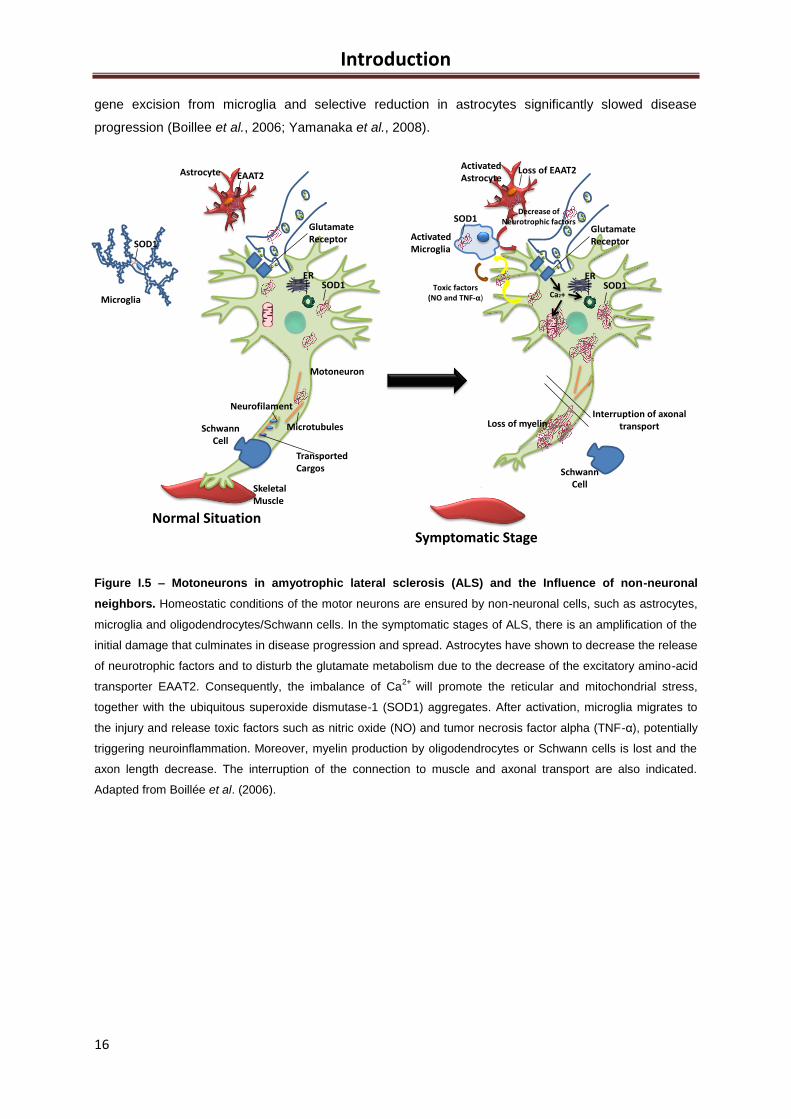
Introduction
16
gene excision from microglia and selective reduction in astrocytes significantly slowed disease
progression (Boillee et al., 2006; Yamanaka et al., 2008).
Figure I.5 – Motoneurons in amyotrophic lateral sclerosis (ALS) and the Influence of non-neuronal
neighbors. Homeostatic conditions of the motor neurons are ensured by non-neuronal cells, such as astrocytes,
microglia and oligodendrocytes/Schwann cells. In the symptomatic stages of ALS, there is an amplification of the
initial damage that culminates in disease progression and spread. Astrocytes have shown to decrease the release
of neurotrophic factors and to disturb the glutamate metabolism due to the decrease of the excitatory amino-acid
transporter EAAT2. Consequently, the imbalance of Ca2+
will promote the reticular and mitochondrial stress,
together with the ubiquitous superoxide dismutase-1 (SOD1) aggregates. After activation, microglia migrates to
the injury and release toxic factors such as nitric oxide (NO) and tumor necrosis factor alpha (TNF-α), potentially
triggering neuroinflammation. Moreover, myelin production by oligodendrocytes or Schwann cells is lost and the
axon length decrease. The interruption of the connection to muscle and axonal transport are also indicated.
Adapted from Boillée et al. (2006).
Symptomatic Stage
Normal Situation
SchwannCell
SOD1
SOD1
Motoneuron
SkeletalMuscle
GlutamateReceptor
ER
Neurofilament
Microtubules
TransportedCargos
Microglia
Astrocyte EAAT2ActivatedAstrocyte
Loss of EAAT2
ActivatedMicroglia
Toxic factors(NO and TNF-α)
GlutamateReceptor
SOD1ER
Ca2+
Decrease ofNeurotrophic factors
Interruption of axonal transportLoss of myelin
SOD1
SchwannCell
Introduction
17
1.4.1. Oligodendrocytes and Schwann cells
Oligodendrocytes in CNS and Schwann cells in the peripheral nervous system are glial cells of
ectodermal origin responsible for the formation and maintenance of myelin sheath in axons, thus
catalyzing the electrical impulse conduction (Siegel et al., 2006). Schwann cells are closely associated
with MN axons, and participate in axonal development and regeneration. Until now, there are a few
reports suggesting that these glial cells have a role on ALS pathogenesis. However, loss of compact
myelin and lamellae detachment, indicators of myelin abnormalities, were observed in SC of pre-
symptomatic SOD1 transgenic rats and aggravated at symptomatic stages (Lasiene and Yamanaka,
2011). Moreover, more pronounced marks of morphological myelin degeneration were observed in
fully symptomatic stages of mSOD1 rats (Niebroj-Dobosz et al., 2007).
In what concerns Schwann cells, there is still some controversy. Interestingly, and in opposite to
what happens in other non-neuronal cells, the elimination of mSOD1G37R specifically in Schwann
cells, not only failed to slow disease progression, but also promoted a substantial acceleration of the
late phase of the disease (Lobsiger et al., 2009). The underlying mechanism suggests a protective
role to mSOD1 which is possibly due to the dismutation activity that can ameliorate some oxidative
damage within the cells (Ilieva et al., 2009). However, recently, Wang and colleagues (2012) found
that knockdown of mSOD1 in Schwann cells of mSODG85R transgenic mice delayed disease onset
and extended survival indicating that mSOD1G85R expression is neurotoxic.
1.4.2 Astrocytes
Astrocytes, also known collectively as astroglia, consist in star-shaped glial cells (Siegel et al.,
2006). They are the most abundant cells of the CNS and have an important role in the support of
neurons, regulation/reuptake of neurotransmitters and ion concentrations in the extracellular space,
neurovascular coupling, and maintenance of a normal blood brain barrier (BBB) and in the tripartite
synapse (Fellin, 2009; Perea et al., 2009; Volterra and Meldolesi, 2005).
Pathological studies have reported that the death of MN in ALS is accompanied by astrogliosis,
characterized by up-regulation of glial fibrillary acidic protein (GFAP) in dorsal and ventral grey matter
and in antero-lateral white matter (Ince et al., 2011). It has been also indicated that the development of
astrogliosis in some models occurs in pre-symptomatic phases, although it varies depending on the
mutation (Vargas et al., 2008).
Astrocytes are essential for the removal of glutamate from the synaptic cleft, mainly through
EAAT2, which has reduced levels in sALS and fALS patients (Van Damme et al., 2007). In addition,
astrocytes expressing mSOD1 have shown to be more toxic than normal reactive astrocytes (Hovden
et al., 2013; Pehar et al., 2004) and to release an insufficient amount of neurotrophic factors crucial to
the neuronal balance, such as glial-derived factor, brain derived neurotrophic factor, cilliary
neurotrophic factor and vascular endothelial growth factor (Dewil et al., 2007; Ekestern, 2004).
Moreover, astrocytes have demonstrated a disturbance of glutamate metabolism in chick SC astroglial
Introduction
18
cultures, probably due to the up-regulation of metabotropic glutamate receptors (Anneser et al., 2004),
which can be the cause of the excitotoxicity in these models.
Also, astrocytes respond to toxic factors in the CSF of ALS patients by undergoing morphological
transformation from flat to process bearing and elevated expression of GFAP, as well as S100A6 and
S100B proteins (Shobha et al., 2010). Recently, a subpopulation of astrocytes from SC of
symptomatic mSOD1G93A rats has revealed a unique pattern of astrocytic markers, aberrant
phenotypic features and an increased proliferation rate (Diaz-Amarilla et al., 2011) that may represent
an unknown astrocytic phenotype associated with later stages of ALS progression.
1.4.3 Microglia
In 1932, Pio del Rio-Hortega introduced the concept of microglia, the lower glial cells, as a defined
cellular element of the CNS. The postulate of del Rio-Hortega states the following: 1) Microglia
penetrates the brain during early development; 2) When they invade the brain, cells have amoeboid
morphology and have a mesodermal origin; 3) Microglia uses vessels and white matter tracts as
guiding structures for migration and enter all brain regions; 4) Here, they transform into a branched,
ramified morphological phenotype in the more mature brain (known today as the resting phenotype);
5) In the mature brain, they are found almost always dispersed; 6) Each cell seems to have a defined
territory; 7) In pathological situation, these cells undergo a transformation; 8) Cell that suffers this
transformation acquire amoeboid morphology similar to the one observed early in development; 9)
These cells have the capacity to migrate, proliferate and phagocytize.
Surprisingly, all of these 9 statements are perfectly valid today (Kettenmann et al., 2011). The
consensus is general: Microglial cells are the resident macrophages of the CNS (Evans et al., 2013),
derived from progenitors that have migrated from the periphery and are from
mesodermal/mesenchymal origin (Chan et al., 2007). Moreover, although microglia are a variety of
macrophage, their appearance are absolutely different (Kettenmann et al., 2011).
In rodents, microglia differentiate from a primitive macrophage population produced by the yolk sac
(Alliot et al., 1999; Ginhoux et al., 2010). Then, in colonization of the brain, its recruitment and
differentiation are suggested to occurring in hematopoietic waves during the embryonic and postnatal
periods (about 10 days in rodents and in the early part of the second trimester in human) (Chan et al.,
2007). In the adult animals there is very little exchange between blood and brain parenchyma.
However, it was reported that after BBB damage, a subpopulation of monocytes can enter the brain
and transform into microglia (Mildner et al., 2007). Moreover, in healthy conditions of intact brain, the
microglial cells exist as a stable population and may derive from two different sources: one, is derived
from progenitors that are of myeloid/mesenchymal origin, but not necessarily monocytes; and the
second population which represents a developmental and transitory form of fetal macrophage (Rezaie
et al., 2005).
Introduction
19
In ALS, the injury by the accumulated debris, excessive aberrant protein and neurodegeneration
(Harry, 2013), triggers activation of resident microglia into a phagocytic phenotype trough a strictly
regulated process. After this activation, microglia migrates to the local of lesion. Here, microglia act as
the first and main form of active immune defense in the CNS. For example, when activated, microglia
is scavenging the CNS for damaged neurons, infectious agents and others threats (Gehrmann et al.,
1995).
Due to the interest of microglial cells in the ALS context, this topic will be further explored in the
next chapter.
1.5 Neuroinflammation: The Breaking Point
Another hallmark of ALS is the neuroinflammation (Figure I.6), which involves glial activation and
infiltration of peripheral immune cells (Papadimitriou et al., 2010).
In spite of the main goal of neuroinflammation to constraint the damage that can have different
origins, activated microglia are capable to release cytotoxic substances such as H2O2 and NO. These
compounds can damage neighbor cells and lead to neuronal death. Over activated microglia can also
secrete proteases that catabolize specific proteins that cause cellular damage, such as TNF-α, while
cytokines such as IL-1 promote demyelination of neurons. Moreover, microglia can harm neurons
through NMDA receptor-mediated processes by secreting glutamate and aspartate. The purpose of
these cytotoxic secretions is to destroy the infected neurons and eliminate the threat, but it can also
cause serious collateral damage (Gehrmann et al., 1995), leading to further neuronal dysfunction and
death, originating a vicious cycle known as neuroinflammation.
By other side, neurons produce several inhibitory signals, as a CD200 (that provides an inhibitory
signal for macrophage lineage in several tissues) and CX3CL1 or fractalkine (a chemokine that attract
microglia and T cells) that can revert the response of microglia and prevent their neurotoxic activation
(Cardona et al., 2006; Hoek et al., 2000; Lewis et al., 2012). Evans and colleagues (2013) verified that
by inducing the knockout of fractalkine receptor (CX3CR1) there was dysregulated microglial
responses.
Similar to what occurs in the ER stress response, the neuroinflammatory process seems to be both
protective and harmful during neurodegeneration (Liao et al., 2012). Several studies reported the
presence of pro-inflammatory cytokines in the CSF of ALS patients (Kuhle et al., 2009) and the lack of
CD4 in mSOD1 mice (Beers et al., 2008), which aggravate neurodegeneration and reinforces that the
inflammatory reactions have an impact on the ALS neurodegeneration (Moser et al., 2013).
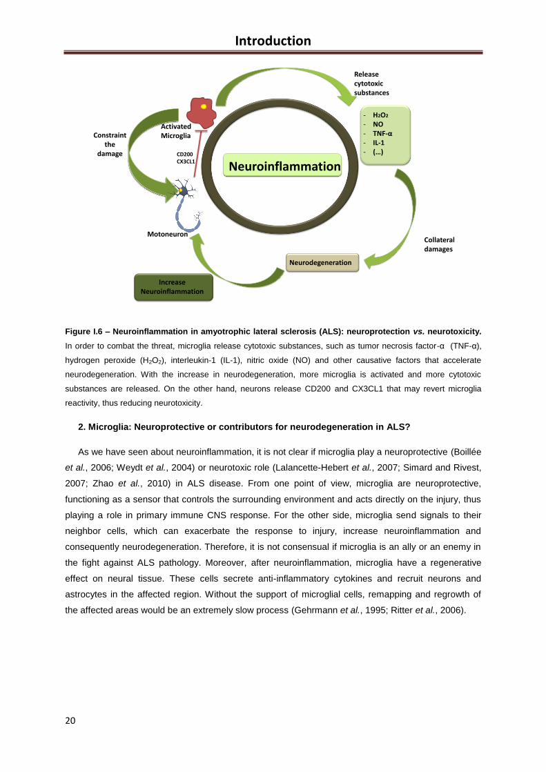
Introduction
20
Figure I.6 – Neuroinflammation in amyotrophic lateral sclerosis (ALS): neuroprotection vs. neurotoxicity.
In order to combat the threat, microglia release cytotoxic substances, such as tumor necrosis factor-α (TNF-α),
hydrogen peroxide (H2O2), interleukin-1 (IL-1), nitric oxide (NO) and other causative factors that accelerate
neurodegeneration. With the increase in neurodegeneration, more microglia is activated and more cytotoxic
substances are released. On the other hand, neurons release CD200 and CX3CL1 that may revert microglia
reactivity, thus reducing neurotoxicity.
2. Microglia: Neuroprotective or contributors for neurodegeneration in ALS?
As we have seen about neuroinflammation, it is not clear if microglia play a neuroprotective (Boillée
et al., 2006; Weydt et al., 2004) or neurotoxic role (Lalancette-Hebert et al., 2007; Simard and Rivest,
2007; Zhao et al., 2010) in ALS disease. From one point of view, microglia are neuroprotective,
functioning as a sensor that controls the surrounding environment and acts directly on the injury, thus
playing a role in primary immune CNS response. For the other side, microglia send signals to their
neighbor cells, which can exacerbate the response to injury, increase neuroinflammation and
consequently neurodegeneration. Therefore, it is not consensual if microglia is an ally or an enemy in
the fight against ALS pathology. Moreover, after neuroinflammation, microglia have a regenerative
effect on neural tissue. These cells secrete anti-inflammatory cytokines and recruit neurons and
astrocytes in the affected region. Without the support of microglial cells, remapping and regrowth of
the affected areas would be an extremely slow process (Gehrmann et al., 1995; Ritter et al., 2006).
Neuroinflammation
Activated MicrogliaConstraint
the damage
- H2O2
- NO- TNF-α- IL-1- (…)
Neurodegeneration
Increase Neuroinflammation
CD200CX3CL1
Motoneuron
Release cytotoxic substances
Collateral damages
Introduction
21
2.1 The different phenotypes
One of the main features of microglial cells is their plasticity, which is a crucial strategy for the
proper functioning of the immune defense of the nervous system (Gehrmann et al., 1995). Microglia
can change their structure based on its current location and function (Figure I.7 A). The different
phenotypes experienced by microglia are defined based on morphological, molecular, and functional
characteristics (Colton and Wilcock, 2010). In physiologic conditions, microglial cells have long
branching processes and a small cellular body (Kettenmann et al., 2011), a condition known as a
“resting state”, where the cell body of ramified microglia remains relatively static. Nevertheless, their
branches are constantly moving and surveying the environment, thus contributing to the maintenance
of the homeostasis, through cross-talk with astrocytes and neurons (Evans et al., 2013; Philips and
Robberecht, 2011). In the resting state, branches have a huge sensitivity to slight changes in
physiological conditions. It is still a matter of debate which is the primary cause that induces the
transformation for "resting" phenotype, although in cell culture studies, astrocyte conditioned medium
increased ramification of cultured blood monocytes (Sievers et al., 1994). Combining astrocyte
conditioned medium with ATP or adenosine yield a phenotype with more extensive ramification,
indicating that purines are not the only ramification-inducing factors of microglia (Wollmer et al., 2001).
Other candidates are cytokines released from astrocytes, such as transforming growth factor-β (TGF-
β), macrophage colony-stimulating factor (M-CSF) and granulocyte macrophage colony stimulating
factor (GM-CSF) (Schilling et al., 2001). Activation of chloride channels also seems to be necessary
for this morphological transformation (Kettenmann et al., 2011).
In case of disturbance of nervous system homeostasis, microglial cells change their
phenotype to an activated stage, which can be divided into M1 and M2 phenotypes (Gordon, 2003;
Henkel et al., 2009). Classical activation of microglia consists in the change to the M1 phenotype,
which is mainly neurotoxic, characterized by the release of pro-inflammatory cytokines, such as TNF-
α, IL-1β and interleukin-12 (IL-12), chemokines, proteases and redox species like NO (Durafourt et al.,
2012; Henkel et al., 2009; Kraft and Harry, 2011; Nakajima et al., 2003; Nayak et al., 2010); and the
alternative activation consist in the change to the M2 phenotype, which is mainly neuroprotective and
is associated with resolution of tissue homeostasis (Hovden et al., 2013), aiming the repair of damage
tissues. The production and release of anti-inflammatory cytokines like IL-4, IL-13, IL-10 and
transforming growth factor-beta (TGF-β) is the predominant induction signal (Colton, 2009; Glezer et
al., 2007; Ledeboer et al., 2000; Suzuki et al., 2005).
During their activation, microglia undergo through some morphological alterations, such as the
thickening and retraction of branches, uptake of major histocompatibility complex (MHC) class I/II
proteins, secretion of cytotoxic factors and secretion of recruitment molecules and pro-inflammatory
signaling molecules.
There are several factors that may activate microglia: cell necrosis factors, glutamate receptor
agonists, lipopolysaccharide (LPS), pro-inflammatory cytokines, and changes in extracellular
potassium (a marker of cellular disruption). Pro-inflammatory mediators including monocyte
chemoattractant protein 1 and IL-8 are present in the CSF of patients with ALS (Mantovani et al.,
2009).
Introduction
22
When activated, microglia can be found in the motor cortex and SC of ALS patients and the
intensity of this activation seems to be related with the severity of upper MN injury. In addition, it is
often associated with infiltration of T lymphocytes (Lasiene and Yamanaka, 2011; Philips and
Robberecht, 2011).
According to Beers and colleagues (2011), until the end-stage of ALS microglia presents a
neuroprotective role, whereupon levels of pro-inflammatory cytokine IL-1β and TNF-α increase, and
levels of NADPH oxidase are increased. Thus, in mSOD1 mice model, during initial stages of ALS,
microglia has an M2 phenotype that supports neuronal survival. Nevertheless, with the advance of the
disease course, microglial activation became skewed towards an M1 phenotype (Lewis et al., 2012).
There is also another phenotype with anti-inflammatory profile, known as acquired deactivation,
that consists in an phenotype with distinct gene profile from alternative activation but which also down-
regulates the response of innate immune system. Since this state is anti-inflammatory, it inhibits the
production of pro-inflammatory cytokines and increase the production of anti-inflammatory cytokines
(Colton, 2009).
Finally, it was discovered the dystrophic or senescent microglia, which has an abnormal
cytoplasmic structure, such as disbranched, atrophic, fragmented or unusually tortuous processes,
with spheroidal or bulbous swellings (Streit, 2006). An hallmark of this phenotype is the fragmentation
of the cytoplasm in microglia, a process called cytorrhexis (Streit and Xue, 2009), which illustrates the
loss of microglia functionality The incidence of this form of microglia increases with the aging and have
been reported in neurodegenerative diseases such as Alzheimer’s disease or schizophrenia,
indicating microglial deterioration in these diseases (Streit, 2006).
2.2 The migration to the injured tissue
The migration is a crucial event for the microglial cells to travel to the lesion sites and is triggered
by the gradient formed by several chemotactic agent, such as purines, neuregulin 1 (NRG1),
complement components, and chemokines such as Chemokine (C-C motif) ligand (CCL2) which is
exclusively expressed in damaged neurons (Figure I.7 B). Adenosine-5'-triphosphate and adenosine
diphosphate (ATP and ADP, respectively) have been widely studied as a chemoattractants of
microglial cells since that these nucleotides can diffuse rapidly and was been shown that they can
induce membrane ruffling and attract microglia (Calvo and Bennett, 2012). This process involves the
G protein-coupled receptor P2Y12 (Honda et al., 2001), which when are knocked out in mice seems to
decrease the ability of microglia to migrate to the site of injury (Haynes et al., 2006).
2.3 Phagocytosis
Phagocytosis is a form of endocytosis involving the vesicular internalization of solid particles, such
as pathological agents or cell debris (Napoli and Neumann, 2009) and is also a process undertaken by
microglia, as they derived from myeloid lineage and share various similarities with peripheral
macrophages (Figure I.7 B).
Introduction
23
Phagocytosis in the CNS is initiated by the release of “find-me” signals, which recruits microglia to
the site of injury. Upon recognition of cell-surface signals on target cells, microglia initiates the uptake
and subsequent responses (Neher et al., 2012). Though phagocytic process, microglia is able to
engulf the debris, such as damaged or apoptotic cells, namely neurons, DNA fragments or plaques,
and secrete either pro- or anti-inflammatory cytokines, depending on the type of receptor that has
been stimulated. As described by Napoli and Neumann (2009), the uptake of apoptotic cells occurs
with production of anti-inflammatory cytokines like TGF-β thus restraining inflammation. However,
microglia can recognize pathogens-associated molecular patterns (PAMPs) of pathogenic agents by
toll like receptors (TLRs) pathway, leading to a release of pro-inflammatory cytokines, such as tumor
necrosis factor-α (TNF-α) or NOS-2 (Napoli and Neumann, 2009). Interestingly, phagocytosis is
impaired in dysfunctional situations. Moreover, this phagocytic microglia interacts with astrocytes and
neural cells to constrain the damage as quickly as possible, avoiding harmful effects to healthy cells
(Aloisi, 2001; Gehrmann et al., 1995).
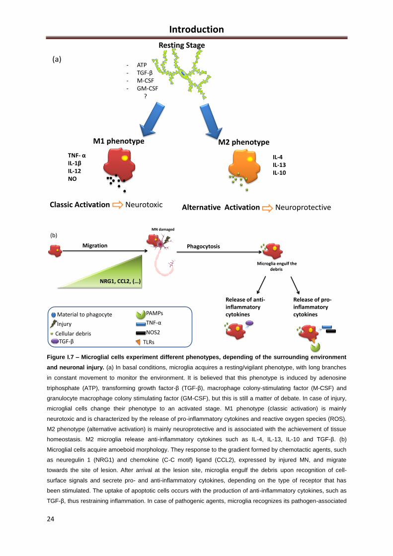
Introduction
24
Figure I.7 – Microglial cells experiment different phenotypes, depending of the surrounding environment
and neuronal injury. (a) In basal conditions, microglia acquires a resting/vigilant phenotype, with long branches
in constant movement to monitor the environment. It is believed that this phenotype is induced by adenosine
triphosphate (ATP), transforming growth factor-β (TGF-β), macrophage colony-stimulating factor (M-CSF) and
granulocyte macrophage colony stimulating factor (GM-CSF), but this is still a matter of debate. In case of injury,
microglial cells change their phenotype to an activated stage. M1 phenotype (classic activation) is mainly
neurotoxic and is characterized by the release of pro-inflammatory cytokines and reactive oxygen species (ROS).
M2 phenotype (alternative activation) is mainly neuroprotective and is associated with the achievement of tissue
homeostasis. M2 microglia release anti-inflammatory cytokines such as IL-4, IL-13, IL-10 and TGF-β. (b)
Microglial cells acquire amoeboid morphology. They response to the gradient formed by chemotactic agents, such
as neuregulin 1 (NRG1) and chemokine (C-C motif) ligand (CCL2), expressed by injured MN, and migrate
towards the site of lesion. After arrival at the lesion site, microglia engulf the debris upon recognition of cell-
surface signals and secrete pro- and anti-inflammatory cytokines, depending on the type of receptor that has
been stimulated. The uptake of apoptotic cells occurs with the production of anti-inflammatory cytokines, such as
TGF-β, thus restraining inflammation. In case of pathogenic agents, microglia recognizes its pathogen-associated
Resting Stage
- ATP- TGF-β- M-CSF- GM-CSF
?
M1 phenotype
Classic Activation Neurotoxic Alternative Activation Neuroprotective
M2 phenotype
TNF- αIL-1βIL-12NO
IL-4IL-13IL-10
(a)
Migration
NRG1, CCL2, (…)
MN damaged
Phagocytosis
Microglia engulf thedebris
Release of anti-inflammatorycytokines
Release of pro-inflammatorycytokines
Cellular debris
Injury
Material to phagocyte
TGF-β TLRs
PAMPs
TNF-α
NOS2
(b)
Introduction
25
molecular patterns (PAMPs) by toll-like receptors (TLRs), leading to the release of pro-inflammatory cytokines,
such as tumor necrosis factor-α (TNF-α), and activation of nitric oxide synthase-2 (NOS2).
2.4 The role of microglia in ALS
The activation of microglia and the infiltration of lymphocytes indicate an inflammatory event in the
CNS pathology of ALS (Carpentier et al., 2008). As previously described, one of the most accepted
hypotheses is based on the formation of protein aggregates as a first dysfunctional cellular process of
ALS that mediates the other ones. Then, microglia, astrocytes and the innate system including
monocytes, macrophages and natural killer cells respond as a first line of defense. It is believed that,
this response is triggered by mSOD1. Then, the adaptive immune system is activated. Throughout the
progression of the disease, CD4+CD25
+ regulatory T cells (Treg, secreting anti-inflammatory
cytokines) and other neuroprotective cells decrease (Kipnis et al., 2004), M2 microglia phenotype
switch to M1 microglia phenotype and the number of CD8+(a receptor predominantly expressed on
the surface of cytotoxic T cells) increase (Hovden et al., 2013). In ALS, the activation of microglial cells
was shown by the discovery of the signal transducer and activator of transcription-3 (STAT3) in post
mortem samples of ALS patients (Shibata et al., 2009). STAT3 is a signal transducer and activator of
transcription that mediates cellular responses to IL-6 family members, in the development,
differentiation, immunity, metabolism and cancer (Grivennikov and Karin, 2010).
Moreover, Meissner and colleagues (2010) concluded that IL-1β is an important pro-inflammatory
cytokine released by microglia in mSOD1G93A mice that activate M1 phenotype, accelerating the
progression of the disease (Meissner et al., 2010). In addition, these events can be reversed by
administration of the IL-1 receptor antagonist. The mSOD1 protein can also act as a powerful activator
of microglia. Indeed, the presence of this mutant protein reduces the expression of neurotrophic
factors such as IGF-1 and BNDF produced by microglia, which will have a deleterious role in ALS MN
(Evans et al., 2013). Using in vivo models, microglial proliferation and activation was shown to occur at
earlier disease stages and to increase with ALS progression (Moisse and Strong, 2006). However,
recently, the elimination of mSOD1 from microglia revealed to not change the onset but to slow
disease progression by 50% (Ferraiuolo et al., 2011).These findings suggest that the onset and the
progression of the disease have different features, which opens new horizons for target driven
therapies.
3. Different models for the study of neurodegeneration in ALS
The study of human neurodegenerative diseases evidences the impossibility to study the cellular
and molecular pathological events in real time or safely and repeatedly remove tissue for analyze
(Turner et al., 2013). The existence of different experimental models in ALS may is considered a
useful tool for providing knowledge at both cellular and molecular levels, to thereby develop more
effective therapies.
In this chapter, we will just focus on the experimental models that will be used in the studies that
comprise the present Thesis, namely MN-like monocultures, SC organotypic cultures and transgenic
Introduction
26
SOD1 animal models. However, there are much more models used in ALS research, such as worms,
flies, fish, mice, rats (Muyderman et al., 2009) and diverse cell types (Cashman et al., 1992).
3.1 Cell models
3.1.1 NSC-34 cells
NSC-34 cell line, a hybrid cell line, is a fusion of MN-enriched embryonic day 12-14 of SC cells with
aminopterin-sensitive mouse neuroblastoma (Cashman et al., 1992). NSC-34 cells, are considered a
model of ALS when transfected with human SOD1 containing the mutation in G93A, revealing some
features of MN degeneration, such as mitochondrial dysfunction (Raimondi et al., 2006) and Golgi
apparatus fragmentation (Gomes et al., 2008). This cell line is used to dissect the complexity of
mechanisms that involve mSOD1 toxicity (Atkin et al., 2006; Cozzolino et al., 2008; Rizzardini et al.,
2005).
The establishment of a cell line of immortalized neurons in culture is a major challenge and due to
its intrinsic properties, such as their null capacity to proliferate when they are completely differentiated.
Thus, the use of a hybrid cell line of neuroblastoma (with high proliferative capacity due to the
presence of the gene N-myc, an oncogene involved in cell proliferation) together with SC MN solved
the problem (Cashman et al., 1992) Morphological and physiological properties of such cells reveal
the suitability of the model when it is considered properties as acetylcholine synthesis, storage and
release; extension of processes; generation of an action potential; formation of contacts with cultured
myotubes and expression of neurofilament proteins among others (Cashman et al., 1992; Tovar et al.,
2009).
When accessing the viability of NSC-34 after exposure to a selection of neurotoxic chemicals, it
was observed that the cells respond to all of them (Durham et al., 1993). The similarity in the
production of the action potential to several ion channel blockers between this cell line and that of
primary MN in culture, corroborate NSC-34 cells as an effective model for studying neurotoxicity in
ALS. Most important is that NSC-34 cells adhere specifically to the leucine-arginine-glutamate motif of
S-laminin, what shows that these hybrid cells uniquely express MN phenotypic features. This property
was not revealed by most neural cell lines (Hunter et al., 1991). However, NSC-34 cells are not
suitable to evaluate the effect of agents on the synaptic transmission (Durham et al., 1993).
It has been recently established in our lab that NSC-34/hSOD1G93A cells reveal features of
mitochondrial dysfunction, energy impairment, oxidative stress, as well as apoptosis and
inflammatory-related processes. All of these events are common processes in the transgenic mice
model and in ALS patients. Studies in our laboratory have also shown that this model may be used to
evaluate the therapeutic efficacy of compounds, such as the glycoursodeoxycholic acid (GUDCA), a
bile acid-glycine conjugate. Based on the overall aspects mentioned, we decided to implement in our
laboratory a model of mixed cultures to analyze the interaction between MN-like cells (NSC-34) and
N9 (a microglial cell line), as will be discussed below.
Introduction
27
3.1.2 Organotypic culture cells
The major problem of the most in vitro models is the difficulty to mimic what actually occurs in the
organism. Although primary cultures and mixed cultures give valuable information on the cell function
and interconnectivity, cellular processes should not be considered isolated, but rather integrated in a
system that involves cell cross-talk and signals from the entire neighborhood. The neurons coexist in a
complex network where the surrounding cells shape the biochemical, electrophysiological and
morphological features between themselves and the remaining cells around (Tovar et al., 2009).
Therefore, the organotypic cultures are a closer model to in vivo, since we do not isolate one cell type
in particular but, instead, we preserve the tissue structure when culturing the entire SC slice.
According to Tovar and colleagues (2009), these sections can be cultured for 3 months, keeping up
the metabolic capacity of MN, such as choline acetyltransferase and acetylcholinesterase activities
(Delfs et al., 1989). Moreover, the use of SC cultured slices have benefits over animal models, such
as the easy access and precise control of the extracellular environment, which makes the model
powerful to screen the efficacy of potential medicines (Su et al., 2011).
3.2 Animal models
Mice are widely used as an animal model in biological sciences, since they react to a disease
induction or treatment as humans. In general, the closer the organism is to the man in the evolutionary
tree, the better will be the model. However, careful is still necessary, since even non-human primates
are not entirely predictive for human outcomes in drug development studies (Kari et al., 2007). In
general, a model is robust when the disease (spontaneous or induced) has similarity with the human
disease and this similarity is significant in the context of the study. Rodents are largely used as a study
model in neurodegenerative diseases because rodents and humans use the same neurotransmitters,
receptors, proteins for synaptic vesicle release and recycling and similar signaling mechanisms.
The recent discovery of ALS-associated mutations in the DNA-binding protein 43 (TDP-43) gene
lead to the generation of alternative animal models of ALS, since these mutations are associated with
both sALS and fALS (Xu et al., 2011). Nevertheless, mutants of human SOD1 mice are still the most
common model to investigate disease pathogenesis (Tovar et al., 2009). The success of mice
expressing mutant proteins associated with fALS (such as mSOD1) has become a powerful tool to
study mechanisms of MN loss, particularly with different forms of SOD1 (Van Den Bosch, 2011). A
huge advance in the ALS research was the discovery that about 20% of fALS cases were due to
mSOD1 (Rosen, 1993). This discovery spurred the creation of a transgenic mice expressing human
mSOD1. It was in 1994 that Gurney and colleagues developed the first transgenic mice expressing
human protein with the G93A mutation, and this was the first obtained lab model based on a known
cause of ALS (Gurney et al., 1994). This transgenic mouse overexpressing mSOD1G93A was obtained
by genetic engineering, inserting multiple copies of human genomic SOD1 into the mouse genome.
Moreover, these mice show symptoms similar to humans such as the progressive hind limb weakness
that culminate in paralysis and ultimately death, as a result of MN injury (Van Den Bosch, 2011).
Introduction
28
Besides these first transgenic mice, several other models that overexpress human SOD1 were
created, but with other mutations (G37R, G85R, G86R, D90A). Interestingly, these new mice have
demonstrated identical phenotype as the mSOD1G93A (Bruijn et al., 1997; Jonsson et al., 2006; Ripps
et al., 1995; Wong et al., 1995) but comparatively are not so often used. Indeed it was in the mSOD1
model that most studies evidenced the non-cell autonomous neurodegeneration (Boillée et al., 2006),
excitotoxicity (Corona et al., 2007), apoptosis (Hervias et al., 2006), protein aggregation (Wood et al.,
2003), axonal dysfunction (Sasaki et al., 2005), mitochondria failure (Cassina et al., 2008) and both
endoplasmic reticulum (Nishitoh et al., 2008) and oxidative stress (Harraz et al., 2008).
The mSOD1 mouse model was also widely used to access new therapies, and minocycline is a
good example of this. This drug was shown to inhibit microglial activation, a property also evidenced
by other studies using two distinct mSOD1 mouse models developed in the same year (Kriz et al.,
2002; Van Den Bosch et al., 2002; Zhu et al., 2002).
In spite that mSOD1 mice and rats are accepted as optimal models to evaluate selective MN death,
they have been also disappointing once no effective drugs in rodents revealed to be effective in
clinical trials with humans (Van Den Bosch, 2011).
4. Therapeutic strategies: Yesterday, today and tomorrow
From the nearly two decades, knowledge about ALS has evolved considerably. Now, we know
more about genetic causes in fALS cases, although not so much about sALS (Venkova-Hristova et al.,
2012). So far, researchers in the ALS field have joined efforts to find an effective therapeutic agent for
the disease. However, to date, the only FDA approved drug is riluzole (Kiernan et al., 2011).
A challenge is still to distinguish which therapies are delaying the onset of the disease versus
prolonging the compensatory pre-clinical period, in which the disease process is progressing in the
absence of clinical signs of motor dysfunction. For example in mSOD1 mice, it is reported that several
anomalies are present before the onset of clinical symptoms (Ferraiuolo et al., 2011), such as
behavioral motor changes (Bories et al., 2007; van Zundert et al., 2008), MN electrophysiological
dysfunction (Mead et al., 2011), mitochondrial swelling and vacuolization (Bendotti et al., 2001) and
transcriptome changes in an attempt to increase MN energy provision (Ferraiuolo et al., 2007).
The progression of the disease constitutes a huge therapeutic problem. For example, patient
groups are very small and often include individuals in different disease stages, making difficult to
obtain strong conclusions from the clinical trials. Moreover, drugs that showed benefits in animals
when administered pre-symptomatically or before ALS onset, failed in humans since that diagnosis is
not confirmed before symptom emergence (Hovden et al., 2013). The setbacks that occur during the
clinical trials led to the conclusion of a combination therapy, as a better solution. However there is still
not an effective combination due to the complexity of the disease. Furthermore, despite the immune
system be considered a promising target, it is possible that the action of at one specific target may be
beneficial in certain ALS phases, while dangerous in others (Hovden et al., 2013).
According to several researchers, a perfect combination cocktail may combine drugs that target
protein aggregation, immune modulation and glutamate excitotoxicity (Hovden et al., 2013). In
summary, research on ALS has been characterized by same success, but also frustration.
Introduction
29
Nevertheless, we must emphasize that scientific progress has been continuous. We are probably
closer to find new hallmarks for clinical success and to further carry that success to other
neurodegenerative disorders (Venkova-Hristova et al., 2012).
4.1 Microglia as a therapeutic target in the future
The complexity of the cross-talk between microglia and neurons and the role of microglia in the
immune system and neuroinflammation suggests that microglia is a very promising target for the
therapeutic advances against ALS progression.
Indeed, studies with the transgenic mice with human mSOD1, have shown that diminishing the
mutant levels of mSOD1 in microglia had little effect on the early disease phase but slowed later
disease progression (Boillée et al., 2006). Furthermore, elimination of microglia using clodronate
liposomes, which specifically target monocyte/macrophage system (such as microglia cells), and
addition of new microglia significantly slowed disease progression and prolonged survival of the
transgenic ALS mice after bone marrow transplantation (BMT). So, replacement of microglia by a
healthier cell may ameliorate neural cell microenvironment; however the mechanisms and functional
implications of this replacement need to be further investigated (Lee et al., 2012). Nevertheless,
improvement by BMT may not be efficient enough since microglia are replaced by tissue-resident
microglia rather than by bone marrow cells (BMCs) (Ohnishi et al., 2009).
4.2 GUDCA: Beneficial effects in ALS
So far, there is no successful drug for ALS treatment and search for novel therapeutic agents
that can prevent or delay MN degeneration is of a great importance. In this context, we propose to
study the efficacy of glycoursodeoxycholic acid (GUDCA), a conjugated species of ursodeoxycholic
acid (UDCA) with glycine, on the prevention of neuronal degeneration in the cellular models of ALS
since it showed ability to counteract neuronal death and synaptic changes in models of
neurodegeneration, such as neonatal unconjugated bilirubin encephalopathy (Silva et al., 2012).
Bile acids are acidic steroids synthesized in hepatocytes from cholesterol. Primary bile acids,
the major constituents of bile, are produced in the liver and when secreted into the intestine, they can
be metabolized into secondary bile acids where they play crucial roles such as solubilization of lipids
(Amaral et al., 2009). Some bile acids are cytotoxic (Bayerdorffer et al., 1993), but others not, due to
small changes in the chemical structure that are found between species (Hofmann and Roda, 1984).
UDCA, for example, helps in the regulation of cholesterol levels and have several functions in the liver,
such as maintenance of mitochondrial integrity, immune-modulation and anti-apoptotic role. During
the process of conjugation in the liver, UDCA can link with taurine or with glycine, originating
tauroursodeoxycholic acid (TUDCA) or GUDCA, respectively. Once GUDCA is formed in larger
amount (close to 80%) (Lazaridis et al., 2001), this conjugate is of major clinical relevance derived
from the utilization of UDCA in several diseases (Brites, 2002). Thus, according to some authors,
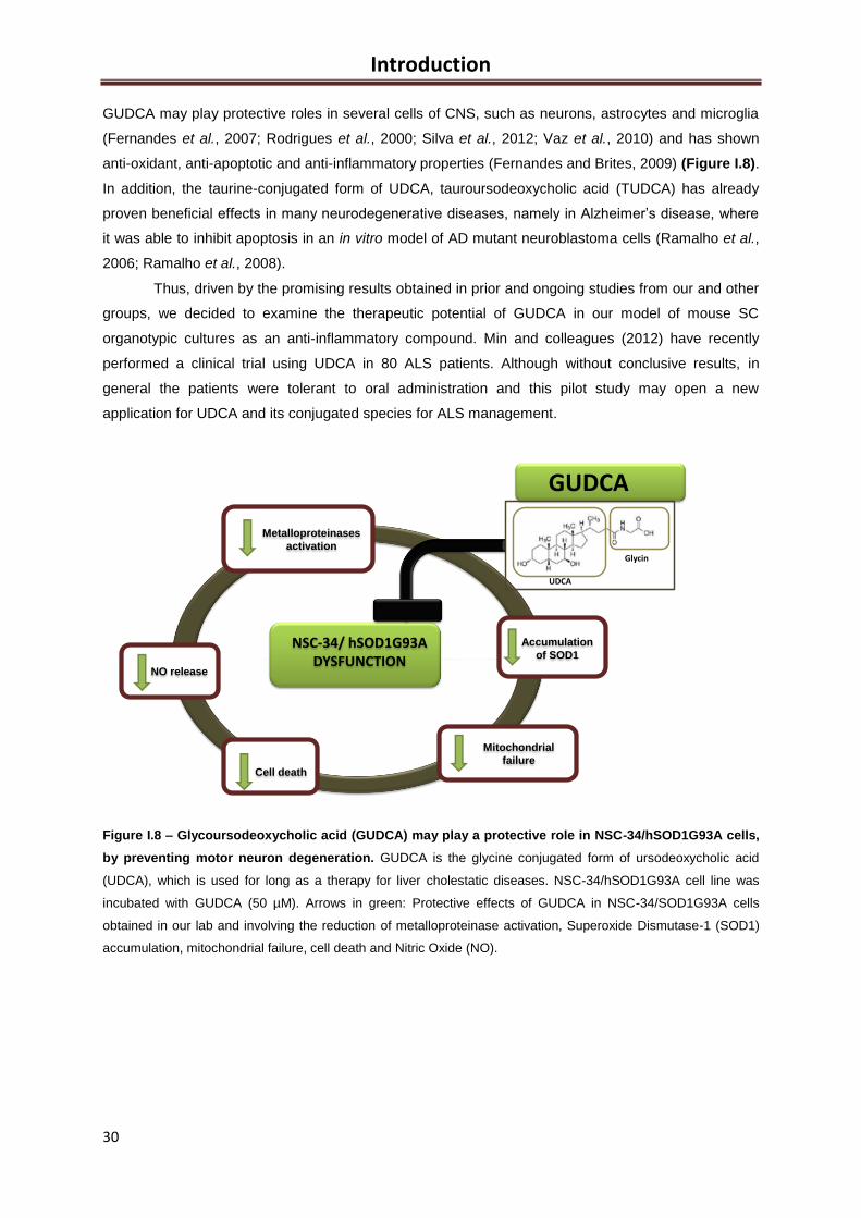
Introduction
30
GUDCA may play protective roles in several cells of CNS, such as neurons, astrocytes and microglia
(Fernandes et al., 2007; Rodrigues et al., 2000; Silva et al., 2012; Vaz et al., 2010) and has shown
anti-oxidant, anti-apoptotic and anti-inflammatory properties (Fernandes and Brites, 2009) (Figure I.8).
In addition, the taurine-conjugated form of UDCA, tauroursodeoxycholic acid (TUDCA) has already
proven beneficial effects in many neurodegenerative diseases, namely in Alzheimer’s disease, where
it was able to inhibit apoptosis in an in vitro model of AD mutant neuroblastoma cells (Ramalho et al.,
2006; Ramalho et al., 2008).
Thus, driven by the promising results obtained in prior and ongoing studies from our and other
groups, we decided to examine the therapeutic potential of GUDCA in our model of mouse SC
organotypic cultures as an anti-inflammatory compound. Min and colleagues (2012) have recently
performed a clinical trial using UDCA in 80 ALS patients. Although without conclusive results, in
general the patients were tolerant to oral administration and this pilot study may open a new
application for UDCA and its conjugated species for ALS management.
Figure I.8 – Glycoursodeoxycholic acid (GUDCA) may play a protective role in NSC-34/hSOD1G93A cells,
by preventing motor neuron degeneration. GUDCA is the glycine conjugated form of ursodeoxycholic acid
(UDCA), which is used for long as a therapy for liver cholestatic diseases. NSC-34/hSOD1G93A cell line was
incubated with GUDCA (50 µM). Arrows in green: Protective effects of GUDCA in NSC-34/SOD1G93A cells
obtained in our lab and involving the reduction of metalloproteinase activation, Superoxide Dismutase-1 (SOD1)
accumulation, mitochondrial failure, cell death and Nitric Oxide (NO).
Metalloproteinases
activation
Cell death
Mitochondrial
failure
NO release
NSC-34/ hSOD1G93A DYSFUNCTION
Accumulation
of SOD1
Glycin
UDCA
GUDCA
Introduction
31
5. Aims
The aims of this thesis are to better understand some of the main molecular hallmarks involved in
motor neuron (MN) degeneration in ALS, as well as to explore the cross-talk between MN and
microglia in ALS, by using three different experimental models: (i) pure MN cultures (NSC-34 cell line);
(ii) mixed MN-microglial cultures (NSC-34/N9 cell lines); (iii) organotypical slices from mice spinal cord
(SC).
Therefore, the specific aims are:
1. To explore the processes involved in MN-mitochondrial dysfunction along ALS progression, namely
mitochondrial dynamics (fusion/fission) and axonal transport (anterograde/retrograde), together with
cell death mechanisms. For this, NSC-34 cell line expressing either human SOD1 wt or mutated in
G93A (NSC-34/hSOD1wt or NSC-34/hSOD1G93A, respectively) will be differentiated for 1-7 days (DIV)
in order to select the mechanisms involved in MN mitochondrial dysfunction in ALS for each stage of
SOD1 accumulation (prior or after SOD1 accumulation, respectively 1 or 4-7 DIV). NSC-34/hSOD1wt
will be used as control.
2. To evaluate the contribution of microglia to MN degeneration in mixed cultures of MN-microglia. For
this we will produce and characterize a mixed culture of either NSC-34/hSOD1wt or NSC-
34/hSOD1G93A with N9 microglia and we will focus on the parameters that will be found altered in the
NSC-34/hSOD1G93A monoculture.
3. To set-up and characterize organotypic cultures obtained from SC of ALS-transgenic mice carrying
the same human SOD1 mutation (TgSOD1-G93A mice). Here, we will test the potential
neuroprotective benefits of glycoursodeoxycholic acid (GUDCA).
Altogether, the final purpose of the project is to explore the mechanisms of MN degeneration in ALS
and the role of microglia on them, to discover intervenient targets and drivers, while assessing
modulatory effects by GUDCA. If we succeed, future research should test ways to design molecules
that although having the benefits of GUDCA better achieve the brain parenchyma.

32
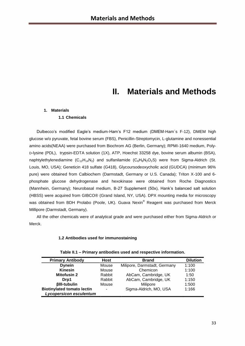
Materials and Methods
33
II. Materials and Methods
1. Materials
1.1 Chemicals
Dulbecco’s modified Eagle’s medium-Ham’s F12 medium (DMEM-Ham´s F-12), DMEM high
glucose w/o pyruvate, fetal bovine serum (FBS), Penicillin-Streptomycin, L-glutamine and nonessential
amino acids(NEAA) were purchased from Biochrom AG (Berlin, Germany); RPMI-1640 medium, Poly-
D-lysine (PDL), trypsin-EDTA solution (1X), ATP, Hoechst 33258 dye, bovine serum albumin (BSA),
naphtylethylenediamine (C12H14N2) and sulfanilamide (C6H8N2O2S) were from Sigma-Aldrich (St.
Louis, MO, USA); Geneticin 418 sulfate (G418), Glycoursodeoxycholic acid (GUDCA) (minimum 96%
pure) were obtained from Calbiochem (Darmstadt, Germany or U.S. Canada); Triton X-100 and 6-
phosphate glucose dehydrogenase and hexokinase were obtained from Roche Diagnostics
(Mannhein, Germany); Neurobasal medium, B-27 Supplement (50x), Hank’s balanced salt solution
(HBSS) were acquired from GIBCO® (Grand Island, NY, USA). DPX mounting media for microscopy
was obtained from BDH Prolabo (Poole, UK). Guava Nexin® Reagent was purchased from Merck
Millipore (Darmstadt, Germany).
All the other chemicals were of analytical grade and were purchased either from Sigma-Aldrich or
Merck.
1.2 Antibodies used for immunostaining
Table II.1 – Primary antibodies used and respective information.
Primary Antibody Host Brand Dilution
Dynein Mouse Milipore, Darmstadt, Germany 1:100 Kinesin Mouse Chemicon 1:100
Mitofusin 2 Rabbit AbCam, Cambridge, UK 1:50 Drp1 Rabbit AbCam, Cambridge, UK 1:150
βIII-tubulin Mouse Milipore 1:500 Biotinylated tomato lectin
Lycopersicon esculentum - Sigma-Aldrich, MO, USA 1:166
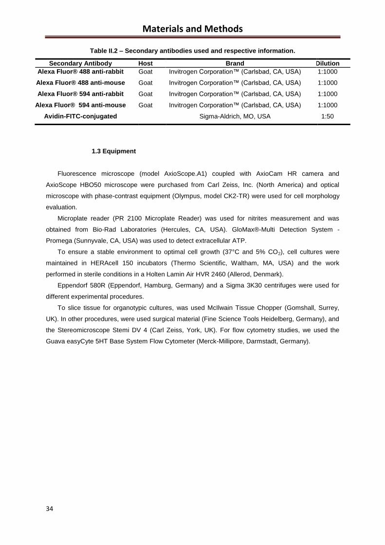
Materials and Methods
34
Table II.2 – Secondary antibodies used and respective information.
Secondary Antibody Host Brand Dilution
Alexa Fluor® 488 anti-rabbit Goat Invitrogen Corporation™ (Carlsbad, CA, USA) 1:1000
Alexa Fluor® 488 anti-mouse Goat Invitrogen Corporation™ (Carlsbad, CA, USA) 1:1000
Alexa Fluor® 594 anti-rabbit Goat Invitrogen Corporation™ (Carlsbad, CA, USA) 1:1000
Alexa Fluor® 594 anti-mouse Goat Invitrogen Corporation™ (Carlsbad, CA, USA) 1:1000
Avidin-FITC-conjugated Sigma-Aldrich, MO, USA 1:50
1.3 Equipment
Fluorescence microscope (model AxioScope.A1) coupled with AxioCam HR camera and
AxioScope HBO50 microscope were purchased from Carl Zeiss, Inc. (North America) and optical
microscope with phase-contrast equipment (Olympus, model CK2-TR) were used for cell morphology
evaluation.
Microplate reader (PR 2100 Microplate Reader) was used for nitrites measurement and was
obtained from Bio-Rad Laboratories (Hercules, CA, USA). GloMax®-Multi Detection System -
Promega (Sunnyvale, CA, USA) was used to detect extracellular ATP.
To ensure a stable environment to optimal cell growth (37°C and 5% CO2), cell cultures were
maintained in HERAcell 150 incubators (Thermo Scientific, Waltham, MA, USA) and the work
performed in sterile conditions in a Holten Lamin Air HVR 2460 (Allerod, Denmark).
Eppendorf 580R (Eppendorf, Hamburg, Germany) and a Sigma 3K30 centrifuges were used for
different experimental procedures.
To slice tissue for organotypic cultures, was used McIlwain Tissue Chopper (Gomshall, Surrey,
UK). In other procedures, were used surgical material (Fine Science Tools Heidelberg, Germany), and
the Stereomicroscope Stemi DV 4 (Carl Zeiss, York, UK). For flow cytometry studies, we used the
Guava easyCyte 5HT Base System Flow Cytometer (Merck-Millipore, Darmstadt, Germany).
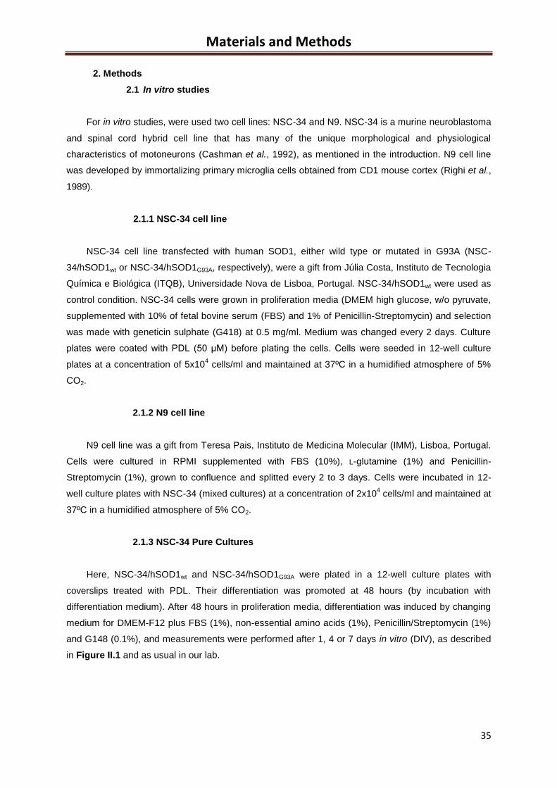
Materials and Methods
35
2. Methods
2.1 In vitro studies
For in vitro studies, were used two cell lines: NSC-34 and N9. NSC-34 is a murine neuroblastoma
and spinal cord hybrid cell line that has many of the unique morphological and physiological
characteristics of motoneurons (Cashman et al., 1992), as mentioned in the introduction. N9 cell line
was developed by immortalizing primary microglia cells obtained from CD1 mouse cortex (Righi et al.,
1989).
2.1.1 NSC-34 cell line
NSC-34 cell line transfected with human SOD1, either wild type or mutated in G93A (NSC-
34/hSOD1wt or NSC-34/hSOD1G93A, respectively), were a gift from Júlia Costa, Instituto de Tecnologia
Química e Biológica (ITQB), Universidade Nova de Lisboa, Portugal. NSC-34/hSOD1wt were used as
control condition. NSC-34 cells were grown in proliferation media (DMEM high glucose, w/o pyruvate,
supplemented with 10% of fetal bovine serum (FBS) and 1% of Penicillin-Streptomycin) and selection
was made with geneticin sulphate (G418) at 0.5 mg/ml. Medium was changed every 2 days. Culture
plates were coated with PDL (50 μM) before plating the cells. Cells were seeded in 12-well culture
plates at a concentration of 5x10⁴ cells/ml and maintained at 37ºC in a humidified atmosphere of 5%
CO2.
2.1.2 N9 cell line
N9 cell line was a gift from Teresa Pais, Instituto de Medicina Molecular (IMM), Lisboa, Portugal.
Cells were cultured in RPMI supplemented with FBS (10%), L-glutamine (1%) and Penicillin-
Streptomycin (1%), grown to confluence and splitted every 2 to 3 days. Cells were incubated in 12-
well culture plates with NSC-34 (mixed cultures) at a concentration of 2x10⁴ cells/ml and maintained at
37ºC in a humidified atmosphere of 5% CO2.
2.1.3 NSC-34 Pure Cultures
Here, NSC-34/hSOD1wt and NSC-34/hSOD1G93A were plated in a 12-well culture plates with
coverslips treated with PDL. Their differentiation was promoted at 48 hours (by incubation with
differentiation medium). After 48 hours in proliferation media, differentiation was induced by changing
medium for DMEM-F12 plus FBS (1%), non-essential amino acids (1%), Penicillin/Streptomycin (1%)
and G148 (0.1%), and measurements were performed after 1, 4 or 7 days in vitro (DIV), as described
in Figure II.1 and as usual in our lab.
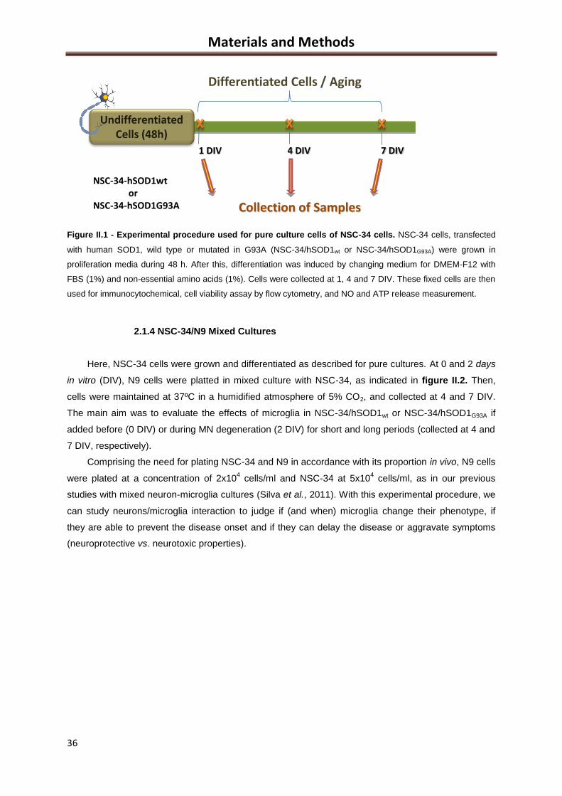
Materials and Methods
36
Figure II.1 - Experimental procedure used for pure culture cells of NSC-34 cells. NSC-34 cells, transfected
with human SOD1, wild type or mutated in G93A (NSC-34/hSOD1wt or NSC-34/hSOD1G93A) were grown in
proliferation media during 48 h. After this, differentiation was induced by changing medium for DMEM-F12 with
FBS (1%) and non-essential amino acids (1%). Cells were collected at 1, 4 and 7 DIV. These fixed cells are then
used for immunocytochemical, cell viability assay by flow cytometry, and NO and ATP release measurement.
2.1.4 NSC-34/N9 Mixed Cultures
Here, NSC-34 cells were grown and differentiated as described for pure cultures. At 0 and 2 days
in vitro (DIV), N9 cells were platted in mixed culture with NSC-34, as indicated in figure II.2. Then,
cells were maintained at 37ºC in a humidified atmosphere of 5% CO2, and collected at 4 and 7 DIV.
The main aim was to evaluate the effects of microglia in NSC-34/hSOD1wt or NSC-34/hSOD1G93A if
added before (0 DIV) or during MN degeneration (2 DIV) for short and long periods (collected at 4 and
7 DIV, respectively).
Comprising the need for plating NSC-34 and N9 in accordance with its proportion in vivo, N9 cells
were plated at a concentration of 2x10⁴ cells/ml and NSC-34 at 5x10
4 cells/ml, as in our previous
studies with mixed neuron-microglia cultures (Silva et al., 2011). With this experimental procedure, we
can study neurons/microglia interaction to judge if (and when) microglia change their phenotype, if
they are able to prevent the disease onset and if they can delay the disease or aggravate symptoms
(neuroprotective vs. neurotoxic properties).
NSC-34-hSOD1wtor
NSC-34-hSOD1G93A
1 DIV 4 DIV 7 DIV
UndifferentiatedCells (48h)
Collection of Samples
Differentiated Cells / Aging
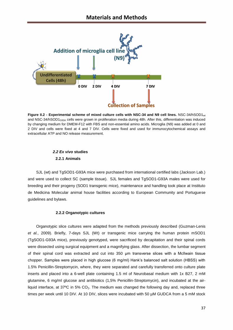
Materials and Methods
37
Figure II.2 - Experimental scheme of mixed culture cells with NSC-34 and N9 cell lines. NSC-34/hSOD1wt
and NSC-34/hSOD1G93A cells were grown in proliferation media during 48h. After this, differentiation was induced
by changing medium for DMEM-F12 with FBS and non-essential amino acids. Microglia (N9) was added at 0 and
2 DIV and cells were fixed at 4 and 7 DIV. Cells were fixed and used for immunocytochemical assays and
extracellular ATP and NO release measurement.
2.2 Ex vivo studies
2.2.1 Animals
SJL (wt) and TgSOD1-G93A mice were purchased from international certified labs (Jackson Lab.)
and were used to collect SC (sample tissue). SJL females and TgSOD1-G93A males were used for
breeding and their progeny (SOD1 transgenic mice), maintenance and handling took place at Instituto
de Medicina Molecular animal house facilities according to European Community and Portuguese
guidelines and bylaws.
2.2.2 Organotypic cultures
Organotypic slice cultures were adapted from the methods previously described (Guzman-Lenis
et al., 2009). Briefly, 7-days SJL (Wt) or transgenic mice carrying the human protein mSOD1
(TgSOD1-G93A mice), previously genotyped, were sacrificed by decapitation and their spinal cords
were dissected using surgical equipment and a magnifying glass. After dissection, the lumbar segment
of their spinal cord was extracted and cut into 350 μm transverse slices with a McIlwain tissue
chopper. Samples were placed in high glucose (6 mg/ml) Hank’s balanced salt solution (HBSS) with
1.5% Penicillin-Streptomycin, where, they were separated and carefully transferred onto culture plate
inserts and placed into a 6-well plate containing 1.5 ml of Neurobasal medium with 1x B27, 2 mM
glutamine, 6 mg/ml glucose and antibiotics (1,5% Penicillin-Streptomycin), and incubated at the air-
liquid interface, at 37ºC in 5% CO2. The medium was changed the following day and, replaced three
times per week until 10 DIV. At 10 DIV, slices were incubated with 50 µM GUDCA from a 5 mM stock
Collection of Samples
0 DIV 2 DIV 4 DIV 7 DIV
Addition of microglia cell line(N9)
UndifferentiatedCells (48h)
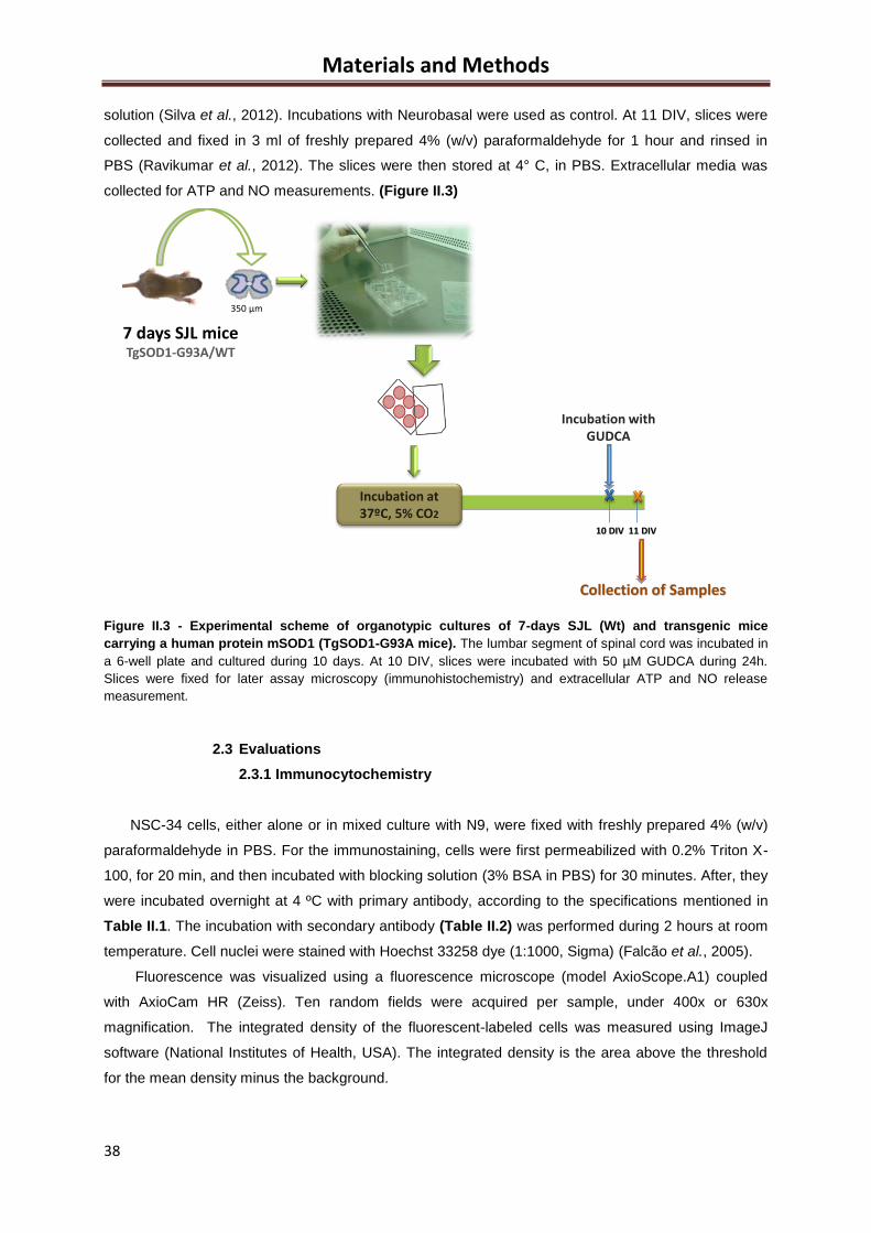
Materials and Methods
38
solution (Silva et al., 2012). Incubations with Neurobasal were used as control. At 11 DIV, slices were
collected and fixed in 3 ml of freshly prepared 4% (w/v) paraformaldehyde for 1 hour and rinsed in
PBS (Ravikumar et al., 2012). The slices were then stored at 4° C, in PBS. Extracellular media was
collected for ATP and NO measurements. (Figure II.3)
Figure II.3 - Experimental scheme of organotypic cultures of 7-days SJL (Wt) and transgenic mice
carrying a human protein mSOD1 (TgSOD1-G93A mice). The lumbar segment of spinal cord was incubated in
a 6-well plate and cultured during 10 days. At 10 DIV, slices were incubated with 50 µM GUDCA during 24h.
Slices were fixed for later assay microscopy (immunohistochemistry) and extracellular ATP and NO release
measurement.
2.3 Evaluations
2.3.1 Immunocytochemistry
NSC-34 cells, either alone or in mixed culture with N9, were fixed with freshly prepared 4% (w/v)
paraformaldehyde in PBS. For the immunostaining, cells were first permeabilized with 0.2% Triton X-
100, for 20 min, and then incubated with blocking solution (3% BSA in PBS) for 30 minutes. After, they
were incubated overnight at 4 ºC with primary antibody, according to the specifications mentioned in
Table II.1. The incubation with secondary antibody (Table II.2) was performed during 2 hours at room
temperature. Cell nuclei were stained with Hoechst 33258 dye (1:1000, Sigma) (Falcão et al., 2005).
Fluorescence was visualized using a fluorescence microscope (model AxioScope.A1) coupled
with AxioCam HR (Zeiss). Ten random fields were acquired per sample, under 400x or 630x
magnification. The integrated density of the fluorescent-labeled cells was measured using ImageJ
software (National Institutes of Health, USA). The integrated density is the area above the threshold
for the mean density minus the background.
10 DIV 11 DIV
Collection of Samples
Incubation at37ºC, 5% CO2
Incubation withGUDCA
350 µm
7 days SJL miceTgSOD1-G93A/WT
Materials and Methods
39
2.3.2 Immunohistochemistry
Slices of 7-days SJL Wt or TgSOD1-G93A mice were placed in blocking solution (2% FBS in
TBS-Triton 10%), for 1h at 25ºC. Then, cells were incubated overnight at 4ºC with primary antibody. In
the second day, after rinsing twice in TBS-Tween (0.05%), for 2x10 minutes at 25ºC, cells were
incubated during 1 h at 25ºC with secondary antibody. After two more washes, slices were washed
with PBS and incubated with DAPI stock 1 mg/ml for 5 minutes. After two washes for 10 minutes with
TBS-Tween (0,05%), slices were dehydrated with ethanol. Cells were then mounted in DPX, on a
microscope slide.
Fluorescence was visualized using a fluorescence microscope (model AxioScope.A1) coupled
with AxioCam HR (Zeiss). Overlapping fields were acquired (under 100x magnification) and merged in
Photoshop Software Massachusetts, USA). Twelve fields were acquired per sample in two
perpendicular axes, with the origin on the central canal of the spinal cord, under 100x magnification.
2.3.3 Quantifying the release of Nitric Oxide
Nitric oxide levels were indirectly quantified by measuring the concentration of nitrites (NO2) in the
extracellular media of NSC-34, either alone or in mixed culture with N9 cells, as well as in organotypic
culture extracellular media. Nitrites are a stable end product of NO.
Cell supernatants free from cellular debris were mixed with Griess reagent [1% (w/v)
sulphanilamide in 5% H3PO4 and 0.1% (w/v) N-1 naphtylethylenediamine, in a proportion of 1:1 (v/v)]
in 96-well tissue culture plates for 10 minutes in the dark, at room temperature. The absorbance at
540 nm was determined using a microplate reader (Bio-Rad Laboratories). A calibration curve was
used for each assay. All samples were measured in duplicate and the mean value was used (Vaz et
al., 2010).
2.3.4 Quantification of extracellular ATP
NSC-34, either alone or in mixed culture with N9 cells, as well as in organotypic culture
extracellular media were treated on ice to prevent degradation of ATP. For the determination of
extracellular ATP levels, the incubation media was collected and treated with 2 M of perchloric acid.
Then, the pH value was neutralized with 4 M KOH solution. To remove cellular debris, the samples
were centrifuged (Eppendorf, 5810R) during 5 min at 10,000 g and 4°C, between the different steps.
ATP levels were determined by an enzymatic assay and fluorescence intensity was quantified using a
GloMax®-Multi Detection System at λem 410-460 nm and λex 365 nm. A calibration curve of ATP was
used for each assay (Vaz et al., 2010).
2.3.5 Detection of Apoptosis/necrosis
After incubation of NSC-34 in12-wells culture plates at 1, 4 and 7DIV, extracellular media was
collected to 2 ml tubes and cells were detached by using a solution of tripsin 1x for 5 min of at 37ºC.
Materials and Methods
40
After adding FBS (to stop the action of trypsin), cells were collected and centrifuged at 700 g during 5
minutes (Eppendorf, 5810R). The supernatant was discharged and the pellet resuspended in 400 μL
of 1% BSA in PBS. The samples were added to 96-wells plates with Nexin Reagent® (Anexin
V/7AAD) and incubated at 20 min, protected from the light. After dilution with 50µl 1% BSA in PBS,
samples were analyzed on a Guava easyCyte 5HT Base System Flow Cytometer (Merck-Millipore).
5000 events per sample were counted. Three populations of cells can be distinguished in this assay:
viable cells (annexin V-PE and 7-AAD negative), early-apoptotic cells (annexin V-PE positive and 7-
AAD negative) and late stages of apoptosis or dead cells (annexin V-PE and 7-AAD positive) (adapted
from (Barateiro et al., 2012).
2.4 Statistical Analysis
Results of at least three different experiments were expressed as mean ± SEM for NSC-34
cultures either isolated or in mixed culture with N9. Comparisons between the different parameters
evaluated in wt and G93A NSC-34 cell line and in organotypic cultures from SC of TgSOD1-G93A or
WT mice were made using two-tailed Student’s t-test for equal or unequal variance, as appropriate.
Comparison of more than two groups in the parameters evaluated in mixed cultures with or without
microglia was done by one-way ANOVA using GraphPad Prism 5 (GraphPad Software, San Diego,
CA, USA) followed by multiple comparisons Bonferroni post-hoc correction. p<0.05 was considered
statistically significant and p<0.01 very significant.
41
III. Results
1. Isolated NSC-34 cells, a MN-like cell model
1.1 Evaluation of cellular viability of differentiated NSC-34 cells, transfected with
mutant SOD1 as a model of motoneuron degeneration in ALS
NSC-34 cell line is a hybrid cell line obtained by fusion of neuroblastoma cells with
motoneuron (MN)-enriched from mice spinal cord cell preparations, and expresses many of the
morphological and physiological properties of MN, such as extension of processes, formation of
contacts with cultured myotubes, synthesis and storage of acetylcholine (ACh), support of action
potentials and expression of neurofilament proteins (Cashman et al.,1992). In our model, we used
NSC-34 cell line that had been transfected either with wild type human SOD1 (NSC-34/hSOD1wt) or
mutated in G93A (NSC-34/hSOD1G93A) (Gomes et al., 2008). In recent work from our lab,
accumulation of mutated SOD1 was shown to occur after 4 days of differentiation (DIV) in NSC-
34/hSOD1G93A cells, together with cell dysfunction (Vaz et al., 2013), which may represent the
progression of MN degeneration in familiar ALS (fALS). Therefore, in our model, we considered three
different time points after NSC-34 cell differentiation that could mimic three stages of MN degeneration
in ALS: (i) 1 DIV – prior to SOD1 accumulation (onset); (ii) 4 DIV – during SOD1 accumulation
(symptomatic) and (iii) 7 DIV – after SOD1 accumulation and cell damage.
We first characterized NSC-34/hSOD1wt or NSC-34/hSOD1G93A in terms of their morphology
and viability at different days of differentiation, in order to establish a timeline of MN degeneration that
mimics ALS progression. As shown in Figure III.1, there is an increase in the number of neurites
along NSC-34/hSOD1wt differentiation, as well as in their extension and ramification, together with a
reduction in cell soma, which was observed through βIII-Tubulin immunostaining. We next analyzed
cellular loss of viability, namely necrosis and apoptosis using flow cytometry. As indicated in Table
III.1, there is almost no variation of total cellular viability after 1 DIV but within time, although not
statistically significant, there is a decrease in cell viability for NSC-34/hSOD1G93A after 4 DIV (~15%) or
after 7 DIV (~22%). Moreover, through the immunostaining for βIII-Tubulin, it was observed that within
the time after differentiation, both cell lines (NSC-34/hSOD1wt or NSC-34/hSOD1G93A) have a
decreased number of cells, and that the fluorescence intensity becomes weaker, indicating less βIII-
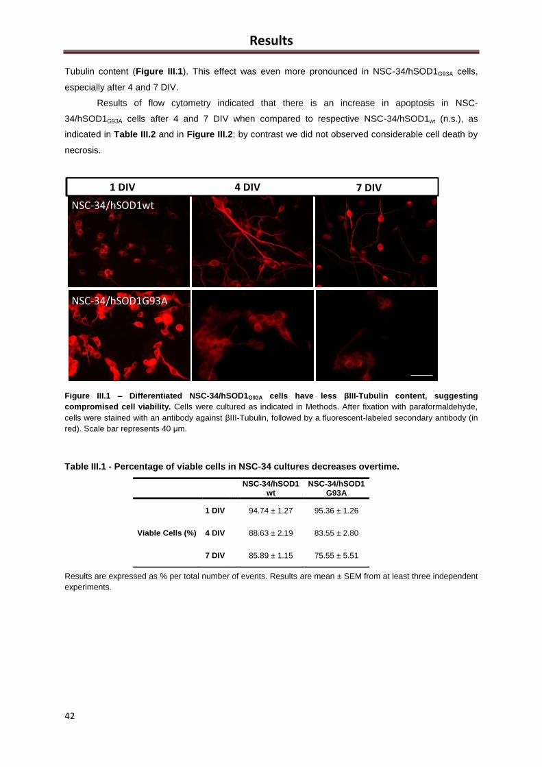
Results
42
Tubulin content (Figure III.1). This effect was even more pronounced in NSC-34/hSOD1G93A cells,
especially after 4 and 7 DIV.
Results of flow cytometry indicated that there is an increase in apoptosis in NSC-
34/hSOD1G93A cells after 4 and 7 DIV when compared to respective NSC-34/hSOD1wt (n.s.), as
indicated in Table III.2 and in Figure III.2; by contrast we did not observed considerable cell death by
necrosis.
Figure III.1 – Differentiated NSC-34/hSOD1G93A cells have less βIII-Tubulin content, suggesting
compromised cell viability. Cells were cultured as indicated in Methods. After fixation with paraformaldehyde,
cells were stained with an antibody against βIII-Tubulin, followed by a fluorescent-labeled secondary antibody (in
red). Scale bar represents 40 μm.
Table III.1 - Percentage of viable cells in NSC-34 cultures decreases overtime.
Results are expressed as % per total number of events. Results are mean ± SEM from at least three independent
experiments.
CX3CR1
CX3CR1
D
CX3CR1
E
CX3CR1
4 DIV 7 DIV
NSC-34/hSOD1wt
NSC-34/hSOD1G93A
1 DIV 4 DIV 7 DIV1 DIV
NSC-34/hSOD1wt
NSC-34/hSOD1 wt
NSC-34/hSOD1 G93A
Viable Cells (%)
1 DIV 94.74 ± 1.27 95.36 ± 1.26
4 DIV 88.63 ± 2.19 83.55 ± 2.80
7 DIV 85.89 ± 1.15 75.55 ± 5.51
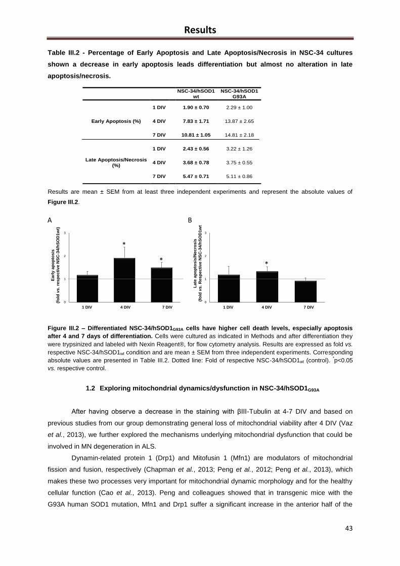
Results
43
A B
0
1
2
3
1 DIV 4 DIV 7 DIV
La
te a
po
pto
sis
/Ne
cro
sis
(fo
ldvs.R
es
pe
cti
ve
NS
C-3
4/h
SO
D1
wt
*
0
1
2
3
1 DIV 4 DIV 7 DIV
Ea
rly
ap
op
tos
is
(fo
ldvs. re
sp
ecti
ve
NS
C-3
4/h
SO
D1w
t)
*
*
Table III.2 - Percentage of Early Apoptosis and Late Apoptosis/Necrosis in NSC-34 cultures
shown a decrease in early apoptosis leads differentiation but almost no alteration in late
apoptosis/necrosis.
Results are mean ± SEM from at least three independent experiments and represent the absolute values of
Figure III.2.
Figure III.2 – Differentiated NSC-34/hSOD1G93A cells have higher cell death levels, especially apoptosis
after 4 and 7 days of differentiation. Cells were cultured as indicated in Methods and after differentiation they
were trypsinized and labeled with Nexin Reagent®, for flow cytometry analysis. Results are expressed as fold vs.
respective NSC-34/hSOD1wt condition and are mean ± SEM from three independent experiments. Corresponding
absolute values are presented in Table III.2. Dotted line: Fold of respective NSC-34/hSOD1wt (control). *p<0.05
vs. respective control.
1.2 Exploring mitochondrial dynamics/dysfunction in NSC-34/hSOD1G93A
After having observe a decrease in the staining with βIII-Tubulin at 4-7 DIV and based on
previous studies from our group demonstrating general loss of mitochondrial viability after 4 DIV (Vaz
et al., 2013), we further explored the mechanisms underlying mitochondrial dysfunction that could be
involved in MN degeneration in ALS.
Dynamin-related protein 1 (Drp1) and Mitofusin 1 (Mfn1) are modulators of mitochondrial
fission and fusion, respectively (Chapman et al., 2013; Peng et al., 2012; Peng et al., 2013), which
makes these two processes very important for mitochondrial dynamic morphology and for the healthy
cellular function (Cao et al., 2013). Peng and colleagues showed that in transgenic mice with the
G93A human SOD1 mutation, Mfn1 and Drp1 suffer a significant increase in the anterior half of the
NSC-34/hSOD1 wt
NSC-34/hSOD1 G93A
Early Apoptosis (%)
1 DIV 1.90 ± 0.70 2.29 ± 1.00
4 DIV 7.83 ± 1.71 13.87 ± 2.65
7 DIV 10.81 ± 1.05 14.81 ± 2.18
Late Apoptosis/Necrosis (%)
1 DIV 2.43 ± 0.56 3.22 ± 1.26
4 DIV 3.68 ± 0.78 3.75 ± 0.55
7 DIV 5.47 ± 0.71 5.11 ± 0.86
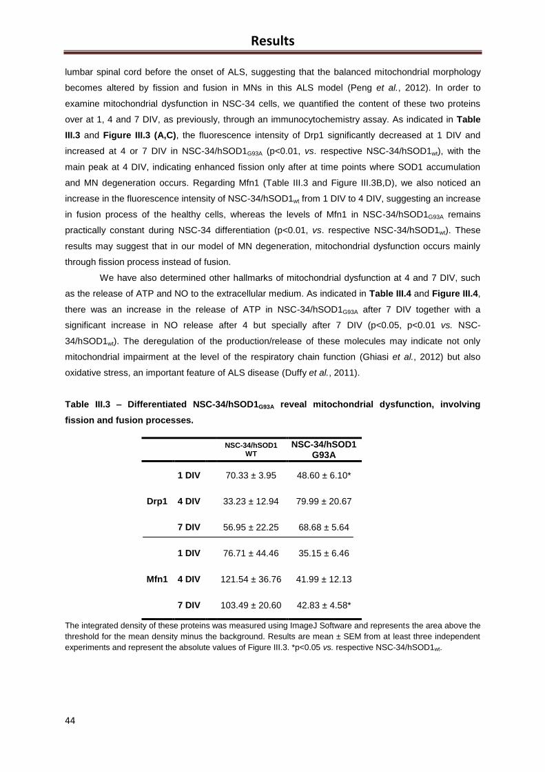
Results
44
lumbar spinal cord before the onset of ALS, suggesting that the balanced mitochondrial morphology
becomes altered by fission and fusion in MNs in this ALS model (Peng et al., 2012). In order to
examine mitochondrial dysfunction in NSC-34 cells, we quantified the content of these two proteins
over at 1, 4 and 7 DIV, as previously, through an immunocytochemistry assay. As indicated in Table
III.3 and Figure III.3 (A,C), the fluorescence intensity of Drp1 significantly decreased at 1 DIV and
increased at 4 or 7 DIV in NSC-34/hSOD1G93A (p<0.01, vs. respective NSC-34/hSOD1wt), with the
main peak at 4 DIV, indicating enhanced fission only after at time points where SOD1 accumulation
and MN degeneration occurs. Regarding Mfn1 (Table III.3 and Figure III.3B,D), we also noticed an
increase in the fluorescence intensity of NSC-34/hSOD1wt from 1 DIV to 4 DIV, suggesting an increase
in fusion process of the healthy cells, whereas the levels of Mfn1 in NSC-34/hSOD1G93A remains
practically constant during NSC-34 differentiation (p<0.01, vs. respective NSC-34/hSOD1wt). These
results may suggest that in our model of MN degeneration, mitochondrial dysfunction occurs mainly
through fission process instead of fusion.
We have also determined other hallmarks of mitochondrial dysfunction at 4 and 7 DIV, such
as the release of ATP and NO to the extracellular medium. As indicated in Table III.4 and Figure III.4,
there was an increase in the release of ATP in NSC-34/hSOD1G93A after 7 DIV together with a
significant increase in NO release after 4 but specially after 7 DIV (p<0.05, p<0.01 vs. NSC-
34/hSOD1wt). The deregulation of the production/release of these molecules may indicate not only
mitochondrial impairment at the level of the respiratory chain function (Ghiasi et al., 2012) but also
oxidative stress, an important feature of ALS disease (Duffy et al., 2011).
Table III.3 – Differentiated NSC-34/hSOD1G93A reveal mitochondrial dysfunction, involving
fission and fusion processes.
The integrated density of these proteins was measured using ImageJ Software and represents the area above the
threshold for the mean density minus the background. Results are mean ± SEM from at least three independent
experiments and represent the absolute values of Figure III.3. *p<0.05 vs. respective NSC-34/hSOD1wt.
NSC-34/hSOD1 WT
NSC-34/hSOD1 G93A
Drp1
1 DIV 70.33 ± 3.95 48.60 ± 6.10*
4 DIV 33.23 ± 12.94 79.99 ± 20.67
7 DIV 56.95 ± 22.25 68.68 ± 5.64
Mfn1
1 DIV 76.71 ± 44.46 35.15 ± 6.46
4 DIV 121.54 ± 36.76 41.99 ± 12.13
7 DIV 103.49 ± 20.60 42.83 ± 4.58*
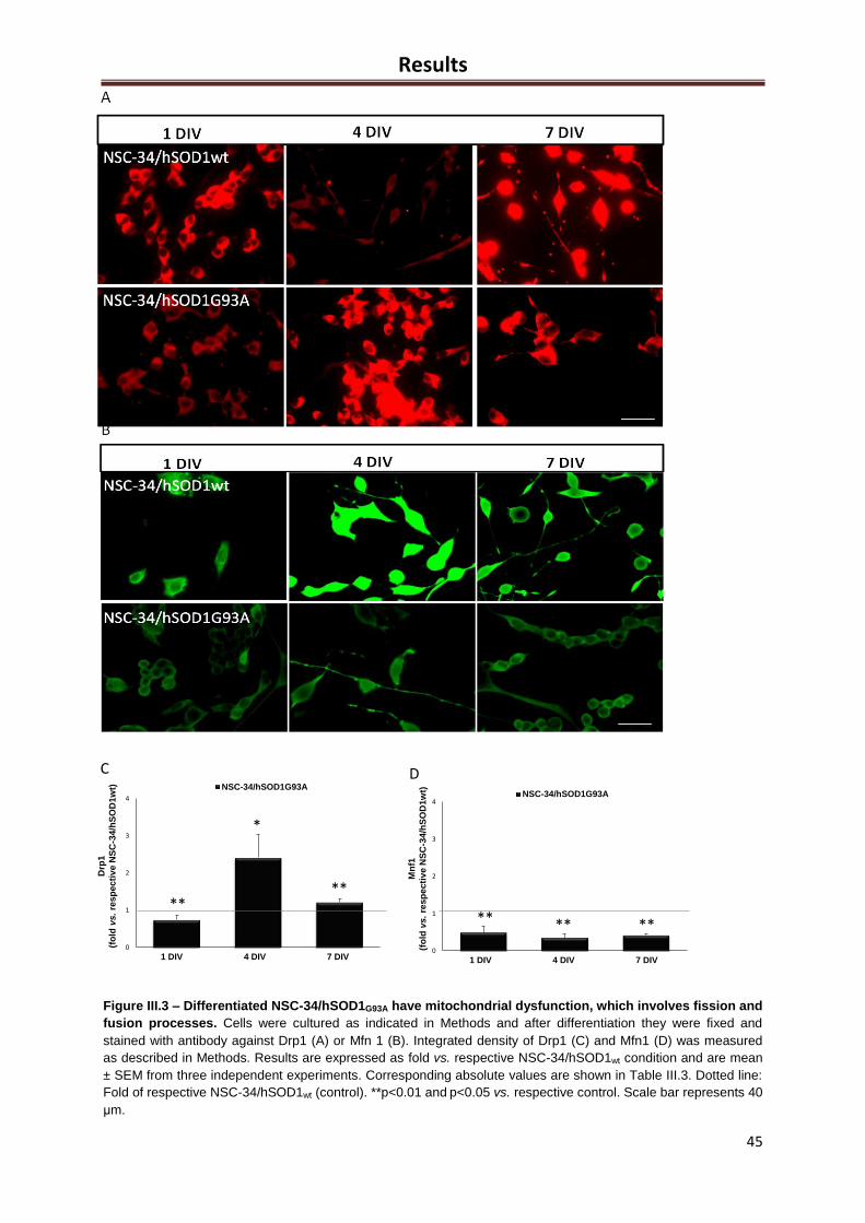
Results
45
0
1
2
3
4
1 DIV 4 DIV 7 DIV
NSC-34/hSOD1G93A
0
1
2
3
4
1 DIV 4 DIV 7 DIV
NSC-34/hSOD1G93A
**
*
**
** ** **
C D
Drp
1
(fo
ldvs. re
sp
ecti
ve
NS
C-3
4/h
SO
D1w
t)
Mn
f1
(fo
ldvs.
resp
ecti
ve
NS
C-3
4/h
SO
D1w
t)
Figure III.3 – Differentiated NSC-34/hSOD1G93A have mitochondrial dysfunction, which involves fission and
fusion processes. Cells were cultured as indicated in Methods and after differentiation they were fixed and
stained with antibody against Drp1 (A) or Mfn 1 (B). Integrated density of Drp1 (C) and Mfn1 (D) was measured
as described in Methods. Results are expressed as fold vs. respective NSC-34/hSOD1wt condition and are mean
± SEM from three independent experiments. Corresponding absolute values are shown in Table III.3. Dotted line:
Fold of respective NSC-34/hSOD1wt (control). **p<0.01 and p<0.05 vs. respective control. Scale bar represents 40
μm.
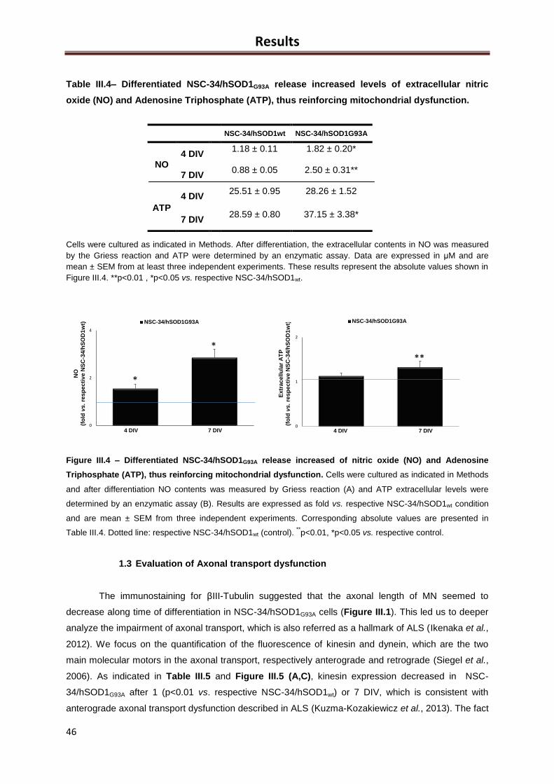
Results
46
Table III.4– Differentiated NSC-34/hSOD1G93A release increased levels of extracellular nitric
oxide (NO) and Adenosine Triphosphate (ATP), thus reinforcing mitochondrial dysfunction.
Cells were cultured as indicated in Methods. After differentiation, the extracellular contents in NO was measured
by the Griess reaction and ATP were determined by an enzymatic assay. Data are expressed in μM and are
mean ± SEM from at least three independent experiments. These results represent the absolute values shown in
Figure III.4. **p<0.01 , *p<0.05 vs. respective NSC-34/hSOD1wt.
Figure III.4 – Differentiated NSC-34/hSOD1G93A release increased of nitric oxide (NO) and Adenosine
Triphosphate (ATP), thus reinforcing mitochondrial dysfunction. Cells were cultured as indicated in Methods
and after differentiation NO contents was measured by Griess reaction (A) and ATP extracellular levels were
determined by an enzymatic assay (B). Results are expressed as fold vs. respective NSC-34/hSOD1wt condition
and are mean ± SEM from three independent experiments. Corresponding absolute values are presented in
Table III.4. Dotted line: respective NSC-34/hSOD1wt (control). **p<0.01, *p<0.05 vs. respective control.
1.3 Evaluation of Axonal transport dysfunction
The immunostaining for βIII-Tubulin suggested that the axonal length of MN seemed to
decrease along time of differentiation in NSC-34/hSOD1G93A cells (Figure III.1). This led us to deeper
analyze the impairment of axonal transport, which is also referred as a hallmark of ALS (Ikenaka et al.,
2012). We focus on the quantification of the fluorescence of kinesin and dynein, which are the two
main molecular motors in the axonal transport, respectively anterograde and retrograde (Siegel et al.,
2006). As indicated in Table III.5 and Figure III.5 (A,C), kinesin expression decreased in NSC-
34/hSOD1G93A after 1 (p<0.01 vs. respective NSC-34/hSOD1wt) or 7 DIV, which is consistent with
anterograde axonal transport dysfunction described in ALS (Kuzma-Kozakiewicz et al., 2013). The fact
0
2
4
4 DIV 7 DIV
NSC-34/hSOD1G93A
0
1
2
4 DIV 7 DIV
NSC-34/hSOD1G93A
*
***
C D
NO
(fo
ldvs.
resp
ecti
ve
NS
C-3
4/h
SO
D1
wt)
Extr
acellu
lar
AT
P
(fo
ldvs.
resp
ecti
ve
NS
C-3
4/h
SO
D1
wt)
B
C
H
NSC-34/hSOD1 WT NSC-34/hSOD1G93A
Nitrites
4 DIV
1.18 ± 0.11
1.82 ± 0.20*
7 DIV 0.88 ± 0.05 2.50 ± 0.31**
ATP
4 DIV
25.51 ± 0.95 28.26 ± 1.52
7 DIV
28.59 ± 0.80 $ 37.15 ± 3.38 *
$
C C
NSC-34/hSOD1wt NSC-34/hSOD1G93A
NO 4 DIV
1.18 ± 0.11 1.82 ± 0.20*
7 DIV 0.88 ± 0.05 2.50 ± 0.31**
ATP
4 DIV 25.51 ± 0.95 28.26 ± 1.52
7 DIV 28.59 ± 0.80 37.15 ± 3.38*
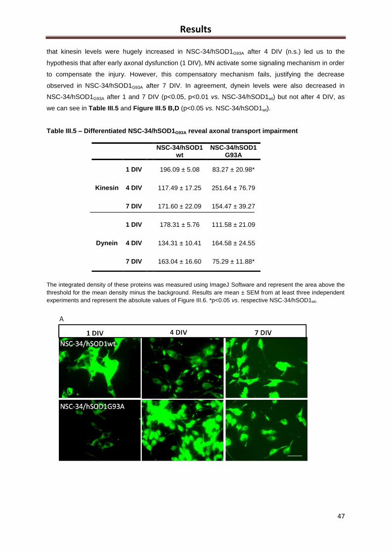
Results
47
that kinesin levels were hugely increased in NSC-34/hSOD1G93A after 4 DIV (n.s.) led us to the
hypothesis that after early axonal dysfunction (1 DIV), MN activate some signaling mechanism in order
to compensate the injury. However, this compensatory mechanism fails, justifying the decrease
observed in NSC-34/hSOD1G93A after 7 DIV. In agreement, dynein levels were also decreased in
NSC-34/hSOD1G93A after 1 and 7 DIV (p<0.05, p<0.01 vs. NSC-34/hSOD1wt) but not after 4 DIV, as
we can see in Table III.5 and Figure III.5 B,D (p<0.05 vs. NSC-34/hSOD1wt).
Table III.5 – Differentiated NSC-34/hSOD1G93A reveal axonal transport impairment
The integrated density of these proteins was measured using ImageJ Software and represent the area above the
threshold for the mean density minus the background. Results are mean ± SEM from at least three independent
experiments and represent the absolute values of Figure III.6. *p<0.05 vs. respective NSC-34/hSOD1wt.
NSC-34/hSOD1 wt
NSC-34/hSOD1 G93A
Kinesin
1 DIV 196.09 ± 5.08 83.27 ± 20.98*
4 DIV 117.49 ± 17.25 251.64 ± 76.79
7 DIV 171.60 ± 22.09 154.47 ± 39.27
Dynein
1 DIV 178.31 ± 5.76 111.58 ± 21.09
4 DIV 134.31 ± 10.41 164.58 ± 24.55
7 DIV 163.04 ± 16.60 75.29 ± 11.88*
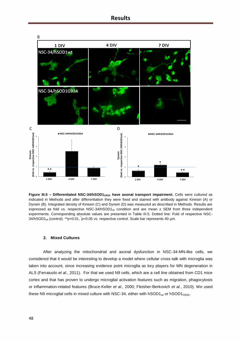
Results
48
Figure III.5 – Differentiated NSC-34/hSOD1G93A have axonal transport impairment. Cells were cultured as
indicated in Methods and after differentiation they were fixed and stained with antibody against Kinesin (A) or
Dynein (B). Integrated density of Kinesin (C) and Dynein (D) was measured as described in Methods. Results are
expressed as fold vs. respective NSC-34/hSOD1wt condition and are mean ± SEM from three independent
experiments. Corresponding absolute values are presented in Table III.5. Dotted line: Fold of respective NSC-
34/hSOD1wt (control). **p<0.01, *p<0.05 vs. respective control. Scale bar represents 40 μm.
2. Mixed Cultures
After analyzing the mitochondrial and axonal dysfunction in NSC-34-MN-like cells, we
considered that it would be interesting to develop a model where cellular cross-talk with microglia was
taken into account, since increasing evidence point microglia as key players for MN degeneration in
ALS (Ferraiuolo et al., 2011). For that we used N9 cells, which are a cell line obtained from CD1 mice
cortex and that has proven to undergo microglial activation features such as migration, phagocytosis
or inflammation-related features (Bruce-Keller et al., 2000; Fleisher-Berkovich et al., 2010). We used
these N9 microglial cells in mixed culture with NSC-34, either with hSOD1wt or hSOD1G93A.
0
1
2
3
4
1 DIV 4 DIV 7 DIV
NSC-34/hSOD1G93A
0
1
2
3
4
1 DIV 4 DIV 7 DIV
NSC-34/hSOD1G93A
***
*
**
C D
Kin
esin
(fo
ldvs.
resp
ecti
ve
NS
C-3
4/h
SO
D1w
t)
Dyn
ein
(fo
ldvs. re
sp
ecti
ve
NS
C-3
4/h
SO
D1w
t)
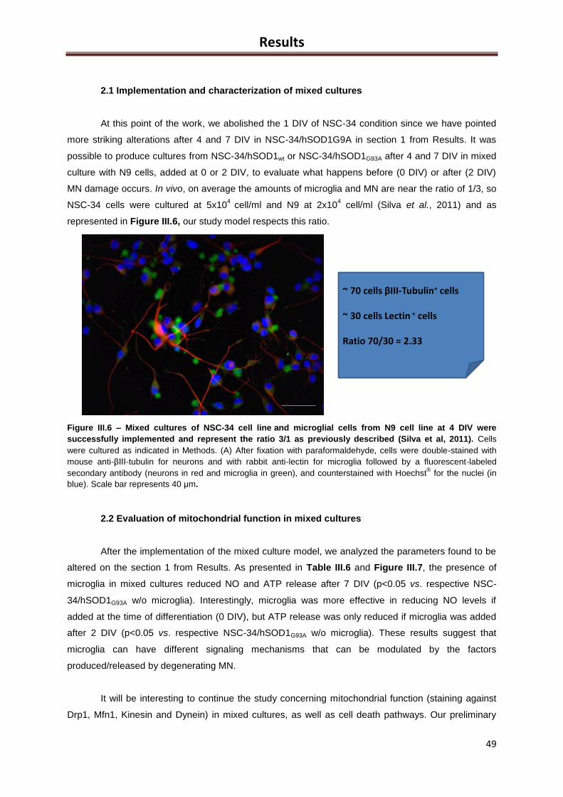
Results
49
~ 70 cells βIII-Tubulin+ cells
~ 30 cells Lectin + cells
Ratio 70/30 = 2.33
2.1 Implementation and characterization of mixed cultures
At this point of the work, we abolished the 1 DIV of NSC-34 condition since we have pointed
more striking alterations after 4 and 7 DIV in NSC-34/hSOD1G9A in section 1 from Results. It was
possible to produce cultures from NSC-34/hSOD1wt or NSC-34/hSOD1G93A after 4 and 7 DIV in mixed
culture with N9 cells, added at 0 or 2 DIV, to evaluate what happens before (0 DIV) or after (2 DIV)
MN damage occurs. In vivo, on average the amounts of microglia and MN are near the ratio of 1/3, so
NSC-34 cells were cultured at 5x10⁴
cell/ml and N9 at 2x10⁴ cell/ml (Silva et al., 2011) and as
represented in Figure III.6, our study model respects this ratio.
Figure III.6 – Mixed cultures of NSC-34 cell line and microglial cells from N9 cell line at 4 DIV were
successfully implemented and represent the ratio 3/1 as previously described (Silva et al, 2011). Cells
were cultured as indicated in Methods. (A) After fixation with paraformaldehyde, cells were double-stained with
mouse anti-βIII-tubulin for neurons and with rabbit anti-lectin for microglia followed by a fluorescent-labeled
secondary antibody (neurons in red and microglia in green), and counterstained with Hoechst® for the nuclei (in
blue). Scale bar represents 40 μm.
2.2 Evaluation of mitochondrial function in mixed cultures
After the implementation of the mixed culture model, we analyzed the parameters found to be
altered on the section 1 from Results. As presented in Table III.6 and Figure III.7, the presence of
microglia in mixed cultures reduced NO and ATP release after 7 DIV (p<0.05 vs. respective NSC-
34/hSOD1G93A w/o microglia). Interestingly, microglia was more effective in reducing NO levels if
added at the time of differentiation (0 DIV), but ATP release was only reduced if microglia was added
after 2 DIV (p<0.05 vs. respective NSC-34/hSOD1G93A w/o microglia). These results suggest that
microglia can have different signaling mechanisms that can be modulated by the factors
produced/released by degenerating MN.
It will be interesting to continue the study concerning mitochondrial function (staining against
Drp1, Mfn1, Kinesin and Dynein) in mixed cultures, as well as cell death pathways. Our preliminary
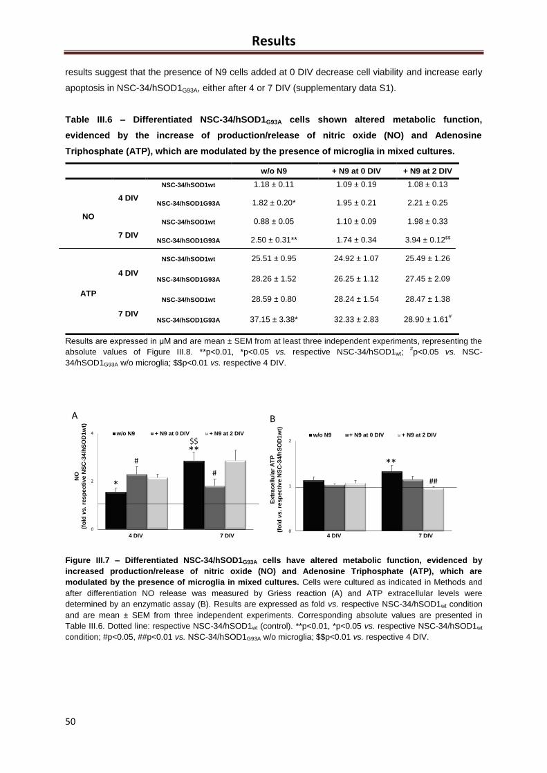
Results
50
results suggest that the presence of N9 cells added at 0 DIV decrease cell viability and increase early
apoptosis in NSC-34/hSOD1G93A, either after 4 or 7 DIV (supplementary data S1).
Table III.6 – Differentiated NSC-34/hSOD1G93A cells shown altered metabolic function,
evidenced by the increase of production/release of nitric oxide (NO) and Adenosine
Triphosphate (ATP), which are modulated by the presence of microglia in mixed cultures.
Results are expressed in μM and are mean ± SEM from at least three independent experiments, representing the
absolute values of Figure III.8. **p<0.01, *p<0.05 vs. respective NSC-34/hSOD1wt; #p<0.05 vs. NSC-
34/hSOD1G93A w/o microglia; $$p<0.01 vs. respective 4 DIV.
Figure III.7 – Differentiated NSC-34/hSOD1G93A cells have altered metabolic function, evidenced by
increased production/release of nitric oxide (NO) and Adenosine Triphosphate (ATP), which are
modulated by the presence of microglia in mixed cultures. Cells were cultured as indicated in Methods and
after differentiation NO release was measured by Griess reaction (A) and ATP extracellular levels were
determined by an enzymatic assay (B). Results are expressed as fold vs. respective NSC-34/hSOD1wt condition
and are mean ± SEM from three independent experiments. Corresponding absolute values are presented in
Table III.6. Dotted line: respective NSC-34/hSOD1wt (control). **p<0.01, *p<0.05 vs. respective NSC-34/hSOD1wt
condition; #p<0.05, ##p<0.01 vs. NSC-34/hSOD1G93A w/o microglia; $$p<0.01 vs. respective 4 DIV.
0
2
4
4 DIV 7 DIV
w/o N9 + N9 at 0 DIV + N9 at 2 DIV
0
1
2
4 DIV 7 DIV
w/o N9 + N9 at 0 DIV + N9 at 2 DIV
##
#
*
**
#
$$
**
A B
NO
(fo
ldvs. re
sp
ecti
ve
NS
C-3
4/h
SO
D1w
t)
Extr
acellu
lar
AT
P
(fo
ldvs. re
sp
ecti
ve
NS
C-3
4/h
SO
D1w
t)
w/o N9 + N9 at 0 DIV + N9 at 2 DIV
NO
4 DIV
NSC-34/hSOD1wt 1.18 ± 0.11 1.09 ± 0.19 1.08 ± 0.13
NSC-34/hSOD1G93A 1.82 ± 0.20* 1.95 ± 0.21 2.21 ± 0.25
7 DIV
NSC-34/hSOD1wt 0.88 ± 0.05 1.10 ± 0.09 1.98 ± 0.33
NSC-34/hSOD1G93A 2.50 ± 0.31** 1.74 ± 0.34 3.94 ± 0.12$$
ATP
4 DIV
NSC-34/hSOD1wt 25.51 ± 0.95 24.92 ± 1.07 25.49 ± 1.26
NSC-34/hSOD1G93A 28.26 ± 1.52 26.25 ± 1.12 27.45 ± 2.09
7 DIV
NSC-34/hSOD1wt 28.59 ± 0.80 28.24 ± 1.54 28.47 ± 1.38
NSC-34/hSOD1G93A 37.15 ± 3.38* 32.33 ± 2.83 28.90 ± 1.61#
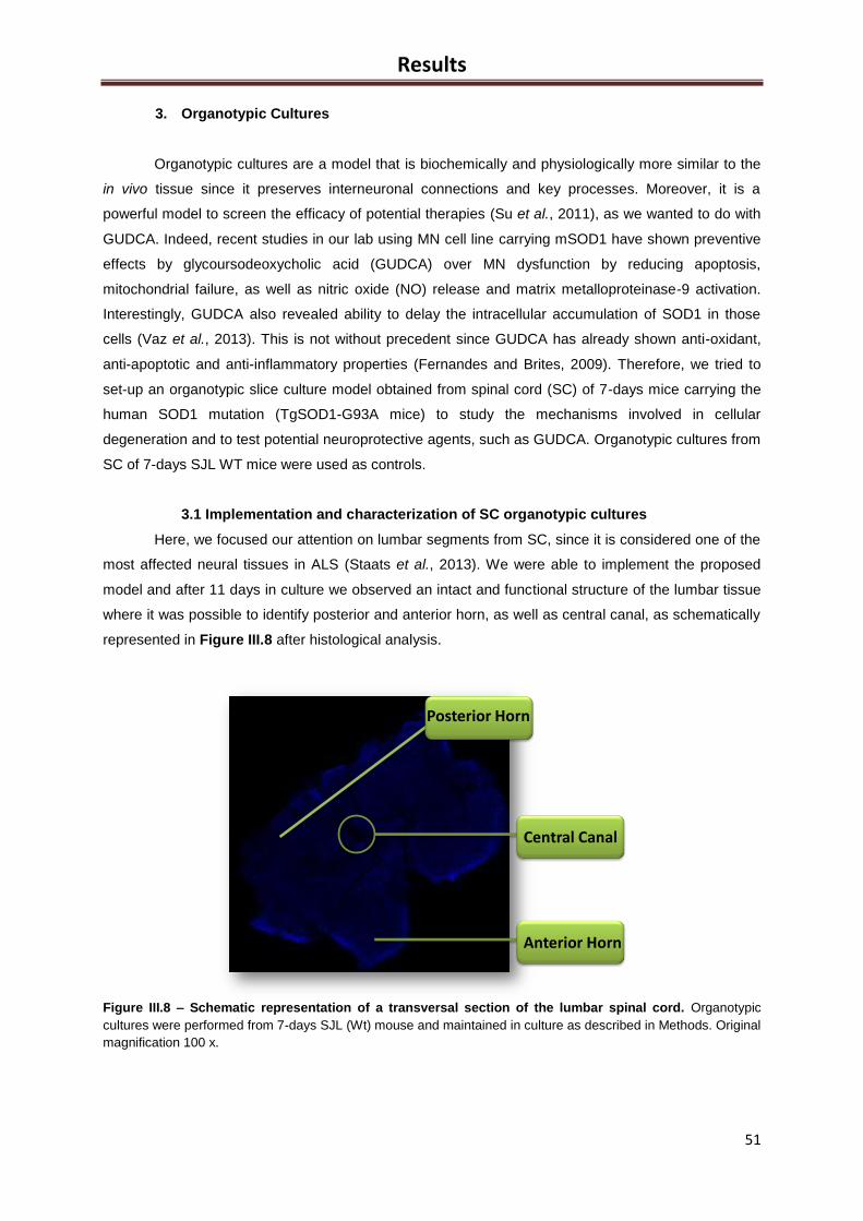
Results
51
Central Canal
Anterior Horn
Posterior Horn
3. Organotypic Cultures
Organotypic cultures are a model that is biochemically and physiologically more similar to the
in vivo tissue since it preserves interneuronal connections and key processes. Moreover, it is a
powerful model to screen the efficacy of potential therapies (Su et al., 2011), as we wanted to do with
GUDCA. Indeed, recent studies in our lab using MN cell line carrying mSOD1 have shown preventive
effects by glycoursodeoxycholic acid (GUDCA) over MN dysfunction by reducing apoptosis,
mitochondrial failure, as well as nitric oxide (NO) release and matrix metalloproteinase-9 activation.
Interestingly, GUDCA also revealed ability to delay the intracellular accumulation of SOD1 in those
cells (Vaz et al., 2013). This is not without precedent since GUDCA has already shown anti-oxidant,
anti-apoptotic and anti-inflammatory properties (Fernandes and Brites, 2009). Therefore, we tried to
set-up an organotypic slice culture model obtained from spinal cord (SC) of 7-days mice carrying the
human SOD1 mutation (TgSOD1-G93A mice) to study the mechanisms involved in cellular
degeneration and to test potential neuroprotective agents, such as GUDCA. Organotypic cultures from
SC of 7-days SJL WT mice were used as controls.
3.1 Implementation and characterization of SC organotypic cultures
Here, we focused our attention on lumbar segments from SC, since it is considered one of the
most affected neural tissues in ALS (Staats et al., 2013). We were able to implement the proposed
model and after 11 days in culture we observed an intact and functional structure of the lumbar tissue
where it was possible to identify posterior and anterior horn, as well as central canal, as schematically
represented in Figure III.8 after histological analysis.
Figure III.8 – Schematic representation of a transversal section of the lumbar spinal cord. Organotypic
cultures were performed from 7-days SJL (Wt) mouse and maintained in culture as described in Methods. Original
magnification 100 x.
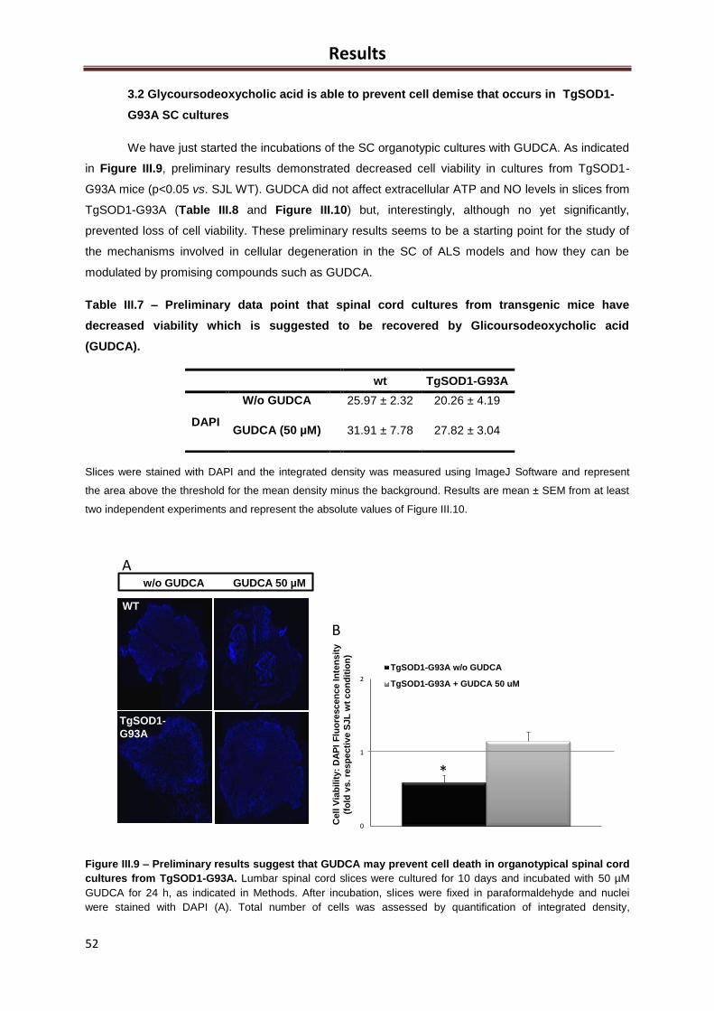
Results
52
wt TgSOD1-G93A
DAPI
W/o GUDCA 25.97 ± 2.32 20.26 ± 4.19
GUDCA (50 µM) 31.91 ± 7.78 27.82 ± 3.04
3.2 Glycoursodeoxycholic acid is able to prevent cell demise that occurs in TgSOD1-
G93A SC cultures
We have just started the incubations of the SC organotypic cultures with GUDCA. As indicated
in Figure III.9, preliminary results demonstrated decreased cell viability in cultures from TgSOD1-
G93A mice (p<0.05 vs. SJL WT). GUDCA did not affect extracellular ATP and NO levels in slices from
TgSOD1-G93A (Table III.8 and Figure III.10) but, interestingly, although no yet significantly,
prevented loss of cell viability. These preliminary results seems to be a starting point for the study of
the mechanisms involved in cellular degeneration in the SC of ALS models and how they can be
modulated by promising compounds such as GUDCA.
Table III.7 – Preliminary data point that spinal cord cultures from transgenic mice have
decreased viability which is suggested to be recovered by Glicoursodeoxycholic acid
(GUDCA).
Slices were stained with DAPI and the integrated density was measured using ImageJ Software and represent
the area above the threshold for the mean density minus the background. Results are mean ± SEM from at least
two independent experiments and represent the absolute values of Figure III.10.
Figure III.9 – Preliminary results suggest that GUDCA may prevent cell death in organotypical spinal cord
cultures from TgSOD1-G93A. Lumbar spinal cord slices were cultured for 10 days and incubated with 50 µM
GUDCA for 24 h, as indicated in Methods. After incubation, slices were fixed in paraformaldehyde and nuclei
were stained with DAPI (A). Total number of cells was assessed by quantification of integrated density,
C
A
WT
TgSOD1-
G93A
w/o GUDCA GUDCA 50 µM
A
0
1
2
4 DIV 7 DIV
TgSOD1-G93A w/o GUDCA
TgSOD1-G93A + GUDCA 50 uM
Cell
Via
bilit
y:
DA
PI
Flu
ore
scen
ce
Inte
nsit
y
(fo
ldv
s. re
sp
ecti
ve
SJL
wt
co
nd
itio
n)
B
*
WT
TgSOD1-
G93A
w/o GUDCA GUDCA 50 µM
A
0
1
2
4 DIV 7 DIV
TgSOD1-G93A w/o GUDCA
TgSOD1-G93A + GUDCA 50 uM
Ce
llV
iab
ilit
y:
DA
PI
Flu
ore
sce
nc
eIn
ten
sit
y
(fo
ldv
s. re
sp
ecti
ve
SJL
wt
co
nd
itio
n)
B
*
WT
TgSOD1-
G93A
w/o GUDCA GUDCA 50 µM
A
0
1
2
4 DIV 7 DIV
TgSOD1-G93A w/o GUDCA
TgSOD1-G93A + GUDCA 50 uM
Cell
Via
bil
ity:
DA
PI
Flu
ore
scen
ce
Inte
ns
ity
(fo
ldv
s. re
sp
ecti
ve
SJ
L w
tc
on
dit
ion
)
B
*
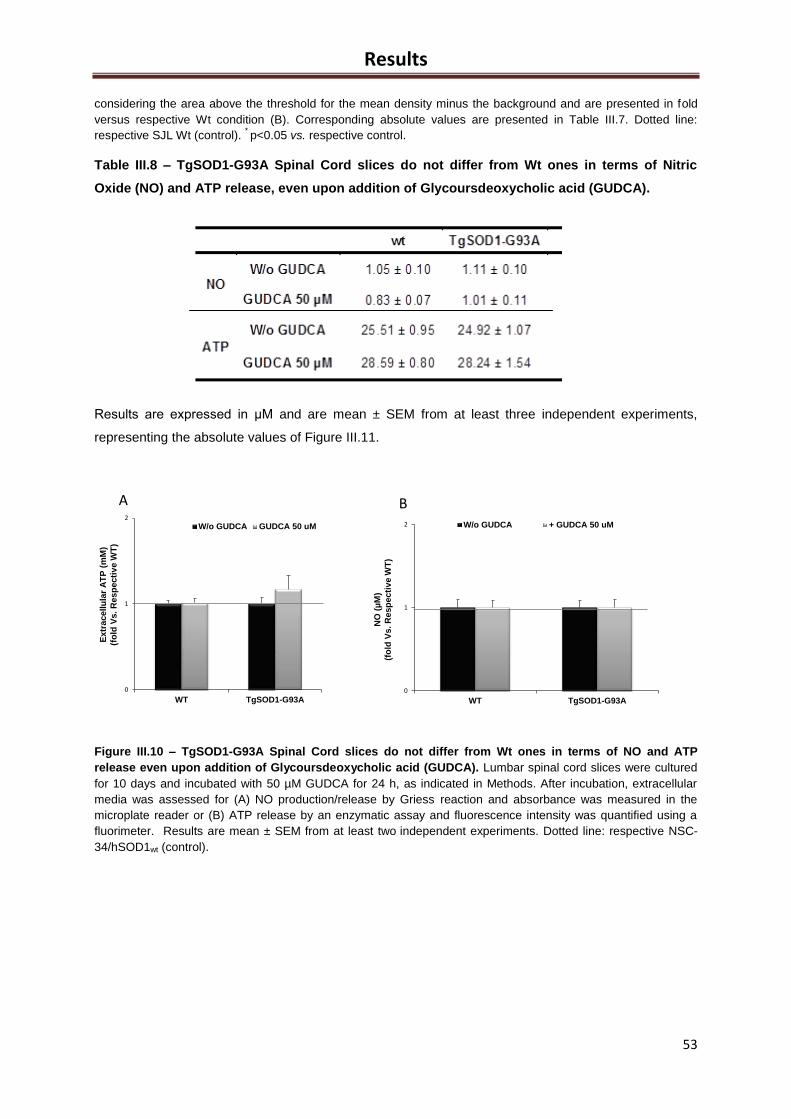
Results
53
considering the area above the threshold for the mean density minus the background and are presented in fold
versus respective Wt condition (B). Corresponding absolute values are presented in Table III.7. Dotted line:
respective SJL Wt (control). * p<0.05 vs. respective control.
Table III.8 – TgSOD1-G93A Spinal Cord slices do not differ from Wt ones in terms of Nitric
Oxide (NO) and ATP release, even upon addition of Glycoursdeoxycholic acid (GUDCA).
Results are expressed in μM and are mean ± SEM from at least three independent experiments,
representing the absolute values of Figure III.11.
Figure III.10 – TgSOD1-G93A Spinal Cord slices do not differ from Wt ones in terms of NO and ATP
release even upon addition of Glycoursdeoxycholic acid (GUDCA). Lumbar spinal cord slices were cultured
for 10 days and incubated with 50 µM GUDCA for 24 h, as indicated in Methods. After incubation, extracellular
media was assessed for (A) NO production/release by Griess reaction and absorbance was measured in the
microplate reader or (B) ATP release by an enzymatic assay and fluorescence intensity was quantified using a
fluorimeter. Results are mean ± SEM from at least two independent experiments. Dotted line: respective NSC-
34/hSOD1wt (control).
0
1
2
WT TgSOD1-G93A
W/o GUDCA GUDCA 50 uM
Extr
acellu
lar
AT
P (
mM
)
(fo
ld V
s. R
esp
ecti
ve W
T)
0
1
2
WT TgSOD1-G93A
W/o GUDCA + GUDCA 50 uM
NO
(µ
M)
(fo
ld V
s. R
esp
ecti
ve W
T)
A B
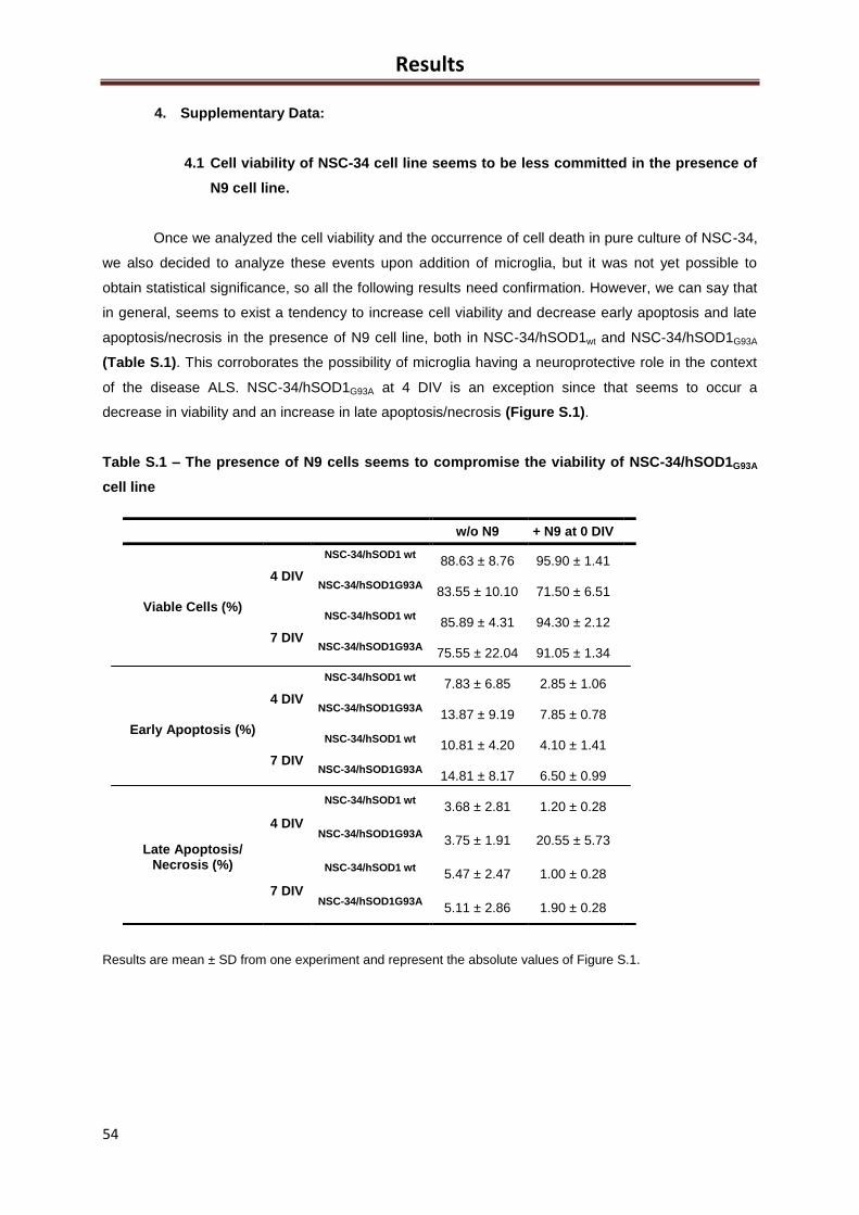
Results
54
4. Supplementary Data:
4.1 Cell viability of NSC-34 cell line seems to be less committed in the presence of
N9 cell line.
Once we analyzed the cell viability and the occurrence of cell death in pure culture of NSC-34,
we also decided to analyze these events upon addition of microglia, but it was not yet possible to
obtain statistical significance, so all the following results need confirmation. However, we can say that
in general, seems to exist a tendency to increase cell viability and decrease early apoptosis and late
apoptosis/necrosis in the presence of N9 cell line, both in NSC-34/hSOD1wt and NSC-34/hSOD1G93A
(Table S.1). This corroborates the possibility of microglia having a neuroprotective role in the context
of the disease ALS. NSC-34/hSOD1G93A at 4 DIV is an exception since that seems to occur a
decrease in viability and an increase in late apoptosis/necrosis (Figure S.1).
Table S.1 – The presence of N9 cells seems to compromise the viability of NSC-34/hSOD1G93A
cell line
Results are mean ± SD from one experiment and represent the absolute values of Figure S.1.
w/o N9 + N9 at 0 DIV
Viable Cells (%)
4 DIV
NSC-34/hSOD1 wt 88.63 ± 8.76 95.90 ± 1.41
NSC-34/hSOD1G93A 83.55 ± 10.10 71.50 ± 6.51
7 DIV
NSC-34/hSOD1 wt 85.89 ± 4.31 94.30 ± 2.12
NSC-34/hSOD1G93A 75.55 ± 22.04 91.05 ± 1.34
Early Apoptosis (%)
4 DIV
NSC-34/hSOD1 wt 7.83 ± 6.85 2.85 ± 1.06
NSC-34/hSOD1G93A 13.87 ± 9.19 7.85 ± 0.78
7 DIV
NSC-34/hSOD1 wt 10.81 ± 4.20 4.10 ± 1.41
NSC-34/hSOD1G93A 14.81 ± 8.17 6.50 ± 0.99
Late Apoptosis/ Necrosis (%)
4 DIV
NSC-34/hSOD1 wt 3.68 ± 2.81 1.20 ± 0.28
NSC-34/hSOD1G93A 3.75 ± 1.91 20.55 ± 5.73
7 DIV
NSC-34/hSOD1 wt 5.47 ± 2.47 1.00 ± 0.28
NSC-34/hSOD1G93A 5.11 ± 2.86 1.90 ± 0.28
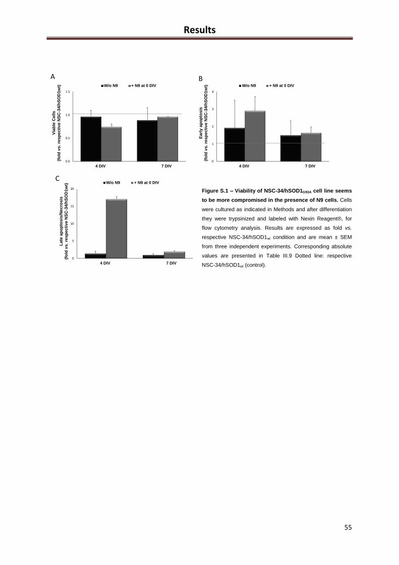
Results
55
0
5
10
15
20
4 DIV 7 DIV
W/o N9 + N9 at 0 DIV
0
1
2
3
4
4 DIV 7 DIV
W/o N9 + N9 at 0 DIV
0.0
0.5
1.0
1.5
4 DIV 7 DIV
W/o N9 + N9 at 0 DIV
A B
C
Via
ble
Cells
(fo
ldvs. re
sp
ecti
ve
NS
C-3
4/h
SO
D1w
t)
Earl
yap
op
tosis
(fo
ldvs. re
sp
ecti
ve
NS
C-3
4/h
SO
D1w
t)
Late
ap
op
tosis
/Necro
sis
(fo
ldvs. re
sp
ecti
ve
NS
C-3
4/h
SO
D1w
t)
Figure S.1 – Viability of NSC-34/hSOD1G93A cell line seems
to be more compromised in the presence of N9 cells. Cells
were cultured as indicated in Methods and after differentiation
they were trypsinized and labeled with Nexin Reagent®, for
flow cytometry analysis. Results are expressed as fold vs.
respective NSC-34/hSOD1wt condition and are mean ± SEM
from three independent experiments. Corresponding absolute
values are presented in Table III.9 Dotted line: respective
NSC-34/hSOD1wt (control).

56
Discussion
57
IV. Discussion
The impossibility to make real-time studies or to remove tissue samples repeatedly from the
patients are limitations for the study of human neurodegenerative diseases (Turner et al., 2013). Thus,
the use of in vitro, ex vivo and in vivo animal models is essential. Among the familial cases in ALS,
approximately 20% are caused by dominantly inherited mutations in the Cu/Zn superoxide dismutase-
1 (SOD1) protein (Musaro, 2010), which provide a basis for many experimental in vitro and in vivo
models including mutations in this enzyme.
This Thesis had as first aim the implementation and characterization of a mixed culture model. Until
now, it is not clear what is the primary cause of motoneurons (MN) degeneration in ALS (Valori et al.,
2013). The MN-like cell line NSC-34 has been widely used for in vitro studies of ALS, once this cell
line present many properties of MN, and also show morphological and physiological properties of
them, like acetylcholine synthesis, storage and release; extension of processes; action potential
generation; formation of contacts with cultured myotubes; expression of neurofilament proteins and
association with neuromuscular synapse-specific basal lamina glycoproteins (Cashman et al., 1992;
Tovar et al., 2009). Moreover, when transfected with human mutations of SOD1 (such as G93A
mutation), these cells acquire some features reported in tissues of ALS patients and in transgenic
mice models, such as mitochondrial dysfunction (Raimondi et al., 2006) and Golgi apparatus
fragmentation (Gomes et al., 2008). Therefore, in the present study, we proposed to investigate the
MN viability in general and the mitochondrial function and axonal transport in particular, in the context
of the neuronal dysfunction in ALS progression. In addition, we aimed to analyze the interaction
between microglia and MN in the context of the disease, in order to better understand the origin of the
MN degeneration. In the last part of the study, we also tested potential neuroprotective agents in ALS
models.
As presented in the Results section, we initiated the work by the study of isolated cultures of NSC-
34. Recently, in our group it was demonstrated that NSC-34/hSOD1G93A cells presented accumulation
of SOD1 after 4 days of differentiation (DIV), together with cell dysfunction (Vaz et al., 2013), which
may represent the progression of MN degeneration in familiar ALS. Thus, here we considered three
different time points after NSC-34 cell differentiation: (i) 1 DIV – prior to SOD1 accumulation (onset);
(ii) 4 DIV – during SOD1 accumulation (symptomatic) and (iii) 7 DIV – after SOD1 accumulation and
MN cell death. Immunostaining assays against βIII-Tubulin could give us an idea of the behavior of
NSC-34/hSOD1wt cells (our control condition) in culture, over the time after differentiation and
evidenced that after 1 day in vitro (DIV), they have few branches and reduced length. After 4 DIV,
Discussion
58
these cells seemed to present a large number of complex synaptic connections between them,
indicating the presence of MN-like cells. However, after 7 DIV, it was an apparent decrease in the
number of connections, as well as a decrease in the number of cells, which may be related to the
stress inherent of the culture. So we decided to examine the cell viability and cell death events of
NSC-34/hSOD1G93A comparing with NSC-34/hSOD1wt in each time point of differentiation. This study,
either by staining against βIII-Tubulin (part of microtubules) and by flow cytometry, revealed that
mutated cells have compromised viability, although with a lower extent through time in culture after
differentiation. Relatively to cell death, we observed preferentially apoptosis rather than necrosis in
NSC-34/hSOD1G93A, which is in accordance with the fact of apoptosis is the most significant event of
death described for ALS disease, according with the generality of authors (Ranganathan and Bowser,
2010).
After observing the occurrence of apoptosis and decreased cell viability in NSC-34/hSOD1G93A
cells, and also because mitochondria plays a role in the apoptotic process, we decided to study more
deeply the mitochondrial dysfunction. We have focus on fission and fusion processes since they are
described to be implicated in neuronal injury and cell death (Barsoum et al., 2006; Liot et al., 2009;
Yuan et al., 2007). For this purpose, NSC-34/hSOD1wt and NSC-34/hSOD1G93A cells were
immunostained against Drp1 and Mfn1, two key proteins in the balance between mitochondrial fission
and fusion, respectively. We observed decreased levels of Drp1 after 1 DIV in NSC-34/hSOD1G93A
(p<0.01 vs. NSC-34/hSOD1wt) but also a significant increase after 4 DIV (p<0.05 vs. NSC-
34/hSOD1wt), indicating enhanced fusion only after at time points where SOD1 accumulation and MN
degeneration occurs. The sudden increase in the expression of Drp1 from 1 to 4 DIV may be linked to
increase in early apoptosis observed also at 4 DIV. In fact, it is described that the overexpression of
Drp1 increases the vulnerability to mitochondrial fragmentation and neuronal cell death (Barsoum et
al., 2006), but how the impairment of these pathways lead to neurodegeneration is still a matter of
debate (Ranieri et al., 2013). By contrast, Mfn1 levels were decreased in NSC-34/hSOD1G93A during
all time after differentiation (p<0.01 vs. NSC-34/hSOD1wt), suggesting that in our model of MN
degeneration, mitochondrial dysfunction occurs through events of fission and fusion. According to
Song and colleagues (2013), this fact can be explained because mitochondrial fusion and fission are
not independent and impact each, so defects in the fusion may result from increased fission rates
(Song et al., 2013).
Since a balance between mitochondrial fusion and fission is required for mitochondrial
homeostasis, we further explored mitochondrial injury. In fact, measurement of ATP and NO levels in
the extracellular media reveled an increase of both molecules in NSC-34/hSOD1G93A (p<0.01, p<0.05
vs. NSC-34/hSOD1wt) and from 4 to 7 DIV, indicating increased production of ROS that will probably
exacerbate oxidative stress. On the other hand, the increased ATP release to the extracellular media
will cause depletion of cell energy because ATP may be not available to be used in the function of
molecular motors that move between the soma and the axons of neurons, necessary for carrying
crucial cargos to cell metabolism and homeostasis, such as mitochondria, neurofilaments and
autophagosomes (Ikenaka et al., 2012; Song et al., 2013). Moreover, the impaired mitochondrial
activity and a consequent decrease in the intracellular availability of ATP will contribute to modify
Discussion
59
calcium homeostasis and ROS production, which may then lead to increased apoptosis (Federico et
al., 2012).
Impaired mitochondrial dynamics has been proposed to trigger axonal degeneration and is
consistent with the “dying back” hypothesis of neuronal projections as a primary event in ALS
pathogenesis (Chan, 2006; Dadon-Nachum et al., 2011; Knott et al., 2008). Thus, it is described the
occurrence of axonal transport dysfunction as a biomarker of disease ALS, associated at
mitochondrial dysfunction and cell death (Song et al., 2013). In fact, we observed that cellular
processes are diminished in -34/hSOD1G93A throughout the differentiation, which is consistent with
studies of Song and colleagues (2013) that described a reduction in neurite length and branching in
neurons with G93A mutation. After 7 DIV cells seem to lose the ability to communicate with each other
by shortening the axons, which may be linked to dysfunction of the carriage along them, with
consequent impossibility of transporting essential molecules throughout the cell. In our model, we
observed that kinesin levels diminished from NSC-34/hSOD1wt to NSC-34/hSOD1G93A at 1 and 7 DIV,
which is consistent with anterograde axonal transport dysfunction. The fact that kinesin levels were
hugely increased in NSC-34/hSOD1G93A after 4 DIV suggests that once more there is a dysfunction
detected by MN-like cells which triggers the activation of a defense mechanism that will be induced to
produce more kinesin to the equilibrium. Regarding dynein, involved in retrograde transport, similar to
what we have seen for kinesin, their expression levels were reduced in NSC-34/hSOD1G93A cells after
1 and 7 DIV (p<0.05, p<0.01 vs. NSC-34/hSOD1wt, respectively). This result indicates that retrograde
transport is also impaired in our model, which is consistent with literature that shows dysfunction of
dynein in different models of ALS (Soo et al., 2011). Therefore, the dysfunction of the two types of
transport does not appear to occur simultaneously at the same extent level. It appears that kinesin
suffers a huge injury first but can be restored after some time, however the compromising of the level
of dynein appears to occur less abruptly but the cells do not significantly recover after 4 DIV,
remaining a deficit in the amount of this molecular motor at 7 DIV, which agrees with the study of
axonal transport in mSOD1 mice model by Shi and colleagues (2010b), who describe the occurrence
of a decreased speed of retrograde transport mediated by dynein in G93A in comparison with WT
mice an early presymptomatic stage. Although Morfini and colleagues (2013) did not find the
anterograde transport affected, there are several reports describing inhibition of both anterograde and
retrograde axonal transport in mSOD1 mice (Bilsland et al., 2010; Perlson et al., 2009; Warita et al.,
1999). Indeed, kinesin and dynein are regulated by phosphorylation of specific subunits and an
abnormal activation of protein kinases (such as p38 MAPK) are described in ALS (Bendotti et al.,
2004; Krieger et al., 2003; Sharma et al., 2010), so in 2009, Morfini and colleagues also published
about the possibility of the pathogenic SOD1 in the activation of kinases.
There is increasing evidence pointing microglia, considered the primary immune cells of the CNS,
as key players for MN protection or instead to MN injury and consequent cell death (Boillée et al.,
2006). Therefore, after the evaluations of cell viability and mitochondrial dynamics performed in
cultures of NSC-34, we next aimed to evaluate these parameters in mixed cultures with microglia. For
that, we implemented and characterized a model of mixed cultures with NSC-34 and N9 (microglia cell
Discussion
60
line), in order to better understand the influence of microglia in these cellular processes and to assess
whether their role is mainly neuroprotective or neurotoxic. In our model of mixed cultures we used
healthy microglia and we evaluated if these cells were able to prevent, promote or restore MN
neurodegeneration, and also to explore long term changes in microglia-MNs cross-talk.
First, we implemented and characterized the mixed culture in our lab with these two types of
cells and we confirmed that N9 cells were able to maintain their capacities when moved from their
proliferative medium to NSC-34 differentiation medium. In fact, N9 cells may be maintained in a similar
medium, with the basis of Dulbecco's Modified Eagle Medium/F12 (Guo et al., 2013). In addition, the
ratio of cells grown initially is held for the 7 days of incubation. This last point is very important once
the purpose of this study with mixed culture is to dissect the crosstalk between these two cell types,
and so it is necessary to mimic as closely as possible the environment to which they are subject in
vivo. Thus, we always kept in mind that the ratio NSC-34:N9 of 3/1 between MN and microglia should
be respected in all mixed cultures (Silva et al., 2011).
After successfully implementation and characterization of mixed cultures, we quantified NO and
ATP in the extracellular media, as in the first part with isolated NSC-34 cells.
We observed increased NO levels released by NSC-34/hSOD1G93A (p<0.01 vs. NSC-34/hSOD1wt).
These findings are in accordance with studies in the cerebrospinal fluid (CSF) and human postmortem
CNS tissue from ALS patients that present biochemical changes reflecting production of reactive
oxygen species and consequent oxidative stress (Ferrante et al., 1997; Shaw et al., 1995; Smith et al.,
1998; Tohgi et al., 1999). In addition, mutated SOD1 has an incorrect folding, which impairs their
antioxidant properties and favors the production of superoxide anion, and consequently peroxynitrite,
ultimately causing tyrosine nitration (Barber and Shaw, 2010). In fact, a product of tyrosine nitration is
3-nitrotyrosine, which is widely detected in the MNs of sporadic cases with ALS (Abe et al., 1997).
Interestingly, when microglia were cultured with NSC-34/hSOD1G93A for longer time (2 or more DIV),
extracellular NO levels were increased. These results suggest microglia activation, with consequent
production and release of NO through an up-regulation of inducible nitric oxide synthase, such as the
one found in microglia co-cultured with neurons after exposure to lipopolysaccharide (LPS) (Zhao et
al., 2004). When NSC-34/hSOD1G93A and microglia were maintained together until 7 DIV, it occurs a
reduction of NO levels, suggesting a switch of microglia activation pattern.
Regarding ATP, we observed higher extracellular ATP levels in NSC-34/hSOD1G93A, which
may also promote activation microglia through purinergic receptors (D'Ambrosi et al., 2009), probably
as an attempt to resolve the extension of the lesion. Moreover, our results showed that interaction
between microglia and NSC-34/hSOD1G93A for longer time (7 DIV) also leads to reduction in
extracellular ATP levels. Indeed, ATP acts as a neuron-to-microglia alarm signal, through cell surface
P2 receptors widely distributed throughout the CNS. In ALS patients (Yiangou et al., 2006), as well as
SOD1 G93A animals (Casanovas et al., 2008), an increased immunoreactivity for P2X was found in SC
microglia. This probably occurs because ATP binds to P2X receptors present in microglia, reducing
their presence in extracellular media. Another aspect that we should keep in mind is that ATP release to
the extracellular media can also function as a way to attract microglial cells, acting as a
Discussion
61
chemoattractant for these cells, which may constitute an attempt to reverse the lesion (Corriden and
Insel, 2012).
After the determinations with mixed cultures of NSC-34/N9, we proceeded to the implementation
and optimization of organotypic cultures of lumbar segments of spinal cord (SC) from 7-days SJL (Wt)
or TgSOD1-G93A mice. This type of cultures was chosen because they have benefits over animal
models include the easy access and the precise control of the extracellular environment. Lumbar SC
was here used once it is considered one of the most affected neural tissues in ALS (Chen et al.,
2010). Preliminary results with Wt and TgSOD1-G93A SC organotypic cultures did not evidence
significant changes in NO release between both models, although a slight decrease in extracellular
ATP of TgSOD1-G93A slices was noticed. SC from TgSOD1-G93A also exhibited a decreased in cell
content.
Organotypic cultures are also a powerful model to screen the efficacy of compounds with potential
neuroprotective effects (Su et al., 2011), as we aimed to do with GUDCA, for which we had previously
demonstrated their neuroprotective action in differentiated NSC-34/hSOD1G93A cells (Vaz et al., 2013),
such as in reducing apoptosis, mitochondrial failure, as well as nitric oxide release and matrix
metalloproteinase-9 activation. This was not without precedent since GUDCA were already shown
anti-oxidant, anti-apoptotic and anti-inflammatory properties (Fernandes and Brites, 2009).
In our model, cell dysfunction was slightly recovered by GUDCA when slices of SC were incubated
with this compound, as well as the decrease of extracellular ATP. However these are still preliminary
data which requires further confirmation.
The main conclusions obtained in this Thesis are shown in Figure IV.1. In summary, mixed
and organotypic cultures were implemented successfully. Regarding isolated NSC-34-MN-like, we
observed impairment of mitochondrial dynamics and axonal transport, together with an increase of NO
and ATP release, as well as apoptotic cell death. The presence of microglia cultured with NSC-
34/hSOD1G93A reduced NO and ATP release. Furthermore, we propose the possible protective
properties of GUDCA, since the incubation with this compound recovered loss of cell viability and
slightly reduced extracellular ATP levels in slices from TgSOD1-G93A. By uncovering different
experimental models, our results contributed to the understanding of some molecular mechanisms
involved in cellular failure during ALS progression, which is fundamental to develop new therapeutic
strategies.
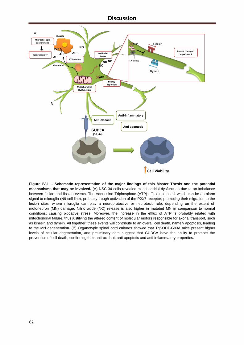
Discussion
62
ATP
NO
NO
Microglia
P2X7
Motoneuron
MitochondrialDysfunction
Microglial cellsrecruitment
Neurotoxicity
ATP releaseATP
ATP
ATP
ATP
Energydepletion
Axonal transportimpairment
Kinesin
Dynein
Swellings
NO
OxidativeStress
NO
A
B
GUDCA(50 µM)
Anti-oxidant
Anti-apoptotic
Anti-inflammatory
Cell Viability
Figure IV.1 – Schematic representation of the major findings of this Master Thesis and the potential
mechanisms that may be involved. (A) NSC-34 cells revealed mitochondrial dysfunction due to an imbalance
between fusion and fission events. The Adenosine Triphosphate (ATP) efflux increased, which can be an alarm
signal to microglia (N9 cell line), probably trough activation of the P2X7 receptor, promoting their migration to the
lesion sites, where microglia can play a neuroprotective or neurotoxic role, depending on the extent of
motoneuron (MN) damage. Nitric oxide (NO) release is also higher in mutated MN in comparison to normal
conditions, causing oxidative stress. Moreover, the increase in the efflux of ATP is probably related with
mitochondrial failure, thus justifying the altered content of molecular motors responsible for axonal transport, such
as kinesin and dynein. All together, these events will contribute to an overall cell death, namely apoptosis, leading
to the MN degeneration. (B) Organotypic spinal cord cultures showed that TgSOD1-G93A mice present higher
levels of cellular degeneration, and preliminary data suggest that GUDCA have the ability to promote the
prevention of cell death, confirming their anti-oxidant, anti-apoptotic and anti-inflammatory properties.
Discussion
63
Future Perspectives
This study provides an increase in our knowledge regarding the pathophysiological events in
the context of ALS. However, there is still a long way to go. It will be important to study more deeply
the dysfunction of axonal transport, in particular at level of neurite length and branching as MNs
degenerate within time. In the context of the mitochondrial dysfunction, it will be important to direct our
studies for the understanding of possible signaling mechanisms that are activated upon MN injury,
which seemed to help in the attempt to return to mitochondrial homeostasis. Therefore, we hope very
soon to proceed with the determinations we made in isolated NSC-34 cultures, now in mixed NSC-
34/N9 cultures. Furthermore, it will be interesting to study more deeply certain pathological events in
organotypical cultures from lumbar SC, including mitochondrial dysfunction and axonal transport, as
well as molecules involved in cellular cross-talk. In addition, further development in the study of
neuroprotective effects of GUDCA would be very interesting, in view of the need to create new
therapeutic strategies for ALS onset or progression, once riluzole, the only therapeutic agent approved
by FDA, which inhibits the glutamate-mediated excitotoxic in neurons can only cause a modest
improvement in the survival of the patients (Kiernan et al., 2011). Finally, the confirmation of these
pathways using transgenic mice carrying G93A mSOD1 will be crucial to get a time point of each
phase of the disease progression and to find biomarkers that allow identifying each of this phase.
Currently, the biggest investment in ALS therapeutic strategies has been at the level of stem cells,
with the aim of replacement the individual mutated cells by healthy ones to become less neurotoxic. In
the same line of thought, (Forostyak et al., 2013) described the grafting of mesenchymal stroma cells
as a way to improve motor and sensory function, modifying the host microenvironment following CNS
injury. In fact, they have already achieved some positive results in clinical trials. There is still much that
is unknown about the primary mechanisms involved in ALS onset and progression, although it is
possible that the use of these stem cells combined with the administration of some neuroprotective
compound (such as GUDCA) will become a common practice therapeutic, allowing rehabilitation of the
patients at motor level.
By uncovering different experimental models, our results provide a strong basis for the
comprehension of the molecular events involved in cellular failure in ALS, which is fundamental to
develop new therapeutic strategies.

64
References
65
V. References
Abe K, Pan LH, Watanabe M, Konno H, Kato T, Itoyama Y. 1997. Upregulation of protein-tyrosine nitration in the anterior horn cells of amyotrophic lateral sclerosis. Neurol Res 19(2):124-8.
Agrawal SK, Fehlings MG. 1997. Role of NMDA and non-NMDA ionotropic glutamate receptors in traumatic spinal cord axonal injury. J Neurosci 17(3):1055-63.
Alberts B, Johnson A, Lewis J, Raff M, Roberts K, Walter P. 2008. Molecular Biology of the Cell. 5th
ed, Garland Science, New York.
Alexander C, Votruba M, Pesch UE, Thiselton DL, Mayer S, Moore A, Rodriguez M, Kellner U, Leo-Kottler B, Auburger G and others. 2000. OPA1, encoding a dynamin-related GTPase, is mutated in autosomal dominant optic atrophy linked to chromosome 3q28. Nat Genet 26(2):211-5.
Alliot F, Godin I, Pessac B. 1999. Microglia derive from progenitors, originating from the yolk sac, and which proliferate in the brain. Brain Res Dev Brain Res 117(2):145-52.
Aloisi F. 2001. Immune function of microglia. Glia 36(2):165-79. Amaral JD, Viana RJ, Ramalho RM, Steer CJ, Rodrigues CM. 2009. Bile acids: regulation of apoptosis by
ursodeoxycholic acid. J Lipid Res 50(9):1721-34. Andersen PM, Al-Chalabi A. 2011. Clinical genetics of amyotrophic lateral sclerosis: what do we really know? Nat
Rev Neurol 7(11):603-15. Anneser JM, Chahli C, Ince PG, Borasio GD, Shaw PJ. 2004. Glial proliferation and metabotropic glutamate
receptor expression in amyotrophic lateral sclerosis. J Neuropathol Exp Neurol 63(8):831-40. Atkin JD, Farg MA, Turner BJ, Tomas D, Lysaght JA, Nunan J, Rembach A, Nagley P, Beart PM, Cheema SS
and others. 2006. Induction of the unfolded protein response in familial amyotrophic lateral sclerosis and association of protein-disulfide isomerase with superoxide dismutase 1. J Biol Chem 281(40):30152-65.
Atkin JD, Farg MA, Walker AK, McLean C, Tomas D, Horne MK. 2008. Endoplasmic reticulum stress and induction of the unfolded protein response in human sporadic amyotrophic lateral sclerosis. Neurobiol Dis 30(3):400-7.
Barateiro A, Vaz AR, Silva SL, Fernandes A, Brites D. 2012. ER stress, mitochondrial dysfunction and calpain/JNK activation are involved in oligodendrocyte precursor cell death by unconjugated bilirubin. Neuromolecular Med 14(4):285-302.
Barber SC, Shaw PJ. 2010. Oxidative stress in ALS: key role in motor neuron injury and therapeutic target. Free Radic Biol Med 48(5):629-41.
Barsoum MJ, Yuan H, Gerencser AA, Liot G, Kushnareva Y, Graber S, Kovacs I, Lee WD, Waggoner J, Cui J and others. 2006. Nitric oxide-induced mitochondrial fission is regulated by dynamin-related GTPases in neurons. EMBO J 25(16):3900-11.
Bayerdorffer E, Mannes GA, Richter WO, Ochsenkuhn T, Wiebecke B, Kopcke W, Paumgartner G. 1993. Increased serum deoxycholic acid levels in men with colorectal adenomas. Gastroenterology 104(1):145-51.
Beers DR, Henkel JS, Xiao Q, Zhao W, Wang J, Yen AA, Siklos L, McKercher SR, Appel SH. 2006. Wild-type microglia extend survival in PU.1 knockout mice with familial amyotrophic lateral sclerosis. Proc Natl Acad Sci U S A 103(43):16021-6.
Beers DR, Henkel JS, Zhao W, Wang J, Appel SH. 2008. CD4+ T cells support glial neuroprotection, slow disease progression, and modify glial morphology in an animal model of inherited ALS. Proc Natl Acad Sci U S A 105(40):15558-63.
Bendotti C, Atzori C, Piva R, Tortarolo M, Strong MJ, DeBiasi S, Migheli A. 2004. Activated p38MAPK is a novel component of the intracellular inclusions found in human amyotrophic lateral sclerosis and mutant SOD1 transgenic mice. J Neuropathol Exp Neurol 63(2):113-9.
Bendotti C, Calvaresi N, Chiveri L, Prelle A, Moggio M, Braga M, Silani V, De Biasi S. 2001. Early vacuolization and mitochondrial damage in motor neurons of FALS mice are not associated with apoptosis or with changes in cytochrome oxidase histochemical reactivity. J Neurol Sci 191(1-2):25-33.
Bilsland LG, Sahai E, Kelly G, Golding M, Greensmith L, Schiavo G. 2010. Deficits in axonal transport precede ALS symptoms in vivo. Proc Natl Acad Sci U S A 107(47):20523-8.
Birgisdottir AB, Lamark T, Johansen T. 2013. The LIR motif - crucial for selective autophagy. J Cell Sci 126(Pt 15):3237-47.
66
Blackburn D, Sargsyan S, Monk PN, Shaw PJ. 2009. Astrocyte function and role in motor neuron disease: a future therapeutic target? Glia 57(12):1251-64.
Boillée S, Vande Velde C, Cleveland DW. 2006. ALS: a disease of motor neurons and their nonneuronal neighbors. Neuron 52(1):39-59.
Boillee S, Yamanaka K, Lobsiger CS, Copeland NG, Jenkins NA, Kassiotis G, Kollias G, Cleveland DW. 2006. Onset and progression in inherited ALS determined by motor neurons and microglia. Science 312(5778):1389-92.
Bories C, Amendola J, Lamotte d'Incamps B, Durand J. 2007. Early electrophysiological abnormalities in lumbar motoneurons in a transgenic mouse model of amyotrophic lateral sclerosis. Eur J Neurosci 25(2):451-9.
Brites D. 2002. Intrahepatic cholestasis of pregnancy: changes in maternal-fetal bile acid balance and improvement by ursodeoxycholic acid. Ann Hepatol 1(1):20-8.
Browne SE, Yang L, DiMauro JP, Fuller SW, Licata SC, Beal MF. 2006. Bioenergetic abnormalities in discrete cerebral motor pathways presage spinal cord pathology in the G93A SOD1 mouse model of ALS. Neurobiol Dis 22(3):599-610.
Bruce-Keller AJ, Keeling JL, Keller JN, Huang FF, Camondola S, Mattson MP. 2000. Antiinflammatory effects of estrogen on microglial activation. Endocrinology 141(10):3646-56.
Bruijn LI, Becher MW, Lee MK, Anderson KL, Jenkins NA, Copeland NG, Sisodia SS, Rothstein JD, Borchelt DR, Price DL and others. 1997. ALS-linked SOD1 mutant G85R mediates damage to astrocytes and promotes rapidly progressive disease with SOD1-containing inclusions. Neuron 18(2):327-38.
Byström R, Andersen PM, Grobner G, Oliveberg M. 2010. SOD1 mutations targeting surface hydrogen bonds promote amyotrophic lateral sclerosis without reducing apo-state stability. J Biol Chem 285(25):19544-52.
Calvo M, Bennett DL. 2012. The mechanisms of microgliosis and pain following peripheral nerve injury. Exp Neurol 234(2):271-82.
Cao Y, Lv G, Wang YS, Fan ZK, Bi YL, Zhao L, Guo ZP. 2013. Mitochondrial fusion and fission after spinal sacord injury in rats. Brain Res 1522:59-66.
Cardona AE, Pioro EP, Sasse ME, Kostenko V, Cardona SM, Dijkstra IM, Huang D, Kidd G, Dombrowski S, Dutta R and others. 2006. Control of microglial neurotoxicity by the fractalkine receptor. Nat Neurosci 9(7):917-24.
Carpentier PA, Duncan DS, Miller SD. 2008. Glial toll-like receptor signaling in central nervous system infection and autoimmunity. Brain Behav Immun 22(2):140-7.
Carri MT, Ferri A, Cozzolino M, Calabrese L, Rotilio G. 2003. Neurodegeneration in amyotrophic lateral sclerosis: the role of oxidative stress and altered homeostasis of metals. Brain Res Bull 61(4):365-74.
Casanovas A, Hernandez S, Tarabal O, Rossello J, Esquerda JE. 2008. Strong P2X4 purinergic receptor-like immunoreactivity is selectively associated with degenerating neurons in transgenic rodent models of amyotrophic lateral sclerosis. J Comp Neurol 506(1):75-92.
Cashman NR, Durham HD, Blusztajn JK, Oda K, Tabira T, Shaw IT, Dahrouge S, Antel JP. 1992. Neuroblastoma x spinal cord (NSC) hybrid cell lines resemble developing motor neurons. Dev Dyn 194(3):209-21.
Cassina P, Cassina A, Pehar M, Castellanos R, Gandelman M, de Leon A, Robinson KM, Mason RP, Beckman JS, Barbeito L and others. 2008. Mitochondrial dysfunction in SOD1G93A-bearing astrocytes promotes motor neuron degeneration: prevention by mitochondrial-targeted antioxidants. J Neurosci 28(16):4115-22.
Chan DC. 2006. Mitochondria: dynamic organelles in disease, aging, and development. Cell 125(7):1241-52. Chan DC. 2012. Fusion and fission: interlinked processes critical for mitochondrial health. Annu Rev Genet
46:265-87. Chan WY, Kohsaka S, Rezaie P. 2007. The origin and cell lineage of microglia: new concepts. Brain Res Rev
53(2):344-54. Chang Y, Kong Q, Shan X, Tian G, Ilieva H, Cleveland DW, Rothstein JD, Borchelt DR, Wong PC, Lin CL. 2008.
Messenger RNA oxidation occurs early in disease pathogenesis and promotes motor neuron degeneration in ALS. PLoS One 3(8):e2849.
Chapman AL, Bennett EJ, Ramesh TM, De Vos KJ, Grierson AJ. 2013. Axonal Transport Defects in a Mitofusin 2 Loss of Function Model of Charcot-Marie-Tooth Disease in Zebrafish. PLoS One 8(6):e67276.
Chen H, Chomyn A, Chan DC. 2005. Disruption of fusion results in mitochondrial heterogeneity and dysfunction. J Biol Chem 280(28):26185-92.
Chen H, Guo Y, Hu M, Duan W, Chang G, Li C. 2010. Differential expression and alternative splicing of genes in lumbar spinal cord of an amyotrophic lateral sclerosis mouse model. Brain Res 1340:52-69.
Clement AM, Nguyen MD, Roberts EA, Garcia ML, Boillee S, Rule M, McMahon AP, Doucette W, Siwek D, Ferrante RJ and others. 2003. Wild-type nonneuronal cells extend survival of SOD1 mutant motor neurons in ALS mice. Science 302(5642):113-7.
Colton C, Wilcock DM. 2010. Assessing activation states in microglia. CNS Neurol Disord Drug Targets 9(2):174-91.
Colton CA. 2009. Heterogeneity of microglial activation in the innate immune response in the brain. J Neuroimmune Pharmacol 4(4):399-418.
Corona JC, Tovar-y-Romo LB, Tapia R. 2007. Glutamate excitotoxicity and therapeutic targets for amyotrophic lateral sclerosis. Expert Opin Ther Targets 11(11):1415-28.
Corriden R, Insel PA. 2012. New insights regarding the regulation of chemotaxis by nucleotides, adenosine, and their receptors. Purinergic Signal 8(3):587-98.
References
67
Costa J, Gomes C, de Carvalho M. 2010. Diagnosis, pathogenesis and therapeutic targets in amyotrophic lateral sclerosis. CNS Neurol Disord Drug Targets 9(6):764-78.
Cox LE, Ferraiuolo L, Goodall EF, Heath PR, Higginbottom A, Mortiboys H, Hollinger HC, Hartley JA, Brockington A, Burness CE and others. 2010. Mutations in CHMP2B in lower motor neuron predominant amyotrophic lateral sclerosis (ALS). PLoS One 5(3):e9872.
Cozzolino M, Amori I, Pesaresi MG, Ferri A, Nencini M, Carri MT. 2008. Cysteine 111 affects aggregation and cytotoxicity of mutant Cu,Zn-superoxide dismutase associated with familial amyotrophic lateral sclerosis. J Biol Chem 283(2):866-74.
D'Ambrosi N, Finocchi P, Apolloni S, Cozzolino M, Ferri A, Padovano V, Pietrini G, Carri MT, Volonte C. 2009. The proinflammatory action of microglial P2 receptors is enhanced in SOD1 models for amyotrophic lateral sclerosis. J Immunol 183(7):4648-56.
Dadon-Nachum M, Melamed E, Offen D. 2011. The "dying-back" phenomenon of motor neurons in ALS. J Mol Neurosci 43(3):470-7.
Das K, Nag C, Ghosh M. 2012. Familial, environmental, and occupational risk factors in development of amyotrophic lateral sclerosis. N Am J Med Sci 4(8):350-5.
Delfs J, Friend J, Ishimoto S, Saroff D. 1989. Ventral and dorsal horn acetylcholinesterase neurons are maintained in organotypic cultures of postnatal rat spinal cord explants. Brain Res 488(1-2):31-42.
Dewil M, Lambrechts D, Sciot R, Shaw PJ, Ince PG, Robberecht W, Van den Bosch L. 2007. Vascular endothelial growth factor counteracts the loss of phospho-Akt preceding motor neurone degeneration in amyotrophic lateral sclerosis. Neuropathol Appl Neurobiol 33(5):499-509.
Diaz-Amarilla P, Olivera-Bravo S, Trias E, Cragnolini A, Martinez-Palma L, Cassina P, Beckman J, Barbeito L. 2011. Phenotypically aberrant astrocytes that promote motoneuron damage in a model of inherited amyotrophic lateral sclerosis. Proc Natl Acad Sci U S A 108(44):18126-31.
Dodson M, Darley-Usmar V, Zhang J. 2013. Cellular metabolic and autophagic pathways: Traffic control by redox signaling. Free Radic Biol Med 63:207-21.
Duan W, Li X, Shi J, Guo Y, Li Z, Li C. 2010. Mutant TAR DNA-binding protein-43 induces oxidative injury in motor neuron-like cell. Neuroscience 169(4):1621-9.
Duffy LM, Chapman AL, Shaw PJ, Grierson AJ. 2011. Review: The role of mitochondria in the pathogenesis of amyotrophic lateral sclerosis. Neuropathol Appl Neurobiol 37(4):336-52.
Durafourt BA, Moore CS, Zammit DA, Johnson TA, Zaguia F, Guiot MC, Bar-Or A, Antel JP. 2012. Comparison of polarization properties of human adult microglia and blood-derived macrophages. Glia 60(5):717-27.
Durham HD, Dahrouge S, Cashman NR. 1993. Evaluation of the spinal cord neuron X neuroblastoma hybrid cell line NSC-34 as a model for neurotoxicity testing. Neurotoxicology 14(4):387-95.
Ekestern E. 2004. Neurotrophic factors and amyotrophic lateral sclerosis. Neurodegener Dis 1(2-3):88-100. Evans MC, Couch Y, Sibson N, Turner MR. 2013. Inflammation and neurovascular changes in amyotrophic lateral
sclerosis. Mol Cell Neurosci 53:34-41. Falcão AS, Fernandes A, Brito MA, Silva RF, Brites D. 2005. Bilirubin-induced inflammatory response, glutamate
release, and cell death in rat cortical astrocytes are enhanced in younger cells. Neurobiol Dis 20(2):199-206.
Federico A, Cardaioli E, Da Pozzo P, Formichi P, Gallus GN, Radi E. 2012. Mitochondria, oxidative stress and neurodegeneration. J Neurol Sci 322(1-2):254-62.
Fellin T. 2009. Communication between neurons and astrocytes: relevance to the modulation of synaptic and network activity. J Neurochem 108(3):533-44.
Fernandes A, Brites D. 2009. Contribution of inflammatory processes to nerve cell toxicity by bilirubin and efficacy of potential therapeutic agents. Curr Pharm Des 15(25):2915-26.
Fernandes A, Vaz AR, Falcao AS, Silva RF, Brito MA, Brites D. 2007. Glycoursodeoxycholic acid and interleukin-10 modulate the reactivity of rat cortical astrocytes to unconjugated bilirubin. J Neuropathol Exp Neurol 66(9):789-98.
Ferraiuolo L, Heath PR, Holden H, Kasher P, Kirby J, Shaw PJ. 2007. Microarray analysis of the cellular pathways involved in the adaptation to and progression of motor neuron injury in the SOD1 G93A mouse model of familial ALS. J Neurosci 27(34):9201-19.
Ferraiuolo L, Kirby J, Grierson AJ, Sendtner M, Shaw PJ. 2011. Molecular pathways of motor neuron injury in amyotrophic lateral sclerosis. Nat Rev Neurol 7(11):616-30.
Ferrante RJ, Browne SE, Shinobu LA, Bowling AC, Baik MJ, MacGarvey U, Kowall NW, Brown RH, Jr., Beal MF. 1997. Evidence of increased oxidative damage in both sporadic and familial amyotrophic lateral sclerosis. J Neurochem 69(5):2064-74.
Fleisher-Berkovich S, Filipovich-Rimon T, Ben-Shmuel S, Hulsmann C, Kummer MP, Heneka MT. 2010. Distinct modulation of microglial amyloid beta phagocytosis and migration by neuropeptides (i). J Neuroinflammation 7:61.
Fornai F, Longone P, Cafaro L, Kastsiuchenka O, Ferrucci M, Manca ML, Lazzeri G, Spalloni A, Bellio N, Lenzi P and others. 2008. Lithium delays progression of amyotrophic lateral sclerosis. Proc Natl Acad Sci U S A 105(6):2052-7.
Forostyak S, Jendelova P, Sykova E. 2013. The role of mesenchymal stromal cells in spinal cord injury, regenerative medicine and possible clinical applications. Biochimie.
Gehrmann J, Matsumoto Y, Kreutzberg GW. 1995. Microglia: intrinsic immuneffector cell of the brain. Brain Res Brain Res Rev 20(3):269-87.
68
Ghiasi P, Hosseinkhani S, Noori A, Nafissi S, Khajeh K. 2012. Mitochondrial complex I deficiency and ATP/ADP ratio in lymphocytes of amyotrophic lateral sclerosis patients. Neurol Res 34(3):297-303.
Ginhoux F, Greter M, Leboeuf M, Nandi S, See P, Gokhan S, Mehler MF, Conway SJ, Ng LG, Stanley ER and others. 2010. Fate mapping analysis reveals that adult microglia derive from primitive macrophages. Science 330(6005):841-5.
Glezer I, Simard AR, Rivest S. 2007. Neuroprotective role of the innate immune system by microglia. Neuroscience 147(4):867-83.
Goetz CG. 2000. Amyotrophic lateral sclerosis: early contributions of Jean-Martin Charcot. Muscle Nerve 23(3):336-43.
Gomes C, Palma AS, Almeida R, Regalla M, McCluskey LF, Trojanowski JQ, Costa J. 2008. Establishment of a cell model of ALS disease: Golgi apparatus disruption occurs independently from apoptosis. Biotechnol Lett 30(4):603-10.
Gordon PH, Cheng B, Katz IB, Pinto M, Hays AP, Mitsumoto H, Rowland LP. 2006. The natural history of primary lateral sclerosis. Neurology 66(5):647-53.
Gordon S. 2003. Alternative activation of macrophages. Nat Rev Immunol 3(1):23-35. Gowing G, Philips T, Van Wijmeersch B, Audet JN, Dewil M, Van Den Bosch L, Billiau AD, Robberecht W, Julien
JP. 2008. Ablation of proliferating microglia does not affect motor neuron degeneration in amyotrophic lateral sclerosis caused by mutant superoxide dismutase. J Neurosci 28(41):10234-44.
Grivennikov SI, Karin M. 2010. Dangerous liaisons: STAT3 and NF-kappaB collaboration and crosstalk in cancer. Cytokine Growth Factor Rev 21(1):11-9.
Guo L, Xing Y, Pan R, Jiang M, Gong Z, Lin L, Wang J, Xiong G, Dong J. 2013. Curcumin Protects Microglia and Primary Rat Cortical Neurons against HIV-1 gp120-Mediated Inflammation and Apoptosis. PLoS One 8(8):e70565.
Gurney ME, Pu H, Chiu AY, Dal Canto MC, Polchow CY, Alexander DD, Caliendo J, Hentati A, Kwon YW, Deng HX and others. 1994. Motor neuron degeneration in mice that express a human Cu,Zn superoxide dismutase mutation. Science 264(5166):1772-5.
Guzman-Lenis MS, Navarro X, Casas C. 2009. Drug screening of neuroprotective agents on an organotypic-based model of spinal cord excitotoxic damage. Restor Neurol Neurosci 27(4):335-49.
Harraz MM, Marden JJ, Zhou W, Zhang Y, Williams A, Sharov VS, Nelson K, Luo M, Paulson H, Schoneich C and others. 2008. SOD1 mutations disrupt redox-sensitive Rac regulation of NADPH oxidase in a familial ALS model. J Clin Invest 118(2):659-70.
Harry GJ. 2013. Microglia during development and aging. Pharmacol Ther. Haynes SE, Hollopeter G, Yang G, Kurpius D, Dailey ME, Gan WB, Julius D. 2006. The P2Y12 receptor regulates
microglial activation by extracellular nucleotides. Nat Neurosci 9(12):1512-9. Henkel JS, Beers DR, Zhao W, Appel SH. 2009. Microglia in ALS: the good, the bad, and the resting. J
Neuroimmune Pharmacol 4(4):389-98. Hervias I, Beal MF, Manfredi G. 2006. Mitochondrial dysfunction and amyotrophic lateral sclerosis. Muscle Nerve
33(5):598-608. Hetz C, Thielen P, Matus S, Nassif M, Court F, Kiffin R, Martinez G, Cuervo AM, Brown RH, Glimcher LH. 2009.
XBP-1 deficiency in the nervous system protects against amyotrophic lateral sclerosis by increasing autophagy. Genes Dev 23(19):2294-306.
Hirokawa N, Niwa S, Tanaka Y. 2010. Molecular motors in neurons: transport mechanisms and roles in brain function, development, and disease. Neuron 68(4):610-38.
Hoek RM, Ruuls SR, Murphy CA, Wright GJ, Goddard R, Zurawski SM, Blom B, Homola ME, Streit WJ, Brown MH and others. 2000. Down-regulation of the macrophage lineage through interaction with OX2 (CD200). Science 290(5497):1768-71.
Hofmann AF, Roda A. 1984. Physicochemical properties of bile acids and their relationship to biological properties: an overview of the problem. J Lipid Res 25(13):1477-89.
Honda S, Sasaki Y, Ohsawa K, Imai Y, Nakamura Y, Inoue K, Kohsaka S. 2001. Extracellular ATP or ADP induce chemotaxis of cultured microglia through Gi/o-coupled P2Y receptors. J Neurosci 21(6):1975-82.
Hovden H, Frederiksen JL, Pedersen SW. 2013. Immune system alterations in amyotrophic lateral sclerosis. Acta Neurol Scand.
Howland DS, Liu J, She Y, Goad B, Maragakis NJ, Kim B, Erickson J, Kulik J, DeVito L, Psaltis G and others. 2002. Focal loss of the glutamate transporter EAAT2 in a transgenic rat model of SOD1 mutant-mediated amyotrophic lateral sclerosis (ALS). Proc Natl Acad Sci U S A 99(3):1604-9.
Hunter DD, Cashman N, Morris-Valero R, Bulock JW, Adams SP, Sanes JR. 1991. An LRE (leucine-arginine-glutamate)-dependent mechanism for adhesion of neurons to S-laminin. J Neurosci 11(12):3960-71.
Ikenaka K, Katsuno M, Kawai K, Ishigaki S, Tanaka F, Sobue G. 2012. Disruption of axonal transport in motor neuron diseases. Int J Mol Sci 13(1):1225-38.
Ilieva H, Polymenidou M, Cleveland DW. 2009. Non-cell autonomous toxicity in neurodegenerative disorders: ALS and beyond. J Cell Biol 187(6):761-72.
Ince PG, Highley JR, Kirby J, Wharton SB, Takahashi H, Strong MJ, Shaw PJ. 2011. Molecular pathology and genetic advances in amyotrophic lateral sclerosis: an emerging molecular pathway and the significance of glial pathology. Acta Neuropathol 122(6):657-71.
Jaarsma D, Teuling E, Haasdijk ED, De Zeeuw CI, Hoogenraad CC. 2008. Neuron-specific expression of mutant superoxide dismutase is sufficient to induce amyotrophic lateral sclerosis in transgenic mice. J Neurosci 28(9):2075-88.
References
69
James DI, Parone PA, Mattenberger Y, Martinou JC. 2003. hFis1, a novel component of the mammalian mitochondrial fission machinery. J Biol Chem 278(38):36373-9.
Jonsson PA, Graffmo KS, Brannstrom T, Nilsson P, Andersen PM, Marklund SL. 2006. Motor neuron disease in mice expressing the wild type-like D90A mutant superoxide dismutase-1. J Neuropathol Exp Neurol 65(12):1126-36.
Kanekura K, Suzuki H, Aiso S, Matsuoka M. 2009. ER stress and unfolded protein response in amyotrophic lateral sclerosis. Mol Neurobiol 39(2):81-9.
Kari G, Rodeck U, Dicker AP. 2007. Zebrafish: an emerging model system for human disease and drug discovery. Clin Pharmacol Ther 82(1):70-80.
Kawamata T, Akiyama H, Yamada T, McGeer PL. 1992. Immunologic reactions in amyotrophic lateral sclerosis brain and spinal cord tissue. Am J Pathol 140(3):691-707.
Kettenmann H, Hanisch UK, Noda M, Verkhratsky A. 2011. Physiology of microglia. Physiol Rev 91(2):461-553. Kiernan MC, Vucic S, Cheah BC, Turner MR, Eisen A, Hardiman O, Burrell JR, Zoing MC. 2011. Amyotrophic
lateral sclerosis. Lancet 377(9769):942-55. Kim J, Kim TY, Cho KS, Kim HN, Koh JY. 2013. Autophagy activation and neuroprotection by progesterone in the
G93A-SOD1 transgenic mouse model of amyotrophic lateral sclerosis. Neurobiol Dis 59C:80-85. King AE, Dickson TC, Blizzard CA, Woodhouse A, Foster SS, Chung RS, Vickers JC. 2011. Neuron-glia
interactions underlie ALS-like axonal cytoskeletal pathology. Neurobiol Aging 32(3):459-69. Kipnis J, Avidan H, Caspi RR, Schwartz M. 2004. Dual effect of CD4+CD25+ regulatory T cells in
neurodegeneration: a dialogue with microglia. Proc Natl Acad Sci U S A 101 Suppl 2:14663-9. Kirby J, Ning K, Ferraiuolo L, Heath PR, Ismail A, Kuo SW, Valori CF, Cox L, Sharrack B, Wharton SB and others.
2011. Phosphatase and tensin homologue/protein kinase B pathway linked to motor neuron survival in human superoxide dismutase 1-related amyotrophic lateral sclerosis. Brain 134(Pt 2):506-17.
Klionsky DJ. 2007. Autophagy: from phenomenology to molecular understanding in less than a decade. Nat Rev Mol Cell Biol 8(11):931-7.
Knott AB, Perkins G, Schwarzenbacher R, Bossy-Wetzel E. 2008. Mitochondrial fragmentation in neurodegeneration. Nat Rev Neurosci 9(7):505-18.
Kong J, Xu Z. 1998. Massive mitochondrial degeneration in motor neurons triggers the onset of amyotrophic lateral sclerosis in mice expressing a mutant SOD1. J Neurosci 18(9):3241-50.
Kraft AD, Harry GJ. 2011. Features of microglia and neuroinflammation relevant to environmental exposure and neurotoxicity. Int J Environ Res Public Health 8(7):2980-3018.
Krieger C, Hu JH, Pelech S. 2003. Aberrant protein kinases and phosphoproteins in amyotrophic lateral sclerosis. Trends Pharmacol Sci 24(10):535-41.
Kriz J, Nguyen MD, Julien JP. 2002. Minocycline slows disease progression in a mouse model of amyotrophic lateral sclerosis. Neurobiol Dis 10(3):268-78.
Kuhle J, Lindberg RL, Regeniter A, Mehling M, Steck AJ, Kappos L, Czaplinski A. 2009. Increased levels of inflammatory chemokines in amyotrophic lateral sclerosis. Eur J Neurol 16(6):771-4.
Kuzma-Kozakiewicz M, Chudy A, Gajewska B, Dziewulska D, Usarek E, Baranczyk-Kuzma A. 2013. Kinesin Expression in the Central Nervous System of Humans and Transgenic hSOD1G93A Mice with Amyotrophic Lateral Sclerosis. Neurodegener Dis 12:71-80.
Kwiatkowski TJ, Jr., Bosco DA, Leclerc AL, Tamrazian E, Vanderburg CR, Russ C, Davis A, Gilchrist J, Kasarskis EJ, Munsat T and others. 2009. Mutations in the FUS/TLS gene on chromosome 16 cause familial amyotrophic lateral sclerosis. Science 323(5918):1205-8.
Lalancette-Hebert M, Gowing G, Simard A, Weng YC, Kriz J. 2007. Selective ablation of proliferating microglial cells exacerbates ischemic injury in the brain. J Neurosci 27(10):2596-605.
Lasiene J, Yamanaka K. 2011. Glial cells in amyotrophic lateral sclerosis. Neurol Res Int 2011:718987. Lazaridis KN, Gores GJ, Lindor KD. 2001. Ursodeoxycholic acid 'mechanisms of action and clinical use in
hepatobiliary disorders'. J Hepatol 35(1):134-46. Ledeboer A, Breve JJ, Poole S, Tilders FJ, Van Dam AM. 2000. Interleukin-10, interleukin-4, and transforming
growth factor-beta differentially regulate lipopolysaccharide-induced production of pro-inflammatory cytokines and nitric oxide in co-cultures of rat astroglial and microglial cells. Glia 30(2):134-42.
Lee JC, Seong J, Kim SH, Lee SJ, Cho YJ, An J, Nam DH, Joo KM, Cha CI. 2012. Replacement of microglial cells using Clodronate liposome and bone marrow transplantation in the central nervous system of SOD1(G93A) transgenic mice as an in vivo model of amyotrophic lateral sclerosis. Biochem Biophys Res Commun 418(2):359-65.
Levine B, Klionsky DJ. 2004. Development by self-digestion: molecular mechanisms and biological functions of autophagy. Dev Cell 6(4):463-77.
Lewis CA, Manning J, Rossi F, Krieger C. 2012. The Neuroinflammatory Response in ALS: The Roles of Microglia and T Cells. Neurol Res Int 2012:803701.
Liao B, Zhao W, Beers DR, Henkel JS, Appel SH. 2012. Transformation from a neuroprotective to a neurotoxic microglial phenotype in a mouse model of ALS. Exp Neurol 237(1):147-52.
Liot G, Bossy B, Lubitz S, Kushnareva Y, Sejbuk N, Bossy-Wetzel E. 2009. Complex II inhibition by 3-NP causes mitochondrial fragmentation and neuronal cell death via an NMDA- and ROS-dependent pathway. Cell Death Differ 16(6):899-909.
Liu Y, Hao W, Dawson A, Liu S, Fassbender K. 2009. Expression of amyotrophic lateral sclerosis-linked SOD1 mutant increases the neurotoxic potential of microglia via TLR2. J Biol Chem 284(6):3691-9.
70
Lobsiger CS, Boillee S, McAlonis-Downes M, Khan AM, Feltri ML, Yamanaka K, Cleveland DW. 2009. Schwann cells expressing dismutase active mutant SOD1 unexpectedly slow disease progression in ALS mice. Proc Natl Acad Sci U S A 106(11):4465-70.
Mackenzie IR, Ansorge O, Strong M, Bilbao J, Zinman L, Ang LC, Baker M, Stewart H, Eisen A, Rademakers R and others. 2011. Pathological heterogeneity in amyotrophic lateral sclerosis with FUS mutations: two distinct patterns correlating with disease severity and mutation. Acta Neuropathol 122(1):87-98.
Mackenzie IR, Rademakers R, Neumann M. 2010. TDP-43 and FUS in amyotrophic lateral sclerosis and frontotemporal dementia. Lancet Neurol 9(10):995-1007.
Mantovani S, Garbelli S, Pasini A, Alimonti D, Perotti C, Melazzini M, Bendotti C, Mora G. 2009. Immune system alterations in sporadic amyotrophic lateral sclerosis patients suggest an ongoing neuroinflammatory process. J Neuroimmunol 210(1-2):73-9.
Maragakis NJ, Dykes-Hoberg M, Rothstein JD. 2004. Altered expression of the glutamate transporter EAAT2b in neurological disease. Ann Neurol 55(4):469-77.
Martin LJ. 1999. Neuronal death in amyotrophic lateral sclerosis is apoptosis: possible contribution of a programmed cell death mechanism. J Neuropathol Exp Neurol 58(5):459-71.
Martin LJ, Al-Abdulla NA, Brambrink AM, Kirsch JR, Sieber FE, Portera-Cailliau C. 1998. Neurodegeneration in excitotoxicity, global cerebral ischemia, and target deprivation: A perspective on the contributions of apoptosis and necrosis. Brain Res Bull 46(4):281-309.
Martin LJ, Liu Z, Chen K, Price AC, Pan Y, Swaby JA, Golden WC. 2007. Motor neuron degeneration in amyotrophic lateral sclerosis mutant superoxide dismutase-1 transgenic mice: mechanisms of mitochondriopathy and cell death. J Comp Neurol 500(1):20-46.
Mead RJ, Bennett EJ, Kennerley AJ, Sharp P, Sunyach C, Kasher P, Berwick J, Pettmann B, Battaglia G, Azzouz M and others. 2011. Optimised and rapid pre-clinical screening in the SOD1(G93A) transgenic mouse model of amyotrophic lateral sclerosis (ALS). PLoS One 6(8):e23244.
Meissner F, Molawi K, Zychlinsky A. 2010. Mutant superoxide dismutase 1-induced IL-1beta accelerates ALS pathogenesis. Proc Natl Acad Sci U S A 107(29):13046-50.
Meredith GE, Totterdell S, Petroske E, Santa Cruz K, Callison RC, Jr., Lau YS. 2002. Lysosomal malfunction accompanies alpha-synuclein aggregation in a progressive mouse model of Parkinson's disease. Brain Res 956(1):156-65.
Mildner A, Schmidt H, Nitsche M, Merkler D, Hanisch UK, Mack M, Heikenwalder M, Bruck W, Priller J, Prinz M. 2007. Microglia in the adult brain arise from Ly-6ChiCCR2+ monocytes only under defined host conditions. Nat Neurosci 10(12):1544-53.
Min JH, Hong YH, Sung JJ, Kim SM, Lee JB, Lee KW. 2012. Oral solubilized ursodeoxycholic acid therapy in amyotrophic lateral sclerosis: a randomized cross-over trial. J Korean Med Sci 27(2):200-6.
Mitchell JD, Borasio GD. 2007. Amyotrophic lateral sclerosis. Lancet 369(9578):2031-41. Mitsumoto H, Santella RM, Liu X, Bogdanov M, Zipprich J, Wu HC, Mahata J, Kilty M, Bednarz K, Bell D and
others. 2008. Oxidative stress biomarkers in sporadic ALS. Amyotroph Lateral Scler 9(3):177-83. Moisse K, Strong MJ. 2006. Innate immunity in amyotrophic lateral sclerosis. Biochim Biophys Acta 1762(11-
12):1083-93. Morfini GA, Bosco DA, Brown H, Gatto R, Kaminska A, Song Y, Molla L, Baker L, Marangoni MN, Berth S and
others. 2013. Inhibition of fast axonal transport by pathogenic SOD1 involves activation of p38 MAP kinase. PLoS One 8(6):e65235.
Morfini GA, Burns M, Binder LI, Kanaan NM, LaPointe N, Bosco DA, Brown RH Jr, Brown H, Tiwari A, Hayward L, Edgar J, Nave KA, Garberrn J, Ataqi Y, Song Y, Pigino G, Brady ST. 2009. Axonal transport defects in neurodegenerative diseases. J Neurosci. 29(41):12776-86.
Morimoto N, Nagai M, Ohta Y, Miyazaki K, Kurata T, Morimoto M, Murakami T, Takehisa Y, Ikeda Y, Kamiya T and others. 2007. Increased autophagy in transgenic mice with a G93A mutant SOD1 gene. Brain Res 1167:112-7.
Moser JM, Bigini P, Schmitt-John T. 2013. The wobbler mouse, an ALS animal model. Mol Genet Genomics 288(5-6):207-29.
Musaro A. 2010. State of the art and the dark side of amyotrophic lateral sclerosis. World J Biol Chem 1(5):62-8. Muyderman H, Hutson PG, Matusica D, Rogers ML, Rush RA. 2009. The human G93A-superoxide dismutase-1
mutation, mitochondrial glutathione and apoptotic cell death. Neurochem Res 34(10):1847-56. Nakajima K, Kohsaka S, Tohyama Y, Kurihara T. 2003. Activation of microglia with lipopolysaccharide leads to
the prolonged decrease of conventional protein kinase C activity. Brain Res Mol Brain Res 110(1):92-9. Napoli I, Neumann H. 2009. Microglial clearance function in health and disease. Neuroscience 158(3):1030-8. Nayak D, Huo Y, Kwang WX, Pushparaj PN, Kumar SD, Ling EA, Dheen ST. 2010. Sphingosine kinase 1
regulates the expression of proinflammatory cytokines and nitric oxide in activated microglia. Neuroscience 166(1):132-44.
Neher JJ, Neniskyte U, Brown GC. 2012. Primary phagocytosis of neurons by inflamed microglia: potential roles in neurodegeneration. Front Pharmacol 3:27.
Niebroj-Dobosz I, Rafalowska J, Fidzianska A, Gadamski R, Grieb P. 2007. Myelin composition of spinal cord in a model of amyotrophic lateral sclerosis (ALS) in SOD1G93A transgenic rats. Folia Neuropathol 45(4):236-41.
Nishitoh H, Kadowaki H, Nagai A, Maruyama T, Yokota T, Fukutomi H, Noguchi T, Matsuzawa A, Takeda K, Ichijo H. 2008. ALS-linked mutant SOD1 induces ER stress- and ASK1-dependent motor neuron death by targeting Derlin-1. Genes Dev 22(11):1451-64.
References
71
Nixon RA. 2007. Autophagy, amyloidogenesis and Alzheimer disease. J Cell Sci 120(Pt 23):4081-91. Nixon RA, Wegiel J, Kumar A, Yu WH, Peterhoff C, Cataldo A, Cuervo AM. 2005. Extensive involvement of
autophagy in Alzheimer disease: an immuno-electron microscopy study. J Neuropathol Exp Neurol 64(2):113-22.
Ohnishi S, Ito H, Suzuki Y, Adachi Y, Wate R, Zhang J, Nakano S, Kusaka H, Ikehara S. 2009. Intra-bone marrow-bone marrow transplantation slows disease progression and prolongs survival in G93A mutant SOD1 transgenic mice, an animal model mouse for amyotrophic lateral sclerosis. Brain Res 1296:216-24.
Panov AV, Kubalik N, Zinchenko N, Ridings DM, Radoff DA, Hemendinger R, Brooks BR, Bonkovsky HL. 2011. Metabolic and functional differences between brain and spinal cord mitochondria underlie different predisposition to pathology. Am J Physiol Regul Integr Comp Physiol 300(4):R844-54.
Papadimitriou D, Le Verche V, Jacquier A, Ikiz B, Przedborski S, Re DB. 2010. Inflammation in ALS and SMA: sorting out the good from the evil. Neurobiol Dis 37(3):493-502.
Parkinson N, Ince PG, Smith MO, Highley R, Skibinski G, Andersen PM, Morrison KE, Pall HS, Hardiman O, Collinge J and others. 2006. ALS phenotypes with mutations in CHMP2B (charged multivesicular body protein 2B). Neurology 67(6):1074-7.
Pasinelli P, Belford ME, Lennon N, Bacskai BJ, Hyman BT, Trotti D, Brown RH, Jr. 2004. Amyotrophic lateral sclerosis-associated SOD1 mutant proteins bind and aggregate with Bcl-2 in spinal cord mitochondria. Neuron 43(1):19-30.
Pasinelli P, Brown RH. 2006. Molecular biology of amyotrophic lateral sclerosis: insights from genetics. Nat Rev Neurosci 7(9):710-23.
Pehar M, Cassina P, Vargas MR, Castellanos R, Viera L, Beckman JS, Estevez AG, Barbeito L. 2004. Astrocytic production of nerve growth factor in motor neuron apoptosis: implications for amyotrophic lateral sclerosis. J Neurochem 89(2):464-73.
Peng L, Men X, Zhang W, Wang H, Xu S, Fang Q, Liu H, Yang W, Lou J. 2012. Involvement of dynamin-related protein 1 in free fatty acid-induced INS-1-derived cell apoptosis. PLoS One 7(11):e49258.
Peng L, Men X, Zhang W, Wang H, Xu S, Fang Q, Liu H, Yang W, Lou J. 2013. Correction: Involvement of Dynamin-Related Protein 1 in Free Fatty Acid-Induced INS-1-Derived Cell Apoptosis. PLoS One 8(8).
Perea G, Navarrete M, Araque A. 2009. Tripartite synapses: astrocytes process and control synaptic information. Trends Neurosci 32(8):421-31.
Perlson E, Jeong GB, Ross JL, Dixit R, Wallace KE, Kalb RG, Holzbaur EL. 2009. A switch in retrograde signaling from survival to stress in rapid-onset neurodegeneration. J Neurosci 29(31):9903-17.
Philips T, Robberecht W. 2011. Neuroinflammation in amyotrophic lateral sclerosis: role of glial activation in motor neuron disease. Lancet Neurol 10(3):253-63.
Pizzasegola C, Caron I, Daleno C, Ronchi A, Minoia C, Carri MT, Bendotti C. 2009. Treatment with lithium carbonate does not improve disease progression in two different strains of SOD1 mutant mice. Amyotroph Lateral Scler 10(4):221-8.
Raimondi A, Mangolini A, Rizzardini M, Tartari S, Massari S, Bendotti C, Francolini M, Borgese N, Cantoni L, Pietrini G. 2006. Cell culture models to investigate the selective vulnerability of motoneuronal mitochondria to familial ALS-linked G93ASOD1. Eur J Neurosci 24(2):387-99.
Ramalho RM, Borralho PM, Castro RE, Sola S, Steer CJ, Rodrigues CM. 2006. Tauroursodeoxycholic acid modulates p53-mediated apoptosis in Alzheimer's disease mutant neuroblastoma cells. J Neurochem 98(5):1610-8.
Ramalho RM, Viana RJ, Low WC, Steer CJ, Rodrigues CM. 2008. Bile acids and apoptosis modulation: an emerging role in experimental Alzheimer's disease. Trends Mol Med 14(2):54-62.
Ranganathan S, Bowser R. 2010. p53 and Cell Cycle Proteins Participate in Spinal Motor Neuron Cell Death in ALS. Open Pathol J 4:11-22.
Ranieri M, Brajkovic S, Riboldi G, Ronchi D, Rizzo F, Bresolin N, Corti S, Comi GP. 2013. Mitochondrial fusion proteins and human diseases. Neurol Res Int 2013:293893.
Rao RV, Ellerby HM, Bredesen DE. 2004a. Coupling endoplasmic reticulum stress to the cell death program. Cell Death Differ 11(4):372-80.
Rao RV, Poksay KS, Castro-Obregon S, Schilling B, Row RH, del Rio G, Gibson BW, Ellerby HM, Bredesen DE. 2004b. Molecular components of a cell death pathway activated by endoplasmic reticulum stress. J Biol Chem 279(1):177-87.
Rao SD, Weiss JH. 2004. Excitotoxic and oxidative cross-talk between motor neurons and glia in ALS pathogenesis. Trends Neurosci 27(1):17-23.
Ravikumar B, Vacher C, Berger Z, Davies JE, Luo S, Oroz LG, Scaravilli F, Easton DF, Duden R, O'Kane CJ and others. 2004. Inhibition of mTOR induces autophagy and reduces toxicity of polyglutamine expansions in fly and mouse models of Huntington disease. Nat Genet 36(6):585-95.
Ravikumar M, Jain S, Miller RH, Capadona JR, Selkirk SM. 2012. An organotypic spinal cord slice culture model to quantify neurodegeneration. J Neurosci Methods 211(2):280-8.
Reaume AG, Elliott JL, Hoffman EK, Kowall NW, Ferrante RJ, Siwek DF, Wilcox HM, Flood DG, Beal MF, Brown RH, Jr. and others. 1996. Motor neurons in Cu/Zn superoxide dismutase-deficient mice develop normally but exhibit enhanced cell death after axonal injury. Nat Genet 13(1):43-7.
Redler RL, Dokholyan NV. 2012. The complex molecular biology of amyotrophic lateral sclerosis (ALS). Prog Mol Biol Transl Sci 107:215-62.
72
Rezaie P, Dean A, Male D, Ulfig N. 2005. Microglia in the cerebral wall of the human telencephalon at second trimester. Cereb Cortex 15(7):938-49.
Righi M, Mori L, De Libero G, Sironi M, Biondi A, Mantovani A, Donini SD, Ricciardi-Castagnoli P. 1989. Monokine production by microglial cell clones. Eur J Immunol 19(8):1443-8.
Ripps ME, Huntley GW, Hof PR, Morrison JH, Gordon JW. 1995. Transgenic mice expressing an altered murine superoxide dismutase gene provide an animal model of amyotrophic lateral sclerosis. Proc Natl Acad Sci U S A 92(3):689-93.
Ritter MR, Banin E, Moreno SK, Aguilar E, Dorrell MI, Friedlander M. 2006. Myeloid progenitors differentiate into microglia and promote vascular repair in a model of ischemic retinopathy. J Clin Invest 116(12):3266-76.
Rizzardini M, Mangolini A, Lupi M, Ubezio P, Bendotti C, Cantoni L. 2005. Low levels of ALS-linked Cu/Zn superoxide dismutase increase the production of reactive oxygen species and cause mitochondrial damage and death in motor neuron-like cells. J Neurol Sci 232(1-2):95-103.
Rodrigues CM, Sola S, Silva R, Brites D. 2000. Bilirubin and amyloid-beta peptide induce cytochrome c release through mitochondrial membrane permeabilization. Mol Med 6(11):936-46.
Rosen DR. 1993. Mutations in Cu/Zn superoxide dismutase gene are associated with familial amyotrophic lateral sclerosis. Nature 364(6435):362.
Rothstein JD, Patel S, Regan MR, Haenggeli C, Huang YH, Bergles DE, Jin L, Dykes Hoberg M, Vidensky S, Chung DS and others. 2005. Beta-lactam antibiotics offer neuroprotection by increasing glutamate transporter expression. Nature 433(7021):73-7.
Sabatelli M, Conte A, Zollino M. 2013. Clinical and genetic heterogeneity of amyotrophic lateral sclerosis. Clin Genet 83(5):408-16.
Sargsyan SA, Monk PN, Shaw PJ. 2005. Microglia as potential contributors to motor neuron injury in amyotrophic lateral sclerosis. Glia 51(4):241-53.
Sarlette A, Krampfl K, Grothe C, Neuhoff N, Dengler R, Petri S. 2008. Nuclear erythroid 2-related factor 2-antioxidative response element signaling pathway in motor cortex and spinal cord in amyotrophic lateral sclerosis. J Neuropathol Exp Neurol 67(11):1055-62.
Sasaki S. 2010. Endoplasmic reticulum stress in motor neurons of the spinal cord in sporadic amyotrophic lateral sclerosis. J Neuropathol Exp Neurol 69(4):346-55.
Sasaki S. 2011. Autophagy in spinal cord motor neurons in sporadic amyotrophic lateral sclerosis. J Neuropathol Exp Neurol 70(5):349-59.
Sasaki S, Warita H, Abe K, Iwata M. 2005. Impairment of axonal transport in the axon hillock and the initial segment of anterior horn neurons in transgenic mice with a G93A mutant SOD1 gene. Acta Neuropathol 110(1):48-56.
Sathasivam S, Shaw PJ. 2005. Apoptosis in amyotrophic lateral sclerosis--what is the evidence? Lancet Neurol 4(8):500-9.
Saxena S, Cabuy E, Caroni P. 2009. A role for motoneuron subtype-selective ER stress in disease manifestations of FALS mice. Nat Neurosci 12(5):627-36.
Schilling T, Nitsch R, Heinemann U, Haas D, Eder C. 2001. Astrocyte-released cytokines induce ramification and outward K+ channel expression in microglia via distinct signalling pathways. Eur J Neurosci 14(3):463-73.
Sharma R, Buras E, Terashima T, Serrano F, Massaad CA, Hu L, Bitner B, Inoue T, Chan L, Pautler RG. 2010. Hyperglycemia induces oxidative stress and impairs axonal transport rates in mice. PLoS One 5(10):e13463.
Shaw PJ, Ince PG, Falkous G, Mantle D. 1995. Oxidative damage to protein in sporadic motor neuron disease spinal cord. Ann Neurol 38(4):691-5.
Shi P, Gal J, Kwinter DM, Liu X, Zhu H. 2010a. Mitochondrial dysfunction in amyotrophic lateral sclerosis. Biochim Biophys Acta 1802(1):45-51.
Shi P, Strom AL, Gal J, Zhu H. 2010b. Effects of ALS-related SOD1 mutants on dynein- and KIF5-mediated retrograde and anterograde axonal transport. Biochim Biophys Acta 1802(9):707-16.
Shibata N, Kakita A, Takahashi H, Ihara Y, Nobukuni K, Fujimura H, Sakoda S, Sasaki S, Iwata M, Morikawa S and others. 2009. Activation of signal transducer and activator of transcription-3 in the spinal cord of sporadic amyotrophic lateral sclerosis patients. Neurodegener Dis 6(3):118-26.
Shobha K, Alladi PA, Nalini A, Sathyaprabha TN, Raju TR. 2010. Exposure to CSF from sporadic amyotrophic lateral sclerosis patients induces morphological transformation of astroglia and enhances GFAP and S100beta expression. Neurosci Lett 473(1):56-61.
Sickles DW, Stone JD, Friedman MA. 2002. Fast axonal transport: a site of acrylamide neurotoxicity? Neurotoxicology 23(2):223-51.
Siegel G, Albers RW, Brady ST, Price DL. 2006. Basic Neurochemistry: Molecular, Cellular and Medical aspects. 7
th ed, Academic Press, USA.
Sievers J, Parwaresch R, Wottge HU. 1994. Blood monocytes and spleen macrophages differentiate into microglia-like cells on monolayers of astrocytes: morphology. Glia 12(4):245-58.
Silva SL, Osório C, Vaz AR, Barateiro A, Falcão AS, Silva RF, Brites D. 2011. Dynamics of neuron-glia interplay upon exposure to unconjugated bilirubin. J Neurochem 117(3):412-24.
Silva SL, Vaz AR, Diogenes MJ, van Rooijen N, Sebastiao AM, Fernandes A, Silva RF, Brites D. 2012. Neuritic growth impairment and cell death by unconjugated bilirubin is mediated by NO and glutamate, modulated by microglia, and prevented by glycoursodeoxycholic acid and interleukin-10. Neuropharmacology 62(7):2398-408.
References
73
Simard AR, Rivest S. 2007. Neuroprotective effects of resident microglia following acute brain injury. J Comp Neurol 504(6):716-29.
Smirnova E, Griparic L, Shurland DL, van der Bliek AM. 2001. Dynamin-related protein Drp1 is required for mitochondrial division in mammalian cells. Mol Biol Cell 12(8):2245-56.
Smith RG, Henry YK, Mattson MP, Appel SH. 1998. Presence of 4-hydroxynonenal in cerebrospinal fluid of patients with sporadic amyotrophic lateral sclerosis. Ann Neurol 44(4):696-9.
Song CY, Guo JF, Liu Y, Tang BS. 2012. Autophagy and Its Comprehensive Impact on ALS. Int J Neurosci 122(12):695-703.
Song W, Song Y, Kincaid B, Bossy B, Bossy-Wetzel E. 2013. Mutant SOD1G93A triggers mitochondrial fragmentation in spinal cord motor neurons: neuroprotection by SIRT3 and PGC-1alpha. Neurobiol Dis 51:72-81.
Soo KY, Farg M, Atkin JD. 2011. Molecular motor proteins and amyotrophic lateral sclerosis. Int J Mol Sci 12(12):9057-82.
Staats KA, Hernandez S, Schonefeldt S, Bento-Abreu A, Dooley J, Van Damme P, Liston A, Robberecht W, Van Den Bosch L. 2013. Rapamycin increases survival in ALS mice lacking mature lymphocytes. Mol Neurodegener 8(1):31.
Streit WJ. 2006. Microglial senescence: does the brain's immune system have an expiration date? Trends Neurosci 29(9):506-10.
Streit WJ, Xue QS. 2009. Life and death of microglia. J Neuroimmune Pharmacol 4(4):371-9. Su T, Paradiso B, Long YS, Liao WP, Simonato M. 2011. Evaluation of cell damage in organotypic hippocampal
slice culture from adult mouse: a potential model system to study neuroprotection. Brain Res 1385:68-76.
Suzuki Y, Claflin J, Wang X, Lengi A, Kikuchi T. 2005. Microglia and macrophages as innate producers of interferon-gamma in the brain following infection with Toxoplasma gondii. Int J Parasitol 35(1):83-90.
Takeuchi H, Kobayashi Y, Ishigaki S, Doyu M, Sobue G. 2002. Mitochondrial localization of mutant superoxide dismutase 1 triggers caspase-dependent cell death in a cellular model of familial amyotrophic lateral sclerosis. J Biol Chem 277(52):50966-72.
Tohgi H, Abe T, Yamazaki K, Murata T, Ishizaki E, Isobe C. 1999. Remarkable increase in cerebrospinal fluid 3-nitrotyrosine in patients with sporadic amyotrophic lateral sclerosis. Ann Neurol 46(1):129-31.
Tovar YRLB, Santa-Cruz LD, Tapia R. 2009. Experimental models for the study of neurodegeneration in amyotrophic lateral sclerosis. Mol Neurodegener 4:31.
Turner MR, Bowser R, Bruijn L, Dupuis L, Ludolph A, McGrath M, Manfredi G, Maragakis N, Miller RG, Pullman SL and others. 2013. Mechanisms, models and biomarkers in amyotrophic lateral sclerosis. Amyotroph Lateral Scler Frontotemporal Degener 14 Suppl 1:19-32.
Valori CF, Brambilla L, Martorana F, Rossi D. 2013. The multifaceted role of glial cells in amyotrophic lateral sclerosis. Cell Mol Life Sci.
Van Damme P, Bogaert E, Dewil M, Hersmus N, Kiraly D, Scheveneels W, Bockx I, Braeken D, Verpoorten N, Verhoeven K and others. 2007. Astrocytes regulate GluR2 expression in motor neurons and their vulnerability to excitotoxicity. Proc Natl Acad Sci U S A 104(37):14825-30.
Van Den Bosch L. 2011. Genetic rodent models of amyotrophic lateral sclerosis. J Biomed Biotechnol 2011:348765.
Van Den Bosch L, Tilkin P, Lemmens G, Robberecht W. 2002. Minocycline delays disease onset and mortality in a transgenic model of ALS. Neuroreport 13(8):1067-70.
Van Den Bosch L, Van Damme P, Bogaert E, Robberecht W. 2006. The role of excitotoxicity in the pathogenesis of amyotrophic lateral sclerosis. Biochim Biophys Acta 1762(11-12):1068-82.
Van Den Bosch L, Vandenberghe W, Klaassen H, Van Houtte E, Robberecht W. 2000. Ca(2+)-permeable AMPA receptors and selective vulnerability of motor neurons. J Neurol Sci 180(1-2):29-34.
van Zundert B, Peuscher MH, Hynynen M, Chen A, Neve RL, Brown RH, Jr., Constantine-Paton M, Bellingham MC. 2008. Neonatal neuronal circuitry shows hyperexcitable disturbance in a mouse model of the adult-onset neurodegenerative disease amyotrophic lateral sclerosis. J Neurosci 28(43):10864-74.
Vance C, Rogelj B, Hortobagyi T, De Vos KJ, Nishimura AL, Sreedharan J, Hu X, Smith B, Ruddy D, Wright P and others. 2009. Mutations in FUS, an RNA processing protein, cause familial amyotrophic lateral sclerosis type 6. Science 323(5918):1208-11.
Vande Velde C, Miller TM, Cashman NR, Cleveland DW. 2008. Selective association of misfolded ALS-linked mutant SOD1 with the cytoplasmic face of mitochondria. Proc Natl Acad Sci U S A 105(10):4022-7.
Vargas MR, Pehar M, Diaz-Amarilla PJ, Beckman JS, Barbeito L. 2008. Transcriptional profile of primary astrocytes expressing ALS-linked mutant SOD1. J Neurosci Res 86(16):3515-25.
Vaz AR, Cunha C, Fernandes A, Brites D. 2013. Exploring motor-neuron degeneration in ALS - interaction with microglia and restoring ability by glycoursodeoxycholic acid. J Neurochem 125:Supplement 1 (See Annex 2).
Vaz AR, Delgado-Esteban M, Brito MA, Bolanos JP, Brites D, Almeida A. 2010. Bilirubin selectively inhibits cytochrome c oxidase activity and induces apoptosis in immature cortical neurons: assessment of the protective effects of glycoursodeoxycholic acid. J Neurochem 112(1):56-65.
Venkova-Hristova K, Christov A, Kamaluddin Z, Kobalka P, Hensley K. 2012. Progress in therapy development for amyotrophic lateral sclerosis. Neurol Res Int 2012:187234.
74
Vijayalakshmi K, Alladi PA, Ghosh S, Prasanna VK, Sagar BC, Nalini A, Sathyaprabha TN, Raju TR. 2011. Evidence of endoplasmic reticular stress in the spinal motor neurons exposed to CSF from sporadic amyotrophic lateral sclerosis patients. Neurobiol Dis 41(3):695-705.
Volterra A, Meldolesi J. 2005. Astrocytes, from brain glue to communication elements: the revolution continues. Nat Rev Neurosci 6(8):626-40.
Vucic S, Lin CS, Cheah BC, Murray J, Menon P, Krishnan AV, Kiernan MC. 2013. Riluzole exerts central and peripheral modulating effects in amyotrophic lateral sclerosis. Brain 136(Pt 5):1361-70.
Vukosavic S, Stefanis L, Jackson-Lewis V, Guegan C, Romero N, Chen C, Dubois-Dauphin M, Przedborski S. 2000. Delaying caspase activation by Bcl-2: A clue to disease retardation in a transgenic mouse model of amyotrophic lateral sclerosis. J Neurosci 20(24):9119-25.
Wang IF, Guo BS, Liu YC, Wu CC, Yang CH, Tsai KJ, Shen CK. 2012. Autophagy activators rescue and alleviate pathogenesis of a mouse model with proteinopathies of the TAR DNA-binding protein 43. Proc Natl Acad Sci U S A 109(37):15024-9.
Wang Q, Johnson JL, Agar NY, Agar JN. 2008. Protein aggregation and protein instability govern familial amyotrophic lateral sclerosis patient survival. PLoS Biol 6(7):e170.
Warita H, Itoyama Y, Abe K. 1999. Selective impairment of fast anterograde axonal transport in the peripheral nerves of asymptomatic transgenic mice with a G93A mutant SOD1 gene. Brain Res 819(1-2):120-31.
Weydt P, Yuen EC, Ransom BR, Moller T. 2004. Increased cytotoxic potential of microglia from ALS-transgenic mice. Glia 48(2):179-82.
Williams TL, Day NC, Ince PG, Kamboj RK, Shaw PJ. 1997. Calcium-permeable alpha-amino-3-hydroxy-5-methyl-4-isoxazole propionic acid receptors: a molecular determinant of selective vulnerability in amyotrophic lateral sclerosis. Ann Neurol 42(2):200-7.
Wollmer MA, Lucius R, Wilms H, Held-Feindt J, Sievers J, Mentlein R. 2001. ATP and adenosine induce ramification of microglia in vitro. J Neuroimmunol 115(1-2):19-27.
Wong PC, Pardo CA, Borchelt DR, Lee MK, Copeland NG, Jenkins NA, Sisodia SS, Cleveland DW, Price DL. 1995. An adverse property of a familial ALS-linked SOD1 mutation causes motor neuron disease characterized by vacuolar degeneration of mitochondria. Neuron 14(6):1105-16.
Wood-Allum C, Shaw PJ. 2010. Motor neurone disease: a practical update on diagnosis and management. Clin Med 10(3):252-8.
Wood JD, Beaujeux TP, Shaw PJ. 2003. Protein aggregation in motor neurone disorders. Neuropathol Appl Neurobiol 29(6):529-45.
Xiao Q, Zhao W, Beers DR, Yen AA, Xie W, Henkel JS, Appel SH. 2007. Mutant SOD1(G93A) microglia are more neurotoxic relative to wild-type microglia. J Neurochem 102(6):2008-19.
Xu YF, Zhang YJ, Lin WL, Cao X, Stetler C, Dickson DW, Lewis J, Petrucelli L. 2011. Expression of mutant TDP-43 induces neuronal dysfunction in transgenic mice. Mol Neurodegener 6:73.
Yamagishi S, Koyama Y, Katayama T, Taniguchi M, Hitomi J, Kato M, Aoki M, Itoyama Y, Kato S, Tohyama M. 2007. An in vitro model for Lewy body-like hyaline inclusion/astrocytic hyaline inclusion: induction by ER stress with an ALS-linked SOD1 mutation. PLoS One 2(10):e1030.
Yamanaka K, Chun SJ, Boillee S, Fujimori-Tonou N, Yamashita H, Gutmann DH, Takahashi R, Misawa H, Cleveland DW. 2008. Astrocytes as determinants of disease progression in inherited amyotrophic lateral sclerosis. Nat Neurosci 11(3):251-3.
Yang I, Han SJ, Kaur G, Crane C, Parsa AT. 2010. The role of microglia in central nervous system immunity and glioma immunology. J Clin Neurosci 17(1):6-10.
Yiangou Y, Facer P, Durrenberger P, Chessell IP, Naylor A, Bountra C, Banati RR, Anand P. 2006. COX-2, CB2 and P2X7-immunoreactivities are increased in activated microglial cells/macrophages of multiple sclerosis and amyotrophic lateral sclerosis spinal cord. BMC Neurol 6:12.
Yuan H, Gerencser AA, Liot G, Lipton SA, Ellisman M, Perkins GA, Bossy-Wetzel E. 2007. Mitochondrial fission is an upstream and required event for bax foci formation in response to nitric oxide in cortical neurons. Cell Death Differ 14(3):462-71.
Zhang X, Li L, Chen S, Yang D, Wang Y, Wang Z, Le W. 2011. Rapamycin treatment augments motor neuron degeneration in SOD1(G93A) mouse model of amyotrophic lateral sclerosis. Autophagy 7(4):412-25.
Zhao W, Beers DR, Henkel JS, Zhang W, Urushitani M, Julien JP, Appel SH. 2010. Extracellular mutant SOD1 induces microglial-mediated motoneuron injury. Glia 58(2):231-43.
Zhao Y, Joshi-Barve S, Barve S, Chen LH. 2004. Eicosapentaenoic acid prevents LPS-induced TNF-alpha expression by preventing NF-kappaB activation. J Am Coll Nutr 23(1):71-8.
Zhu S, Stavrovskaya IG, Drozda M, Kim BY, Ona V, Li M, Sarang S, Liu AS, Hartley DM, Wu DC and others. 2002. Minocycline inhibits cytochrome c release and delays progression of amyotrophic lateral sclerosis in mice. Nature 417(6884):74-8.
Zuchner S, Mersiyanova IV, Muglia M, Bissar-Tadmouri N, Rochelle J, Dadali EL, Zappia M, Nelis E, Patitucci A, Senderek J and others. 2004. Mutations in the mitochondrial GTPase mitofusin 2 cause Charcot-Marie-Tooth neuropathy type 2A. Nat Genet 36(5):449-51.
Annexes
Annex 1
Annexes
(a)
Annexes
(b)
Annexes
Annex 2
Annexes