Embed Size (px)
Citation preview
Kidney
Dissection of the Golgi Complex. I.
Monensin Inhibits the Transport of Viral Membrane Proteins
from Medial to Trans Golgi Cisternae in Baby Hamster
Cells Infected with Semliki Forest Virus
GARETH GRIFFITHS, PAUL QUINN, and GRAHAM WARREN European Molecular Biology Laboratory, 6900 Heidelberg, Federal Republic of Germany
ABSlRACI Baby hamster kidney (BHK) cells were infected with Semliki Forest virus (SFV) and, 2 h later, were treated for 4 h with 10/~M monensin. Each of the four to six flattened cisternae in the Golgi stack became swollen and separated from the others. Intracellular transport of the viral membrane proteins was almost completely inhibited, but their synthesis continued and they accumulated in the swollen Golgi cisternae before the monensin block. In consequence, these cisternae bound large numbers of viral nucteocapsids and were easily distinguished from other swollen cisternae such as those after the block. These intracellular capsid-binding membranes (ICBMs) were not stained by cytochemical markers for endoplasmic reticulum (ER) (glucose-6-phosphatase) or trans Golgi cisternae (thiamine pyrophosphatase, acid phospha- tase) but were labeled by Ricinus communis agglutinin I (RCA) in thin, frozen sections. Since this lectin labels only Golgi cisternae in the middle and on the trans side of the stack (Griffiths, G., R. Brands, B. Burke, D. Louvard, and G. Warren, 1982, I. Cell Biol., 95:781-792), we conclude that ICBMs are derived from Golgi cisternae in the middle of the stack, which we term medial cisternae. The overall movement of viral membrane proteins appears to be from cis to trans Golgi cisternae (see reference above), so monensin would block movement from medial to the trans cisternae. It also blocked the trimming of the high-mannose oligosaccharides bound to the viral membrane proteins and their conversion to complex oligosaccharides. These functions presumably reside in trans Golgi cisternae. This is supported by data in the accompanying paper, in which we also show that fatty acids are covalently attached to the viral membrane proteins in the cis or medial cisternae. We suggest that the Golgi stack can be divided into three functionally distinct compartments, each comprising one or two cisternae. The viral membrane proteins, after leaving the ER, would all pass in sequence from the cis to the medial to the trans compartment.
As viral membrane proteins are transported from their site of synthesis in the rough endoplasmic reticulum (ER) to the cell surface, they pass through the stacks of flattened cisternae that constitute the central feature of the Golgi complex (3, 5). The mechanism of transport and precise route taken through the Golgi stack are still unknown, but transport is accompanied by a precise sequence of structural changes in the transported protein and its attached oligosaccharides (10-12, 15, 18, 25, 31, 34). If these changes reflect the movement of proteins from one part of the stack to another, it would be important to determine precisely where these changes occur. We would then gain a
THE JOURNAL OF CELt BIOLOGY - VOLUME 96 MARCH 1983 835-850 © The Rockefel ler Univers i ty Press • 0021-9525/83/03/0835/16 $1.00
better understanding of how the Golgi stack functions. One approach which was very useful in early metabolic
studies was to block the pathway with a specific inhibitor (see e.g. 16). The intermediate before the block accumulated and could then be isolated and characterized. For the intracellular transport pathway, an equivalent inhibitor would cause the transported protein to accumulate in the membrane compart- ment before the block. If this membrane compartment could then be isolated, or at least distinguished, from other membrane compartments, the structural changes in the transported pro- teins could be related to the precise point reached on the
835
on October 21, 2016
Dow
nloaded from
Published March 1, 1983
intracellular transport pathway. Unfortunately, most of the inhibitors of intracellular transport also inhibit protein synthe- sis so that no protein accumulates. There are, however, excep- tions, the most useful being the Na ÷ ionophores of which monensin is an example (26). It blocks transport at some point in the Golgi complex but has no significant effect on protein synthesis (37, 38). It has previously proven difficult to locate the precise site of the block because monensin destroys the characteristic morphology of the Golgi stacks; the flattened cisternae become swollen and separated from each other.
Johnson and Schlessinger (13) noted that cells infected with Sindbis virus and then treated with monensin contained a class of swollen Golgi cisternae that were covered by viral nucleo- capsids. They suggested that these were intermediates on the transport pathway. It occurred to us that these structures were in fact the membrane compartment before the monensin block. The viral membrane proteins were accumulating there and in consequence the membranes bound viral nucleocapsids, a proc- ess that would normally occur only at the plasma membrane. It no longer mattered that monensin disrupted the typical Golgi morphology; the bound nucleocapsids clearly distinguished these Golgi cisternae from others in the stack.
Using this fortuitous observation, we set out to identify the site at which monensin blocks transport. We have used Semliki Forest virus (SFV), a virus closely related to Sindbis, which is affected in a similar way by monensin (14). Using a combina- tion of cytochemistry, immunocytochemistry, and biochemis- try, we have been able to determine the site within the Golgi stack at which monensin blocks transport of the viral mem- brane proteins.
MATERIALS AND METHODS
Cells, Virus, and M o n e n s i n : Baby hamster kidney (BHK)-21 cells and the RiCa-14 line derived from it 09 ) were grown and infected with SFV as described previously (5, 8). They were normally used 6 h after infection.
Monensin (Eli Lilly, Indianapolis, IN) was stored as a l0 mM solution in absolute ethanol at - 8 0 ° C for up to 3 too. It was diluted directly into the medium used to bathe the cells. Uninfected BHK cells were treated for 4 h with l0 ~M monensin. Infected BHK cells were treated in the same way but the drug was added 2 h after the start of infection.
I.abelingStudies: Labeling with :~S-methionine, [aH]mannose, and the preparation of labeled oligosaccharides were performed as described previously (5).
Electron Microscopy: The techniques for preparing cells for electron microscopy, both for conventional Epon embedding and for frozen section immunocytochemistry, including double-labehng experiments, have been de- scribed previously (5, 8, 9).
Quantitation of Double-labeling Experiments: Thin, frozen sections of infected BHK ceils, with and without monensin treatment, were
labeled with anti-spike protein antibodies and Ricinus communis agglutinin I (RCA) and anti-RCA antibodies. These antibodies were then visualized using protein A conjugated to either 5- or 12-nm colloidal gold particles. The radius of a virus particle was -225 .~ (excluding the spike proteins which were normally not visible in frozen sections), and both sizes of gold were found within 250 A of the membrane of budding or budded virus. Hence, all gold particles within 500 A of the center of the virus were counted as labeling the viral profile. When groups of viral profiles were found, the total number of gold particles found within the overlapping 1,000-A diameter circles was averaged over the number of viral particles seen. Such groups were regarded as a single count for data analysis. Results were expressed as gold particles per viral profile plus-or-minus standard deviation of the mean.
Cytochemistry: For all cytochemical preparations the cells were fixed in 0.5% glutaraldebyde in 100 mM PIPES, pH 7.0, containing 5% (wl/vol) sucrose for 30 min, washed in the same buffer containing 10% (wt/vol) sucrose, and then briefly washed in the buffer used for the subsequent cytochemical incubation.
GLUCOSE 6-PHOSPHATASE; The procedure used was a slight modification of that developed by Wachstein and Meisel (40). The medium was made up by dissolving 0.19 gm of glucose-6-phosphate (Sigma Chemical Co., St. Louis, MO) in l0 ml of 80 mM Tris-maleate buffer, pH 6.5, and then slowly adding 80 #1 of a 12% (wt/vol) solution of lead nitrate. The optimal incubation time was 120 min at room temperature. Control incubations were without substrate.
T H I A M I N E P ¥ R O P H O S P H A T A S E : This was the standard method of Novikoff and Goldfisher (22). To l0 ml of 80 mM Tris-maleate buffer, pH 7.2, 9 mg of thiamine pyrophosphate (HCI salt; Sigma Chemical CoO was added, followed by 50 #l of IM MnCl2 and then, slowly, 80 #l of a 12% (wt/vol) lead nitrate solution. Optimal incubation time was 45 min at 37°C. Control incubations were without substrate.
ACID PHOSPHATASE: The incubation medium was the same as that used previously (7). To 25 ml of 50 mM sodium acetate buffer, pH 5.0, 0.25 ml of 12% (wt/vol) lead nitrate solution was added and then, slowly, 2.5 ml of a 3% (wt/vol) glycerophosphate (Sigma Chemical Co.) solution. Optimal incubation time was 45 rain at 37°C. Control incubations were either without substrate or with the addition of l0 mM NaF.
NICOTINAMIDE ADENINE DINUCLEOTIDE PHOSPHATASE: This was as- sayed as described by Smith (33). The incubation mixture consisted of I-2 mM nicotinamide adenine dinucleotide phosphate (NADP) (Sigma Chemical Co.), 40 mM sodium acetate buffer, pH 5.0, 4 mM lead acetate, and 5% (wt/vol) sucrose. Incubation times from 30 min to 3 h at 37°C were used. Control incubations were carried out without substrate.
Post-incuba tion Processing: After incubation the cells, still on the petri dishes, were washed for 5 rain in the buffer used for the cytochemical incubation and then briefly in 80 mM sodium cacodylate, pH 7.2, followed by 2% (wt/vol) osmium tetroxide solution in 80 mM sodium cacodylate for 15 min at room temperature. Following three brief rinses in the cacodylate buffer, the cells were dehydrated in ethanol and the monolayer was brought off the dish using propylene oxide (8) and embedded in Epon 812.
RESU LTS
Intracellular Capsid-binding Membranes Appear in Infected Cells Treated with Monensin
A cross-section through the peri-nuclear region of a BHK cell revealed the typical organization of the Golgi complex.
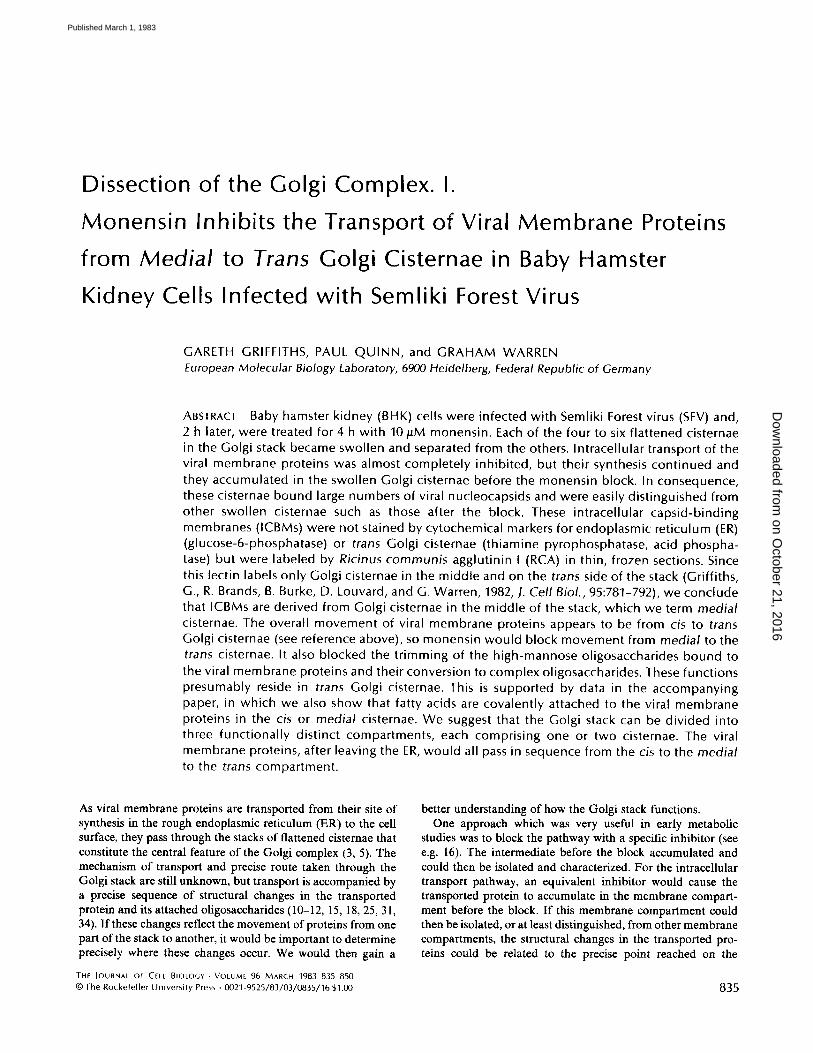
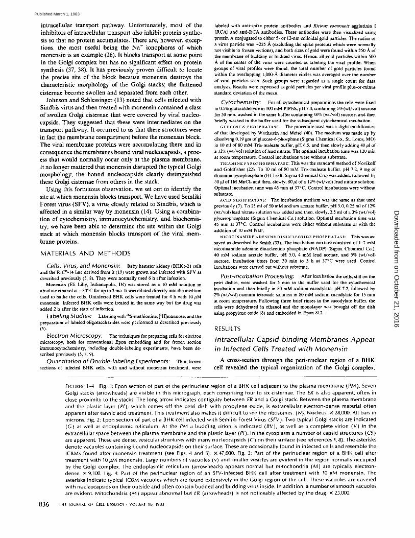
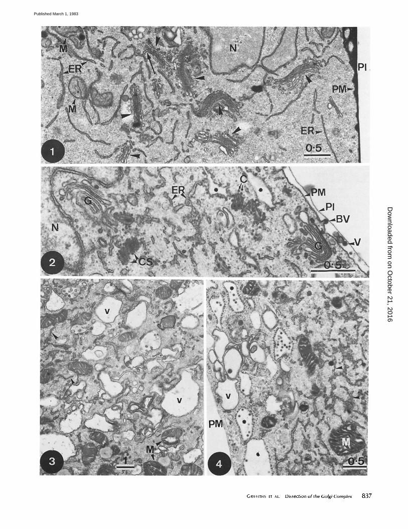
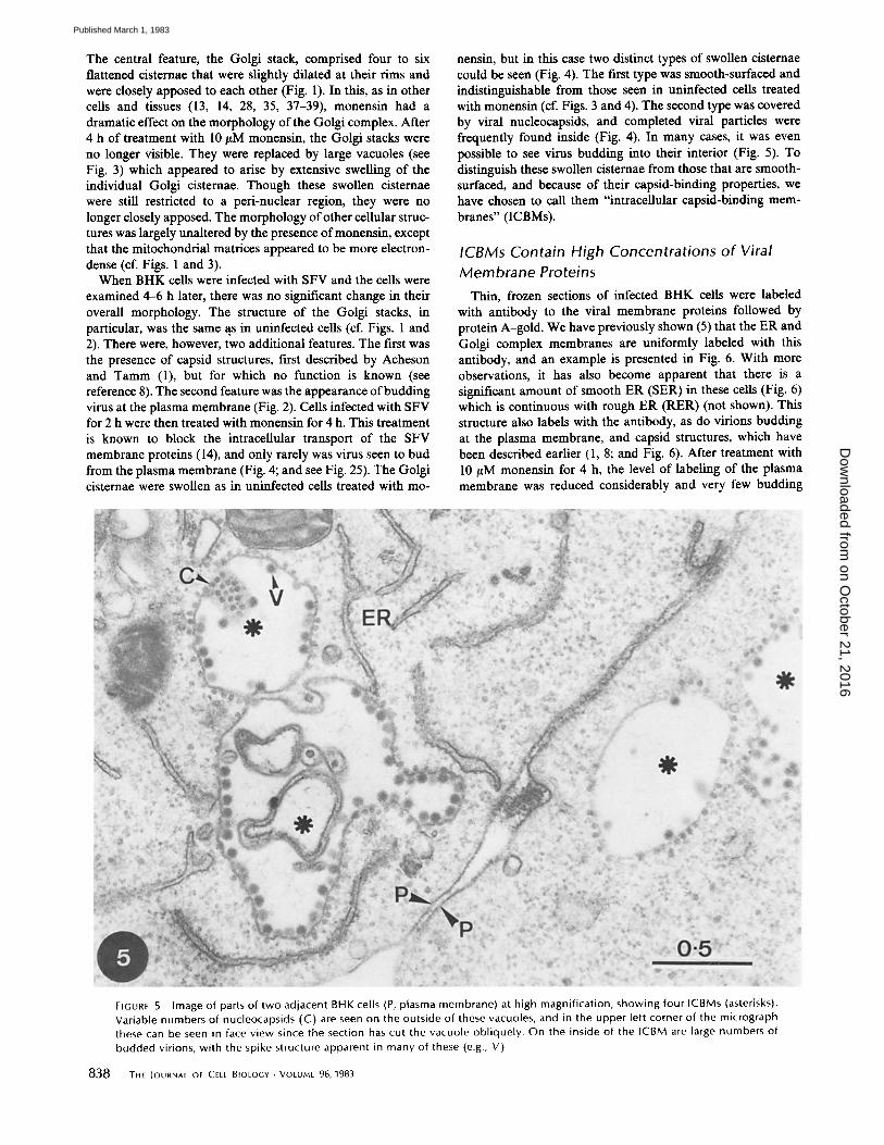
FIGURES 1-4 Fig. 1: Epon section of part of the perinuclear region of a BHK cell adjacent to the plasma membrane (PM). Seven Golgi stacks (arrowheads) are visible in this micrograph, each comprising four to six cisternae. The ER is also apparent, often in close proximity to the stacks. The long arrow indicates contiguity between ER and a Golgi stack. Between the plasma membrane and the plastic layer (PI), which comes off the petri dish with propylene oxide, is extracellular electron-dense material often apparent after tannic acid treatment. This treatment also makes it difficult to see the ribosomes. (N), Nucleus. x 28,000. All bars in microns. Fig. 2: Epon section of part of a BHK ceil infected with Semliki Forest Virus (SFV). Two typical Golgi stacks are indicated (G) as well as endoplasmic reticulum. At the PM a budding virion is indicated (BY), as well as a complete virion (V) in the extracellular space between the plasma membrane and the plastic layer (PI). In the cytoplasm a number of capsid structures (CS) are apparent. These are dense, vesicular structures with many nucleocapsids (C) on their surface (see references 1, 8). The asterisks denote vacuoles containing bound nucleocapsids on their surface. These are occasionally found in infected cells and resemble the ICBMs found after monensin treatment (see Figs. 4 and 5). x 47,000. Fig. 3: Part of the perinuclear region of a BHK cell after treatment with 10/LM monensin. Large numbers of vacuoles (v) and smaller vesicles are evident in the region normally occupied by the Golgi complex. The endoplasmic reticulum (arrowheads) appears normal but mitochondria (M) are typically electron- dense, x 9,100. Fig. 4: Part of the perinuclear region of an SFV-infected BHK cell after treatment with 10 /~M monensin. The asterisks indicate typical ICBM vacuoles which are found extensively in the Golgi region of the cell. These vacuoles are covered with nucleocapsids on their outside and often contain budded and budding virus inside. In addition, a number of smooth vacuoles are evident. Mitochondria (M) appear abnormal but ER (arrowheads) is not noticeably affected by the drug. x 25,000.
836 THE lournAt o~ CELL BIOLOGY-VOLUME 96, 1983
on October 21, 2016
Dow
nloaded from
Published March 1, 1983
GRIFFITH5 ET AL. Dissection of the Golgi Complex 837
on October 21, 2016
Dow
nloaded from
Published March 1, 1983
The central feature, the Golgi stack, comprised four to six flattened cisternae that were slightly dilated at their rims and were closely apposed to each other (Fig. 1). In this, as in other cells and tissues (13, 14, 28, 35, 37-39), monensin had a dramatic effect on the morphology of the Golgi complex. After 4 h of treatment with I0 gM monensin, the Golgi stacks were no longer visible. They were replaced by large vacuoles (see Fig. 3) which appeared to arise by extensive swelling of the individual Golgi cisternae. Though these swollen cisternae were still restricted to a peri-nuclear region, they were no longer closely apposed. The morphology of other cellular struc- tures was largely unaltered by the presence of monensin, except that the mitochondrial matrices appeared to be more electron- dense (cf. Figs. 1 and 3).
When BHK cells were infected with SFV and the cells were examined 4-6 h later, there was no significant change in their overall morphology. The structure of the Golgi stacks, in particular, was the same as in uninfected cells (cf. Figs. 1 and 2). There were, however, two additional features. The first was the presence of capsid structures, first described by Acheson and Tamm (1), but for which no function is known (see reference 8). The second feature was the appearance of budding virus at the plasma membrane (Fig. 2). Cells infected with SFV for 2 h were then treated with monensin for 4 h. This treatment is known to block the intracellular transport of the SFV membrane proteins (14), and only rarely was virus seen to bud from the plasma membrane (Fig. 4; and see Fig. 25). The Golgi cisternae were swollen as in uninfected cells treated with mo-
nensin, but in this case two distinct types of swollen cisternae could be seen (Fig. 4). The first type was smooth-surfaced and indistinguishable from those seen in uninfected cells treated with monensin (cf. Figs. 3 and 4). The second type was covered by viral nucleocapsids, and completed viral particles were frequently found inside (Fig. 4). In many cases, it was even possible to see virus budding into their interior (Fig. 5). To distinguish these swollen cisteruae from those that are smooth- surfaced, and because of their capsid-binding properties, we have chosen to call them "intracellular capsid-binding mem- branes" (ICBMs).
ICBMs Contain High Concentrations of Viral Membrane Proteins
Thin, frozen sections of infected BHK cells were labeled with antibody to the viral membrane proteins followed by protein A-gold. We have previously shown (5) that the ER and Golgi complex membranes are uniformly labeled with this antibody, and an example is presented in Fig. 6. With more observations, it has also become apparent that there is a significant amount of smooth ER (SER) in these cells (Fig. 6) which is continuous with rough ER (RER) (not shown). This structure also labels with the antibody, as do virions budding at the plasma membrane, and capsid structures, which have been described earlier (1, 8; and Fig. 6). After treatment with 10/tM monensin for 4 h, the level of labeling of the plasma membrane was reduced considerably and very few budding
FIGURE 5 Image of parts of two adjacent BHK cells (P, plasma membrane) at high magnification, showing four ICBMs (asterisks). Variable numbers of nucleocapsids (C) are seen on the outside of these vacuoles, and in the upper left corner of the micrograph these can be seen in face view since the section has cut the vacuole obliquely. On the inside of the ICBM are large numbers of budded virions, with the spike structure apparent in many of these {e.g., V).
838 THE Iournat OF CELL BIOLOGY. VOLUME 96, 1983
on October 21, 2016
Dow
nloaded from
Published March 1, 1983
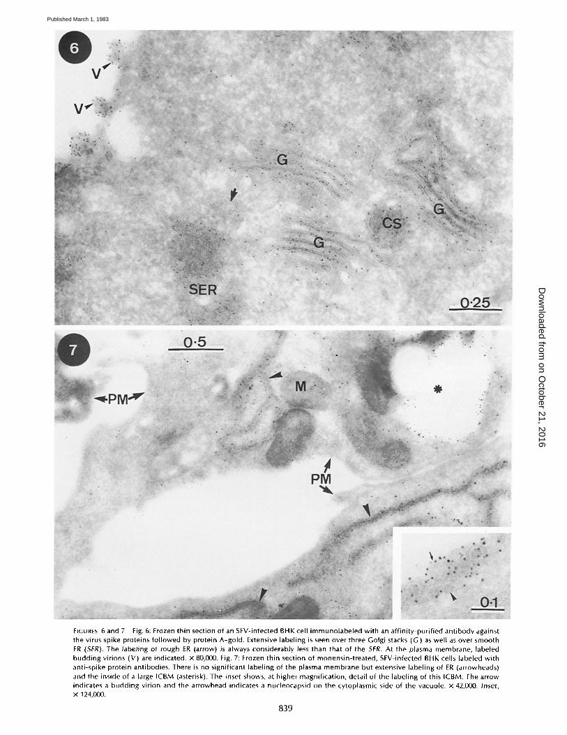
FIGURES 6and7 Fig. 6: Frozen thin section of an SFV-infected BHK cell immunolabeled with an affinity-purified antibody against the virus spike proteins followed by protein A-gold. Extensive labeling is seen over three Golgi stacks (G) as well as over smooth ER (SER). The labeling of rough ER (arrow) is always considerably less than that of the SER. At the plasma membrane, labeled budding virions (V) are indicated, x 80,000. Fig. 7: Frozen thin section of monensin-treated, SFV-infected BHK cells labeled with anti-spike protein antibodies. There is no significant labeling of the plasma membrane but extensive labeling of ER (arrowheads) and the inside of a large ICBM (asterisk). The inset shows, at higher magnification, detail of the labeling of this ICBM. The arrow indicates a budding virion and the arrowhead indicates a nucleocapsid on the cytoplasmic side of the vacuole, x 42,000. Inset, x 124,000.
839
on October 21, 2016
Dow
nloaded from
Published March 1, 1983
profiles were seen (Fig. 7; and see Fig. 25). The labeling of the whole ER appeared to be higher than that seen in untreated infected cells. ICBMs were heavily labeled (Fig. 7, inset), the density of labeling being much higher than that seen in the Golgi membranes of infected cells not treated with monensin. Smooth-surfaced vacuoles of unknown origin were labeled, but the level of labeling was much less than that of ICBMs (see Fig. 27).
ICBMs ARE DERIVED FROM GOLGI CISTERNAE IN THE MIDDLE OF THE STACK
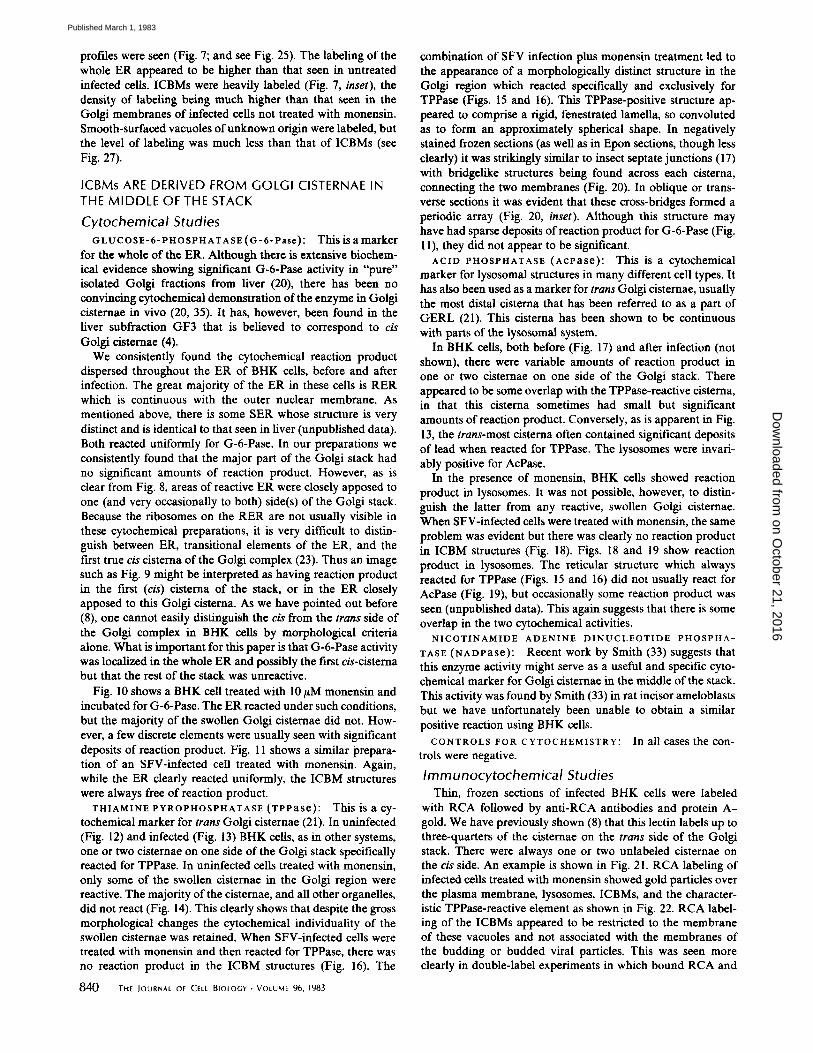
Cytochemical Studies G L U C O S E - 6 - P H O S P H A T A S E ( G - 6 - P a s e ) : This is a marker
for the whole of the ER. Although there is extensive biochem- ical evidence showing significant G-6-Pase activity in "pure" isolated Golgi fractions from liver (20), there has been no convincing cytochemical demonstration of the enzyme in Golgi cisteruae in vivo (20, 35). It has, however, been found in the liver subfraction GF3 that is believed to correspond to cis Oolgi cisternae (4).
We consistently found the cytochemical reaction product dispersed throughout the ER of BHK ceils, before and after infection. The great majority of the ER in these cells is RER which is continuous with the outer nuclear membrane. As mentioned above, there is some SER whose structure is very distinct and is identical to that seen in liver (unpublished data). Both reacted uniformly for G-6-Pase. In our preparations we consistently found that the major part of the Golgi stack had no significant amounts of reaction product. However, as is clear from Fig. 8, areas of reactive ER were closely apposed to one (and very occasionally to both) side(s) of the Golgi stack. Because the ribosomes on the RER are not usually visible in these cytochemical preparations, it is very difficult to distin- guish between ER, transitional elements of the ER, and the first true cis cisterua of the Golgi complex (23). Thus an image such as Fig. 9 might be interpreted as having reaction product in the first (cis) cisterua of the stack, or in the ER closely apposed to this Golgi cisterna. As we have pointed out before (8), one cannot easily distinguish the cis from the trans side of the Golgi complex in BHK cells by morphological criteria alone. What is important for this paper is that G-6-Pase activity was localized in the whole ER and possibly the first cis-cistema but that the rest of the stack was unreactive.
Fig. 10 shows a BHK cell treated with 10/tM monensin and incubated for G-6-Pase. The ER reacted under such conditions, but the majority of the swollen Golgi cisternae did not. How- ever, a few discrete elements were usually seen with significant deposits of reaction product. Fig. 11 shows a similar prepara- tion of an SFV-infected cell treated with monensin. Again, while the ER clearly reacted uniformly, the ICBM structures were always free of reaction product.
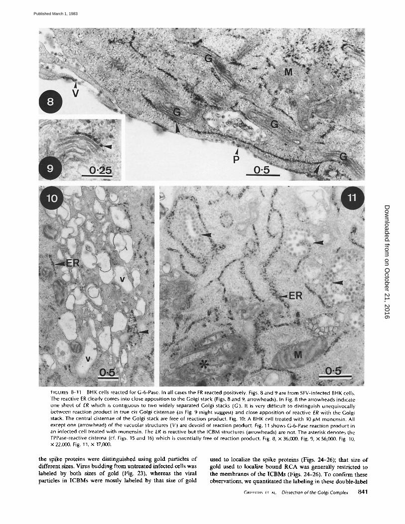
THIAMINE PYROPHOSPHATASE (TPvase): This is a cy- tochemical marker for trans Golgi cistemae (21). In uninfected (Fig. 12) and infected (Fig. 13) BHK cells, as in other systems, one or two cisteruae on one side of the Golgi stack specifically reacted for TPPase. In uninfected cells treated with monensin, only some of the swollen cisternae in the Golgi region were reactive. The majority of the cisternae, and all other organelles, did not react (Fig. 14). This clearly shows that despite the gross morphological changes the cytochemical individuality of the swollen cisteruae was retained. When SFV-infected cells were treated with monensin and then reacted for TPPase, there was no reaction product in the ICBM structures (Fig. 16). The
840 THe JournAL OF CeLt BIOLOGY-VOLUme 96, 1983
combination of SFV infection plus monensin treatment led to the appearance of a morphologically distinct structure in the Golgi region which reacted specifically and exclusively for TPPase (Figs. 15 and 16). This TPPase-positive structure ap- peared to comprise a rigid, fenestrated lamella, so convoluted as to form an approximately spherical shape. In negatively stained frozen sections (as well as in EPOn sections, though less clearly) it was strikingly similar to insect septate junctions (17) with bridgelike structures being found across each cisterua, connecting the two membranes (Fig. 20). In oblique or trans- verse sections it was evident that these cross-bridges formed a periodic array (Fig. 20, inset). Although this structure may have had sparse deposits of reaction product for G-6-Pase (Fig. 11), they did not appear to be significant.
ACID PHOSPHATASE (AcPase): This is a cytochemical marker for lysosomal structures in many different cell types. It has also been used as a marker for trans Golgi cisternae, usually the most distal cisterna that has been referred to as a part of GERL (21). This cisterua has been shown to be continuous with parts of the lysosomal system.
In BHK cells, both before (Fig. 17) and after infection (not shown), there were variable amounts of reaction product in one or two cisternae on one side of the Golgi stack. There appeared to be some overlap with the TPPase-reactive cisterna, in that this cisterna sometimes had small but significant amounts of reaction product. Conversely, as is apparent in Fig. 13, the trans-most cisterna often contained significant deposits of lead when reacted for TPPase. The lysosomes were invari- ably positive for AcPase.
In the presence of monensin, BHK cells showed reaction product in lysosomes. It was not possible, however, to distin- guish the latter from any reactive, swollen Golgi cisternae. When SFV-infected cells were treated with monensin, the same problem was evident but there was clearly no reaction product in ICBM structures (Fig. 18). Figs. 18 and 19 show reaction product in lysosomes. The reticular structure which always reacted for TPPase (Figs. 15 and 16) did not usually react for AcPase (Fig. 19), but occasionally some reaction product was seen (unpublished data). This again suggests that there is some overlap in the two cytochemical activities.
N I C O T I N A M I D E A D E N I N E D I N U C L E O T I D E P H O S P H A -
TASE (NADPaSe)." Recent work by Smith (33) suggests that this enzyme activity might serve as a useful and specific cyto- chemical marker for Golgi cisternae in the middle of the stack. This activity was found by Smith (33) in rat incisor ameloblasts but we have unfortunately been unable to obtain a similar positive reaction using BHK cells.
CONTROLS FOR CYTOCHEMISTRY: In all cases the con- trois were negative.
Immunocytochemical S t u d i e s
Thin, frozen sections of infected BHK ceils were labeled with RCA followed by anti-RCA antibodies and protein A - gold. We have previously shown (8) that this lectin labels up to three-quarters of the cisternae on the trans side of the Golgi stack. There were always one or two unlabeled cisternae on the cis side. An example is shown in Fig. 21. RCA labeling of infected cells treated with monensin showed gold particles over the plasma membrane, lysosomes, ICBMs, and the character- istic TPPase-reactive element as shown in Fig. 22. RCA label- hag of the ICBMs appeared to be restricted to the membrane of these vacuoles and not associated with the membranes of the budding or budded viral particles. This was seen more clearly in double-label experiments in which bound RCA and
on October 21, 2016
Dow
nloaded from
Published March 1, 1983
FIGURES 8-11 BHK cells reacted for G-6-Pase. In all cases the ER reacted positively. Figs. 8 and 9 are from SFV-infected BHK cells. The reactive ER clearly comes into close apposition to the Golgi stack (Figs. 8 and 9, arrowheads). In Fig. 8 the arrowheads indicate one sheet of ER which is contiguous to two widely separated Golgi stacks (G). It is very difficult to distinguish unequivocally between reaction product in true cis Golgi cisternae (as Fig. 9 might suggest) and close apposition of reactive ER with the Gotgi stack. The central cisternae of the Golgi stack are free of reaction product. Fig. 10: A BHK cell treated with 10/tM monensin. All except one (arrowhead) of the vacuolar structures (V) are devoid of reaction product. Fig. 11 shows G-6-Pase reaction product in an infected cell treated with monensin. The ER is reactive but the ICBM structures (arrowheads) are not. The asterisk denotes the TPPase-reactive cisterna (cf. Figs. 15 and 16) which is essentially free of reaction product. Fig. 8, X 36,000. Fig. 9, X 56,000. Fig. 10, X 22,000. Fig. 11, X 37,000.
the spike proteins were distinguished using gold particles of different sizes. Virus budding from untreated infected cells was labeled by both sizes of gold (Fig. 23), whereas the viral particles in ICBMs were mostly labeled by that size of gold
used to localize the spike proteins (Figs. 24-26); that size of gold used to localize bound RCA was generally restricted to the membranes of the ICBMs (Figs. 24-26). To confirm these observations, we quantitated the labeling in these double-label
G m F F I t H S er a t Dissection of the Golgi Complex 84"1
on October 21, 2016
Dow
nloaded from
Published March 1, 1983
FIGURES 12-16 BHK cells reacted for TPPase. Fig. 12 is an image of a BHK cell, showing typical reaction product in one or two cisternae (arrowheads) on one side of the Golgi stack. This micrograph also indicates the difficulty in seeing any clear morphological difference between the two sides of the Golgi stack. Fig. 13 shows, in an SFV-infected BHK cell, a rare example where the ER adjacent to the obliquely sectioned Golgi stack (arrowhead) is free of ribosomes on the cis side of the Golgi stack, the one or two cisternae reactive for TPPase being located on the opposite side. Fig. 14 shows TPPase reaction product in two vacuoles (arrowheads) in a BHK cell after treatment with 10/,tM monensin. The other vacuoles (v) are free of reaction product. Figs. 15 and 16 show BHK cells infected with SFV and treated with 10/~M monensin. In Fig. 15 the typical collapsed TPPase-positive cistema is shown. Arrowheads indicate unreactive ER. In Fig. 16 the large arrowheads indicate parts of the TPPase-positive element. The ICBM (asterisk) has no reaction product. The small arrows within the ICBM indicate budding virions. Fig. 12, x 57,000. Fig. 13, x 34,000. Fig. 14, x 23,000, Fig. 15, x 34,000. Fig. 16, X 39,000.
experiments and the results are presented in Table I. Gold labeling of spike proteins was essentially the same whether the virus was budding from the plasma membrane of untreated cells or from the membrane of the ICBMs. In contrast, the level of RCA labeling of budding (or budded) virus in ICBMs was
only one-third of that of virus budding from the cell surface. Furthermore, the RCA labeling of virus in ICBMs was to a large extent an unavoidable artifact of the method used to quantitate the labeling. Since all gold particles within 500/~ of the virus center were counted, gold particles labeling the ICBM
842 ]-HE IOURN^t OF CELt BIOLOGY. VOLUME 96, 1983
on October 21, 2016
Dow
nloaded from
Published March 1, 1983
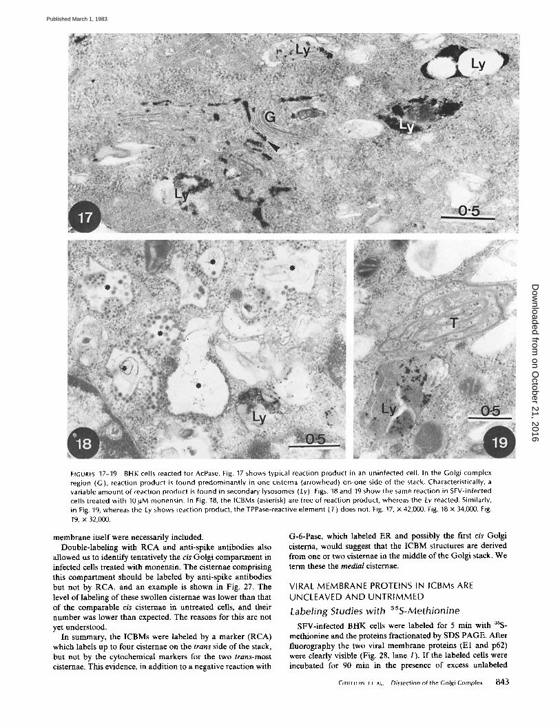
FrGURES 17-19 BHK cells reacted for AcPase. Fig. 17 shows typical reaction product in an uninfected cell. In the Golgi complex region (G), reaction product is found predominantly in one cisterna (arrowhead) on,one side of the stack. Characteristically, a variable amount of reaction product is found in secondary lysosomes (Ly). Figs. 18 and 19 show the same reaction in SFV-infected cells treated with 10 #M monensin. In Fig. 18, the ICBMs (asterisk) are free of reaction product, whereas the Ly reacted. Similarly, in Fig. 19, whereas the Ly shows reaction product, the TPPase-reactive element (T) does not. Fig. 17, x 42,000. Fig. 18 x 34,000. Fig. 19, x 32,000.
membrane itself were necessarily included. Double-labeling with RCA and anti-spike antibodies also
allowed us to identify tentatively the cis Golgi compartment in infected cells treated with monensin. The cisternae comprising this compartment should be labeled by anti-spike antibodies but not by RCA, and an example is shown in Fig. 27. The level of labeling of these swollen cisternae was lower than that of the comparable cis cisternae in untreated cells, and their number was lower than expected. The reasons for this are not yet understood.
In summary, the ICBMs were labeled by a marker (RCA) which labels up to four cisternae on the trans side of the stack, but not by the cytochemical markers for the two trans-most cisternae. This evidence, in addition to a negative reaction with
G-6-Pase, which labeled ER and possibly the first cis Golgi cisterna, would suggest that the ICBM structures are derived from one or two cisternae in the middle of the Golgi stack. We term these the medial cisternae.
V I R A L M E M B R A N E P R O T E I N S IN I C B M s ARE
U N C L E A V E D A N D U N T R I M M E D
Labeling Studies wi th 35S-Methionine
SFV-infected BHK cells were labeled for 5 min with 35S- methionine and the proteins fractionated by SDS PAGE. After fluorography the two viral membrane proteins (El and p62) were clearly visible (Fig. 28, lane 1). If the labeled cells were incubated for 90 rain in the presence of excess unlabeled
GRIF~IrHS E, AL. Dissection of lhe Golgi Complex 843
on October 21, 2016
Dow
nloaded from
Published March 1, 1983
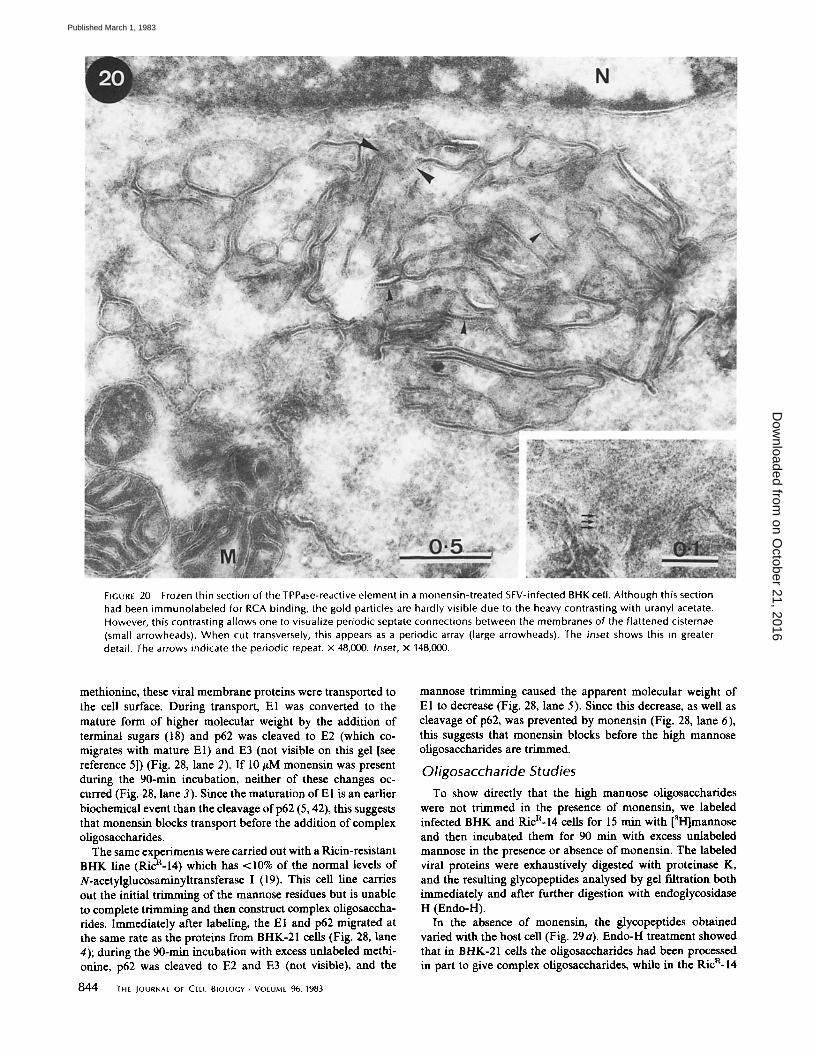
FIGURE 20 Frozen thin section of the TPPase-reactive element in a monensin-treated SFV-infected BHK cell. Although this section had been immunolabeled for RCA binding, the gold particles are hardly visible due to the heavy contrasting with uranyl acetate. However, this contrasting allows one to visualize periodic septate connections between the membranes of the flattened cisternae (small arrowheads). When cut transversely, this appears as a periodic array (large arrowheads). The inset shows this in greater detail. The arrows indicate the periodic repeat, x 48,000. Inset, x 148,000.
methionine, these viral membrane proteins were transported to the cell surface. During transport, E1 was converted to the mature form of higher molecular weight by the addition of terminal sugars (18) and p62 was cleaved to E2 (which co- migrates with mature El) and E3 (not visible on this gel [see reference 5]) (Fig. 28, lane 2). If 10/~M monensin was present during the 90-min incubation, neither of these changes oc- curred (Fig. 28, lane 3). Since the maturation of El is an earlier biochemical event than the cleavage of p62 (5, 42), this suggests that monensin blocks transport before the addition of complex oligosaccharides.
The same experiments were carried out with a Ricin-resistant BHK line (RicR-14) which has <10% of the normal levels of N-acetylglucosaminyltransferase I (19). This cell line carries out the initial trimming of the mannose residues but is unable to complete trimming and then construct complex oligosaccha- rides. Immediately after labeling, the E I and p62 migrated at the same rate as the proteins from BHK-21 cells (Fig. 28, lane 4); during the 90-min incubation with excess unlabeled methi- onine, p62 was cleaved to E2 and E3 (not visible), and the
844 THE IOURNAL OF CELL BIOLOGY • VOLUME 96, 1983
mannose trimming caused the apparent molecular weight of El to decrease (Fig. 28, lane 5). Since this decrease, as well as cleavage of p62, was prevented by monensin (Fig. 28, lane 6), this suggests that monensin blocks before the high mannose oligosaccharides are trimmed.
Oligosaccharide Studies To show directly that the high marmose oligosaccharides
were not trimmed in the presence of monensin, we labeled infected BHK and RicR-14 cells for 15 min with [aH]marmose and then incubated them for 90 rain with excess unlabeled man_nose in the presence or absence of monensin. The labeled viral proteins were exhaustively digested with proteinase K, and the resulting glycopeptides analysed by gel filtration both immediately and after further digestion with endoglycosidase H (Endo-H).
In the absence of monensin, the glycopeptides obtained varied with the host cell (Fig. 29 a). Endo-H treatment showed that in BHK-21 cells the oligosaccharides had been processed in part to give complex oligosaccharides, while in the Ric a- 14
on October 21, 2016
Dow
nloaded from
Published March 1, 1983
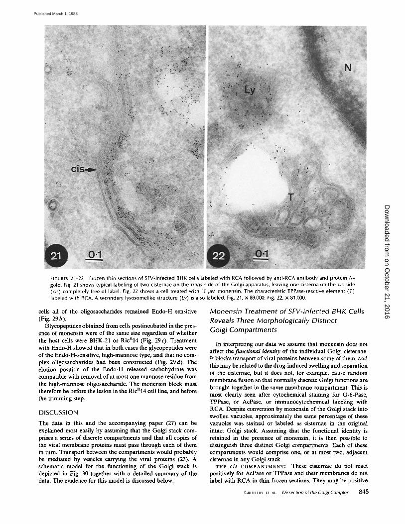
FIGURES 21-22 Frozen thin sections of SFV-infected BHK cells labeled with RCA fol lowed by anti-RCA ant ibody and protein A- gold. Fig. 21 shows typical labeling of two cisternae on the trans side of the Golgi apparatus, leaving one cisterna on the cis side (cis) completely free of label. Fig. 22 shows a cell treated with 10 #M monensin. The characteristic TPPase-reactive element (T) labeled with RCA. A secondary lysosomelike structure (Ly) is also labeled. Fig. 21, x 89,000. Fig. 22, x 81,000.
cells all of the oligosaccharides remained Endo-H sensitive (Fig. 29 b).
Glycopeptides obtained from cells postincubated in the pres- ence of monensin were of the same size regardless of whether the host cells were BHK-21 or RicR14 (Fig. 29c). Treatment with Endo-H showed that in both cases the glycopeptides were of the Endo-H-sensitive, high-mannose type, and that no com- plex oligosac~harides had been constructed (Fig. 29 d). The elution position of the Endo-H released carbohydrate was compatible with removal of at most one mannose residue from the high-mannose oligosaccharide. The monensin block must therefore be before the lesion in the RicRl4 cell line, and before the trimming step.
DISCUSSION
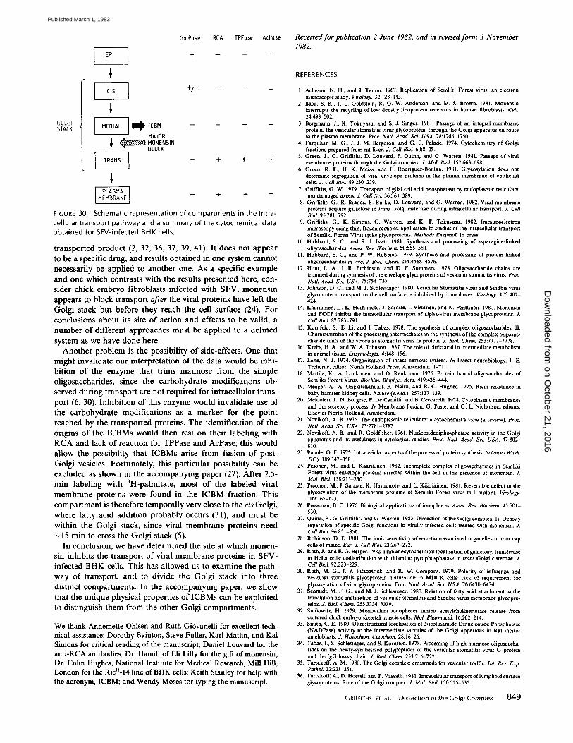
The data in this and the accompanying paper (27) can be explained most easily by assuming that the Golgi stack com- prises a series of discrete compartments and that all copies of the viral membrane proteins must pass through each of them in turn. Transport between the compartments would probably be mediated by vesicles carrying the viral proteins (23). A schematic model for the functioning of the Golgi stack is depicted in Fig. 30 together with a detailed summary of the data. The evidence for this model is discussed below.
Monensin Treatment of 5FV-infected BHK Cells
Reveals Three Morphologically Distinct Golgi Compartments
In interpreting our data we assume that monensin does not affect the functional identity of the individual Golgi cisternae. It blocks transport of viral proteins between some of them, and this may be related to the drug-induced swelling and separation of the cisternae, but it does not, for example, cause random membrane fusion so that normally discrete Golgi functions are brought together in the same membrane compartment. This is most clearly seen after cytocbemical staining for G-6-Pase, TPPase, or AcPase, or immunocytochemical labeling with RCA. Despite conversion by monensin of the Golgi stack into swollen vacuoles, approximately the same percentage of these vacuoles was stained or labeled as cisternae in the original intact Golgi stack. Assuming that the functional identity is retained in the presence of monensin, it is then possible to distinguish three distinct Golgi compartments. Each of these compartments would comprise one, or at most two, adjacent cisternae in any Golgi stack.
THE cis COMPARTMENT" These cisternae do not react positively for AcPase or TPPase and their membranes do not label with RCA in thin frozen sections. They may be positive
GRIFFItHS el ^t. Dissection of the GolgiComplex 845
on October 21, 2016
Dow
nloaded from
Published March 1, 1983
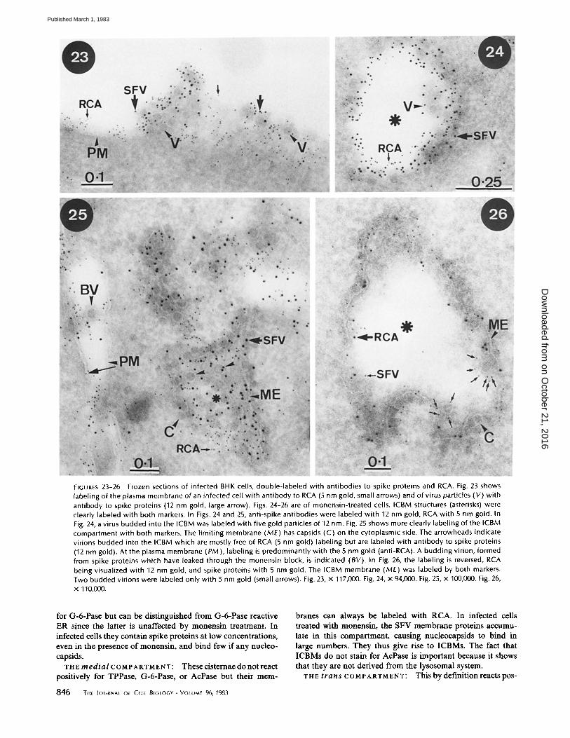
FIGURES 23-26 Frozen sections of infected BHK cells, double-labeled with antibodies to spike proteins and RCA. Fig. 23 shows labeling of the plasma membrane of an infected cell wi th ant ibody to RCA (5 nm gold, small arrows) and of virus particles (V) with antibody to spike proteins (12 nm gold, large arrow). Figs. 24-26 are of monensin-treated cells. ICBM structures (asterisks) were clearly labeled with both markers. In Figs. 24 and 25, anti-spike antibodies were labeled with 12 nm gold, RCA with 5 nm gold. In Fig. 24, a virus budded into the ICBM was labeled with five gold particles of 12 nm. Fig. 25 shows more clearly labeling of the ICBM compartment with both markers. The l imit ing membrane (ME) has capsids (C) on the cytoplasmic side. The arrowheads indicate virions budded into the ICBM which are mostly free of RCA (5 nm gold) labeling but are labeled with ant ibody to spike proteins (12 nm gold). At the plasma membrane (PM), labeling is predominantly with the 5 nm gold (anti-RCA). A budding virion, formed from spike proteins which have leaked through the monensin block, is indicated (BY). In Fig. 26, the labeling is reversed, RCA being visualized with 12 nm gold, and spike proteins with 5 nm gold. The ICBM membrane (ME) was labeled by both markers. Two budded virions were labeled only with 5 nm gold (small arrows). Fig. 23, x 117,000. Fig. 24, x 94,000. Fig. 25, x 100,000. Fig. 26, x 110,000.
for G-6-Pase but can be distinguished from G-6-Pase reactive ER since the latter is unaffected by monensin treatment. In infected ceils they contain spike proteins at low concentrations, even in the presence of monensin, and bind few if any nucleo- capsids.
T H E m e d i a I c o M P A R T M E N T : These cistemae do not react positively for TPPase, G-6-Pase, or AcPase but their mem-
846 Y,t JOURNAL Of CEtt 81OtOGV • VOLUME 96, 1983
branes can always be labeled with RCA. In infected cells treated with monensin, the SFV membrane proteins accumu- late in this compartment, causing nucleocapsids to bind in large numbers. They thus give rise to ICBMs. The fact that ICBMs do not stain for AcPase is important because it shows that they are not derived from the lysosomal system.
THE t r a n s COMPARTMENT: This by defmition reacts pos-
on October 21, 2016
Dow
nloaded from
Published March 1, 1983
itively for TPPase and AcPase, and the membranes can also be labeled with RCA in thin, frozen sections. The TPPase-positive cisterna assumes a striking fenestrated appearance in infected cells treated with monensin and can be identified even in the absence of the TPPase reaction. The reasons for this appear- ance, and the periodic cross-bridges revealed in thin frozen sections, are unknown. Relatively low concentrations of SFV membrane proteins are found in these membranes in the presence of monensin, and occasional nucleocapsid binding is seen.
Di f fe ren t Go lg i Funct ions Are Found in
D i f f e ren t Compar tmen ts
Our data strongly suggest that monensin blocks the move- ment of SFV membrane proteins from the medial to the trans
compartment. It also prevents the trimming of the high-man- nose oligosaccharides bound to the viral proteins and the
TABLE I
Quantitation of Double-labeling with RCA (AuS) and anti-SFV (Au12) over Budding and Budded Virus Particles *
No. of gold particles/ viral profile
Membrane Anti-SFV RCA
Plasma membrane of normal infected 10.5 _+ 3.5 5.9 + 2.9 cells
ICBMs 8.1 + 3.4 1.9 _+ 1.5
* 50 budded virus profiles per count.
construction of complex oligosaccharides which is known to occur within the Golgi stack (5). These Golgi functions should therefore be restricted to the trans Golgi compartment. This is consistent with our previous work (8) and that of others, notably the recent work by Roth and Berger (29). They showed that one of the enzymes responsible for the construction of complex oligosaccharides, galactosyl transferase, co-localizes with TPPase in trans Golgi cisternae.
Covalent attachment of fatty acids to the viral membrane proteins was not blocked by monensin in this or in other viral systems (13). It is also an earlier event than construction of complex oligosaccharides (31). This would restrict fatty acyla- tion to the cis or medial compartments. This has been confirmed by separating the medial compartments from the others on sucrose gradients as shown in the accompanying paper (27).
Viral Proteins M o v e f rom the cis to the med ia l
to the trans Compar tmen ts
SFV membrane proteins appear to move from cis to trans
Golgi cisternae (8). In this paper we provide strong evidence that monensin blocks the movement from the medial to the trans compartment. Since almost all of the viral proteins ac- cumulate in the medial compartment (producing ICBMs), it is unlikely that there is movement directly from the cis to the trans compartment. It is reasonable to conclude that proteins in the cis compartment must move via the medial compartment to reach the trans one. This argues against a transport pathway, proposed for secretory proteins, which allows a product leaving the ER in a vesicle to enter any cisterna of the Golgi stack, or even bypass the Golgi stack altogether (23).
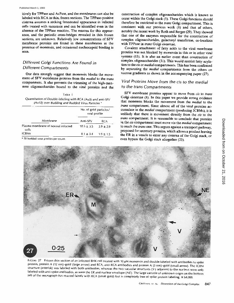
FIGURE 27 Frozen thin section of an infected BHK cell treated with 10#M monensin and double-labeled with antibodies to spike protein, protein A (12 nm)-gold (large arrow) and RCA, anti-RCA antibodies and protein A (5 nm)-gold (small arrow). The ICBM structure (asterisk) was labeled with both antibodies, whereas the two vacuolar structures (V) adjacent to the nucleus were only labeled with anti-spike antibodies, as were the ER and nuclear envelope (NE). The large vacuole of unknown origin on the bottom left of the micrograph has reacted faintly with RCA (small gold) but is completely free of spike protein labeling, x 64,000.
GrlmtHs et AL. Dissection of the Golgi Complex 847
on October 21, 2016
Dow
nloaded from
Published March 1, 1983
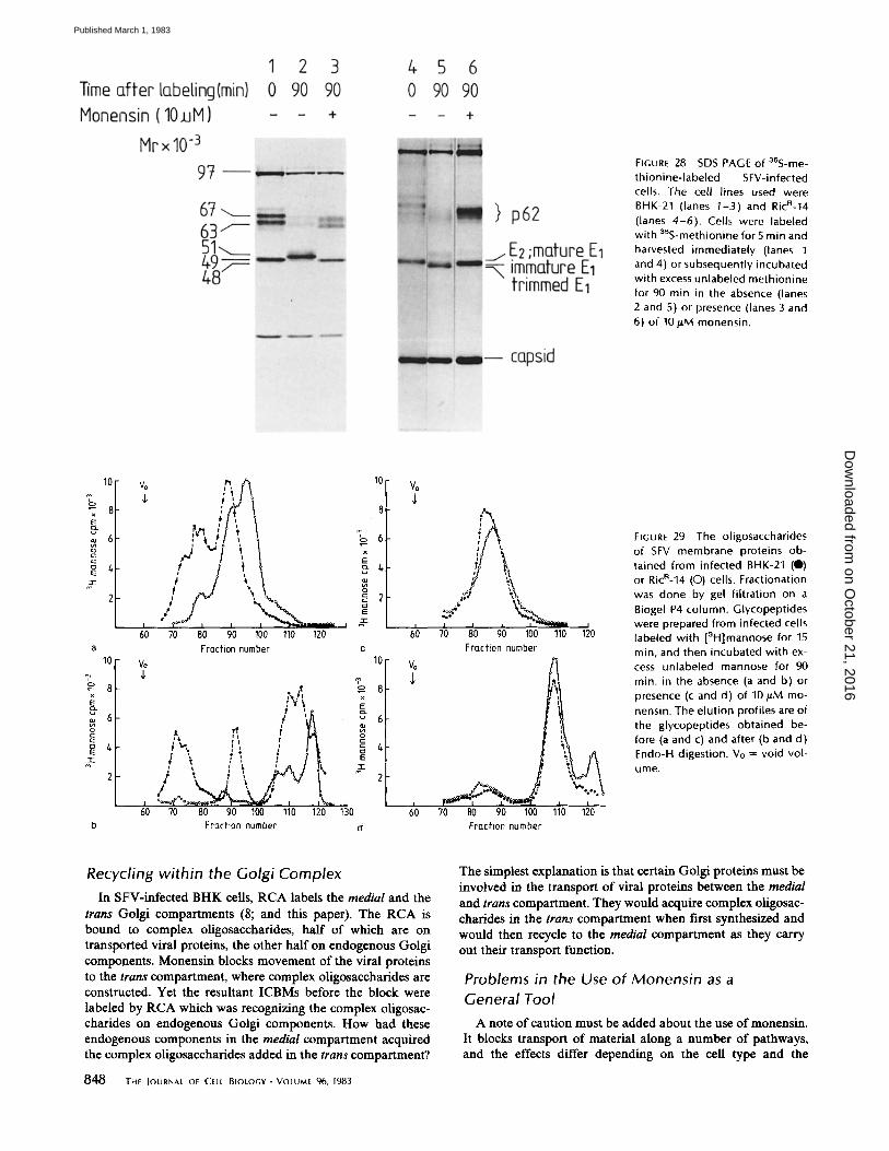
FIGURE 28 SDS PAGE of a~S-me- thionine-labeled SFV-infected cells. The cell lines used were BHK-21 (lanes I-3) and RicR-14 (lanes 4-6). Cells were labeled with 35S-methionine for5 min and harvested immediately (lanes I and 4) or subsequently incubated with excess unlabeled methionine for 90 min in the absence (lanes 2 and 5) or presence (lanes 3 and 6) of 10/.tM monensin.
10
% x 8
6
a
10
8 x
o
2
vo
f ' o
70 80 90 100 110 120 Fraction number
x E Q.
,,p
i - • E T i o ~
; , ,~ f =o
70 fl0 90 100 110 120 130 Fraction number d
I0 Vo
8
6
2
6b
10 Vo
8
6
2
6~0
i
70 80 90 100 110 120 Fraction number
/1 70 80 90 100 110 120
Frucfion number
FIGUR[ 29 The oligosaccharides of SFV membrane proteins ob- tained from infected BHK-21 (0) or RicR-14 (O) cells. Fractionation was done by gel f i l trat ion on a Biogel P4 column. Glycopeptides were prepared from infected cells labeled with [3H]mannose for 15 rain, and then incubated with ex- cess unlabeled mannose for 90 rain. in the absence (a and b) or presence (c and d) of I 0 / IM mo- nensin. The elut ion profiles are of the glycopeptides obtained be- fore (a and c) and after (b and d) Endo-H digestion. Vo = void vol-
ume.
Recycling within the Golgi Complex
In SFV-infected BHK cells, RCA labels the medial and the trans Golgi compartments (8; and this paper). The RCA is bound to complex ohgosaccharides, half of which are on transported viral proteins, the other half on endogenous Golgi components. Monensin blocks movement of the viral proteins to the trans compartment, where complex oligosaccharides are constructed. Yet the resultant ICBMs before the block were labeled by RCA which was recognizing the complex oligosac- charides on endogenous Golgi components. How had these endogenous components in the medial compartment acquired the complex oligosaccharides added in the trans compartment?
848 l-HE IOURNAt OF CELL BIOLOGY • VOLUME 96, 1983
The simplest explanation is that certain Golgi proteins must be involved in the transport of viral proteins between the medial
and trans compartment. They would acquire complex oligosac- charides in the trans compartment when ftrst synthesized and would then recycle to the medial compartment as they carry out their transport function.
Problems in the Use of Monensin as a General Tool
A note of caution must be added about the use ofmonensin. It blocks transport of material along a number of pathways, and the effects differ depending on the cell type and the
on October 21, 2016
Dow
nloaded from
Published March 1, 1983
I
f i 6 Pose RCA TPPose
+ -- _
AcPase Received for publication 2 June 1982, and in revised form 3 November 1982.
STAcKGOLGI ~ i ~ ICBM -- + -- --
MAJOR MONENSIN BLOCK
- - + + +
1 _ + - - - -
FIGURE 30 Schematic representation of compartments in the intra- cellular transport pathway and a summary of the cytochemical data obtained for SFV-infected BHK cells.
transported product (2, 32, 36, 37, 39, 41). It does not appear to be a specific drug, and results obtained in one system cannot necessarily be applied to another one. As a specific example and one which contrasts with the results presented here, con- sider chick embryo fibroblasts infected with SFV; monensin appears to block transport after the viral proteins have left the Golgi stack but before they reach the cell surface (24). For conclusions about its site of action and effects to be valid, a number of different approaches must be applied to a defined system as we have done here.
Another problem is the possibility of side-effects. One that might invalidate our interpretation of the data would be inhi- bition of the enzyme that trims mannose from the simple oligosaccharides, since the carbohydrate modifications ob- served during transport are not required for intracellular trans- port (6, 30). Inhibition of this enzyme would invalidate use of the carbohydrate modifications as a marker for the point reached by the transported proteins. The identification of the origins of the ICBMs would then rest on their labeling with RCA and lack of reaction for TPPase and AcPase; this would allow the possibility that ICBMs arise from fusion of post- Golgi vesicles. Fortunately, this particular possibility can be excluded as shown in the accompanying paper (27). After 2.5- min labeling with 3H-palmitate, most of the labeled viral membrane proteins were found in the ICBM fraction. This compartment is therefore temporally very close to the cis Golgi, where fatty acid addition probably occurs (31), and must be within the Golgi stack, since viral membrane proteins need ~ 15 min to cross the Golgi stack (5).
In conclusion, we have determined the site at which monen- sin inhibits the transport of viral membrane proteins in SFV- infected BHK cells. This has allowed us to examine the path- way of transport, and to divide the Golgi stack into three distinct compartments. In the accompanying paper, we show that the unique physical properties of ICBMs can be exploited to distinguish them from the other Golgi compartments.
We thank Annemette Ohlsen and Ruth Giovanelli for excellent tech- nical assistance; Dorothy Bainton, Steve Fuller, Karl Matlin, and Kai Simons for critical reading of the manuscript; Daniel Louvard for the anti-RCA antibodies; Dr. Hamill of Eli Lilly for the gift of monensin; Dr. Colin Hughes, National Institute for Medical Research, Mill Hill, London for the Ric a- 14 line of BHK cells; Keith Stanley for help with the acronym, ICBM; and Wendy Moses for typing the manuscript.
REFERENCES
1. Acheson, N. H., and I. Tamm. 1967. Replication of Semliki Forest virus: an electron microscopic study. Virology. 32:128-143.
2. Basu, S. K., J. L. Goldstein, R. G. W. Anderson, and M. S. Brown. 1981. Monensin interrupts the recycling of low density lipoprotein receptors in human fibroblasts. Cell. 24:493-502.
3. Bergmann, J., K. Tokuyasu, and S. J. Singer. 1981. Passage of an integral membrane protein, the vesicular stomatitis virus glycoprotein, through the Golgi apparatus en route to the plasma membrane. Proc. Nail Acad. Sei. USA. 78:1746-1750.
4. Farquhar, M. G., J. J. M. Bergeron, and G. E. Palade. 1974. Cytochemistry of Golgi fractions prepared from rat liver..L Cell Biol. 60:8-25.
5. Green, J., G. Griffiths, D. Louvard, P. Quinn, and G. Warren. 1981. Passage of viral membrane proteins through the Golgi complex. J. Mol. BioL 152:663-698.
6. Green, R. F., H. K. Meiss, and E. Rodriguez-Boulan. 1981. Glycosylation does opt determine segregation of viral envelope proteins in the plasma membrane of epithelial cells. J. Cell BioL 89:230-239.
7. Griffiths, G. W. 1979. Transport of glial cell acid phnsphatase by endoplasmic reticalum into damaged axons. J. Cell ScL 36:361-389.
8. Griffiths, G., R. Brands, B. Burke, D. Louvard, and G. Warren. 1982. Viral membrane proteins acquire galactose in trans Golgi cisternae during intracellular transport. J. Cell Biol. 95:781-792.
9. Grifliths, G., K. Simons, G. Warren, and K. T. Tokuyasu. 1982. lmmunoelectron microscopy using thin, frozen sections: application to studies of the intracellular transport of Semliki Forest Virus spike glycoproteins. Methods Enzymol. In press.
10. Hubbard, S. C., and R. J. Ivatt. 1981. Synthesis and processing of asparagine-linked oligosaccharides. Annu. Rev. Biochem. 50:555 583.
1 I. Hubbard, S. C., and P. W. Robbins. 1979. Synthesis and processing of protein linked oligosaccharides in vivo. £ BioL Chem. 254:4568~1576.
12. Hunt, L. A., J. R. Etchinson, and D. F. Summers. 1978. Olignsaccharide chains are trimmed during synthesis of the envelope glycoproteins of vesicular stomatitis virus. Proc. Natl. Acad. ScL USA. 75:754-758.
13. Johnson, D. C., and M. J. Schlessinger. 1980. Vesicular Stomatitis virus and Sindbis virus glycoprotein transport to the cell surface is inhibited by ionophores. Virology. 103:407- 424.
14. K~iari~iinen, L., K. Hashimoto, J. Saraste, I. Virtanen, and K. Penttinen. 1980. Monensin and FCCP inhibit the intracellular transport of alpha-virus membrane glycoproteins..L Cell Biol. 87:783-791.
15. Kornfeld, S., E. Li, and I. Tabas. 1978. The synthesis of complex ollgosaccharides. II. Characterization of the processing intermediates in the synthesis of the complex oligosac- charide units of the vesicular stomatitis virus G protein. J. Biol. Chem. 253:7771-7778.
16. Krebs, H. A., and W. A. Johnson. 1937. The role of citric acid in intermediate metabolism in animal tissue. Enzymologia. 4:148-156.
17. Lane, N. J. 1974. Organisation of insect nervous system. In Insect neurobiology. J. E. Treheme, editor. North Holland Press, Amsterdam. I-7 I.
18. Mattila, K., A. Luukonen, and O. Renkonen. 1976. Protein bound oligosaccharides of Semliki Forest Virus. Biochim. Biophys. A eta. 419:435-444.
19. Meager, A., A. Ungkitchanukit, R. Nairn, and R. C. Hughes. 1975. Ricin resistance in baby hamster kidney cells. Nature (Lond). 257:137 139.
20. Meldolesi, J., N. Borgese, P. De Camilli, and B. Ceccarelli. 1978. Cytoplasmic membranes and the secretory process. In Membrane Fusion. G. Poste, and G. L. Nicholson, editors. Elsevier North Holland, Amsterdam.
21. Novikoff, A. B. 1976. The endoplasmic reticuhim: a cytochemist's view (a review). Proc. Natl. Acad. ScL USA. 73:2781-2787.
22. Novikoff, A. B., and R. Goldfisher. 1961. Nucleosidedipliosphatase activity in the Golgi apparatus and its usefulness in cytological studies. Proe. NatL Acad Sci. USA. 47:802- 810.
23. Palade, G. E. 1975. Intraceitular aspects of the process of protein synthesis. Science { Wash. DC). 189:347-358.
24. Pesonen, M., and L. K~iari~iinen. 1982. Incomplete complex oligosaccharides in Semliki Forest virus envelope proteins arrested within the cell in the presence of monensin. J. Mol. Biol. 158:213 230.
25. Pesonen, M., J. Saraste, K. Hashimoto, and L. K~i~iri~.inen. 1981. Reversible defect in the glycosylation of the membrane proteins of Semliki Forest virus ts-I mutant. Virology. 109:165 173.
26. Pressman, B. C. 1976. Biological applications of ionophores. Annu. Bey. Biochem. 45:501- 530.
27. Quinn, P., G. Griffiths, and G. Warren. 1983. Dissection of the Golgi complex. IL Density separation of specific Goigi functions in virally infected cells treated with monensin. J. Cell Biol. 96:851-856.
28. Robinson, D. E. 1981. The ionic sensitivity of secretion-associated organelles in root cap cells of maize. Eur. J. Cell Biol. 23:267-272.
29. Roth, J., and E. G. Berger. 1982. lmmunocytochemical locallsation ofgalactosyl transfecase in HeLa cells: codistribution with thiamine pyrophosphatase in trans Golgi cisternae. J. Cell Biol. 92:223-229.
30. Roth, M. G., J. P. Fitzpatrick, and R. W. Compans. 1979. Polarity of inlluenza and vesicular stomatitis glycoprotein maturation in MDCK cells: lack of requirement for glycosylation of viral glycoproteins. Proe. NatL Acad $cL USA. 76:6430~434.
3 I. Schmidt, M. F. G., and M. J. Schlesinger. 1980. Relation of fatty acid attachment to the translation and maturation of vesicular stomatitis and Sindbis virus membrane glycopro- teins. J. BioL Chem. 255:3334-3339.
32. Smilowitz, H. 1979. Monovalent ionophores inhibit acetylcholinesterase release from cultured chick embryo skeletal muscle cells. Mol. PharmacoL 16:202 214.
33. Smith, C. E. 1980. Ultrastructural locallsation of Nicutinamide Dinucleotide Phosphatase (NADPase) activity to the intermediate saccules of the Golgi apparatus in Rat incisor ameloblasts. J. Histochem. Cytochem. 28:16-26.
34. Tabas, 1., S. ScMesinger, and S. Korufeld. 1978. Processing of liigh mannose oligosaccha- rides on the newly-synthesized polypeptides of the vesicular stomatitis virus G protein and the lgG heavy chain. £ Biol. Chem. 253:716-722.
35. Tartakoff, A. M. 198@ The Golgi complex: crossroads for vesicular traffic. Int. Rev. Exp. Pathol. 22:228-251.
36. Tartakoff, A., D. Hoessli, and P. Vassalli. 1981. lntracellular transport of lymphoid surface glycoproteins. Role of the Golgi complex. ,L MoL BioL 150:525-535.
GRtFFIIHS Er At. Dissec t i on o f the Golg i Complex 849
on October 21, 2016
Dow
nloaded from
Published March 1, 1983
37. Tartakoff, A., and P. Vassalli. 1977. Plasma cell unmunoglobulin secretion. Arrest is accompanied by alterations in the Golgi complex. J. Exp. Med. 146:1332-1345.
38. Tartakoff, A., and P. VassaUi. 1978. Comparative studies of intracellular transport of secretory proteins. J. Cell Biol. 79:694-707.
39. Vladutio, G. D., and M. C. Rattazzi. 1980. The effect of monensin on fl-hexosaminidase transport in normal and I cell fibroblasts. Biochem. J. 192:813-820.
40. Wachstein, M., and E. Meisel. 1956. On the histochemical demonstration of gincose-6- phos~hatase. Z Histochem. Cfllochem. 4:592-596.
41. Wilcox, D. K., R. P. Kitson, and C. C. Widnell. 1982. Inhibition of pinocytosis in rat embryo fibroblasts treated with monensin. Z Cell Biol. 92:859-864.
42. Ziemiecki, A., H. Garoff, and K. Simons. 1980. Formation of the Semliki Forest virus membrane glycoprotein complexes in the infected cell. Z Gen. ViroL 50:113-123.
850 THe Journal oe CELL BIOLOGY. VOLUME 96, 1983
on October 21, 2016
Dow
nloaded from
Published March 1, 1983
![A Golgi-Released Subpopulation of the Trans-Golgi · A Golgi-Released Subpopulation of the Trans-Golgi Network Mediates Protein Secretion in Arabidopsis1[OPEN] Tomohiro Uemura,a,b,2,3,4](https://img.pdfslide.net/doc/110x75/5eda9f5a09f66a09130ba5a1/a-golgi-released-subpopulation-of-the-trans-golgi-a-golgi-released-subpopulation.jpg)