Embed Size (px)
Citation preview
1
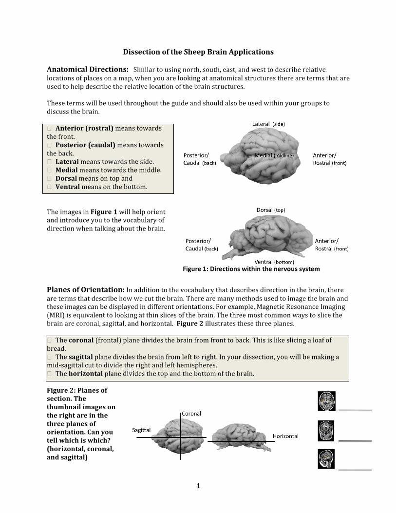
Dissection of the Sheep Brain Applications Anatomical Directions: Similar to using north, south, east, and west to describe relative locations of places on a map, when you are looking at anatomical structures there are terms that are used to help describe the relative location of the brain structures. These terms will be used throughout the guide and should also be used within your groups to discuss the brain. � Anterior (rostral) means towards the front. � Posterior (caudal) means towards the back. � Lateral means towards the side. � Medial means towards the middle. � Dorsal means on top and � Ventral means on the bottom. The images in Figure 1 will help orient and introduce you to the vocabulary of direction when talking about the brain.
Planes of Orientation: In addition to the vocabulary that describes direction in the brain, there are terms that describe how we cut the brain. There are many methods used to image the brain and these images can be displayed in different orientations. For example, Magnetic Resonance Imaging (MRI) is equivalent to looking at thin slices of the brain. The three most common ways to slice the brain are coronal, sagittal, and horizontal. Figure 2 illustrates these three planes. � The coronal (frontal) plane divides the brain from front to back. This is like slicing a loaf of bread. � The sagittal plane divides the brain from left to right. In your dissection, you will be making a mid-‐sagittal cut to divide the right and left hemispheres. � The horizontal plane divides the top and the bottom of the brain. Figure 2: Planes of section. The thumbnail images on the right are in the three planes of orientation. Can you tell which is which? (horizontal, coronal, and sagittal)
Figure 1: Directions within the nervous system

2
Examination of the exterior of the brain The first part of the dissection is examination of the exterior surface of the brain. This part requires no actual cutting. As you identify the listed structures, note how their location is related to other parts of the brain. Examine the exterior of the entire brain. You will notice the two symmetrical cerebral hemispheres, the cerebellum, and the brainstem. The two cerebral hemispheres are separated by the longitudinal fissure (Figure 3).
Figure 3: Exterior regions of the sheep brain.
Fun Fact! The brain has two hemispheres with “contralateral” (opposite side) control of motion and sensation. That is, the right hemisphere generally controls motion and processes sensation from the left half of the body. You might have heard of “right brain” and “left brain” functions or even personalities. This is a myth. The two hemispheres share 96% functional similarity and most differences between right and left brain functions are biases, rather than capabilities. One important difference, however, is that in most individuals the left hemisphere controls speech. You Try It! Identify the directions rostral, caudal, dorsal, ventral, lateral, and medial on your sheep brain. Select the correct answer for the following questions:
1. In the sheep brain, the cerebellum is (dorsal or ventral) to the cerebral hemispheres.
2. Which hemisphere controls movement of the right arm? (right or left hemisphere)

3
3. A patient comes to the emergency room with impaired motor ability on the right side of her body; the physician also notices that her speech is impaired. The physician suspects that the patient suffered from a stroke, which causes the loss of blood flow to certain brain region.
a. The stroke most likely occurred in the (right or left) cerebral hemisphere.
b. How do the patient’s symptoms indicate that this is the region of the stroke? The Four Lobes of the Brain With your sheep brain, identify the major lobes of the brain: the frontal, temporal, parietal and occipital (Figure 4).
Figure 4: The lobes of the brain.
Talk About It: Lobe Damage Research Many functions of the cerebral cortex are distributed among multiple brain regions. However, some functions are associated primarily with portions of one lobe. Much of the information about the four lobes has been obtained using research on patients with damage to one of the lobes.
The occipital lobe is responsible for vision. Damage to this lobe can result in a “field cut” in both eyes, regardless of which side it occurs on, and hallucinations.
Patients with damage to the frontal lobe have significant changes to their personality and social behavior as well as a decrease in creativity and divergent thinking. The famous case of Phineas Gage, a railroad worker who suffered from an injury to his left frontal lobe when a large iron rod protruded his skull in 1845, sparked doctors’ interests in cerebral localization and was perhaps the first case suggesting that brain damage can affect personality and behavior. Gage’s physician Dr. John Martyn Harlow stated in 1868, after Gage’s death, that “The equilibrium or balance, so to speak, between his intellectual faculties and animal propensities, seems to have been destroyed.”
The parietal lobe takes the sensory information from a single perception and then integrates these singular perceptions into a complex network of perceptions that creates the world around us. Patients with damage to the parietal lobe can have difficulties in determining spatial relationships as well as a distorted body image.
Finally, the temporal lobe is responsible for a multitude of functions. Notably, damage to the temporal lobe can result in a loss of recognition of words and faces as well as the inability to categorize words or pictures, depending on which side is damaged. Long-‐term memory retention is also affected by temporal lobe damage.

4
The Cerebral Cortex The cortex has “wrinkles” called sulci (grooves; singular: sulcus) and gyri (ridges; singular: gyrus). The human brain is characterized by many sulci and gyri, but other species have fewer. The sheep brain, for example, has fewer than the human brain, and the rat and mouse brains are almost entirely smooth. Sulci and gyri increase the total surface area of the brain.
Figure 5: comparative brain sizes across species. Image from Jason G. Goldman http://scienceblogs.com/thoughtfulanimal/2010/09/the_fate_of_the_alamogordo_chi.php Depending on the brain, you may be able to see one or two of the three layers of the meninges. The meninges are the protective covering of the brain and spinal cord. There are three layers: the dura mater, the tough protective outer layer that lines the skull (this will likely have been removed from your sheep brains), the middle arachnoid layer with blood vessels, and the pia mater that lines the brain. The thin pia mater follows the sulci and gyri of the brain surface and is likely still attached to the brain. However, this layer is thin and may be hard to distinguish from the surface of the brain itself.
Before identifying structures on the ventral surface of the brain, you will need to carefully remove the arachnoid layer and pia mater. Using your tweezers, carefully pull these layers away from the brain. Use care because the brain tissue is soft and easily damaged. (Figure 6)
Talk About It! Looking at the different images of brains, what can be concluded about the relationship between the amount of sulci and gyri and brain surface area? What role could these structures play in overall brain functions?
Which lobe(s) will help you park the car? ______________________________ Which lobe(s) will help you learn new words and remember them? _______________________ Which lobe(s) will help you solve problems? ________________________________
Figure 6: Carefully remove the meninges from the brain.

5
Clinical Connection: Hematoma Head injury from impact can sometimes result in the accumulation of blood around the brain, or hematoma. Brain imaging techniques allow visualization of fluid accumulation and the patterns of this accumulation can inform physicians about the location of the bleeding within the layers of the meninges. Blood can accumulate between any of the layers, or between the dura and the skull. For example, a lens shaped pattern of blood accumulation indicates that blood is pooling between the dura and the skull. This is called an epidural (above the dura) hematoma. People with epidural hematomas are referred to as patients who “talk and die” because they have a brief lucid phase followed by rapid deterioration. Being able to quickly image the brain after head injury, identify the pattern of blood accumulation, and then treat the head injury is essential. In contrast, a subdual hematoma (blood accumulation under the dura, between the dura mater and the arachnoid layer) appears as a crescent shape in brain images. Subdural hematomas can occur immediately following head injury, or can accumulate slowly many weeks or even months after injury. Finally, in subarachnoid hematomas (between the pia and the arachnoid layers), the blood can pool along the ridges of the sulci and gyri because the underlying pia layer so closely follows the contours of the brain.
In
what plane of section are the images above? An 8 year old boy comes into the emergency room with his parents. He hit his head while playing baseball and, though he says that he feels ok, his parents are still worried. The physician requests a brain scan that shows a crescent-‐shaped pattern of blood accumulation. Is this hemorrhage above the dura mater (epidural) or below the dura mater (subdural)? Should the physician be concerned, or send the child home since he seems fine? Why? Once the protective layers of the meninges are removed, look at the ventral side of the brain. You will see the pons, the medulla, and the cerebellum (Figure 8). While the arrows point to one side of the brain, you can assume bilateral symmetry within the brain.
Figure 7: Epidural (left), subdural (center), and subarachnoid (right) hematomas. The pattern of blood accumulation is limited by the position of the bleeding between the layers of the meninges. Blood or fluid appears white in these images. (Images from Loyola Stritch School of Medicine) http://www.meddean.luc.edu/lumen/meded/radio/curriculum/neurology/ic_hemorrhage2.htm)
6
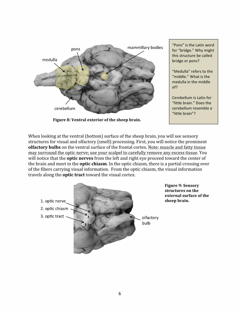
Figure 8: Ventral exterior of the sheep brain.
When looking at the ventral (bottom) surface of the sheep brain, you will see sensory structures for visual and olfactory (smell) processing. First, you will notice the prominent olfactory bulbs on the ventral surface of the frontal cortex. Note: muscle and fatty tissue may surround the optic nerve; use your scalpel to carefully remove any excess tissue. You will notice that the optic nerves from the left and right eye proceed toward the center of the brain and meet in the optic chiasm. In the optic chiasm, there is a partial crossing over of the fibers carrying visual information. From the optic chiasm, the visual information travels along the optic tract toward the visual cortex.
Figure 9: Sensory structures on the external surface of the sheep brain.
“Pons” is the Latin word for “bridge.” Why might this structure be called bridge or pons? “Medulla” refers to the “middle.” What is the medulla in the middle of? Cerebellum is Latin for “little brain.” Does the cerebellum resemble a “little brain”?
7
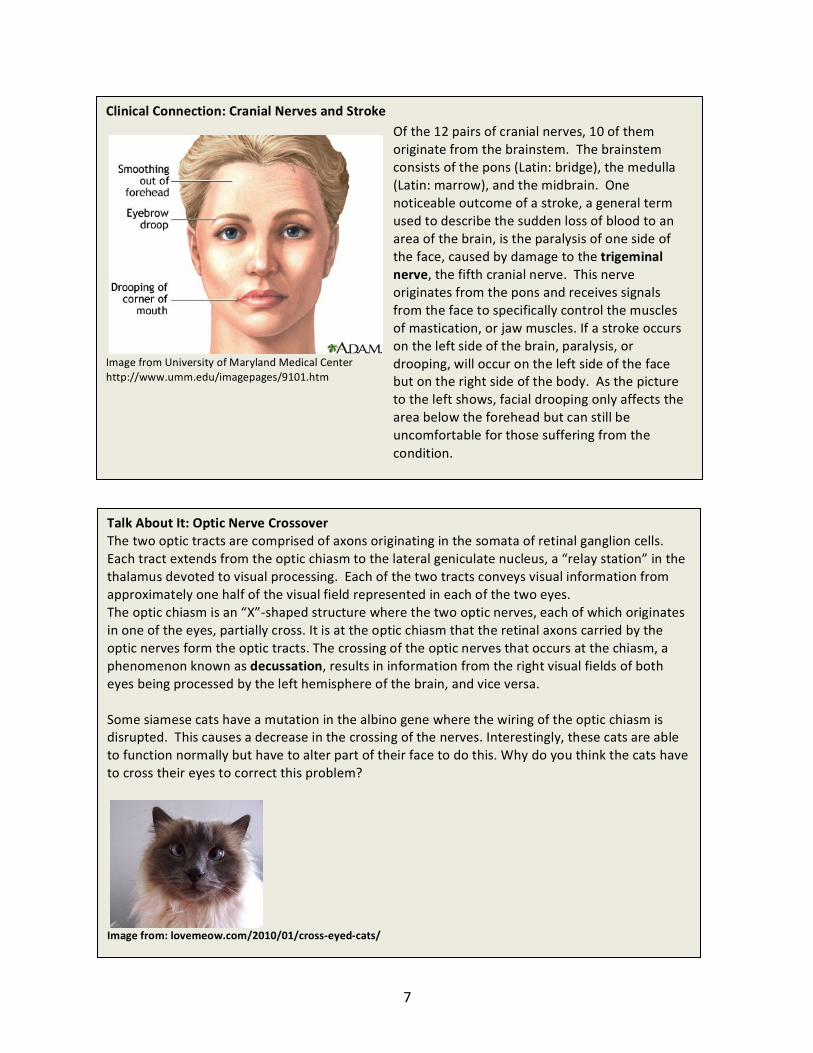
Clinical Connection: Cranial Nerves and Stroke
Image from University of Maryland Medical Center http://www.umm.edu/imagepages/9101.htm
Talk About It: Optic Nerve Crossover The two optic tracts are comprised of axons originating in the somata of retinal ganglion cells. Each tract extends from the optic chiasm to the lateral geniculate nucleus, a “relay station” in the thalamus devoted to visual processing. Each of the two tracts conveys visual information from approximately one half of the visual field represented in each of the two eyes. The optic chiasm is an “X”-‐shaped structure where the two optic nerves, each of which originates in one of the eyes, partially cross. It is at the optic chiasm that the retinal axons carried by the optic nerves form the optic tracts. The crossing of the optic nerves that occurs at the chiasm, a phenomenon known as decussation, results in information from the right visual fields of both eyes being processed by the left hemisphere of the brain, and vice versa. Some siamese cats have a mutation in the albino gene where the wiring of the optic chiasm is disrupted. This causes a decrease in the crossing of the nerves. Interestingly, these cats are able to function normally but have to alter part of their face to do this. Why do you think the cats have to cross their eyes to correct this problem?
Image from: lovemeow.com/2010/01/cross-‐eyed-‐cats/
Of the 12 pairs of cranial nerves, 10 of them originate from the brainstem. The brainstem consists of the pons (Latin: bridge), the medulla (Latin: marrow), and the midbrain. One noticeable outcome of a stroke, a general term used to describe the sudden loss of blood to an area of the brain, is the paralysis of one side of the face, caused by damage to the trigeminal nerve, the fifth cranial nerve. This nerve originates from the pons and receives signals from the face to specifically control the muscles of mastication, or jaw muscles. If a stroke occurs on the left side of the brain, paralysis, or drooping, will occur on the left side of the face but on the right side of the body. As the picture to the left shows, facial drooping only affects the area below the forehead but can still be uncomfortable for those suffering from the condition.

8
Talk About It: Language Processing Neurosurgeon Paul Broca first identified Broca’s area following observations of his patient Mr. Tan. Mr.Tan's true identity is unknown; he was called "Tan" by the workers at the hospital where he died, because "tan" was the only thing he ever said (as he was suffering from severe Broca's aphasia, which is characterized by very limited verbal production). When he died in 1861, Broca dissected his brain and found a tumor in the left hemisphere. After observing this fact in subsequent autopsies of people suffering from similar type of aphasia, Paul Broca concluded that "the faculty for articulate language" was located in the left hemisphere of the brain. Broca's area is responsible for language output and production of words. Wernicke's area is an important language center located in the left hemisphere of the brain. While the precise location of the Wernicke's area is not very clear, it is known to be located around the lateral sulcus, posterior section of the superior temporal gyrus. This area was discovered by Carl Wernicke, a German neurologist, who observed that people with lesions at this location could speak, but their speech was often incoherent and made no sense. Wernicke's area is responsible for processing of language input (words that we hear). Broca's area and Wernicke's area are connected by a large bundle of nerve fibers.
Figure 10: Broca's and Wernike's areas control languge processing. Label the corresponding regions in the sheep brain, above (note that these regions may have different functions in sheep and humans). MR images from Brain Voyager Brain Tutor software: http://www.brainvoyager.com/downloads/downloads.html
Do you think the sheep brain will have a Broca and/or Wernicke’s area(s)? Why or why not? Are there limitations to using the sheep brain as a model for human brains? If so, what are they?

9
Examination of the Mid-‐Sagittal Cut Different cuts allow different views of brain structures. Some structures are easier to visualize with coronal or sagittal sections. Together with your exploration of the brain surface anatomy, making a series of cuts will allow you to become more familiar with the three-‐dimensional structure of the brain. 1. Before making your first cut, place the brain so that you are looking at the dorsal (top) side. Using your hands, not the scalpel, gently pull the two hemispheres of the brain away from the midline. You don’t want to rip anything, but you should notice that the two hemispheres can be pulled apart slightly, and looking into the longitudinal fissure. You will see a structure that connects the two hemispheres: the corpus callosum. Clinical Connection: Callosumectomy for the Treatment of Epilepsy The corpus callosum is a series of axons that connects neurons in the two hemispheres. Historically, treatment of severe epilepsy sometimes included surgically severing the corpus callosum. This surgery is not widely used in modern times but if used, only 1/3 of the corpus callosum is severed. In infants, functional plasticity can be achieved in greater amounts than in adults, which means that our brain has the ability to remarkably recover after injury, especially when the injury occurs at a younger age. 2. Make a mid-‐sagittal cut. Holding the brain flat and level cut along the longitudinal fissure all the way through the brain. In this cut, you can see the lateral ventricles, the third ventricle, and the cerebral aqueduct (connects the third and fourth ventricle). (Figure 11)
Figure 11: Mid-‐sagittal view of the ventricular structures in the sheep brain.
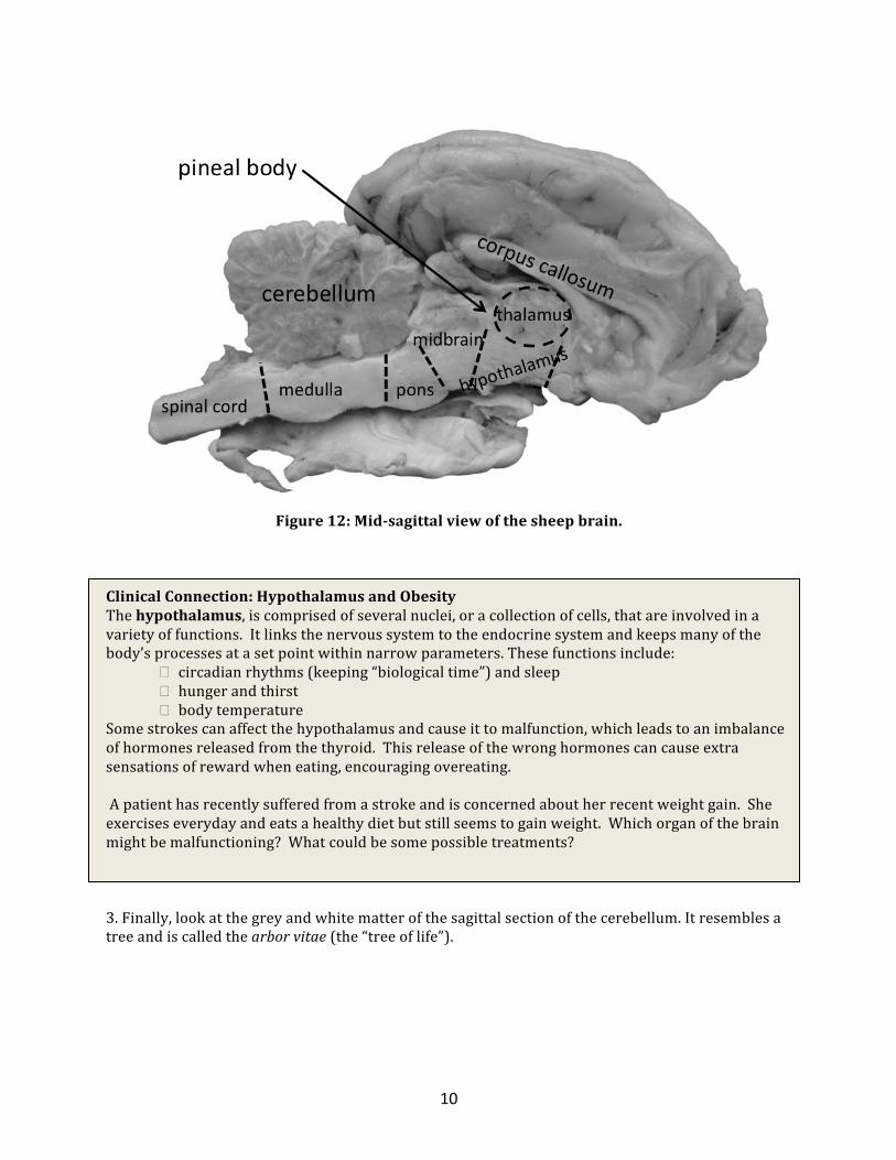
Recall that you were able to see the top of the corpus callosum between the hemispheres before even cutting the brain. The corpus callosum is a band of axons that connects the two sides of the central nervous system. You can also see the pineal body, the thalamus, and the hypothalamus (Figure 12).
10
Figure 12: Mid-‐sagittal view of the sheep brain.
Clinical Connection: Hypothalamus and Obesity The hypothalamus, is comprised of several nuclei, or a collection of cells, that are involved in a variety of functions. It links the nervous system to the endocrine system and keeps many of the body’s processes at a set point within narrow parameters. These functions include: � circadian rhythms (keeping “biological time”) and sleep
� hunger and thirst � body temperature
Some strokes can affect the hypothalamus and cause it to malfunction, which leads to an imbalance of hormones released from the thyroid. This release of the wrong hormones can cause extra sensations of reward when eating, encouraging overeating. A patient has recently suffered from a stroke and is concerned about her recent weight gain. She exercises everyday and eats a healthy diet but still seems to gain weight. Which organ of the brain might be malfunctioning? What could be some possible treatments? 3. Finally, look at the grey and white matter of the sagittal section of the cerebellum. It resembles a tree and is called the arbor vitae (the “tree of life”).

11
You Try It! Can you distinguish the white and gray matter? What is contained in each? As our brains age, and in some diseases, neuronal cell bodies in the cortical gray matter can be lost. How do you think this will affect the sulci and gyri? How does the presence of sulci and gyri affect the surface area of the brain? Examination of the coronal cuts Coronal cuts separate the front and the back of the brain (like slicing a loaf of bread). You will make a series coronal cuts starting from the anterior (front) of the brain and working back. Each section should be about 1 cm thick. The more slices, the more that you can see, however, slices that are very thin are difficult to work with and are very fragile. Set the slices out and try to identify the sections that correspond to the two images below. First, identify the basal ganglia (Figure 13). The landmarks that will help you to identify the basal ganglia are the corpus callosum, and the anterior commisure.
Figure 13: Coronal section showing the basal ganglia. Coronal sections modified from EA Gallman Sheep Brain Dissection Guide
12
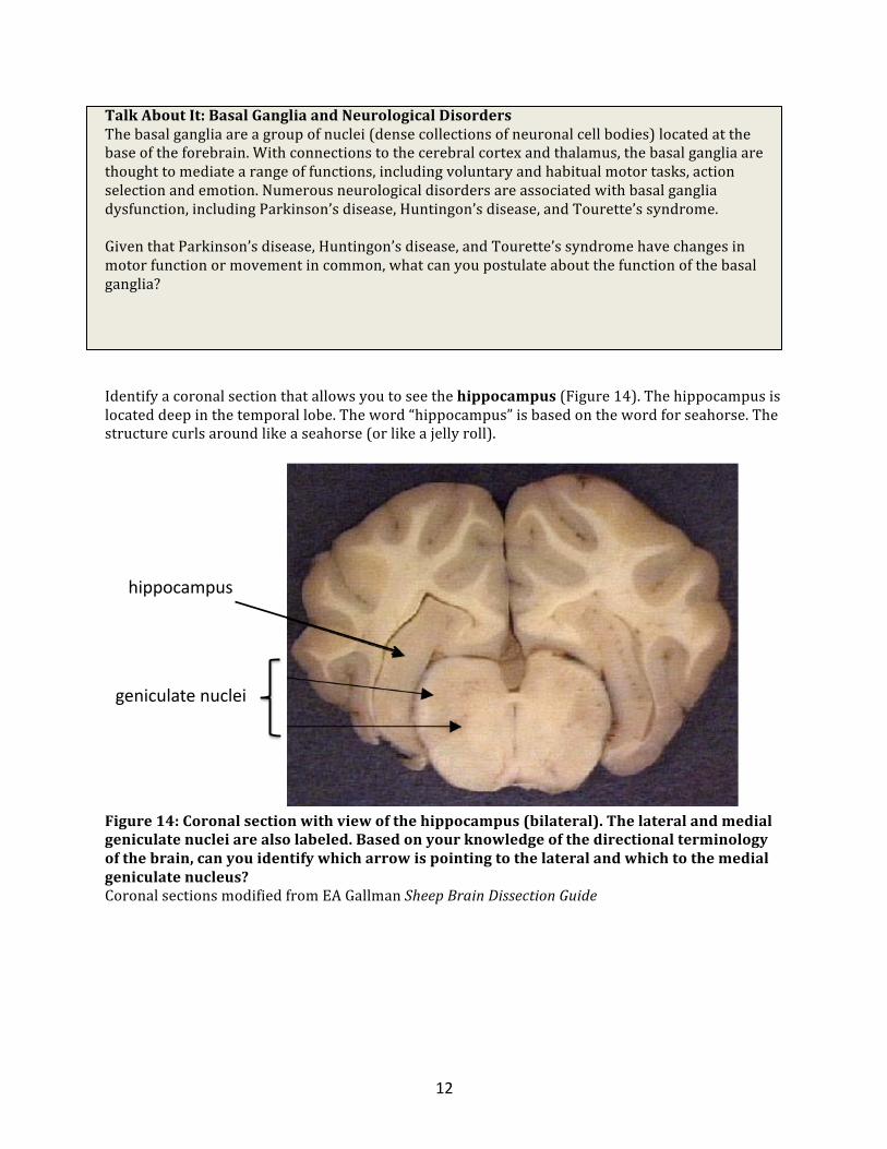
Talk About It: Basal Ganglia and Neurological Disorders The basal ganglia are a group of nuclei (dense collections of neuronal cell bodies) located at the base of the forebrain. With connections to the cerebral cortex and thalamus, the basal ganglia are thought to mediate a range of functions, including voluntary and habitual motor tasks, action selection and emotion. Numerous neurological disorders are associated with basal ganglia dysfunction, including Parkinson’s disease, Huntingon’s disease, and Tourette’s syndrome. Given that Parkinson’s disease, Huntingon’s disease, and Tourette’s syndrome have changes in motor function or movement in common, what can you postulate about the function of the basal ganglia? Identify a coronal section that allows you to see the hippocampus (Figure 14). The hippocampus is located deep in the temporal lobe. The word “hippocampus” is based on the word for seahorse. The structure curls around like a seahorse (or like a jelly roll).
Figure 14: Coronal section with view of the hippocampus (bilateral). The lateral and medial geniculate nuclei are also labeled. Based on your knowledge of the directional terminology of the brain, can you identify which arrow is pointing to the lateral and which to the medial geniculate nucleus? Coronal sections modified from EA Gallman Sheep Brain Dissection Guide
!"##$%&'#()*
+,-"%(.&/,*-(%.,"*

13
Talk About It! Memory How do we remember to ride a bike? Or where we live? Are these memories stored in the same place? It turns out that different types of memory are stored in different regions of the brain. For example, procedural memories, which allow us to remember how to hold a pencil, are stored in the brainstem or cerebellum. Memories that are emotional, such as remembering how you felt when you got a good grade on a test, are stored in the hippocampus. Finally, the hippocampus is responsible for declarative memories, those that can recall information that was necessary for you to do well on your test. Each of these regions is responsible for helping us perform our daily tasks and helping you to get a good grade on a test! Research has shown that jugglers, who need particularly attuned motor memory, and London cabbies, who must memorize extensive street maps, each have a particular region of their brain that is enlarged. Can you speculate as to which region is enlarged in the jugglers and the London cabbies?