Embed Size (px)
Citation preview

Disseminated Histoplasmosis and the Urinary Histoplasmosis Antigen# ^ D a v i d G a l l a g h e r , M D
HistoplasmosisHistoplasma capsulatum is a dimorphic fungus which is prevalent in
certain areas of North America and Latin America. In the United States thisfungus is found in fertile, humid river valleys such as the Missouri,Mississippi, and Ohio valleys (1). The organism apparently thrives in soilsupplemented by bird and bat droppings. In its mycelial stage it forms micro-and macro-conidial spores which are infectious upon inhalation. Outdooractivities in which soil containing the fungus is disturbed carry a higher thanusual risk for infection (2). Histoplasma enters the host usually via the'respiratory tract by inhalation of spores and, depending on a variety of factors,may cause one of several clinical infectious outcomes. Most infections withHistoplasma are acute, self limited, sometimes asymptomatic, and resolve ontheir own without specific treatment. It is estimated that 95% of Histoplasmainfections are of this type (1). The fungus can also result in a chronicpulmonary histoplasmosis which may or may not involve pulmonary cavityformation. The chronic pulmonary type of infection may or may not progress,with most non-cavitary cases resolving spontaneously. Similar to the chronicpulmonary infectious form of histoplasmosis is mediastinal fibrosis in whichprior histoplasmal infection results in excessive mediastinal granulomaformation and scarring, causing a mediastinal mass effect (2). The most feared
(f^ type of Histoplasma infection is the disseminated form. Disseminatedhistoplasmosis is a relatively uncommon form of the infection occurring in0.02% to 0.5% of all acute cases. The organism has been reported in many bodysites in disseminated disease especially lung, blood, bone marrow, lymphnodes, mucous membranes, and others (3).
Patients with AIDS as well as other immunosuppressed patients havean increased risk for developing disseminated histoplasmosis rather than theother self limited or chronic forms. However, there is also a subset of patientswith disseminated histoplasmosis with no underlying immunosuppression.It is theorized that these patients have some sort of intrinsic defect in theircellular immune systems which predisposes them to develop thedisseminated form of the disease (2). The clinical course of disseminatedhistoplasmosis can be especially rapid and morbid involving septicemia,respiratory difficulties, hypotension, renal failure, hepatic failure,coagulopathies, encephalopathy, meningitis, and death (4). There is obviouslya need for quick and accurate diagnosis in patients with disseminatedhistoplasmosis so that appropriate antifungal treatment can be started earlyenough to prevent some of the morbidity and sequelae.
Laboratory DiagnosisThere are three basic approaches in the laboratory to diagnosing
histoplasmosis. Culture of the organism on fungal media from various bodyf^ sites is viewed as the gold standard of diagnosis. On Sabouraud's dextrose agar
at 25 degrees C Histoplasma will grow rope-like hyphae with spherical micro-(0^ and macro-conidia. The young macroconidia are at first smooth, but as they
age they become "tuberculate" developing knob-like projections on theirsurface (5). Exoantigen testing on the colonies can be performed to confirmthe diagnosis. In disseminated disease cultures taken from various body sitesduring the course of illness have been positive in 77% to 88% of patients withthe highest yields reported from the bone marrow (77%-84%), tissue(lung,lymph nodes, etc.: 69.2%-100%), sputum (70%-79%), urine (76%), andblood (52%-71%) (6,7). However, the major drawback to culture is the longperiod it takes to grow Histoplasma. Histoplasma has a reported growth rateof 5 to 45 days (5) with most authors reporting 3 to 4 weeks to grow and isolatethe organism using conventional culture techniques. In a patient withdisseminated disease with a quickly worsening clinical condition the wait forculture results may be too long. This may be changed in light of recent reportsof blood cultures for Histoplasma using the Dupont Isolator - lysiscentrifugation blood culture system. In one report of blood cultures whichactually grew out the organism the mean recovery time was 8 days versus 24days for conventional biphasic blood culture method (8). Using the Isolatormethod one could theoretically grow out Histoplasma in about 8 days in amajority of disseminated patients.
The second major method in diagnosing disseminated Histoplasmainfection is histopathological examination of biopsy specimens. Tissues orspecimens examined for the organism include bone marrow, buffy coat of
f* peripheral blood, lung, liver, oral mucosa, and other tissues. Specimen slidesare usually stained by Gomori's Methenamine Silver (GMS) stain to show theorganisms. Several authors have reported that biopsy and histopathologicalexamination of specimens by silver stains was the means of intial diagnosisin 43% to 68% of patients (7,9,10). Of those culture positive patients reportedby Paya et al 88% had Histoplasma organisms seen on pathologicalexamination of tissue biopsies. Of course, the major drawbacks of thisdiagnostic technique is ifs dependency on the pathologist's experience inidentifying Histoplasma in tissue sections and it may involve an invasivesurgical procedure to obtain tissue in a critically ill patient.
The third approach to diagnosing disseminated Histoplasma isserology. Serologic tests for Histoplasma involve complement fixation andimmunodiffusion. In complement fixation the patient's serum is testedseparately for antibodies against the yeast antigen and mycelial antigen(histoplasmin). Antibodies against the yeast antigen occur earlier in thecourse of the disease than those against the mycelial antigen and reach ahigher titer (11). The cutoff titers used mosj often in the literature forpositivity are >= 1:32 for strong presumptive evidence of infection and = 1:8or 1:16 for presumptive evidence of infection (6). Immunodiffusion involvestesting the patient's serum for the H and M antibodies against histoplasminon a double diffusion agar plate and looking for lines of identity withreference samples. Any lines of identity seen for the H and M bands are
f*^ viewed as a positive result (12). Both these serological tests are used together

JfP^v
in testing and a positive result by either CF or ID is viewed as a overallpositive test. By using these serological tests in this way, sensitivities of 81%to 90% have been reported for disseminated Histoplasmosis. The specificityfor disseminated histoplasmosis is much harder to figure, however. Therehave been reports of background seropositivity (false positives) forHistoplasma of 15% (13). Skin testing with histoplasmin, past infections withHistoplasma, and cross reactions with other fungal antibodies have all beenreported to cause false positive results, thereby lowering specificity (11).
Radioimmunoassay for Histoplasma capsulatum antigenIn light of the limitations of culture, histopathological examination,
and serology in the diagnosis of disseminated histoplasmosis there is muchinterest in developing a noninvasive, accurate, and quick test for diagnosis.Just such an assay was reported by Wheat et al in 1986 (14) in which theyreported measuring an antigen of Histoplasma in the urine and serum ofpatients with various forms of histoplasmosis. Patients were identified ashaving histoplasmosis by positive cultures and/or positive serologies and ofthose patients with the diagnosis urine and serum specimens were obtained.The assay was a double antibody "sandwich" radioimmunoassay. Polyclonalantibodies were prepared by injecting Histoplasma yeast cells into rabbits andthen the IgG fraction purified out. Unlabeled IgG antibody was then coatedonto polystyrene test tubes and the serum or urine aliquot (undiluted) wasadded. After a wash step, the second radiolabeled IgG antibody fraction wasthen added to the tubes and the amount of histoplasma antigen present wasquantified by scintillation counting (see schematic diagram). These rawcounts were converted to "radioimmunassay units" (RU) by dividing thecount by 1.5*mean value of normals. This meant that the cutoff for positivitywas a count 50% higher than the mean count for normals and any valuesabove 1.0 RU were positive. The normal values were obtained from twolaboratory employees.
A n t i - SHistoplasma 'antigen
Histoplasmaantigen
RadiolabeledAnti- Histoantigen
/$$pp\ Sandwich RIA for Histoplasma Antigen
The assay appeared to work much better for patients with disseminated^ disease rather than the acute self-limited or chronic forms. In 22 episodes ofdisseminated disease (16 patients; 10 initial presentations, 6 relapsing patients)
antigen was detected in the urine of 20 cases giving a sensitivity of 91%. Of 295controls with various fungal and bacterial infections in the lungs and urinarytract none of the urines were positive for the histoplasma antigen giving aspecificity approaching 100%. The antigen was only present in the serum inhalf of the disseminated cases, giving a sensitivity of 50% for serum. Theassay did not perform well for other forms of histoplasmosis givingsensitivities of 19% for acute self-limited disease, 6% for pulmonary cavitarydisease, and 50% for the granulomatous sarcoid-like histoplasmosis. Of note,antigenuria did appear to fall after treatment in 89% of cases and rise inrelapse in 89% of cases. As for reproducibility, 21 positive urine specimenswere retested and 18 (86%) continued to be positive with a correlation ofinitial test and retest values of R=0.946. 20 initially negative specimens wereretested and all continued to be negative.
Wheat et al have also reported on the use of the antigen in patientswith Histoplasma meningitis (15). In evaluating Histoplasma meningitis,CSF of patients were assayed for the Histoplasma antigen and found in 4 of 12patients giving a sensitivity of 33% (15). Of interest also was a false positiveresult in a patient with coccidiodal meningitis indicating a possible crossreacting antigen between C. immitis and H. capsulatum.
Using the urinary Histoplasma antigen assay in patients with AIDS and(^ disseminated histoplasmosis Wheat et al have reported similar values to
their initial report (16). In 61 cases of AIDS and disseminated histoplasmosis59 patients had elevated levels of urinary Histoplasma antigen giving asensitivity of 96.7%, slightly higher than the initial report. The antigen levelsalso fell during treatment in 90% of patients and increased in all relapsedcases. In the report involving the AIDS patients the authors did note anunpublished observation of 4 false positive results in the urine of 25 patientswith blastomycosis.
Realizing that an assay which uses a radioimmunometric approachwould only be able to be performed by a limited number of laboratoriesWheat et al developed and tested two ELISA based assays which would bemore practical for clinical commercial laboratories (17). The assays usedalkaline phosphatase (AP) or horseradish peroxidase (HRP) as the enzymeslinked to the antibodies against histoplasma antigen and the end reaction wasmeasured photometrically. Apart from the replacement of the radioactiveIodide molecule with AP or HRP the assays were exactly the same as thepreviously published RIA assay. In dissenjinated histoplasmosis theseurinary ELISAs performed similar to the urinary RIA. The HRP ELISA hadsensitivity of 94.7% (18/19 patients) and specificity of 97.6% (40/41 controls).The AP ELISA had sensitivity of 89.5% (17/19 patients) and specificity of 92.7%(38/41 controls). In testing these assays false positive results were noted inlcontrol with blastomycosis, 1 control with candidemia, and 1 control with noknown coexisting infections.

* p k P r o b l e m s w i t h a s s a yThe urinary Histoplasma antigen as described by Wheat et al in the
articles mentioned appears to be a sensitive and specific assay fordisseminated histoplasmosis which can give an answer faster than any of themore conventional methods. However, there are some problems in currentlyusing this RIA test for diagnosis. In their study designs the authors screenedfor patients with disseminated histoplasmosis by looking at all patients withpositive cultures for Histoplasma or positive serologies and then obtainedurine and serum from these patients and ran the RIA Histoplasma test onthem. This is in a sense a retrospective evaluation of their assay since thesepatients already had the diagnosis of disseminated histoplasmosis and by thetime the serum/urine samples were obtained they were much farther alongin their disease course. A prospective study needs to be done in which theserum/urine samples are drawn during the initial workup for disseminatedhistoplasmosis. Also, in their reports the authors don't adequately describethe clinical criteria for dictating which patients had disseminated disease. It ispossible that what another group would call disseminated disease the Wheatgroup would call chronic or self-limited. There is therefore the risk that thedisseminated patients in the studies could have only far-gone widespreaddissemination and this possible preselection of patients would raise thesensitivity of the assay. Another problem which was raised in theaccompanying editorial (18) to the original paper is that there are at least five
f^ serotypes of Histoplasma capsulatum which differ geographically (19) and theassay developed in Indiana by Wheat et al may have different sensitivities todifferent antigenic serotypes across the USA. In addition the total number ofdisseminated histoplasmosis patients tested by urinary RIA in these reports islow and it would be more convincing if a higher number of patients weretested. Unfortunately this ideal situation will be hard to attain asdisseminated histoplasmosis is a relatively rare disease. The false positivesreported for the urinary RIA are relatively low in number with the specificityfor the assay probably in the 98% to 100% range. However, more attentionprobably needs to be paid to the number of false positives and theirassociation. False positive reactions have been reported in patients withblastomycosis, coccidioidomycosis, and candidemia (AP ELISA), all of whichare probably due to cross reactions of the anti-Histoplasma capsulatum with acommon fungal cell wall antigen. The false positive that occurred in a patientwith no known concurrent infections is also of concern even though itoccurred with the AP ELISA test rather than the RIA.
Another difficulty with using this asgay which was mentioned briefly isthat it is radioisotope based assay which requires specialized facilities,including difficult waste disposal, which most clinical laboratories would liketo avoid. As mentioned, the Wheat group have reported on an ELISA (HRP)for the urinary histoplasma antigen which appears promising with similarsensitivities and specificities to the RIA, yet it is not yet available
f1 commercially. The urinary Histoplasma antigen assay is as of now only
<r
available through one reference laboratory (Wheat). This brings up manyissues involving quality assurance in that there is no other laboratory(including the CDC) with which you could compare results with.
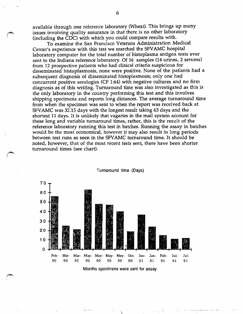
To examine the San Francisco Veterans Administration MedicalCenter's experience with this test we searched the SFVAMC hospitallaboratory computer for the total number of histoplasma antigen tests eversent to the Indiana reference laboratory. Of 16 samples (14 urines, 2 serums)from 12 prospective patients who had clinical criteria suspicious fordisseminated histoplasmosis, none were positive. None of the patients had asubsequent diagnosis of disseminated histoplasmosis; only one hadconcurrent positive serologies (CF 1:64) with negative cultures and no firmdiagnosis as of this writing. Turnaround time was also investigated as this isthe only laboratory in the country performing this test and this involvesshipping specimens and reports long distances. The average turnaround timefrom when the specimen was sent to when the report was received back atSFVAMC was 32.15 days with the longest result taking 63 days and theshortest 11 days. It is unlikely that vagaries in the mail system account forthese long and variable turnaround times, rather, this is the result of thereference laboratory running this test in batches. Running the assay in batcheswould be the most economical, however it may also result in long periodsbetween test runs as seen in the SFVAMC turnaround time. It should benoted, however, that of the most recent tests sent, there have been shorterturnaround times (see chart).
Turnaround time (Days)
Feb- Mar- Mar- May- May- May- May- Oct- Jan- Jan- Feb- Jul- Jul-9 0 9 0 9 0 9 0 9 0 9 0 9 0 9 0 9 1 9 1 9 1 9 1 9 1
/0$^\
Months specimens were sent for assay

SummaryThe urinary Histoplasma antigen assay for disseminatedhistoplasmosis is a promising test. There is the capability for a quick andaccurate result for critically ill patients with possible disseminatedhistoplasmosis and this would significantly lower the morbidity andmortality associated with the disease. However, there are some seriouslimitations in currently using this test. There has of yet been no report ofprospective evaluation of the assay which is essential to remove the biasedpatient selection in the previous articles. The fact that different antigenicserotypes exist across different geographical areas needs to be adressed; wouldthis assay recognize all serotypes? Quality assurance is a problem in that sincethis is the only laboratory performing the assay there is no way of checkingthe results as is done for every other sendout test. Finally, the longturnaround time (32 days avg.) because of batch running makes this test nobetter than culture which takes 2 to 4 weeks. The report on the ELISAsdeveloped by Wheat et al is promising (especially the HRP ELISA) sinceamong other attributes it is non-radioimmunometric. If the ELISA could bedeveloped and commercially available many of the problems mentionedwith the RIA could be resolved and there would be a potentially excellent testfor the diagnosis of disseminated histoplasmosis.
/fPK

BIBLIOGRAPHY
1.Roberts GD. Laboratory Methods in Basic Mycology in Bailey & Scott'sDiagnostic Microbiology. Baron EJ & Finegold SM ed. 8th edition, 1990, pgs.681-775
2.Wheat LJ. Diagnosis and management of histoplasmosis.Eur J Clin Micr InfDis, 8(5):480-490,1989
3.Roberts GD, Goodman NL, Land GA, et al. Detection and Recovery of Fungiin Clinical Specimens in Manual of Clinical Microbiology. Lennette EH ed.4th edition, 1985, pgs. 500-513
4.Wheat LJ. Disseminated histoplasmosis in the acquired immune deficiencysyndrome: Clinical findings, diagnosis and treatment, and review of theliterature.Medicine, 69(6):361-374,1990
5.Gray LD, Roberts GD. Laboratory diagnosis of systemic fungal diseases.InfDis Clin NA, 2(4):779-803, Dec 1988
6.Wheat LT.Histoplasmosis.Inf Dis Clin NA, 2(4):850, Dec 1988
7.Paya CV, Roberts GD, Cockerill FR III. Laboratory methods for the diagnosisof disseminated histoplasmosis: Clinical importance of the lysis-centrifugation blood culture technique. Mayo Clin Proceed, 62:480-485,1987
8.Bille J, Stockman L, Roberts GD, et al. Evaluation of a lysis centrifugationsystem for recovery of yeasts and filamentous fungi from blood. J Clin Micro,18:469-471,1983
9.Davies SF, McKenna RW, Sarosi GA. Trephine biopsy of the bone marrowin disseminated histoplasmosis. Am J Med, 67:617-622,1979
10.Kauffman CA, Israel KS, Smith JW, et al. Histoplasmosis inimmunosuppressed patients. Am J Med, 64:923-932,1978
H.deRepentigny L. Serological techniques for diagnosis of fungal infection.Eur J Clin Micro Inf Dis, 8(4):362-375, April 1989
12.Kaufman L, Reiss E. Serodiagnosis of Fungal Disease in Manual of ClinicalMicrobiology. Lennette EH ed. 4th edition, 1985. pages 924-944
13.Terry PB, Rosenow EC, Roberts GD. False-positive complement fixationserology in histoplasmosis: a retrospective study. JAMA, 239:2453-2456,1978
/iffp^v,

14.Wheat LJ, Kohler RB, Tewari RP. Diagnosis of disseminatedhistoplasmosis by detection of Histoplasma capsulatum antigen in serum andurine specimens. NEJM, 314(2):83-88, Jan 9,1986
15.Wheat LJ, Kohler RB, Tewari RP, et al. Significance of Histoplasma antigenin the cerebrospinal fluid of patients with meningitis. Arch Int Med, 149:302-304, Feb 1989
16.Wheat LJ, Connolly-Stringfield P, Kohler RB. Histoplasma capsulatumpolysaccharide antigen detection in diagnosis and management ofdisseminated histoplasmosis in patients with acquired immunodeficiencysyndrome. Am Jour Med, 87:396-400, Oct 1989
17.Zimmerman SE, Connolly-Stringfield P, Wheat LJ, et al. Comparison ofsandwich solid-phase radioimmunoassay and two enzyme-linkedimmunosorbent assays for detection of Histoplasma capsulatumpolysaccharide antigen.Tour Inf Dis, 160(4):678-685, Oct 1989
18.Drutz DJ. Antigen detection in fungal infections (Editorial). NEJM,314(2):115-117,Jan91986
19.Kaufman L, Blumer S. Occurrence of serotypes among Histoplasmacapsulatum strains. J Bacteriology, 91:1434-1439,1966
jSfl^s