Embed Size (px)
Citation preview

Interplay between Cyclase-Associated Protein, Cofi lin, Profi lin and Twinfi lin in Actin Dynamics
INSTITUTE OF BIOTECHNOLOGYRESEARCH PROGRAM IN CELL AND MOLECULAR BIOLOGY AND DIVISION OF BIOCHEMISTRY AND BIOTECHNOLOGYDEPARTMENT OF BIOSCIENCESFACULTY OF BIOLOGICAL AND ENVIRONMENTAL SCIENCES ANDHELSINKI GRADUATE PROGRAM IN BIOTECHNOLOGY AND MOLECULAR BIOLOGYUNIVERSITY OF HELSINKI
MAARIT MAKKONEN
DISSERTATIONES BIOCENTRI VIIKKI UNIVERSITATIS HELSINGIENSIS 39/2013

Interplay Between Cyclase-Associated Protein, Cofi lin, Profi lin and Twinfi lin in Ac n Dynamics
Maarit Makkonen
Institute of BiotechnologyResearch Program in Cell and Molecular Biology
andDivision of Biochemistry and Biotechnology
Department of BiosciencesFaculty of Biological and Environmental Sciences
andHelsinki Graduate Program in Biotechnology and Molecular Biology
University of Helsinki
ACADEMIC DISSERTATION
To be presented for public examination with the permission of the Faculty of Biological and Environmental Sciences of the University of Helsinki, in lecture hall 1 of the Viikki
C-building (Latokartanonkaari 5), on the 5th of December 2013, at 12 o’clock noon.

Supervisor Professor Pekka Lappalainen Institute of Biotechnology University of Helsinki, Finland
Reviewers Professor Olli Carpén Department of Pathology University of Turku, Finland
Assistant professor Sanna Lehtonen Department of Pathology Haartman Institute University of Helsinki, Finland
Th esis committee members Docent Maria Vartiainen Institute of Biotechnology University of Helsinki, Finland
Docent Pirta Hotulainen Neuroscience Center University of Helsinki, Finland
Custodian Professor Jukka Finne Department of Biosciences Division of Biochemistry and Biotechnology University of Helsinki, Finland
Opponent Adjunct professor Aki Manninen Oulu Center for Cell-Matrix Research Department of Medical Biochemistry and Molecular Biology Institute of Biomedicine Faculty of Medicine University of Oulu, Finland
Cover fi gure: A snapshot of a mouse NIH3T3 fi broblast cell expressing GFP-CAP1 (green) and stained with Alexa Fluor 568 phalloidin to visualize fi lamentous actin (red) and a drawing illustrating the interactions of CAP.
Layout: Tinde PäivärintaISBN 978-952-10-9517-7 (paperback)ISBN 978-952-10-9518-4 (PDF; http://ethesis.helsinki.fi )ISSN 1799-7372
Unigrafi aHelsinki, 2013

”Askel ja pysähdys kerrallaanyksi nurin yksi oikein
etsi etä joka hehkuu,polkua joka puhuu”
- A. W. Yrjänä

TABLE OF CONTENTS
LIST OF ORIGINAL PUBLICATIONSABSTRACTABBREVIATIONS1. INTRODUCTION ........................................................................................................ 1
1.1. Actin .................................................................................................................... 1 1.1.1. Structure of actin ....................................................................................................1 1.1.2. Actin fi lament dynamics .......................................................................................1
1.2. Actin based processes in animal cells .................................................................. 2 1.2.1. Muscle cell contraction ..........................................................................................2 1.2.2. Cell migration .........................................................................................................4 1.2.3. Endocytosis .............................................................................................................5 1.2.4. Cell division ............................................................................................................6 1.2.5. Additional functions of actin in animal cells ......................................................6
1.3. Th e actin cytoskeleton in unicellular organisms ................................................. 7 1.3.1. Th e actin cytoskeleton in budding yeast .............................................................7 1.3.2. Th e actin cytoskeleton in apicomplexan parasites .............................................8
1.4. Actin binding proteins ....................................................................................... 10 1.4.1. Actin fi lament nucleating proteins .....................................................................10 1.4.2 Actin fi lament capping proteins .........................................................................11 1.4.3. Actin fi lament crosslinking proteins ..................................................................12 1.4.4. Actin monomer pool regulating proteins .........................................................12
1.4.4.1. Actin depolymerizing factor (ADF)/cofi lins .......................................12 1.4.4.2. Twinfi lins .................................................................................................14 1.4.4.3. Profi lins ....................................................................................................15
1.4.5. Cyclase-associated proteins (CAPs) ...................................................................16 1.4.5.1. Structure of CAP .....................................................................................16 1.4.5.2. Biochemical properties of CAP.............................................................17 1.4.5.3. Cellular role of CAP ...............................................................................18 1.4.5.4. Physiological role of CAP ......................................................................19

2. AIMS OF THE STUDY .............................................................................................. 203. MATERIALS AND METHODS ................................................................................. 214. RESULTS AND DISCUSSION ................................................................................... 22
4.1. CAPs interact with ADF/cofi lins, profi lin and twinfi lin to regulate actin dynamics ..................................................................................... 22
4.1.1. CAPs bind ADF/cofi lin, profi lin and actin through conserved mechanisms (I, II) .............................................................................22
4.1.1.1. Identifi cation of ADF/cofi lin binding site in yeast CAP (I) ............22 4.1.1.2. Identifi cation of cofi lin-actin and actin binding site in mammalian CAP1 (II) ..........................................................................23 4.1.1.3. Identifi cation of two independent profi lin binding sites in mammalian CAP1 (II) ..........................................................................23
4.1.2. Mammalian CAP binds ATP-actin with high affi nity (II) ..............................23 4.1.3. Catalyzing nucleotide exchange is the most conserved function of CAP ....24
4.1.3.1. Th e C-terminal domain of CAP1 is suffi cient for catalyzing nucleotide exchange (II) .......................................................................24 4.1.3.2. Plasmodium falciparum CAP catalyzes nucleotide exchange on actin monomers (II) ...............................................................................25
4.1.4. Mammalian CAP1 interacts with twinfi lin-ADP-G-actin complex (Makkonen et al., unpublished) .........................................................................26 4.1.5. Model how CAPs regulate actin dynamics in nonmuscle cells ......................27
4.2. Actin fi lament length in muscle sarcomeres is controlled by cofi lin-2 ............. 27 4.2.1. Identifi cation of the ATP-G-actin binding site at cofi lin-2 (III) ....................30 4.2.2. Cofi lin-2 does not show actin isoform specifi city in ATP-G-actin binding (III) ...................................................................................31
5. CONCLUDING REMARKS AND FUTURE PERSPECTIVES ................................... 326. ACKNOWLEDGEMENTS ........................................................................................... 357. REFERENCES ............................................................................................................... 37

LIST OF ORIGINAL PUBLICATIONS
Th is thesis is based on three original articles (I-III) which are referred to in the text by their roman numerals.
I. Quintero-Monzon, O., Jonasson, E. M., Bertling, E., Talarico, L., Chaudhry, F., Sihvo, M., Lappalainen, P., and Goode, B. L. (2009) Reconstitution and dissection of the 600-kDa Srv2/CAP complex: roles for oligomerization and cofi lin-actin binding in driving actin turnover. Th e Journal of biological chemistry 284, 10923-10934
II. Makkonen, M., Bertling, E., Chebotareva, N. A., Baum, J., and Lappalainen, P. (2013) Mammalian and malaria parasite cyclase-associated proteins catalyze nucleotide ex-change on G-actin through a conserved mechanism. Th e Journal of biological chemis-try 288, 984-994
III. Kremneva, E., Makkonen, M., Skwarek-Maruszewska, A., Gateva, G., Michelot, A., Dominguez, R., and Lappalainen, P. (2013) Cofi lin-2 controls actin fi lament length in muscle sarcomeres. submitted manuscript
In addition, some unpublished data “Mammalian CAP1 interacts with twinfi lin-ADP-G-ac-tin complex” (Makkonen et al., unpublished) will be presented.
Contributions:I. Maarit Makkonen, together with Enni Bertling, carried out experiments for fi gure
2 (C-D) and fi gure 5 (D-G) and purifi ed proteins used in these experiments. Maarit Makkonen together with Enni Bertling also cloned some of the plasmids used in as-says mentioned above.
II. Maarit Makkonen planned, conducted and analysed the results of >80% of the ex-periments and prepared all fi gures. Maarit Makkonen wrote the manuscript together with Pekka Lappalainen.
III. Maarit Makkonen planned, performed the experiments and analysed the results for fi gures 1 (A-B), 2 (C), 6 (A-D) and S6 (A-B) and prepared the corresponding fi gures.

ABSTRACT
Several fundamental biological processes rely on actin. Th e ability of actin to form dynamic networks is crucial for processes including cell migration, endocytosis and cell division in unicellular and multicellular organisms. Furthermore, in sarcomeres of muscle cells, actin and myosin form interdigitating networks responsible for muscle contraction.
Actin is highly conserved and abundant protein in all eukaryotic cells. It exists as monomeric (G-actin) and fi lamentous (F-actin) forms, which are in balance and strictly regulated by plethora of actin binding proteins. Th ese proteins regulate the assembly, disassembly, branching and capping of actin fi laments to produce actin networks that are able to undergo changes rapidly according to cell’s needs. Among the most central actin binding proteins are cyclase-associated protein (CAP), actin depolymerizing factor (ADF)/cofi lin, profi lin and twinfi lin, which are under investigation in this study. CAP was fi rst identifi ed as a protein associated with adenylyl cyclase. Later it was realized that this function is not conserved from yeast to mammals and that CAP has more important role related to actin dynamics. Loss of CAP in yeast cells results in abnormalities in actin distribution, and in CAP-defi cient mammalian cells decreased cell motility is observed. In nonmuscle cells CAP localizes to dynamic actin structures like lamellipodia together with actin and cofi lin. Furthermore, lack of the muscle-specifi c isoform of mammalian CAP results in disruption of sarcomeric structure and dilated cardiomyopathy in knockout mice.
CAP is known to bind G-actin and accelerate actin turnover together with ADF/cofi lin, which is the severing and depolymerizing agent in actin fi lament regulation. CAP is suggested to release cofi lin from actin monomers, thus recycling it for new rounds of depolymerization. Furthermore, CAP has been shown to accelerate nucleotide exchange in actin monomers and this is crucial for building dynamic actin structures. Profi lin sequesters actin monomers and promotes their assembly to the fi lament. It also catalyzes the nucleotide exchange on actin monomers to promote their assembly to fi lament ends. CAP is known to interact with profi lin, but the exact mechanism how these proteins work together is not understood. Twinfi lin sequesters actin monomers and caps actin fi lament ends preventing their growth. It also binds another fi lament capping protein (CP) but the overall mechanism how twinfi lin regulates actin turnover has remained elusive.
In this study, we reveal that interactions with actin monomers, ADF/cofi lin and profi lin are conserved in CAPs from yeast to mammals. Unexpectedly, we observed that mammalian CAP has higher affi nity for ATP-actin than yeast CAP, and that mammalian CAP has two independent profi lin binding sites whereas yeast CAP has only one. We also demonstrate a novel function for the ‘mini-CAP’ from apicomplexan parasite as a nucleotide exchange promoting factor. Th e malaria parasite CAP comprises only the C-terminal ADP-actin binding site suggesting that this domain is crucial and harbors the most conserved function of CAPs. Th ese fi ndings together enlighten our knowledge of how CAPs regulate actin dynamics together with ADF/cofi lin and profi lin.
We also revealed that CAP, twinfi lin and ADP-actin form a ternary complex. We demonstrate this novel interaction by two independent assays, native PAGE and supernatant depletion pull-down assay. We demonstrate that both ADF-homology (ADF-H) domains of twinfi lin are capable of forming a complex with CAP and actin. Th e mechanism and biological role of this interaction remain to be solved in future.
In addition to studies with nonmuscle proteins, we expanded our research to muscle-specifi c ADF/cofi lin. Many actin-binding proteins have muscle-specifi c isoforms in addition to nonmuscle ones, but very little is known about these isoforms. We studied the muscle-specifi c

cofi lin-2 and noted that the levels of cofi lin-2 increased during sarcomere maturation while cofi lin-1 amounts remained constant. We compared the actin binding abilities of muscle-specifi c cofi lin-2 and nonmuscle cofi lin-1 and show that cofi lin-2 binds ATP-actin with higher affi nity than cofi lin-1. Importantly, we identifi ed a specifi c cluster of residues on the surface of cofi lin-2 that is responsible for its high-affi nity interactions with ATP-actin. Th us, our studies suggest that this region functions as a ‘nucleotide sensor’ in ADF/cofi lins. Th erefore, a specifi c ADF/cofi lin isoform with high affi nity for ATP-actin evolved to regulate actin dynamics in thin fi laments of sarcomeres. Th e roles of other muscle-specifi c proteins are under particular interest and subject of future research.
In summary, the fi ndings of this study reveal the mechanisms by which CAP regulates actin dynamics together with ADF/cofi lin, profi lin and twinfi lin. Furthermore, this study elucidates yet rather unknown actin regulation by muscle-specifi c cofi lin-2.

ABBREVIATIONS
Abp1 Actin binding protein 1ADF Actin depolymerizing factorADF-H Actin depolymerizing factor homologyADP Adenosine diphosphateAip1 Actin interacting protein 1Arp Actin related protein ATP Adenosine triphosphateBAR BIN/Amphiphysin/RvsCAP Cyclase-associated proteincDNA Complementary DNACobl Cordon-bleuCof Cofi linCP Capping proteinDNA Deoxyribonucleic acidDTT DithiothreitolF-actin Filamentous actinFAK Focal adhesion kinaseFH Formin homology G-actin Globular actinGMF Glia maturation factorGRABP Gelsolin-related actin binding proteinGST Glutathione S-transferaseGTP Guanosine triphosphate hnRNPs Heterogenous nuclear ribonucleoproteinsIMC Inner membrane complexKd Dissociation constantkD KilodaltonLmod LeiomodinMAL Megakaryocytic acute leukemiaMKL1 Megakaryoblastic leukemia 1mRNA Messenger RNAMRTF-A Myocardin related transcription factor A NBD 7-Chloro-4-nitrobenzeno-2-oxa-1,3-diazoleNPF Nucleation-promoting factorPAGE Polyacrylamide gel electrophoresisPfCAP Plasmodium falciparum CAPPi Inorganic phosphatePI(4,5)P2 Phosphatidylinositol 4,5-bisphosphatePP1 Polyproline region 1PP2 Polyproline region 2Prof Profi linRas Rat sarcoma RhoA Ras homolog gene family member ARNA Ribonucleic acidRNAi RNA interference

SDS-PAGE Sodium dodecyl sulfate polyacrylamide gel electrophoresisSEM Standard error of the meanSH3 Src homology 3 domainSrc SarcomaSRF Serum response factorTIRF Total internal refl ection fl uorescenceWASP Wiskott-Aldrich syndrome proteinWH2 WASP homology domain 2WIP WASP-interacting proteinXAC Xenopus laevis ADF/cofi lin

1
1. INTRODUCTION
1.1. Ac nActin is highly conserved protein found in all eukaryotic cells. It is among the most abundant proteins in most cells. Striated muscle cells have the highest amounts of actin, which can constitute 20% of total muscle protein. Actin is restricted to only eukaryotes, but it has been suggested that an ancestral actin exists in prokaryotes because bacterial proteins such as MreB show striking similarity to actin when comparing their fi lamentous structures (reviewed in dos Remedios et al., 2003). In eukaryotic cells, the ability of actin to form polarized fi laments and to interact with a number of actin-binding proteins allows it to perform diff erent functions dependent on the cell type, as will be discussed in chapters 1.2. and 1.3.
1.1.1. Structure of ac n
Actin exists in monomeric (G-actin) and fi lamentous (F-actin) forms in cells. Each actin monomer comprises of two domains, which can be further subdivided into two subdomains yielding four subdomains altogether (Kabsch et al., 1990). Each domain consists of similar motif with alternating β-sheets and α-helixes, suggesting that a gene duplication might have taken place (Kabsch et al., 1990).
Actin monomer binds either ADP or ATP complexed with divalent cation Mg2+ or Ca2+ through a nucleotide binding cleft located in the center of the monomer, between the four subdomains (reviewed in Kabsch & Holmes, 1995). Binding of nucleotide and divalent cation aff ects the conformation of the actin molecule by domain rotations. Th is is also case with numerous ligands that actin has, including all the actin-binding proteins which control the conformation of actin and regulate the actin fi lament turnover (reviewed in Schuler, 2001). Th e affi nity of actin for ATP is higher than for ADP. Ca2+-actin binds
ATP with 200-fold affi nity compared to ADP whereas Mg2+-actin binds ATP with 4-fold better affi nity than ADP (Kinosian et al., 1993). Considering the fact that cells have much higher concentration of Mg2+ compared to Ca2+, monomeric actin mainly exists as Mg2+-ATP-actin inside the cells.
1.1.2. Ac n fi lament dynamics
Th e ability to polymerize from monomers into fi laments is the key process involved in all actin structures and processes. Polymerization from scratch is unfavorable since the newly formed actin dimers are more likely to dissociate than associate with another monomer to form a trimer. However, once the trimer has been formed, actin rapidly polymerizes and fi lament elongates. Th e conditions inside the cells including high salt (KCl > 50 mM) and Mg2+ concentrations favour polymerization. Th e ends of one fi lament diff er from each other as actin monomers are orientated so that their subdomains 1 and 3 are facing towards the other end and subdomains 2 and 4 towards the other. Th ese ends are named as barbed and pointed ends, respectively, according to their arrowhead-like pattern when decorated with myosins (reviewed in dos Remedios et al., 2003).
Except for associating, actin monomers also dissociate continuously from both ends establishing a steady state where both fi laments and monomers are in equilibrium. ATP-actin monomers associate more likely to barbed ends of the fi laments than pointed ends, resulting in fi lament elongation mainly from the barbed end. In steady state, the actin concentration left in solution as monomers is called the critical concentration, and it is 0.1 μM at the barbed end and 0.6 μM at the pointed end (reviewed in Pollard et al., 2000).
In actin fi lament, ATP-actin monomers rapidly undergo ATP hydrolysis yielding ADP-Pi-actin, which later become ADP-actin when γ-phosphate dissociates. ADP-actin subunits then dissociate at the pointed end and monomeric ADP-actins undergo a nucleotide
Introduction

2
exchange process where they are “re-charged” with ATP. ATP-actin monomers are then ready for new rounds of polymerization where they are added to the barbed ends of fi laments (reviewed in Pollard & Borisy, 2003). Th is ATP-hydrolysis driven cycle is called treadmilling and illustrated in Figure 1. Treadmilling provides actin fi laments characteristic to perform their multiple dynamic functions in cellular processes. Treadmilling per se is comparatively slow process and diff erent steps of the cycle are regulated by an array of actin binding proteins, which will be discussed in more detail in chapter 1.4.
1.2. Ac n based processes in animal cellsMany biological processes are actin-dependent. In muscle cells, actin cooperates with myosin in sarcomeres to constrict the cell, and in nonmuscle cells actin constitutes the skeletal framework determining cell shape and movement. Besides actin, the cytoskeleton also includes microtubules and intermediate
fi laments. Th e cytoskeletal actin fi laments provide mechanical strength and motility for animal cells, whereas microtubules take care of vesicle transport and intermediate fi laments function to resist mechanical forces. Also vesicle internalization and cell division are actin-dependent. All these processes are somewhat complicated including numerous proteins involved, but actin is one of the key players in all of these actions and they are introduced below. Furthermore, additional functions of actin in nucleus are discussed in chapter 1.2.5.
1.2.1. Muscle cell contrac on
Striated myofi brils found from muscle cells in heart and skeletal muscle are examples of extremely well organized actin networks. In these cells, actin and myosin, accompanied by plethora of other proteins, are organized into specialized units called sarcomeres, which produce force for muscle contraction. Muscle contraction is a result of molecular interactions between these muscle-specifi c
Introduction
Figure 1. Treadmilling of actin. ATP-actin monomers associate to the barbed (fast-growing) end of the actin fi lament and undergo rapid ATP hydrolysis. Subsequently the phosphates of the ATP-Pi-actins dissociate, followed by ADP-actin monomer disassembly at the pointed (slow-growing) end of the fi lament. Th e nucleotides in ADP-actin momomers are exchanged to ATPs, resulting newly “charged” ATP-actin monomer pool to be added to the barbed end.

3
proteins that are precisely aligned and tightly regulated, but not static, since they constantly undergo dynamic turnover (reviewed in Sanger & Sanger, 2008).
By light microscopy, several types of stripes are visible in muscle cells resulting from diff erent fi lament systems linked together (reviewed in Clark et al., 2002). Th ese include dark A bands containing thick myosin II fi laments and light I bands containing thin fi laments composed of actin but not myosin. In the middle of the I bands are Z discs,
which connect adjacent sarcomeric units together thus serving as boundaries between them. α-actinin is the best characterized and major component of the Z disc, but to date, also several other proteins have been identifi ed, which all together form a complex network that not only have a structural role in sarcomeres but are also involved in signal transduction (reviewed in Faulkner et al., 2001 and Clark et al., 2002). Th e structure of muscle sarcomere is illustrated in Figure 2.
Introduction
Figure 2. Th e organization of muscle sarcomeres. Sarcomere is a functional unit stretching from Z disc to another Z disc. Th e interdigitating thin actin fi laments and thick myosin fi laments constitute the main components of the sarcomere. Actin fi lament barbed ends are facing the Z discs and pointed ends are towards the M line. Other proteins including tropomyosin, nebulin, tropomodulin, CapZ and titin regulate the dynamics of the thin and thick fi laments to produce muscle contraction. In contracting sarcomere, myosin heads walk along the actin fi laments moving them towards the M line and decreasing the H zone width.

4
Th e thick myosin fi laments interdigitate with thin actin fi laments in A band, except in the middle region, which is called H zone. Furthermore, special proteins in the H zone form M line in the middle of the sarcomere. In addition to actin and myosin, giant proteins titin and nebulin form additional fi lament systems, which are parallel to thin fi laments and attached to the Z discs. Titin is anchored to Z discs via its N-terminus, spanning the I- and A bands, whereas its C-terminal end overlaps with the M line (Wang et al., 1979). Th e enormous size and elastic elements enables titin to act as a molecular spring in maintaining the sarcomeric structure. Nebulin, in turn, extends from Z discs to the pointed ends of the thin fi laments and has been suggested to determine the lengths of the thin fi laments (Kruger et al., 1991).
Th in actin fi laments extend from Z discs to the H zone. Th ey are accompanied by troponins and tropomyosins, which together regulate the force-generating interactions between myosin and actin in a Ca2+-dependent manner (reviewed in Vale & Milligan, 2000 and Craig & Lehman, 2001). Tropomyosins also stabilize thin fi laments preventing their depolymerization from the pointed ends (Broschat 1990). Th e heads of the myosin II molecules at the A band are bipolarly orientated and interact with actin fi laments to drive muscle contraction. Th e tail regions of myosins form M line, which is free from thin fi laments. However, several proteins located in the M line have been identifi ed, e.g. myomesin and M-protein (Obermann et al., 1996).
Th in fi laments are capped at barbed ends by CapZ (Casella et al., 1987; Hug et al., 1992), which is also called as capping protein (CP) in nonmuscle cells. At pointed ends, fi laments are capped by tropomodulin, which binds to tropomyosin and actin molecules blocking elongation and depolymerization at pointed ends of thin fi laments (Weber et al., 1994). Th e lengths of the thin fi laments are extremely precise and constant within a myofi bril, and several mechanisms have been
suggested to regulate their length including capping proteins and ‘nebulin ruler’ (reviewed in Littlefi eld & Fowler, 2008 and Gokhin & Fowler, 2013). Also other molecules, e.g. actin polymerizing, depolymerizing, severing and/or sequestering proteins might be involved in determining the thin fi lament length. In consequence of many proteins involved, the mechanism of this regulation remains still unsolved.
1.2.2. Cell migra on
It is crucial for many cell types to perform directional motility. For example, in developing embryo, closure of wound, immune responses, neuronal path-fi nding and metastatic cancer cells the ability to migrate is essential. In nonmuscle cells, actin is able to produce force without any motor proteins by polymerizing against the cell membrane to push it forward (reviewed Rottner and Stradal, 2011).
Th e mechanism by which cells migrate is universal and includes four steps (reviewed in Ridley et al., 2003). First, cell polarizes and produces a protrusion at the leading edge. Protrusions can be large lamellipodia or thinner fi lopodia oriented towards the direction of migration. Second, the protrusions are attached to the substratum, either extracellular matrix or other cells, via adhesion molecules. Th ird, the rear of the cell is retracted and fourth, it is detached from substratum. Th is cycle is repeated continuously in crawling-type of motility, although some details may diff er according to cell type.
At the leading edge, actin fi laments are oriented diff erently depending on the protrusion type. In fi lopodia, the fi laments are assembled as parallel bundles, whereas in lamellipodia they form branched networks (reviewed in Ridley, 2011). Th e actin dynamics at the leading edge of moving cell is explained by a dendritic-nucleation model (reviewed in Pollard et al., 2000). In the model, new actin fi laments are created by the Arp2/3 complex,
Introduction

5
which is activated by Wiskott-Aldrich syndrome protein (WASP) family proteins. Th e elongating fi laments push the plasma membrane forward establishing a protrusion. Th e elongation of individual fi laments is soon restricted by capping protein, and new Arp2/3 complexes are needed for initiating new branches to the actin “bush”. Hydrolysis of ATP and dissociation of phosphate in actin fi laments triggers ADF/cofi lins to sever and depolymerize older fi laments yielding ADP-actin monomers, which are converted to ATP-actin monomers by profi lin. Maintaining a large pool of monomeric actin is essential for rapid fi lament growth and some actin binding proteins are involved in maintenance of the large monomer pool. Th is is important, because the concentration of actin monomers needed is above the critical concentration for actin fi lament assembly. Th is task is taken care by e.g. profi lin and thymosin-β4, which both bind actin monomers and compete with each other for binding. However, from these two proteins only profi lin is able to elongate actin fi lament barbed ends when bound to ATP-actin (reviewed in Pollard et al., 2000). In continuously moving cells, the assembly and disassembly of actin fi laments are in balance, controlled by mechanisms described above.
Th e attachment of protrusion to the substratum is mediated by specifi c receptors found in cells. Th e major family of these receptors are integrins, which link the extracellular matrix to actin fi laments via adaptor proteins. Adhesions serve as traction points or “feet” by which cell is able to grab the substratum. Actin together with myosin generates contractile force that is transmitted to adhesion sites, allowing cell to move forward (reviewed in Ridley et al., 2003 and Rottner and Stradal, 2011). Finally, to be able to proceed, cell needs to disassemble the adhesions at rear to promote rear retraction (reviewed in Webb et al., 2002).
1.2.3. Endocytosis
Cellular uptake is fundamental process for the normal function of many cell types. Cells use endocytosis e.g. to dispose of pathogens or damaged cells, signaling with other cells and take up nutrients or potential antigens. Endocytosis includes many types of cellular uptake such as phagocytosis, macropinocytosis, clathrin-mediated endocytosis and caveolae-mediated endocytosis, from which the clathrin-mediated endocytosis will be discussed here in more detail, because it is the best characterized form of endocytosis (reviewed in Doherty & McMahon, 2009).
Clathrin-mediated endocytosis is a complex process involving many proteins with diff erent properties, including endocytic coat proteins and regulators of actin assembly. Endocytosis includes several processes that occur sequentially, specifi c proteins functioning in particular phases. First, endocytic proteins, including clathrin, are recruited on the plasma membrane forming an endocytic coat, which is surrounded by proteins that activate Arp2/3 to nucleate new actin fi laments. Next, actin polymerization starts and fi laments are crosslinked and attached to the coat, and this is accompanied by increase in curvature of membrane and elongation of the invagination. Barbed ends of actin fi laments are pointing towards the membrane, and promote invagination, elongation and fi ssion. It is not understood how actin fi laments generate force to invaginate the vesicle and what is the role of myosins in this process, as they are found to localize to endocytic sites. Aft er elongation, membrane scission occurs. In mammalian cells, dynamin together with actin, several actin-binding proteins and BIN/Amphiphysin/Rvs (BAR) domain proteins seem to be the key players in this event. In yeast, dynamin is less critical while another protein, Vps1, is suggested to play more important role triggering the scission (reviewed in Kaksonen et al., 2006 and Mooren et al., 2012).
Introduction

6
Although it is evident that actin is crucial in clathrin-mediated endocytosis in yeast, the need of actin in endocytosis in mammalian cells is less clear. Th ere is evidence that actin may play a role in several stages of endocytosis but, some data suggest that this role is not obligatory (Yarar et al., 2005; Fujimoto et al., 2000). Yeast cells have higher turgor pressure due to their cell walls, which has been suggested to result in the diff erent requirements for actin in yeast and mammals (Aghamohammadzadeh & Ayscough, 2009). Supporting the need of actin in endocytosis in mammalian cells, actin has been shown to elongate tubular necks of clathrin pits together with BAR domain proteins (Ferguson et al., 2009). Actin fi laments also form actin cortex associated with the plasma membrane, generating cortical tension. Th is might inhibit endocytosis but, however, the results of the role of cortical tension and actin in endocytosis are somewhat confusing and need still further investigations (reviewed in Mooren et al., 2012).
1.2.4. Cell division
Cytokinesis is the last step in the cell cycle, where two cells are physically separated from each other. In eukaryotes, cytokinesis occurs by an universal mechanism involving actomyosin contractile ring, but there are diff erences between diff erent species like yeast and animal cells. Th e budding yeast cytokinesis is discussed briefl y in chapter 1.3.1. while the cytokinesis in metazoan cells is discussed here.
Th e fi rst phase of cytokinesis is to determine the place for the cleavage furrow. Th is occurs slightly diff erently depending on the cell type, but it takes place in the beginning of anaphase by astral microtubules, the central spindle or both (reviewed in Glotzer, 2004). Th e formation of cleavage furrow also requires activation of GTPase RhoA, although it seems not to be critical in determining the division plane (reviewed in Balasubramanian et al., 2004).
Th e next step is the formation of actomyosin ring. Th e activation of RhoA results in polymerization of actin by formins and activation of myosin by phosphorylation by either inhibiting of myosin phosphatase or phosphorylating myosin light chain (Kawano et al., 1999; Piekny & Mains, 2002; Watanabe et al., 2008). Also other actin binding proteins like α-actinin and fi lamin are found to be involved in formation of contractile ring (Fujiwara et al., 1978; Nunnally et al., 1980). Th e actin fi laments are orientated towards several directions in contractile ring, that is, they have mixed polarities. Th e constriction of the ring is suggested to occur via mechanism related to muscle sarcomeres including several contractile modules in series around the ring, but more information is needed to build a reliable model for constriction. Th is is because contractile rings diff er from muscle sarcomeres being more complicated with non-organized actin and myosin (reviewed in Pollard, 2010). In addition, the ring apparatus is disassembled during constriction, most likely by ADF/cofi lins (Gunsalus et al., 1995).
Aft er constriction, cytokinesis is fi nalized by membrane remodeling event, where the plasma membrane of the dividing cell is subdivided into two. Th is requires delivery of new membranes by targeted secretion to compensate the increased cell surface area of the daughter cells (reviewed in Balasubramanian et al., 2004).
1.2.5. Addi onal func ons of ac n in animal cells
Actin is traditionally considered as a cytoplasmic protein, but it is important to note that actin is also found from the nucleus, where it has a potentially crucial role in regulation of gene expression. Several functions have been described for actin in transcription, including binding to chromatin remodeling complexes together with actin related proteins (Arps), associating with all three RNA polymerases and binding to specifi c heterogenous nuclear ribonucleoproteins
Introduction

7
(hnRNPs) which regulate transcription and pre-mRNA processing (reviewed in Farrants, 2008 and Skarp & Vartiainen, 2010). Actin has also a force-generating role in nucleus and it has been suggested to drive the movement of individual chromosomal loci or even the whole chromosomes. Th is force-generated movement is based on either actin polymerization or cooperation with nuclear myosin I (reviewed in Skarp & Vartiainen, 2010).
Actin is also linked to several proteins that regulate transcription. Th ese proteins include e.g. myocardin related transcription factor A (MRTF-A, also known as megakaryocytic acute leukemia, MAL, or megakaryoblastic leukemia 1, MKL1) and gelsolin family proteins (reviewed in Skarp & Vartiainen, 2010). MAL is a co-activator of the serum response factor (SRF), which controls many immediate-early genes encoding e.g. signaling and cytoskeletal proteins. Actin regulates MAL by controlling its nuclear import, export and activation in nucleus, which eventually aff ect the expression of cytoskeletal proteins including actin itself (Vartiainen et al., 2007). Gelsolin and several gelsolin-related actin binding proteins (GRABPs) regulate actin dynamics in the cytoplasm. However, they also act as co-activators for several nuclear receptors e.g. androgen, estrogen and thyroid hormone receptors which mediate the expression of their target genes (reviewed in Archer et al., 2005).
Actin distribution between nucleus and cytoplasm is tightly regulated and highly dynamic. Controlling nuclear actin levels in cells is important for effi cient transcription. Actin can rapidly move in and out of the nucleus and the transport in both directions is active. Exportin 6 mediates the export of actin from nucleus whereas importin 9 is required for importing actin to the nucleus. Interestingly, importin 9 also interacts with cofi lin, and this interaction appears to be crucial for actin import (Dopie et al., 2012).
Th e organization of actin in the nucleus is not completely understood yet. Studies where actin polymerization was inhibited suggested that polymerization is needed for transcription but not necessarily for all nuclear functions of actin. Based on several studies, actin is suggested to exist in three diff erent populations in the nucleus (Huet et al., 2012). Th e two populations with fastest and second fastest turnover rates have been implicated to correspond to free actin monomers and polymers, respectively. Th e third and largest (~ 60% of total nuclear actin) population with slowest turnover rate is suggested to be involved in chromatin remodeling, transcription regulation and mRNA processing. However, more investigations are needed to reveal the signifi cance and mechanism of nuclear actin in diff erent cellular processes in distinct cell types.
1.3. The ac n cytoskeleton in unicellular organisms1.3.1. The ac n cytoskeleton in budding yeast
In 1984, it was established in two publications that yeast cells have two distinct actin structures, cortical dots called patches and cytoplasmic fi bers called cables (Adams & Pringle, 1984; Kilmartin & Adams, 1984). Adams and coworkers also found actin to be concentrated in the neck region connecting the mother cell and the bud, which was later confi rmed as third actin structure, contractile actomyosin ring (Bi et al., 1998; Lippincott & Li, 1998).
Actin patches are now known to mediate endocytosis (discussed in chapter 1.2.3.). Th ese actin structures mature during diff erent endocytic stages and they display diff erent types of movement depending on the stage of endocytosis (Kaksonen et al., 2003). Aft er fi rst actin-independent phase of endocytosis, recruitment of endocytic patch components to the cell cortex, follows the slow actin-dependent movement. In this stage, Arp2/3-dependent actin polymerization occurs
Introduction

8
driving vesicle formation and internalization (Kaksonen et al., 2003). Aft er slow motility phase, patch undergoes transition to rapid movement stage, in which the newly formed vesicle is released and moves inwards from the cell cortex (Kaksonen et al., 2003). Th e vesicle is transported to endosomal sorting compartments along actin cables, which mediate the retrograde fl ow in yeast cells (Huckaba et al., 2004).
In yeast cells, the polarized growth and organelle segregation are mediated by the actin cytoskeleton, unlike in larger eukaryotic cells, in which microtubules perform this function. Pruyne et al. (1998) showed, by using mutant strain defective for tropomyosin, that the actin cables mediate the delivery of secretory vesicles for polarization of cell growth. It is important to note that although both actin cables and cortical patches are polarized in yeast cells, it is the cables which are required for targeted secretion, not the patches (Pruyne et al., 1998). Type V myosins function as motors to deliver diff erent cargos along actin cables (reviewed in Bretscher, 2003). Transport of post-Golgi secretory vesicles, which facilitate polarized cell growth, is dependent on Myo2. Also transport of cell organelles including vacuole, Golgi, nucleus and peroxisomes to daughter cell occurs in Myo2-dependent manner. Another type V myosin expressed in budding yeast, Myo4, uses actin cable tracks similarly to Myo2 but instead of vesicles and organelles it transports daughter-specifi c mRNA into the bud (reviewed in Bretscher, 2003). Unlike actin patches, cables are assembled in Arp2/3-independent manner by actin nucleating proteins formins and profi lin (reviewed in Moseley & Goode, 2006).
In yeast cytokinesis, two diff erent F-actin structures at the bud neck are visible during anaphase. First, actin cables are orientated towards the bud neck in mother and daughter cell, targeting the secretion along the axis of polarity (reviewed in Pruyne et al., 2004). Second, a contractile actomyosin ring forms when a ring of F-actin associates with Myo1p ring, which appears already before
bud emergence (Bi et al., 1998; Lippincott & Li, 1998). Th e actomyosin ring constricts to facilitate the neck closure and then disappears. F-actin ring, as well as patches and cables, undergo rapid turnover demonstrated by studies using an actin monomer sequestering drug, latrunculin A. Th us, a set of actin-binding proteins including e.g. ADF/cofi lins, actin interacting protein 1 (Aip1), CAP, profi lin and twinfi lin are involved promoting the actin turnover in yeast (reviewed in Moseley & Goode, 2006). Th e S. cerevisiae actin-binding proteins that are known to be involved in actin dynamics are listed in Table 1.
1.3.2. The ac n cytoskeleton in apicomplexan parasites
Th e phylum Apicomplexa includes several protozoan parasites that require a host cell in order to survive and replicate. Invading to host cells results in severe diseases in host organism depending on the parasite, e.g. toxoplasmosis (Toxoplasma), enteritis (Cryptosporidium) or malaria (Plasmodium) (reviewed in Frénal & Soldati-Favre, 2009). Plasmodium falciparum, which is responsible of causing malaria, is transmitted to humans via mosquitoes. Parasite moves through bloodstream to liver where it multiplies. Later, the parasite enters to red blood cells where they proliferate and further spread the infection (reviewed in Cowman & Crabb, 2006).
Apicomplexan parasites display a unique type of movement called gliding motility, which is based on actomyosin system, to access the host cells. Apicomplexans have multi-layered membrane-structure including plasma membrane and inner membrane complex (IMC) that consists of the inner and outer membranes. Th e compartment between the plasma membrane and IMC contains most of the proteins generating the gliding motility, including actin and myosin, which are linked to both membrane systems. Th e movement is suggested to occur by IMC-bound myosins walking along actin fi laments, which are
Introduction

9
linked to cell surface through adhesion molecules (reviewed in Baum et al., 2006).
Th e actin fi laments in apicomplexan parasites are somewhat diff erent compared to eukaryotes. Studies on T. gondii revealed that in vitro, their actin fi laments are 10 times shorter than rabbit actin fi laments and unstable, probably due to instability of fi laments itself
and actin-binding proteins (Schmitz et al., 2005; Sahoo et al., 2006). Th e cytosolic actin in parasites most probably exists primarily as monomeric form (Dobrowolski et al., 1997). However, T. gondii actin has 3-4-fold lower critical concentration than conventional actins and thus polymerizes readily into fi laments (Sahoo et al., 2006). Th ese characteristics
Introduction
Table 1. Comparison of actin binding proteins found from yeast S. cerevisiae and malaria parasite P. falciparum.
Functional class Homologue Protein in S. cerevisiae
Protein in P. falciparum
Monomer treadmilling Aip1 Aip1CAP Srv2 PfC-CAPCofi lin Cof1 PfADF1, PfADF2Gelsolin/Villin *GMF GMFProfi lin Pfy1 PfPfnTwinfi lin Twf1(unknown) Bud6/Aip3
Nucleation Arp2/3 complex Arp2/3 complex **Eps15 Pan1Formin Bni1, Bnr1 PfFormin1, PfFormin2mAbp1 Abp1WASp Las17/Bee1
F-actin capping Capping protein Cap1/Cap2 PfCPα, PfCPβCrosslinking and bundling Calponin Scp1
Coronin Crn1 Pf coroninFimbrin Sac6IQGAP Iqg1/Cyk1Tropomyosin Tpm1, Tpm2(unknown) Abp140
Myosin Myosin Myo1 – Myo5 Pfmyo-A – Pfmyo-F
Other HIP1R Sla2WIP Vrp1
* P. falciparum has two gelsolin-like domains which are most likely functionally unrelated. ** ARPC1 subunit only. Data for the table has been collected from reviews by Baum et al. (2006), Moseley & Goode (2006) and Schüler & Matuschewski (2006). P. falciparum myosins were described in Chaparro-Olaya et al. (2005) and GMF was characterized by Gandhi et al. (2010) and Nakano et al. (2010).

10
are most likely applied to all apicomplexan parasites, making their actins well suited for gliding motility where rapid turnover of fi laments is needed.
Th e protein repertoire regulating actin in apicomplexan parasites is remarkably limited compared to that of other eukaryotes (reviewed in Baum et al., 2006). Th e actin binding proteins found from apicomplexan P. falciparum compared to ones found from S.cerevisiae are listed in Table 1. However, it is still possible that parasites perform their cell processes by some compensatory proteins that are still unknown. Th ree main classes of proteins regulating actin dynamics are found from apicomplexan and include ADF/cofi lin, profi lin, and CAP (reviewed in Olshina et al., 2012). Th e lack of Arp2/3 complex and its regulators in apicomplexan parasites suggests that the most probable fi lament nucleator in these organisms is formin (Baum et al., 2008). Coronin is the only known crosslinking protein found from apicomplexan, whereas numerous actin bundling proteins found from yeasts and animals are absent. Filament barbed end capping is most likely taken care by heterodimeric CapZ since it is the only known capping protein found from apicomplexan (reviewed in Baum et al., 2006).
P. falciparum has two ADF/cofi lin isoforms, PfADF1 and PfADF2, from which only PfADF1 is expressed throughout the life cycle (Schüler et al., 2005). Unlike other eukaryotic ADF/cofi lins, PfADF1 was detected to bind actin monomers and not fi laments, and stimulate nucleotide exchange on actin monomer instead of inhibition, which is characteristic to other ADF/cofi lins (Schüler et al., 2005). However, recent studies provided evidence that ADF1 in P. falciparum and T. gondii are capable of severing actin fi laments (Mehta & Sibley, 2010; Wong et al., 2011).
Apicomplexan seem to have only a single profi lin, which binds and sequesters actin monomers, as shown for P. falciparum and T. gondii profi lins (Kursula et al., 2008; Plattner et al., 2008). T. gondii, P. falciparum and C.
parvum profi lins also promote actin assembly at barbed ends (Plattner et al., 2008). Th ere is very little information whether apicomplexan profi lins catalyze nucleotide exchange on actin monomers similarly to other profi lins. Th ere is one study demonstrating that T. gondii profi lin, unexpectedly, reduces nucleotide exchange instead of promoting it (Kucera et al., 2010).
Apicomplexans have one CAP homologue, which completely lacks the N-terminal and WASP homology domain 2 (WH2) domains that are present in other eukaryotes (Hliscs et al., 2010). C. parvum CAP has been shown to sequester actin monomers inhibiting actin polymerization (Hliscs et al., 2010), but other possible activities of C. parvum or other apicomplexan CAPs are unknown.
1.4. Ac n binding proteinsActin fi lament treadmilling allows constant reorganization of the fi lamentous networks that produce force and allow cell movement. However, actin fi lament treadmilling and fi lament reorganization are very slow processes without any cooperative proteins. A plethora of actin binding proteins have been identifi ed so far, and they are able to speed up the treadmilling process by ~200-fold (reviewed in Dos Remedios et al., 2003). Th ese proteins can aff ect the fi lament reorganization by various mechanisms, like generating free barbed ends for polymerization (by uncapping, severing or nucleating), capping existing fi lament ends, maintaining actin monomer pool (actin sequestering proteins) or crosslinking actin fi laments.
1.4.1. Ac n fi lament nuclea ng proteins
New actin fi laments are needed in cells in order to build actin networks in cytoplasm. Th e formation of nuclei required for initiating actin fi lament elongation is kinetically unfavorable process, which is also inhibited by various actin monomer sequestering proteins.
Introduction

11
Specifi c nucleating proteins are thus needed for speeding up the nucleation process.
Formins are a large family of actin-binding proteins that are able to nucleate new actin fi laments. Th ey also alter the fi lament elongation rate and “walk” along the growing fi lament remaining at the barbed end. Th e ability to alter the elongation or depolymerization varies between diff erent formins. All formins compete with barbed-end capping proteins by inhibiting their function and thus allowing fi lament elongation at the barbed ends (reviewed in Higgs, 2005).
Formins contain a formin homology 1 (FH1) domain, which is variable in length and rich in proline, and a highly conserved formin homology 2 (FH2) domain. Together these domains regulate actin fi lament elongation (Castrillon & Wasserman, 1994; Higgs & Peterson, 2005). FH2 domain is a dimer forming a donut-shape structure that wraps around actin polymer in the barbed end and moves along the fi lament during elongation (Xu et al., 2004). FH1 is suggested to be unstructured and it serves as a binding site for profi lin. Binding of profi lin to actin inhibits nucleation and monomer addition to the pointed ends but not to the barbed ends. However, profi lin binding to the FH1 domain of formin enhances addition of profi lin-actin to the barbed end resulting fi lament elongation (reviewed in Higgs, 2005).
In contrast to formins, Arp2/3 is able to produce branched actin fi laments. Th e branched actin networks are needed at the leading edge of the moving cell to push the cell membrane forward. Arp2/3 consists of seven subunits, from which two are actin-related proteins 2 and 3 (Arp2, Arp3) that are stabilized by fi ve other subunits. Arp2 and Arp3 mimic actin monomers and serve as a nuclei for the new daughter fi lament which branches off from mother fi lament at a 70° angle (reviewed in Pollard, 2007). Arp2/3 alone is ineffi cient in nucleating new fi laments, but binding to existing actin fi lament enhances its nucleation activity.
Activation of Arp2/3 is initiated by nucleation-promoting factors (NPFs), which is a group of proteins belonging to several protein families (reviewed in Campellone & Welch, 2010). Also phosphorylation in Arp2 is needed for activation by NPFs (LeClaire et al., 2008).
Th e more recently identifi ed actin nucleation proteins include Spire, Cordon-bleu (Cobl) and Leiomodin (Lmod), which all contain WH2 domains that bind G-actin. Spire has four WH2 domains and linker, which cooperatively bind to total of four actin monomers (Quinlan et al., 2005). Cobl has three WH2 domains and polyproline regions and recently it has been shown to have only weak fi lament nucleating and strong fi lament severing activity (Ahuja et al., 2007; Husson et al., 2011). Lmod is a muscle-specifi c nucleating protein that has a domain organization resembling that of tropomodulins, except the C-terminus which has a WH2 domain and polyproline region. Lmod has been shown to be a strong actin nucleator in muscle cells and important for sarcomere organization in cardiomyocytes (Chereau et al., 2008).
1.4.2 Ac n fi lament capping proteins
Actin fi lament capping is essential for cell movement. Capping of fi lament barbed ends results in shorter fi laments, which are more effi cient in pushing cell membrane. Furthermore, capping of fi laments can target fi lament assembly towards desired direction (reviewed in Pollard & Borisy, 2003). Several barbed-end capping proteins are known, from which gelsolin and heterodimeric capping protein are the best characterized and discussed here. Only one family of pointed-end capping proteins, tropomodulins, is known to exist. Tropomodulins have low affi nity for F-actin alone, but the affi nity increases when F-actin is decorated by tropomyosin (Weber et al., 1994).
Eight proteins belonging to gelsolin superfamily have been characterized in mammals (reviewed in Nag et al., 2013). Th ese all contain variable number (three to
Introduction

12
six) of gelsolin domains, which are structures including β-sheet sandwiched between two α-helixes (Burtnick et al., 1997). Binding of calcium ions changes the conformation of gelsolin and activates it (Choe et al., 2002). Th ere are three major actin-binding sites in gelsolin, which are exposed in activation by calcium ions (reviewed in Nag et al., 2013). In addition to capping, gelsolin is also able to sever actin fi laments. Th us, gelsolin is effi cient protein to dissolve actin gels by severing fi laments and remaining in the fi lament end as a cap therefore preventing fi lament growth and reannealing (reviewed in Dos Remedios et al., 2003).
Heterodimeric capping protein (CP) is found in almost all eukaryotic cells. It consists of α and β subunits, which have similar secondary and tertiary structures despite the lack of amino acid sequence similarity (Yamashita et al., 2003). CP caps fi lament barbed ends with high affi nity (Kd ~ 0.1–1nM) by two independent actin binding sites located in the C-temini of the α and β subunits (Wear et al., 2003). CP is known to interact with another actin-binding protein twinfi lin, but the interaction between these two proteins does not aff ect their actin binding properties (Falck et al., 2004). 1.4.3. Ac n fi lament crosslinking proteins
Connecting actin fi laments together is important for cell to obtain rigid three-dimensional actin structures. A variety of proteins have been identifi ed to crosslink actin fi laments in cells, including for example α-actinin, fascin, fi mbrin, fi lamin, some myosins and spectrin. From these proteins, α-actinin is one of the best characterized and therefore discussed here as an example.
α-actinin has a molecular structure of an anti-parallel dimer bearing high elasticity and strength. Each monomer consists of an N-terminal actin binding domain followed by four spectrin repeats and two EF-hand regions (reviewed in Broderick & Winder, 2002). In muscle cells, α-actinin localizes
to the Z discs of sarcomers linking together actin fi laments of adjacent sarcomers. In nonmuscle cells, α-actinin is found from stress fi bers, lamellipodia and diff erent adhesion sites in cell-cell and cell-matrix contacts. In addition to actin, α-actinin interacts with a variety of diff erent proteins in stress fi bers and adhesion sites including e.g. transmembrane proteins and receptors. Also several regulatory molecules like phophotidylinositol lipids and tyrosine kinases bind to α-actinin, regulating its activity (reviewed in Otey & Carpen, 2004).
1.4.4. Ac n monomer pool regula ng proteins
1.4.4.1. Ac n depolymerizing factor (ADF)/cofi linsTh e fi rst protein belonging to the ADF/cofi lin family was identifi ed from chick embryo brain as a 19 kD protein that was observed to depolymerize actin fi laments (Bamburg et al., 1980). Aft er this, a large group of homologous proteins with various names (ADF, cofi lin, destrin, actophorin, coactosin, twinstar, unc-60, XAC) have been identifi ed from numerous eukaryotic organisms studied (reviewed in Bamburg, 1999). Studies on diff erent organisms show that ADF/cofi lins are fundamental for all eukaryotes since defi ciency of ADF/cofi lin causes lethality and defects in cytokinesis and muscle assembly in Drosophila and C. elegans (reviewed in Poukkula et al., 2011). Today the mammalian ADF/cofi lin protein family is considered to consist of three kind of proteins, ADF, cofi lin-1 and cofi lin-2, from which cofi lin-1 is expressed in most cell types whereas cofi lin-2 is expressed mainly in striated muscles and ADF in epithelial cells (Ono et al., 1994; Vartiainen et al., 2002).
ADF/cofi lins consist of a single ADF-H domain, which is known to bind both G-actin and F-actin (Lappalainen et al., 1998). By binding to actin monomers ADF/cofi lins inhibit the nucleotide exchange (Nishida et al., 1985; Hayden et al., 1993; Kardos et al., 2009). Most ADF/cofi lins prefer ADP-G-actin binding over ATP-G-actin (Kd = 0.02-0.15
Introduction

13
μM and 0.6-8.0 μM, respectively), although mammalian cofi lin-2 and chick ADF bind both ADP- and ATP-G-actin with similar affi nities (reviewed in Poukkula et al., 2011).
ADF/cofi lins bind cooperatively to fi lamentous actin, preferentially ADP-F-actin, and enhance the fi lament treadmilling rate by disassembling actin fi laments (Figure 3). Th e mechanism of fi lament disassembly is controversial, since there are studies showing cofi lin-induced depolymerization of actin from the pointed ends and fi lament fragmentation by severing of ADP-actin fi laments (Carlier et al., 1997; Andrianantoandro & Pollard, 2006; Gandhi et al., 2009). Binding of ADF/cofi lin changes the actin fi lament structure by inducing a twist in the fi lament, which enhances fi lament’s fragmentation (McGough et al., 1997; Galkin
et al., 2001). ADF/cofi lins also contribute to “fi lament aging” by accelerating the Pi release from ADP-Pi-actin and this activity has been shown to spread allosterically to distal sites that are not occupied with ADF/cofi lins (Blanchoin & Pollard, 1999; Suarez et al., 2011). In addition, ADF/cofi lins are able to contribute actin fi lament organization by promoting debranching of Arp2/3-induced daughter fi laments and also potentially nucleating of new fi laments by stabilizing actin dimers (Blanchoin et al., 2000; Andrianantoandro & Pollard, 2006).
Th e activity of ADF/cofi lins is regulated by pH, phosphorylation and binding of phosphoinositides and other proteins including Aip1, coronin and cyclase-associated protein (Rodal et al., 1999; Cai et al., 2007; Gandhi et al., 2009; Moriyama & Yahara, 2002).
Introduction
Figure 3. Th e functions of ADF/cofi lin and profi lin in actin dynamics. ADF/cofi lins bind to actin fi laments and sever and depolymerize them into ADP-actin monomers. ADF/cofi lins are dissociated from ADP-actin monomers and recycled for new rounds of severing/depolymerization. Profi lins catalyze nucleotide exchange from ADP to ATP on actin monomers and sequester ATP-actin monomers. In the presence of barbed ends, profi lin assembles actin monomers to the fast-growing ends of the fi laments.

14
ADF/cofi lin is pH-sensitive since elevation of pH above 7.1 enhances its depolymerizing activity (Yonezawa et al., 1985; Hayden et al., 1993). Inactivation of ADF/cofi lins can occur by two diff erent mechanisms, either binding of phosphatidylinositol 4,5-bisphosphate (PI(4,5)P2) or by phosphorylation of Ser-3 (Yonezawa et al., 1990; Gorbatyuk et al., 2006; Morgan et al., 1993; Agnew et al., 1995; Moriyama et al., 1996).
In budding yeast, mutations in cofi lin causes enlarged cells with abnormal cortical actin patches and defects in actin polymerization (Lappalainen & Drubin, 1997). In mammalian cells, cofi lin displays dynamic localization at the lamellipodium (Lai et al., 2008). Inactivation of ADF or cofi lin-1results in abnormal stress fi bers, defects in cytokinesis and impaired cell movement as well as diminished actin turnover, demonstrating that ADF/cofi lins are crucial for the normal function of the cell (Hotulainen et al., 2005).
1.4.4.2. Twinfi linsFirst twinfi lin isoform was identifi ed as a novel tyrosine kinase named A6, which seemed to be conserved in diff erent species (Beeler et al., 1994). However, subsequent studies could not confi rm the protein kinase activity but instead, a homologous protein from yeast was shown to sequester actin monomers (Goode et al., 1998). To date, there are three diff erent twinfi lin isoforms found from mammals. Twinfi lin-1 and twinfi lin-2a are expressed in most tissues, whereas twinfi lin-2b expression is restricted to skeletal muscle and heart (Vartiainen et al., 2003; Nevalainen et al., 2009).
Twinfi lins consist of two ADF-H domains separated by a central linker region and followed by a C-terminal tail region (reviewed in Palmgren et al., 2002). All twinfi lin isoforms bind actin monomers with higher affi nity for ADP-G-actin than for ATP-G-actin (Palmgren et al., 2001; Ojala et al., 2002; Nevalainen et al., 2009). Both ADF-H domains of twinfi lin bind actin independently, but the C-terminal ADF-H domain has ~ 10-fold
higher affi nity for ADP-G-actin compared to N-terminal ADF-H domain (Ojala et al., 2002). Twinfi lins bind heterodimeric capping protein through their C-terminal tail region which, at least in yeast, is essential for correct localization of twinfi lin, but does not aff ect the actin monomer binding of twinfi lin (Palmgren et al., 2001; Falck et al., 2004). Interestingly, twinfi lins have been reported to sever actin fi laments in low pH and capping protein seems to inhibit this action (Moseley et al., 2006). Mammalian twinfi lin, but not yeast or Drosophila twinfi lin, also caps actin fi lament barbed ends and both ADF-H domains are needed for this activity (Helfer et al., 2006; Paavilainen et al., 2007). Th e biochemical functions of twinfi lin described above are illustrated in Figure 4. Twinfi lins are regulated by PI(4,5)P2, which seems to inhibit actin binding but not CP binding of twinfi lin (Palmgren et al., 2001; Vartiainen et al., 2003; Falck et al., 2004).
In budding yeast, twinfi lin localizes mainly to the cytoplasm but is also enriched in cortical actin patches (Goode et al., 1998; Palmgren et al., 2001). Yeast cells defi cient for twinfi lin are viable, but the combination of twinfi lin defi ciency with cofi lin or profi lin mutants results in synthetic lethality (Goode et al., 1998; Wolven et al., 2000). Similarly, in mammalian cells, twinfi lin shows strong cytoplasmic localization but in addition, twinfi lin is found from actin-rich cell processes and fi lopodia (Vartiainen et al., 2000; Vartiainen et al., 2003). Twinfi lin localization is regulated by small GTPases Cdc42 and Rac1, which target twinfi lin-1 to cell-cell contacts and membrane ruffl es, respectively (Vartiainen et al., 2000; Vartiainen et al., 2003). All twinfi lin isoforms are present in cardiomyocytes where twinfi lin-1 and twinfi lin-2b are found to enrich in myofi brils (Nevalainen et al., 2009).
Twinfi lin has also been studied in Drosophila, where twinfi lin and cofi lin have been shown to have genetic interaction. Mutations in Drosophila twinfi lin resulted in defects in bristle morphology, axon growth,
Introduction

15
neurotransmission and border cell migration (Wahlström et al., 2001; Wang et al., 2010).
1.4.4.3. Profi linsProfi lin was fi rst characterized from calf spleen as a protein that co-crystallizes with actin and inhibits actin monomers to polymerize (Carlsson et al., 1977). Later profi lins have been identifi ed in all organisms studied and several isoforms are found to exist. Profi lin I is expressed in most tissues except skeletal muscle, whereas profi lin II is expressed mostly in brain but also in skeletal muscle, uterus and kidney (Honore et al., 1993; Witke et al., 1998). At least mouse profi lin II has two isoforms due to alternative splicing; profi lin IIA which binds actin similarly to profi lin I, and profi lin IIB which does not bind actin (Di Nardo et al., 2000). Th e less characterized profi lin III and profi lin IV are testis-specifi c (Hu et al., 2001;
Obermann et al., 2005). Profi lins have multiple binding partners and functions related to actin dynamics, membrane traffi cking, nuclear transport and neuronal plasticity (reviewed in Birbach, 2008).
Profi lin has binding sites for actin and polyproline sequences, which are distinct from each other allowing simultaneous binding of these two ligands (Schutt et al., 1993; Mahoney et al., 1997). Profi lins also bind phosphoinositides and PI(4,5)P2 is known to regulate both actin and polyproline binding of profi lin (Lassing & Lindberg, 1985; Lambrechts et al., 1997).
Profi lin binds ATP-G-actin with higher affi nity (Kd = 0.1μM) than ADP-G-actin (Kd = 0.5 μM) and many profi lins are able to accelerate the exchange of nucleotide bound to actin monomer thus “recharging” actin monomers for new rounds of polymerization
Introduction
Figure 4. Th e functions of twinfi lin in actin dynamics. Th e N-terminal ADF-H domain of twinfi lin binds ADP-actin monomers with ~10-fold lower affi nity than C-terminal ADF-H domain. ADP-actin monomer is suggested to fi rst bind to the N-terminal ADF-H domain of twinfi lin, resulting in conformational change and then transferring to C- terminal ADF-H domain (1). Interaction with capping protein is crucial for the localization of twinfi lin at least in yeasts. Twinfi lin binds capping protein through C-terminal tail region, and this interaction does not inhibit the ADP-actin monomer binding of twinfi lin (2). Twinfi lin also caps barbed ends of the actin fi laments through its both ADF-H domains, the N-terminal ADF-H domain binding to terminal actin subunit and C-terminal ADF-H to the side of the fi lament (3). In acidic conditions, twinfi lin has been shown to sever actin fi laments but the mechanism for this activity has not been clarifi ed (4).

16
(Vinson et al., 1998; Mockrin & Korn, 1980). In the absence of barbed ends, profi lin sequesters actin monomers but when barbed ends are available, profi lin-actin complex associates with growing fi lament ends promoting actin fi lament elongation, as demonstrated in Figure 3 (Tilney et al., 1983; Pantaloni & Carlier, 1993). In addition, these multi-functional proteins inhibit the nucleation of new actin fi laments and change the critical concentration of actin, an issue that has been widely discussed and debated in literature over the years (reviewed in Yarmola & Bubb, 2009). Moreover, profi lin interacts through its polyproline-binding domain with a wide variety of ligands that cooperate with profi lins in diff erent cellular processes (reviewed in Witke, 2004).
Th e importance of profi lin function in actin-based motility has been shown both in vitro by reconstituting the motility machinery and also in vivo in diff erent organisms (Loisel et al., 1999). In single cell organisms, the decreased levels of profi lin lead to reduced growth and cell motility, abnormal phenotype and defects in actin distribution and cytokinesis (Haarer et al., 1990; Haugwitz et al., 1994). Studies in cultured fi broblasts and Drosophila cells show that profi lin localizes to actin-rich and highly dynamic lamellipodia and is needed in lamellipodia formation (Buss et al., 1992; Rogers et al., 2003). In multicellular organisms fl y and mouse, disruption of profi lin gene is lethal (Verheyen & Cooley, 1994; Witke et al., 2001).
1.4.5. Cyclase-associated proteins (CAPs)
CAP (also called Srv2 in budding yeast) was fi rst identifi ed from S. cerevisiae as a 70 kD protein associated with adenylyl cyclase (Field et al., 1988). Th is protein was named as cyclase-associated protein and although it seemed to be needed for adenylyl cyclase activation by Ras, also other functions were suspected (Field et al., 1990; Fedor-Chaiken et al., 1990). Later, the importance of CAP in
interaction between Ras and adenylyl cyclase has been questioned and since this interaction is not conserved in mammals CAPs are instead thought to have a more important function as actin cytoskeleton regulating proteins (Wang et al., 1992; reviewed in Hubberstey and Mottillo, 2002). To date, CAPs are found from all eukaryotes studied including yeast, plants and mammals and they regulate actin dynamics through a complicated mechanism, which still needs to be defi ned.
1.4.5.1. Structure of CAPCAPs in eukaryotes are composed of N-terminal and C-terminal domains, which are separated by a central region containing two polyproline-rich repeats and a WH2 domain (Figure 5). Th e N-terminal domain is the least conserved region in CAPs over the species, while C-terminal domain is the most conserved. Identities between S.cerevisiae and human N-terminal, middle and C-terminal domains are 25%, 43% and 38%, respectively (Matviw et al., 1992).
Th e structures of N-terminal domain obtained from Dictyostelium discoideum CAP revealed that amino acids 51-226 of N-CAP form a bundle of six antiparallel α-helixes while the structure of the fi rst 50 amino acids could not be solved, perhaps because of the random-coil conformation (Ksiazek et al., 2003, Mavoungou et al., 2004). However, this most extreme N-terminal sequence has been suggested to have a coiled-coil structure based its amino acid sequence (Nishida et al., 1998). Studies on yeast and human CAPs showed that the C-terminal domain of the protein consists of antiparallel β-stands, which form a six-coil β-helix that dimerizes by domain swapping between the last two strands (Dodatko et al., 2004). Also the N-terminal domains are found to dimerize and full-length CAP oligomerizes forming high molecular weight complexes (Hubberstey et al., 1996; Ksiazek et al., 2003; Dodatko et al., 2004; Yusof et al., 2005; Yusof et al., 2006; Zelicof et al., 1996).
Introduction

17
Interestingly, CAPs found in apicomplexan parasites are small ‘mini-CAPs’ that resemble the C-terminal domain of other CAPs (Hliscs et al., 2010).
In the middle of the two domains lies the WH2 domain, which is generally known to bind monomeric actin and is highly conserved in CAPs from yeast to mammals (reviewed in Paunola et al., 2002). WH2 domain is fl anked by two polyproline regions (PP1 and PP2), from which the fi rst is more conserved than the latter.
1.4.5.2. Biochemical proper es of CAPBoth S. cerevisiae and S. pombe CAPs are known to have a unique property to interact with adenylyl cyclase via their N-terminal domains (Gerst et al., 1991; Kawamukai et al., 1992). However, the signifi cance of this interaction has been questioned (Wang et al., 1992). In S. cerevisiae CAP, the fi rst 36 amino acids in N-terminal domain forming a coiled-coil structure are responsible of adenylyl cyclase binding (Nishida et al., 1998). Human or S. pombe CAPs cannot suppress the phenotype associated with Ras-adenylyl cyclase pathway that results from loss of N-terminal S. cerevisiae CAP, suggesting
Introduction
Figure 5. Th e domain structure and known functions of yeast, mammalian and apicomplexan CAPs. Most CAPs have a similar domain structure in which the N-terminal and C-terminal halves constitute independent α-helical and β-sheet structures, respectively. Between these domains lie two polyproline regions (PP1 and PP2) and a WH2 domain. However, apicomplexan CAPs consist only of the β-sheet domain. Th e known functions of each domain are listed in the fi gure.

18
limited functional conservation of this domain (Matviw et al., 1992). Since the binding to adenylyl cyclase has not been conserved in mammals, the role of coiled-coil domain in mammalian CAPs remains unsolved.
Th e N-terminal domain was shown to interact with cofi lin-actin complex whereas it neither binds actin nor cofi lin alone (Moriyama and Yahara, 2002). Th e N-terminal domain of CAP has been shown to accelerate the turnover of F-actin mediated by cofi lin and thus CAP has been proposed to dissociate cofi lin from cofi lin-actin complexes for further severing and depolymerization (Moriyama and Yahara, 2002; Balcer et al., 2003). In recent studies, S. cerevisiae and human CAPs were also suggested to assist cofi lin in severing actin fi laments (Chaudhry et al., 2013; Normoyle & Brieher, 2012). However, the mechanism how these properties link together remains unknown.
Downstream of CAP’s N-terminal domain are located the two polyproline regions and the WH2 domain. Th e fi rst polyproline region is highly conserved between species. In yeast CAP, PP1 but not PP2, binds profi lin, and mutation of only two prolines at PP1 region disrupt this binding (Bertling et al., 2007). In addition, binding site of profi lin does not overlap with the ADP-actin binding site, suggesting that profi lin, ADP-actin and CAP form a complex (Bertling et al., 2007). Before the present study, it has not been shown whether the PP1 in mammalian CAP binds profi lin similarly than yeast CAP.
In S. cerevisiae, the second polyproline region has been shown to interact with the Src homology 3 domain (SH3) domain of actin binding protein 1 (Abp1), and this interaction localizes CAP to the cortical actin cytoskeleton (Freeman et al., 1996; Lila & Drubin, 1997). Abp1 also links CAP to actin fi laments at least in S. cerevisiae (Balcer et al., 2003). Th ere are no reports of this interaction in mammals, probably because the PP2 region seems not to be highly conserved from yeast to higher eukaryotes.
In the middle of the polyproline regions lies the WH2 domain, which is generally known to bind actin monomers in diff erent proteins (reviewed in Paunola et al., 2002). However, there are confl icting results about actin binding by the yeast CAP WH2 domain. In a study carried out by Mattila et al. (2004), only weak actin binding was detected for the WH2 domain, and mutations in WH2 domain in C-terminal CAP caused a small decrease in ATP-actin binding. On the contrary, Chaudhry et al. (2010) showed that WH2 domain binds both ATP- and ADP-actin with same affi nity and is important for the function of CAP in vitro and in cells. Before the present study, there was no information about actin binding properties of mammalian CAP1 WH2 domain.
Th e C-terminal half of CAP sequesters actin monomers and accelerates nucleotide exchange from ADP to ATP on actin monomers (Freeman et al., 1995; Moriyama & Yahara, 2002). Th e C-terminal domain of S. cerevisiae CAP binds ADP-actin with high affi nity (Kd = 0.02 μM) and the ADP-actin binding residues were mapped to the C-terminal β-sheet structure, although some additional residues towards the N-terminal domain enhance the binding affi nity (Mattila et al., 2004). It remains still unknown if these residues responsible for ADP-actin binding are conserved in mammalian CAP and whether mammalian CAP binds also ATP-actin, since yeast C-terminal CAP has been shown to bind ATP-actin only with modest affi nity (Kd 1.9 μM, Mattila et al., 2004). Th e domain structure and function of each domain of CAP are summarized in Figure 5. Th e fi gure shows a comparison of known functions between yeast, mammalian and apicomplexan CAPs.
1.4.5.3. Cellular role of CAPSince CAP was initially found from S. cerevisiae, the very fi rst studies on CAP in living cells were performed with yeast cells. Deletion of CAP from yeast was observed to yield two independent phenotypes: one
Introduction

19
is loss of responsiveness to activated Ras and the other includes abnormalities in cell morphology and growth (Field et al., 1990; Gerst et al., 1991). Th e N-terminal domain of CAP seems to be needed for Ras responsiveness while C-terminal domain was observed mainly to be responsible for normal cell morphology and growth (Gerst et al., 1991; Kawamukai et al., 1992). Th e loss of CAP in yeast results also random budding and abnormal actin distribution (Vojtek et al., 1991).
In mouse fi broblasts, CAP localizes to lamellipodia and dorsal ruffl es together with actin and cofi lin (Vojtek & Cooper, 1993; Moriyama & Yahara, 2002; Bertling et al., 2004). Knockdown of CAP1 from mouse cells resulted in larger and less polarized cells containing thick stress fi bers and decreased cell motility (Bertling et al., 2004). Also endocytosis and actin turnover were diminished and cofi lin was observed to aggregate together with actin (Bertling et al., 2004). On the contrary, studies conducted with HeLa cells showed that knocking down of CAP1 results in an increased cell movement (Zhang et al., 2013). However, authors also found CAP1 to interact with focal adhesion kinase (FAK), which may explain the confl icting results, since HeLa cells are not strongly adherent and thus an increase in adhesion molecules can enhance cell motility (Zhang et al., 2013).
1.4.5.4. Physiological role of CAPIn Dictyostelium, CAP localizes to the edges of moving cells. When CAP expression is restricted, cells show abnormal size and defects in cytokinesis, growth, endocytosis, cell movement, polarization and F-actin organization (Gottwald et al., 1996; Noegel et al., 1999; Noegel et al., 2004).
CAPs are also found in plants. In cotton (Gossypium hirsutum), CAP is expressed mostly in young fi bers and it might be involved in cell elongation in the fi bers (Kawai et al., 1998). In Arabidopsis, CAP is expressed
in roots, leaves, fl owers and stems and its overexpression results growth abnormalities such as smaller leaves and petioles (Chaudhry et al., 2007; Barrero et al., 2002). Arabidopsis CAP has been detected to bind both ADP- and ATP-actin and accelerate nucleotide exchange on actin monomers (Chaudhry et al., 2007).
Th ere are two CAP isoforms in C. elegans, named CAS-1 and CAS-2. CAS-1 localizes to the M lines in C. elegans body wall muscle and its depletion causes aggregation of F-actin and cofi lin (Nomura et al., 2012). Th e biochemical properties of CAS-1 and CAS-2 have been described to diff er from each other at some extent. Both C-terminal halves are able to bind G-actin, but only C-CAS-2 is able to promote nucleotide exchange independently since C-CAS-1 requires some additional parts in the N-terminal domain (Nomura et al., 2012; Nomura & Ono, 2013). Th e N-terminal halves of both CAS-1 and CAS-2 bind G-actin, and in addition CAS-1 binds cofi lin-actin complex while this property has not been reported for CAS-2 (Nomura et al., 2012; Nomura & Ono, 2013).
CAP proteins are also known to play developmental role in Drosophila. CAP is needed in Drosophila oocyte polarization and eye disc development by preventing excess actin polymerization (Benlali et al., 2000; Baum et al., 2000).
In mammals, two CAP isoforms (named CAP1 and CAP2) are found and they share 64% identity at the amino acid level (Yu et al., 1994). In rat and mouse, CAP1 is expressed in most tissues except skeletal muscle while CAP2 expression is mainly restricted to heart, skeletal muscle and brain (Swiston et al., 1995; Bertling et al., 2004). In developing cardiomyocytes and undiff erentiated cardiac muscle cells, CAP2 was observed to localize to the nucleus whereas in mature muscle cells CAP2 localizes to M lines and I bands in myofi brils (Peche et al., 2007). Deletion of CAP2 in mouse results in heart problems with disarrayed sarcomers and dilated cardiomyopathy (Peche et al., 2013).
Introduction

20
Aims of the Study
2. AIMS OF THE STUDY
Th e actin binding properties of CAP, ADF/cofi lin and profi lin have been studied for a few decades already, but the overall understanding how these proteins cooperate together in regulating actin dynamics is still elusive. Moreover, the cellular functions of these proteins have been studied mainly in nonmuscle cells and whole organisms, but their muscle-specifi c isoforms are still poorly characterized. Th is study aims to reveal how CAP, ADF/cofi lin, profi lin and twinfi lin together regulate actin dynamics in vitro and also broaden our understanding on the functions of the muscle-specifi c isoforms of actin-binding proteins.
Th e specifi c aims of the study are:
1. To reveal whether the ADF/cofi lin, profi lin and actin binding sites are conserved in CAPs from yeast to mammals.
2. To elucidate the mechanism of CAP-promoted nucleotide exchange on actin.3. To reveal the mechanism that is responsible for the high-affi nity ATP-actin binding of the
muscle-specifi c ADF/cofi lin isoform, cofi lin-2.4. To test whether CAP also interacts with twinfi lin, which like ADF/cofi lins is composed of
ADF-H domains.

21
Materials and Methods
3. MATERIALS AND METHODS
Th e methods Maarit Makkonen personally used in this study are listed below. Th e publications in which the methods have been used and described in detail are indicated below by roman numbers.
Method PublicationSite-directed mutagenesis and plasmid construction I, II, IIIRecombinant protein expression and purifi cation I, II, III, unpublished dataRabbit skeletal muscle actin purifi cation I, II, III, unpublished data7-Chloro-4-nitrobenzeno-2-oxa-1,3-diazole(NBD)-G-actinbinding assay
I, II, III
Nucleotide exchange assay I, IISupernatant depletion pull-down assay I, II, unpublished dataTryptophan fl uorescence spectroscopy IIPyrene-labeled actin polymerization assay IINative PAGE II, unpublished dataIsolation of neonatal rat cardiomyocytes IIIWestern blotting III

22
4. RESULTS AND DISCUSSION
4.1. CAPs interact with ADF/cofi lins, profi lin and twinfi lin to regulate ac n dynamics Effi cient treadmilling of actin fi laments requires rapid recycling and “recharging” of actin monomers. Th is can be executed by ADF/cofi lin, profi lin, CAP and twinfi lin in concert. ADF/cofi lins are known to disassemble actin fi laments through depolymerization and/or severing (Carlier et al., 1997; Gandhi et al., 2009). Profi lin catalyzes the nucleotide exchange on actin monomers and also adds actin monomers to the growing barbed ends of fi laments (Mockrin & Korn, 1980; Tilney et al., 1983). Among other activities, twinfi lin binds actin monomers preferring ADP-G-actin over ATP-G-actin (Palmgren et al., 2001). We suggest that the missing link connecting all these proteins is CAP, which brings together cofi lin, profi lin, twinfi lin and actin, thus ensuring suffi cient treadmilling rate required for actin-dependent cellular processes.
4.1.1. CAPs bind ADF/cofi lin, profi lin and ac n through conserved mechanisms (I, II)
4.1.1.1. Iden fi ca on of ADF/cofi lin binding site in yeast CAP (I)An earlier study showed that the N-terminal region of CAP interacts with cofi lin, but the exact location at the actin-binding site was unknown (Moriyama & Yahara, 2002). Another study on yeast CAP showed that it binds ADP-actin with high affi nity and ATP-actin with low affi nity, and pinpointed the ADP-actin binding activity to the C-terminal region of CAP (Mattila et al., 2004).
We studied the function of N-terminal region of yeast CAP by reconstituting CAP-actin complex (publication I, Figure 1). We showed that the N-terminal coiled coil domain mediates oligomerization of yeast CAP (publication I, Figures 2A-B and 6). However, oligomerization is not critical but optimizes
the function of CAP in vitro (publication I, Figure 2E-F). In vivo studies on budding yeast showed that deletion of the coiled coil domain resulted in partial defects in cell growth and morphology (publication I, Figure 6).
Consistent with an earlier study with yeast C-CAP (Mattila et al., 2004), we found that full length yeast CAP binds ADP-actin with high affi nity (Kd = 56 nM) and ATP-actin with low affi nity (Kd = 2 μM) (publication I, Figure 2C). Th e actin binding activity is in the C-terminal region of CAP, whereas the N-terminal region does not have any eff ect on nucleotide exchange in actin monomer (publication I, Figure 2D). We mutated several solvent-exposed residues at the N-terminal region of yeast CAP and observed impaired cell growth and abnormal cell morphology with two alleles Srv2-90 and Srv2-91 (publication I, Figures 3 and 4). We purifi ed Srv2-90 and Srv2-91 mutant proteins from E. coli and showed that these mutants had also diminished eff ect on promoting actin turnover in vitro (publication I, Figure 5A-C). Srv2-90 and Srv2-91 mutant proteins also displayed impaired binding ability for cofi lin-actin complex in supernatant depletion pull-down assays (publication I, Figure 5D-F). Th is shows that the N-terminal domain of yeast CAP binds cofi lin-actin complex, consistent with the earlier study with human CAP (Moriyama & Yahara, 2002). Furthermore, we identifi ed specifi c residues in yeast CAP, which are critical for this interaction (publication I, Figure 5F).
Reciprocally, we also mapped the CAP-binding site on cofi lin by testing the ability of two cofi lin mutants to bind yeast CAP in presence of ADP-actin. In an earlier study these mutations, Cof1-5 and Cof1-9, caused growth defects in budding yeast without aff ecting actin binding (Lappalainen et al., 1997). Th us, the authors of this study suggested that these residues are important for some other unknown activity of cofi lin. We revealed by supernatant depletion pull-down assay that these mutations abolish the CAP-
Results and Discussion

23
binding activity of cofi lin indicating that these residues, which are non-overlapping with the actin binding surface of cofi lin (Lappalainen et al., 1997), are responsible for interaction of cofi lin with yeast CAP (publication I, Figure 5G-H). Th is confi rms that cofi lin can bind both actin and CAP simultaneously, thus forming a ternary complex to accelerate actin turnover.
Several studies have provided evidence to support the idea that the main function of CAP is to accelerate cofi lin-mediated actin turnover by recycling cofi lin from ADP-actin monomers and catalyzing the nucleotide exchange in actin monomers (Moriyama & Yahara, 2002; Balcer et al., 2003; Chaudhry et al., 2007). Our fi ndings showing that N-terminal region of yeast CAP has an important role in actin turnover by forming a complex together with cofi lin and ADP-actin fi ts in this model very well. However, two recent studies suggested that CAP also directly severs actin fi laments together with cofi lin (Normoyle & Brieher, 2012; Chaudhry et al., 2013) indicating that the function of this protein may be more complex.
4.1.1.2. Iden fi ca on of cofi lin-ac n and ac n binding site in mammalian CAP1 (II)Since both human CAP1 and yeast CAP bind cofi lin-actin complex via N-terminal α-helical domain (Moriyama & Yahara, 2002; publication I, Figure 5) and the residues critical for the binding have been mapped in yeast CAP (publication I, Figure 5), we asked whether the corresponding surface-exposed residues would be responsible for cofi lin-actin binding also in mouse CAP1. We thus mutated four conserved amino acids on the surface of α-helical domain of mouse CAP1 (publication II, Figure 1), which were shown to be critical for cofi lin-actin binding in yeast CAP in publication I. Mutations in these residues also disrupted the cofi lin-mediated fi lament severing activity of CAP in yeast (Chaudhry et al., 2013). Our supernatant depletion pull-down assay shows that, like human and yeast
CAPs, mouse N-CAP1 binds cofi lin-actin complexes but not cofi lin alone without actin (publication II, Figure 7). Furthermore, the mutated N-CAP1 showed no binding activity for cofi lin-actin complexes confi rming that the cofi lin-actin binding site of CAP is conserved from budding yeast to mammals (publication II, Figure 7).
4.1.1.3. Iden fi ca on of two independent profi lin binding sites in mammalian CAP1 (II)Yeast CAP is known to interact with profi lin through its fi rst polyproline region and mutation of only two prolines abolishes this binding activity (Bertling et al., 2007). We tested whether this binding site is conserved in mouse CAP1 by using a fragment of CAP1 containing the two polyproline regions. Th is CAP1 fragment bound profi lin with similar affi nity (Kd ~ 1 μM; publication II, Figure 8A) as previously reported for yeast C-CAP (Bertling et al., 2007). However, the three proline-to-alanine substitutions (mutPP1) did not aff ect signifi cantly profi lin binding (publication II, Figure 8B). Th e fi rst polyproline region of mouse CAP1 contains more prolines than yeast CAP and thus we tested if mutation of another three prolines (mutPP2) downstream of mutPP1 would aff ect the profi lin binding of CAP1. Interestingly, neither mutation mutPP1 nor mutPP2 aff ected profi lin binding alone, but when combined together, they almost completely abolished profi lin binding of CAP1 (publication II, Figure 8B-D). Th ese data show that, unlike yeast CAP, mouse CAP1 has two adjacent profi lin binding sites that can bind profi lin independently from each other. Whether one CAP molecule can bind one or two profi lins at a time, needs still further investigation.
4.1.2. Mammalian CAP binds ATP-ac n with high affi nity (II)
Yeast CAP has been shown to bind ADP-actin with high affi nity (Kd = 18 nM) whereas its affi nity for ATP-actin is only modest (Kd =
Results and Discussion

24
1.4-1.9 μM) (Mattila et al., 2004; Chaudhry et al., 2010). We studied the actin binding properties of mouse CAP1 and found that CAP1 binds ADP-actin with high affi nity (Kd = 50 nM), similarly to yeast CAP (publication II, Figure 2A-B). Interestingly, we also revealed that its affi nity for ATP-actin is considerably higher (Kd = 380 nM) compared to yeast CAP (publication II, Figure 3A). Several specifi c residues responsible for ADP-actin binding have been mapped in yeast C-CAP and mutations in the WH2 domain of yeast CAP were shown impair the weak ATP-actin binding (Mattila et al., 2004; Chaudhry et al., 2010). We tested if the corresponding mutations in mouse CAP1 would aff ect the actin binding properties, and found that mutation of six residues in C-CAP1 β-sheet structure abolished ADP-actin binding while ATP-actin binding was not aff ected (publication II, Figures 2D and 3C). Furthermore, we found that mutation of only one amino acid in WH2 domain (K271A) disrupted the ATP-actin binding of CAP1 while ADP-actin binding ability was similar to the one of wild type CAP1 (publication II, Figures 3B and 2C). Th ese data shows that unlike yeast CAP, mouse CAP1 binds both ADP-and ATP-actin with high affi nity. Furthermore, these data reveal that the ADP-actin binding site is conserved in C-terminal region of yeast and mammalian CAPs and that the ATP-actin binding site is located at the WH2 domain at mouse CAP1.
Next we examined more closely the WH2 domain itself and tested whether this domain alone, without C-terminal β-sheet structure, would bind ATP-actin. Th e construct containing merely WH2 domain and both polyproline regions was found to bind ATP-actin with relatively high affi nity (Kd = 730 nM) suggesting that WH2 domain alone is suffi cient for ATP-actin binding in mouse CAP1 (publication II, Figure 3D). Th e WH2 domain of CAP1 also inhibited nucleotide exchange from ε-ATP to ATP in actin monomer (publication II, Figure 3F), which is
in line with previous observations with yeast CAP (Chaudhry et al., 2010). Since previous studies showed that the WH2 domain of yeast CAP inhibits actin polymerization (Chaudhry et al., 2010), we tested the eff ect of mouse CAP1 WH2 domain on actin polymerization. In contrast to WH2 domain of yeast CAP, the WH2 domain of mouse CAP did not aff ect actin polymerization thus allowing actin monomers to be added to barbed ends meanwhile CAP1 was bound to the monomer (publication II, Figure 3E). Th is contradictory result may arise from diff erent experimental setups, but more likely and intriguing option is that yeast and mammalian CAPs have slightly diff erent ways to assist the actin fi lament turnover.
4.1.3. Catalyzing nucleo de exchange is the most conserved func on of CAP
4.1.3.1. The C-terminal domain of CAP1 is suffi cient for catalyzing nucleo de exchange (II)Th e C-terminal half of CAP is known to catalyze nucleotide exchange from ADP to ATP on actin monomers (Moriyama & Yahara, 2002; Chaudhry et al., 2010). In order to study the roles of ADP- and ATP-actin binding sites on nucleotide exchange activity of CAP1, we performed the nucleotide exchange assay from ADP to ATP with our wild-type and mutated C-CAP1 constructs. As expected, wild type C-CAP1 accelerated the nucleotide exchange on actin monomers (publication II, Figure 4A). Interestingly, mutation in WH2 domain ATP-actin binding site did not aff ect this nucleotide exchange activity. On the contrary, mutations in β-sheet domain ADP-actin binding site diminished the eff ect of C-CAP1 to catalyze nucleotide exchange (publication II, Figure 4B). Th is is somewhat contradictory to earlier results with yeast CAP, which was proposed to require also the WH2 domain for accelerating nucleotide exchange on cofi lin-bound actin monomer (Chaudhry et al., 2010). However, the constructs used in studies by Chaudhry et al. (2010) were slightly diff erent
Results and Discussion

25
than in ours since they used full length yeast CAP. Furthermore, the mutations that diminished the nucleotide exchange activity were more extensive, including replacement of four amino acids to alanines, whereas in our study only one lysine-to-alanine substitution was needed to abolish the ATP-actin binding.
To further confi rm the dispensable role of WH2 domain on nucleotide exchange, we performed experiments with the C-terminal region of yeast CAP lacking the amino acids critical for ATP-actin binding. Importantly, also this deletion construct was suffi cient in accelerating nucleotide exchange on actin monomers, albeit in slightly higher concentration (publication II, Figure 4C). Th is could be explained by an earlier report of yeast CAP, where the β-sheet structure alone was found to be suffi cient for moderate affi nity ADP-actin binding, whereas additional sequences upstream of this domain were needed for high-affi nity binding (Mattila et al., 2004). Collectively, these results show that C-terminal β-sheet structure of CAP is needed and suffi cient for nucleotide exchange from ADP to ATP on actin monomers in yeast and mammals, while the ATP-actin binding activity of the WH2 domain is not required for this activity.
4.1.3.2. Plasmodium falciparum CAP catalyzes nucleo de exchange on ac n monomers (II)Compared to yeast, plants and animals, CAP-proteins found from apicomplexan parasites are much smaller comprising only the C-terminal β-sheet domain. Apicomplexan CAPs are not well characterized yet, but the structure of Cryptosporidium parvum CAP has been solved and the protein was shown to sequester G-actin (Vedadi et al., 2007; Hliscs et al., 2010). However, the role of apicomplexan CAPs in actin dynamics is largerly unknown.
We produced and purifi ed Plasmodium falciparum CAP (PfCAP) and examined its activity in actin dynamics in vitro. For our surprise, PfCAP did not migrate properly
on SDS-PAGE, instead most of the protein remained on the top of the gel (publication II, Figure 5A, left panel). Th is could provide an explanation for problems reported in purifi cation of PfCAP in earlier study (Hliscs et al., 2010). When loaded on nondenaturating PAGE, PfCAP migrated as a single band (publication II, Figure 5A, right panel) and the identity of PfCAP was confi rmed by mass spectrometry. Our analytical ultracentrifugation experiment showed that PfCAP dimerizes in solution (publication II, Figure 5B), consistent with earlier reports on the C-terminal domains of yeast, mammalian and Cryptosporidium parvum CAPs (Zelicof et al., 1996; Hubberstey et al., 1996; Hliscs et al., 2010).
We were unable to detect the direct binding of PfCAP to G-actin purifi ed from rabbit, probably because rabbit and P. falciparum actins share only ~ 76 %, identity. However, PfCAP was found to catalyze the nucleotide exchange from ADP to ATP on actin monomers and from this assay we were also able to determine that PfCAP binds rabbit actin with ~ 370 nM affi nity (publication II, Figure 6). Although the affi nity of PfCAP for rabbit actin is somewhat lower compared to yeast and mammalian CAPs, these results show that PfCAP, lacking the N-terminal domains of yeast and animal CAPs, can effi ciently catalyze nucleotide exchange on actin monomers.
Many profi lins are also known to catalyze nucleotide exchange, but they bind ADP-actin with lower affi nity compared to CAPs (Kd = 0.5 μM for profi lin, Kd = 0.02-0.05 μM for CAPs, Vinson et al., 1998; Mattila et al., 2004; publication II). Furthermore, it is not likely that only profi lins would be responsible for exchanging the nucleotide since e.g. yeast profi lins exhibit low nucleotide exchange activity and plant profi lins have no detectable activity (Perelroizen et al., 1996; Eads et al., 1998). Th us, these data together with our results support our hypothesis that nucleotide exchange activity, which resides
Results and Discussion

26
on the C-terminal β-sheet domain of CAP, is the most conserved function of CAPs over the species. Furthermore, CAPs rather that profi lins probably have the main responsibility for recharging ADP-actin monomers with ATP in living cells.
4.1.4. Mammalian CAP1 interacts with twinfi lin-ADP-G-ac n complex (Makkonen et al., unpublished)
Twinfi lins bind ADP-G-actin through specifi c residues located in their two ADF-H domains and these sites correspond to ADP-G-actin sites in ADF/cofi lins (Palmgren et al., 2001; Paavilainen et al., 2008). Since twinfi lin seems to have somewhat higher affi nity for ADP-G-actin than ADF/cofi lins and cells have relatively high cellular concentration of twinfi lin, it has been suggested that twinfi lin competes with ADF/cofi lin for ADP-G-actin (Ojala et al., 2002). Based on these observations combined with results showing that CAPs bind cofi lin-actin complexes (Moriyama & Yahara, 2002, publication I, publication II), we hypothesized that N-terminal domain of CAP could possibly bind also twinfi lin-actin complexes. To strengthen our hypothesis, the searches in Molecular INTeraction database (MINT) suggested that these proteins interact, based on in vivo screen in S. cerevisiae (Tarassov et al., 2008). To test our assumption, we produced and purifi ed full-length mouse twinfi lin and tested whether it binds mouse N-CAP1 in the presence of ADP-G-actin by native PAGE. We loaded these proteins alone and mixed together on nondenaturating gel. Importantly, the mixture containing all three proteins migrated as a single band, whereas in the absence of actin, both twinfi lin and CAP migrated as separate bands. Th is demonstrates that in the presence of actin, N-CAP1 indeed binds twinfi lin and actin forming a ternary complex (Figure 6A, indicated by two adjacent asterisks). We confi rmed the ternary complex formation in physiological salt conditions by a supernatant depletion pull-down assay. A clear decrease of twinfi lin in the supernatant
was visible in the samples containing N-CAP1 and actin, compared to the samples lacking actin (Figure 6B, marked by an asterisk). Th is further confi rms that N-CAP1 binds twinfi lin-ADP-actin complex. However, the signifi cance of this binding and possible relation on cofi lin-ADP-G-actin-N-CAP1 complex formation remains unresolved.
Both ADF-H domains of twinfi lin are able to bind actin (Goode et al., 1998; Palmgren et al.,. 2001), so next we asked which ADF-H domain is responsible for the ternary complex formation. We produced and purifi ed both N-terminal and C-terminal ADF-H domains of twinfi lin and tested the complex formation by native gel assay. Our results revealed that both ADF-H domains are able to form a complex with N-CAP1 and ADP-actin independently from each other (Figure 7A). In the case of N-terminal ADF-H domain, the complex between actin and twinfi lin was not clearly visible (Figure 7A, left panel, lane 4), probably due to lower affi nity of the N-terminal ADF-H domain of twinfi lin for ADP-actin compared to the C-terminal domain (Ojala et al., 2002).
To compare the affi nities of full-length twinfi lin and its N- and C-terminal ADF-H domains for CAP1, we performed a supernatant depletion pull-down assay for N- and C-terminal twinfi lin domains. We noted that the decrease of either N- or C-twinfi lin in supernatant was less clear, although some decrease was visible (Figure 7B and 7C, decreased twinfi lin band is marked by an asterisk). We quantifi ed the twinfi lin band intensities on gels and according to these results, the full-length twinfi lin is effi ciently depleted leaving only ~ 20 % of twinfi lin in supernatant, whereas ~ 60-70 % of both of the N- and C-terminal ADF-H domains remained in the supernatant (Figure 7D). Th is shows that although the both halves of twinfi lin are able to form ternary complex with N-CAP1 and ADP-actin, they are less effi cient in complex formation compared to full-length twinfi lin. Further investigations are needed to
Results and Discussion
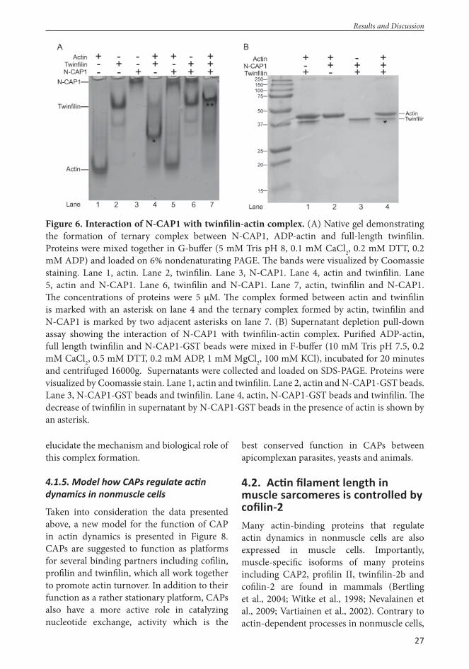
27
elucidate the mechanism and biological role of this complex formation.
4.1.5. Model how CAPs regulate ac n dynamics in nonmuscle cells
Taken into consideration the data presented above, a new model for the function of CAP in actin dynamics is presented in Figure 8. CAPs are suggested to function as platforms for several binding partners including cofi lin, profi lin and twinfi lin, which all work together to promote actin turnover. In addition to their function as a rather stationary platform, CAPs also have a more active role in catalyzing nucleotide exchange, activity which is the
best conserved function in CAPs between apicomplexan parasites, yeasts and animals.
4.2. Ac n fi lament length in muscle sarcomeres is controlled by cofi lin-2Many actin-binding proteins that regulate actin dynamics in nonmuscle cells are also expressed in muscle cells. Importantly, muscle-specifi c isoforms of many proteins including CAP2, profi lin II, twinfi lin-2b and cofi lin-2 are found in mammals (Bertling et al., 2004; Witke et al., 1998; Nevalainen et al., 2009; Vartiainen et al., 2002). Contrary to actin-dependent processes in nonmuscle cells,
Results and Discussion
Figure 6. Interaction of N-CAP1 with twinfi lin-actin complex. (A) Native gel demonstrating the formation of ternary complex between N-CAP1, ADP-actin and full-length twinfi lin. Proteins were mixed together in G-buff er (5 mM Tris pH 8, 0.1 mM CaCl2, 0.2 mM DTT, 0.2 mM ADP) and loaded on 6% nondenaturating PAGE. Th e bands were visualized by Coomassie staining. Lane 1, actin. Lane 2, twinfi lin. Lane 3, N-CAP1. Lane 4, actin and twinfi lin. Lane 5, actin and N-CAP1. Lane 6, twinfi lin and N-CAP1. Lane 7, actin, twinfi lin and N-CAP1. Th e concentrations of proteins were 5 μM. Th e complex formed between actin and twinfi lin is marked with an asterisk on lane 4 and the ternary complex formed by actin, twinfi lin and N-CAP1 is marked by two adjacent asterisks on lane 7. (B) Supernatant depletion pull-down assay showing the interaction of N-CAP1 with twinfi lin-actin complex. Purifi ed ADP-actin, full length twinfi lin and N-CAP1-GST beads were mixed in F-buff er (10 mM Tris pH 7.5, 0.2 mM CaCl2, 0.5 mM DTT, 0.2 mM ADP, 1 mM MgCl2, 100 mM KCl), incubated for 20 minutes and centrifuged 16000g. Supernatants were collected and loaded on SDS-PAGE. Proteins were visualized by Coomassie stain. Lane 1, actin and twinfi lin. Lane 2, actin and N-CAP1-GST beads. Lane 3, N-CAP1-GST beads and twinfi lin. Lane 4, actin, N-CAP1-GST beads and twinfi lin. Th e decrease of twinfi lin in supernatant by N-CAP1-GST beads in the presence of actin is shown by an asterisk.

28
Results and Discussion
Figure 7. Th e abilities of N- and C-terminal ADF-H domains of twinfi lin to form complex with N-CAP1 and actin. (A) Native gel assays demonstrating that both N-terminal and C-terminal ADF-H domains are able to form a complex with N-CAP1 and actin. Proteins were mixed together in G-buff er (5 mM Tris pH 8, 0.1 mM CaCl2, 0.2 mM DTT, 0.2 mM ADP) and loaded on 6% nondenaturating PAGE. Th e bands were visualized by Coomassie staining. Th e concentrations of proteins were 5 μM. Left gel shows the result of the assay carried out with N-terminal ADF-H domain of twinfi lin, N-CAP1 and ADP-actin and the gel on the right hand side represents data from C-terminal ADF-H domain of twinfi lin, N-CAP1 and ADP-actin. In left gel, the complex formed by actin, N-twinfi lin and N-CAP1 is indicated by an asterisk on lane 7. In gel on the right side, the faint band in lane 4 demonstrates the complex formed by actin and C-twinfi lin and is marked by an asterisk, whereas the complex formed by actin, C-twinfi lin and N-CAP1 is marked with two adjacent asterisks on lane 7. (B,C) Supernatant depletion pull-down assay showing the interaction of N-CAP1 with ADP-actin and N-twinfi lin (B) or C-twinfi lin (C). Purifi ed ADP-actin, twinfi lin and N-CAP1-GST beads were mixed in F-buff er (10 mM Tris pH 7.5, 0.2 mM CaCl2, 0.5 mM DTT, 0.2 mM ADP, 1 mM MgCl2, 100 mM KCl), incubated for 20 minutes and centrifuged 16000g. Supernatants were collected and loaded on SDS-PAGE. Proteins were visualized by Coomassie stain. Lane 1, actin and twinfi lin. Lane 2, actin and N-CAP1-GST beads. Lane 3, N-CAP1-GST beads and twinfi lin. Lane 4, actin, N-CAP1-GST beads and twinfi lin. Th e decrease of twinfi lin in supernatant by N-CAP1-GST beads in the presence of actin is shown by an asterisk. (D) Quantifi cation of the amounts of full length, N- and C-twinfi lin in the supernatant in pull-down assays shown in Figures 6B (full length), 7B (N-twinfi lin) and 7C (C-twinfi lin). Error bars represent SEM of three independent experiments.

29
Results and Discussion
Figure 8. A model for CAP function in actin dynamics. (A) Apicomplexan ‘mini-CAPs’, consisting only of the β-sheet domain, catalyze nucleotide exchange from ADP to ATP on actin monomer generating ATP-actin monomers for polymerization. (B) Mammalian CAPs are multifunctional proteins promoting actin turnover in concert with cofi lin and profi lin. CAP binds cofi lin-ADP-actin complexes through the N-terminal domain or, as suggested for yeast CAP, the N-terminal domain might accelerate cofi lin-mediated actin fi lament severing. ADP-actin is transferred from N-terminus to C-terminus, releasing cofi lin for new rounds of depolymerization/severing. C-terminal domain of CAP catalyzes the exchange of nucleotide in actin monomer and newly “recharged” actin is transferred to the central WH2 domain, which binds ATP-actin with high affi nity. Alternatively, ATP-actin can be handed to profi lin, which binds to fi rst polyproline region of CAP. Th us ATP-actin monomer can be transported to the fi lament barbed end by either profi lin or CAP. (C) Mammalian CAP interacts also with twinfi lin-ADP-actin complexes through N-terminal domain. Twinfi lin binds ADP-actin monomers and transfers them to CAP forming a ternary complex similarly as cofi lin. Both ADF-H domains of twinfi lin are capable to form the complex independently. Followed by complex formation, C-terminus of CAP most likely binds the ADP-actin monomer thus releasing twinfi lin from N-CAP, similarly as cofi lin, and the subsequent events are most likely to occur similarly as demonstrated in panel B. Th e fi gure is modifi ed version from publication II.
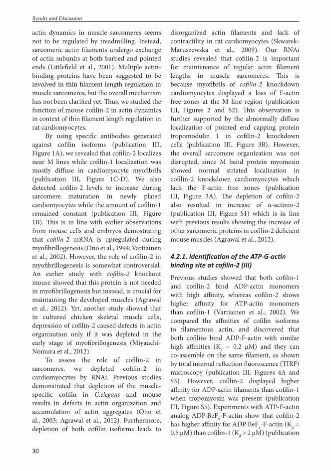
30
actin dynamics in muscle sarcomeres seems not to be regulated by treadmilling. Instead, sarcomeric actin fi laments undergo exchange of actin subunits at both barbed and pointed ends (Littlefi eld et al., 2001). Multiple actin-binding proteins have been suggested to be involved in thin fi lament length regulation in muscle sarcomeres, but the overall mechanism has not been clarifi ed yet. Th us, we studied the function of mouse cofi lin-2 in actin dynamics in context of thin fi lament length regulation in rat cardiomyocytes.
By using specifi c antibodies generated against cofi lin isoforms (publication III, Figure 1A), we revealed that cofi lin-2 localizes near M lines while cofi lin-1 localization was mostly diff use in cardiomyocyte myofi brils (publication III, Figure 1C-D). We also detected cofi lin-2 levels to increase during sarcomere maturation in newly plated cardiomyocytes while the amount of cofi lin-1 remained constant (publication III, Figure 1B). Th is is in line with earlier observations from mouse cells and embryos demostrating that cofi lin-2 mRNA is upregulated during myofi brillogenesis (Ono et al., 1994; Vartiainen et al., 2002). However, the role of cofi lin-2 in myofi brillogenesis is somewhat controversial. An earlier study with cofi lin-2 knockout mouse showed that this protein is not needed in myofi brillogenesis but instead, is crucial for maintaining the developed muscles (Agrawal et al., 2012). Yet, another study showed that in cultured chicken skeletal muscle cells, depression of cofi lin-2 caused defects in actin organization only if it was depleted in the early stage of myofi brillogenesis (Miyauchi-Nomura et al., 2012).
To assess the role of cofi lin-2 in sarcomeres, we depleted cofi lin-2 in cardiomyocytes by RNAi. Previous studies demonstrated that depletion of the muscle-specifi c cofi lin in C.elegans and mouse results in defects in actin organization and accumulation of actin aggregates (Ono et al., 2003; Agrawal et al., 2012). Furthermore, depletion of both cofi lin isoforms leads to
disorganized actin fi laments and lack of contractility in rat cardiomyocytes (Skwarek-Maruszewska et al., 2009). Our RNAi studies revealed that cofi lin-2 is important for maintenance of regular actin fi lament lengths in muscle sarcomeres. Th is is because myofi brils of cofi lin-2 knockdown cardiomyocytes displayed a loss of F-actin free zones at the M line region (publication III, Figures 2 and S2). Th is observation is further supported by the abnormally diff use localization of pointed end capping protein tropomodulin 1 in cofi lin-2 knockdown cells (publication III, Figure 3B). However, the overall sarcomere organization was not disrupted, since M band protein myomesin showed normal striated localization in cofi lin-2 knockdown cardiomyocytes which lack the F-actin free zones (publication III, Figure 3A). Th e depletion of cofi lin-2 also resulted in increase of α-actinin-2 (publication III, Figure S1) which is in line with previous results showing the increase of other sarcomeric proteins in cofi lin-2 defi cient mouse muscles (Agrawal et al., 2012).
4.2.1. Iden fi ca on of the ATP-G-ac n binding site at cofi lin-2 (III)
Previous studies showed that both cofi lin-1 and cofi lin-2 bind ADP-actin monomers with high affi nity, whereas cofi lin-2 shows higher affi nity for ATP-actin monomers than cofi lin-1 (Vartiainen et al., 2002). We compared the affi nities of cofi lin isoforms to fi lamentous actin, and discovered that both cofi lins bind ADP-F-actin with similar high affi nities (Kd ~ 0.2 μM) and they can co-assemble on the same fi lament, as shown by total internal refl ection fl uorescence (TIRF) microscopy (publication III, Figures 4A and S3). However, cofi lin-2 displayed higher affi nity for ADP-actin fi laments than cofi lin-1 when tropomyosin was present (publication III, Figure S5). Experiments with ATP-F-actin analog ADP·BeFx-F-actin show that cofi lin-2 has higher affi nity for ADP·BeFx-F-actin (Kd = 0.5 μM) than cofi lin-1 (Kd > 2 μM) (publication
Results and Discussion

31
Results and Discussion
III, Figure 4B). Furthermore, cofi lin-2 more effi ciently disassembles ADP·BeFx-F-actin than cofi lin-1, suggesting that cofi lin-2 is more effi cient in interacting with ATP-actin fi laments compared to cofi lin-1 (publication III, Figures 4C-D and S4A).
Next, we wanted to identify the residues responsible for diff erent actin binding properties of cofi lin-1 and cofi lin-2. We found that replacement of three conserved residues on the actin binding surface of cofi lin-1 by corresponding cofi lin-2 residues (E141D, E142D, V143I) increased the cofi lin-1 disassembly activity for ADP·BeFx-F-actin (publication III, Figures 5 and S4B). Next we tested whether this “cofi lin-1 to cofi lin-2” mutation would aff ect the ATP-actin monomer binding ability. Importantly, our experiments revealed that these mutations signifi cantly enhanced the ATP-actin monomer binding affi nity of cofi lin-1 (from Kd ~ 230 nM to Kd ~ 20 nM; publication III, Figure 6A-B). Th is increase in ATP-G-actin binding was even further enhanced by replacing one additional cofi lin-1 residue by a cofi lin-2 specifi c residue (A137V). We also tested these replacements vice versa, replacing residues 141-143 on cofi lin-2 by cofi lin-1-specifi c residues, and observed diminished binding affi nity for ATP-G-actin (from Kd ~ 30 nM to Kd ~ 110 nM; publication III, Figure 6C-D). Th us, a specifi c cluster of residues is important for the high affi nity of cofi lin-2 for ATP-actin.
Th ese results, together with observations of cofi lin-2 localization to near M line and phenotype resulting from depletion of cofi lin-2, suggest cofi lin-2 to be well suited for promoting disassembly of actin fi laments from pointed ends in muscle sarcomeres. Due to both actin fi lament disassembly and assembly
taking place at the M line region, actin fi lament ends at these sites are expected to contain both ATP- and ADP-subunits. Th us, cofi lin-2, with its specifi c ATP-actin binding activity is well suited to drive actin dynamics at these sites. It still remains examined, how cofi lin-2 localizes to the M line region in myofi brils. One candidate for localizing cofi lin-2 is CAP, which has a nonmuscle isoform CAP1 and muscle specifi c isoform CAP2 (Bertling et al., 2004). Since CAP1 is known to interact with cofi lin-1 in nonmuscle cells (Moriyama & Yahara, 2002) and CAP2 has been found to localize to M lines in cardiomyocyte myofi brils (Peche et al., 2007), it is possible that CAP2 interacts with cofi lin-2 in muscle sarcomeres thus localizing it near M lines.
4.2.2. Cofi lin-2 does not show ac n isoform specifi city in ATP-G-ac n binding (III)
Since cofi lin-2 has higher affi nity for ATP-actin than cofi lin-1, and the actin we used in our assays was α-actin from skeletal muscle, we wanted to assess whether this specifi c actin isoform caused diff erent binding preferences of cofi lins. We tested the binding of β/γ-ATP-G-actin versus α-ATP-G-actin and noted that cofi lin-2 binds both actin isoforms with higher affi nity than cofi lin-1 (publication III, Figure S6). However, the affi nities of both cofi lins for muscle actin were somewhat higher than for nonmuscle actin. Th ese results show that variations in specifi c cofi lin residues have not evolved to increase the affi nities of cofi lins towards certain actin isoforms, but rather to distinguish between ATP- and ADP-nucleotide bound to actin monomers.

32
Th is study provides new evidence for actin regulation in cells conducted by CAP, cofi lin, profi lin and twinfi lin. In publications I and II we identifi ed the conserved residues on the surface of yeast and mammalian CAPs that are involved in cofi lin binding and also mapped the binding site of CAP on the surface of yeast cofi lin. Th e complex formation between CAP, cofi lin and ADP-actin has been shown earlier by Moriyama and Yahara (2002), but the exact binding sites were not known before this study. Localizing the binding interfaces allows further investigations and speculations about the mechanism and biological signifi cance of the complex formation. In future, structural studies will be needed to reveal the mechanism by which these proteins interact with each other. In addition, rescue experiments in mammalian cells would shed light into the cellular roles of these protein interactions.
Since CAPs are multifunctional proteins with several binding partners, the signifi cance of diff erent activities in diff erent organisms is diffi cult to elucidate. In publication II we show that C-terminal halves of yeast, mouse and malaria parasite P. falciparum CAPs are capable and suffi cient for exchanging nucleotide on actin monomers. Th is characteristic is the fi rst, and so far the only known function for PfCAP. From these results, we hypothesize that promoting nucleotide exchange on actin monomers is the most conserved function of CAPs. Profi lin is traditionally considered to be responsible for exchanging nucleotide on actin monomers. However, this assumption needs to be reconsidered now and we suggest that mainly CAP is responsible for nucleotide exchange to promote actin turnover in cells. We also reveal that profi lin binding by CAP is conserved from yeast to mammals, as shown in publication II. However, in contrast to the yeast protein, mammalian CAP1 has two independent profi lin binding sites. Further studies will be needed to reveal how profi lin and CAP function together and what is the
signifi cance of each protein in actin dynamics and whether they cooperate or compensate each other’s actions.
For our surprise, mammalian CAP unlike yeast CAP, binds also ATP-actin with high affi nity through WH2 domain as shown in publication II. Th is fi nding is unexpected and leads us to speculate whether yeast and mammalian CAP functions diff er from each other. Th is study is the fi rst one to show that mammalian CAP1 binds also ATP-actin with high affi nity and this can be crucial for its function. Furthermore, we show in publication II that unlike yeast CAP, mammalian CAP1 WH2 domain allows actin polymerization when bound to actin monomer. Th ese fi ndings suggest that, before further investigations, yeast and mammalian CAPs should not be assumed to share exactly same functional mechanisms in actin regulation. Although the main function of CAPs in cells is most probably to accelerate actin turnover, CAPs from diff erent species might carry out this action through slightly diff erent ways.
Our unpublished data (Makkonen et al., unpublished) revealed a completely new interaction partner, twinfi lin, which binds CAP together with ADP-actin in complex. Importantly, this is fi rst study to show that these two proteins interact together directly. Th is interaction is notable since the functions of CAP and twinfi lin are not completely understood yet. In our unpublished data we show that N-CAP1 forms ternary complex together with twinfi lin and ADP-G-actin, and that both ADF-H domains of twinfi lin are capable of the complex formation. However, full-length twinfi lin containing both ADF-H domains is more eff ective in forming this complex. Th is interaction is a useful starting point to create new hypothesizes when aiming to understand the roles of CAP and twinfi lin in regulating actin dynamics. Crystallization and structure determination would reveal the binding mechanism and importance of two
Concluding Remarks and Future Perspectives
5. CONCLUDING REMARKS AND FUTURE PERSPECTIVES

33
separate ADF-H domains of twinfi lin in the complex formation.
Another important question is whether the binding site for N-CAP1 on surface of twinfi lin corresponds to conserved N-CAP1 binding site on ADF/cofi lin surface (publication I). To test this, we plan to design a twinfi lin construct bearing mutations that correspond to cofi lin mutants Cof1-5 and Cof1-9 that are defective in yeast CAP binding (publication I) and test whether such mutants are able to form a complex with ADP-G-actin and N-CAP1. Vice versa, it would be interesting to investigate whether the binding site for twinfi lin-actin complex on the surface of N-CAP1 corresponds to the binding site mapped for cofi lin-actin complex (publication I, publication II). Th is could be tested by our mouse mutation construct N-CAP1 mutα (publication II) e.g. by supernatant depletion pull-down assay.
It would be also useful to determine whether cofi lin-actin and twinfi lin-actin compete with each other for binding to N-CAP1. Th e role of twinfi lin-actin binding to CAP1 should also be investigated more closely since the cellular roles of cofi lin and twinfi lin seem to be somewhat overlapping. Th is is supported by observations made with yeast, where deletion of twinfi lin alone did not result strong phenotype but, when combined to cofi lin mutant, resulted in synthetic lethality (Goode et al., 1998). It has been shown earlier that human N-CAP1 accelerates the nucleotide exchange from ADP to ATP on actin monomer in the presence of cofi lin (Moriyama and Yahara, 2002). Th e possible overlapping roles of twinfi lin and cofi lin could be examined similarly by observing the eff ect of N-CAP1 to accelerate the nucleotide exchange in the presence of twinfi lin. Also, it would be interesting to perform an F-actin turnover assay with N-CAP1, cofi lin and twinfi lin and test whether addition of twinfi lin would make a diff erence in the turnover rate accelerated by CAP1 and cofi lin.
We show that cofi lin-2 localizes near M lines and disassembles actin fi laments on muscle sarcomeres thus regulating the fi lament length (publication III). Th ere is very little information about the muscle-specifi c isoforms of actin binding proteins and how they regulate actin dynamics in muscle cells. Cofi lin is a fundamental actin cytoskeleton regulating protein in nonmuscle cells and we show that cofi lin-2 has also a crucial function in muscle cells. Th e regulation of sarcomeric length is essential for proper function of sarcomere in contracting muscle and thus the mechanism and proteins involved in this regulation are necessary to elucidate. Th is study broadens our understanding about the thin fi lament length regulation, but still the overall mechanism remains largerly unknown and additional studies are needed to fully understand how the actin dynamics is regulated in muscle cells. For example, other muscle-specifi c actin binding protein isoforms should be examined more closely.
It is already known that mammalian CAP2, as well as C.elegans muscle-specifi c CAP, localize to M lines (Peche et al., 2007; Nomura et al., 2012). Recently published results from CAP2 knockout mouse showed that in the absence of CAP2, mice have disarrayed sarcomeres in cardiomyocytes and they develop severe heart defects and dilated cardiomyopathy (Peche et al., 2013). Th e interaction of CAP2 and cofi lin-2 in muscle cells would be important to study. First, the expression of CAP1 and CAP2 proteins in muscle cells should be tested and this could be studied by Western blot. It would be interesting to fi nd out if these CAP isoforms are diff erentially expressed during maturation like we showed for the two cofi lin isoforms. If both isoforms are present, it would be informative to determine if they localize similarly to cofi lin isoforms. To fi nd out if muscle-specifi c isoforms cofi lin-2 and CAP2 interact with each other and whether they prefer the muscle-specifi c isoform over nonmuscle isoform, one could apply similar
Concluding Remarks and Future Perspectives

34
pull-down experiments as used for cofi lin-1 and CAP1 in publication II of this study. Th ese experiments would perhaps give us more information about cofi lin-2 localization and function near M lines.
Th e roles of twinfi lin-2b and profi lin II in muscle cells would be also interesting to reveal. Since yeast and mammalian CAPs are known to interact with profi lin (Bertling et al., 2007; publication II) and we show in our
unpublished data that mammalian CAP1 interacts with twinfi lin (Makkonen et al., unpublished) it would be enlightening to examine the potential interactions of muscle-specifi c profi lin II and twinfi lin-2b with CAP2, as described above for cofi lin-2 and CAP2 interaction. Further studies concerning these interactions could give us valuable information and further widen our knowledge about actin fi lament regulation in muscle cells.
Concluding Remarks and Future Perspectives

35
Acknowledgements
6. ACKNOWLEDGEMENTS
Th is study was carried out between years 2007 and 2013 at the Institute of Biotechnology (BI), University of Helsinki, with fi nancial support from the Helsinki Graduate Program in Biotechnology and Molecular Biology (GPBM).
I warmly thank my supervisor professor Pekka Lappalainen for taking me into his active lab team and supervising my thesis work. You have created a great team spirit in the Lappalainen group and I feel privileged for getting to work in such relaxed but inspiring atmosphere. I am happy for your enthusiasm and encouragement towards my project (which I myself wanted to give up many times) and scientifi c discussions in the corridor. I am also grateful for your always positive attitude. For my surprise, several times my crappy results have turned into good ones aft er showing them to Pekka.
I want to express my gratitude for my thesis pre-examiners professor Olli Carpén and assistant professor Sanna Lehtonen for going through my thesis in a tight schedule and giving me excellent comments. I also wish to thank thank my thesis committee members docents Maria Vartiainen and Pirta Hotulainen for their contribution and advice concerning my thesis project.
Professor Kari Keinänen and other personnel (especially student advisor Pirjo Nikula-Ijäs) of the division of Biochemistry and Biotechnology in the Department of Biosciences are acknowledged for the help provided in diff erent stages of my thesis process. I have been lucky to belong to the graduate program GPBM, which I would like to thank for providing funding and other support for my thesis work. I would like to thank the personnel including director Hannes Lohi, scientifi c coordinator Erkki Raulo and communications offi cer Anita Tienhaara for organizing useful courses and valuable help in all practical things.
I wish to thank the people in the Institute of Biotechnology for providing me such a great facilities to carry on my thesis work. I express my gratitude to the former and current directors of the institute, professors Mart Saarma and Tomi Mäkelä, respectively. Also administrative unit, IT support (Iikka) and equipment care people (Pauli, Petri, Repe) are acknowledged for providing solutions in everyday practicalities. I would like to thank the several core facilities at BI (DNA Sequencing and Genomics Laboratory, Proteomics Unit, Light Microscopy Unit, Protein Crystallisation Facility and Media Kitchen) for fruitful collaboration. Marc Baumann’s Proteomics Unit in Meilahti is acknowledged for identifying my diffi cult protein. Tinde Päivärinta is acknowledged for an excellent thesis layout.
I am grateful for all my collaborators and co-authors for their contributions for the papers included in my thesis. Especially I want to thank Enni Bertling for teaching me all the lab skills and techniques from cloning to imaging and being my personal mentor, Elena Kremneva and Aneta Skwarek-Maruszewska for our fruitful collaboration in cardiomyocyte project and Dr Jake Baum for bringing the fascinating malaria parasite CAP in my life and thesis project. Markku is thanked for taking care of CAP and twinfi lin in future.
I am deeply grateful for all former and present people working in the Lappalainen lab. I want to thank the “old” lab setup including Enni, Pirta, Pieta, Ansku, Ville, Elisa, Aneta, Miia, Juha and Perttu for taking me into your scientifi c and sporty team! My special thanks goes to Enni who

36
Acknowledgements
patiently supervised me and shared her knowledge about science, dogs and life in general. My warmest thanks goes to the “newer” setup including all people who have came to our group aft er me (and some have already left ): Hongxia, Sari, Geri, Martina, Minna, Markku, Elena, Yaming, Yosuke, Nalle, Essi, Nitai, Kirsi, Juho and Anna-Liisa. Honestly you have been the best colleagues ever. I have no words to tell you how much it means to have you guys around me to share my disappointing results, occational successes, coff ee breaks and all the discussions about work and other life.
I also want to thank the former and present people in the Vartiainen and Jäntti labs for creating such a pleasant athmosphere to work in the 5th fl oor. No matter is it weekday or weekend, day or night, always there is someone working in our fl oor which makes me feel so cozy. Especially I would like to thank my dear friends Johanna and Kaisa, who shared not only coff ee breaks but also dog activities with me. I am unspeakably thankful to get friends like you, which for sure will remain even though I leave from Viikki.
Haluaisin kiittää kaikkia ystäviäni arvokkaasta tuesta ja siitä, että edes yritätte esittää kiinnostusta väitöskirjatyötäni kohtaan. Kiitos teille lukuisista mökkireissuista joista palaan aina väsyneenä mutta onnellisena. Kiitän myös omaa tee-se-itse-matkatoimistoamme (erityisesti Pekka I ja Riikka) erinomaisesta matkustusseurasta, teidän kanssanne voin (lähes) turvallisin mielin lähteä maailman ääriin keräämään uusia elämyksiä. Erityiskiitos Riikalle että lenkkeilytät minut viikoittain ja samalla toimit terapeuttinani. Hannu, kiitos että säännöllisesti rentoutat mieleni samppanjalla .
Haluaisin lämpimästi kiittää kaikkia perheenjäseniäni heidän tuestaan näiden vuosien aikana. Haluan kiittää sekä äitiä ja Pekkaa että siskojani Suvia, Iida-Mariaa, Marjaanaa ja Rosannaa siitä että olette olemassa ja muodostatte minulle perheen ja kodin jonne tulla käymään. Kiitän erityisesti äitiäni, joka löytyy aina puhelinsoiton päästä, oli kyse sitten pienestä tai suuresta murheesta. Kiitos että olet antanut meidän lasten kulkea omia polkujamme pakottamatta mihinkään tiettyyn suuntaan, mutta kuitenkin tukien taustalla koko ajan. Se on tärkeää jotta jaksaa kulkea tien loppuun asti, kiitos. Kiitän myös isääni perheineen kiinnostuksesta väitöskirjaani kohtaan ja kannustuksesta opinnoissa eteenpäin sekä heidän tarjoamiaan aterioita jotka ovat aina erinomaisia! Haluan myös lämpimästi kiittää appivanhempiani Arjaa ja Hannua, joiden vilpittömästä avusta ja vieraanvaraisuudesta olen lukemattomia kertoja saanut nauttia. Kiitos myös tieteellisistä kysymyksistänne ja jatkuvasta mielenkiinnosta työtäni kohtaan, saatte ajoittain minut tuntemaan tekeväni jotain tärkeää.
Lopuksi haluaisin kiittää rakasta aviomiestäni Anttia. Pari viime vuotta eivät ole olleet meille aivan helppoja ja oletkin saanut kannettavaksesi enemmän harmeja kuin olisin toivonut. Lämmin halaus kotiintullessa muistuttaa aina minua siitä mikä on oikeasti tärkeää. Saatuani tämän urakan päätökseen lupaan antaa vapautuvan aikani lyhentämättömänä sinulle ja koirallemme Nukalle. Olen pohjattoman onnellinen ja kiitollinen siitä, että jopa kaikkein synkimpinä epätoivon hetkinä minun ei ole koskaan tarvinnut epäillä seisotko rinnallani. Kiitos.
Helsinki, October 2013

37
References
7. REFERENCESAdams, A. E., and Pringle, J. R. (1984) Relationship of actin and tubulin distribution to bud growth in wild-type and morphogenetic-mutant Saccharomyces cerevisiae. Th e Journal of cell biology 98, 934-945
Aghamohammadzadeh, S., and Ayscough, K. R. (2009) Diff erential requirements for actin during yeast and mammalian endocytosis. Nature cell biology 11, 1039-1042
Agnew, B. J., Minamide, L. S., and Bamburg, J. R. (1995) Reactivation of phosphorylated actin depolymerizing factor and identifi cation of the regulatory site. Th e Journal of biological chemistry 270, 17582-17587
Agrawal, P. B., Joshi, M., Savic, T., Chen, Z., and Beggs, A. H. (2012) Normal myofi brillar development followed by progressive sarcomeric disruption with actin accumulations in a mouse Cfl 2 knockout demonstrates requirement of cofi lin-2 for muscle maintenance. Human molecular genetics 21, 2341-2356
Ahuja, R., Pinyol, R., Reichenbach, N., Custer, L., Klingensmith, J., Kessels, M. M., and Qualmann, B. (2007) Cordon-bleu is an actin nucleation factor and controls neuronal morphology. Cell 131, 337-350
Andrianantoandro, E., and Pollard, T. D. (2006) Mechanism of actin fi lament turnover by severing and nucleation at diff erent concentrations of ADF/cofi lin. Molecular cell 24, 13-23
Archer, S. K., Claudianos, C., and Campbell, H. D. (2005) Evolution of the gelsolin family of actin-binding proteins as novel transcriptional coactivators. BioEssays : news and reviews in molecular, cellular and developmental biology 27, 388-396
Balasubramanian, M. K., Bi, E., and Glotzer, M. (2004) Comparative analysis of cytokinesis in budding yeast, fi ssion yeast and animal cells. Current biology : CB 14, R806-818
Balcer, H. I., Goodman, A. L., Rodal, A. A., Smith, E., Kugler, J., Heuser, J. E., and Goode, B. L. (2003) Coordinated regulation of actin fi lament turnover by a high-molecular-weight Srv2/CAP complex, cofi lin, profi lin, and Aip1. Current biology : CB 13, 2159-2169
Bamburg, J. R. (1999) Proteins of the ADF/cofi lin family: essential regulators of actin dynamics. Annual review of cell and developmental biology 15, 185-230
Bamburg, J. R., Harris, H. E., and Weeds, A. G. (1980) Partial purifi cation and characterization of an actin depolymerizing factor from brain. FEBS letters 121, 178-182
Barrero, R. A., Umeda, M., Yamamura, S., and Uchimiya, H. (2002) Arabidopsis CAP regulates the actin cytoskeleton necessary for plant cell elongation and division. Th e Plant cell 14, 149-163
Baum, B., Li, W., and Perrimon, N. (2000) A cyclase-associated protein regulates actin and cell polarity during Drosophila oogenesis and in yeast. Current biology : CB 10, 964-973
Baum, J., Papenfuss, A. T., Baum, B., Speed, T. P., and Cowman, A. F. (2006) Regulation of apicomplexan actin-based motility. Nature reviews. Microbiology 4, 621-628
Baum, J., Tonkin, C. J., Paul, A. S., Rug, M., Smith, B. J., Gould, S. B., Richard, D., Pollard, T. D., and Cowman, A. F. (2008) A malaria parasite formin regulates actin polymerization and localizes to the parasite-erythrocyte moving junction during invasion. Cell host & microbe 3, 188-198
Beeler, J. F., LaRochelle, W. J., Chedid, M., Tronick, S. R., and Aaronson, S. A. (1994) Prokaryotic expression cloning of a novel human tyrosine kinase. Molecular and cellular biology 14, 982-988
Benlali, A., Draskovic, I., Hazelett, D. J., and Treisman, J. E. (2000) act up controls actin polymerization to alter cell shape and restrict Hedgehog signaling in the Drosophila eye disc. Cell 101, 271-281
Bertling, E., Hotulainen, P., Mattila, P. K., Matilainen, T., Salminen, M., and Lappalainen, P. (2004) Cyclase-associated protein 1 (CAP1) promotes cofi lin-induced actin dynamics in mammalian nonmuscle cells. Molecular biology of the cell 15, 2324-2334
Bertling, E., Quintero-Monzon, O., Mattila, P. K., Goode, B. L., and Lappalainen, P. (2007) Mechanism and biological role of profi lin-Srv2/CAP interaction. Journal of cell science 120, 1225-1234
Bi, E., Maddox, P., Lew, D. J., Salmon, E. D., McMillan, J. N., Yeh, E., and Pringle, J. R. (1998) Involvement of an actomyosin contractile ring in Saccharomyces cerevisiae cytokinesis. Th e Journal of cell biology 142, 1301-1312

38
Birbach, A. (2008) Profi lin, a multi-modal regulator of neuronal plasticity. BioEssays : news and reviews in molecular, cellular and developmental biology 30, 994-1002
Blanchoin, L., and Pollard, T. D. (1999) Mechanism of interaction of Acanthamoeba actophorin (ADF/Cofi lin) with actin fi laments. Th e Journal of biological chemistry 274, 15538-15546
Blanchoin, L., Pollard, T. D., and Mullins, R. D. (2000) Interactions of ADF/cofi lin, Arp2/3 complex, capping protein and profi lin in remodeling of branched actin fi lament networks. Current biology : CB 10, 1273-1282
Bretscher, A. (2003) Polarized growth and organelle segregation in yeast: the tracks, motors, and receptors. Th e Journal of cell biology 160, 811-816
Broderick, M. J., and Winder, S. J. (2002) Towards a complete atomic structure of spectrin family proteins. Journal of structural biology 137, 184-193
Broschat, K. O. (1990) Tropomyosin prevents depolymerization of actin fi laments from the pointed end. Th e Journal of biological chemistry 265, 21323-21329
Burtnick, L. D., Koepf, E. K., Grimes, J., Jones, E. Y., Stuart, D. I., McLaughlin, P. J., and Robinson, R. C. (1997) Th e crystal structure of plasma gelsolin: implications for actin severing, capping, and nucleation. Cell 90, 661-670
Buss, F., Temm-Grove, C., Henning, S., and Jockusch, B. M. (1992) Distribution of profi lin in fi broblasts correlates with the presence of highly dynamic actin fi laments. Cell motility and the cytoskeleton 22, 51-61
Cai, L., Marshall, T. W., Uetrecht, A. C., Schafer, D. A., and Bear, J. E. (2007) Coronin 1B coordinates Arp2/3 complex and cofi lin activities at the leading edge. Cell 128, 915-929
Campellone, K. G., and Welch, M. D. (2010) A nucleator arms race: cellular control of actin assembly. Nature reviews. Molecular cell biology 11, 237-251
Carlier, M. F., Laurent, V., Santolini, J., Melki, R., Didry, D., Xia, G. X., Hong, Y., Chua, N. H., and Pantaloni, D. (1997) Actin depolymerizing factor (ADF/cofi lin) enhances the rate of fi lament turnover: implication in actin-based motility. Th e Journal of cell biology 136, 1307-1322
Carlsson, L., Nystrom, L. E., Sundkvist, I., Markey, F., and Lindberg, U. (1977) Actin polymerizability is infl uenced by profi lin, a low molecular weight protein in non-muscle cells. Journal of molecular biology 115, 465-483
Casella, J. F., Craig, S. W., Maack, D. J., and Brown, A. E. (1987) Cap Z(36/32), a barbed end actin-capping protein, is a component of the Z-line of skeletal muscle. Th e Journal of cell biology 105, 371-379
Castrillon, D. H., and Wasserman, S. A. (1994) Diaphanous is required for cytokinesis in Drosophila and shares domains of similarity with the products of the limb deformity gene. Development (Cambridge, England) 120, 3367-3377
Chaparro-Olaya, J., Margos, G., Coles, D. J., Dluzewski, A. R., Mitchell, G. H., Wasserman, M. M., and Pinder, J. C. (2005) Plasmodium falciparum myosins: transcription and translation during asexual parasite development. Cell motility and the cytoskeleton 60, 200-213
Chaudhry, F., Breitsprecher, D., Little, K., Sharov, G., Sokolova, O., and Goode, B. L. (2013) Srv2/cyclase-associated protein forms hexameric shurikens that directly catalyze actin fi lament severing by cofi lin. Molecular biology of the cell 24, 31-41
Chaudhry, F., Guerin, C., von Witsch, M., Blanchoin, L., and Staiger, C. J. (2007) Identifi cation of Arabidopsis cyclase-associated protein 1 as the fi rst nucleotide exchange factor for plant actin. Molecular biology of the cell 18, 3002-3014
Chaudhry, F., Little, K., Talarico, L., Quintero-Monzon, O., and Goode, B. L. (2010) A central role for the WH2 domain of Srv2/CAP in recharging actin monomers to drive actin turnover in vitro and in vivo. Cytoskeleton (Hoboken, N.J.) 67, 120-133
Chereau, D., Boczkowska, M., Skwarek-Maruszewska, A., Fujiwara, I., Hayes, D. B., Rebowski, G., Lappalainen, P., Pollard, T. D., and Dominguez, R. (2008) Leiomodin is an actin fi lament nucleator in muscle cells. Science (New York, N.Y.) 320, 239-243
Choe, H., Burtnick, L. D., Mejillano, M., Yin, H. L., Robinson, R. C., and Choe, S. (2002) Th e calcium activation of gelsolin: insights from the 3A structure of the G4-G6/actin complex. Journal of molecular biology 324, 691-702
References

39
Clark, K. A., McElhinny, A. S., Beckerle, M. C., and Gregorio, C. C. (2002) Striated muscle cytoarchitecture: an intricate web of form and function. Annual review of cell and developmental biology 18, 637-706
Cowman, A. F., and Crabb, B. S. (2006) Invasion of red blood cells by malaria parasites. Cell 124, 755-766
Craig, R., and Lehman, W. (2001) Crossbridge and tropomyosin positions observed in native, interacting thick and thin fi laments. Journal of molecular biology 311, 1027-1036
Di Nardo, A., Gareus, R., Kwiatkowski, D., and Witke, W. (2000) Alternative splicing of the mouse profi lin II gene generates functionally diff erent profi lin isoforms. Journal of cell science 113 Pt 21, 3795-3803
Dobrowolski, J. M., Niesman, I. R., and Sibley, L. D. (1997) Actin in the parasite Toxoplasma gondii is encoded by a single copy gene, ACT1 and exists primarily in a globular form. Cell motility and the cytoskeleton 37, 253-262
Dodatko, T., Fedorov, A. A., Grynberg, M., Patskovsky, Y., Rozwarski, D. A., Jaroszewski, L., Aronoff -Spencer, E., Kondraskina, E., Irving, T., Godzik, A., and Almo, S. C. (2004) Crystal structure of the actin binding domain of the cyclase-associated protein. Biochemistry 43, 10628-10641
Doherty, G. J., and McMahon, H. T. (2009) Mechanisms of endocytosis. Annual review of biochemistry 78, 857-902
Dopie, J., Skarp, K. P., Rajakyla, E. K., Tanhuanpaa, K., and Vartiainen, M. K. (2012) Active maintenance of nuclear actin by importin 9 supports transcription. Proceedings of the National Academy of Sciences of the United States of America 109, E544-552
dos Remedios, C. G., Chhabra, D., Kekic, M., Dedova, I. V., Tsubakihara, M., Berry, D. A., and Nosworthy, N. J. (2003) Actin binding proteins: regulation of cytoskeletal microfi laments. Physiological reviews 83, 433-473
Eads, J. C., Mahoney, N. M., Vorobiev, S., Bresnick, A. R., Wen, K. K., Rubenstein, P. A., Haarer, B. K., and Almo, S. C. (1998) Structure determination and characterization of Saccharomyces cerevisiae profi lin. Biochemistry 37, 11171-11181
Falck, S., Paavilainen, V. O., Wear, M. A., Grossmann, J. G., Cooper, J. A., and Lappalainen, P. (2004) Biological role and structural mechanism of twinfi lin-capping protein interaction. Th e EMBO journal 23, 3010-3019
Farrants, A. K. (2008) Chromatin remodelling and actin organisation. FEBS letters 582, 2041-2050
Faulkner, G., Lanfranchi, G., and Valle, G. (2001) Telethonin and other new proteins of the Z-disc of skeletal muscle. IUBMB life 51, 275-282
Fedor-Chaiken, M., Deschenes, R. J., and Broach, J. R. (1990) SRV2, a gene required for RAS activation of adenylate cyclase in yeast. Cell 61, 329-340
Ferguson, S. M., Raimondi, A., Paradise, S., Shen, H., Mesaki, K., Ferguson, A., Destaing, O., Ko, G., Takasaki, J., Cremona, O., E, O. T., and De Camilli, P. (2009) Coordinated actions of actin and BAR proteins upstream of dynamin at endocytic clathrin-coated pits. Developmental cell 17, 811-822
Field, J., Nikawa, J., Broek, D., MacDonald, B., Rodgers, L., Wilson, I. A., Lerner, R. A., and Wigler, M. (1988) Purifi cation of a RAS-responsive adenylyl cyclase complex from Saccharomyces cerevisiae by use of an epitope addition method. Molecular and cellular biology 8, 2159-2165
Field, J., Vojtek, A., Ballester, R., Bolger, G., Colicelli, J., Ferguson, K., Gerst, J., Kataoka, T., Michaeli, T., Powers, S., and et al. (1990) Cloning and characterization of CAP, the S. cerevisiae gene encoding the 70 kd adenylyl cyclase-associated protein. Cell 61, 319-327
Freeman, N. L., Chen, Z., Horenstein, J., Weber, A., and Field, J. (1995) An actin monomer binding activity localizes to the carboxyl-terminal half of the Saccharomyces cerevisiae cyclase-associated protein. Th e Journal of biological chemistry 270, 5680-5685
Freeman, N. L., Lila, T., Mintzer, K. A., Chen, Z., Pahk, A. J., Ren, R., Drubin, D. G., and Field, J. (1996) A conserved proline-rich region of the Saccharomyces cerevisiae cyclase-associated protein binds SH3 domains and modulates cytoskeletal localization. Molecular and cellular biology 16, 548-556
Frenal, K., and Soldati-Favre, D. (2009) Role of the parasite and host cytoskeleton in apicomplexa parasitism. Cell host & microbe 5, 602-611
References

40
Fujimoto, L. M., Roth, R., Heuser, J. E., and Schmid, S. L. (2000) Actin assembly plays a variable, but not obligatory role in receptor-mediated endocytosis in mammalian cells. Traffi c (Copenhagen, Denmark) 1, 161-171
Fujiwara, K., Porter, M. E., and Pollard, T. D. (1978) Alpha-actinin localization in the cleavage furrow during cytokinesis. Th e Journal of cell biology 79, 268-275
Galkin, V. E., Orlova, A., Lukoyanova, N., Wriggers, W., and Egelman, E. H. (2001) Actin depolymerizing factor stabilizes an existing state of F-actin and can change the tilt of F-actin subunits. Th e Journal of cell biology 153, 75-86
Gandhi, M., Achard, V., Blanchoin, L., and Goode, B. L. (2009) Coronin switches roles in actin disassembly depending on the nucleotide state of actin. Molecular cell 34, 364-374
Gandhi, M., Smith, B. A., Bovellan, M., Paavilainen, V., Daugherty-Clarke, K., Gelles, J., Lappalainen, P., and Goode, B. L. (2010) GMF is a cofi lin homolog that binds Arp2/3 complex to stimulate fi lament debranching and inhibit actin nucleation. Current biology : CB 20, 861-867
Gerst, J. E., Ferguson, K., Vojtek, A., Wigler, M., and Field, J. (1991) CAP is a bifunctional component of the Saccharomyces cerevisiae adenylyl cyclase complex. Molecular and cellular biology 11, 1248-1257
Glotzer, M. (2004) Cleavage furrow positioning. Th e Journal of cell biology 164, 347-351
Gokhin, D. S., and Fowler, V. M. (2013) A two-segment model for thin fi lament architecture in skeletal muscle. Nature reviews. Molecular cell biology 14, 113-119
Goode, B. L., Drubin, D. G., and Lappalainen, P. (1998) Regulation of the cortical actin cytoskeleton in budding yeast by twinfi lin, a ubiquitous actin monomer-sequestering protein. Th e Journal of cell biology 142, 723-733
Gorbatyuk, V. Y., Nosworthy, N. J., Robson, S. A., Bains, N. P., Maciejewski, M. W., Dos Remedios, C. G., and King, G. F. (2006) Mapping the phosphoinositide-binding site on chick cofi lin explains how PIP2 regulates the cofi lin-actin interaction. Molecular cell 24, 511-522
Gottwald, U., Brokamp, R., Karakesisoglou, I., Schleicher, M., and Noegel, A. A. (1996) Identifi cation of a cyclase-associated protein (CAP) homologue in Dictyostelium discoideum and characterization of its interaction with actin. Molecular biology of the cell 7, 261-272
Gunsalus, K. C., Bonaccorsi, S., Williams, E., Verni, F., Gatti, M., and Goldberg, M. L. (1995) Mutations in twinstar, a Drosophila gene encoding a cofi lin/ADF homologue, result in defects in centrosome migration and cytokinesis. Th e Journal of cell biology 131, 1243-1259
Haarer, B. K., Lillie, S. H., Adams, A. E., Magdolen, V., Bandlow, W., and Brown, S. S. (1990) Purifi cation of profi lin from Saccharomyces cerevisiae and analysis of profi lin-defi cient cells. Th e Journal of cell biology 110, 105-114
Haugwitz, M., Noegel, A. A., Karakesisoglou, J., and Schleicher, M. (1994) Dictyostelium amoebae that lack G-actin-sequestering profi lins show defects in F-actin content, cytokinesis, and development. Cell 79, 303-314
Hayden, S. M., Miller, P. S., Brauweiler, A., and Bamburg, J. R. (1993) Analysis of the interactions of actin depolymerizing factor with G- and F-actin. Biochemistry 32, 9994-10004
Helfer, E., Nevalainen, E. M., Naumanen, P., Romero, S., Didry, D., Pantaloni, D., Lappalainen, P., and Carlier, M. F. (2006) Mammalian twinfi lin sequesters ADP-G-actin and caps fi lament barbed ends: implications in motility. Th e EMBO journal 25, 1184-1195
Higgs, H. N. (2005) Formin proteins: a domain-based approach. Trends in biochemical sciences 30, 342-353
Higgs, H. N., and Peterson, K. J. (2005) Phylogenetic analysis of the formin homology 2 domain. Molecular biology of the cell 16, 1-13
Hliscs, M., Sattler, J. M., Tempel, W., Artz, J. D., Dong, A., Hui, R., Matuschewski, K., and Schuler, H. (2010) Structure and function of a G-actin sequestering protein with a vital role in malaria oocyst development inside the mosquito vector. Th e Journal of biological chemistry 285, 11572-11583
Honore, B., Madsen, P., Andersen, A. H., and Leff ers, H. (1993) Cloning and expression of a novel human profi lin variant, profi lin II. FEBS letters 330, 151-155
References

41
Hotulainen, P., Paunola, E., Vartiainen, M. K., and Lappalainen, P. (2005) Actin-depolymerizing factor and cofi lin-1 play overlapping roles in promoting rapid F-actin depolymerization in mammalian nonmuscle cells. Molecular biology of the cell 16, 649-664
Hu, E., Chen, Z., Fredrickson, T., and Zhu, Y. (2001) Molecular cloning and characterization of profi lin-3: a novel cytoskeleton-associated gene expressed in rat kidney and testes. Experimental nephrology 9, 265-274
Hubberstey, A., Yu, G., Loewith, R., Lakusta, C., and Young, D. (1996) Mammalian CAP interacts with CAP, CAP2, and actin. Journal of cellular biochemistry 61, 459-466
Hubberstey, A. V., and Mottillo, E. P. (2002) Cyclase-associated proteins: CAPacity for linking signal transduction and actin polymerization. FASEB journal : offi cial publication of the Federation of American Societies for Experimental Biology 16, 487-499
Huckaba, T. M., Gay, A. C., Pantalena, L. F., Yang, H. C., and Pon, L. A. (2004) Live cell imaging of the assembly, disassembly, and actin cable-dependent movement of endosomes and actin patches in the budding yeast, Saccharomyces cerevisiae. Th e Journal of cell biology 167, 519-530
Huet, G., Skarp, K. P., and Vartiainen, M. K. (2012) Nuclear actin levels as an important transcriptional switch. Transcription 3, 226-230
Hug, C., Miller, T. M., Torres, M. A., Casella, J. F., and Cooper, J. A. (1992) Identifi cation and characterization of an actin-binding site of CapZ. Th e Journal of cell biology 116, 923-931
Husson, C., Renault, L., Didry, D., Pantaloni, D., and Carlier, M. F. (2011) Cordon-Bleu uses WH2 domains as multifunctional dynamizers of actin fi lament assembly. Molecular cell 43, 464-477
Kabsch, W., and Holmes, K. C. (1995) Th e actin fold. FASEB journal : offi cial publication of the Federation of American Societies for Experimental Biology 9, 167-174
Kabsch, W., Mannherz, H. G., Suck, D., Pai, E. F., and Holmes, K. C. (1990) Atomic structure of the actin:DNase I complex. Nature 347, 37-44
Kaksonen, M., Sun, Y., and Drubin, D. G. (2003) A pathway for association of receptors, adaptors, and actin during endocytic internalization. Cell 115, 475-487
Kaksonen, M., Toret, C. P., and Drubin, D. G. (2006) Harnessing actin dynamics for clathrin-mediated endocytosis. Nature reviews. Molecular cell biology 7, 404-414
Kardos, R., Pozsonyi, K., Nevalainen, E., Lappalainen, P., Nyitrai, M., and Hild, G. (2009) Th e eff ects of ADF/cofi lin and profi lin on the conformation of the ATP-binding cleft of monomeric actin. Biophysical journal 96, 2335-2343
Kawai, M., Aotsuka, S., and Uchimiya, H. (1998) Isolation of a cotton CAP gene: a homologue of adenylyl cyclase-associated protein highly expressed during fi ber elongation. Plant & cell physiology 39, 1380-1383
Kawamukai, M., Gerst, J., Field, J., Riggs, M., Rodgers, L., Wigler, M., and Young, D. (1992) Genetic and biochemical analysis of the adenylyl cyclase-associated protein, cap, in Schizosaccharomyces pombe. Molecular biology of the cell 3, 167-180
Kawano, Y., Fukata, Y., Oshiro, N., Amano, M., Nakamura, T., Ito, M., Matsumura, F., Inagaki, M., and Kaibuchi, K. (1999) Phosphorylation of myosin-binding subunit (MBS) of myosin phosphatase by Rho-kinase in vivo. Th e Journal of cell biology 147, 1023-1038
Kilmartin, J. V., and Adams, A. E. (1984) Structural rearrangements of tubulin and actin during the cell cycle of the yeast Saccharomyces. Th e Journal of cell biology 98, 922-933
Kinosian, H. J., Selden, L. A., Estes, J. E., and Gershman, L. C. (1993) Nucleotide binding to actin. Cation dependence of nucleotide dissociation and exchange rates. Th e Journal of biological chemistry 268, 8683-8691
Kruger, M., Wright, J., and Wang, K. (1991) Nebulin as a length regulator of thin fi laments of vertebrate skeletal muscles: correlation of thin fi lament length, nebulin size, and epitope profi le. Th e Journal of cell biology 115, 97-107
Ksiazek, D., Brandstetter, H., Israel, L., Bourenkov, G. P., Katchalova, G., Janssen, K. P., Bartunik, H. D., Noegel, A. A., Schleicher, M., and Holak, T. A. (2003) Structure of the N-terminal domain of the adenylyl cyclase-associated protein (CAP) from Dictyostelium discoideum. Structure (London, England : 1993) 11, 1171-1178
References

42
Kucera, K., Koblansky, A. A., Saunders, L. P., Frederick, K. B., De La Cruz, E. M., Ghosh, S., and Modis, Y. (2010) Structure-based analysis of Toxoplasma gondii profi lin: a parasite-specifi c motif is required for recognition by Toll-like receptor 11. Journal of molecular biology 403, 616-629
Kursula, I., Kursula, P., Ganter, M., Panjikar, S., Matuschewski, K., and Schuler, H. (2008) Structural basis for parasite-specifi c functions of the divergent profi lin of Plasmodium falciparum. Structure (London, England : 1993) 16, 1638-1648
Lai, F. P., Szczodrak, M., Block, J., Faix, J., Breitsprecher, D., Mannherz, H. G., Stradal, T. E., Dunn, G. A., Small, J. V., and Rottner, K. (2008) Arp2/3 complex interactions and actin network turnover in lamellipodia. Th e EMBO journal 27, 982-992
Lambrechts, A., Verschelde, J. L., Jonckheere, V., Goethals, M., Vandekerckhove, J., and Ampe, C. (1997) Th e mammalian profi lin isoforms display complementary affi nities for PIP2 and proline-rich sequences. Th e EMBO journal 16, 484-494
Lappalainen, P., and Drubin, D. G. (1997) Cofi lin promotes rapid actin fi lament turnover in vivo. Nature 388, 78-82
Lappalainen, P., Fedorov, E. V., Fedorov, A. A., Almo, S. C., and Drubin, D. G. (1997) Essential functions and actin-binding surfaces of yeast cofi lin revealed by systematic mutagenesis. Th e EMBO journal 16, 5520-5530
Lappalainen, P., Kessels, M. M., Cope, M. J., and Drubin, D. G. (1998) Th e ADF homology (ADF-H) domain: a highly exploited actin-binding module. Molecular biology of the cell 9, 1951-1959
Lassing, I., and Lindberg, U. (1985) Specifi c interaction between phosphatidylinositol 4,5-bisphosphate and profi lactin. Nature 314, 472-474
LeClaire, L. L., 3rd, Baumgartner, M., Iwasa, J. H., Mullins, R. D., and Barber, D. L. (2008) Phosphorylation of the Arp2/3 complex is necessary to nucleate actin fi laments. Th e Journal of cell biology 182, 647-654
Lila, T., and Drubin, D. G. (1997) Evidence for physical and functional interactions among two Saccharomyces cerevisiae SH3 domain proteins, an adenylyl cyclase-associated protein and the actin cytoskeleton. Molecular biology of the cell 8, 367-385
Lippincott, J., and Li, R. (1998) Sequential assembly of myosin II, an IQGAP-like protein, and fi lamentous actin to a ring structure involved in budding yeast cytokinesis. Th e Journal of cell biology 140, 355-366
Littlefi eld, R., Almenar-Queralt, A., and Fowler, V. M. (2001) Actin dynamics at pointed ends regulates thin fi lament length in striated muscle. Nature cell biology 3, 544-551
Littlefi eld, R. S., and Fowler, V. M. (2008) Th in fi lament length regulation in striated muscle sarcomeres: pointed-end dynamics go beyond a nebulin ruler. Seminars in cell & developmental biology 19, 511-519
Loisel, T. P., Boujemaa, R., Pantaloni, D., and Carlier, M. F. (1999) Reconstitution of actin-based motility of Listeria and Shigella using pure proteins. Nature 401, 613-616
Mahoney, N. M., Janmey, P. A., and Almo, S. C. (1997) Structure of the profi lin-poly-L-proline complex involved in morphogenesis and cytoskeletal regulation. Nature structural biology 4, 953-960
Mattila, P. K., Quintero-Monzon, O., Kugler, J., Moseley, J. B., Almo, S. C., Lappalainen, P., and Goode, B. L. (2004) A high-affi nity interaction with ADP-actin monomers underlies the mechanism and in vivo function of Srv2/cyclase-associated protein. Molecular biology of the cell 15, 5158-5171
Matviw, H., Yu, G., and Young, D. (1992) Identifi cation of a human cDNA encoding a protein that is structurally and functionally related to the yeast adenylyl cyclase-associated CAP proteins. Molecular and cellular biology 12, 5033-5040
Mavoungou, C., Israel, L., Rehm, T., Ksiazek, D., Krajewski, M., Popowicz, G., Noegel, A. A., Schleicher, M., and Holak, T. A. (2004) NMR structural characterization of the N-terminal domain of the adenylyl cyclase-associated protein (CAP) from Dictyostelium discoideum. Journal of biomolecular NMR 29, 73-84
McGough, A., Pope, B., Chiu, W., and Weeds, A. (1997) Cofi lin changes the twist of F-actin: implications for actin fi lament dynamics and cellular function. Th e Journal of cell biology 138, 771-781
Mehta, S., and Sibley, L. D. (2010) Toxoplasma gondii actin depolymerizing factor acts primarily to sequester G-actin. Th e Journal of biological chemistry 285, 6835-6847
References

43
Miyauchi-Nomura, S., Obinata, T., and Sato, N. (2012) Cofi lin is required for organization of sarcomeric actin fi laments in chicken skeletal muscle cells. Cytoskeleton (Hoboken, N.J.) 69, 290-302
Mockrin, S. C., and Korn, E. D. (1980) Acanthamoeba profi lin interacts with G-actin to increase the rate of exchange of actin-bound adenosine 5’-triphosphate. Biochemistry 19, 5359-5362
Mooren, O. L., Galletta, B. J., and Cooper, J. A. (2012) Roles for actin assembly in endocytosis. Annual review of biochemistry 81, 661-686
Morgan, T. E., Lockerbie, R. O., Minamide, L. S., Browning, M. D., and Bamburg, J. R. (1993) Isolation and characterization of a regulated form of actin depolymerizing factor. Th e Journal of cell biology 122, 623-633
Moriyama, K., Iida, K., and Yahara, I. (1996) Phosphorylation of Ser-3 of cofi lin regulates its essential function on actin. Genes to cells : devoted to molecular & cellular mechanisms 1, 73-86
Moriyama, K., and Yahara, I. (2002) Human CAP1 is a key factor in the recycling of cofi lin and actin for rapid actin turnover. Journal of cell science 115, 1591-1601
Moseley, J. B., and Goode, B. L. (2006) Th e yeast actin cytoskeleton: from cellular function to biochemical mechanism. Microbiology and molecular biology reviews : MMBR 70, 605-645
Moseley, J. B., Okada, K., Balcer, H. I., Kovar, D. R., Pollard, T. D., and Goode, B. L. (2006) Twinfi lin is an actin-fi lament-severing protein and promotes rapid turnover of actin structures in vivo. Journal of cell science 119, 1547-1557
Nag, S., Larsson, M., Robinson, R. C., and Burtnick, L. D. (2013) Gelsolin: Th e tail of a molecular gymnast. Cytoskeleton (Hoboken, N.J.) 70, 360-384
Nakano, K., Kuwayama, H., Kawasaki, M., Numata, O., and Takaine, M. (2010) GMF is an evolutionarily developed Adf/cofi lin-super family protein involved in the Arp2/3 complex-mediated organization of the actin cytoskeleton. Cytoskeleton (Hoboken, N.J.) 67, 373-382
Nevalainen, E. M., Skwarek-Maruszewska, A., Braun, A., Moser, M., and Lappalainen, P. (2009) Two biochemically distinct and tissue-specifi c twinfi lin isoforms are generated from the mouse Twf2 gene by alternative promoter usage. Th e Biochemical journal 417, 593-600
Nishida, E., Muneyuki, E., Maekawa, S., Ohta, Y., and Sakai, H. (1985) An actin-depolymerizing protein (destrin) from porcine kidney. Its action on F-actin containing or lacking tropomyosin. Biochemistry 24, 6624-6630
Nishida, Y., Shima, F., Sen, H., Tanaka, Y., Yanagihara, C., Yamawaki-Kataoka, Y., Kariya, K., and Kataoka, T. (1998) Coiled-coil interaction of N-terminal 36 residues of cyclase-associated protein with adenylyl cyclase is suffi cient for its function in Saccharomyces cerevisiae ras pathway. Th e Journal of biological chemistry 273, 28019-28024
Noegel, A. A., Blau-Wasser, R., Sultana, H., Muller, R., Israel, L., Schleicher, M., Patel, H., and Weijer, C. J. (2004) Th e cyclase-associated protein CAP as regulator of cell polarity and cAMP signaling in Dictyostelium. Molecular biology of the cell 15, 934-945
Noegel, A. A., Rivero, F., Albrecht, R., Janssen, K. P., Kohler, J., Parent, C. A., and Schleicher, M. (1999) Assessing the role of the ASP56/CAP homologue of Dictyostelium discoideum and the requirements for subcellular localization. Journal of cell science 112 ( Pt 19), 3195-3203
Nomura, K., Ono, K., and Ono, S. (2012) CAS-1, a C. elegans cyclase-associated protein, is required for sarcomeric actin assembly in striated muscle. Journal of cell science 125, 4077-4089
Nomura, K., and Ono, S. (2013) ATP-dependent regulation of actin monomer-fi lament equilibrium by cyclase-associated protein and ADF/cofi lin. Th e Biochemical journal 453, 249-259
Normoyle, K. P., and Brieher, W. M. (2012) Cyclase-associated protein (CAP) acts directly on F-actin to accelerate cofi lin-mediated actin severing across the range of physiological pH. Th e Journal of biological chemistry 287, 35722-35732
Nunnally, M. H., D’Angelo, J. M., and Craig, S. W. (1980) Filamin concentration in cleavage furrow and midbody region: frequency of occurrence compared with that of alpha-actinin and myosin. Th e Journal of cell biology 87, 219-226
References

44
Obermann, H., Raabe, I., Balvers, M., Brunswig, B., Schulze, W., and Kirchhoff , C. (2005) Novel testis-expressed profi lin IV associated with acrosome biogenesis and spermatid elongation. Molecular human reproduction 11, 53-64
Obermann, W. M., Gautel, M., Steiner, F., van der Ven, P. F., Weber, K., and Furst, D. O. (1996) Th e structure of the sarcomeric M band: localization of defi ned domains of myomesin, M-protein, and the 250-kD carboxy-terminal region of titin by immunoelectron microscopy. Th e Journal of cell biology 134, 1441-1453
Ojala, P. J., Paavilainen, V. O., Vartiainen, M. K., Tuma, R., Weeds, A. G., and Lappalainen, P. (2002) Th e two ADF-H domains of twinfi lin play functionally distinct roles in interactions with actin monomers. Molecular biology of the cell 13, 3811-3821
Olshina, M. A., Wong, W., and Baum, J. (2012) Holding back the microfi lament--structural insights into actin and the actin-monomer-binding proteins of apicomplexan parasites. IUBMB life 64, 370-377
Ono, K., Parast, M., Alberico, C., Benian, G. M., and Ono, S. (2003) Specifi c requirement for two ADF/cofi lin isoforms in distinct actin-dependent processes in Caenorhabditis elegans. Journal of cell science 116, 2073-2085
Ono, S., Minami, N., Abe, H., and Obinata, T. (1994) Characterization of a novel cofi lin isoform that is predominantly expressed in mammalian skeletal muscle. Th e Journal of biological chemistry 269, 15280-15286
Otey, C. A., and Carpen, O. (2004) Alpha-actinin revisited: a fresh look at an old player. Cell motility and the cytoskeleton 58, 104-111
Paavilainen, V. O., Hellman, M., Helfer, E., Bovellan, M., Annila, A., Carlier, M. F., Permi, P., and Lappalainen, P. (2007) Structural basis and evolutionary origin of actin fi lament capping by twinfi lin. Proceedings of the National Academy of Sciences of the United States of America 104, 3113-3118
Paavilainen, V. O., Oksanen, E., Goldman, A., and Lappalainen, P. (2008) Structure of the actin-depolymerizing factor homology domain in complex with actin. Th e Journal of cell biology 182, 51-59
Palmgren, S., Ojala, P. J., Wear, M. A., Cooper, J. A., and Lappalainen, P. (2001) Interactions with PIP2, ADP-actin monomers, and capping protein regulate the activity and localization of yeast twinfi lin. Th e Journal of cell biology 155, 251-260
Palmgren, S., Vartiainen, M., and Lappalainen, P. (2002) Twinfi lin, a molecular mailman for actin monomers. Journal of cell science 115, 881-886
Pantaloni, D., and Carlier, M. F. (1993) How profi lin promotes actin fi lament assembly in the presence of thymosin beta 4. Cell 75, 1007-1014
Paunola, E., Mattila, P. K., and Lappalainen, P. (2002) WH2 domain: a small, versatile adapter for actin monomers. FEBS letters 513, 92-97
Peche, V., Shekar, S., Leichter, M., Korte, H., Schroder, R., Schleicher, M., Holak, T. A., Clemen, C. S., Ramanath, Y. B., Pfi tzer, G., Karakesisoglou, I., and Noegel, A. A. (2007) CAP2, cyclase-associated protein 2, is a dual compartment protein. Cellular and molecular life sciences : CMLS 64, 2702-2715
Peche, V. S., Holak, T. A., Burgute, B. D., Kosmas, K., Kale, S. P., Wunderlich, F. T., Elhamine, F., Stehle, R., Pfi tzer, G., Nohroudi, K., Addicks, K., Stockigt, F., Schrickel, J. W., Gallinger, J., Schleicher, M., and Noegel, A. A. (2013) Ablation of cyclase-associated protein 2 (CAP2) leads to cardiomyopathy. Cellular and molecular life sciences : CMLS 70, 527-543
Perelroizen, I., Didry, D., Christensen, H., Chua, N. H., and Carlier, M. F. (1996) Role of nucleotide exchange and hydrolysis in the function of profi lin in action assembly. Th e Journal of biological chemistry 271, 12302-12309
Piekny, A. J., and Mains, P. E. (2002) Rho-binding kinase (LET-502) and myosin phosphatase (MEL-11) regulate cytokinesis in the early Caenorhabditis elegans embryo. Journal of cell science 115, 2271-2282
Plattner, F., Yarovinsky, F., Romero, S., Didry, D., Carlier, M. F., Sher, A., and Soldati-Favre, D. (2008) Toxoplasma profi lin is essential for host cell invasion and TLR11-dependent induction of an interleukin-12 response. Cell host & microbe 3, 77-87
Pollard, T. D. (2007) Regulation of actin fi lament assembly by Arp2/3 complex and formins. Annual review of biophysics and biomolecular structure 36, 451-477
Pollard, T. D. (2010) Mechanics of cytokinesis in eukaryotes. Current opinion in cell biology 22, 50-56
References

45
Pollard, T. D., Blanchoin, L., and Mullins, R. D. (2000) Molecular mechanisms controlling actin fi lament dynamics in nonmuscle cells. Annual review of biophysics and biomolecular structure 29, 545-576
Pollard, T. D., and Borisy, G. G. (2003) Cellular motility driven by assembly and disassembly of actin fi laments. Cell 112, 453-465
Poukkula, M., Kremneva, E., Serlachius, M., and Lappalainen, P. (2011) Actin-depolymerizing factor homology domain: a conserved fold performing diverse roles in cytoskeletal dynamics. Cytoskeleton (Hoboken, N.J.) 68, 471-490
Pruyne, D., Legesse-Miller, A., Gao, L., Dong, Y., and Bretscher, A. (2004) Mechanisms of polarized growth and organelle segregation in yeast. Annual review of cell and developmental biology 20, 559-591
Pruyne, D. W., Schott, D. H., and Bretscher, A. (1998) Tropomyosin-containing actin cables direct the Myo2p-dependent polarized delivery of secretory vesicles in budding yeast. Th e Journal of cell biology 143, 1931-1945
Quinlan, M. E., Heuser, J. E., Kerkhoff , E., and Mullins, R. D. (2005) Drosophila Spire is an actin nucleation factor. Nature 433, 382-388
Ridley, A. J. (2011) Life at the leading edge. Cell 145, 1012-1022
Ridley, A. J., Schwartz, M. A., Burridge, K., Firtel, R. A., Ginsberg, M. H., Borisy, G., Parsons, J. T., and Horwitz, A. R. (2003) Cell migration: integrating signals from front to back. Science (New York, N.Y.) 302, 1704-1709
Rodal, A. A., Tetreault, J. W., Lappalainen, P., Drubin, D. G., and Amberg, D. C. (1999) Aip1p interacts with cofi lin to disassemble actin fi laments. Th e Journal of cell biology 145, 1251-1264
Rogers, S. L., Wiedemann, U., Stuurman, N., and Vale, R. D. (2003) Molecular requirements for actin-based lamella formation in Drosophila S2 cells. Th e Journal of cell biology 162, 1079-1088
Rottner, K., and Stradal, T. E. (2011) Actin dynamics and turnover in cell motility. Current opinion in cell biology 23, 569-578
Sahoo, N., Beatty, W., Heuser, J., Sept, D., and Sibley, L. D. (2006) Unusual kinetic and structural properties control rapid assembly and turnover of actin in the parasite Toxoplasma gondii. Molecular biology of the cell 17, 895-906
Sanger, J. M., and Sanger, J. W. (2008) Th e dynamic Z bands of striated muscle cells. Science signaling 1, pe37
Schmitz, S., Grainger, M., Howell, S., Calder, L. J., Gaeb, M., Pinder, J. C., Holder, A. A., and Veigel, C. (2005) Malaria parasite actin fi laments are very short. Journal of molecular biology 349, 113-125
Schuler, H. (2001) ATPase activity and conformational changes in the regulation of actin. Biochimica et biophysica acta 1549, 137-147
Schuler, H., and Matuschewski, K. (2006) Regulation of apicomplexan microfi lament dynamics by a minimal set of actin-binding proteins. Traffi c (Copenhagen, Denmark) 7, 1433-1439
Schuler, H., Mueller, A. K., and Matuschewski, K. (2005) A Plasmodium actin-depolymerizing factor that binds exclusively to actin monomers. Molecular biology of the cell 16, 4013-4023
Schutt, C. E., Myslik, J. C., Rozycki, M. D., Goonesekere, N. C., and Lindberg, U. (1993) Th e structure of crystalline profi lin-beta-actin. Nature 365, 810-816
Skarp, K. P., and Vartiainen, M. K. (2010) Actin on DNA-an ancient and dynamic relationship. Cytoskeleton (Hoboken, N.J.) 67, 487-495
Skwarek-Maruszewska, A., Hotulainen, P., Mattila, P. K., and Lappalainen, P. (2009) Contractility-dependent actin dynamics in cardiomyocyte sarcomeres. Journal of cell science 122, 2119-2126
Suarez, C., Roland, J., Boujemaa-Paterski, R., Kang, H., McCullough, B. R., Reymann, A. C., Guerin, C., Martiel, J. L., De la Cruz, E. M., and Blanchoin, L. (2011) Cofi lin tunes the nucleotide state of actin fi laments and severs at bare and decorated segment boundaries. Current biology : CB 21, 862-868
Swiston, J., Hubberstey, A., Yu, G., and Young, D. (1995) Diff erential expression of CAP and CAP2 in adult rat tissues. Gene 165, 273-277
Tarassov, K., Messier, V., Landry, C. R., Radinovic, S., Serna Molina, M. M., Shames, I., Malitskaya, Y., Vogel, J., Bussey, H., and Michnick, S. W. (2008) An in vivo map of the yeast protein interactome. Science (New York, N.Y.) 320, 1465-1470
References

46
Tilney, L. G., Bonder, E. M., Coluccio, L. M., and Mooseker, M. S. (1983) Actin from Th yone sperm assembles on only one end of an actin fi lament: a behavior regulated by profi lin. Th e Journal of cell biology 97, 112-124
Vale, R. D., and Milligan, R. A. (2000) Th e way things move: looking under the hood of molecular motor proteins. Science (New York, N.Y.) 288, 88-95
Vartiainen, M. K., Guettler, S., Larijani, B., and Treisman, R. (2007) Nuclear actin regulates dynamic subcellular localization and activity of the SRF cofactor MAL. Science (New York, N.Y.) 316, 1749-1752
Vartiainen, M., Ojala, P. J., Auvinen, P., Peranen, J., and Lappalainen, P. (2000) Mouse A6/twinfi lin is an actin monomer-binding protein that localizes to the regions of rapid actin dynamics. Molecular and cellular biology 20, 1772-1783
Vartiainen, M. K., Mustonen, T., Mattila, P. K., Ojala, P. J., Th esleff , I., Partanen, J., and Lappalainen, P. (2002) Th e three mouse actin-depolymerizing factor/cofi lins evolved to fulfi ll cell-type-specifi c requirements for actin dynamics. Molecular biology of the cell 13, 183-194
Vartiainen, M. K., Sarkkinen, E. M., Matilainen, T., Salminen, M., and Lappalainen, P. (2003) Mammals have two twinfi lin isoforms whose subcellular localizations and tissue distributions are diff erentially regulated. Th e Journal of biological chemistry 278, 34347-34355
Vedadi, M., Lew, J., Artz, J., Amani, M., Zhao, Y., Dong, A., Wasney, G. A., Gao, M., Hills, T., Brokx, S., Qiu, W., Sharma, S., Diassiti, A., Alam, Z., Melone, M., Mulichak, A., Wernimont, A., Bray, J., Loppnau, P., Plotnikova, O., Newberry, K., Sundararajan, E., Houston, S., Walker, J., Tempel, W., Bochkarev, A., Kozieradzki, I., Edwards, A., Arrowsmith, C., Roos, D., Kain, K., and Hui, R. (2007) Genome-scale protein expression and structural biology of Plasmodium falciparum and related Apicomplexan organisms. Molecular and biochemical parasitology 151, 100-110
Verheyen, E. M., and Cooley, L. (1994) Profi lin mutations disrupt multiple actin-dependent processes during Drosophila development. Development (Cambridge, England) 120, 717-728
Vinson, V. K., De La Cruz, E. M., Higgs, H. N., and Pollard, T. D. (1998) Interactions of Acanthamoeba profi lin with actin and nucleotides bound to actin. Biochemistry 37, 10871-10880
Vojtek, A., Haarer, B., Field, J., Gerst, J., Pollard, T. D., Brown, S., and Wigler, M. (1991) Evidence for a functional link between profi lin and CAP in the yeast S. cerevisiae. Cell 66, 497-505
Vojtek, A. B., and Cooper, J. A. (1993) Identifi cation and characterization of a cDNA encoding mouse CAP: a homolog of the yeast adenylyl cyclase associated protein. Journal of cell science 105 ( Pt 3), 777-785
Wahlstrom, G., Vartiainen, M., Yamamoto, L., Mattila, P. K., Lappalainen, P., and Heino, T. I. (2001) Twinfi lin is required for actin-dependent developmental processes in Drosophila. Th e Journal of cell biology 155, 787-796
Wang, D., Zhang, L., Zhao, G., Wahlstrom, G., Heino, T. I., Chen, J., and Zhang, Y. Q. (2010) Drosophila twinfi lin is required for cell migration and synaptic endocytosis. Journal of cell science 123, 1546-1556
Wang, J., Suzuki, N., and Kataoka, T. (1992) Th e 70-kilodalton adenylyl cyclase-associated protein is not essential for interaction of Saccharomyces cerevisiae adenylyl cyclase with RAS proteins. Molecular and cellular biology 12, 4937-4945
Wang, K., McClure, J., and Tu, A. (1979) Titin: major myofi brillar components of striated muscle. Proceedings of the National Academy of Sciences of the United States of America 76, 3698-3702
Watanabe, S., Ando, Y., Yasuda, S., Hosoya, H., Watanabe, N., Ishizaki, T., and Narumiya, S. (2008) mDia2 induces the actin scaff old for the contractile ring and stabilizes its position during cytokinesis in NIH 3T3 cells. Molecular biology of the cell 19, 2328-2338
Wear, M. A., Yamashita, A., Kim, K., Maeda, Y., and Cooper, J. A. (2003) How capping protein binds the barbed end of the actin fi lament. Current biology : CB 13, 1531-1537
Webb, D. J., Parsons, J. T., and Horwitz, A. F. (2002) Adhesion assembly, disassembly and turnover in migrating cells -- over and over and over again. Nature cell biology 4, E97-100
Weber, A., Pennise, C. R., Babcock, G. G., and Fowler, V. M. (1994) Tropomodulin caps the pointed ends of actin fi laments. Th e Journal of cell biology 127, 1627-1635
Witke, W. (2004) Th e role of profi lin complexes in cell motility and other cellular processes. Trends in cell biology 14, 461-469
References

47
Witke, W., Podtelejnikov, A. V., Di Nardo, A., Sutherland, J. D., Gurniak, C. B., Dotti, C., and Mann, M. (1998) In mouse brain profi lin I and profi lin II associate with regulators of the endocytic pathway and actin assembly. Th e EMBO journal 17, 967-976
Witke, W., Sutherland, J. D., Sharpe, A., Arai, M., and Kwiatkowski, D. J. (2001) Profi lin I is essential for cell survival and cell division in early mouse development. Proceedings of the National Academy of Sciences of the United States of America 98, 3832-3836
Wolven, A. K., Belmont, L. D., Mahoney, N. M., Almo, S. C., and Drubin, D. G. (2000) In vivo importance of actin nucleotide exchange catalyzed by profi lin. Th e Journal of cell biology 150, 895-904
Wong, W., Skau, C. T., Marapana, D. S., Hanssen, E., Taylor, N. L., Riglar, D. T., Zuccala, E. S., Angrisano, F., Lewis, H., Catimel, B., Clarke, O. B., Kershaw, N. J., Perugini, M. A., Kovar, D. R., Gulbis, J. M., and Baum, J. (2011) Minimal requirements for actin fi lament disassembly revealed by structural analysis of malaria parasite actin-depolymerizing factor 1. Proceedings of the National Academy of Sciences of the United States of America 108, 9869-9874
Xu, Y., Moseley, J. B., Sagot, I., Poy, F., Pellman, D., Goode, B. L., and Eck, M. J. (2004) Crystal structures of a Formin Homology-2 domain reveal a tethered dimer architecture. Cell 116, 711-723
Yamashita, A., Maeda, K., and Maeda, Y. (2003) Crystal structure of CapZ: structural basis for actin fi lament barbed end capping. Th e EMBO journal 22, 1529-1538
Yarar, D., Waterman-Storer, C. M., and Schmid, S. L. (2005) A dynamic actin cytoskeleton functions at multiple stages of clathrin-mediated endocytosis. Molecular biology of the cell 16, 964-975
Yarmola, E. G., and Bubb, M. R. (2009) How depolymerization can promote polymerization: the case of actin and profi lin. BioEssays : news and reviews in molecular, cellular and developmental biology 31, 1150-1160
Yonezawa, N., Nishida, E., Iida, K., Yahara, I., and Sakai, H. (1990) Inhibition of the interactions of cofi lin, destrin, and deoxyribonuclease I with actin by phosphoinositides. Th e Journal of biological chemistry 265, 8382-8386
Yonezawa, N., Nishida, E., and Sakai, H. (1985) pH control of actin polymerization by cofi lin. Th e Journal of biological chemistry 260, 14410-14412
Yu, G., Swiston, J., and Young, D. (1994) Comparison of human CAP and CAP2, homologs of the yeast adenylyl cyclase-associated proteins. Journal of cell science 107 ( Pt 6), 1671-1678
Yusof, A. M., Hu, N. J., Wlodawer, A., and Hofmann, A. (2005) Structural evidence for variable oligomerization of the N-terminal domain of cyclase-associated protein (CAP). Proteins 58, 255-262
Yusof, A. M., Jaenicke, E., Pedersen, J. S., Noegel, A. A., Schleicher, M., and Hofmann, A. (2006) Mechanism of oligomerisation of cyclase-associated protein from Dictyostelium discoideum in solution. Journal of molecular biology 362, 1072-1081
Zelicof, A., Protopopov, V., David, D., Lin, X. Y., Lustgarten, V., and Gerst, J. E. (1996) Two separate functions are encoded by the carboxyl-terminal domains of the yeast cyclase-associated protein and its mammalian homologs. Dimerization and actin binding. Th e Journal of biological chemistry 271, 18243-18252
Zhang, H., Ghai, P., Wu, H., Wang, C., Field, J., and Zhou, G. L. (2013) Mammalian Adenylyl Cyclase-associated Protein 1 (CAP1) Regulates Cofi lin Function, the Actin Cytoskeleton, and Cell Adhesion. Th e Journal of biological chemistry 288, 20966-20977
References