Embed Size (px)
Citation preview
DISSERTATIONES MEDICINAE UNIVERSITATIS TARTUENSIS110
DISSERTATIONES MEDICINAE UNIVERSITATIS TARTUENSIS110
EVALUATION OF TECHNOLOGICALAND FUNCTIONAL PROPERTIES
OF THE NEW PROBIOTICLACTOBACILLUS FERMENTUM ME-3
EPP SONGISEPP
TARTU UNIVERSITY
P R E S S
Department of Microbiology, University of Tartu, Estonia Dissertation is accepted for the commencement of the degree of Doctor of Medical Sciences on May 18, 2005 by the Council of the Faculty of Medicine, University of Tartu, Estonia Opponent: Professor Seppo Salminen PhD, Department of Biochemistry and
Food Chemistry, University of Turku, Finland Commencement: June 20, 2005 Publication of this dissertation is granted by University of Tartu ISSN 1024–395X ISBN 9949–11–084–X (trükis) ISBN 9949–11–085–8 (PDF) Autoriõigus Epp Songisepp, 2005 Tartu Ülikooli Kirjastus www.tyk.ee Tellimus nr 220

To my mother

7
CONTENTS LIST OF ORIGINAL PUBLICATIONS........................................................ 10 ABBREVIATIONS ........................................................................................ 11 INTRODUCTION.......................................................................................... 13 LITERATURE REVIEW ............................................................................... 14
1. Functional foods and probiotics ........................................................... 14 2. Lactic acid bacteria............................................................................... 16
2.1. Taxonomy of Lactobacillus spp. .................................................. 16 2.2. Metabolism ................................................................................... 17
3. Prebiotics.............................................................................................. 22 4. Synbiotics ............................................................................................. 23 5. Strategy of selection of probiotic strains .............................................. 23
5.1. Strain origin, identification and typing......................................... 24 5.2. Safety assessment ......................................................................... 26 5.3. Functional features ....................................................................... 27 5.4. Clinical testing.............................................................................. 29
5.4.1. Oxidative stress markers of the human body...................... 31 5.5. Technological aspects of probiotics .............................................. 32
5.5.1. Product manufacturing........................................................ 32 5.5.2. Viability of probiotics and interaction with starter
cultures ............................................................................... 33 6. Origin and history of Lactobacillus fermentum ME-3 ......................... 34 7. Unsolved problems............................................................................... 34
AIMS OF THE STUDY ................................................................................. 35 MATERIALS AND METHODS ................................................................... 36
8. Origin of bacterial strains ..................................................................... 37 8.1. Lactobacillus strains ..................................................................... 37 8.2. Identification of Lactobacillus fermentum ME-3 ......................... 37 8.3. Basic characteriation of Lactobacillus fermentum ME-3 ............. 39 8.4. Pathogenic target bacteria............................................................. 39
9. Properties of L. fermentum ME-3......................................................... 40 9.1. Resistance to low pH, bile and heat in vitro ................................. 40 9.2. Growth in cell suspensions ........................................................... 41 9.3. Antagonistic activity...................................................................... 41
10. Growth and survival in different products............................................ 43 10.1. Preparation and survival in probiotic cheese.............................. 43
8
10.2. Preparation and survival in fermented goat milk ....................... 44 10.3. Preparation and survival in HELLUS fermented milk products 44
11. Total antioxidative activity of ME-3 .................................................... 45 11.1. In vitro testing of antioxidative activity ..................................... 45 11.2. Measurement of selected oxidative stress markers
in humans ................................................................................... 46 12. Detection of health effects of ME-3 ..................................................... 46
12.1. Design of human volunteer trials ............................................... 46 12.1.1. Safety study with probiotic capsule. .............................. 46 12.1.2. Functional efficacy trials................................................ 47
12.2. Microbiological analyses of feces ............................................... 48 12.3 Identification of lactobacilli ......................................................... 48
13. Statistical Analysis ............................................................................... 49 RESULTS AND DISCUSSION..................................................................... 50
14. Basic characterization of L. fermentum ME-3...................................... 50 15. Technological properties of L. fermentum ME-3 ................................. 52
15.1. Acid tolerance ............................................................................ 52 15.2. Dependence of heat resistance on pH.......................................... 52 15.3. Growth in cell suspensions.......................................................... 54 15.5. Antagonistic activity.................................................................... 55
15.5.1. Antagonistic activity between L. fermentum ME-3 and starter cultures .............................................. 55
15.5.2. Antagonistic activity between L. fermentum ME-3 and non-starter lactobacilli.................................. 56
15.6. Survival in milk products ............................................................ 57 15.6.1. Probiotic cheese ............................................................. 57 15.6.2. Fermented goat milk ...................................................... 57 15.6.3. Fermented milk products................................................ 57
15.7. Survival and growth of L. fermentum ME-3 in different juices ....................................................................... 58
16. Functional properties of L. fermentum ME-3 ....................................... 58 16.1. Stability in GI conditions ............................................................ 58 16.2. Stability of probiotic properties of L. fermentum
ME-3 in products ....................................................................... 60 16.2.1. Probiotic cheese ............................................................. 60 16.2.2. Fermented milk products................................................ 61 16.2. 3. Capsule and juices......................................................... 63
17. Health effects of ME-3: human volunteer trials ................................... 64 17.1. Safety trial with probiotic capsule.............................................. 64 17.2. Functional efficacy trials with fermented goat milk and
capsules ...................................................................................... 65

9
GENERAL DISCUSSION ............................................................................. 69 18. Technological properties of L. fermentum ME-3 ................................. 69
18.1. Suitability of ME-3 for various delivery vehicles...................... 69 18.1.1. Impact on s the sensory properties of the
product........................................................................... 69 18.1.2. Survival in products ....................................................... 70 18.1.3. Stress responses.............................................................. 71
19. Stability of the functional properties of ME-3 in products................... 73 19.1. Antioxidative activity .................................................................. 73 19.2. Antagonistic activity.................................................................... 74
20. Health effects of ME-3 ......................................................................... 75 CONCLUSIONS ............................................................................................ 80 REFERENCES ............................................................................................... 82 SUMMARY IN ESTONIAN ......................................................................... 91 ACKNOWLEDGMENTS .............................................................................. 95 PUBLICATIONS ........................................................................................... 97 CURRICULUM VITAE................................................................................. 171

10
LIST OF ORIGINAL PUBLICATIONS This thesis is based on the following original publications. Additional data are also presented. I Mikelsaar, M., Zilmer, M., Kullisaar, T., Annuk, H. and Songisepp, E.
Strain of microorganism Lactobacillus fermentum ME-3 as novel anti-microbial and antioxidative probiotic. International Patent application 2001, WO03002131 (http://ep.espacenet.com).
II Annuk, H., Shchepetova, J., Kullisaar, T., Songisepp, E., Zilmer, M.,
Mikelsaar, M. Characterization of intestinal lactobacilli as putative probiotic candidates. Journal Applied Microbiology 94, 403–412 (2003).
III Songisepp, E., Kullisaar, T., Hütt, P., Elias, P., Brilene, T., Zilmer, M.,
Mikelsaar, M. A New Probiotic Cheese with Antioxidative and Anti-microbial Activity. Journal of Dairy Science 87, 2017–2023 (2004).
IV Kullisaar, T., Songisepp, E., Mikelsaar, M., Zilmer, K., Vihalemm, T., Zil-
mer, M. Antioxidative probiotic fermented goats milk decreases oxidative stress-mediated atherogenicity in human subjects. British Journal of Nutrition 90, 449–456 (2003).
V Songisepp, E., Kals, J., Kullisaar, T., Hütt, P., Mändar, R., Zilmer, M.,
Mikelsaar, M. Evaluation of the functional efficacy of a probiotic in healthy volunteers. Nutrition Journal, submitted.
11
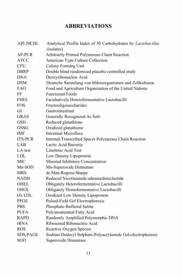
ABBREVIATIONS
API 50CHL Analytical Profile Index of 50 Carbohydrates by Lactobacillus
(isolates) AP-PCR Arbitrarily Primed Polymerase Chain Reaction ATCC American Type Culture Collection CFU Colony Forming Unit DBRP Double blind randomized placebo controlled study DNA Deoxyribonucleic Acid DSM Deutsche Sammlung von Mikroorganismen und Zellkulturen FAO Food and Agriculture Organization of the United Nations FF Functional Foods FHEL Facultatively Heterofermentative Lactobacilli FOS Fructooligosaccharides GI Gastrointestinal GRAS Generally Recognized As Safe GSH Reduced glutathione GSSG Oxidized glutathione IMF Intestinal Microflora ITS-PCR Internal-Transcribed Spacer Polymerase Chain Reaction LAB Lactic Acid Bacteria LA-test Linolenic Acid Test LDL Low Density Lipoprotein MIC Minimal Inhibitory Concentration Mn-SOD Mn-Superoxide Dismutase MRS de Man-Rogosa-Sharpe NADH Reduced Nicotinamide-adeninedinucleotide OHEL Obligately Heterofermentative Lactobacilli OHOL Obligately Homofermentative Lactobacilli Ox LDL Oxidized Low Density Lipoprotein PFGE Pulsed-Field Gel Electrophoresis PBS Phosphate-Buffered Saline PUFA Polyunsaturated Fatty Acid RAPD Randomly Amplified Polymorphic DNA rRNA Ribosomal Ribonucleic Acid ROS Reactive Oxygen Species SDS-PAGE Sodium Dodecyl Sulphate-Polyacrylamide Gel-electrophoresis SOD Superoxide Dismutase
12
TAA Total Antioxidative Activity TAS Total Antioxidative Status TGSH total glutathione WHO World Health Organization WIPO World Intellectual Property Organization

13
INTRODUCTION
During the past decades lifestyle in the developed industrial countries has changed, regarding living standard, hygiene, diet, usage of antibiotics and other antimicrobial substances. Prevalence of chronic diseases like different allergies and gut-associated diseases (e.g. ulcerative colitis, Crohn disease, inflammatory bowel disease) are of rising importance in the industrial world today. The balance of gut microbiota is considered to grant colonization resistance against infectious agents and promote antiallergenic processes, stimulate immune defense and reduce hypersensitivity reactions, incl. food allergy (Isolauri et al, 2001). Probiotics, live beneficial microbes, are aimed to improve the imbalance of the indigenous microbiota. In Europe, public interest in probiotics started to increase at the end of the last century and is still growing. About 65% of the European functional food market is covered nowadays with dairy probiotic products (Stanton et al., 2001).
In Estonia, investigation of lactobacilli in the human microbiota reached the high level at University of Tartu already in the second half of the 20th century (Voronina, 1968; Mikelsaar, 1969; Lenzner, 1973) and has proceeded till nowadays (Naaber, 1997; Mikelsaar et al., 1998; Annuk, 2002; Mikelsaar et al., 2002; Naaber and Mikelsaar, 2004). The technological aspects of food related lactobacilli have been thoroughly studied at Tallinn Technical University (Kask, 2003; Laht, 2003). Since 2001, the Department of Microbiology of the Uni-versity of Tartu has participated in the EU 5th Framework Programme PROEUHEALTH (The Food, Gastrointestinal Tract Functionality and Human Health Cluster), a cooperative investigation of several European universities aimed to develop new probiotics.
The Lactobacillus fermentum strain ME-3 (previously designated as 822-1-1 and E-3) of healthy human origin was isolated from an Estonian 1-year-old child during studies on development of allergy in two differently industrialized countries like Estonia and Sweden (Björksten et al., 1994; Sepp, 1998). Its probiotic properties – antioxidative and antimicrobial activity were assessed at the University of Tartu, Department of Microbiology and Depart-ment of Biochemistry (Annuk et al., 1999; Mikelsaar et al., 2001). The strain is deposited in the culture collection (Deutsche Sammlung von Mikroorganismen und Zellkulturen (DSMZ), DSM 14241 and patented (Application No. 0356/ 01PV to the Estonian Patent Agency). The International Bureau of the World Intellectual Property Organization (WIPO) approved the patent application WO 03002131) and the WIPO experts recognized its novelty in 2003.
In order to be used as functional food or food additive, labeled and mar-keted according to EU regulations (Commission of the European Communities, 2003) the scientifically proven health claims of L. fermentum ME-3 are necessary. The present thesis specified the technical applications of the strain in different food products, its functional properties and the functional efficacy of L. fermentum ME-3 in healthy human volunteers.
14
LITERATURE REVIEW
1. Functional foods and probiotics Although the primary purpose of food is to provide enough nutrients to fulfil body requirements, various functions of the body are modulated by diet. In order to compensate for deficiency of certain nutrients in the diet due to changes in nutritional habits of developed industrial countries, the concept of functional food has been developed. A food can be regarded as functional if it is satisfactorily demonstrated to affect beneficially one or more target functions in the body, beyond adequate nutritional effect, in a way which is relevant to either an improved state of health and well-being and/or reduction of disease risk " (ILSI Europe, 1999)
Functional food (FF) is intended for a population generally in normal health and must demonstrate beneficial effects in amounts that are usually consumed in the diet. FF is a natural food, to which a component has been added/removed or a food in which the bioavailability of the components has been modified by technological or biotechnological means (Korhonen, 2002). FF can be classified into different groups according to their effect: fat replacers, probiotics, pre-biotics and dietary fibres, antioxidants, vitamins, polyphenols, plant sterols, polyunsaturated fatty acids and minerals.
The most promising targets for FF are the GI functions and particularly control of nutrient bioavailability (Roberfroid, 2000). However, FF can affect different systems in the body: GI functions (e.g. balanced colonic microflora, control of transit time and mucosal motility, bowel habits; modulation of epithelial cell proliferation, balance of redox and antioxidant systems, meta-bolism of macronutrients, especially amino acids, carbohydrates and fatty acids).
The term “functional food” originates from the 1980s (Sanders, 1999). In 1991, a legal status to functional foods was granted in Japan, indicating foods for special health use. The first FF probiotic fermented milk drink Yakult has been available in Japan already since 1935 (Karimi and Peña, 2003).
The term “probiotic” was derived from Greek and means “for life.” Since the first reference to the positive effecs of beneficial bacteria (Vergin, 1954), different definitions have been proposed for probiotics. Fuller (1992) defined a probiotic as “a live microbial feed supplement, which beneficially affects the host animal by improving its intestinal microbial balance.” According to the expert panel commissioned by the Food and Agriculture Organization of the United Nations (FAO) and the World Health Organization (WHO) the present-day interpretation of probiotics is “live microorganisms which when adminis-tered in adequate amounts confer a health benefit on the host” (FAO/WHO, 2002).
Probiotics may be administered as a component of FF or as food additives (e.g. capsules, tablets).
15
Some authors have intepreted probiotics as “microbial cell preparations or components of microbial cells that have a beneficial effect on the health and well-being of the host.” Bacterial cell-wall components, heat-killed whole cells or metabolites can have a specific probiotic effect, for example, improvement of lactose digestion or treatment of acute or chronic diarrhoea (Ouwehand and Salminen, 1998; Romond et al., 1998; Salminen et al., 1999; Simakachorn et al., 2000; Xiao et al., 2002). Inactivation of probiotics by heat, UV or γ-irra-diation sustains more or less their ability to adhere to the intestinal mucus in vitro (Ouwehand et al., 2000). Adherence is considered one of the selection criteria and pre-requisite for probiotic effects. It has been demonstrated in vitro that above mentioned inactivation did not negatively affect the ability of probiotics to bind carcinogens. Yet it is still debatable as, whether a probiotic must definitely be alive upon digestion (Ouwehand et al., 1999; Sanders and Huis int Veld, 1999).
Common probiotics include: 1) Lactobacilli such as Lactobacillus acido-philus, L. johnsonii, L. casei, L. delbrueckii ssp. bulgaricus, L. reuteri, L. brevis, L. cellobiosus, L. curvatus, L. fermentum, L. plantarum; 2) Gram-positive cocci such as Lactococcus lactis ssp. cremoris, Streptococcus salivarius ssp. Thermo-philus, Enterococcus faecium, S. diaacetylactis, S. intermedius; and 3) Bifido-bacteria such as Bifidobacterium bifidum, B. adolescentis, B. animalis, B. infantis, B. longum, B. thermophilum (Collins et al., 1998; Gibson, 1999; Mercenier et al., 2002). Also other microbial species, besides lactic acid bacteria (LAB), like Bacillus subtilis, Propionibacterium spp. and yeasts (Saccharomyces boulardii) have been accepted and used as probiotics (Chukeatirote, 2002; Jan et al. 2002).
The mechanism of the action of probiotics (e.g. bifidobacteria and lacto-bacilli) relies on their metabolic end products, mainly organic acids may lower the human gut pH at which pathogenic microbes are not able to compete effectively. Other factors are occupation of normal colonization sites by probiotics, competition for available nutrients and production of antimicrobial substances. The second generation of probiotics is genetically modified micro-organisms providing the host with some necessary components, e.g. production of immunomodulators (e.g. interleukines) or Helicobacter pylori and rotavirus antigens (Mercenier et al, 2004).
Probiotic products may be conventional foods (yoghurt, cheese, milk) (Holzapfel et al., 2001; Temmerman et al., 2002; Yeung et al., 2002) consumed for nutritional purposes, but also for the probiotic effect; food supplement/ fermented milks or ”medical foods” (e.g. food formulation is a delivery vehicle for probiotics or their fermentation endproducts are the primary purpose); dietary supplements: capsules, tablets, liquids, powder (Ross, 2000, Kaur et al., 2002; Temmerman et al., 2002). Probiotic preparations used as food supplement can consist of one single strain (e.g. Yacult, Japan – L. casei Sirota) or there are mixed cultures of two (e.g. Bacilac, Belgium – L. acidophilus plus L. rhamnosus) or even more (e.g. food supplement VSL-3, Italy containes 8 LAB species) strains.
16
2. Lactic acid bacteria
2.1. Taxonomy of Lactobacillus spp. Lactic acid bacteria are Gram-positive non-sporing bacteria, which are physiologically diverse and include the genera of Lactobacillus, Leuconostoc, Pediococcus, Streptococcus, Enterococcus, Lactococcus, Oenococcus, Weis-sella, Carnobacterium, Tetragenococcus, Vagococcus and Bifidobacterium (Kandler and Weiss, 1986; Klein et al., 1998).
The classification of LAB has mainly remained unchanged since the work of Orla-Jensen (1919). According to growth temperature, the genus Lacto-bacillus has been divided into three subgenera termed “Thermobacterium”, “Streptobacterium”, and “Betabacterium” (Fig. 1). In addition, lactobacilli are divided into three biochemically diverse groups based on their fermentation pathways of different carbon sources (pentoses and hexoses) as follows: obligately homofermentative lactobacilli (OHOL), facultatively heterofermen-tative lactobacilli (FHEL), and obligately heterofermentative lactobacilli (OHEL) (Kandler and Weiss, 1986; Hammes and Vogel, 1995; Klein et al., 1998).
However, the type of carbohydrate fermentation as the traditional basis of grouping of LAB is not strictly related to the evolution of the organisms. Phylogenetic classification and identification on the species level relies today largely on molecular methods based on highly conserved regions in a microbial genome like 16S ribosomal DNA (16S rDNA) genes (Song et al., 2000). The studies based on 16S rRNA sequences have shown close relationships between the genera Lactobacillus, Leuconostoc, Pediococcus, Oenococcus and Weis-sella.
The principal groupings today are summarised in Table 1 as follows: (1) the L. delbrueckii group, which contains several obligately homofermentative lactobacilli (L. delbrueckii with subspecies, L. gasseri, L. acidophilus, L. helve-ticus, L. johnsonii and L. jensenii) and a few facultatively heterofermentative lactobacilli; (2) the L. casei – Pediococcus group, comprising the remaining obligately homofermentative, all heterofermentative and most of the facul-tatively heterofermentative lactobacilli; and (3) the Leuconostoc group inclu-ding the species from the above mentioned genera (Stiles and Holzapfel, 1997 modified after Kandler and Weiss 1986; Hammes et al., 1992; Collins et al., 1998; Klein et al., 1998; Gomes and Malcata, 1999; Sanders, 1999; Holzapfel et al., 2001; Mercenier et al, 2002).
17
2.2. Metabolism Lactobacilli have complex nutritional requirements for organic substrates, nutritional requirements for amino acids, peptides, vitamins, salts, fatty acid or fatty acid esters, minerals and fermentable carbohydrates (Kandler and Weiss, 1986). Lactobacilli can adapt to various environmental and nutritional conditions and change their metabolism accordingly. Metabolically, lactobacilli are mostly microaerophilic, but they are able to grow at variable oxygen tension from aerobic to anaerobic.
Most species prefer mesophilic growth temperatures; optimum temperature is generally between 30–40°C. Further, lactobacilli grow best in a slightly acidic environment, optimal growth pH from 5.5 to 6.2.
Lactobacilli are capable to degrade different carbohydrates and related compounds, while the end products are dependent on the fermentation type of the species (Fig. 1). Lactic acid is the predominant end product, however, under certain conditions additional products may be acetate, ethanol, succinate or CO2 (Botazzi, 1983; Kandler and Weiss, 1986; Hammes et al., 1992; Klein et al., 1998).
At the enzyme level, lactobacilli of the OHOL and OHEL groups differ with respect to presence or absence of fructose diphosphate (FDP) aldolase or phosphoketolase. Lactobacilli from the OHOL group do not possess FDP aldolase and are thus unable to ferment pentoses. On the other hand, the representatives of the OHEL group possess phosphoketolase to break down pentoses, yielding equimolar amounts of lactic and acetic acids. However, the FHEL group of lactobacilli, possesses an inducible phosphoketolase with pentoses acting as inducers. They are thus able to ferment pentoses upon adaptation to lactic acid and acetic acid, whereas hexoses are homofermen-tatively metabolised (Kandler and Weiss, 1986; Axelsson, 1998).
The main end product of hexoses – glycolysis or the Embden-Meyerhof pathway (homolactic fermentation) – is lactic acid, characteristic of the lacto-bacilli of both the FHEL and OHOL groups. Hexoses other than glucose enter the major pathways after isomerization and/or phosphorylation. In addition to lactic acid, other end products are also produced (CO2, acetate, ethanol), mainly by OHEL (Axelsson, 1998).

18
Glucose fermentation
D-, L-, DL- D-, L-, DL- DL-lactic acid, lactic acid lactic acid CO2, acetic acid,
ethanol Growth at 45°C + + ± Growth at 15°C –(+)* +(–)** +(–)** Fermentation of hexoses + + + Gas from glucose – + + Fermentation of pentoses – + + Gas from pentoses – + + NH3 from arginine –(+)* – +(–)** Subgenera Thermobacterium Streptobacterium BetabacteriumFermentation (OHOL group) (FHEL group) (OHEL group)pathway Incl. L. fermentum *Mostly negative, with a few exceptions; **mostly positive, with a few exceptions Figure 1. Differentiation of lactobacilli after Botazzy, 1983, modified. Different LAB species may use different pathways depending on conditions and enzymatic capacity (Kandler and Weiss, 1986; Axelsson, 1998). Change of LAB metabolism in response to various conditions results in production of different end products. Mostly, it can be attributed to altered pyruvate meta-bolism. Pyruvate, intermediately formed in both above-mentioned pathways, may partly undergo several conversions, producing aroma compounds like diacetyl and acetoin (2,3-butanediol) or acetate, formate and ethanol.
In some LAB species or strains carbohydrates (especially sucrose) may contribute to formation of dextrans (slime), important in yoghurt production.
Protein utilisation. Lactobacilli have very limited capacity to synthesize amino acids from inorganic nitrogen sources, depending on amino acids present in the growth environment (Axelsson, 1998; Christensen et al., 1999).
19
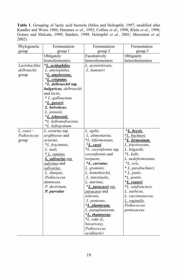
Table 1. Grouping of lactic acid bacteria (Stiles and Holzapfel, 1997; modified after Kandler and Weiss 1986; Hammes et al., 1992; Collins et al., 1998; Klein et al., 1998; Gomes and Malcata, 1999; Sanders, 1999; Holzapfel et al., 2001; Mercenier et al, 2002).
Phylogenetic group
Fermentation group 1
Fermentation group 2
Fermentation group 3
Obligately homofermenters
Facultatively heterofermenters
Obligately heterofermenters
Lactobacillus delbrueckii group
*L. acidophilus, L. amylophilus, *L. amylovorus, *L. crispatus, *L. delbrueckii ssp. bulgaricus, delbrueckii and lactis, * L. gallinarium, *L. gasseri, L. helveticus, L. jensenii, *L. johnsonii, *L. kefiranofraciens, *L. kefirgranum
L. acetotolerans, L. hamsteri
L. casei – Pediococcus group
L. aviarius ssp. araffinosus and aviarius, *L. fraciminis, L. mali, * L. ruminis, L. salivarius ssp. salicinus and salivarius, L. sharpae, Pediococcus damnosus, P. dextrinum, P. parvulus
L. agilis, L. alimentarius, *L. bifermentans, *L. casei, *L. coryniformis ssp. coryniformis and torquens, *L. curvatus, L. graminis, L. homohiochii, L. intestinalis, L. murinus, *L. paracasei ssp. paracasei and tolerans, L. pentosus, *L. plantarum, L. paraplantarum, *L. rhamnosus, *L. sake (L. bavaricus), Pediococcus acidilactici
*L. brevis, *L. buchneri, *L. fermentum, L. fructivorans, L. hilgardii, *L. kefir, L. malefermentans, *L. oris, * L. parabuchneri, * L. panis, *L. pontis, *L. reuteri, *L. sanfrancisco, L. suebicus, L. vaccinostercus, L. vaginalis, Pediococcus pentosaceus
20
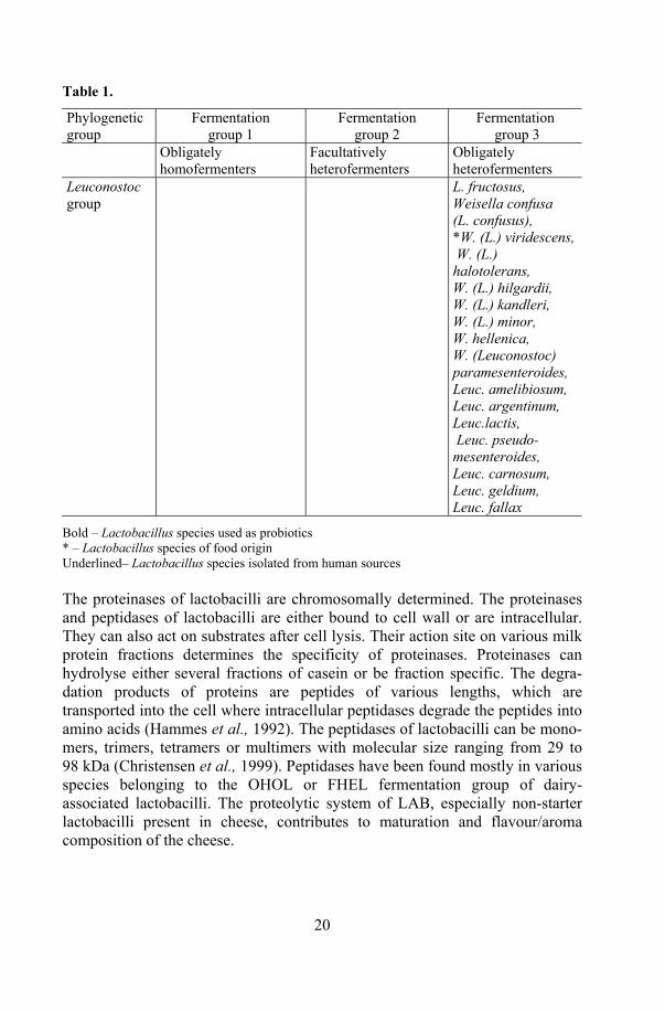
Table 1.
Phylogenetic group
Fermentation group 1
Fermentation group 2
Fermentation group 3
Obligately homofermenters
Facultatively heterofermenters
Obligately heterofermenters
Leuconostoc group
L. fructosus, Weisella confusa (L. confusus), *W. (L.) viridescens, W. (L.) halotolerans, W. (L.) hilgardii, W. (L.) kandleri, W. (L.) minor, W. hellenica, W. (Leuconostoc) paramesenteroides, Leuc. amelibiosum, Leuc. argentinum, Leuc.lactis, Leuc. pseudo-mesenteroides, Leuc. carnosum, Leuc. geldium, Leuc. fallax
Bold – Lactobacillus species used as probiotics * – Lactobacillus species of food origin Underlined– Lactobacillus species isolated from human sources The proteinases of lactobacilli are chromosomally determined. The proteinases and peptidases of lactobacilli are either bound to cell wall or are intracellular. They can also act on substrates after cell lysis. Their action site on various milk protein fractions determines the specificity of proteinases. Proteinases can hydrolyse either several fractions of casein or be fraction specific. The degra-dation products of proteins are peptides of various lengths, which are transported into the cell where intracellular peptidases degrade the peptides into amino acids (Hammes et al., 1992). The peptidases of lactobacilli can be mono-mers, trimers, tetramers or multimers with molecular size ranging from 29 to 98 kDa (Christensen et al., 1999). Peptidases have been found mostly in various species belonging to the OHOL or FHEL fermentation group of dairy-associated lactobacilli. The proteolytic system of LAB, especially non-starter lactobacilli present in cheese, contributes to maturation and flavour/aroma composition of the cheese.
21
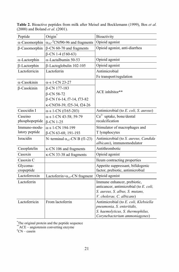
Table 2. Bioactive peptides from milk after Meisel and Bocklemann (1999), Bos et al. (2000) and Boland et al. (2001).
Peptide Origin* Bioactivity α-Casomorphin αS1-#CNf90-96 and fragments Opioid agonist β-Casomorphin β-CN 60-70 and fragments Opioid agonist, anti-diarrhea β-CN 1-4 (f 60-63) α-Lactorphin α-Lactalbumin 50-53 Opioid agonist β-Lactorphin β-Lactoglobulin 102-105 Opioid agonist Lactoferricin Lactoferrin Antimicrobial Fe transport/regulation α-Casokinin α-s 1-CN 23-27
β-CN 177-183 β-CN 58-72 β-CN f 6-14, f7-14, f73-82
β-Casokinin
κ-CNf38-39, f25-34, f24-26
ACE inhibitor**
Casocidin I α-s 1-CN (f165-203) Antimicrobial (to E. coli, S. aureus) Caseino phosphopeptide
α-s 1-CN 43-58; 59-79 β-CN 1-25
Ca2+ uptake, bone/dental recalcification
Immuno-modu-latory peptide
α-s 1-CN 194-199 β-CN 63-68; 191-193
Stimulator of macrophages and T lymphocytes
Isracidin N–terminal αs1-CN B (f1-23) Antimicrobial (to S. aureus, Candida albicans), immunomodulator
Casoplatelin κ-CN 106 and fragments Antithrombotic Casoxin κ-CN 33-38 ad fragments Opioid agonist Casoxin C Ileum contracting properties Glycoma-cropeptide
Appetite suppressant, bifidogenic factor, prebiotic, antimicrobial
Lactoferroxin Lactoferrin+αs1-CN fragment Opioid agonist Lactoferrin Immune enhancer, prebiotic,
anticancer, antimicrobial (to E. coli, S. aureus, S. albus, S. mutans, V. cholerae, C. albicans)
Lactoferricin From lactoferrin Antimicrobial (to E. coli, Klebsiella pneumonia, S. enteritidis, S. haemolyticus, S. thermophilus, Corynebacterium ammoniagenes)
*The original protein and the peptide sequence **ACE – angiotensin converting enzyme #CN – casein

22
On the other hand, amino acids, especially arginine, present in the growth environment (e.g. originating from the primary breakdown of milk casein during cheese ripening), can be used as an alternative energy source by lactobacilli (Laht, 2003). Energy is derived through substrate level phosphorylation, ornithine, CO2 and NH3 being the end products of the process (Axelsson, 1998). The ability of a lactobacillus species to split arginine and the appearance of NH3 in the growth environment can be used as one of the parameters for the fermentation group and species level identification of Lactobacillus spp (Fig. 1).
The breakdown of casein by lactobacilli during milk fermentation or by human digestive enzymes after consumption produces a variety of hormone-like substances or bioactive peptides. Different health promoting activities of bioactive peptides have been described (Table 2).
Different aspects of metabolism are important in elaborating technical aspects for probiotic strains to be incorporated into different products. Products in which bioactive peptides are used are rare in the market.
In the European market, the Valio’s bioactive peptides mediated blood pressure lowering milk-based drink Evolus® is available. The bioactive peptides in this product are generated by L. helveticus during fermentation (Seppo et al., 2002, Seppo et al., 2003).
3. Prebiotics Prebiotics are defined by Gibson and Roberfroid (1995) as “nondigestible food ingredients that target certain components within the microbiota of the human large intestine”. These prebiotics are fermented by one or a limited number of potentially beneficial bacteria form the resident colonic microflora. A prebiotic is expected to improve the composition of the colonic microbiota and through this serve as beneficial to the host health (Gibson, 1999).
The two basic types of fermentations taking place in the gut are saccharolytic fermentation and proteolytic fermentation. The main end products of carbohydrate metabolism are the short chain fatty acids: acetate, propionate and butyrate. These may be further metabolised systematically or locally to generate energy for the host. The end products of the proteolytic fermentation include more or less toxic compounds as amines, ammonia and phenolic compounds. Fermentation in the gut can be modulated towards saccharolytic by prebiotic consumption.
Research into prebiotics stems from interest in the dietary fibre shown since the beginning of the 1970s. Much of the interest is aimed at non-digestible oligosaccharides (fructooligosaccharides, trans-galactooligosaccharides, iso-maltooligosaccharides, xylooligosaccharides, soyoligosaccharides, glucooli-gosaccharides and lactosucrose).

23
More than 36 000 plants worldwide contain FOS; some common sources of inulin are onion (2–6%), garlic (9–16%), leek (3–10%), banana (0.3–0.7%), asparagus (10–15%), Jerusalem artichokes (15–20%), chicory (13–20%), and even wheat (1–4%). Yet the levels are too low for a significant GI tract effect (Crow, 2004). Consumption of more than 4 grams of FOS daily is needed to induce changes in LAB levels in the gut, though estimated daily consumption differs in the US and Europe (Roberfroid, 2000; Gibson, 2001).
Prebiotics are increasingly used in development of new food product, e.g. drinks, yoghurts, biscuits and table spreads (Gibson and Roberfroid, 1995; Gibson, 1999). Several prebiotics are available in Europe.
The positive effects of prebiotic consumption are: improvement of bowel habit; reduction of diarrhoea and constipation; modulation of lipid metabolism by normalizing cholesterol values; reduction of osteoporosis by improved mineral absorption; reduction of allergy risk through immune system modulation; reduction of colon cancer risk (Roberfroid, 2000; Conway, 2001). Unfortunately, many of the above mentioned health claims still require further research.
4. Synbiotics Bifidobacteria and lactobacilli are the most frequent target organisms for prebiotics. These genera are most commonly used as probiotics too. Probiotics and prebiotics used in synergistic combination are termed synbiotics. Synbiotics are mixtures that improve the survival and implantation of live microbial dietary supplements in the GI tract, either by stimulating growth or by metabolically activating the health promoting bacteria (Kaur et al., 2002).
Although there is growing interesting development of new FF with synbiotics, combination of prebiotics and probiotics into a synbiotic has been studied to a limited extent and needs further investigations, because of the afore mentioned different substrate requirements for individual probiotic LAB species and strains. Only a few human studies have been carried out on the effectiveness of synbiotics (Morelli et al., 2003).
5. Strategy of selection of probiotic strains Introducing a new probiotic into the market involves a step-wise process in order to obtain a functional and safe product. Though the genus Lactobacillus has a Generally Recognized As Safe (GRAS) status and a long history of safe use for food fermentation, several criteria must be taken into consideration to select and evaluate a concrete putative probiotic Lactobacillus strain (Collins et al., 1998; FAO/WHO, 2002; Reid et al., 2003). The properties of a putative
24
probiotic must be thoroughly described in vitro as well as in vivo animal studies and in clinical trials (Fig. 2).
Selection criteria for probiotics are an area of much debate and should be taken into account when defining appropriate strains. The following criteria have been suggested for use in probiotic strain selection (Sanders and Huis in`t Veld, 1999; Saarela et al., 2000; FAO/WHO, 2002; Reid et al., 2003):
1. General aspects (origin, identity) 2. Safety 3. Functional features 4. Technological aspects
Today few probiotics have been tested according to all recommended aspects in the scheme by FAO/WHO.
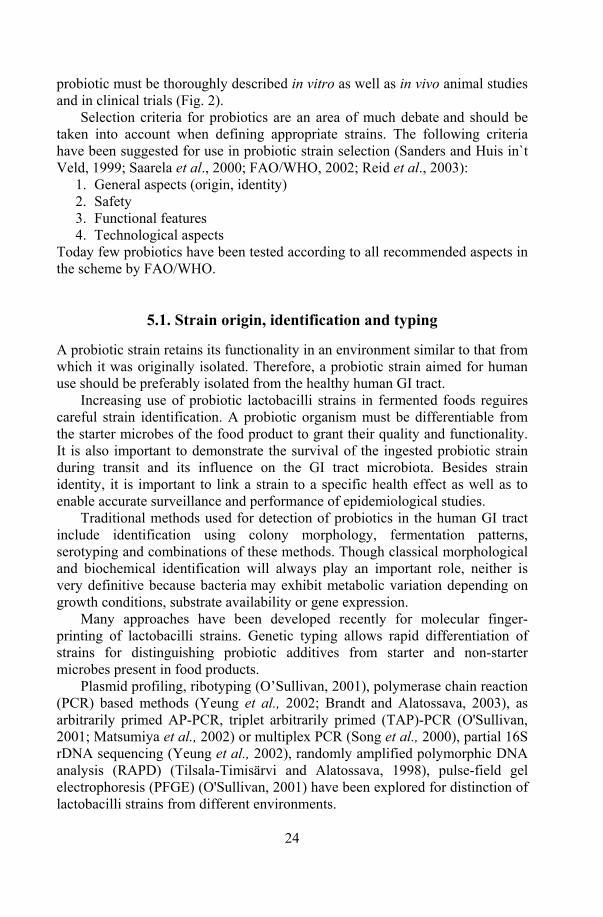
5.1. Strain origin, identification and typing A probiotic strain retains its functionality in an environment similar to that from which it was originally isolated. Therefore, a probiotic strain aimed for human use should be preferably isolated from the healthy human GI tract.
Increasing use of probiotic lactobacilli strains in fermented foods reguires careful strain identification. A probiotic organism must be differentiable from the starter microbes of the food product to grant their quality and functionality. It is also important to demonstrate the survival of the ingested probiotic strain during transit and its influence on the GI tract microbiota. Besides strain identity, it is important to link a strain to a specific health effect as well as to enable accurate surveillance and performance of epidemiological studies.
Traditional methods used for detection of probiotics in the human GI tract include identification using colony morphology, fermentation patterns, serotyping and combinations of these methods. Though classical morphological and biochemical identification will always play an important role, neither is very definitive because bacteria may exhibit metabolic variation depending on growth conditions, substrate availability or gene expression.
Many approaches have been developed recently for molecular finger-printing of lactobacilli strains. Genetic typing allows rapid differentiation of strains for distinguishing probiotic additives from starter and non-starter microbes present in food products.
Plasmid profiling, ribotyping (O’Sullivan, 2001), polymerase chain reaction (PCR) based methods (Yeung et al., 2002; Brandt and Alatossava, 2003), as arbitrarily primed AP-PCR, triplet arbitrarily primed (TAP)-PCR (O'Sullivan, 2001; Matsumiya et al., 2002) or multiplex PCR (Song et al., 2000), partial 16S rDNA sequencing (Yeung et al., 2002), randomly amplified polymorphic DNA analysis (RAPD) (Tilsala-Timisärvi and Alatossava, 1998), pulse-field gel electrophoresis (PFGE) (O'Sullivan, 2001) have been explored for distinction of lactobacilli strains from different environments.

25
Besides molecular DNA-based typing, extraction of whole-cell proteins, followed by sodium dodecyl sulphate-polyacrylamide gel-electrophoresis (SDS-PAGE) separation, has been found to be a reliable and rapid way to characterize a large number of strains (Reuter et al., 2002).
Strain identification by phenotypic and genotypic methods • Genus, species, strain • Deposit strain in international culture collection
Functional characterization • In vitro tests • Animal studies
Safety assessment • In vitro and/or animal • Phase 1 human study
Preferably second Independent DBPC study to confirm results
Probiotic
Phase 3, effectiveness trial is appropriate to compare probiotics with standard treatment of a specific condition
Labelling • Contents - genus, species, strain designation • Minimum numbers of viable bacteria at the end of shelf-life • Proper storage conditions • Corporate contact details for consumer information
Technological properties:Survival in product manufacturing and shelf life
Double blind, randomised, placebo-controlled (DBPC) phase 2 human trial or other appropriate design with sample size and primary outcome appropriate to determine if strain/product is efficacious
Figure 2. FAO and WHO (2002) guidelines for probiotics in food, modified
26
5.2. Safety assessment Though lactobacilli and bifidobacteria are historically associated with food, they are normal commensals of the mammalian microflora and their pathogenic potential is considered quite low. However, as probiotics are, after all, viable microorganisms, there is the possibility that they could cause infections in immunocompromised host.
Lactobacillus spp. related systemic (Saxelin et al., 1996; Soleman et al., 2003) and local infections (Mackay et al., 1999; Rautio et al., 1999) have been reported in several studies. Other species used as probiotics (such as ente-rococci, yeasts) pose greater threat than LAB (Sanders and Huis in`t Veld, 1999; Reid et al., 2003). Precautions should be considered for persons with lowered immune functions.
Before incorporating a probiotic strain into a food product, it should be tested for safety, to exclude, for example, haemolytic activity and toxin production in vitro or on animal models. Besides, post-market epidemiological surveillance of adverse effects should be carried out (FAO/WHO, 2002).
Safety aspects associated with probiotic microbes include the following specifications (Saarela et al., 2000): healthy human origin, non-pathogenicity (no thrombocytic activity; no degradation of host mucins, no platelet aggre-gation properties), no history of association with diseases; not deconjugating bile salts, no transmissible antibiotic resistance.
Bile acids synthesized in the liver and excreted into the duodenum in the conjugated form can be chemically modified (deconjugated) in the colon by GI microbes. Though both conjugated bile and deconjugated bile have anti-microbial properties, the deconjugated form is more toxic to microbes (Floch et al., 1972; Stewart et al., 1986). Therefore, it is considered important that the consumed probiotic lacks the ability to deconjugate bile. The properties of probiotics to resist bile as well as their conjugated bile salt hydrolase activity have been until now considered independent properties (Moser and Savage, 2001).
There exists highly varied range of species-specific natural antibiotic resistance among lactobacilli and bifidobacteria (Yazid et al., 2000; Mändar et al., 2001; Danielsen and Wind, 2003), mostly non-transmissible. Though plas-mid based antibiotic resistance is not very common among lactobacilli strains, it still can occur. On one hand, the antibiotic resistance of a probiotic Lacto-bacillus strain is favourable, as probiotics are often consumed after antibiotic therapy to establish the microbial balance. On the other hand, this arises the need to confirm whether the antibiotic resistance of the probiotic strain is of chromosomal origin or it is carried by plasmid and is therefore putatively transferable (Salminen et al., 1998; Saarela et al., 2000).
27
5.3. Functional features Several functional aspects are important while selecting a novel probiotic strain (Saarela et al., 2000). The probiotic strain must essentially be able to survive in the GI tract (e.g. tolerate acid and bile, adhere to epithelial cells) in order to be effective in therapeutic actions and carry on normal metabolic activity after consumption.
Bile and acid tolerance. Environmental factors like low pH in the stomach, presence of bile acid in the duodenum and intestinal enzymes affect the viability as well as the adhesive properties of a probiotic introduced into the human GI tract (Ouwehand et al., 2001). Mechanical factors that may affect the binding and temporal persistence of a probiotic strain in vivo are peristalsis and mucus secretion. Probiotic strains that are able to survive and grow at the physiological levels of bile and low pH in vitro are more likely to survive in the intestinal transit.
Adhesion and antimicrobial activity. Among human microbiota, lactobacilli are considered the important colonization resistance-granting bacteria that fight infectious agents. Adhesion to host gut epithelial cells and intestinal mucus is an important property of a probiotic strain for temporary colonization of the GI tract and stimulation of beneficial effects. Adhesion of a probiotic strain might be closely related to one of the functionally beneficial properties of probiotics – antimicrobial activity. Probiotic microorganisms can coaggregate with pathogens or attach to enterocytes (competitive exclusion) and thus inhibit the binding of enteric pathogens to the intestinal mucosa. Another strategy of competitive exclusion of a pathogen is the production of inhibitory compounds like bacteriocins (antibacterial proteins), low molecular mass non-proteinaceous compounds like different acids (lactic acid, acetic acid, succinic acid) and toxic oxygen metabolites like hydrogen peroxide, which in combination with the lactose peroxidase–thiocyanate milk system exerts a bacteriocidal effect on most pathogens (Klaenhammer, 1988; Mishra and Lambert, 1996; Helander et al., 1997). The antagonistic activity of a probiotic strain against pathogens such as Clostridium difficile, Salmonella sp., Helicobacter pylori, Listeria mono-cytogenes, Escherichia coli should be detected in different milieus according to the action site of a certain strain (Annuk, 2002; Naaber et al., 2004; Hütt et al., 2005).
Immunostimulatory properties. By selecting a probiotic strain the immunostimulatory effect of the strain has to be taken into account. It has been proved in human studies that probiotics can have positive effects on the immune system of their host, which has not been linked with inflammatory response, or any other harmful effects (Salminen et al., 1998; Saarela et al., 2000). However, there are differences between probiotic bacteria in respect to their immunomodulatory properties, which should be evaluated for a particular probiotic strain.
28
Antimutagenic and anticarcinogenic properties. Another desired health-promoting properties of microbes (those of food origin or members of the intestinal microflora) are the ability to neutralize carcinogenic or mutagenic compounds through modulating the procarcinogenic enzymes of the gut, through suppression of tumours by immune stimulation or through binding and degradating carcinogens (Saarela et al., 2000). Although the antimutagenic and anticarcinogenic properties of probiotic microorganisms have been demonstra-ted in vitro and on animal models of elucidation, the problem reguires further clinical evidence.
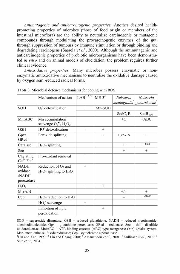
Antioxidative properties. Many microbes possess enzymatic or non-enzymatic antioxidative mechanisms to neutralize the oxidative damage caused by oxygen semi-reduced radical forms. Table 3. Microbial defence mechanisms for coping with ROS.
Mechanism of action LAB1, 2, 3 ME-34 Neisseria meningitidis5
Neisseria gonorrhoeae5
+ Mn-SOD SOD O2• detoxification
SodC, B SodB low MntABC Mn accumulation
scavenge O2•–, H2O2
+C +ABC
GSH HO• detoxification + + Gpx/ GRed
Peroxide splitting + + gpx A –
Catalase H2O2 splitting + +high Sco + + Chelating Cu2+ Fe2+
Pro-oxidant removal +
NADH oxidase /NADH peroxidase
Reduction of O2 and H2O2 splitting to H2O
+
H2O2 + + MsrA/B +/– + Ccp H2O2 reduction to H2O – +Anaer HO2
• scavenge + Inhibition of lipid
peroxidation + +
SOD – superoxide dismutase, GSH – reduced glutathione, NADH – reduced nicotinamide-adeninedinucleotide; Gpx – glutathione peroxidase; GRed – reductase; Sco – thiol: disulfide oxidoreductase; MntABC – ATB-binding cassette (ABC)-type manganese (Mn) uptake system; Msr– methionine sulfoxide reductase; Ccp – cytochrome c peroxidase. 1Lin and Yen, 1999; 2 Lin and Chang 2000; 3 Amanatidou et al., 2001; 4 Kullisaar et al., 2002; 5 Seib et al., 2004.
29
Especially pathogens have to cope with the defence mechanisms of the host and have therefore developed various strategies forsurvival in the conditions of oxidative stress and for scavenging reactive oxygen species (ROS) and nitric oxide species (Seib et al., 2004).
Although, there is increasing evidence regarding the antioxidative effect of LAB in vitro, the high values seem to be a strain-specific property among lactobacilli (Lin and Yen, 1999a; Lin and Yen, 1999b; Lin and Chang, 2000; Stecchini et al., 2001). And although different antioxidative ways to maintain ROS have been discovered in both pathogens and lactobacilli, as described in Table 3, the mechanisms require further clarification. Besides, little information is available about clinical trials where the effect of antioxidative probiotics on human health parameters is evaluated.
5.4. Clinical testing The most important proof of probiotic functional efficacy and safety can be tested with volunteer trials and clinical studies on children and adults. Both volunteer and clinical trials should be randomized and double-blinded.
Clinical evaluations are usually designed to compare a test strain to a placebo or control; rarely the efficacy of more than one strain is compared (Sanders and Huis in`t Veld, 1999; Pathmakanthan et al., 2000). Clinical trials for probiotic evaluation are divided into three phases (Fig. 2): phase one (safety), phase two (efficacy) and phase three (effectiveness) (FAO/WHO, 2002; Reid et al., 2003).
Phase one clinical studies on healthy human volunteers are closely associated with safety evaluation of the tested probiotic strain. A phase-one study is carried out to test whether a probiotic is well tolerated, and to establish the putative side effects of consumption of a certain strain containing a probiotic product.
Phase-two studies compare the efficacy of probiotic strain/product with that of a placebo on healthy human volunteers. The format of probiotic delivery and the proper dose of the probiotic are calculated in the course of this phase. During clinical studies, pharmacological properties of a probiotic strain are evaluated, such as survival and activity in the human intestine, fecal recovery, and dose-response relationship. There have been tested the significant improvement in health condition, well-being, quality of life or reduced risk of disease (Reid et al., 2003). However, most the above parameters are subjective and therefore relatively difficult to evaluate objectively.
The main aim of clinical trials is to assess the effectiveness of a probiotic product together with a standard therapy for a particular disease. Usually the study group receives a probiotic as an adjunct to standard therapy, while the control group is treated according to a standard therapy protocol (Reid, 2001).

30
There are several more or less scientifically proved health effects of probiotics, which cover improvement of different functions: like alleviation of lactose intolerance and constipation, treatment and prevention of GI disorders, reduction of diarrhoeas of different origin and food allergy, reduction of risk of various diseases like colon cancer or atherosclerosis, regulation of immuno-modulatory effects and cholesterol control (Ouwehand and Salminen, 1998; Salminen, 2001).
Several confounding factors (Table 4) may be important and shoud be considered in administering a probiotic. Table 4. Factors affecting the performance of a probiotic strain in GI tract (Goldin et al., 1992; Reid, 2001; Naaber and Mikelsaar, 2004).
Modulator Effect Intrinsic properties of the strain Antagonistic activity
Bacteriocins Antioxidative activity
Metabolic status of the probiotic strain during ingestion
Metabolically active Metabolically inactive (frozen, powdered)
Ecosystem in different parts of host GI tract
Low pH in stomach Bile Intestinal enzymes Individuality of indigenous microflora Peristalsis Mucus
Dose At least 109 CFU Duration of administration At least one week Time and way of consumption Active part of the day
Before bedtime During meals Between meals Nature of food Growth promoting, “bifidogenic” factor Protection by buffering capacity Interference of adhesion Fermentation products Presence of supportive cultures
Milk Nature of the probiotic carrier Capsule Coating
Protectants However, several above modulators need to be defined more precisely by specific research.
31
5.4.1. Oxidative stress markers of the human body Oxidation is essential to living organisms for energy production. Reactive oxygen species (ROS) are either free radicals, reactive anions containing oxygen atoms, or molecules containing oxygen atoms that can either produce free radicals or are chemically activated by them, for example hydroxyl radical, superoxide, hydrogen peroxide, and peroxynitrite. The main source of ROS in vivo is aerobic respiration, although ROS are also produced by oxidation of fatty acids, stimulation of phagocytosis by pathogens or lipopolysaccharides and tissue specific enzymes. ROS are important in the metabolism of various compounds. However, the abundance of ROS generated within the body from different external and internal sources (UV, pollutants or O2 involving bio-chemical reactions in vivo) cause oxidative stress (Vervaart and Knight, 1996; Diplock et al., 1998). Oxidative stress is imposed on cells as a result of one of the three factors: 1) an increase in oxidant generation, 2) a decrease in antioxi-dant protection, or 3) a failure to repair oxidative damage. The main damage to cells results from the ROS-induced alteration of macromolecules such as polyunsaturated fatty acids in membrane lipids, essential proteins, and DNA.
Additionally, oxidative stress and ROS are considered to be important in pathophysiology of a variety of human diseases, such as some forms of cancer, cardiovascular diseases, rheumatoid arthritis, and aging or neurodegenerative diseases like Alzheimer's disease or Parkinson's disease (Diplock et al., 1998; Eisen, 2002; Esch and Stefano, 2002).
Since ROS are very short-lived, it is very difficult to measure them and their effect directly. There are several methods for measurement of oxidative stress markers from human blood and urine. Polyunsaturated fatty acid (PUFA) containing lipids (e.g. in cell membranes) are very susceptible to peroxidation upon exposure to ROS (Vervaart and Knight, 1996). Lipid peroxidation is a pathophysiological process causing apoptosis, but it may also be involved in tissue damage in inflammation, ageing, cancer, and toxicity of xenobiotics (Shan et al., 1990; Karelson et al., 2001). Isoprostanes are prostaglandines-like compounds that are produced upon lipid peroxidation. Human body fluids (e.g. urine) usually contain low levels of F2-isoprostanes (8-epi-prostaglandin F2α) that arise by ROS oxidation of phospholipides containing arachidonic acid (Diplock et al., 1998). Peroxidation of arachidonyl phospholipids results in formation of the positional peroxyl isomers of arachidonic acid. These inter-mediates lead to formation of isoprostanes (Morrow et al., 1992). Measurement of F2-isoprostanes represents a useful approach to assessment of lipid peroxi-dation and oxidative stress in vivo of the whole body (Diplock et al., 1998).
Glutathione (L-γ-glutamyl-L-cysteinylglycine) is a tripeptide composed of cysteine, glutamic acid and glycine, which have two biologically important structural features: a thiol (SH) group and γ-glutamyl linkage (Shan et al., 1990). In humans it is present in the millimolar range mainly in the red blood cells, liver, pancreas, kidneys, spleen, eyes, lungs and intestinal cells (Meister

32
and Anderson, 1983). In cells, total glutathione can be free or bound to proteins. Free glutathione is present mainly in its reduced form, which can be converted to the oxidised form during oxidative stress, and can be reverted to the reduced form by the action of enzyme glutathione reductase. The GSH is the crucial cellular non-enzymatic antioxidant. The oxidised form of glutathione (GSSG) becomes toxic even at low levels; therefore the glutathione red-ox ratio (GSSG/GSH) is maintained as low as possible in the cells (Pastore et al., 2003). In the case of inflammation this balance is shifted towards the oxidized form, indicating non-physiological intracellular oxidative stress. Measurement of the various forms of glutathione concentrations in biological samples is important for the understanding of GSH homeostasis in health and disease. Because blood glutathione concentrations may reflect the glutathione status in other less accessible tissues, measurements of both GSH and GSSG in blood have been considered a useful indicator of disease risk in humans. Low GSH and a high GSSG/GSH ratio have been found in the blood of patients with various diseases (Pastore et al., 2003).
5.5. Technological aspects of probiotics The aspects related to the production of probiotic food and food additives are of utmost importance in providing products of good biological and technological quality. Technological aspects include: viability during processing, good sensory properties, phage resistance and stability in final formulation and during storage
5.5.1. Product manufacturing Difficulties with the production of probiotic products are due to the human origin of probiotic strains. Several challenges can arise because the environment within the human GI tract and that in food may be quite different.
Fermented dairy products, especially yoghurts and yoghurt-like products are most widely used probiotic carriers (Sanders and Huis in’t Veld, 1999; Holzapfel et al., 2001; Yeung et al., 2002). There is a technological reason for this: many dairy products have already been optimized for survival of starter lactobacilli and are relatively easily adapted to grant survival of probiotic strains as well. Cheese is used as a probiotic vehicle to a less extent than fermented milk products. Additionally, probiotics can be applied in non-dairy foods such as juices and cereals or in a freeze-dried form in special formulations like capsules, powders and tablets.
The fermentation technology used during strain or product manufacturing is of major importance for microbiological stability. During product manu-facturing the chemical composition of the fermentation medium (availability of nutrients, carbohydrate source, and presence of inhibitors in the food matrix
33
such as NaCl), cultivation conditions (inoculation level, incubation temperature, fermentation time) or final acidity and flavour additives can affect the probiotic strain. Also subsequent handling of the product (e.g. cooling the product after fermentation) and packaging (Lee and Wong, 1998, De Vuyst, 2000) can affect the viability of the probiotic strain.
At the end of fermentation and during shelf life of products several stressors occur simultaneously (e.g. carbon source starvation combined with low pH) (Champomier-Vergès et al., 2002). The property of a probiotic strain to tolerate very low or high temperatures and/or dehydration is quite important, as probiotic cultures as food additives are mostly produced in a frozen and freeze-dried or spray-dried form. Heat tolerance of a probiotic strain favours its survival in conditions of temperature variatons during product manufacturing when technology foresees a short-term heat treatment (e. g. spray-drying) of a raw material with added probiotic cultures. These technological properties are strain-specific and need to be evaluated separately for every strain.
5.5.2. Viability of probiotics and interaction with starter cultures Stability of commercial probiotic strains is important in ensuring that stated levels of viable cells are delivered in probiotic products. According to the FAO/WHO, the suggested minimum numbers of probiotic bacteria at the end of the products shelf life and at the time of consumption should be minimally 106–107 CFU per g of food (De Vuyst, 2000; Reid, 2001). The minimum thera-peutic dose per day is 108–109 viable cells (Reid, 2001), which can be gained through consumption of 100 g of the product. Besides, a probiotic strain should not only be viable but also maintain its probiotic characteristics throughout product manufacturing and storage.
As microbial interactions can be either beneficial or antagonistic, the suitability of a probiotic strain with starter microbes should be tested befo-rehand in order to obtain the most suitable combination for a particular product and to avoid undesirable changes in the composition of the product`s microflora during manufacture and storage.
Heterofermentative LAB as weak lactic acid producers can create some unwanted by-products as glucose is metabolised to both lactic acid and acetic acids. The latter gives an undesirable “vinegary” sharp taste. Carbon dioxide, produced by heterofermentative strains, may disrupt the food matrix. Therefore, it is important that the probiotic culture used in fermented products contributes to good sensory properties, e.g. absence of off-flavour or texture.
To avoid problems with slow acidification and formation of unwanted byproducts, as well as to control the flavour and aroma of the product, probiotic bacteria are combined with a support or starter culture suited for the fermen-tation of the specific product (Sanders and Huis in’t Veld, 1999; Saxelin et al., 1999; Saarela et al., 2000).
34
In selecting a suitable starter, its negative impact on probiotic survival should also be taken into consideration. Survival of a probiotic may be influenced by the metabolites of the starter cultures such as lactic or acetic acid, hydrogen peroxide or bacterocins (Saarela et al., 2000).
6. Origin and history of Lactobacillus fermentum ME-3 Strain origin. The Lactobacillus fermentum strain ME-3 (previously designated as 822-1-1 and E-3) was isolated from a fecal sample of one-year-old healthy Estonian child during a comparative study of the lactoflora of Estonian and Swedish children and some of the properties have been described in different previous studies (Sepp et al., 1997; Mikelsaar et al., 2001; Annuk, 2002; Mikelsaar et al., 2002; Annuk et al., 2003). The L. fermentum ME-3 is depo-sited in the culture collection (Deutsche Sammlung von Mikroorganismen und Zellkulturen (DSMZ), DSM 14241).
Antimicrobial activity. ME-3 has strong antimicrobial activity against Gram-positive and Gram-negative entero- and uropathogens (Annuk et al., 1999) and moderate activity against Helicobacter pylori (Hütt et al., 2005). L. fermentum ME-3 has been tested for production of H2O2 in a qualitative assay as well as by a quantitative method (Kullisaar et al., 2002).
Antioxidative properties. The cells and cell lysate of L. fermentum ME-3 have high antioxidative potency. The cells have Mn-superoxide dismutase (Mn-SOD) actvity, contain reduced glutathione and scavenge hydroxyl and peroxyl radicals. In addition, the cells of ME-3 have high values of total antioxidative activity (TAA%) (Kullisaar et al., 2002).
7. Unsolved problems Several “bottle-necks” are still unsolved in probiotic development and application. Besides the mentioned strain origin and safety, problems connected with the technological accuracy of production of FF or food additives containing particular probiotic strains have to be efficiently solved. Only in a few investigations the technological conditions for the processing of a probiotic product are selected as a result of profound research.
Selection of markers characterizing specific probiotic-induced functional processes of host is of utmost importance. To prove probiotic health claims, there are no definite indications to measure the positive effect on the physiological and biochemical indices of human health. It has not yet been assessed if the presence of a probiotic strain in feces could be an important marker for a probiotic positive effect.
Moreover, no systematic studies have been performed to approve the functional efficacy of different probiotic formulations on the antioxidative defence system of the healthy host.
35
AIMS OF THE STUDY The general goal of the research was to assess if the Lactobacillus fermentum strain ME-3 is suitable as a component of functional food or food additive with antioxidative health claim in normal population. The following objectives were designed to be achieved: 1. To assess in vitro various technological properties of L. fermentum ME-3:
• effect of high acidity on the viability of the strain; • heat resistance of the strain in neutral and acidic environments; • survival in limited nutritional conditions; • possible interactions between ME-3 and starter cultures or non-starter
lactobacilli; • viability of L. fermentum ME-3 in various fermented milk products, in
juices and in capsules.
2. To study the viability of L. fermentum ME-3 on the basis of a simulated gastrointestinal digestion model with pepsin, hydrochloric acid, pancreatin and bile. 3. To estimate the stability of the functional properties of L. fermentum ME-3 in different fermented milk products, juices and capsulated formulations. 4. To evaluate the safety and health improvement properties of ME-3 in human volunteer trials with healthy persons:
• detect the tolerability of the strain and possible side effects; • estimate the recovery of the strain in the human gastrointestinal tract by
bacteriological and molecular methods; • measure the effect of ME-3 consumption on total fecal lactoflora count; • measure the effect on reduction of oxidative stress in the humans through
testing the key markers of blood and urine; • optimize the dose of the probiotic strain in two different formulations.

36
MATERIALS AND METHODS A summary of the materials and methods used in this study is presented in Table 5 and, in addition, a detailed description is available in the following section and, for particular cases, in Papers I to V. Table 5. Study subjects and microbial strains.
Study subjects Type of study Presented in: Intrinsic antibiotic resistance Paper I Production of metabolites by fermentation Paper I Antimicrobial activity in vitro against 3 E. coli strains, S. Typhimurium, S. Enteritidis, S. aureus, S. sonnei
Paper I Paper II
Lactic acid, acetic acid, HCl, pancreatin, pepsin and bile tolerance in vitro Heat resistance in skim milk in physiological saline and in different juices
The strain of Lactobacillus fermentum ME-3 Growth on starter Probat 505 and supportive
culture L. plantarum LB-4 suspensions in limited nutritional conditions
Present study
Survival of probiotic additive L. fermentum ME-3 throughout product’s shelf life Stability of antimicrobial activity of L. fermentum ME-3 reisolates from products against 3 E. coli strains, S. Typhimurium, S. Enteritidis, S. aureus, S. sonnei
Probiotic fermented milk products of the HELLUS brand Stability of total antioxidative activity of L.
fermentum ME-3 reisolates from products
Present study
Survival of probiotic additive L. fermentum ME-3 throughout storage and ripening of cheese Stability of antimicrobial activity of L. fermentum ME-3 throughout storage and ripening of cheese against 3 E. coli strains, S. Typhimurium, S. Enteritidis, S. aureus, S. sonnei
Probiotic cheese based on smear-ripened semi-soft cheese Pikantne
Stability of total antioxidative activity of L. fermentum ME-3 throughout storage and ripening of cheese
Paper III
Survival of probiotic additive L. fermentum ME-3 throughout cheese storage and ripening
Probiotic open-texture cheese Atleet
Stability of total antioxidative activity of L. fermentum ME-3 throughout storage and ripening of cheese
Present study
37
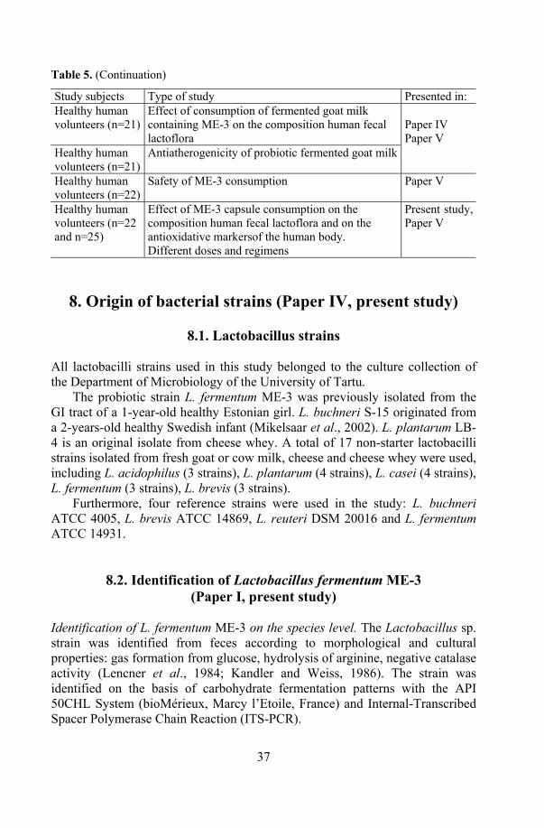
Table 5. (Continuation) Study subjects Type of study Presented in: Healthy human volunteers (n=21)
Effect of consumption of fermented goat milk containing ME-3 on the composition human fecal lactoflora
Healthy human volunteers (n=21)
Antiatherogenicity of probiotic fermented goat milk
Paper IV Paper V
Healthy human volunteers (n=22)
Safety of ME-3 consumption Paper V
Healthy human volunteers (n=22 and n=25)
Effect of ME-3 capsule consumption on the composition human fecal lactoflora and on the antioxidative markersof the human body. Different doses and regimens
Present study, Paper V
8. Origin of bacterial strains (Paper IV, present study)
8.1. Lactobacillus strains All lactobacilli strains used in this study belonged to the culture collection of the Department of Microbiology of the University of Tartu.
The probiotic strain L. fermentum ME-3 was previously isolated from the GI tract of a 1-year-old healthy Estonian girl. L. buchneri S-15 originated from a 2-years-old healthy Swedish infant (Mikelsaar et al., 2002). L. plantarum LB-4 is an original isolate from cheese whey. A total of 17 non-starter lactobacilli strains isolated from fresh goat or cow milk, cheese and cheese whey were used, including L. acidophilus (3 strains), L. plantarum (4 strains), L. casei (4 strains), L. fermentum (3 strains), L. brevis (3 strains).
Furthermore, four reference strains were used in the study: L. buchneri ATCC 4005, L. brevis ATCC 14869, L. reuteri DSM 20016 and L. fermentum ATCC 14931.
8.2. Identification of Lactobacillus fermentum ME-3 (Paper I, present study)
Identification of L. fermentum ME-3 on the species level. The Lactobacillus sp. strain was identified from feces according to morphological and cultural properties: gas formation from glucose, hydrolysis of arginine, negative catalase activity (Lencner et al., 1984; Kandler and Weiss, 1986). The strain was identified on the basis of carbohydrate fermentation patterns with the API 50CHL System (bioMérieux, Marcy l’Etoile, France) and Internal-Transcribed Spacer Polymerase Chain Reaction (ITS-PCR).

38
ITS-PCR, followed by enzymatic restriction Taq I was used to confirm the identification of the species. The DNA extraction from Lactobacillus isolates was prepared as described by Alander et al. (1999) using lysozyme (Serva, Sweden; 20 mg/ml), mutanolysin (Sigma; 0.5 mg/ml) and proteinase K solutions (Fermentas, Lithuania; 14.6 mg/ml) and the work was carried out with the help of J. Shchepetova (extraordinary research fellow at the Department of Microbiology, University of Tartu). The DNA amplification was performed according to Jacobsen et al. (1999) in a reaction volume of 50 µl containing 1xTaq polymerase buffer (Fermentas, Lithuania), 1.5U Taq polymerase (Fermentas), 0.5 µM of each primer (16S–1500F and 23S–32R; DNA Techno-logy AS) (Jacobsen et al., 1999), 200 µM deoxynucleoside triphosphates, 2 mM MgCl2 and 2 µl of extracted DNA.
Subsequently, the PCR product was restricted as described by Zhong et al. (1998) using a Taq I restriction enzyme (Fermentas). DNA fragments were separated by electrophoresis (1.5h, 100 V) on a 2% agarose gel in a 1xTBE [Tris(Hydroxymethyl)aminomethane-borate/disodium ethylendiamine tetra-acetate] buffer. A size marker 100 bp DNA Ladder Plus (Fermentas) was also separated simultaneously on the same gel.
The banding pattern of the isolates was visually compared with that of the above-mentioned Lactobacillus reference strains.
Rapid identification of L. fermentum ME-3. Colonies of less than 24-hours-old culture of ME-3 are relatively big, flat and rough with irregular edges and of different morphology. In the Gram preparation more elongated and yet undivided cells can be seen compared to the older culture. Over 24-hours-old colonies of ME-3 are greyish-white, convex with regular edges. Microscopic evaluation after Gram staining shows regular, Gram-positive plump rods, which are variable in length, mostly occurring in parallel pairs.
ME-3-like colonies were reisolated from different environments (feces, food products). For rapid identification of ME-3, three biochemical tests – gas from glucose, growth at 15°C and lysozyme production was carried out by a modified scheme of Lencner et al. (1984). The latter was elaborated for rapid differentiation of L. fermentum species from the other representatives of the OHEL group.
Lysozyme production was detected on a modified de Man-Rogosa-Sharpe (MRS) medium containing 10 % of the inactivated Micrococcus lysodeikticus culture. The transparent zone around selected potential ME-3 colonies, grown in microaerobic environment, indicated positive lysozyme production due to cell wall lysis of micrococci by enzyme (Lenzner and Lenzner, 1982). Growth at 15°C was estimated in MRS broth after 7 days of incubation. Turbidity in the growth medium was considered a positive test result.
The identification of strain L. fermentum ME-3 was confirmed by AP-PCR.
39
8.3. Basic characteriation of Lactobacillus fermentum ME-3 (Papers I, II, III)
The antibiotic resistance of L. fermentum ME-3 was determined in
cooperation with Dr. R. Mändar, Department of Microbiology. Ampicillin, cefoxitin, gentamicin, ciprofloxacin, tetracycline, ofloxacin, aztreonam, trimethoprim-sulfamethoxazole and vancomycin susceptibility of the strain was tested using antibiotic strips of the E-test (AB Biodisk, Solna, Sweden). A thioglycolate broth for suspending bacteria (McFarland 0.5 turbidity standard), Wilkins-Chalgren (Oxoid Ltd. Basingstoke, Hampshire, UK) agar plates with 5% horse blood was used (Mändar et al., 2001). After 36h of incubation at 37°C microaerobically the MIC-s (minimal inhibitory concentration) and breakpoints (susceptible/resistant) were determined in accordance with the NCCLS guideline (Tenover et al., 1999).
Two antibiotics – erythromycin and metronidazole – were tested using the Kirby-Bauer disk-diffusion test. BBL (Becton Dickinson, Cockeysville, USA) Sensi-Disk Susceptibility Test Disks were used (erythromycin 15µg, metronidazole 5µg). The inoculums, agar plates and incubation conditions were similar to those used for MIC tests.
The profile of metabolites. The production of organic acids and ethanol (mg/ml) was estimated by gas chromatography as described by Holdeman et al. (1977) in cooperation with J. Shchepetova. The gas chromatograph (Hewlett-Packard model 6890, USA) was equipped with a hydrogen flame ionization detector and an auto sampler (model 7683). The HP Chemical Station for GC System (A.06 revision) was used. Analyses were performed following cultivation of the Lactobacillus in modified the MRS broth for 48h in a 10% CO2 environment.
8.4. Pathogenic target bacteria The reference strains of Escherichia coli K12, E. coli ATCC 700336, E. coli ATCC 700414, Shigella sonnei ATCC 25931, Staphylococcus aureus B46, Salmonella Enteritidis ATCC 13076 and two clinical isolates of Salmonella enterica ssp. enterica serovar. Typhimurium were used as the target bacteria.

40
9. Properties of L. fermentum ME-3
9.1. Resistance to low pH, bile and heat in vitro (Paper V, present study)
Resistance to low pH. The ability of L. fermentum ME-3 to tolerate low pH was tested in hydrochloric (HCl), lactic and acetic acid environments.
L. fermentum ME-3 was precultured on a MRS agar medium. The overnight grown cells were harvested and suspended in saline (0.9% NaCl). The amount of 0.5 ml of suspension, according to McFarland 4 standard suspension (109 CFU/ml), was inoculated into 4.5 ml of the MRS broth, pH adjusted from 2.0 to 4.0 with either HCl, DL-lactic or acetic acid.
Bile tolerance. Bile tolerance of L. fermentum ME-3 was detected in the MRS broth with 0.3 to 2.0% of ox gall (Sigma-Aldrich Chemie GmbH, Stein-heim, Germany). Survival was detected on the MRS agar medium after 2, 4, 6, 8 and 24h. The samples were serially diluted in saline, plated on MRS agar and incubated for 24h in a variable atmosphere incubator (IG 150, Jouan, France) with the following microaerobic atmosphere CO2/O2/N2: 10/5/85 at 37°C.
Survival in simulated gastric digestion. The cumulative effect of low pH, digestive enzymes and bile was tested in simulated gastric digestion. The cell suspension of the overnight L. fermentum ME-3 culture was prepared as described above and added to 10 ml of phosphate-buffered saline (PBS) with 1.5 M NaCl (Naxo Ltd, Tartu, Estonia) and pepsin (3g/l, Sigma, EC3.4.23.1), where pH was adjusted to pH2.5 with HCl.
In parallel the effect of simulated gastric duodenal juice on the capsulated lyophilized L. fermentum ME-3 culture was tested with two different methods. One capsule was dissolved in 4.5 ml of pepsin containing PBS (pH2.5). Second, the content of a capsule was added to the mixture together with 0.3ml of skim milk to simulate consumption of milk. All variants were incubated at 37°C for 3 hours, after which the cells were centrifuged and resuspended in PBS (pH 7.5) containing 0.1% of pancreatin (BMP Production Gmbh, 19370 Parchirn, Germany) and 0.1% of ox gall (Sigma-Aldrich Chemie GmbH, Steinheim, Germany) and incubated for 3 more hours. Aliquots were taken for enumeration of viable cells at 0, 1.5h, 3h, 4.5h and 6 h from the beginning of the trial. The harvested cells of the overnight L. fermentum ME-3 culture (109 CFU/ml) in a PBS buffer (pH7.5) without enzymes and bile served as control.
Heat resistance. ME-3 was precultured overnight on the MRS agar medium. The amount of 0.17 ml of ME-3 cell suspension according to McFarland 4 was inoculated into 1.5 ml of 10% reconstituted sterile skim milk (Oxoid Ltd. Basingstoke, Hampshire, UK) or into 0.9% NaCl preheated to required temperatures (73°C, 85°C or 95°C). The samples were taken after 5 and 10 minutes.

41
The heat resistance of ME-3 in an acidic environment was estimated in two ways. The strain was precultured on the MRS agar medium and the suspension adjusted to McFarland 4 in physiological saline was prepared as described above. The survival of ME-3 in three different juices: multivitamin nectar (pH 3.57), orange juice (pH 3.85) and tropical drink with carrot (pH 3.05), produced by Tallinn Dairy Ltd. from natural juice concentrates (no stabilizers) was estimated during 4 minutes at 96°C.
Additionally, the heat resistance of a frozen (–80°C) ME-3 skim milk suspension (5x10⁹ CFU/ml) in three juices was estimated as follows. The frozen suspension was melted at room temperature (22°C). The melted suspension was added to a juice (0,4 ml/l) at different times (0h, 1h, 2h, 3h) after melting and the mixture was kept at 96°C during 15 s. ME-3 mixture in juice without heat treatment served as a control.
All experiments were carried out in a dry-block thermostat (Biosan TDB120, MyLab, SeouLin Bioscience Ltd, Seoul, Korea).
All samples were cooled, serially diluted in saline, plated on the MRS agar and incubated for 48 h at 37°C microaerobically.
9.2. Growth in cell suspensions (Present study) L. fermentum ME-3, lactococci from the starter culture of Pikantne cheese Probat 505 (Danisco A/S, Copenhagen, Denmark) and L. plantarum LB-4 were precultured for 24h on the modified MRS agar medium without triammonium-citrate and sodium-acetate (pH 7.2) microaerobically. Two suspensions in 0.9% NaCl (9.9 ml each), adjusted to McFarland 4, were prepared from the cheese starter culture and L. plantarum LB-4. One of the suspensions of either culture was thermally inactivated by boiling for 2 minutes and cooled.
All suspensions were inoculated with 0.1 ml L. fermentum ME-3 suspension (105 CFU/ml) to a final count of about 103 CFU/ml. The suspensions were incubated for 7 days at 37°C and the changes in cell count were evaluated by making serial dilution from the suspensions and inoculating 0.1 ml of each dilution on the modified MRS agar medium. A suspension of ME-3 in physiological saline served as control. Different isolated were identified by colony morphology and were microscopically evaluated after Gram staining.
9.3. Antagonistic activity (Papers I, II, III, present study) On agar, the antagonistic activity of the L. fermentum ME-3 culture and its isolates from fermented milk products, cheese and capsules against pathogens was assessed using a streak line method on the modified MRS agar medium without triammonium-citrate and sodium-acetate (pH 7.2). A single line of ME-
42
3 culture, grown in the MRS broth for 48h, was seeded in the middle of an agar plate and cultivated for 48h at 37°C in a 10% CO2 environment and subsequently inactivated using chloroform gases for 2h. The target pathogens were cultured in a peptone broth (CM9 Unipath Basingstoke, Hampshire, U.K.) for 24h at 37°C and seeded in duplicate perpendicular to the streak line of lactobacillus. Following incubation of the plates at 37°C for 24h, the width of the zone of inhibition (mm) of the target bacteria extending from the culture line of the lactobacilli seeded in the middle of the agar plate was measured (Mikelsaar et al., 1987). The width of the growth inhibition zone 0 to 13.0 mm was considered low; 13.0 to 25.0 mm – intermediate and >25.0mm – high (Hütt et al., 2005).
The antagonistic activity of the non-starter lactobacilli isolates from cheese and cheese whey, cow milk or goat milk was measured using the same method. Non-starter lactobacilli were precultured in the MRS broth for 48h and the test was performed on the MRS agar medium. The plates were incubated at 37°C microaerobically for 48h. The inhibitory activity of L. fermentum ME-3 on the growth of the cheese starters (Probat 505 and starter mix CHN 11, CHN 19 and Flora Danica, Christian Hansen Holding AS, Denmark) and the yoghurt starter Jo-Mix VK 1-30 (Danisco A/S, Copenhagen, Denmark) was tested on modified MRS (without either tri-ammonium-citrate or sodium-acetate, pH 7.2). All starters were tested as mixed cultures and no different species were isolated beforehand.
Milk and broth. The antagonistic activity of L. fermentum ME-3 in milk was assessed against starter cultures and against the pathogens E. coli K12, S. sonnei ATCC 25931, S. aureus B46, and two strains of Salmonella Typhimurium. The target pathogens were cultured in a peptone broth.
The starter cultures were precultured in sterilized (120°C for 15 min) milk (fat content 2.5%) for 48h at 37°C. L. fermentum ME-3 was precultured as described above. Milk was inoculated with equal aliquots of ME-3 and starter culture suspensions adjusted to McFarland 4 (109 CFU/ml) and co-incubated. Serial dilutions were made and plated onto the modified MRS agar medium and incubated microaerobically. The identification of Lactobacillus and cocci was microscopically confirmed by cell morphology after Gram staining.
The survival of target pathogens was detected on peptone agar aerobically for 24 h at 37°C. Different colonies were counted.
The antagonistic activity of ME-3 against pathogens in broth was determined in the modified MRS broth and in milk. Equal aliquots of pathogens and L. fermentum ME-3 suspensions were co-incubated. Subsequently, the number of colony forming units (CFU/ml) of pathogens was semi-quantitatively determined on peptone agar (Gould, 1965).

43
10. Growth and survival in different products Capsule. Gelatine coated capsules were manufactured by the Tallinn Pharmaceutical Company Ltd. The probiotic capsules contained freeze-dried L. fermentum ME-3 (109 CFU) with the addition of 250 mg of saccharose and microcellulose. Identical placebo capsules contained only saccharose and microcellulose. All capsules were stored at +4°C.
Survival of ME-3 in capsule was measured by dissolving the content of one capsule aseptically in 2 ml of a 0.9% NaCl solution. The suspension was vortexed, serially diluted and plated 0.1 ml on the MRS agar medium (Oxoid Ltd. Basingstoke, Hampshire, U.K.). The plates were incubated for 48 hours at 37°C microaerobically (10% CO2) and the colonies were counted. Viable cell count per capsule was calculated.
Juices. The ability of L. fermentum ME-3 to grow in different juices was estimated in multivitamin nectar, orange juice and tropical drink with carrot produced by Tallinn Dairy Ltd.
A suspension according to McFarland 4 was prepared from L. fermentum ME-3, precultured for 24 h microaerobically on the MRS agar. The juices were inoculated with L. fermentum ME-3 to a final count of about 5x10⁵ CFU/ml and incubated for 48 h at 37°C. Changes in cell count were evaluated at the beginningof the trial, at 24h and 48h by the serial dilution method as described above.
Survival of L. fermentum ME-3 in the juices was estimated during one month. The juices were inoculated with an approximate concentration of viable cells 107 CFU/ml of the strain and stored at 4°C. Samples were taken at 48 h, 2 weeks and one month from the beginning of the trial, and were serially diluted as described above.
10.1. Preparation and survival in probiotic cheese (Paper III, present study)
The Vana-Kuuste Dairy Ltd developed a probiotic cheese, based on the Estonian open texture smear-ripened semi-soft cheese Pikantne as described in Paper III.
Probiotic cheese, based on the Estonian open texture semi-hard cheese Atleet (a cheese from the Svecia group) was developed by Võru Juust Ltd. from 11.7 tons of whole milk with 170 l of the precultured starter mix CHN 11, CHN 19 and Flora Danica (Christian Hansen Holding AS, Denmark) containing Lactococcus lactis ssp. lactis; Lactococcus lactis ssp. cremoris; Lactococcus lactis ssp. lactis biovar diacetylactis; Leuconostoc mesenteroides ssp. cremorsi. The amount of 0.4% of L. fermentum ME-3 (109 CFU/ml) was added together with the starter.
44
Inoculation rate for both cheeses with ME-3 was 108 CFU per gram of cheese. The ripening of Pikantne cheese lasts 30 days and the shelf life is also 30 days. For Atleet cheese the respective periods are 45 days and 8 months. The survival of the strain ME-3 in both probiotic cheeses and its number per gram during ripening and storage was analysed on the 10th, 24th, 38th, 54th and 66th day of cheese preparation for Pikantne cheese and on the 12th, 20th, 29th, 40th, 63rd, 118th, 217th and 242nd day for probiotic Atleet cheese.
10.2. Preparation and survival in fermented goat milk (Papers IV, V, present study)
Combining the probiotic ME-3 strain with two supportive lactobacilli cultures of different origin allowed development of experimental fermented probiotic goat milk. L. buchneri S1-5 tempered the specific taste of goat milk. L. plantarum LB-4 was included as an efficient producer of exopolysaccharides, which gives the fermented milk cream-like consistence and delicate acidity. L. fermentum ME-3 was used as the probiotic additive. Each Lactobacillus strain was incubated for 48 h in the MRS medium at 37°C in microaerobic conditions. Goat milk was inoculated with a 2% mixture of Lactobacillus strains and incubated at 37ºC for 24 hours. The ready product was cooled and stored at 4°C. The count of probiotic L. fermentum ME-3 amounted to 3x10⁹ CFU/ml of the product.
To measure the viable cell count of ME-3 in fermented goat milk, samples were taken at the end of fermentation (and before cooling the product), after 24h, 48h, 72h and 7 days of preparation when the product was stored at 4˚C. The amount of 0.5 ml of fermented milk was serially diluted in saline and plated on the MRS agar medium and incubated for 48 h at 37°C in microaerobic conditions.
10.3. Preparation and survival in HELLUS fermented milk products (Present study)
HELLUS is a brand name for four different fermented milk products (yoghurt, kefir, quark, sour cream), containing L. fermentum ME-3 as a probiotic additive, produced by Tallinn Dairy Ltd.
Yoghurts (fat content 3%) were produced using 0.1% of the thermophilic starter cultures Lactobacillus delbrueckii ssp. bulgaricus and Streptococcus thermophilus. Yoghurts with four different flavours (flavour additive content 15%) were prepared: “Rhubarb-oat” with rhubarb-oat jam; “Pasha” with apricot-orange jam and 8% of raisins; “Wild berries” with youngberry, blueberry and raspberry jam

45
or “Cowberry-muesli” containing whole grains of wheat, barley and oat and cowberry jam.
Using the mesophilic cultures of Lactococcus lactis ssp. lactis and L. lactis ssp. cremoris as the starters, three different flavoured quarks (fat content 4%) were produced with 19% of different jams: “Tropical” with peach jam, “Raspberry” and”Wild strawberry.”
Mesophilic aromatic L. lactis ssp. lactis, L. lactis ssp. cremoris, L. lactis ssp diacetylactis and Leuconostoc mesenteroides ssp. cremoris were used as the sour cream (fat content 20%) starter cultures.
Mixture of mesophilic lactic acid bacteria L. lactis ssp. lactis, L. lactis ssp. cremoris, L. lactis ssp diacetylactis and Leuconostoc mesenteroides ssp. Cremoris, Lactobacillus kefir, two yeasts Klyveromyces marxianus and Saccharomyces unisporus were used as starter cultures for the production of kefir (fat content 2.5%). All starters originated from Danisco A/S.
The freeze probiotic ME-3 culture (5x10⁹ CFU per ml of saline) was activated in a small amount of milk and added in vat simultaneously with starter cultures. The dose depended on the product’s fat content: 400 ml per ton for yog-hurt and kefir, or 500 ml per ton of product for sour cream and quark dessert.
The viability of ME-3 in HELLUS brand products (yoghurt, kefir, quark dessert, sour cream) was analyzed at different times of the product’s shelf life. The products were analysed during 6 months. The shelf life of kefir and quark is 15 days that of yoghurt and sour cream 20 days. The amount of 0.5 ml of a product was serially diluted in saline and plated on the MRS agar medium and incubated for 48 h at 37°C in microaerobic conditions.
11. Total antioxidative activity of ME-3
11.1. In vitro testing of antioxidative activity (Paper III, present study)
The antioxidative activity of L. fermentum ME-3 was measured in vitro. The antioxidative influence of ME-3 was estimated in body fluids in trials with healthy human volunteers. All further work was carried out in cooperation with Dr. T. Kullisaar from the Department of Biochemistry, University of Tartu.
The linolenic acid test (LA-test) was used to assess the total antioxidative activity (TAA) of L. fermentum ME-3, the reisolates of ME-3 from cheese; HELLUS brand fermented milk products, probiotic capsule and juices following the procedure as described in Paper III.
The TAA of samples was expressed as the percentageof inhibition of the linolenic acid (LA) peroxidation per sample. The high numerical value (%) indicates the high total antioxidative activity of the sample. Peroxidation of LA-standard in isotonic saline served as control.
46
11.2. Measurement of selected oxidative stress markers in humans (Paper IV, V)
In urine, changes in the concentrations of the oxidative stress marker
8-isoprostanes (ng/ml) were assessed by a competitive enzyme-linked immu-noassay (ELISA) (BIOXYTECH 8-Isoprostane Assay, Cat No 21019) as described in Paper IV.
Blood serum was analysed for total antioxidative activity TAA, total antioxidative status TAS and glutathione red-ox (GSSG/GSH). The TAA of the serum was assessed by the linolenic acid test (LA-test) described in Paper IV. The TAS of the serum was measured with a commercially available kit (TAS, Randox Laboratories Ltd. Ardmore, UK) as described in Paper IV, water-soluble vitamin E (Trolox) serving as the standard. This method is based on inhibition of absorbance of the ferrylmyoglobin radicals of 2,2’-azinobis-ethylbenzothiazoline 6-sulfonate (ABTS+) generated by activation of met-myoglobin peroxidase with H2O2.
The cellular oxidative stress markers as total glutathione and oxidized glutathione were measured using the method of Griffith (1980) as described in Paper IV. Glutathione content was calculated on the basis of a standard curve generated using a known concentration of glutathione. The amount of GSH (µg/ml) was calculated as a difference between total glutathione (TGSH) and GSSG (TGSH– GSSG = GSH). The glutathione red/ox ratio was expressed as GSSG/GSH.
12. Detection of health effects of ME-3
12.1. Design of human volunteer trials (Papers IV, V)
12.1.1. Safety study with probiotic capsule. A double-blind, 10-day randomized placebo controlled study (DBRP) was carried out with 7 men and 15 women, mean age 42 years (range 24–64) to evaluate the possible side effects of L. fermentum ME-3 consumption. Healthy volunteers were selected according to their self-assessment and wish to participate in the study and were randomly allocated by the computer. The members of the study group (3 male and 8 female) took daily three probiotic capsules containing 109CFU per capsule (daily dose 3x10⁹ CFU); the placebo group (4 males and 7 females) received identical capsules without any probiotic strain. The participants of the trial were daily questioned about their general welfare, intestinal function (gut gas production, stool frequency) and putative adverse effects. Fecal samples were collected before and after the trial to assess the survival of the probiotic strain in the human GI tract and the effect of the
47
strain on the fecal lactoflora. Urine samples were collected before and after the trial to estimate the effect of ME-3 consumption on 8-isoprostanes concentrations.
12.1.2. Functional efficacy trials Two healthy volunteer (n 45) trials (an open placebo controlled study and a DBRP study) were carried out to evaluate the functional efficacy of L. fermentum ME-3 in the human body. The inclusion criteria included the wish to participate, absence of known health problems and absence of medical conditions requiring drug therapy, non-use of other yoghurts or special diets. The subjects with a history of GI tract disease or food allergy, use of any antimicrobial agent within the last month or use of any regular concomitant medication were excluded. The blood samples (6 ml) from the antecubital vein, and fecal and urine samples were collected before and at the end of all clinical trials.
Open placebo controlled fermented goat milk trial. The study participants were 5 men and 16 women, mean age 50 years (range 35–60). During three weeks of the trial, the study group (3 men and 13 women) consumed daily 150 ml of fermented goat milk. The average daily dose of the probiotic Lacto-bacillus strain was 3x10¹¹CFU per person. The control group (1 man and 4 women) consumed the same dose of fresh goat milk.
Probiotic capsule trial. The DBRP study was carried out as follows. The study group consisted of 15 men and 9 women, mean age 52 years (range 40–60) who were allocated according to their wish to participate and divided randomly into two groups by an independent person who used a computer program. The members of the study group (8 male and 4 female) took three probiotic containing capsules (2.6x10⁸CFU per capsule) two times daily (daily dose 1.5x10⁹CFU) during three weeks. The placebo group (7 men and 5 women) received identical capsules without the probiotic strain. Participants were recruited for endothelium testing by pulse wave analysis and by analysis of the biomarkers (total of 17 parameters). Changes in the selected biomarkers after 3-week consumption of ME-3 are described in the present study. This study was carried out in cooperation with the Dept. of Biochemistry and the centre of endothelial research of Tartu University Clinics.
The fecal samples of all participants for assessing changes in the fecal lactoflora and the persistence of the ingested probiotic strain were collected before and at the end of the trials. Several laboratory indices of blood and urine were measured before and after the consumption of ME-3. Changes in human body oxidative stress markers as total antioxidative activity (TAA); total antioxidative status (TAS) and glutathione red-ox ratio (GSSG/GSH) from blood serum and 8-isoprostanes in urine were reported.
48
The participants of all trials, including the safety study gave informed consent to the study protocols approved by the Ethics Committee of the University of Tartu.
12.2. Microbiological analyses of feces (Papers IV, V)
The fecal samples were collected at the beginning (day 0) and at the end of the trials (day 21 in the case of the goat milk trial with healthy volunteers, day 10 in the case of safety trial and day 21 in the case of the probiotic capsule efficacy trial) and stored at –80°C unti analysed. The serial dilutions of the weighed fecal samples were prepared with phosphate buffer (pH 7.2) and 0.05 ml of each dilution was plated onto the MRS agar medium (Mikelsaar et al. 1972). The plates were incubated at 37°C for 4 days microaerobically. Representative colonies were selected on the basis of colony morphology, cell microscopy and Gram staining. Detection level was 1x10³ CFU/g feces.
The relative amount of lactobacilli colonizing the GI tract of the persons in the study groups were expressed as a proportion of total count (%), using the Bioquant statistical program (Mändar et al., 1992), which gives output data as an absolute count (log10 CFU/g) for every micro-organism and the percentage of different species in the total count.
12.3 Identification of lactobacilli (Papers III, IV, V, present study)
Lactobacillus sp. isolates from different environments (fermented milk products, cheese and feces) were identified according to their morphology and the properties of the culture: carbohydrate fermentation patterns, gas formation from glucose, hydrolysis of arginine, negative catalase activity (Lencner et al., 1984; Kandler and Weiss, 1986).
Putative ME-3 isolates were typed according to arbitrarily primed poly-merase chain reaction (AP-PCR). Genomic DNA was extracted from 24h old cultures, cultivated on MRS agar microaerobically with the QIAamp DNA Mini Kit 50 (QIAGEN GmbH., Hilden, Germany) according to the manufacturer`s instructions. AP-PCR typing was done with two primers: ERIC1R (5'-ATGTAAGCTCCT GGGGATTCAC-3') and ERIC2 (5'-AAGTAAGTGACTGGGGTGAGCG -3') (DNA Technology A/S, Aarhus, Denmark). A 30 µl volume of the reaction mixture consisted of 10xPCR buffer (Fermentas, Vilnius, Lithuania), 2.5 mM MgCl2 (Fermentas, Vilnius, Lithuania), 200µM deoxynucleoside triphosphate mixture (dATP, dGTP, dTTP and dCTP, Amersham Pharmacia Biotech, Freiburg, Germany) 0.60µg of each
49
primer and 2.5U Taq DNA Polymerase (Fermentas, Vilnius, Lithuania,) and 5 µl of extracted DNA according to Matsumiya et al. (2002). The PCR mixture was subjected to thermal cycling 35 cycles of denaturation at 95°C for 1 min, annealing at 35°C for 1 min, and extension at 74°C for 2 min, with a final extension at 74°C for 5 min with the PTC-200 thermal cycler (Eppendorf AG, Hamburg, Germany). The PCR products were separated by electrophoresis in a horizontal 2% agarose gel containing 0.1µl/ml ethidium bromide in a Tris-acetic acid–EDTA (TAE) buffer (40mM Tris, 20mM boric acid, 1mM EDTA, pH 8.3) (Bio-Rad Laboratories, Hercules, USA) at constant voltage of 120V. A 1kb ladder (GeneRuler, Fermentas, Vilnius, Lithuania) was used as the base pair size marker. The banding patterns of the isolates were visualised with UV light and compared with those of the L. fermentum ME-3 strain.
13. Statistical Analysis The computer program Sigma Stat for Windows 2.0 (Jandel Coprporation, USA) was applied. The count of the fecal lactoflora was compared by using Student’s t-test and Mann-Whitney rank sum test. Changes in oxidative stress markers of blood sera (TAA, TAS and glutathione red-ox ratio) and urine (8-isoprostanes) were evaluated by Student’s t-test, paired t-test and the Mann-Whitney rank sum test. The choice of the tests was made automatically according to the distribution of the data. Both microbial and biochemical markers are presented as mean and standard deviation.
One-way ANOVA test was performed to compare the effect of different formulations on TAA, TAS and the fecal lactoflora.
Differences were considered statistically significant if the value was p <0.05.

50
RESULTS AND DISCUSSION
14. Basic characterization of L. fermentum ME-3 L. fermentum ME-3 was identified on the species level according to the API CHL 50 System kit (ID% 99.6, one test contra). The strain ferments the following sugars: ribose, galactose, D-glucose, D-fructose, D-mannose, esculine, maltose, lactose, melibiose, saccharose, D-raffinose, D-tagatose and gluconate. The identification of ME-3 on the species level was confirmed by ITS-PCR (Fig. 3, a) and by AP-PCR (Fig. 3, b).
1 2 3 4 5 6 7 8 (a) (b) 1 2 3 4 5 6 7
Figure 3. Identification of ME-3 on the species level according to (a) ITS-PCR: Starting from left (1) fecal isolate; (2) L. fermentum ME-3; (3) fecal isolate; L. fermentum ATCC 14931; (4) isolate from fermented milk; (5) L. reuteri DSM 20016; fecal isolate, (8) DNA 100 kb Ladder Plus and (b) according to AP-PCR on the species level Starting from left (1) DNA 1 kb Ladder; (2) L. buchneri ATCC 4005; (3) L. brevis ATCC 14869; (4) L. reuteri DSM 20016; (5) L. fermentum ATCC 14931; (6) L. fermentum ME-3 The production of organic acids (mg/ml) in different growth environments was estimated by gas chromatography. L. fermentum ME-3 has heterofermentative metabolism, the fermentation profile being dependent on the environmental conditions (Table 6).
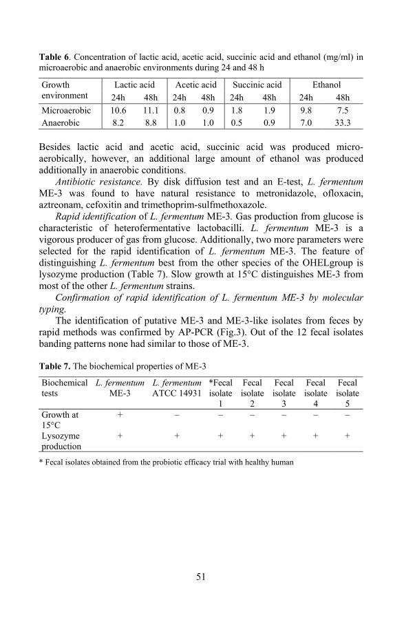
51
Table 6. Concentration of lactic acid, acetic acid, succinic acid and ethanol (mg/ml) in microaerobic and anaerobic environments during 24 and 48 h
Lactic acid Acetic acid Succinic acid Ethanol Growth environment 24h 48h 24h 48h 24h 48h 24h 48h Microaerobic 10.6 11.1 0.8 0.9 1.8 1.9 9.8 7.5 Anaerobic 8.2 8.8 1.0 1.0 0.5 0.9 7.0 33.3
Besides lactic acid and acetic acid, succinic acid was produced micro-aerobically, however, an additional large amount of ethanol was produced additionally in anaerobic conditions.
Antibiotic resistance. By disk diffusion test and an E-test, L. fermentum ME-3 was found to have natural resistance to metronidazole, ofloxacin, aztreonam, cefoxitin and trimethoprim-sulfmethoxazole.
Rapid identification of L. fermentum ME-3. Gas production from glucose is characteristic of heterofermentative lactobacilli. L. fermentum ME-3 is a vigorous producer of gas from glucose. Additionally, two more parameters were selected for the rapid identification of L. fermentum ME-3. The feature of distinguishing L. fermentum best from the other species of the OHELgroup is lysozyme production (Table 7). Slow growth at 15°C distinguishes ME-3 from most of the other L. fermentum strains.
Confirmation of rapid identification of L. fermentum ME-3 by molecular typing.
The identification of putative ME-3 and ME-3-like isolates from feces by rapid methods was confirmed by AP-PCR (Fig.3). Out of the 12 fecal isolates banding patterns none had similar to those of ME-3. Table 7. The biochemical properties of ME-3
Biochemical tests
L. fermentum ME-3
L. fermentum ATCC 14931
*Fecalisolate
1
Fecal isolate
2
Fecal isolate
3
Fecal isolate
4
Fecal isolate
5 Growth at 15°C
+ – – – – – –
Lysozyme production
+ + + + + + +
* Fecal isolates obtained from the probiotic efficacy trial with healthy human

52
1 2 3 4 5 6 7 8 9 10 11 12 13 14
Figure 4. Distinguishing of L. fermentum ME-3 from the other L. fermentum strains isolated from feces. Starting from left (1) DNA 1 kb Ladder; (2) L. fermentum ME-3; (3-14) fecal lactobacillus isolates obtained from the probiotic capsule efficacy trial participants
15. Technological properties of L. fermentum ME-3 (Present study)
15.1. Acid tolerance
In food products, survival of a probiotic strain may be influenced by acidic stress caused by accumulation of the metabolic end products of starters or by strain itself.
During 24h, L. fermentum ME-3 tolerated well both lactic acid and acetic acid at pH 4.5 and pH 4. In more acidic conditions (pH<3.5), a decrease in cell count from 1 log (lactic acid) to 1.5 log (acetic acid) was seen after 6 hours. Thus, ME-3 was able to retain its viable count for at least 6 h in an environment with relatively low pH values caused by lactic or acetic acid.
15.2. Dependence of heat resistance on pH
The heat resistance of L. fermentum ME-3 was tested to evaluate the potential use of the strain in probiotic juices, where technology foresees pasteurisation and addition of the strain to hot juice. The ability of ME-3 to resist high temperatures was tested in skim milk at pH 7.0 and in different juices at pH 3.05…3.85.
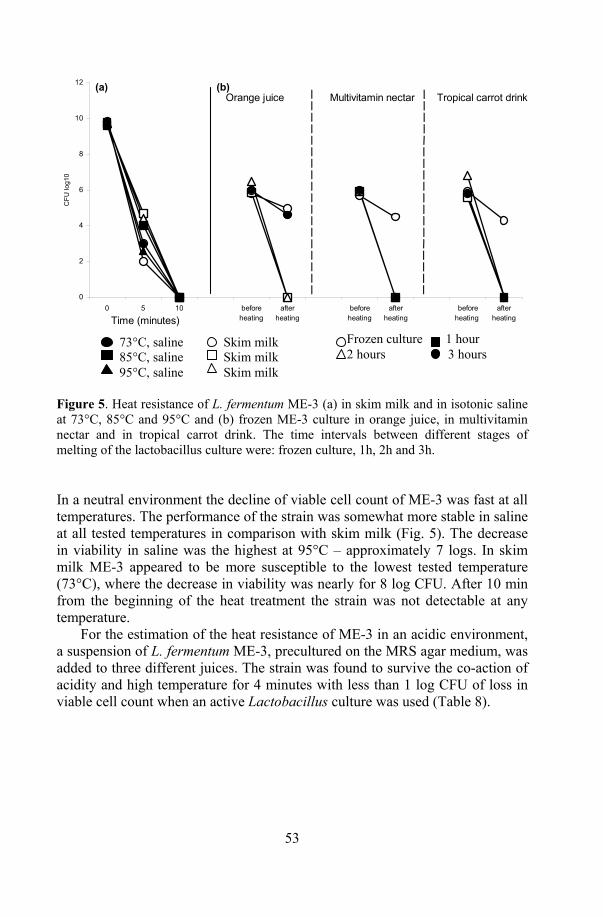
53
0
2
4
6
8
10
12
0 5 10 beforeheating
afterheating
beforeheating
afterheating
beforeheating
afterheating
CFU
log1
0
Time (minutes)
Orange juice Multivitamin nectar Tropical carrot drink(a) (b)
Frozen culture 1 hour 2 hours 3 hours
73°C, saline 85°C, saline 95°C, saline
Skim milk Skim milk Skim milk
Figure 5. Heat resistance of L. fermentum ME-3 (a) in skim milk and in isotonic saline at 73°C, 85°C and 95°C and (b) frozen ME-3 culture in orange juice, in multivitamin nectar and in tropical carrot drink. The time intervals between different stages of melting of the lactobacillus culture were: frozen culture, 1h, 2h and 3h.
In a neutral environment the decline of viable cell count of ME-3 was fast at all temperatures. The performance of the strain was somewhat more stable in saline at all tested temperatures in comparison with skim milk (Fig. 5). The decrease in viability in saline was the highest at 95°C – approximately 7 logs. In skim milk ME-3 appeared to be more susceptible to the lowest tested temperature (73°C), where the decrease in viability was nearly for 8 log CFU. After 10 min from the beginning of the heat treatment the strain was not detectable at any temperature.
For the estimation of the heat resistance of ME-3 in an acidic environment, a suspension of L. fermentum ME-3, precultured on the MRS agar medium, was added to three different juices. The strain was found to survive the co-action of acidity and high temperature for 4 minutes with less than 1 log CFU of loss in viable cell count when an active Lactobacillus culture was used (Table 8).
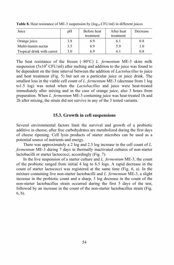
54
Table 8. Heat resistance of ME-3 suspension by (log10 CFU/ml) in different juices
Juice pH Before heat treatment
After heat treatment
Decrease
Orange juice 3.8 6.9 6.1 0.8 Multivitamin nectar 3.5 6.9 5.9 1.0 Tropical drink with carrot 3.0 6.9 6.1 0.8
The heat resistance of the frozen (–80°C) L. fermentum ME-3 skim milk suspension (5x10⁹ CFU/ml) after melting and addition to the juice was found to be dependent on the time interval between the addition of Lactobacillus to juice and heat treatment (Fig. 5) but not on a particular juice or juice drink. The smallest loss in the viable cell count of L. fermentum ME-3 (decrease from 1 log to1.5 log) was noted when the Lactobacillus and juice were heat-treated immediately after mixing and in the case of orange juice, also 3 hours from preparation. When L. fermentum ME-3 containing juice was heat-treated 1h and 2h after mixing, the strain did not survive in any of the 3 tested variants.
15.3. Growth in cell suspensions Several environmental factors limit the survival and growth of a probiotic additive in cheese, after free carbohydrates are metabolized during the first days of cheese ripening. Cell lysis products of starter microbes can be used as a potential source of nutrients and energy.
There was approximately a 2 log and 2.5 log increase in the cell count of L. fermentum ME-3 during 7 days in thermally inactivated cultures of non-starter lactobacilli or starter lactococci, accordingly (Fig. 7).
In the live suspension of a starter culture and L. fermentum ME-3, the count of the probiotic ranged from initial 4 log to 6.5 logs. A rapid decrease in the count of starter lactococci was registered at the same time (Fig. 6, a). In the mixture containing live non-starter lactobacilli and L. fermentum ME-3, a slight increase in the probiotic count and a sharp, 3 log decrease in the count of the non-starter lactobacillus strain occurred during the first 5 days of the test, followed by an increase in the count of the non-starter lactobacillus strain (Fig. 6, b).

55
Figure 6. Growth of L. fermentum ME-3 in limited nutritional conditions on the cell suspension of non-starter lactobacillus (NSLAB) (a) and lactococci (b)
15.5. Antagonistic activity
15.5.1. Antagonistic activity between L. fermentum ME-3 and
starter cultures The compatibility of the probiotic strain to starter organisms should be assessed in order to avoid undesirable changes in the composition of the product`s microflora and unwanted changes in the sensory properties of the product.
0123456789
10
0 1 2 5 6 7
Time (days)
log1
0
ME-3 + inactivated NSLABviable NSLAB (+ME-3)ME-3+ (viable NSLAB)control (ME-3)
0123456789
0 1 2 5 6 7
Time (hours)
log1
0
ME-3 + inactivated starter viable starter (+ME-3)ME-3 (+ viable starter)control (ME-3)
(a)
(b)
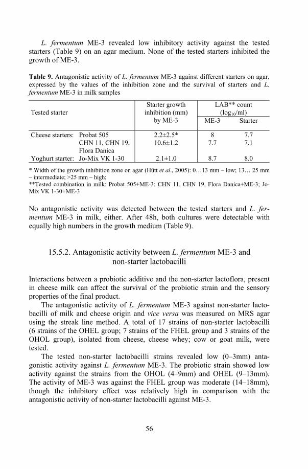
56
L. fermentum ME-3 revealed low inhibitory activity against the tested starters (Table 9) on an agar medium. None of the tested starters inhibited the growth of ME-3. Table 9. Antagonistic activity of L. fermentum ME-3 against different starters on agar, expressed by the values of the inhibition zone and the survival of starters and L. fermentum ME-3 in milk samples
LAB** count (log10/ml)
Tested starter
Starter growth inhibition (mm)
by ME-3 ME-3 Starter
Cheese starters: Probat 505 2.2±2.5* 8 7.7 CHN 11, CHN 19,
Flora Danica 10.6±1.2 7.7 7.1
Yoghurt starter: Jo-Mix VK 1-30 2.1±1.0 8.7 8.0
* Width of the growth inhibition zone on agar (Hütt et al., 2005): 0…13 mm – low; 13… 25 mm – intermediate; >25 mm – high; **Tested combination in milk: Probat 505+ME-3; CHN 11, CHN 19, Flora Danica+ME-3; Jo-Mix VK 1-30+ME-3
No antagonistic activity was detected between the tested starters and L. fer-mentum ME-3 in milk, either. After 48h, both cultures were detectable with equally high numbers in the growth medium (Table 9).
15.5.2. Antagonistic activity between L. fermentum ME-3 and
non-starter lactobacilli Interactions between a probiotic additive and the non-starter lactoflora, present in cheese milk can affect the survival of the probiotic strain and the sensory properties of the final product.
The antagonistic activity of L. fermentum ME-3 against non-starter lacto-bacilli of milk and cheese origin and vice versa was measured on MRS agar using the streak line method. A total of 17 strains of non-starter lactobacilli (6 strains of the OHEL group; 7 strains of the FHEL group and 3 strains of the OHOL group), isolated from cheese, cheese whey; cow or goat milk, were tested.
The tested non-starter lactobacilli strains revealed low (0–3mm) anta-gonistic activity against L. fermentum ME-3. The probiotic strain showed low activity against the strains from the OHOL (4–9mm) and OHEL (9–13mm). The activity of ME-3 was against the FHEL group was moderate (14–18mm), though the inhibitory effect was relatively high in comparison with the antagonistic activity of non-starter lactobacilli against ME-3.
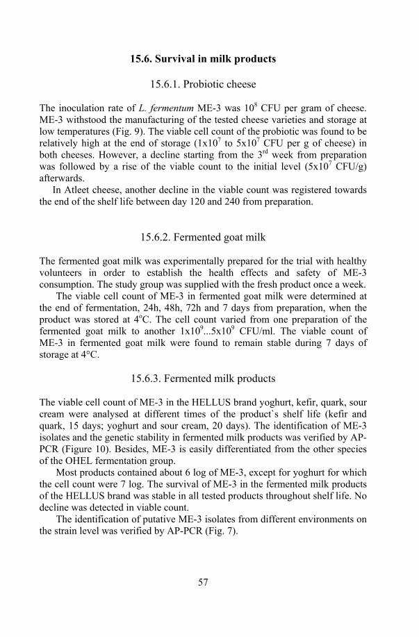
57
15.6. Survival in milk products
15.6.1. Probiotic cheese The inoculation rate of L. fermentum ME-3 was 108 CFU per gram of cheese. ME-3 withstood the manufacturing of the tested cheese varieties and storage at low temperatures (Fig. 9). The viable cell count of the probiotic was found to be relatively high at the end of storage (1x10⁷ to 5x10⁷ CFU per g of cheese) in both cheeses. However, a decline starting from the 3rd week from preparation was followed by a rise of the viable count to the initial level (5x10⁷ CFU/g) afterwards.
In Atleet cheese, another decline in the viable count was registered towards the end of the shelf life between day 120 and 240 from preparation.
15.6.2. Fermented goat milk The fermented goat milk was experimentally prepared for the trial with healthy volunteers in order to establish the health effects and safety of ME-3 consumption. The study group was supplied with the fresh product once a week.
The viable cell count of ME-3 in fermented goat milk were determined at the end of fermentation, 24h, 48h, 72h and 7 days from preparation, when the product was stored at 4oC. The cell count varied from one preparation of the fermented goat milk to another 1x10⁹...5x109 CFU/ml. The viable count of ME-3 in fermented goat milk were found to remain stable during 7 days of storage at 4°C.
15.6.3. Fermented milk products
The viable cell count of ME-3 in the HELLUS brand yoghurt, kefir, quark, sour cream were analysed at different times of the product`s shelf life (kefir and quark, 15 days; yoghurt and sour cream, 20 days). The identification of ME-3 isolates and the genetic stability in fermented milk products was verified by AP-PCR (Figure 10). Besides, ME-3 is easily differentiated from the other species of the OHEL fermentation group.
Most products contained about 6 log of ME-3, except for yoghurt for which the cell count were 7 log. The survival of ME-3 in the fermented milk products of the HELLUS brand was stable in all tested products throughout shelf life. No decline was detected in viable count.
The identification of putative ME-3 isolates from different environments on the strain level was verified by AP-PCR (Fig. 7).
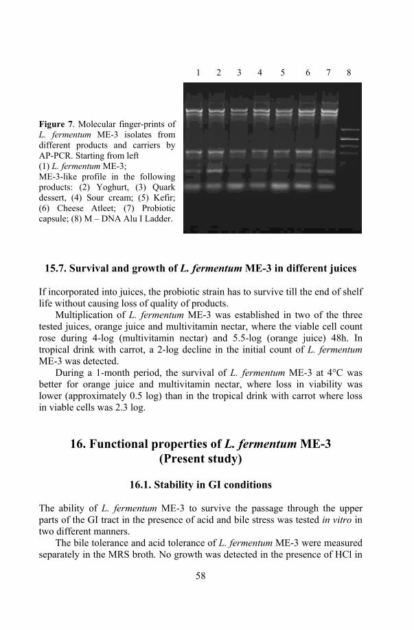
58
Figure 7. Molecular finger-prints of L. fermentum ME-3 isolates from different products and carriers by AP-PCR. Starting from left (1) L. fermentum ME-3; ME-3-like profile in the following products: (2) Yoghurt, (3) Quark dessert, (4) Sour cream; (5) Kefir; (6) Cheese Atleet; (7) Probiotic capsule; (8) M – DNA Alu I Ladder.
15.7. Survival and growth of L. fermentum ME-3 in different juices If incorporated into juices, the probiotic strain has to survive till the end of shelf life without causing loss of quality of products.
Multiplication of L. fermentum ME-3 was established in two of the three tested juices, orange juice and multivitamin nectar, where the viable cell count rose during 4-log (multivitamin nectar) and 5.5-log (orange juice) 48h. In tropical drink with carrot, a 2-log decline in the initial count of L. fermentum ME-3 was detected.
During a 1-month period, the survival of L. fermentum ME-3 at 4°C was better for orange juice and multivitamin nectar, where loss in viability was lower (approximately 0.5 log) than in the tropical drink with carrot where loss in viable cells was 2.3 log.
16. Functional properties of L. fermentum ME-3 (Present study)
16.1. Stability in GI conditions
The ability of L. fermentum ME-3 to survive the passage through the upper parts of the GI tract in the presence of acid and bile stress was tested in vitro in two different manners.
The bile tolerance and acid tolerance of L. fermentum ME-3 were measured separately in the MRS broth. No growth was detected in the presence of HCl in
1 2 3 4 5 6 7 8

59
the environment, though ME-3 tolerated well as low as pH 2.5 without loss in viability during 24h. At pH2.0 the strain survived well for 6 hours. L. fermentum ME-3 was found to tolerate all the tested bile concentrations without loss in viable count. The acid and bile tolerance of the active overnight culture and freeze-dried capsulated culture of L. fermentum ME-3 (Fig. 8) was tested on simulated gastric digestion model, applying low pH and pepsin followed by bile and pancreatin.
Different behaviours of L. fermentum ME-3 were noted on the comparison of two tests. The viable count of ME-3 were relatively stable at least for 6 h at low pH and even longer at different bile concentrations in MRS, a decrease of 0.5-log to 1.5-log of viable count of ME-3 was noticed during first 1.5 h from the beginning at the presence pH2.5 and pepsin.
0
1
2
3
4
5
6
7
8
9
0 1,5 3 4,5 6
Time (hours)
log1
0
controlME-3capsulecapsule+skim milk
Figure 8. Survival of L. fermentum ME-3 on the basis of the in vitro gastric digestion model at pH2.5 in an HCl and pepsin environment (solid line), dashed line shows following treatment with 0.15% bile and 0.1% pancreatin. At the end of the experiment, freeze-dried ME-3 with skim milk proved to be the most stable and was found to resist better the stresses according to the the in vitro gastro-intestinal model compared with the active overnight ME-3 culture and the capsulated probiotic without skim milk.
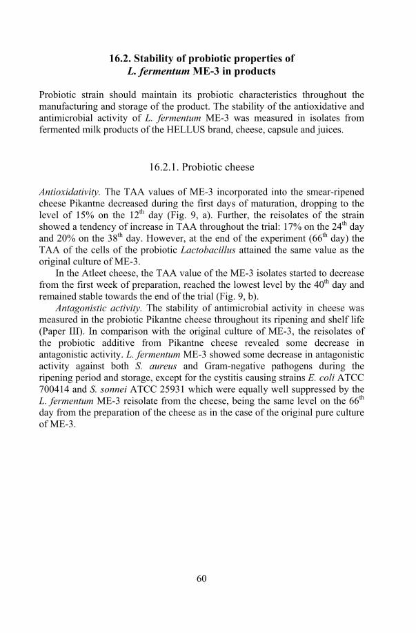
60
16.2. Stability of probiotic properties of L. fermentum ME-3 in products
Probiotic strain should maintain its probiotic characteristics throughout the manufacturing and storage of the product. The stability of the antioxidative and antimicrobial activity of L. fermentum ME-3 was measured in isolates from fermented milk products of the HELLUS brand, cheese, capsule and juices.
16.2.1. Probiotic cheese Antioxidativity. The TAA values of ME-3 incorporated into the smear-ripened cheese Pikantne decreased during the first days of maturation, dropping to the level of 15% on the 12th day (Fig. 9, a). Further, the reisolates of the strain showed a tendency of increase in TAA throughout the trial: 17% on the 24th day and 20% on the 38th day. However, at the end of the experiment (66th day) the TAA of the cells of the probiotic Lactobacillus attained the same value as the original culture of ME-3.
In the Atleet cheese, the TAA value of the ME-3 isolates started to decrease from the first week of preparation, reached the lowest level by the 40th day and remained stable towards the end of the trial (Fig. 9, b).
Antagonistic activity. The stability of antimicrobial activity in cheese was measured in the probiotic Pikantne cheese throughout its ripening and shelf life (Paper III). In comparison with the original culture of ME-3, the reisolates of the probiotic additive from Pikantne cheese revealed some decrease in antagonistic activity. L. fermentum ME-3 showed some decrease in antagonistic activity against both S. aureus and Gram-negative pathogens during the ripening period and storage, except for the cystitis causing strains E. coli ATCC 700414 and S. sonnei ATCC 25931 which were equally well suppressed by the L. fermentum ME-3 reisolate from the cheese, being the same level on the 66th day from the preparation of the cheese as in the case of the original pure culture of ME-3.
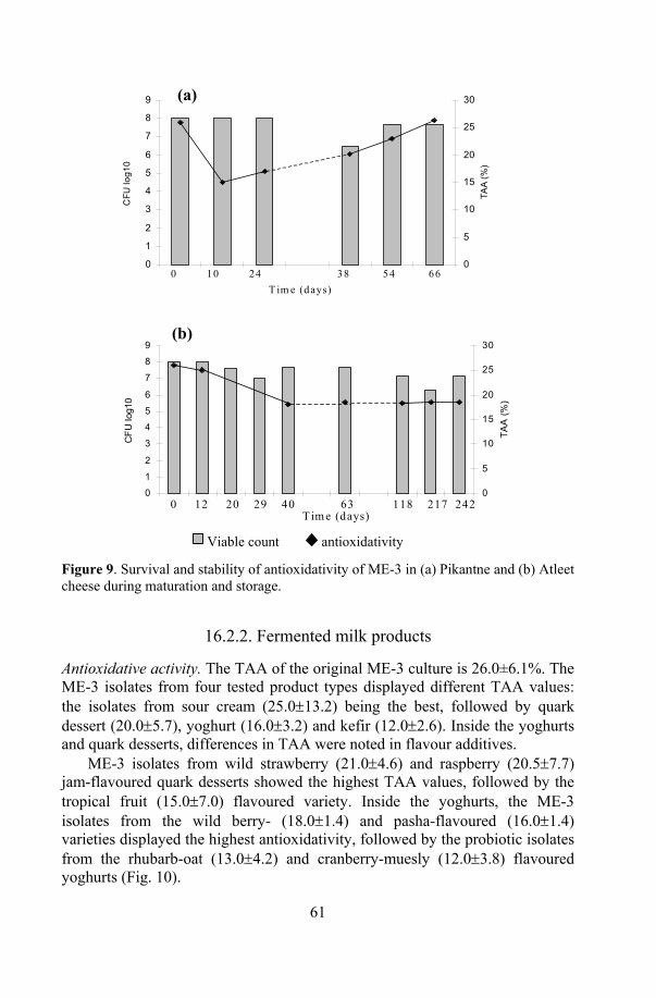
61
0
1
2
3
4
5
6
7
8
9
1 2 3 4 5 6 7
CFU
log1
0
0
5
10
15
20
25
30
TAA
(%)
0 10 24 38 54 66T im e (days)
(a)
0
1
2
3
4
5
6
7
8
9
1 2 3 4 5 6 7 8 9 10 11
CFU
log1
0
0
5
10
15
20
25
30
TAA
(%)
0 12 20 29 40 63 118 217 242Tim e (days)
(b)
Viable count antioxidativity
Figure 9. Survival and stability of antioxidativity of ME-3 in (a) Pikantne and (b) Atleet cheese during maturation and storage.
16.2.2. Fermented milk products Antioxidative activity. The TAA of the original ME-3 culture is 26.0±6.1%. The ME-3 isolates from four tested product types displayed different TAA values: the isolates from sour cream (25.0±13.2) being the best, followed by quark dessert (20.0±5.7), yoghurt (16.0±3.2) and kefir (12.0±2.6). Inside the yoghurts and quark desserts, differences in TAA were noted in flavour additives.
ME-3 isolates from wild strawberry (21.0±4.6) and raspberry (20.5±7.7) jam-flavoured quark desserts showed the highest TAA values, followed by the tropical fruit (15.0±7.0) flavoured variety. Inside the yoghurts, the ME-3 isolates from the wild berry- (18.0±1.4) and pasha-flavoured (16.0±1.4) varieties displayed the highest antioxidativity, followed by the probiotic isolates from the rhubarb-oat (13.0±4.2) and cranberry-muesly (12.0±3.8) flavoured yoghurts (Fig. 10).
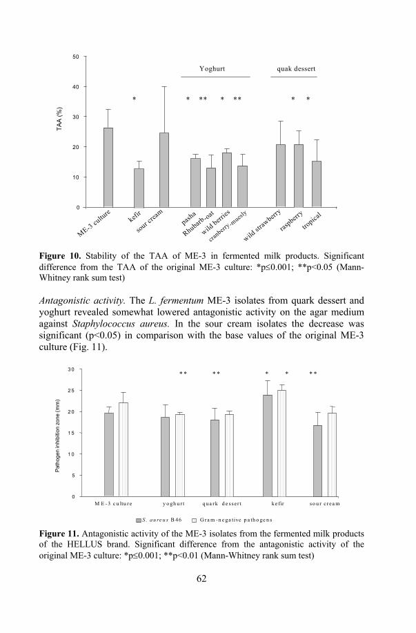
62
TAA
(%)
ME-3 cultu
rekefi
r
sour crea
mpash
a
wild berr
ies
Rhubarb-oat
cranberr
y -muesl
y
raspberr
y
tropica
l
* * ** * ** * *
Yoghurt quak dessert
0
10
20
30
40
50
wild str
awberr
y
Figure 10. Stability of the TAA of ME-3 in fermented milk products. Significant difference from the TAA of the original ME-3 culture: *p≤0.001; **p<0.05 (Mann-Whitney rank sum test)
Antagonistic activity. The L. fermentum ME-3 isolates from quark dessert and yoghurt revealed somewhat lowered antagonistic activity on the agar medium against Staphylococcus aureus. In the sour cream isolates the decrease was significant (p<0.05) in comparison with the base values of the original ME-3 culture (Fig. 11).
M E - 3 c u ltu r e y o gh u r t q ua r k d e s se r t ke fir so u r c rea m
* * * * * * * *
Path
ogen
inhi
bitio
n zo
ne (m
m)
0
5
1 0
1 5
2 0
2 5
3 0
S . a u re u s B 46 G ra m - n e ga tive p a th o ge n s Figure 11. Antagonistic activity of the ME-3 isolates from the fermented milk products of the HELLUS brand. Significant difference from the antagonistic activity of the original ME-3 culture: *p≤0.001; **p<0.01 (Mann-Whitney rank sum test)
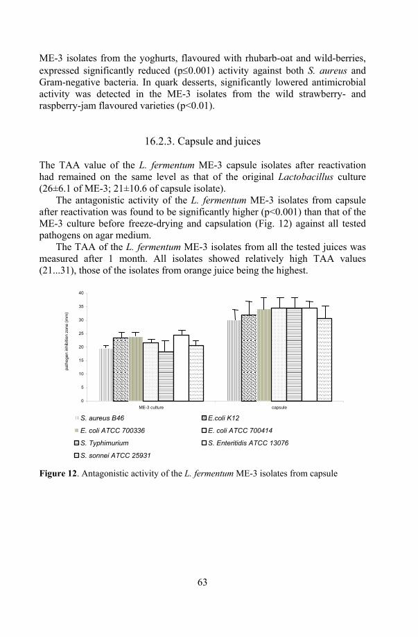
63
ME-3 isolates from the yoghurts, flavoured with rhubarb-oat and wild-berries, expressed significantly reduced (p≤0.001) activity against both S. aureus and Gram-negative bacteria. In quark desserts, significantly lowered antimicrobial activity was detected in the ME-3 isolates from the wild strawberry- and raspberry-jam flavoured varieties (p<0.01).
16.2.3. Capsule and juices The TAA value of the L. fermentum ME-3 capsule isolates after reactivation had remained on the same level as that of the original Lactobacillus culture (26±6.1 of ME-3; 21±10.6 of capsule isolate).
The antagonistic activity of the L. fermentum ME-3 isolates from capsule after reactivation was found to be significantly higher (p<0.001) than that of the ME-3 culture before freeze-drying and capsulation (Fig. 12) against all tested pathogens on agar medium.
The TAA of the L. fermentum ME-3 isolates from all the tested juices was measured after 1 month. All isolates showed relatively high TAA values (21...31), those of the isolates from orange juice being the highest.
0
5
10
15
20
25
30
35
40
ME-3 culture capsule
S. aureus B46 E.coli K12
E. coli ATCC 700336 E. coli ATCC 700414
S. Typhimurium S. Enteritidis ATCC 13076
S. sonnei ATCC 25931
path
ogen
inhi
bitio
n zo
ne (m
m)
Figure 12. Antagonistic activity of the L. fermentum ME-3 isolates from capsule
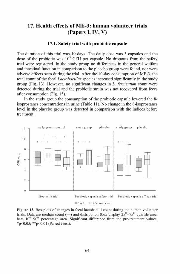
64
17. Health effects of ME-3: human volunteer trials (Papers I, IV, V)
17.1. Safety trial with probiotic capsule
The duration of this trial was 10 days. The daily dose was 3 capsules and the dose of the probiotic was 109 CFU per capsule. No dropouts from the safety trial were registered. In the study group no differences in the general welfare and intestinal function in comparison to the placebo group were found, nor were adverse effects seen during the trial. After the 10-day consumption of ME-3, the total count of the fecal Lactobacillus species increased significantly in the study group (Fig. 13). However, no significant changes in L. fermentum count were detected during the trial and the probiotic strain was not recovered from feces after consumption (Fig. 15).
In the study group the consumption of the probiotic capsule lowered the 8-isoprostanes concentrations in urine (Table 11). No change in the 8-isoprostanes level in the placebo group was detected in comparison with the indices before treatment.
CFU
log 10
G oat m ilk tria l P rob iotic capsule safety tr ia l P rob iotic capsu le effica y tr ia l
0
2
4
6
8
1 0
1 2 study group control study group placeb o study group p laceb o
D ay 0 A fter treatm en t
* * * * *
* *
Figure 13. Box plots of changes in fecal lactobacilli count during the human volunteer trials. Data are median count (—) and distribution (box display 25th–75th quartile area, bars 10th–90th percentage area. Significant difference from the pre-treatment values: *p<0.05; **p<0.01 (Paired t-test).
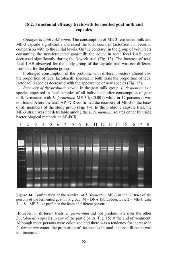
65
18.2. Functional efficacy trials with fermented goat milk and capsules
Changes in total LAB count. The consumption of ME-3 fermented milk and
ME-3 capsule significantly increased the total count of lactobacilli in feces in comparison with to the initial levels. On the contrary, in the group of volunteers consuming the non-fermented goat-milk the count in total fecal LAB even decreased significantly during the 3-week trial (Fig. 13). The increase of total fecal LAB observed for the study group of the capsule trial was not different from that for the placebo group.
Prolonged consumption of the probiotic with different vectors altered also the proportion of fecal lactobacilli species: in both trials the proportion of fecal lactobacilli species decreased with the appearance of new species (Fig. 15).
Recovery of the probiotic strain. In the goat milk group, L. fermentum as a species appeared in fecal samples of all individuals after consumption of goat milk fermented with L. fermentum ME-3 (p<0.001) while in 12 persons it was not found before the trial. AP-PCR confirmed the recovery of ME-3 in the feces of all members of the study group (Fig. 14). In the probiotic capsule trial, the ME-3 strain was not detectable among the L. fermentum isolates either by using bacteriological methods or AP-PCR. 1 2 3 4 5 6 7 8 9 10 11 12 13 14 15 16 17 18
Figure 14. Confirmation of the survival of L. fermentum ME-3 in the GI tract of the persons of the fermented goat milk group. M – DNA 1kb Ladder, Line 2 – ME-3, Line 3…18 – ME-3 like profile in the feces of different persons. However, in different trials, L. fermentum did not predominate over the other Lactobacillus species in any of the participants (Fig. 15) at the end of treatment. Although more persons were colonized and there was a tendency for increase in L. fermentum count, the proportion of the species in total lactobacilli count was not increased.
66
Antioxidative effect of ME-3. The consumption of ME-3 in both formu-lations had a positive effect on the blood oxidative stress markers TAA and TAS (Table 10).
Consumption of goat milk and fermented goat milk enhanced TAA and TAS in both the study and control groups. There was an additional increase in both indices in the fermented goat milk group, which was not statistically significant. A significant increase in TAA and TAS values occurred also during the consumption of the probiotic strain in the capsulated form. No change was detected in the placebo group.
Compared with the baseline values, the consumption of ME-3 in goat milk reduced urine 8-isoprostanes concentrations but no changes were seen in the probiotic capsule trial (Table 10).
The decrease in the glutathione red-ox ratio was significant in both the study and control groups in the goat milk trial. The goat milk fermented with L. fermentum ME-3 had no statistically significant additional effect. When the probiotic was consumed in the capsulated form, no significant decrease was noted in the glutathione red-ox ratio (Table 10).
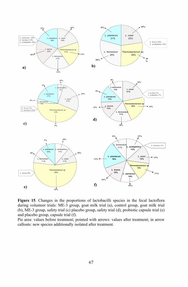
67
a)
L. brevis 20%
L. fermentum 21%
Thermobacterium sp 21%.
L. plantarum 18%
L. casei 20%
12%12%
10%
13%
13%
L. salivarius 16%L. buchneri 14%L. acidophilus 10%
c)
Therm obacterium sp. 43%
L. plantarum 21%
L. casei 27%L. ferm entum
4%
L. acidophilus 5%
9%
29%
9%
0%
29%
L. brevis 2%L. buchneri 22%
e)
Thermobcterium sp 55%
L. casei 11%
L. plantarum 12%
L. acidophilus 11%
L. fermentum 11%
15%
34%
33%
1%
8%
L. brevis 9%
L. plantarum 21%
L. fermentum 29%
L. casei 22%
Thermobacterium sp. 28%
L. brevis 22%L. acidophilus 14%
18%0%
18%28%
L. plantarum 14%
L.salivarius 12%
Thermobacterium sp. 22%L. brevis
14%
L. fermentum 17%
L. casei21%
24%
24%0%
0%
12%
20%
L. buchneri 3%L. acidophilus 17%
L. plantarum 19%
L. acidophilus 13%
L. fermentum 11%
L. casei 14%
L. salivarius 14%
L. brevis 15%
Thermobacterium sp. 14%
15%0%
13%
16%
19%
17%
15%
L. buchneri 5%
Figure 15. Changes in the proportions of lactobacilli species in the fecal lactoflora during volunteer trials: ME-3 group, goat milk trial (a), control group, goat milk trial (b), ME-3 group, safety trial (c) placebo group, safety trial (d), probiotic capsule trial (e) and placebo group, capsule trial (f). Pie area: values before treatment; pointed with arrows: values after treatment; in arrow callouts: new species additionally isolated after treatment.
a) b)
c) d)
e) f)
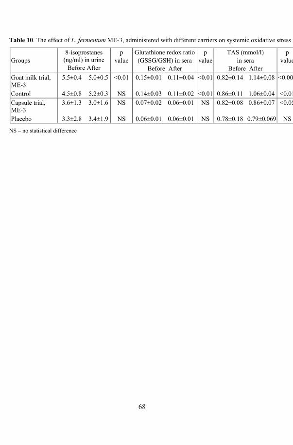
68
Table 10. The effect of L. fermentum ME-3, administered with different carriers on systemic oxidative stress markers (mean ± SD).
Groups
8-isoprostanes (ng/ml) in urine
Before After
p value
Glutathione redox ratio(GSSG/GSH) in sera
Before After
p value
TAS (mmol/l) in sera
Before After
p value
TAA (%) in sera
Before After
P value
Goat milk trial, ME-3
5.5±0.4 5.0±0.5 <0.01 0.15±0.01 0.11±0.04 <0.01 0.82±0.14 1.14±0.08 <0.005 38.0±3.0 45.0±3.0 0.01
Control 4.5±0.8 5.2±0.3 NS 0.14±0.03 0.11±0.02 <0.01 0.86±0.11 1.06±0.04 <0.01 41.0±6.0 45.0±5.0 0.01 Capsule trial, ME-3
3.6±1.3 3.0±1.6 NS 0.07±0.02 0.06±0.01 NS 0.82±0.08 0.86±0.07 <0.05 37.8±3.0 39.5±3.3 0.005
Placebo 3.3±2.8 3.4±1.9 NS 0.06±0.01 0.06±0.01 NS 0.78±0.18 0.79±0.069 NS 38.9±2.5 38.9±3.3 NS
NS – no statistical difference
69
GENERAL DISCUSSION A probiotic strain meets different challenges on its way to the target site in the human body. Therefore, a putative probiotic should be tested beforehand in vitro, only after which its suitability could be confirmed in human trials.
The purpose of the present study was to evaluate the effect of different environments on the functional properties and viability of L. fermentum ME-3. The safety and efficacy of ME-3 consumption during healthy volunteer trials were assessed.
L. fermentum ME-3 possesses two main probiotic properties: antimicrobial activity and antioxidative activity. The stability of these functional properties of L. fermentum ME-3 in the course of technological process was evaluated using elaborated simple tests by us.
There are three main technological aspects concerning suitability of probiotic strains for incorporation into food products:
1. Probiotic strains must be suitable for incorporation into various foods without a negative impact on activity product`s sensory properties.
2. Probiotic strains must be suitable for manufacturing under industrial conditions.
3. Probiotic strains must retain viability and stable functionality during storage in food products.
The main functional aspects concerning a probiotic in host are as follows:
1. Probiotic strain has to survive in the GI tract 2. Probiotic strain has to express some measurable human health promoting
properties.
18. Technological properties of L. fermentum ME-3
18.1. Suitability of ME-3 for various delivery vehicles
18.1.1. Impact on s the sensory properties of the product Lactobacilli using the heterofermentative pathway can produce unwanted by-products like acetic acid or carbon dioxide, which may negatively affect the texture or flavour and aroma formation of a certain food product. L. fermentum ME-3 belongs to the obligately heterofermentative group of lactobacilli and is a vigourous producer of carbon dioxide from glucose as well as a relatively efficient producer of lactic acid and acetic acid. Therefore the suitability of ME-3 as a probiotic additive for incorporation into different fermented milk products was tested in the present study. ME-3 was incorporated during pilot plant experiments into two cheese varieties, and into yoghurt, kefir, quark

70
dessert and sour cream. No negative impact on the organoleptic quality of any product was detected. All products were tested by food experts and proved to be commercially acceptable, while no off-flavour or -texture, caused by ME-3 metabolism was noticed.
18.1.2. Survival in products
Probiotic products are carriers of strain’s which should enter the human GI tract alive to express their specific functional properties.
The survival of the strain was satisfactory in all above products, although slight differences were detected between different products. The viability of ME-3 in fermented milk products of the HELLUS brand was found to be stable throughout the entire product`s shelf life. In yoghurts the cell count higher than 7 logs were detected already one week after production, although the initial amount of inoculation has been the lowest. Obviously, the strain multiplied during the manufacturing and/or storage of yoghurt. All other products contained about 6 logs of ME-3. Somewhat lower count compared with the initial inoculation rate was detected only for quark desserts. Apparently, a quark dessert environment does not support the survival of the strain.
The survival of L. fermentum ME-3 in both cheese varieties was similar (107 to 5x10⁷ CFU/g of cheese). The property to tolerate lower temperatures distinguishes ME-3 from some other L. fermentum strains. This made possible for the strain to survive at a relatively low temperature of cheese ripening and even lower temperature of storage (6o–8°C) for long time (maximum 8 months). The behaviour of L. fermentum ME-3 was similar in both cheese varieties in spite of differences in technological process, maturation time as well as in the duration of shelf life: a decline in viable count was noted at almost the same time from cheese preparation, followed by a rise in cell count afterwards. This phenomenon can be explained by the reorganization of the metabolism on other potential growth substrates by ME-3 after the depletion of milk sugars shortly after preparation of cheese. Non-starter lactobacilli can use sugars released via the enzymatic hydrolysis of casein, lactate, free amino acids or a variety of other compounds present in the cheese environment as the alternative energy source for carbohydrates (Laht, 2003). Like most heterofermenters, L. fermentum ME-3 utilizes arginine and is probably able to use it as an additional energy source. Besides, according to our data, ME-3 is able to survive by using the microbial cell lysis products of cheese starters. ME-3 also expresses weak proteolytic activity, however, this issue needs further investigation.
Additionally, the potential use of ME-3 for the production of probiotic juices was tested in laboratory scale studies. During 1-month refrigerated storage of probiotic juice, ME-3 retained its viability in orange juice and in multivitamin nectar; at the same time a loss of viability was detected in the
71
tropical drink with carrot. The cause for this could be the higher acidity and the chemical composition (the juice contained 9% mixture of orange, lemon and carrot juices) of the latter product in comparison with 100% of orange juice or multinectar drink, a mixture containing six different juices.
Although good survival during refrigerated storage in orange juice and multivitamin nectar is a favourable feature, multiplication of L. fermentum ME-3 in the same juices may potentially negatively affect the quality parameters like the taste and appearance of the product in the future, when the consumer stores probiotic juice at room temperature.
18.1.3. Stress responses Any extreme change in environmental conditions causes stress for an organism. Probiotic lactobacilli can be affected by various stresses due to industrial process and following storage in the final product. Therefore, the resistance of L. fermentum ME-3 to different possible stresses: heat and cold shock, acidic environment and nutrient limitation, was assessed.
In food products the survival of a probiotic strain might be influenced by the acidic stress caused by a decrease in pH due to fermentation of sugars by starters or by the strain itself. Typical acidity of cultured milk is between pH3.5 and 4.5, containing 0.5–1.5% (w/v) lactic acid. The H+ ions can cause a decrease in the viability of cells. Acidity has negative impact on bacterial physiology by altering enzymatic activities, leading to dissipation of the proton motive force and expression of acid response proteins (Champomier-Vergès et al., 2002). Lactobacilli have an intrinsic property to tolerate relatively lower pH values in comparison with some other microbes especially pathogens.
Nutrient limitation. In cheese, non-starter lactobacilli are submitted to growth-restricting conditions, like carbon source starvation after free carbohydrates are metabolized during the first days of cheese ripening. For non-starter bacteria, probiotic additives included other energy sources like microbial metabolites and microbial cell lysis products are available. It is proposed that the rate of starter cultures cell lysis might influence the growth of the secondary microflora as well as of probiotic additives during ripening of cheese (Rapposch et al., 1999). In present study the ability of ME-3 to grow in microbial cell suspensions as a potential source of nutrients and energy was tested on an intact, thermally inactivated cheese starter and on non-starter lactobacilli suspensions. We found that ME-3 was able to survive and grow in limited nutritional conditions in vitro and to use microbial cell lysis products as an energy source at least during 7 days. This could be one explanation for the multiplication of the strain in cheese during its ripening and storage.
Co-action of stresses. A probiotic strain can be submitted to several stresses at the same time. The resistance of ME-3 to the co-action of sharp temperature changes and low pH was tested in 3 juice drinks (pH ranging from 3.05 to 3.85)
72
during different time intervals. Differences in pH or in the composition of juices did not seem to affect the resistance of the strain to sharp temperature variation. The active ME-3 strain tolerated well the co-action of acidity and high temperature for 4 minutes at 96°C with a loss in viable cell count less than 1 log. Surprisingly, the heat treatment at the same temperature and even lower values (73°C and 85°C) was less tolerated in neutral conditions at pH 7.0 in skim milk and in physiological saline, where the decrease in the viable cell count was sharper (from 5 log to 8 log) compared with the juices. In both above described tests the culture submitted to heat treatment was in the active form. Probably, the chemical composition of these juices enhanced the stress tolerance of ME-3 more than reconstituted skim milk or saline.
The survival of the strain was tested in even more harsh conditions: frozen skim milk suspension was let melt at room temperature and was then added to the juices at different times and kept at 96°C during 15 seconds. The test was to clarify the optimal time interval for the maximum possible survival of the frozen culture during the production of probiotic juice, when added to pasteurized juice. The lowest loss in the viable cell count (decrease from 1 log to1.5 log) of ME-3, comparable that in the above heat treatment test with an active culture, was noted when Lactobacillus and juice mixture were heat-treated immediately just after mixing and, in the case of orange juice, also 3 hours after the preparation. On the other hand, the time 1–2h interval from between the melting of the frozen culture and heat treatment appeared to be lethal for the strain.
Cold shock is known to alter the liquid crystalline nature of the cytoplasmic membrane by transforming it to a gel phase state, DNA supercoiling and mRNAs encoding cold shock response proteins (Champomier-Vergès et al., 2002). The actual stage of recovery from cold shock seems to be crucial for the resistance of varying stresses affecting the strain. Sublethal exposure to one stress is known to induce genetic responses in bacteria that can lead to elevated tolerance and gives cross protection against other stresses (Klaenhammer and Kullen, 1999). This could be one factor that affected the survival of ME-3 during following heat challenge.
Heat resistance was tested to evaluate the possibility to produce ME-3 containing probiotic juices, where technology foresees addition of the strain to freshly pasteurised juice and following cooling of the final product. The results show that despite some decrease in viable count, ME-3 could even survive pasteurization in juices. Some technical advantages like encapsulation of the strain can increase the viability of the strain and make pasteurized juices suitable carrier`s for ME-3.
73
19. Stability of the functional properties of ME-3 in products
19.1. Antioxidative activity
The antioxidative activity among lactic acid bacteria seems to be strain specific. Kaizu with colleauges (1993) showed that only about 3% of tested strains revealed remarkable antioxidative properties.
L. fermentum ME-3 expresses high antioxidative activity in vitro. The strain retained its high antioxidative property in all fermented milk products, though in most products it was somewhat lower than the initial value of ME-3 original culture. In the HELLUS brand, the antioxidativity of ME-3 was clearly influenced by the acidity of a particular product and a flavour additive.
Acidity. The products tested by us can be categorized into acidic (yoghurts, titrable acidity up to 1.1%), slightly acidic (sour cream, quark, 0.5%) and acid-alcoholic (kefir). Lactic acid content appears to affect the TAA of ME-3, as in products with lower acidity like sour cream and quark desserts the strain expressed higher antioxidativity compared with yoghurts. The relatively lower TAA values, of kefir, compared to the majority of other HELLUS brand products can be due to kefir`s acidity and higher ethanol content. According to literature, ethyl alcohol content in kefir can be up to 1% (Botazzi, 1983; Otles and Cagindi, 2003).
Flavour additive. Besides lactic acid, flavour additives influenced antioxidativity. The isolatef of ME-3 from yoghurts with cranberry-muesly and rhubarb-oat flavours revealed the lowest TAA values. L. fermentum possesses Mn-SOD, an enzyme related to the antioxidativity of the strain (Kullisaar et al., 2002). Fibre- and oxalate rich additives (e.g. oat, wheat, barley, raisins and rhubarb) or low-pH additives (e.g. orange jam in pasha flavoured yoghurt) have the ability to absorb or enhance the absorbance of divalent microelements including Mn. Through this; the activity of Mn-SOD is probably lowered. Milling of additive cereals (e.g. rhubarb-oat yoghurt additive) increases the microelement-binding reaction.
Cheese environment appeared to be suitable for ME-3, as the probiotic strain survived well the cheese manufacturing and retained both its viability and high antioxidativity. The tested cheese varieties Atleet and Pikantne were similar with the respect to fat content (45% and 50% in dry matter, respectively), but differed in all acpects of the manufacture and length of shelf-life. The incorporation of ME-3 into cheese probably causes stress to the probiotic as its cell count and TAA decreased was during cheese ripening. The unique property to tolerate low temperatures distinguishes ME-3 from the other L. fermentum strains (Hammes et al., 1992; Kandler and Weiss, 1986) and makes possible for the strain to survive and multiply at relatively low temperature of cheese ripening and even lower temperature of storage. The
74
TAA values, which decreased rapidly during the initial phase of the maturation of Pikantne cheese, gained the high values of the original ME-3 culture towards the end of shelf-life. In Atleet cheese, on the other hand, the TAA values remained stable after the initial drop to the value of 18%. Fluctuations in viable count and TAA values probably reflected the adaptation of the probiotic strain to a certain cheese environment.
The TAA of the L. fermentum ME-3 isolates from all tested juices was measured after 1 month of refrigerated storage. All isolates were found to reveal relatively high TAA values (21... 31%), the isolates from orange juice having the highest value. Although the survival of the strain in tropical carrot drink was the lowest among the tested juices, the loss in viable count was compensated by stable antioxidative activity, which remained at the same level as that of the initial ME-3 culture.
Surprisingly, freeze-drying did not affect negatively the TAA of the strain, although the range of the antioxidative activity of the ME-3 isolates from capsule was more variable during several following isolation tests, than that of the isolates from food products. Still, the mean value of TAA was only somewhat lower (21±11%) than the base value of the Lactobacillus culture.
19.2. Antagonistic activity
The antagonistic activity of the ME-3 isolates from cheese and majority of the HELLUS brand products against tested pathogens was significantly decreased in comparison to the original probiotic culture. No correlation was detected between products titrable acidity and antimicrobial activity. However, the flavour additives seemed to influence also the antimicrobial properties of the strain.
The exceptions were the ME-3 kefir isolates the antagonistic activity of which was significantly higher. The starter mixture of kefir consisted of seven species of microbes, representing 4 different genera: in addition to LAB (Leuconostoc, different lactococci and Lactobacillus) two species of yeasts, Klyveromyces marxianus and Saccharomyces unisporus were present. Interestingly, the same lactococci species were present also in the sour cream starter mix, but pathogen inhibition by the ME-3 isolates from this product type remained lower. Therefore, the higher number of different microbes compared with the other starters and probably especially the presence of yeasts in the kefir starter may have stimulated the antagonistic activity of ME-3 to remain high. Kefir has been used empirically to relieve gastrointestinal disorders. Though the high antagonistic activity of ME-3 gives additional food safety value to the kefir concerning occasional food contaminating pathogens, it remains unknown if the property persists in also inside the human GI tract, providing putative defence against enteric infections.
75
In fermented milk products the interactions between the starter microbes (and, in a cheese environment, additionally non-starter lactobacilli species) and the probiotic additive are among main factors influencing the viability and functional properties of the probiotic strain. It has been shown that probiotic strains can be more suppressive towards starters than vice versa (Vinderola et al., 2002), which in turn might have negative impact on the quality of the product. Our data of the suppressive activity of ME-3 on starter microbes revealed any negative interactions with the tested starters in vitro. Moreover, all fermented milk products produced in the pilot plant experiment were of commercial grade. Lack of antagonistic activity against different starter cultures seems to be a strain-specific property of ME-3, making it easy to incorporate the strain as a probiotic additive into different food products.
The antagonistic activity of the ME-3 isolates from capsule against the tested pathogens was found to be significantly stronger than the respective basic values of the probiotic culture before freeze-drying and capsulation. Probably the inoculation of the inactive culture into a nutritious environment abruptly reactivated the strain, the cells recovered from injuries caused by freeze-drying and that in turn stimulated the expression of higher probiotic properties.
According to our results, L. fermentum ME-3 withstood the manufacturing process, while technological handling did not affect negatively the probiotic properties of the strain.
20. Health effects of ME-3 The stability of strain-specific properties plays an important role in the fate of the ingested probiotic strain. A probiotic strain is supposed to survive, transiently persist in the GI tract of the host and reach high numbers of viable count in the targeted part of the gut. Probiotic strains react differently while facing challenges in the GI tract.
L. fermentum ME-3 expressed in vitro good characteristics for survival and performance in the human GI tract: the strain survived in the presence of solely ox gall or at low pH as well as in simulated gastric and intestinal juices without remarkable loss in viability. The health effects and possible side effects as well as the dose responses of a potential probiotic strain are usually first evaluated in trials with healthy volunteers and only then in clinical trials with patients suffering from a particular health problem.
Mostly non-transmissible natural antibiotic resistance exists among lactobacilli (Yazid et al., 2000; Mändar et al., 2001; Danielsen and Wind, 2003). Though plasmid based antibiotic resistance is not very common among lactobacilli strains, it still can occur. L. fermentum ME-3 possesses intrinsic resistance metronidazole, ofloxacin, aztreonam, cefoxitin and trimethoprim-
76
sulfmethoxazole. The localization (chromosomal or plasmid origin) of these antibiotic resistance enconing genes need further investigations
In present study, no possible side effects of ME-3 consumption were noted in the trial where healthy volunteers consumed capsulated ME-3 in a daily dose of 3x10⁹ CFU during 10 days. Besides, during the marketing period of the HELLUS products since year 2003, no reports, concerning any side effects are documented either being an indirect proof of the safety of L. fermentum ME-3containing foods for consumers.
Interactions between the probiotic strain and the indigenous microbiota of the host. Mutual interactions take place between a probiotic strain and the host’s indigenous microbiota in the small intestine. It is documented that ingestion of a certain probiotic may cause changes in the fecal flora by increasing the total number of a particular genus like lactobacilli, bifidobacteria or enterococci (Sepp et al., 1993; Alander et al., 1999; Brigidi et al., 2001; Cesena et al., 2001). In the present study, ingestion of ME-3 caused a significant increase in the total count of fecal LAB in comparison with the initial count. In all three trials the increase was almost the same (more than ten times) in spite of the probiotic formulation or daily dose. Competition for nutrients and attachment sites as well as the influence of metabolites can cause changes in the fecal flora. Besides, substances secreted by one microorganism could stimulate the growth of the other microorganisms. It has been shown that even cell-free milk products fermented with probiotics can enhance the viable number of some genera of fecal LAB (Romond et al., 1998). The increase in total LAB count in feces in the present study could be due to some metabolites secreted by ME-3 into the GI tract, which were used as a substrate by other LAB. Though significant increase in total fecal count of lactobacilli was detected in all trials with healthy subjects in the present study, there occurred some correlation between the probiotic ME-3 or/and the delivery vehicle in association with increased number of certain Lactobacillus species.
To measure the disease risk reduction by probiotic, the main pathogenic mechanisms of a particular disease have to be known and suitable markers for measurement selected. In development of atherosclerosis the oxidative stress and related factors play an important role (Witzum, 1994, Pihl et al., 2003). Concerning reduction of infectious risk, Truusalu and colleagues proved on the basis of a mice model, that freeze-dried ME-3, administered with water suppressed Salmonella infection. This was associated with the increased count of lactobacilli and reduction of excessive oxidative stress caused by pathogen in the intestinal mucosa (Truusalu et al., 2004). In our volunteer studies the increased count of lactobacilli, accompanied by the stability of the antagonistic activity of ingested ME-3 and non-predominance in the total lactoflora seem valuable for optimization of the gut microflora balance and provision of putative defense against enteric infections.
In the present study, two dose response trials were carried out with healthy persons consuming either capsulated ME-3 (daily dose 1.5x10⁹CFU) or ME-3
77
containing fermented goat milk (daily dose 3x10¹¹) in comparison with goat milk with the aim to clarify if the daily dose during 3 weeks was enough to achieve some improvement in selected oxidative stress markers. Six markers were chosen to evaluate the positive effect of the strain on human health. Two microbiological markers – fecal recovery of the strain as an indicator of its survival in the human GI tract and changes in total LAB count affected by probiotic consumption – were determined. Four biochemical markers were chosen concerning the possible change in whole human body oxidative stress markers resulting from probiotic consumption. Urine 8-isoprostanes were measured as indirect marker for lipid peroxidation. The state of the lipid fraction of the antioxidative defence system of the human blood was evaluated by TAA and TAS evaluated the state of the water-soluble fraction of the human blood. The glutathione redox ratio, as a marker indicating intracellular oxidative stress, and the balance between the reduced form and the oxidized form of glutathione were measured from the sera.
The mode of probiotic administration. One important effecter on the successful performance of the ingested strain is the mode of administration of the probiotic into the human GI tract. Milk as a delivery vehicle has a dual effect on the probiotic additive: buffering capacity of milk protects the viability of the strain against stomachs’ acidic conditions in the stomach. On the other hand, according to the investigations of Ouwehand et al. (2001) milk, especially milk with higher fat content, can reduce the adhesive properties of a strain (although the negative effect seems to be strain-dependent) and remove the strain from the GI tract with feces. According to literature, the fecal recovery of probiotic strains is higher when consumed with fermented milk/whey (mostly 90…100%) in comparison with relatively poorer (25…86%) fecal recovery, when they are administered in the freeze-dried form in capsules (Goldin et al., 1992; Jacobsen, et al., 1999; Mattila-Sandholm et al., 1999; Brigidi et al., 2001; Hattaka et al., 2003). Moreover, in addition to the protective effect, which affects the survival of the ingested probiotic, milk contains natural ”lactogenic” factors like lactose, minerals, vitamins and other components as bioactive petides, which enhance the metabolic activity of the ingested probiotic strain in the GI tract.
Therefore, the high fecal recovery of a probiotic strain after consumption can reflect not only the viability and good survival of a particular strain in the GI tract, but also it’s reduced adhesion. The fecal recovery of administered ME-3 was different when it was administered in fermented goat milk or in capsules. The strain recovered in the feces of all participants of the fermented goat milk trial, but it remained below levels detectable by classical bacteriological methods when it was consumed in the capsulated form.
Probiotic dose. The daily dose is another factor affecting the presence of the strain in feces. The dose–response study with capsulated Lactobacillus rhamnosus GG showed colonisation of feces with a daily consumption of 109 CFU (Saxelin et al., 1991). The best recovery of the ingested strain was
78
noted with a daily dose starting from 1010 viable cells, however, there may be strain-specificity concerning the dose response of different probiotic strains. In the present study, the highest daily dose of the capsulated formulation of ME-3 was up to 3x10⁹ CFU. The strain was below detection level in the feces of the participants of both volunteer trials. Yet, its presence in gut was proved by the positive antioxidative effect on blood but not on urine indices. Probably, if administered in moderate quantities, as in the case of capsule trials in the present study, a majority of the ingested cells of the probiotic strain attach to the upper parts of the GI tract and the strain is undetectable in feces by classical bacteriological methods. On the other hand, when ingested with higher daily doses, the surplus of the unadhered strain is probably washed out.
Time of consumption. The time of consumption of the probiotic is important for the survival of the strain: the probiotic was ingested before, after or during the meal, during the active part of day or in the evening immediately before sleep when digestion has slowed down. In the case, the time of contact of the ingested probiotic with gastric juice is longer, which definitely affects the viability of the strain. In the present study, the capsules were distributed to all participants at the beginning of the trial (day 0) and no prescriptions were made concerning probiotic ingestion time or way (i.e. during or between meals). In the fermented goat milk trial, the daily dose was given to the participants during the active part of the day.
Ecological conditions of the digestive tract. Fecal persistence does not necessarily reflect the fate of the ingested probiotic strain in the small intestine, as ecological conditions of the upper parts of the digestive tract differ from those of the lower parts (distal colon, large bowel). It has been demonstrated that the human cecal flora differs quantitatively and qualitatively from the fecal flora, LAB count being 3.5 times higher in the cecum (Marteau et al., 2001). Besides, the microbes present in feces represent mostly the luminal flora, which probably differs from the mucosal flora. Alander and co-workers (1999) demonstrated that the ingested probiotic (LGG) persisted in the colonic mucosa even after its disappearance from fecal samples. Therefore, although ME-3 did not recover in the feces of the participants of the probiotic capsule trial even after 3 weeks of consumption, we can still presume that it survived passage even in the case of a low dose and attached to the intestinal mucosa, which was confirmed with positive changes in the health markers for antioxidative status.
Metabolic status of the probiotic strain. In the present study, ME-3 was administered to healthy volunteers in two different formulations: in fermented goat milk, i.e. in the metabolically active form, and as a lyophilised culture, i.e. inactive form, in gelatine capsules. As a result of consumption of ME-3 with either vehicle no adverse side effects were detected.
According to the results of the present investigation, fermented milk appeared a more suitable carrier for ME-3 in comparison with the food supplement, enhancing the effect of ME-3 in the gut. Consumption of fermented goat milk, containing ME-3, lowered significantly the oxidative stress markers
79
for all members of the study group. Besides the strain, the vehicle itself has a positive effect on human health. On the other hand, the certain strain, the chemical composition of milk and the probiotic fermentation products in milk ingested together with the live probiotic may also play important role for in vivo health effects of the probiotic. The aspect needs further investigation in relation to ME-3.
Impact of ME-3 on selected antioxidativity markers of the healthy humans. The present study shows strong significant association between the mode of formulation of the probiotic and the expression of its functional properties inside the healthy host. The antioxidative potential of the food supplement containing ME-3 was excellent, as reisolates of the strain from capsule expressed significantly higher TAA in comparison with the base values of the strain in vitro, still the shifts in the TAA markers in blood serum were less pronounced in comparison with ME-3 fermented goat milk.
Although a significant increase in the total antioxidative status was detected after longer consumption of capsules, another parameter of antioxidativity, reduction in 8-isoprostanes in urine, was not so clearly expressed. The explanation for this may be in the different responses of the subjects to capsule consumption.
The reduction of the glutathione red-ox ratio was detected after the consumption of fermented by ME-3 goat milk but not when consumed in the capsule. More pronounced positive shifts in oxidative stress markers for former volunteers can be due to the synergistic effect of the probiotic and the substrate.
According to the results of safety and functional efficacy trials with pro-biotic capsules, the daily dose (1.5x10⁹–3x10⁹ CFU) was obviously moderate: ME-3 was below detection level in fecal samples by bacteriological methods. On the other hand, a clear improvement in the laboratory indices of the antioxidative defense system of a healthy host was documented in all trials. Additionally, the amount of total fecal lactoflora was increased. All this serves as an indirect proof for the viability and functioning of lactobacilli in the host when ingested in either formulation. The daily dose of 109 CFU can be sufficient for improvement of antioxidative parameters when used during long term, and through this, for prevention of the range of the diseases related to oxidative stress.
Surely, in order to achieve clearer health effects to be applied in prevention and treatment of different oxidative stress induced diseases some new trials must be carried out. The trials of this study with freeze-dried capsulated L. fermentum ME-3 should be considered as pilot studies, though helping to bring some clarity to the dose and health effects of the antimicrobial and antioxidative probiotic strain.
The effects of L. fermentum ME-3 on selected oxidative stress markers of humans with other formulations, like with yoghurt or cheese must also be carried out in comparison with the consumption of the regular product without the probiotic.
80
CONCLUSIONS The present study evaluates survival in the GI tract, safety, stability of functional and technological properties of the probiotic strain Lactobacillus fermentum ME-3 in vitro and in human volunteer trials in accordance with the FAO/WHO guidelines with the next main results: 1. Survival of L. fermentum ME-3 in the GI tract was proved on the basis of the
ability to resist acid, bile and digestive enzymes. The recovery of ME-3 from the human GI tract was confirmed by elaborated simple rapid phenotypic and molecular identification methods in volunteers consuming goat milk fermented with ME-3 with the daily dose of 3x10¹¹CFU. However, no recovery was detected after the consumption of freeze-dried ME-3 (the daily dose 3x10⁹CFU). Thus, the efficacy of recovery seems to depend on the daily dose and on the supportive properties of formulations consumed.
2. The good tolerance of L. fermentum ME-3 consumption and its safety were
confirmed by the absence of any side effects in volunteer trial participants. Moreover, the two-year market period of ME-3 products (HELLUS brand) without reports on any adverse effects indicates its safe use.
3. High antioxidative and antimicrobial activity values of L. fermentum ME-3
in different carriers (fermented milk products, juices, capsules) prove the stability of its functional properties in food and food additives.
4. L. fermentum ME-3 is well suited for the technological processing of
fermented milk products and cheese without negatively affecting their quality and commercial grade. Various useful properties of ME-3 have granted its adaptation to different carriers: absence of antagonistic activity between the probiotic strain and commonly used starters of milk products; ability to reorganize metabolism by deficiency of nutrients; ability to tolerate harsh environmental conditions as high temperature and acidity; survival at viable count levels recommended for probiotics in various products.
5. Increased fecal lactobacilli count was detected in all participants of human
volunteer studies irrespective of the type of the probiotic formulation. This increase, accompanied by the stability of the antagonistic activity of ingested ME-3 and non-predominance in the total lactoflora seems valuable for optimization of the gut microflora balance and provision of putative defense against enteric infections.
6. In healthy volunteers, the antioxidative effect of L. fermentum ME-3
consumption was proved by positive effect the oxidative stress markes in

81
sera (total antioxidative activity, TAA of lipid fractions, total antioxidative status, TAS of water fractions, glutathione redox ratio GSSG/GSH) and urine 8-isoprostanes. However, after 3-week consumption of ME-3 in fer-mented goat milk, positive changes were detected in all measured oxidative stress-related indices (8-isoprostanes, GSSG/GSH, TAA and TAS), while in the capsule trial only blood serum TAA and TAS values got increased. Thus, for detection of the antioxidative effect of a probiotic, the testing of several oxidative stress parameters of the healthy humans is necessary.
7. Our experimental and volunteer studies indicate that L. fermentum ME-3 has
the principal characteristics necessary for its application as a probiotic component for functional food and food supplement in normal population. The specific health claims include the augmentation of the human defence system against oxidative stress.

82
REFERENCES Alander, M., Satokari, R., Korpela, R., Saxelin, M., Vilpponen-Salmela, T., Mattila-
Sandholm, T. and von Wright, A. Persistence of colonization of human colonic mucosa by a probiotic strain, Lactobacillus rhamnosus GG, after oral consumption. Appl. Environ. Microbiol. 65, 351–354 (1999).
Annuk, H. Selection of medicinal plants and intestinal lactobacilli as antimicrobial components for functional foods. Dissertation, Tartu (2002).
Annuk, H., Vainumäe, I., Aimla, K., Ausmees, K., Kullisaar, T., Kask, S. and Mikelsaar M. Antagonistic activity of Lactobacilli against urinary Escherichia coli. Microecol. Therapy. 28, 169–175 (1999).
Annuk, M., Zilmer, M. and Fellström, B. Endothelium-depedent vasodilatation and oxidative stress in chronic renal failure: impact on cardiovascular disease. Kidney Int. 84, 50–53 (2003).
Axelsson, L. Lactic Acid Bacteria: Classification and Physiology. In: Lactic Acid Bacteria (S. Salminen and A. von Wright, eds.). Marcel Dekker, New York, pp. 1–72 (1998).
Björksten, B. Risk factors in early childhood for development of atopic diseases. Allergy. 49, 400–407 (1994).
Boland, M., MacGibbon, A. and Hill, J. Designer milks for the new millennium. Livestock Production Science. 72, 99–109 (2001).
Bos, C., Gaudichon, C. and Tomé, D. Nutritional and physiological criteria in the assessment of milk protein quality for humans. J. Am. Coll. Nutr. 19, 191–205 (2000).
Bottazzi, V. Other fermented dairy products. Food and feed production with micro-organisms. In: Biotechnology. (G. Reed, ed.) Verlag Chemie, Weinhein, 5, pp. 315−364 (1983).
Brandt, K. and Alatossava, T. Specific identification of certain probiotic Lactobacillus rhamnosus strain with PCR primers based on phage-related sequences. Int. J. Food Microbiol. 84, pp. 189–196 (2003).
Brigidi, P., Vitali, B., Swennen, E., Bazzocchi, G. and Matteuzzi, D. Effects of probiotic administration upon the composition and enzymatic activity of human fecal microbiota in patients with irritable bowel syndrome or functional diarrhea. Res. Microbiol. 152, 735–741 (2001).
Cesena, C., Moreli, L., Alander, T., Siljander, T., Tuomola, E., Salminen, S., Mattila-Sandholm, T., Vipponen-Salmela, T. and von Wright, A. Lactobacillus crispatus and its nonaggregating mutant in human colonization trials. J. Dairy Sci. 84, 1001–1010 (2001).
Champomier-Vergès, M.-C., Manguin, E., Mistou, M.-I., Anglade, P. and Chich, J.-F. Lactic acid bacteria and proteomics: current knowledge and perspectives. J. Chromatogra. 771, 329–342 (2002).
Christensen, J.E, Dudley, E.G., Pederson, J.A. and Steele, J.L. Peptidases and amino acid catabolism in lactic acid bacteria. Antonie van Leeuwenhoek. 76, 217−246 (1999).
Chukeatirote, E. Potential use of probiotics. Songklanakarin Sci. Technol. 25, 275–282 (2002).
83
Collins, J.K., Thornton, G. and Sullivan, G.O. Selection of probiotic strains for human application. Int. Dairy J. 8, 487–490 (1998).
Commission of the European Communities. Proposal for a regulation of the European Parliament and of the Council on nutrition and health claims made on foods. Brussels, 2003 Retrieved from: http://europa.eu.int/eur-lex/en/com/pdf/2003/ com2003_0424 en01.pdf
Conway, P.L. Prebiotics and human health: the state-of-the-art and future perspectives. Scand. J. Nutr. 45, 13–21 (2001).
Corsetti, A., De Angelis, M., Dellaglio, F., Paparella, A., Fox, P.F., Settanni, L. and Gobbetti, M. Characterization of sourdough lactic acid bacteria on genotypic and cell-wall protein analysis. J. Appl. Microbiol. 94, 641–654 (2003).
Crow, D. Inulin – a comprehensive scientific review. Retrieved April 28, 2004 from http://members.shaw.ca/duncancrow/inulin_review.html
Cusick, S.M. and O'Sullivan, D.J. Use of a single, triplicate arbitrarly primed –PCR procedure for molecular fingerprinting of lactic acid bacteria. Appl. Environ. Microbiol. 66, 2227–2231 (2000).
Esch, T. and Stefano, G.B. Proinflammation: a common denominator or initiator of different patopfysiological disease processes. Med. Sci. Monit. 2002. Retrieved from: http://www.MedSciMonit.com/pub/vol_8/no_5/2686.pdf
FAO/WHO. Guidelines for the evaluation of probiotics in food. Food and Agriculture Organization of the United Nations and World Health Organization Working Group Report. London, Ontario, Canada, 2002. Retrieved from: http://www.fao.org/es/ESN/Probio/probio.htm.
Floch, M.H., Binder, H.J., Filburn, B. and Gershengoren, W. The effect of bile acids on intestinal microflora. Am. J. Clin. Nutr. 25, 1418–14126 (1972).
Fuller, R. Probiotics in human medicine. Gut 32, 439–442 (1991). Danielsen, M. and Wind, A. Susceptibility of Lactobacillus spp. to antimicrobial agents.
Int. J. Food Microbiol. 82, 1–11(2003). De Angelis, M., Corsetti, A., Tosti, N., Rossi, J., Corbo, M.R. and Gobbetti, M.
characterization of non-starter lactic acid bacteria from Italian ewe cheeses on phenotypic, genotypic, and cell wall protein analyses. Appl. Env. Microbiol. 67, 2011–2020 (2001).
De Vuyst, L. Technology aspects related to the application of functional starter cultures. Food Technol. Biotech. 38, 105–112 (2000).
Diplock, A.T., Charleux, J.-L., Crozier-Willi, G., Kok, F. J., Rice-Evans, C., Roberfroid, M., Stahl, W. and Viňa-Ribes, J. Functional food science and defence against reactive oxidative species. Br. J. Nutr. 80, 77S–112S (1998).
Eisen, M. Pathogenesis of contact dermatitis: participation of oxidative stress. Dissertation, Tartu (2002).
Gibson, G.R. Dietary modulation of the human gut microflora using the prebiotics oligofructose and inulin. J. Nutr. 129, 1438S–1441S (1999).
Gibson, G.R. and Roberfroid, M.B. Dietary modulation of the human colonic microflora: introducing the concept of prebiotics. J. Nutr. 125, 1401–1412 (1995).
Gibson, G. Prebiotics for improved gut health. Retrieved November 20, 2001 from: http://www.ifis.org/forum/ /March_2001/prebiotics4health_real.html
Goldin, B.R., Gorbach, S. L., Saxelin, M., Barakat, S., Gualtieri, I. and Salminen, S. Survival of Lactobacillus species (strain GG) in human gastrointestinal tract. Dig. Dis. Sci. 37, 121–128 (1992).
84
Gomes, A.M.P. and Malcata, F.X. Bifidobacterium spp and Lactobacillus acidophilus: biological, biochemical, technological and therapeutical properties relevant for use as probiotics. Trends Food Sci Technology. 10, 139–157 (1999).
Gould, J.C. Quantity and quality in the diagnosis of urinary tract infections. Brit. J. Urol. 37, 7–11 (1965).
Griffith, O.W. Determination of glutathione and glutathione disulfide using glutathione reductase and 2-vinylpyridine. Anal. Biochem. 106, 207–212 (1980).
Hammes, W.P. and Vogel, R.F. The genus Lactobacillus. In: The Genera of Lactic Acid Bacteria (B. J. B. Wood and W.H. Holzapfel, eds.). Chapman & Hall, London, Vol. 2, pp. 19–54 (1995).
Hammes, W.P., Weiss, N. and Holzapfel, W. The genera Lactobacillus and Carnobacterium. In: The Procaryotes (A. Balows, H.S. Trüpes, M. Drowkin and K.-H. Schleifer, eds.). Springer-Verlag, Heidelberg, New York, Vol. 2, pp. 1535–1594 (1992).
Hattaka, K., Martio, J., Korpela, M., Herranen, M., Poussa, T., Laasanen, T., Saxelin, M. and Vapaatalo, H. Effects of probotic therapy on the activity and activation of mild rheumatroid arthritis – a pilot study. Scand. J. Rheumatol. 32, 211–215 (2003).
Helander, I.M., von Wright, A. and Mattila-Sandholm, T.M. Potential of lactic acid bacteria and novel antimicrobials against Gram-negative bacteria. Trends Food Sci. 8, 146–150 (1997).
Heller, K.J. Probiotic bacteria in fermented foods: product characteristics and starter organisms. Am. J. Clin. Nutr. 73, 374S–379S (2001).
Holdeman, L.V., Cato, E.P., and Moore, W.E.C. Anaerobe Laboratory Manual. Virginia Polytechnic Institute and State Laboratory 4th edn. Virginia, Blacksburg (1977).
Holzapfel, H.W., Haberer, P., Geisen, R., Björkroth, J. and Schillinger, U. Taxonomy and important features of probiotic microorganisms in food and nutrition. Am. J. Clin. Nutr. 73, 365S–373S (2001).
Hütt, P., Lõivukene, K., Kullisaar, T., Shchepetova, J and Mikelsaar, M. Antagonistic activity of probiotic lactic acid abcteria and bifidobacteria against entero- and uropathogens. J. Appl. Microbiol. (2005) submitted.
ILSI Europe. Scientific concepts of functional foods in Europe: consensus document. Br. J. Nutr. 81, 1S–27S (1999).
Jacobsen, C.N., Rosenfeldt Nielsen, V., Hayford, A. E., Møller, P. L., Michaelsen, K. F., Pærregaard, A., Sandström, B., Tvede, M. and Jakobsen, M. Screening of probiotic activities of forty−seven strains of lactobacillus ssp by in vitro techniques and evaluation of the colonization ability of five selected strains in humans. Appl. Environ. Microbiol. 65, 4949−4956 (1999).
Jan, G., Leverrier, P., Proudy, I. and Roland, N. Survival and beneficial effects of propionibacteria in the human gut: in vivo and in vitro investigations. Lait. 82, 131–144 (2002).
Isolauri, E., Sütas, Y., Kakaanpää, P., Arvilommi, H. and Salminen, S. Probiotics: effects of immunity. Am. J. Clin. Nutr. 73, 444S–450S (2001).
Jenkins, J. K., Harper, W. J. and Courtney, P. D. Genetic diversity of Swiss cheese starter cultures assessed by pulse field gell elecrophoresis. Lett. Appl. Microbiol. 35, 423–427 (2002).
85
Kaizu, M., Sasaki, M., Nakajama, H., and Y. Suzuki. Effect of antioxidative lactic acid bacteria on rats fed a diet deficient in vitamin E. J. Dairy Sci. 76, 2493–2499 (1993).
Kandler O. and Weiss N. Regular non-sporing Gram-positive rods.In: Bergey`s Manual of Systematic Bacteriology. (P.H.A. Sneath, N.S. Mair and M.E. Sharpe eds.), Williams and Wilkins, Baltimore, Vol. 2, pp. 1209–1234 (1986).
Karelson, E., Bogdanovic, N., Garlind, A., Winbland, B., Zilmer, K., Kullisaar, T., Vihalemm, T., Kairane, C. and Zilmer, M. The cerebrocortical areas in normal brain aging and in Alheimer’s disease: noticeable differences in the lipid peroxidation level and in antioxidant defence. Neurochem. Res. 26, 353–361 (2001).
Karimi, O. and Peña, A.S. Probiotics: isolated bacteria strain or mixtures of different strains? Drugs Today. 39, 565–597 (2003).
Kaur, I.P., Chopra, K. and Saini, A. Probiotics: potential pharmaceutical applications. Eur. J. Pharm. Sci. 15, 1–9 (2002).
Kask, S. Identification and characterization of dairy-related Lactobacillus. Dissertation, Tallinn, (2003).
Klaenhammer, T.R. Bacteriocins of lactic acid bacteria. Biochimie. 70, 337–379 (1988). Klaenhammer, T.R. and Kullen, M.J. Selection and design of probiotics. Int. J. Food
Microbiol. 50, 45–57 (1999). Klein, G., Pack, A., Bonaparte, C. and Reuter, G. Taxonomy and physiology of
probiotic lactic acid bacteria. Int. J. Food Microbiol. 41, 103–125 (1998). Korhonen, H. Technology options for new nutritional concepts. Int. J. Dairy Technol.
55, 79–88 (2002). Kullisaar T, Zilmer M, Mikelsaar M, Vihalemm T, Annuk H, Kairane C, Kilk A. Two
antioxidative lactobacilli strains as promising probiotics. Int. J. Food Microbiol. 72, 215–224 (2002).
Laht, T.-M. Influence of microstructure of the curd on enzymatic and microbiological processes in Swiss-type cheese. Dissertation, Tallinn (2003).
Lee, Y-K. and Wong, S-F. Stability of lactic acid bacteria in fermented milk. In: Lactic Acid Bacteria. Microbiology and Functional Aspects. (S. Salminen and A. von Wright, eds.) Marcel Dekker Inc, New York, pp.103−115 (1998).
Lencner, A.A. Lactobacilli of human microflora. Dissertation, Tartu (1973). Lencner, A.A., Lencner, Ch.P., Mikelsaar, M., Tjuri, M.E., Toom, M.A., Väljaots,
M.E., Šilov, V.M., Liz´ko, N.N., Legenikov, V. I., and Reznikov I. M. Die quantitative Zusammensetzung der Laktoflora des Verdauungstraktes vor und nach kosmischen Flügen unterschiedlicher Dauer. Die Nahrung. 28, 607–613 (1984).
Lenzner H.P. and Lenzner A.A. Lysozyme activity and susceptibility of Lactobacilli to lysozyme of human microflora lactobacilli. Mikrobielle Umwelt und antimikrobielle Massnahmen. (J. A. Barth, ed.) Verlag, Leipzig, pp. 73–81 (1982).
Lin, M.-Y. and Chang, F.-J. Antioxidative effect of intestinal bacteria Bifidobacterium longum ATCC 15708 and Lactobacillus acidophilus ATCC 4356. Dig. Dis. Sci. 45, 1617–1622 (2000).
Lin, M.-Y. and Yen, C.-L. Antioxidative ability of lactic acid bacteria. J. Agric. Food Chem. 47, 1460–1466 (1999a).
Lin, M.-Y. and Yen, C.-L. Inhibition of lipid peroxidation by Lactobacillus acidophilus and Bifidobacterium longum. J. Agric. Food Chem. 47, 3661–3664 (1999b).
86
Mackay, A.D., Taylor, M.B., Kibbler, C.C. and Hamilton-Miller, J.M.T. Lactobacillus endocarditis caused by a probiotic organism. Clin. Microbiol. Infect. 5, 290–292 (1999).
Maeno, M., Yamamoto, N. and Takano, T. Identification of anti-hypertensive peptides from casein hydrolysate produced by a proteinase from Lactobacillus helveticus CP790. J. Dairy Sci. 73, 1316–1321 (1996).
Marteau, P., Pochart, P., Dore, J., Bera-Maillet, C., Bernalier, A. and Corthier, G. Comparative study of bacterial groups within the cecal and fecal microbiota. Appl. Env. Microbiol. 67, 4939–4942 (2001).
Mattila-Sandholm, T., Blum, S., Collins, J.K., Crittenden, R., de Vos, W., Dunne, C. et al. Probiotics: towards demonstrating efficacy. Trends Food Sci. Technol. 10, 393–399 (1999).
Matsumiya, Y., Kato, N. and Watanabe, K. Molecular epidemiological study of vertical transmission of vaginal Lactbacillus species from mothers to newborn infants in Japanese, by arbitrarily primer polymerase chain reaction. J. Infect. Chemother. 8, 43–49 (2002).
Meisel, H. and Bocklemann, W. Bioactive peptides encrypted in milk proteins: proteolytic activation and tropho−functional properties. In: Proceedings of the sixth symposium on lactic acid bacteria: genetics, metabolism and applications. 19−23 September 1999. Veldhoven. (W. N. Konings, O. P. Kuipers, and J. Huis in`t Veld., eds.) Kluver Academic Publishers, The Netherlands. pp.207−215 (1999).
Meister, A. and Anderson, M.E. Glutathione. Annu. Rev. Biochem. 52, 711–760 (1983).
Mercenier, A., Pavan, S. and Pot, B. Probiotics as biotherapeutic agents: present knowledge and future prospects. Curr. Pharm. Des. 8, 99–110 (2002).
Mercenier, A., Hols, P., Roussel, Y., Perez-Martinez, G., Buesa, J., Wilks, M., Pozzi G., Remaut, E., Morelli, L., Grangette, C., Monedero, V., Palumbo, E. Foligne, B. Steidler, L. and Nutten, S. Screening and construction of probiotic strains with enhanced protective properties against intestinal disorders. Microb. Ecol Health Dis. 16, 86–95 (2004).
Mikelsaar, M. Lactobacilli in the fecal microflora by some noninfectious diseases of GI tract. Acad. Dissertation, Tartu (1969) (in Russian).
Mikelsaar, M., Lenzner A and Goljanova L.A. Methods for the estimation of the quantitative composition of fecal microflora. Laboratornoje delo 1, 41–45 (1972) (in Russian)
Mikelsaar, M., Türi, M., Lenzner, H., Kolts, K., Kirch, R. and Lenzner, A. Interrelations between mucosal and luminal microflora of gastrointestine. Die Nahrung. 31, 449–456 (1987).
Mikelsaar, M., Mändar, R. and Sepp, E. Lactic acid microflora in the human microbial ecosystem. In Lactic Acid Bacteria. (S. Salminen and A. von Wright, eds.) pp. 279–342. New York: Marcel Dekker (1998).
Mikelsaar, M., Kullisaar, T, Zilmer, M. Antagonistic and antioxidative activity of lactobacilli and survival in oxidative milieu. Am. J. Clin. Nutr. 73, 45 (2001).
Mikelsaar, M., Annuk, H., Shchepetova, J., Mändar, R., Sepp, E., and Björkstén, B. Intestinal Lactobacilli of Estonian and Swedish children. Microb. Ecol. Health Dis. 14, 75–80 (2002).
87
Mikelsaar, M., Mändar, R., Sepp, E., Annuk, H. Human lactic acid microflora and its role in the welfare of the host. In: Lactic Acid Bacteria. (S. Salminen, von Wright, A. and A. Ouwehand, eds.) Marcel Dekker Inc., pp. 453–506 (2004).
Miller, R.A. and Britigan, B.E. Role of oxidants in microbial pathophysiology. Clin. Microecol. Revs. 1–18 (1997).
Mishra, C. and Lambert, J. Production of antimicrobial substances by probiotics. Asia Pac. J. Clin. Nutr. 5, 20–24 (1996).
Morelli, L., Zonenschain, D., Callegari, M.L., Grossi, E., Maisano, F. and Fusillo, M. Assessment of a new symbiotic preparation in healthy volunteers: survival, persistence of probiotic strains and its effect on the indigenous flora. Nutr. J. 2, 11 (2003). Retrieved from: http://www.nutritionj.com/content/2/1/11
Morrow, J.D., Awad, J.A., Boss, H.J., Blair, I.A. and Roberts, L.J. Non-cyclooxygenase-derived prostanoids (F2-isoprostanes) are formed in situ on phospholipids. Proc. Natl. Acad. Sci. 89, 10721–10725 (1992).
Moser, S.A. and Savage, D.C. Bile salt hydrolase activity and resistance to toxicity of conjugated bile salts are unrelated properties in Lactobacilli. Appl. Environ. Microbiol. 67, 3476–3480 (2001).
Mändar, R., Mändar, H. and Mikelsaar, M. Bioquant−a program for evaluation of fecal microbiocenosos. 1. Baltic Congress of Laboratory Medicine. Clin. Chem. Lookout. 56, 46 (1992).
Mändar R, Lõivukene K, Hütt P, Karki T, Mikelsaar M. Antibacterial susceptibility of intestinal lactobacilli of healthy children. Scand. J. Infect. Dis. 33, 344–349 (2001).
Naaber, P. Clostridium difficile infections and microbial ecology. Dissertation, Tartu (1997).
Naaber, P. and Mikelsaar, M. Interactions between lactobacilli and antibiotic-associated diarrhea. Adv. Appl. Microbiol. 54, 231–260 (2004).
Naaber, P., Smidt, I., Štšepetova, J., Brilene, T., Annuk, H. and Mikelsaar, M. Inhibition of Clostridium difficile strains by intestinal Lactobacillus species. J. Med. Microbiol. 53, 551–554 (2004).
Orla-Jensen, S. Lactic Acid Bacteria. Host and Son, Copenhagen (1919). O ̉Sullivan, D. Screening of intestinal microflora for effective probiotic bacteria. J.
Agric. Food Chem. 49, 1751–1760 (2001). Otles, S. and Cagindi, O. Kefir: a probiotic dairy-composition, nutritional and
therapeutic aspects. Pakistan J. Nutr. 2, 54–59 (2003). Ouwehand, A. and Salminen, S. The health effects of cultured milk products with viable
and non-viable bacteria. Int. Dairy J. 8, 749–758 (1998). Ouwehand, A., Kirjavainen, P. V., Schrott, C. and Salminen, S. Probiotics: mechanics
and established effects. Int. Dairy J. 9, 43–52 (1999). Ouwehand, A., Tölkkö, S., Kulmala, J., Salminen, S. and Salminen, E. Adhesion of
inactivated probiotic strains to intestinal mucus. Lett. Appl. Microbiol. 31, 82–86 (2000).
Ouwehand, A., Tuomola, E. M., Tölkkö, S. and Salminen, S. Assessment of adhesion of novel probiotic strains to human mucus. Int. J. Food Microbiol. 64, 119–126 (2001).
Pastore, A., Federici, G., Bertini, E. and Piemonte, F. Analysis of glutathione in redox and detoxification. Clin. Chim. Acta. 333, 19–39 (2003).
88
Pathmakanthan, S., Meance, S. and Edwards, C. A. Probiotics: a review of human studies to date and methodological approaches. Microb. Ecol. Health Dis. 2, 10−30 (2000).
Pihl, E., Zilmer, K., Kullisaar, T., Kairane, C., Pulges, A. and Zilmer, M. High-sensitive C-reactive protein level and oxidative stress-related status in former athletes in relation to traditional cardiovascular risk factors. Atherosclerosis 171, 321–326 (2003).
Rapposch, S., Eliskases-Lechner, F. and Ginzinger, W. Growth of facultatively heterofermentative Lactobacilli on starter cell suspensions. Appl. Environ. Micro-biol. 65, 5597–5599 (1999)
Rautio, M., Jousimies-Somer, H., Kauma, H., Pietarinen, I., Saxelin, M., Tynkkinen, S. and Koskela, M. Liver abscess due to a Lactobacillus rhamnosus strain indistinguishable from L. rhamnosus strain GG. Clin. Infect. Dis. 28, 1159–60 (1999).
Reid, G., Jass, J., Sebulsky, M. T. and McCormick, J. K. Potential uses of probiotics in clinical practice. Clin. Microbiol. Rev. 16, 658–672 (2003).
Reuter, G., Klein, G. and Goldberg, M. Identification of probiotic cultures in food samples. Can. J. Microbiol. 34, 344–351 (2002).
Roberfroid, M.B. Prebiotics and probiotics: are they functional foods? Am. J. Clin. Nutr. 71, 1682S–1687S (2000).
Romond, M.-B., Ais, A., Guillemot, F., Bounouader, R., Cortot, A. and Romond, C. Cell-free whey from milk fermented with Bifidobacterium breve C50 used to modify the colonic microflora of healthy subjects. J. Dairy Sci. 81, 1229–1235 (1998).
Ross, S. Functional foods: the Food and Drug Administration perspective. Am. J. Clin. Nutr. 71, 1735S–1738S (2000).
Saarela, M., Morgensen, G., Fondén, R., Mättö, J. and Mattila-Sandholm, T. Probiotic bacteria: safety, functional and technological properties. J. Biotechnol. 84, 197–215 (2000).
Salminen, S., Von Wrght, A., Morelli, L., Marteau, P., Brassart, D., de Vos, W.M., Fonden, R., Saxelin, M., Collins, K., Mogensen, G., Birkeland, S.-E. and Mattila-Sandholm, T. Demonstration of safety of probiotics – a review. Int. J. Food Microbiol. 44, 93–106 (1998).
Salminen, S., Ouwerhand, A., Benno, Y. and Lee, Y. K. Probiotics: how should they be defined? Trends Food Sci. Technol. 10, 107–110 (1999).
Salminen, S. Human studies on probiotics: aspects of scientific documentation. Scand. J. Nutr. 45, 8–12 (2001).
Sanders, M. E. Probiotics. Food Technol. 53, 67–77 (1999). Sanders, M. E. and Huis in`t Veld J. Bringing a probiotic−containing functional food to
the market: microbiological, product, regulatory and labeling issues. In: Proceedings of the sixth symposium on lactic acid bacteria: genetics, metabolism and applications, 19−23 September 1999, Veldhoven, (W. N. Konings, O.P. Kuipers, and J. Huis in`t Veld, eds.) Kluver Academic Publishers, The Netherlands. pp. 293−316 (1999).
Saxelin, M., Chuang, N.H., Chassy, B., Rautelin, H., Salminen, S. and Gorbach, S.L. Lactobacilli and bacteremia in Finland, 1989 – 1992. Clin. Infect. Dis. 22, 564–566 (1996).
89
Saxelin, M., Grenov, B., Svensson, U; Fonden, R; Reniero, R. and Mattila-Sandholm, T. The technology of probiotics. Trends Food Sci. Tehcnol. 10, 387–392 (1999).
Saxelin, M., Elo, S., Salminen, S and Vaapatalo, H. Dose response colonisation of feces after oral administration of Lactobacillus casei strain GG. Microb. Ecol. Health Dis. 4, 209–214 (1991).
Shan, X., Aw, T.Y. and Jones, D.P. Glutathione-dependent protection against oxidative injury. Pharmacol. Ther. 47, 61–71 (1990).
Sepp, E., Mikelsaar, M., and Salminen, S. Effect of administration of Lactobacillus casei strain GG on the gastrointestinal microbiota of newborns. Microb. Ecol. Health Dis. 6, 309–314 (1993).
Sepp E. Formation of intestinal microbial ecosystem in children. Dissertation, Tartu, (1998).
Sepp, E., Julge, K., Vasar, M., Björksten, B. and Mikelsaar M. Intestinal microflora of Estonian and Swedish infants. Acta Paediatr. 86, 956–961 (1997).
Sepp, E., Naaber, P., Voor, T., Mikelsaar, M. and Björksten, B. Development of intestinal microflora during the first month of life in Estonian and Swedish infants. Microb. Ecol. Health Dis. 12, 22–26 (2000).
Seppo, L., Kerojoki, O, Suomalainen, T. and Korpela, R. The effect of a Lactobacillus helveticus LBK-16H fermented milk on hypertension- a pilot study on humans. Milchwissenschaft. 57, 124–127 (2002).
Seppo, L., Jauhiainen, T., Poussa, T. and Korpela, R.A fermented milk high in bioactive peptides has blood pressure-lowering effect in hypertensive subjects. Am. J.Clin. Nutr. 77, 326–330 (2003).
Schillinger, U. Isolation and identification of lactobacilli from novel-type probiotic and mild yoghurts and their stability during refrigerated storage. Int. J. Food Microbiol. 47, 79–87 (1999).
Simakachon, N., Pichaipat, V., Rithiopornpaisarn, P., Kongkaew, C., Tongpradit, P. and Varavithya, W. Clinical evaluation of the addition of lyophilized, heat-killed Lactobacillus acidophilus LB to oral rehydration therapy in the treatment of acute diarrhea in children. J. Pediatr. Gastroenterol. Nutr. 30, 68–72 (2000).
Soleman, N., Laferl, H., Kneifel, W., Tucek, G., Budschedl, E., Weber, H., Pichler, H. and Mayer, H. K. How safe is safe? – A case of Lactobacillus paracasei ssp. paracasei endocarditis and discussion of the safety of lactic acid bacteria. Scand. J. Infect. Dis. 35, 759–762 (2003).
Song, Y-L., Kato, N., Liu, C.-X., Matsumiya, Y., Kato, H. and Watanabe, K. Rapid identification of 11 human intestinal Lactobacillus species by multiplex PCR assays using group- and species-specific primers derived from 16S-23S rRNA intergenic spacer region and its flanking 23S rRNA. FEMS Microbiol. Lett. 187, 167–173 (2000).
Stanton, C., Gardiner, G., Meehan, H., Collins, K., Fitzgerald, G., Lynch, P. B. and Ross, R. P. Market potential for probiotics. Am. J. Clin. Nutr. 73, 476–483 (2001).
Stecchini, M.L., Del Torre, M. and Munari, M. Determination of peroxyl-radical-scavenging of lactic acid bacteria. Int. J. Food Microbiol. 64, 183–188 (2001).
Stewart, L., Pellegrini, C. A. and Way, L. W. Antibacterial activity of bile acids against common biliary tract organisms. Sugr. Forum. 37, 157–159 (1986).
Stiles, M.E. and Holzapfel, W.H. Lactic acid bacteria of foods and their current taxonomy. Int. J. Food Microbiol. 36, 1–29 (1997).

90
Temmerman, R., Pot, B., Huys, G. and Swings, J. Identification and antibiotic suscepti-bility of bacterial isolates from probiotic products. Int. J. Food Microbiol. 81, 1–10 (2002).
Tenover, F.C., Jorgensen, J.H., and Turnidge, I.D. Antimicrobial agents and susceptibility testing. In: Manual of clinical microbiology (Murray, P.R., Baron, E. J., Pfaller, M. A. et al., eds.) ASM Press, Washington DC. pp. 1467–662 (1999).
Tilsala-Timisärvi, A. and Alatossava, T. Strain-specific identification of probiotic Lactobacillus rhamnosus with randomly amplified polymorphic DNA-derived PCR primers. Appl. Environ. Microbiol. 64, 816–4819 (1998).
Truusalu, K., Naaber, P., Kullisaar, T., Tamm, H., Mikelsaar, R.-H., Zilmer, K., Rehema, A., Zilmer, M. and Mikelsaar, M. The influence of antibacterial and antioxidative probiotic lactobacilli on gut mucosa in a mouse model of Salmonella infection. Microb. Ecol. Health Dis. 16, 180–187 (2004).
Tuomola, E., Crittenden, R., Playne, M., Isolauri, E. and Salminen, S. Quality assurance criteria for probiotic bacteria. Am. J. Clin. Nutr. 73, 393S–398S (2001).
Xanthopoulos, V., Ztaliou, I., Gaier, W., Tzanetakis, N. and Litopoulou-Tzanetaki, E. Differentiation of Lactobacillus isolates from infant feces by SDS-PAGE and rRNA-targeted oligonucleotide probes. J. Appl. Microbiol. 87, 743–749 (1999).
Xiao, S. D., Zhang, D.Z., Lu, H., Jiang, S.H., Liu, H.Y., Wang, G.S., Xu, G.M., Zhang, Z.B., Lin, G.J., Wang, G.L. Multicenter randomised controlled trial of heat-killed Lactobacillus acidophilus LB in patients with chronic diarrhea. Chin. J. Dig. Dis. 3, 167–171 (2002).
Vergin, F. Anti- und Probiotica. Hippocrates. 25, 16–119 (1954). Vervaart, P. and Knight, K.R. Oxidative stress and the cell. Clin. Biochem. Rev. 17, 3–
16 (1996). Vinderola, C.G., Mocchiutti, P. and Reinheimer, J.A. Interactions among lactic acid
starter and probiotic bacteria used for fermented dairy products. J. Dairy Sci. 85, 721–728 (2002).
Voronina, M. Lactobacilli of human stomach microflora. Dissertation, Tartu (1968). Wetzel, K., Menzel, M. and Heller, K. J. Stress response in Lactococcus lactis and
Streptococcus thermophilus induced by carbon starvation. Kieler Milchwirtschaft Forsch. Ber. 51, 319–332 (1999).
Witzum, J.L. The oxidation hypothesis of atherosclerosis. Lancet 344, 793–795 (1994). Yazid, M.A., Ali, A. M., Shuhaimi, M., Kalaivaani, V., Rokiah, M.Y. and Reezal, A.
Antimicrobial susceptibility of bifidobacteria. Lett. Appl. Microbiol. 31, 57–62 (2000).
Yeung, P.S.M., Sanders, M.E., Kitts, C.L., Cano, R. and Tong, P.S. Species-specific identification of commercial strains. J. Dairy Sci. 85, 1039–1051 (2002).
Zhong, W., Millsap, K., Bialkowska-Hobrzanska, H. and Reid, G. Differentiation of Lactobacillus species by molecular typing. Appl. Environ. Microbiol. 64, 2418–2423 (1998).
91
SUMMARY IN ESTONIAN
Uudse probiootikumi Lactobacillus fermentum ME-3 tehnoloogiliste ja funktsionaalsete omaduste hindamine
Probiootikum on inimeselt pärinev elus mittepatogeenne mikroob, mille manustamine on tervisele kasulik. Kõige sagedamini on probiootikumideks laktobatsillid, mis on aastatuhandeid olnud inimtoidu koostisosaks. Kuigi neid mikroobe peetakse üldiselt tervisele ohututeks, on iga potentsiaalset pro-biootikumi vaja põhjalikult testida. Vajalik on tagada probiootikumi tarbimise ohutus, aga ka tervistavate omaduste avaldumine peremeesorganismis. FAO ja WHO 2002 aastal soovitatud astmeline skeem probiootikumide hindamiseks hõlmab vastava mikroobi üldist iseloomustamist in vitro, funktsionaalsete omaduste väljaselgitamist ja ohutuse tuvastamist loommudelil. Järgneb tüve tervistava toime hindamine ning sobiva doosi väljaselgitamine kliinilistes katsetustes, esmalt tervetel vabatahtlikel ja hiljem haigetel.
Lisaks eelnevale on oluline hinnata ka potentsiaalse probiootikumi sobivust tehnoloogiliseks käitlemiseks ja säilimist tootes. Sellele olulisele aspektile ei ole seni küllaldaselt tähelepanu pööratud. Samuti on vähe andmeid probiootikumi tervistavate omaduste seosest probiootikumi erinevate kandjatega (piimatooted, kapseldatud toidulisandid).
Probiootikumi tervistava toime eelduseks on tema eluvõime säilimine peale seedekulgla läbimist. Samas on vajalik leida kergesti määratavaid inim-organismi füsioloogilisi, biokeemilisi ja/või immunoloogilisi näitajaid, et objektiivselt hinnata manustatud probiootikumi tervistava toime olemust.
Eesti ja Rootsi tervete laste laktobatsillide võrdleva uuringu käigus isoleeriti 1995. aastal Lactobacillus fermentum tüvi ME-3 (DSM 14241, algselt tähistatud 822-1-1 ja E-3), mille mõningaid olulisi omadusi on eelnevates publikat-sioonides kirjeldatud. ME-3 iseloomustab tugev antioksüdatiivne aktiivsus, tüvi tekitab Mn-superoksiidi dismutaasi, lõhustab hüdroksüülradikaali ja on võimeline alandama glutatiooni redoks-suhet (GSH/GSSG). Lisaks on ME-3 antimikroobse toimega Gram-positiivsete ja -negatiivsete entero- ja uropato-geenide ning Helicobacter pylori suhtes.
Uurimistöö eesmärgid ja ülesanded Uurimuse üldiseks eesmärgiks oli hinnata, kas L. fermentum tüvi ME-3 kui antioksüdatiivsete tervistavate omadustega mikroob on sobiv kasutamiseks tervetel inimestel probiootikumina kas funktsionaalse toidu komponendina ja/või toidulisandina.
92
Uurimustöö ülesanded: 1. Hinnata in vitro L. fermentum ME-3 erinevate omaduste sobivust tehnoloogiliseks käitlemiseks:
• kõrge happesuse toimet tüve elulemusele; • kõrgete temperatuuride taluvust happelises ja neutraalses keskkonnas; • elulemust toitainete defitsiidi tingimustes • toimet piimatoodetes kasutatavate starterkultuuride ja teiste toidust
pärinevate laktobatsillide suhtes • elulemust erinevates fermenteeritud piimatoodetes, mahlades ja kapslites.
2. Hinnata L. fermentum ME-3 elulemust seedetrakti mudelis pepsiini, soolhappe, pankreatiini ja sapi manulusel. 3. Selgitada L. fermentum ME-3 funktsionaalsete omaduste püsivust erinevates fermenteeritud piimatoodetes, mahlades ja kapslites. 4. Hinnata ME-3 manustamise ohutust ja tervistavaid omadusi tervetel täiskasvanud vabatahtlikel katseisikutel selgitades:
• manustamise talutavust ja kaasneda võivaid kõrvaltoimeid; • tüve eritumist peale seetrakti läbimist bakterioloogiliste ja molekulaarsete
meetodite abil; • toimet rooja laktobatsillidele hulgale ja liigilisele koostisele; • toimet mõningatele vere ja uriini oksüdatiivse stressi markeritele; • probiootilise tüve optimaalset doosi kahes erinevas vormis
manustamisel.
Materjal ja meetodid L. fermentum ME-3 samastati liigi tasemel laktobatsillide samastamiskitiga (API CHL 50) ja molekulaarselt ITS-PCR abil võrdluses tüüptüvedega. Tüve tasemel samastamine toimus bakterioloogiliselt füsioloogilis-biokeemiliste omaduste ning molekulaarselt AP-PCR alusel. L. fermentum ME-3 meta-boliitide hulgad (äädik-, piim-, merivaikhape ja etanool) määrati gaaskromato-graafilisel meetodil MRS vedelsöötmes. Antibiootikumtundlikkust selgitati diskdifusiooni ja E-testide meetodil.
ME-3 antimikroobsete omaduste stabiilsuse uurimiseks Esherichia coli, Shigella sonnei, Staphylococcus aureus'e, Salmonella enteritidis’e tüvede suhtes ning ME-3 ja juuretise kultuuride/mittestartermikroobide vastastikuse toime uurimiseks kasutati agardifusiooni ning antagonistliku aktiivsuse määramist vedelas kasvukeskkonnas (piimasöötmes, lihapeptoonpuljongis). Antioksüdant-sust in vitro määrati linoleenhappe testi (TAA) abil. Kliinilistes katsetustes kasutati vereseerumi oksüdatiivse stressi markerite määramiseks Randox kitti,
93
linoleenhappe ja glutatiooni redoks testi ning ELISA testi uriinist 8-isoprostaanide määramiseks.
L. fermentum ME-3 elulemust piim- ja äädikhappe keskkonnas määrati 24 tunni jooksul erinevatel pH väärtustel. Kõrgete temperatuuride (73oC, 85oC ja 95oC) taluvust hinnati happelises (Tallinna Piimatööstuse, TPT mahlad) ja neutraalses keskkonnas (lõss), ME-3 elulemust “HELLUS” sarja piimatoodetes, Vana-Kuuste Piimaühistu pikantses Tervisejuustus ja Võru Juustutööstuse Atleet juustus ning TPT mahlades (apelsini mahl, multivitamiini nektar ja troopiline jook porgandiga) toodete säilivusaja jooksul. Tüve elulemust toitainete defitsiidi tingimustes määrati fosfaatpuhvris, mis sisaldas ainult surmatud või elusaid juuretise ja mittestartermikroobe.
Seedeensüümide koostoimet ME-3 elulemusele uuriti pepsiini ja soolhapet ning järgnevalt sappi ja pankreatiini sisaldavas fosfaatpuhvris.
ME-3 ohutust inimesele hinnati 10 päeva jooksul 22 tervel vabatahtlikul manustades tüve probiootiliste kapslitena päevases annuses 3x10⁹ mikroobi-rakku. ME-3 antioksüdantset toimet ja probiootikumi optimaalset doosi hinnati manustades tüve 45 tervele vabatahtlikule probiootiliste kapslite või fermen-teeritud kitsepiimana erinevates päevastes doosides (vastavalt 109 või 1011 mikroobirakku) kolme nädala jooksul.
Probiootilise tüve elulemust inimese seedetraktis ja laktobatsillide hulga ja liikide muutusi määrati katse eel ja lõpus erinevate rooja lahjenduste väljakülvidega MRS söötmele. Isoleeritud laktobatsillid samastati füsioloogilis- biokeemiliste omaduste alusel. ME-3 taasisolaate roojast samastati moleku-laarselt kasutades AP-PCR. Oksüdatiivse stressi markeritena määrati vere-seerumist TAA, TAS ja GSSG/GSH ning uriinist 8-isoprostaanid.
Uurimistöö tulemused ja järeldused Käesolevas uurimuses iseloomustati L. fermentum ME-3 ohutust, eluvõimelisust seedetraktis, funktsionaalsete ja tehnoloogiliste omaduste stabiilsust in vitro ja katsetes tervete vabatahtlikega, toetudes FAO/WHO soovitatud skeemile. 1. ME-3 vastupidavust seedetraktis valitsevatele tingimustele tõestati in vitro
sapi, happestressi ning seedeensüümide koostoimel. Tüve eritus pärast seedetrakti läbimist leidis kinnitust ka tervete vabatahtlikega läbiviidud katsetuses fermenteeritud kitsepiimaga päevase doosi 3x10¹¹ mikroobirakku korral: ME-3 oli katseisikute roojast leitav väljatöötatud fenotüüpiliste kiirtestide ning molekulaarsete meetodite abil, kuid tüvi ei olnud leitav ME-3 kapsleid manustanud isikute roojast (päevane doos 3x10⁹ mikroobirakku). Seega sõltub probiootikumi eritus roojaga tõenäoliselt nii sissevõetavast doosist kui ka selle kandja toetavatest omadustest.
2. L. fermentum ME-3 oli katseisikutele hästi talutav ning selle manustamisega
vabatahtlikele katseisikutele ei kaasnenud ebasoovitavaid kõrvaltoimeid.
94
ME-3 ohutuse kaudseks kinnituseks on kaubandusvõrgus saadaolevate Tallinna Piimatööstus AS “HELLUS” sarja toodete kõrvaltoimeteta tarbimine elanikkonnas kahe aasta jooksul.
3. L. fermentum ME-3 isolaatide kõrged antioksüdatiivsed ja antimikroobsed
väärtused erinevates kandjates (fermenteeritud piimatooted, mahlad, kapslid) tõestavad mikroobi funktsionaalsete omaduste püsivust toidus ja toidulisandites.
4. L. fermentum ME-3 on tehnoloogiliseks käitlemiseks sobiv, sest tüvi ei
mõjutanud negatiivselt fermenteeritud piimatoodete ja juustu kvaliteeti ning kommertsiaalset väärtust. Tüvi ME-3 ei vähenda Eestis enimkasutatavate juuretiste eluvõimet; on võimeline kohandama oma ainevahetust toitainete defitsiidi tingimustele ning on suuteline taluma võimalikke tootmisprotsessis ja toidus esinevaid stressoreid, nagu kõrgendatud temperatuur või happesus. Toidus säilib L. fermentum ME-3 probiootikumidele ettenähtud hulgas.
5. Sõltumata probiootikumi manustamisvormist täheldati kõikide katseisikute
rooja laktobatsillide hulga suurenemist. See leid koos L. fermentum ME-3 antimikroobsete omadustega ja mittedomineerimine seedetraktis peale manustamist loob võimaluse seedefloora tasakaalustamiseks ning pakub võimalikku kaitset seedeinfektsioonide vastu.
6. ME-3 manustamisel saavutati tervetel vabatahtlikel vereseerumis (totaalne
antioksüdatiivne aktiivsus lipiidses fraktsioonis, TAA, totaalne antioksüda-tiivne staatus vesifraktsioonis, TAS ja glutatiooni redokssuhe GSSG/GSH) ning uriini (8-isoprostaanid) oksüdatiivse stressi parameetrites positiivseid nihkeid, mis tõestab ME-3 antioksüdatiivset toimet inimorganismile. ME-3 sisaldava fermenteeritud kitsepiima joomine 3 nädala jooksul parandas oluliselt kõiki meie poolt mõõdetud oksüdatiivse stressi parameetreid (8-isoprostaane, GSSG/GSH, TAA ja TAS), seevastu sama ajaperioodi jooksul ME-3 sissevõtmisel probiootiliste kapslitena muutusid oluliselt vaid TAA ja TAS. Seega on probiootikumi antioksüdantsete toime hindamiseks oluline mõõta samaaegselt mitmeid inimorganismi oksüdatiivse stressi para-meetreid.
7. Meie eksperimentaalsed ja tervete vabatahtlikega läbiviidud uurimused
tõestavad, et L. fermentum ME-3 omab põhilisi tunnuseid, mille alusel ta sobib kasutamiseks probiootikumina, kas funktsionaalse toidu või toidu-lisandina. L. fermentum ME-3 tervistavad omadused on suunatud inimorga-nismi antioksüdantsete kaitsesüsteemide tugevdamisele.

95
ACKNOWLEDGMENTS This study was carried out at the Department of Microbiology of University of Tartu, Estonia. The study was supported by the grant from the Estonian Science Foundation (base fundings 0418 and 0411) and Estonian Technology Agency (funding No. 07/2002) and EU QLRT-2001-00135. I wish to express my sincere gratitude to my supervisor Professor Marika Mikelsaar, for giving me the opportunity to work in the field of microbiology, for her advice and also her great support throughout my studies. My heartest thanks to prof. Mihkel Zilmer for his support, brilliant ideas and guidance. I want to thank associated professor Priit Elias for technological advising. Special thanks to Tiiu Kullisaar for her great optimism and belief in Lactobacillus fermentum ME-3. I thank co-authors of my publications for their valuable help and also all my colleagues who have contributed to the completion of this thesis. I am grateful for the advice of Professor Raivo Uibo and Professor Enn Seppet serving as referees of my thesis. I am sincerely grateful to Mai Laanes for her support and practical advising and to Enely Alas for the excellent technical assistance. My heartest thanks to Professor Irja Lutsar, Pirje Hütt, Kai Truusalu, Reet Mändar for critical reading the manuscript and valuable comments. Great thanks to Ester Jaigma and Irja Roots for the profound and competent revision of the manuscript. I express my gratitude to the whole collective of Starter St OY, especially to Üllas Jaaska for support and patience throughout all the years. I wish to thank all my friends for their interest and encouragement Thanks to my mother for everything...
PUBLICATIONS
Mikelsaar, M., Zilmer, M., Kullisaar, T., Annuk, H. and Songisepp, E. Strain of microorganism Lactobacillus fermentum ME-3 as novel antimicrobial
and antioxidative probiotic. International Patent application 2001, WO03002131
(http://ep.espacenet.com).
Annuk, H., Shchepetova, J., Kullisaar, T., Songisepp, E., Zilmer, M., Mikelsaar, M. Characterization of intestinal lactobacilli as putative probiotic candidates.
Journal Applied Microbiology 94, 403–412 (2003).
Songisepp, E., Kullisaar, T., Hütt, P., Elias, P., Brilene, T., Zilmer, M., Mikelsaar, M. A New Probiotic Cheese with Antioxidative and Antimicrobial Activity.
Journal of Dairy Science 87, 2017–2023 (2004).
Kullisaar, T., Songisepp, E., Mikelsaar, M., Zilmer, K., Vihalemm, T., Zilmer, M. Antioxidative probiotic fermented goats milk decreases oxidative stress-mediated
atherogenicity in human subjects. British Journal of Nutrition 90, 449–456 (2003).
Songisepp, E., Kals, J., Kullisaar, T., Hütt, P., Mändar, R., Zilmer, M., Mikelsaar, M. Evaluation of the functional efficacy of a probiotic in healthy volunteers.
Nutrition Journal, submitted.

1
EVALUATION OF THE FUNCTIONAL EFFICACY OF AN ANTIOXIDATIVE PROBIOTIC
IN HEALTHY VOLUNTEERS
Epp Songisepp1†, Jaak Kals2†, Tiiu Kullisaar2†, Reet Mändar1†, Pirje Hütt1†, Mihkel Zilmer2†, Marika Mikelsaar1*
1Department of Microbiology, University of Tartu, 50411 Tartu, Estonia 2Department of Biochemistry, University of Tartu, 50411 Tartu, Estonia
*Corresponding author †These authors contributed equally to this work
JK: [email protected] TK: [email protected] RM: [email protected]
PH: [email protected] MZ: [email protected]
MM: [email protected] ABSTRACT Backround: In persons without clinical symptom it is difficult to assess an impact of probiotics regarding its effect on health. We evaluated the functional efficacy of the probiotic Lactobacillus fermentum ME-3 in healthy volunteers by measuring the influence of two different formulations on intestinal lactoflora, fecal recovery of the probiotic strain and oxidative stress markers of blood and urine after 3 weeks consumption. Methods: Two studies with healthy adults were performed; altogether 45 randomly allocated persons consumed either the ME-3 capsules/placebo or fermented goat milk/goat milk in a daily of dose 9.2 to 11.8 log CFU respec-tively for 3 weeks. The fecal lactoflora composition, fecal ME-3 recovery, effect of the consumption on intestinal lactoflora, and oxidative stress markers of blood (total antioxidative activity; total antioxidative status and glutathione red-ox ratio) and urine (8-isoprostanes) was measured. Results: ME-3 was well tolerated and a significant increase in total fecal lactobacilli yet no predominance of ME-3 was detected in all study groups. Fecal recovery was documented by molecular methods only in fermented milk group, however the significant improvement of blood TAA and TAS indices
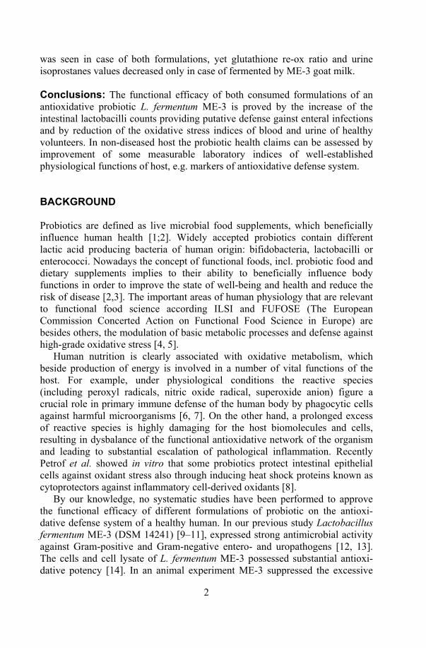
2
was seen in case of both formulations, yet glutathione re-ox ratio and urine isoprostanes values decreased only in case of fermented by ME-3 goat milk. Conclusions: The functional efficacy of both consumed formulations of an antioxidative probiotic L. fermentum ME-3 is proved by the increase of the intestinal lactobacilli counts providing putative defense gainst enteral infections and by reduction of the oxidative stress indices of blood and urine of healthy volunteers. In non-diseased host the probiotic health claims can be assessed by improvement of some measurable laboratory indices of well-established physiological functions of host, e.g. markers of antioxidative defense system. BACKGROUND Probiotics are defined as live microbial food supplements, which beneficially influence human health [1;2]. Widely accepted probiotics contain different lactic acid producing bacteria of human origin: bifidobacteria, lactobacilli or enterococci. Nowadays the concept of functional foods, incl. probiotic food and dietary supplements implies to their ability to beneficially influence body functions in order to improve the state of well-being and health and reduce the risk of disease [2,3]. The important areas of human physiology that are relevant to functional food science according ILSI and FUFOSE (The European Commission Concerted Action on Functional Food Science in Europe) are besides others, the modulation of basic metabolic processes and defense against high-grade oxidative stress [4, 5].
Human nutrition is clearly associated with oxidative metabolism, which beside production of energy is involved in a number of vital functions of the host. For example, under physiological conditions the reactive species (including peroxyl radicals, nitric oxide radical, superoxide anion) figure a crucial role in primary immune defense of the human body by phagocytic cells against harmful microorganisms [6, 7]. On the other hand, a prolonged excess of reactive species is highly damaging for the host biomolecules and cells, resulting in dysbalance of the functional antioxidative network of the organism and leading to substantial escalation of pathological inflammation. Recently Petrof et al. showed in vitro that some probiotics protect intestinal epithelial cells against oxidant stress also through inducing heat shock proteins known as cytoprotectors against inflammatory cell-derived oxidants [8].
By our knowledge, no systematic studies have been performed to approve the functional efficacy of different formulations of probiotic on the antioxi-dative defense system of a healthy human. In our previous study Lactobacillus fermentum ME-3 (DSM 14241) [9–11], expressed strong antimicrobial activity against Gram-positive and Gram-negative entero- and uropathogens [12, 13]. The cells and cell lysate of L. fermentum ME-3 possessed substantial antioxi-dative potency [14]. In an animal experiment ME-3 suppressed the excessive
3
oxidative stress reaction caused by Salmonella infection in intestinal mucosa and thus improved the gut mucosal antioxidative status [15]. The antioxidative effect of L. fermentum ME-3 on human body oxidative stress markers was confirmed by our pilot study with fermented goat milk [16].
The aim present study was to evaluate the functional efficacy of the probiotic strain L fermentum ME-3 in the human GIT of healthy volunteers. The fecal recovery, effect of two different formulations on total fecal lactoflora and oxidative stress markers of blood and urine were compared after 3 weeks consumption. METHODS Formulations The efficacy of two different formulations (experimental fermented goat milk and probiotic capsules) on the human body oxidative stress markers was evaluated.
Lactobacillus fermentum ME-3, a probiotic strain of healthy human intestinal origin (17), has been identified by biochemical and molecular methods [9]. The patent application has been submitted to the Estonian Patent Agency (Application No. 0356/01PV) as well as to the International Bureau of WIPO (Application No. WO03002131) [11]. L. fermentum ME-3 was used as freeze-dried powder in capsulated form and in fermented milk.
Capsules. Gelatine coated capsules were manufactured by the Tallinn Pharmaceutical Company. The freshly prepared probiotic capsules contained 9.0 log CFU of L. fermentum ME-3 per capsule in addition to 250 mg of saccha-rose and microcellulose. Identical placebo capsules contained only saccharose and microcellulose. All capsules were stored at +4°C.
Survival of ME-3 in capsule. Survival of ME-3 in capsule was monitored during 12 months at +4°C. The content of one capsule was dissolved aseptically in 2 ml of 0.9% NaCl solution. The suspension was vortexed, serially diluted and seeded 0.1 ml on MRS agar medium (OXOID, U.K.) and incubated 48 hours at 37°C microaerobically (10% CO2). The number of colonies was coun-ted and the viable cell count in capsule was calculated.
Experimental fermented milk. Three different lots of experimental fermented goat milk was prepared for the 3-week trial with healthy volunteers in order to establish the health effects of ME-3 consumption. The study group was supplied with fresh product once a week. Experimental fermented milk was prepared as described previously [16] by combining the probiotic strain with two supportive lactobacilli cultures L. plantarum LB-4 and L. buchneri S-15. L. buchneri strain S1-5 decreased the specific taste of the goat milk. L. plantarum LB-4 was included as a strong producer of exopolysaccharides, which gives the fermented milk a cream-like consistence and delightful acidity. The goat milk was

4
inoculated with 2% mixture of Lactobacillus strains and incubated at 37ºC for 24 hours. The product, ready to use, was cooled and stored at 4°C.
Survival of L. fermentum ME-3 in fermented goat milk. To measure the viable cell count of ME-3 in fermented goat milk, samples were taken at the end of fermentation (before cooling the product), and after 24h, 32h, 48h and 7 days from the preparation, when the product was stored at 4°C. The amount of 0.5 ml of the fermented milk was serially diluted in saline and plated on MRS agar medium and incubated for 48 h at 37°C in microaerobic conditions. DESIGN OF HUMAN VOLUNTEER TRIALS Two healthy volunteer (n 45) trials, particularly open placebo controlled (OPC) study and double blind randomised placebo controlled (DBRP) study were carried on to evaluate the functional efficacy of L. fermentum ME-3 in the human body. The inclusion criteria included the wish to participate, no known health problems, and no medical conditions requiring drug therapy, no other yoghurts or no special diets. The subjects with a history of GIT disease, food allergy and acute infection, use of any antimicrobial agent within the last month or use of any regular concomitant medication were excluded. The members of the trial were daily questioned about their general welfare, intestinal function (general welfare, gut gas production, stool frequency) and putative adverse effects. The withdrawal criteria from the trials included acute infections during the study. Reasons for dropout were the unwillingness to proceed with the study or relocation to new area. The blood samples (6 ml) from the antecubital vein, fecal and urine samples were collected before and at the end of all clinical trials. Participants of all trials gave informed consent to the study protocols approved by the Ethical Committee of Tartu University.
Open placebo controlled fermented goat milk trial. The study participants were 5 men and 16 women, mean age 50 years (range 35–60). During three weeks of the trial the study group (3 males and 13 females) consumed daily 150 ml fermented goat milk. The daily dose of probiotic Lactobacillus strain was 11.2 to 11.8 log CFU per person.
The control group (1 male and 4 females) consumed the same dose of fresh goat milk.
Probiotic capsule trial. A DBRP study was carried out as follows. The study group consisted of 15 men and 9 women, mean age 52 years (range 40–60) allocated according to their wish to participate and randomly divided by an independent person and computer program for two groups. The study group members (8 males and 4 females) took three probiotic containing capsules (8.4 log CFU per capsule) two times daily (the daily dose 9.2 log CFU) during three weeks. The placebo group (7 males and 5 females) received identical capsules without the probiotic strain.
5
Fecal samples of all participants to assess change in fecal lactoflora and the persistence of the ingested probiotic strain were collected before and at the end of trial. Several laboratory indices of blood and urine were measured before and after the consumption of ME-3. Here we report on changes in human body oxidative stress markers as total antioxidative activity (TAA), total antioxidative status (TAS) and glutathione red-ox ratio (GSH/GSSG) from blood serum and 8-isoprostanes in urine. Microbiological analyses of feces The total count of lactobacilli and the count of L. fermentum were evaluated in fecal samples. The fecal samples were collected at day 0 and 21 in both trials. Samples were kept at –80°C before analyzed. Serial dilutions (10–2–10–9) of the weighed fecal samples were prepared with phosphate buffer (pH 7.2) and 0.05 ml of aliquots was seeded onto MRS agar medium [17]. The plates were incubated at 37°C for 4 days microaerobically in 10% CO2 environment (incubator IG 150, Jouan, France). The catalase negative colonies were selected on the basis of typical for LAB colony morphology, cells microscopy and Gram staining.
The count of Lactobacillus species was expressed in log10 colony forming units per gram feces (log10 CFU/g) and percentage (relative share) in the total count of lactobacilli. The detection level of lactobacilli was a 3.0 log CFU/g feces.
The relative amount of L. fermentum, colonizing the gastrointestinal tract of persons in the study groups was expressed as a proportion of the total count (%), using the Bioquant program [18]). The program gives output data for every microorganism as an absolute count (log10 CFU/g) and their percentage in the total count with its normal values. AP-PCR TYPING The putative ME-3 isolates were typed by arbitrarily primed polymerase chain reaction (AP-PCR). Genomic DNA was extracted from 24h old cultures, cultivated on MRS agar microaerobically with the QIAamp DNA Mini Kit 50 (QIAGEN GmbH., Hilden, Germany) according to the manufacturers instructions. AP-PCR typing was done with two primers: ERIC1R (5'-ATGTAAGCTCCT GGGGATTCAC-3') and ERIC2 (5'-AAGTAAGTGACTGGGGTGAGCG -3') (DNA Technology A/S, Aarhus, Denmark). A 30 µl volume of reaction mixture consisted of 10xPCR buffer (Fermentas, Vilnius, Lithuania), 2.5 mM MgCl2 (Fermentas, Vilnius, Lithuania), 200µM deoxynucleoside triphosphate mixture (dATP, dGTP, dTTP and dCTP, Amersham Pharmacia Biotech, Freiburg, Germany) 0,60µg of each primer and 2.5U Taq DNA Polymerase (Fermentas, Vilnius, Lithuania,) and 5
6
µl of extracted DNA according to Matsumiya et al. [19]. The PCR mixture was subjected to thermal cycling 35 cycles of denaturation at 95°C for 1 min, annealing at 35°C for 1 min, and extension at 74°C for 2 min, with a final extension at 74°C for 5 min with the PTC-200 thermal cycler (Eppendorf AG, Hamburg, Germany). The PCR products were separated by electrophoresis in a horizontal 2% agarose gel containing 0.1 µl/ml ethidium bromide in Tris-acetic acid–EDTA (TAE) buffer (40mM Tris, 20mM boric acid, 1mM EDTA, pH 8.3) (Bio-Rad Laboratories, Hercules, USA) at constant voltage of 120V. A 1kb ladder (GeneRuler, Fermentas, Vilnius, Lithuania) was used as a base pair size marker. The banding patterns of isolates were visualized with UV light and compared with that of L. fermentum ME-3 strain. Measurement of human body oxidative stress status In urine the changes of the oxidative stress marker 8-isoprostanes concent-rations (ng/ml) were assessed by a competitive enzyme-linked immunoassay (ELISA) (BIOXYTECH 8-Isoprostane Assay, Cat No 21019) as described previously [16].
Blood serum was analysed for total antioxidative activity TAA, total antioxidative status TAS and GSSG/GSH. TAA of the serum was assessed by the linolenic acid test (LA-test) described previously [16]. This test evaluates the ability of the sample to inhibit lipid peroxidation. TAS of the serum was measured with a commercially available kit (TAS, Randox Laboratories Ltd. Ardmore, UK) as described elsewhere [16], water-soluble vitamin E (Trolox) serving as a standard. This method is based on the inhibition of the absorbance of the ferrylmyoglobin radicals of 2,2’-azinobis-ethylbenzothiazoline 6-sulfonate (ABTS+) generated by activation of metmyoglobin peroxidase with H2O2.
The cellular oxidative stress markers as total glutathione and oxidized glutathione were measured using the method of Griffith [20] as described elsewhere [16]. The glutathione content was calculated on the basis of a standard curve generated with known concentration of glutathione. Amount of GSH (µg/ml) was calculated as a difference between the total glutathione and GSSG (total glutathione – GSSG). The glutathione red/ox ratio was expressed as GSH/GSSG. STATISTICAL ANALYSIS The computer program Sigma Stat for Windows 2.0 (Jandel Coprporation, USA) was applied. The counts of fecal lactoflora were compared by using Student’s t-test and Mann-Whitney rank sum test. Changes in oxidative stress markers of blood sera (TAA, TAS and glutathione red-ox ratio) and urine (8-isoprostanes) were evaluated by Student’s t-test, paired t-test and Mann-

7
Whitney rank sum test. The choice of tests was made automatically according to the distribution of the data. Both microbial and biochemical markers were given as mean and standard deviation.
One-way ANOVA test was performed to compare the effect of different formulation on TAA, TAS and fecal lactoflora parameters.
Differences were considered statistically significant if the value was p <0.05.
RESULTS Survival of ME-3 in formulations In capsule after approximately 1-log drop after one week from the production of the capsules, the viable count of the probiotic strain remained stable at the level of 8.4 log CFU per capsule. Additional results have shown at +4°C the stability of the freeze-dried capsulated culture at least 17 months from the production. In fermented goat-milk the cell count of the probiotic strain varied insigni-ficantly from 9.0 to 9.7 log CFU/ml from one preparation to the other. The viable count of ME-3 in the fermented goat milk was found to remain stable at least during 7 days of storage at 4°C. Human volunteer trials No dropouts were registered during volunteer trials, yet one participant was withdrawn from the probiotic capsule trial due to acute respiratory viral infection. Besides, no adverse affects in general welfare or changes in GI functionality were assessed during the trial.
Changes in total LAB count. The consumption of both ME-3 fermented milk and ME-3 capsule significantly increased the total count of lactobacilli in feces as compared to the initial levels (Fig. 1). In opposite, in the group of volunteers consuming non-fermented goat-milk there was even a decrease in total LAB counts during the 3-week trial and no changes were found in capsule placebo group. Additional increase of lactobacilli counts was found only in persons consuming fermented goat milk.
Recovery of the probiotic strain. In goat milk group L. fermentum as a species appeared in fecal samples of all individuals (n=16) after consumption of fermented goat milk (Table 1). The AP-PCR confirmed the recovery of ME-3 in the feces of all study group members (Fig. 2). However, different trials the strain did not perform the predominant Lactobacillus species in total lactobacilli count in participants in (Table 1), though there was a tendency for increase in L. fermentum counts. In the probiotic capsule trial the strain ME-3 was not detectable between L. fermentum isolates by AP-PCR.
8
Antioxidative health effect of ME-3. The consumption of ME-3 in both formulations had a positive effect on the blood oxidative stress markers as TAA and TAS (Fig. 3). Consumption of goat milk and fermented goat milk enhanced TAA and TAS in the study and control group. There was a significant additional increase (6% and 9% respectively) in both indices in the fermented goat milk group. Significant increase in TAA and TAS values occurred during the consumption of the probiotic strain in capsulated form. No changes were detected in the placebo group. Additional effect of probiotic consumption in capsulated form was 4% for TAA and 2.5 % for TAS.
The effect of goat-milk consumption on the TAA and TAS values was significantly higher (p<0.001) than by the consumption of the capsulated probiotic (Fig. 2).
The decrease of the glutathione red-ox ratio was significant in both groups: the study group (from 0.15±0.01 to 0.11±0.04 µg/ml, p<0.01) and control (from 0.14±0.03 to 0.11±0.02 µg/ml, p<0.01) in the goat milk trial (Kullisaar et al. 2003). The fermented goat milk containing L. fermentum ME-3 had no statistically significant additional effect. When the probiotic was consumed in capsulated form, no significant decrease was noticed in the glutathione red-ox ratio. The additional effect of ME-3 fermented goat milk consumption was 6%.
Compared with the baseline values, the consumption of ME-3 in goat milk reduced urine 8-isoprostanes concentrations from 5.5±0.4 before to 5.0±0.5 ng/ml after the treatment (p<0.01) [16]. No changes in urine 8-isoprostanes concentrations were seen between baseline and end of the capsule trial (from 3.59±1.3 to 3.0±1.6 ng/ml). DISCUSSION We have assessed the functional efficacy of the antimicrobial and antioxidative probiotic L. fermentum ME-3 for healthy host. First of all the safety of the L. fermentum strain ME-3 was confirmed as no adverse side effects were registered in volunteers. Even relatively high (>1011 CFU) doses of consumed ME-3 had no negative impact on the hosts’ general well-being. Lactobacillus fermentum as species, used in various food applications, has a well-established history of safe use and is evaluated as GRAS according to the Food and Drug Administration of the USA [21].
Second, a clear improvement of laboratory indices of antioxidative defense system of a healthy host was documented, using both formulations as fermented by L. fermentum ME-3 goat-milk and probiotic capsules. This effect was simultaneous with the increase of intestinal lactoflora of healthy volunteers even without necessity for fecal recovery of the strain. In the human population, persons without clinical symptoms have still a quite different health status, including stability, capacity and potency of antioxidative defence to counteract sufficiently to oxidative stress-caused adverse effects [7]. If a probiotic is able
9
to exhibit a positive functionality on oxidative stress-related indices, it helps both to stabilize and promote the potency of the whole body antioxidative defence system in subclinical situations without disease symptoms. That in turn may have an impact for lowering the risk of atherosclerotic damage of blood vessels associated with several cardiovascular and neurodegenerative diseases [22–24].
In our study of healthy volunteers for validation of the antioxidative functionality of probiotic, four well-known oxidative stress markers of blood and urine were chosen. Urine 8-isoprostanes reflect the whole body oxidative stress-load, normally present at low concentrations in human body fluids [25]. We found that both formulations of ME-3 lowered their concentration as compared to individual baseline values in our clinical settings. This may be understood as indirect evidence for suppression of LDL oxidation [26, 27]. The state of the lipid fraction (including also LDL) in the antioxidative defence system of the blood is evaluated by TAA. TAS on the other hand reflects more the antioxidativity of the water-soluble fraction of the human blood. Among the measured blood sera markers both the TAA and TAS values were also reduced in the two different study groups. However, there was found a significantly lower improvement of TAA and TAS values in cases of capsule than fermented goat-milk where the recovery o the strains was assessed by AP-PCR. The AP-PCR is an easily performed technique very effective for identification of potential ME-3 fecal isolates.
Similarly, the reduction of the glutathione red-ox ratio was detected after the consumption of fermented by ME-3 goat-milk but not with the capsule. The crucial non-enzymatic cellular antioxidant is GSH [28] present in the millimolar range mainly in the red blood cells, liver, pancreas, kidneys, spleen, eyes, lungs and intestinal cells [29]. The oxidized form of glutathione becomes even at low concentrations toxic, and therefore in the cells the glutathione red-ox ratio is kept as low as possible. In the case of inflammation this balance is shifted towards the oxidized form, indicating non-physiological intracellular oxidative stress. Thus, our study shows that there is a good association between the mode of formulation of probiotic and expression of its functional properties inside the healthy host. Particularly, the explanation for more expressed positive shifts in oxidative stress markers of former volunteers could be due to the synergistic effect of the probiotic and the substrate. Milk is not just a carrier for the probiotic Lactobacillus strain, but contains natural “lactogenic” factors like lactose, minerals, vitamins and other components that enhance the metabolic activity of ingested probiotic strain in GIT. Besides, a variety of bioactive peptides (e.g. casomorphins, lactorphins, casokinins, etc.) revealed in milk [30–32] may have the antioxidative potency. This was proved by some antioxidative effect also in persons consuming non-fermented goat milk. The composition of goat milk differs from cow milk, containing several biomolecules, which by consumption may also contribute to some additional antioxidative effect, as discussed elsewhere [16]. Therefore, the provisional FAO regulations [33]
10
suggesting the need for health claims by specified formulations of probiotic seem to be of the utmost importance.
Additionally, in our study with experimental fermented milk the average daily dose of L. fermentum ME-3 being 11.5 logs CFU was clearly higher than that of capsule (max 9.5 log CFU). It is possible that the dose excesses the amount of bacteria necessary for interacting with intestinal mucosa and the unattached lactobacilli are excreted with faeces. The finding of Saxelin and colleagues confirmed that the fecal recovery of the probiotic strain started from the consumption of more than 9.0 log CFU daily doses of capsulated LGG [34]. To our surprise, in the present study the similar dose did not result in faecal recovery of the strain.
It is possible that the ME-3 strain germinated mainly in some upper parts of intestinal tract where the advantageous conditions for survival and metabolic activity of probiotic lactobacilli were present. Using molecular tools, Marteau et al. showed that lactobacilli figuring only 7% of fecal microflora performed up to 30% of microbial communities in human colon [35]. If administered in lower quantities as in case of capsule trial, ME-3 did not reach the detectable level in fecal samples. Yet, its presence in gut was proved by the positive antioxidative health effect in blood but not in urine. Therefore it is understandable that the higher load of metabolically active probiotic bacteria in goat-milk resulted also in their fecal recovery and the highest impact on the oxidative stress indices.
Moreover, in our study the positive impact of ME-3 consumption on the host lactoflora was proved by the increase of fecal lactobacilli counts in all participants of human volunteer studies. In experimental settings the high counts of intestinal lactobacilli have been shown as an important defensive factor against enteral infections [36, 37]. Though up to now the period of consumption of probiotics has not been defined, the 3-week ingestion of fermented goat-milk and capsule seemed enough for reaching the aims.
It is important to mention that after consumption of ME-3, a strain with high antagonistic activity, neither the species nor the strain predominated among total lactoflora. This shows a well-granted microbial balance inside the gut, which cannot be disturbed by high load of probiotic bacteria. Apparently, the interconnected advanced metabolism of large gut microbiota keeps the proportions of different species quite stable. Some other investigators have obtained similar results showing the proportional increase of different microbial populations (bifidobacteria, coliforms) after administration of Lactobacillus sp. probiotic [38, 39].
Thus, the functional efficacy of different formulations of antiinfectious and antioxidative probiotic L. fermentum ME-3 were proved both by the increase of the lactobacilli counts providing putative defense against infectious agents in gut and by reduction of the oxidative stress indices of blood and urine of healthy volunteers. Further, Phase III studies evaluating the efficacy of ME-3 as adjunct to conventional therapy in patients with atherosclerotic damages and a high-grade oxidative stress are ongoing.

11
CONCLUSIONS In non-diseased host, the probiotic health claims can be assessed by improvement of some measurable laboratory indices of well-established physiological functions of organism. In our case, the possibility for augmentation of the antioxidative defence system by the probiotic L. fermentum ME-3 in normal population can be proposed. COMPETING INTERESTS Marika Mikelsaar, Mihkel Zilmer, Tiiu Kullisaar, Heidi Annuk (Hynes) and Epp Songisepp are sharing the Estonian patent application: no. EE 2001 00356 29.06.01 and International Patent application: no. WO03002131. AUTHORS’ CONTRIBUTIONS Epp Songisepp, and Pirje Hütt have been in charge of the microbiological analysis. The former has been also in charge of analysing the results and writing of the manuscript. Jaak Kals was responsible for performance and management of the volunteer trials. Tiiu Kullisaar has been in charge of the biochemical analysis and writing the manuscript. Mihkel Zilmer has conducted the biochemical estimations and writing the manuscript. Reet Mändar has been in charge of the molecular analysis and revising the manuscript. Marika Mikelsaar is the main conductor of the L. fermentum ME-3 research; for this paper she has been in charge of the clinical trial design and writing the manuscript. ACKNOWLEDGEMENTS The study was supported by a grant from the Estonian Science Foundation (base funding 0418 and 5327) and Estonian Technology Agency funding 01103 and EU QLRT-2001-00135.
We are sincerely grateful to Dr. Irja Lutsar for critical reading of the manuscript, Dr. Heidi Hynes, Mrs. Eha-Maie Laanes and Miss Enely Alas for excellent technical assistance.

12
REFERENCES 1. McFarland L: A review of evidences of health claims for biotherapeutic agents.
Microb Ecol Health Dis 2000, 12: 65–76. 2. Salminen S: Human studies on probiotics: aspects of scientific documentation.
Scand J Nutr 2001, 45: 8–12. 3. Hardy G: Nutraceuticals and functional foods: introduction and meaning.
Nutrition 2000, 16: 688–697. 4. ILSI Europe: Scientific concepts of functional foods in Europe: consensus
document. Br J Nutr 1999, 81: 1–27. 5. Ashwell M: Concepts of functional foods. In ILSI Europe Concise monograph
series, ILSI Press; 2002, 40 S: 7894–10. 6. Diplock AT, Charleux J-L, Crozier-Willi G, Kok FJ, Rice-Evans C, Roberfroid M,
Stahl W and Viňa-Ribes J: Functional food science and defence against reactive oxidative species. Br J Nutr 1998, 80: 77–112.
7. Halliwell B and Gutteridge JMC: Free radicals in biology and medicine. New York: Oxford University Press; 1999.
8. Petrof EO, Kojima K, Ropeleski MJ, Musch MW, Tao Y, De Simone C and Chang EB: Probiotics inhibit nuclear factor kappa-B and induce heat shock proteins in colonic epithelial cells through proteasome inhibition. Gastroenterology 2004, 127: 1474–1487.
9. Annuk H, Vainumäe I, Aimla K, Ausmees K, Kullisaar T, Kask S and Mikelsaar M: Antagonistic activity of Lactobacilli against urinary Escherichia coli. Microecol Therapy 1999, 28: 169–175.
10. Mikelsaar M, Annuk H, Shchepetova J, Mändar R, Sepp E and Björkstén B: Intestinal Lactobacilli of Estonian and Swedish children. Microb Ecol Health Dis 2002, 14: 75–80.
11. Mikelsaar M, Zilmer M, Kullisaar T, Annuk H and Songisepp E: Strain of microorganism Lactobacillus fermentum ME-3 as novel antimicrobial and antioxidative probiotic. International Patent application 2001, WO03002131 [http://ep.espacenet.com]
12. Annuk H: Selection of medicinal plants and intestinal lactobacilli as antimicrobial components for functional foods. PhD thesis. Tartu University, Microbiology Department; 2002.
13. Songisepp E, Kullisaar T, Hütt P, Elias P, Brilene T, Zilmer M and Mikelsaar M: A New Probiotic Cheese with Antioxidative and Antimicrobial Activity. J Dairy Sci 2004, 87: 2017–2023.
14. Kullisaar T, Zilmer M, Mikelsaar M, Vihalemm T, Annuk H, Kairane C and Kilk A: Two antioxidative lactobacilli strains as promising probiotics. Int J Food Microbiol 2002, 72: 215–224.
15. Truusalu K, Naaber P, Kullisaar T, Tamm H, Mikelsaar R-H, Zilmer K, Rehema A, Zilmer M and Mikelsaar M: The influence of antibacterial and antioxidative probiotic lactobacilli on gut mucosa in a mouse model of Salmonella infection. Micr Ecol Health Dis 2004, 16: 180–187.
16. Kullisaar T, Songisepp E, Mikelsaar M, Zilmer K, Vihalemm T and Zilmer M: Antioxidative probiotic fermented goats milk decreases oxidative stress-mediated atherogenicity in human subjects. Br J Nutr 2003, 90: 449–456.
13
17. Mikelsaar M, Lenzner A and Goljanova LA: Methods for the estimation of the quantitative composition of fecal microflora. Laboratornoje delo 1972, 1: 41–45.
18. Mändar R, Mändar H and Mikelsaar M: Bioquant−a program for evaluation of fecal microbiocenosos. Clin Chem Lookout 1992, 56: 46.
19. Matsumiya Y, Kato N and Watanabe K: Molecular epidemiological study of vertical transmission of vaginal Lactobacillus species from mothers to newborn infants in Japanese, by arbitrarily primer polymerase chain reaction. J Infect Chemother 2002, 8: 43–49.
20. Griffith OW: Determination of glutathione and glutathione disulfide using glutathione reductase and 2-vinylpyridine. Anal Biochem 1980, 106: 207–212.
21. Food and Drug Administration: Rules and regulations. [http://a257.g.akamaitech.net/7/257/2422/15nov20040800/edocket.access.gpo.gov/2004/pdf/04-25243.pdf].
22. Witzum JL: The oxidation hypothesis of atherosclerosis. Lancet 1994, 344: 793–795.
23. Bogdanovic N, Zilmer M, Zilmer K, Rehema A and Karelson E: The Swedish APP670/671 Alzheimer’s disease mutation: The first evidence for strikingly increased oxidative injury in the temporal inferior cortex. Dementia Geriatric Cognitive Disorders 2001, 12: 364–370.
24. Pihl E, Zilmer K, Kullisaar T, Kairane C, Pulges A and Zilmer M: High-sensitive C-reactive protein level and oxidative stress-related status in former athletes in relation to traditional cardiovascular risk factors. Atherosclerosis 2003, 171: 321–326.
25. Hata AN and Breyer RM: Pharmacology and signaling of prostaglandin receptors: Multiple roles in inflammation and immune modulation. Pharmacol Ther 2004, 103: 147–66.
26. O’Brien RC, Luo M, Balazs N and Mecuri J: In vitro and vivo antioxidant properties of gliclazide. J Diabetes Complic 2000, 14: 201–206.
27. Sanchez-Moreno C, Dashe JF, Scott T, Thaler D, Folstein MF and Martin A: Decreased levels of plasma vitamin C and increased concentration of inflammatory and oxidative stress markers after sroke. Stroke 2003, 35: 163.
28. Pastore A, Federici G, Bertini E and Piemonte F: Analysis of glutathione: implication in redox and detoxification. Clinica Chimica Acta 2003, 333: 19–39.
29. Meister A and Anderson ME: Glutathione. Ann Rev Biochem 1983, 52: 711–760. 30. Meisel H and Bockelmann W: Bioactive peptides encrypted in milk proteins:
proteolytic activation and tropho−functional properties. In Proceedings of the sixth symposium on lactic acid bacteria: genetics, metabolism and applications. Edited by Konings, WN, Kuipers OP and Huis in`t Veld J. Veldhoven: Kluver Academic Publishers; 1999: 207−215.
31. Bos C, Gaudichon C and Tomé D: Nutritional and physiological criteria in the assessment of milk protein quality for humans. J Am Colledge Nutr 2000, 19: 191–205.
32. Boland M, MacGibbon A and Hill J: Designer milks for the new millennium. Livestock Prod Sci 2001, 72: 99–109.
33. Food Agriculture Organization: Guidelines for the evaluation of probiotics in food. [http://www.who.int/foodsafety/fs_management/en/probiotic_guidelines.pdf].
14
34. Saxelin M, Elo S, Salminen S and Vaapatalo H: Dose response colonisation of faces after oral administration of Lactobacillus casei strain GG. Micr Ecol Health Dis 1991, 4: 209–214.
35. Marteau P, Pochart P, Dore J, Bera-Maillet C, Bernalier A and Corthier G: Comparative study of bacterial groups within the cecal and fecal microbiota. Appl Env Microb 2001, 67: 4939–4942.
36. Kelleher SL, Casas I, Carbajal N and Lonnerdal B: Supplementation of infant formula with the probiotic Lactobacillus reuteri and zinc: impact on entericinfection and nutrition in infant rhesus monkeys. J. Pediatr Gastr Nutr 2002, 35: 162–168.
37. Cruchet S, Obregon MC, Salazar G, Diaz E and Gotteland M: Effect of the ingestion of dietary product containing Lactobacillus johnsonii La1 on Helicobacter pylori colonization in children. Nutrition 2003, 19: 716–721.
38. Sepp E, Mikelsaar M and Salminen S: Effect of administration of Lactobacillus casei strain GG on the gastrointestinal microbiota of newborns. Micr Ecol Health Dis 1993, 6: 309–314.
39. Morelli L, Zonenschain D, Callegari ML, Grossi E, Maisano F and Fusillo M: Assessment of a new symbiotic preparation in healthy volunteers: survival, persistence of probiotic strains and its effect on the indigenous flora. Nutr J 2003, 2:11 [http://www.nutritionj.com/content/2/1/11].
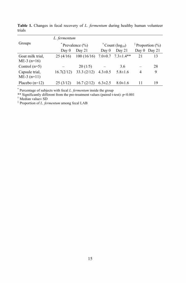
15
Table 1. Changes in fecal recovery of L. fermentum during healthy human volunteer trials
L. fermentum Groups * Prevalence (%)
Day 0 Day 21
† Count (log10) Day 0 Day 21
‡ Proportion (%)Day 0 Day 21
Goat milk trial, ME-3 (n=16)
25 (4/16) 100 (16/16) 7.0±0.7 7.3±1.4** 21 13
Control (n=5) – 20 (1/5) – 3.6 – 28 Capsule trial, ME-3 (n=11)
16.7(2/12) 33.3 (2/12) 4.3±0.5 5.8±1.6 4 9
Placebo (n=12) 25 (3/12) 16.7 (2/12) 6.3±2.5 8.0±1.6 11 19 * Percentage of subjects with fecal L. fermentum inside the group ** Significantly different from the pre-treatment values (paired t-test): p<0.001 † Median value± SD ‡ Proportion of L. fermentum among fecal LAB

16
Study group control study group placebo
0
2
4
6
8
10
12
Goat milk trial Probiotic capsule trial
1 2 1 2 1 2 1 2
CFU
log
10
*‡
* *
Figure 1. Increase of total fecal counts of lactobacilli in healthy volunteers consuming of ME-3 in fermented goat milk and probiotic capsule.
1 – Day 0, 2 – Day 21 Significantly different from pre-treatment values (Student’s t-test): * p<0.05; Significantly different from control (Student’s t-test): ‡ p=0.01
M 1 2 3 4 5 6 7 8 9 10 11 12 13 14 15 16 17
Figure 2. Confirmation for the survival of L. fermentum ME-3 in GIT in subjects receiving ME-3 fermented goat milk by AP-PCR in a horizontal 2% agarose gel.
From the left: M – molecular weight marker, Line 2 – ME-3, Line 3…17 – ME-3 like profiles from feces of goat milk trial study group participants

17
TAA
(%)
Study group control study group placebo
30
40
50
60
Goat milk trial Probiotic capsule trial
1 2 1 2 1 2 1 2
‡
* * * * * *TA
S(m
mol
/l)
0
1
2
Goat milk trial Probiotic capsule trial
0.5
1.5
1 2 1 2 1 2 1 2
Study group control study group placebo
‡
** ***
a)
b)
Figure 3. Effect of ME-3 consumption in fermented goat milk and capsules on human blood oxidative stress markers a) TAA (%) and b) TAS (mmol/l) 1 – Day 0, 2 – Day 21 Significantly different from pre-treatment values: *p<0.05 (paired t-test); **p≤0.01 (Student’s t-test and paired t-test); ME-3 goat milk effect different from the effect of the ME-3 in capsule-form (ANOVA): ‡p≤0.001
171
CURRICULUM VITAE
Epp Songisepp
Citizenship: Estonian Born: November 17, 1965, Võru
Address: Ravila 19, 50411 Tartu, Estonia Phone: +372 374 175
Fax: +372 374 172 E-mail: [email protected]
Education 1973–1984 Võru Secondary School No 1. 1984–1989 University of Tartu, Faculty of Biology and Geography, Biology 1999–2001 University of Tartu, Medical Faculty, Department of Micro-
biology M. Sci student of biomedicine
Special courses 2001 4 months in Japan, Osaka Municipal Technical Research Institute
Professional employment 1989–1990 Estonian Biocentre, technician 1990–1994 Ldt Estar Strip, microbiologist 1994– Starter ST OÜ, microbiologist and production manager 2002–2005 University of Tartu, Medical Faculty, Department of Micro-
biology, extraordinary research fellow 2005– R/D Centre of Healthy Milk Products Ltd, research fellow
Scientific work The main subject of the research work has concerned starter microbes and probiotic lactobacilli, their technological and functional properties. 6 scientific publications and 15 presentations at the congresses

172
CURRICULUM VITAE
Epp Songisepp
Kodakondsus: Eesti Sünniaeg ja koht: November 17, 1965, Võru
Aadress : Ravila 19, 50411 Tartu, Eesti Tel: +372 374 175 Fax: +372 374 172
E-mail: [email protected]
Haridus 1973–1984 Võru I keskkool 1984–1989 Tartu Ülikool, bioloogia-geograafia teaduskond, bioloogia 1999–2001 Tartu Ülikool, arstiteaduskond, mikrobioloogia instituut,
magistrant
Erialane enesetäiendus 2002 4 kuud Jaapan, Osaka Munitsipaalne Tehnikaülikool
Erialane teenistuskäik 1989–1990 Eesti Biokeskus, laborant 1990–1994 AS Estar Strip, mikrobioloog 1994– Starter ST OÜ, mikrobioloog, tootmisjuht 2002–2005 Tartu Ülikool, Arstiteaduskond, mikrobioloogia instituut, era-
korraline teadur 2005– OÜ Tervisliku Piima Biotehnoloogiate Arenduskeskus, teadur
Teadustöö Peamiseks uurimisvaldkonnaks on juuretised ja probiootilised laktobatsillid ning nende tehnoloogilised ja funktsionaalsed omadused 6 teaduspublikatsiooni, 15 konverentsiteesi