Embed Size (px)
Citation preview
Dissociative Photoionization of Methyl Thiocyanate, CH3SCN, in the Proximity of the Sulfur2p Edge
Emiliano Cortes,† Mauricio F. Erben,† Mariana Gerones,† Rosana M. Romano,† andCarlos O. Della Vedova*,†,‡
CEQUINOR (UNLP-CONICET, CCT La Plata), Departamento de Quımica, Facultad de Ciencias Exactas,UniVersidad Nacional de La Plata, CC 962, La Plata (CP 1900), Republica Argentina and Laboratorio deSerVicios a la Industria y al Sistema Cientıfico (LaSeISiC) (UNLP-CIC-CONICET),Camino Centenario e/505 y 508, (1903) Gonnet, Republica Argentina
ReceiVed: August 13, 2008; ReVised Manuscript ReceiVed: NoVember 12, 2008
The dissociative photoionization of gaseous CH3SCN has been investigated at the S 2p core level usingtime-of-flight mass spectrometry and synchrotron radiation. The total ion yield spectrum could be successfullyassigned by comparison with available data from electron energy loss spectra. The relative abundances of theionic fragments and their kinetic energy release values were obtained from both PEPICO (photoelectronphotoion coincidence) and PEPIPICO (photoelectron photoion photoion coincidence) spectra. The dynamicsof the ionic fragmentation of S 2p excited CH3SCN is dominated by the rupture of both carbon-sulfur bonds.This process may be related with electronic excitations from the ground electronic state to vacant σ* molecularorbitals.
Introduction
Organosulfur compounds have attracted much attention, andseveral outstanding reviews covering the chemistry of thiocy-anates (RSCN) and isothiocyanates (RNCS) can be found inthe chemical literature.1-3 Simple alkylated species (R ) alkyl)are very well-known molecules. For example, methyl isothio-cyanate, CH3NCS, is widely used as an agricultural fumigant,and much attention in recent years has been devoted tounderstanding the effect of this application on the atmosphericbalance.4 Much information is available for both methyl isomersin their fundamental, ionic, and excited electronic states, andthe CH3SCN T CH3NCS isomerization equilibrium has beenstudied in depth.2,3
The molecular structure of isothiocyanate (CH3SCN) has beendetermined experimentally using microwave spectroscopy,5,6 andhigh-level quantum chemical calculations, including CCSD7 andQCISD8 methods, are available for this species. In addition, itsvibrational properties have been studied,9 and infrared spectrain several common solvation environments have recently beenreported.10
The plasma chemistry of transient species in methyl thiocy-anate discharges has been semiquantitatively studied usingspectroscopic techniques.11 The behavior of both isomers, oftenhaving been irradiated with VUV synchrotron photons, is quitesimilar as determined from emission spectra of the NCSradical.12 In addition, the emission spectra of the radical NCSproduced by low-energy electron impact on both isomers havebeen measured.13 These studies have been further complementedby laser-induced fluorescence spectroscopy, which succeededin evaluating the photodissociation process using both 248 and193 nm wavelengths.14,15 The gas-phase ion chemistry ofCH3NCS and CH3SCN has been investigated by pulsed ICR
techniques, and their proton affinities are known to be 193.0 (0.4 and 192.6 ( 0.5 kcal/mol, respectively.16
Photoelectron spectra of CH3SCN and CH3NCS were firstrecorded by Neijzen et al.,17 and a molecular orbital assignmentfor the outer valence electron distribution for both species wasproposed. However, this initial assignment was later revised byPasinszki et al.18 based on ab initio quantum chemical calcula-tions, high-resolution HeI spectra, and HeI/HeII band intensityratios. Moreover, the ionization process of CH3SCN andCH3NCS upon collision with metastable He*(23S) has beenstudied by collision-energy-resolved Penning ionization electronspectroscopy.19
Of particular interest for the present work, Hitchcock et al.20
reported optical oscillator strengths for C 1s, N 1s, and S 2pinner shell excitations in CH3SCN as derived from electronenergy loss spectra. The S 2p region of the spectrum shows thepresence of very structured transitions below the ionizationpotential. These sharp structures have largely been explainedin terms of excitations from S 2p electrons to vacant π* andσ*CS orbitals.
Our research group has quite recently started studying theproperties of shallow and inner core level electrons in sulfe-nylcarbonyl compounds. Penta-atomic FC(O)SCl21,22 and ClC-(O)SCl23 species have been studied using synchrotron radiationin the 100-1000 eV range, and their ionic fragmentation afterelectronic decay has been analyzed. We also studied othermembers of this family such as CH3C(O)SH24 andCH3OC(O)SCl.25 Most recently, we succeeded in analyzing theelectronic structure and ionic dissociation induced by photonabsorption in the outermost valence region of sulfur-containingspecies. This study used a combined experimental approach thatincludes HeI photoelectron spectroscopy and photoionizationunder the action of synchrotron radiation in the 10-22.5 eVregion.26-28
Following these studies, we became interested in anothersimple sulfur-containing compound which presents the advan-tage of possessing earlier related studies using the inner-shell
* To whom correspondence should be addressed. E-mail: [email protected].
† Universidad Nacional de La Plata.‡ Laboratorio de Servicios a la Industria y al Sistema Cientıfico.
J. Phys. Chem. A 2009, 113, 564–572564
10.1021/jp807230s CCC: $40.75 2009 American Chemical SocietyPublished on Web 12/22/2008
electron energy loss spectrum (ISEELS) technique. Here wereport a study of the photon impact excitation and dissociationdynamics of CH3SCN exited at the S 2p level using synchrotronradiation. To our knowledge, the inner shell electronic propertiesand ionic fragmentation of photon-excited CH3SCN have notbeen previously described.
Experimental Section
Hazards: Methyl thiocyanate is a moderately toxic liquid.When heated to decomposition or in contact with mineral acidsit emits highly toxic fumes.
Synchrotron radiation was used at the Laboratorio Nacionalde Luz Sıncrotron (LNLS), Campinas, Sao Paulo, Brazil.29
Linearly polarized light monochromatized by a toroidal gratingmonochromator (available at the TGM beam line in the range12-300 eV)30 intersects the effusive gaseous sample inside ahigh-vacuum chamber with a base pressure in the range of 10-8
mbar. During the experiments the pressure was maintainedbelow 5 × 10⁻⁶ mbar. The resolution power is better than 400in the TGM beam-line at the LNLS. The energy calibration wasestablished by means of the S 2p f 6a1g and S 2p f 2t2g
absorption resonances in SF6.31 The intensity of the emergentbeam was recorded with a light-sensitive diode. The ionsproduced by interaction of the gaseous sample with the lightbeam were detected using a time-of-flight (TOF) mass spec-trometer of the Wiley-McLaren type for both PEPICO andPEPIPICO measurements.32,33 This instrument was constructedat the Institute of Physics, Brasilia University, Brasilia, Brazil.34
The axis of the TOF spectrometer was perpendicular to thephoton beam and parallel to the plane of the storage ring.Electrons were accelerated to a multichannel plate (MCP) andrecorded without energy analysis. This event starts the flighttime determination process of the corresponding ion, which isconsequently accelerated to another MCP. The characteristicsand performance of this electron-ion coincidence TOF spec-trometer have been recently reported.35
The average kinetic-energy release (KER) values of thefragments were calculated from the coincidence spectra byassuming an isotropic distribution of the fragments, that theyare perfectly space focused, and that the electric field appliedin the extraction region is uniform.36 Under these conditionsthe energy released in the fragmentation process can bedetermined from the peak width (fwhm).37,38 Deviations fromideal conditions always increase the peak width; thus, the valuescalculated are upper bounds. Santos et al.39 measured the argonmass spectrum under very similar experimental conditions, anda peak width value of 0.05 eV was achieved for the Ar+ ion.Because the broadening in argon can only be the result ofthermal energy and instrumental broadening, this value repre-sents a good estimation for the instrumental resolution. More-over, KER values have been determined from the projection ofthe PEPIPICO islands in the corresponding time domain foreach ion involved in the coincidence. The sum of theseindividual KER values is reported in this work and gives anestimate for the energy release occurring in the double iondissociation process.
The sample of CH3SCN was obtained from commercialsources (Aldrich, estimated purity better than 97%). The liquidsample was purified by repeated trap-to-trap vacuum distillation.The purity of the compound in both vapor and liquid phaseswas checked by IR and 1H NMR spectroscopies, respectively.
Results and Discussion
The orbital assignment of methyl thiocyanate can be brieflydescribed as follows. The CH3SCN molecule in the ground
electronic state (X1A1) belongs to the CS symmetry point group.All canonical molecular orbitals of type a′ are σ orbitals lyingin the molecular plane, while those of type a′′ are π orbitals.The 38 electrons are then arranged in 19 doubly occupiedorbitals in the independent particle description and distributedaccording to the following configuration: Core electrons,[S 1s]2[N 1s]2[C 1s]4[S 2s]2[S 2p]6; Valence electrons,(a′)2(a′)2(a′)2(a′)2(a′′ )2(a′)2(a′)2(a′)2(a′′ )2(a′)2(a′′ )2.
The HOMO can be visualized as an orbital having a′′symmetry, nominally localized on the sulfur atom occupied bylone-pair electrons. Its vertical ionization potential value is 10.13eV.17 The HOMO-1 and HOMO-2 orbitals are assigned toboth πSCN orbitals of the thiocyanate group. The simple modelsdescribe the thiocyanate moiety with a formal triple bond inthe CtN bond, i.e., -S-CtN. Thus, two different bonds withπ symmetry are expected. The bent C-S-C geometry ofCH3SCN removes the degeneracy of both π CtN molecularorbitals, which are classified as a′ (πSCN) and a′′ (πSCN) molecularorbitals. The following two occupied orbitals are assigned topredominantly nonbonding a′ orbitals, occupied by lone pairsnominally belonging to sulfur and nitrogen atoms, respectively.18
Total Ion Yield Spectra (TIY). Neither the calculated northe experimental photoabsorption spectrum for CH3SCN in thesulfur 2p edge energy region has been reported so far in theliterature. The TIY spectra were obtained by recording the countrates of the total ions while the photon energy is scanned. Athigh photon energies corresponding to shallow- and core-shellelectronic levels the quantum yield for molecular ionization isquite likely tending to unity. Consequently, detection of theparent and fragment ions as a function of the incident photonenergy is a powerful method to be used as a complement toabsorption spectroscopy.40
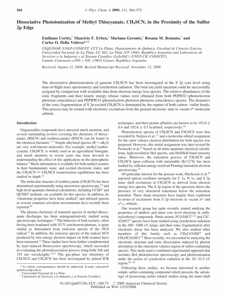
The TIY spectrum of CH3SCN, measured near the S 2p edge,is shown in Figure 1. Below the S 2p threshold the spectrum isdominated by a group of well-defined signals centered at 164.4,165.7, 167.2, 168.5, and 170.0 eV. These resonant transitionsshould correspond to dipole-allowed transitions that involveexcitations of a 2p electron to an antibonding molecular orbital.The TIY spectrum of CH3SCN is remarkably similar to theinner-shell electron energy loss spectrum (ISEELS) reported byHitchcock et al.20,41 It is well established that spectra obtainedby ISEELS in scattering regimes where electric-dipole transi-tions dominate are equivalent to optical X-ray absorption
Figure 1. Total ion yield spectrum (2) and oscillator strengths derivedfrom dipole-regime electron energy loss spectroscopy20 (∆) forCH3SCN. The ISEELS curve is shifted by 0.2 au.
Dissociative Photoionization of Methyl Thiocyanate J. Phys. Chem. A, Vol. 113, No. 3, 2009 565
spectra.42 As observed in Figure 1, intense and well-defined pre-edge features are present in both TIY and ISEELS spectra ofCH3SCN. The better resolution obtained in the present caseallows clearly identifying two weak transitions at 164.4 and170.0 eV, reported as shoulders in the former ISEELS spectrum.
Following the proposed assignment for S 2p transitions forCH3SCN20 the main features in ISEELS spectra have beenassigned to states associated with (S2p, π*SCN) and (S2p, σ*CS)configurations. According to the angular momentum selectionrules, the final state should have mainly either d or s character.Therefore, the L-edge spectra probe the sulfur d-orbital contri-butions to the molecular orbitals, which are sensitive to the moredistant environment around the sulfur atom.43,44
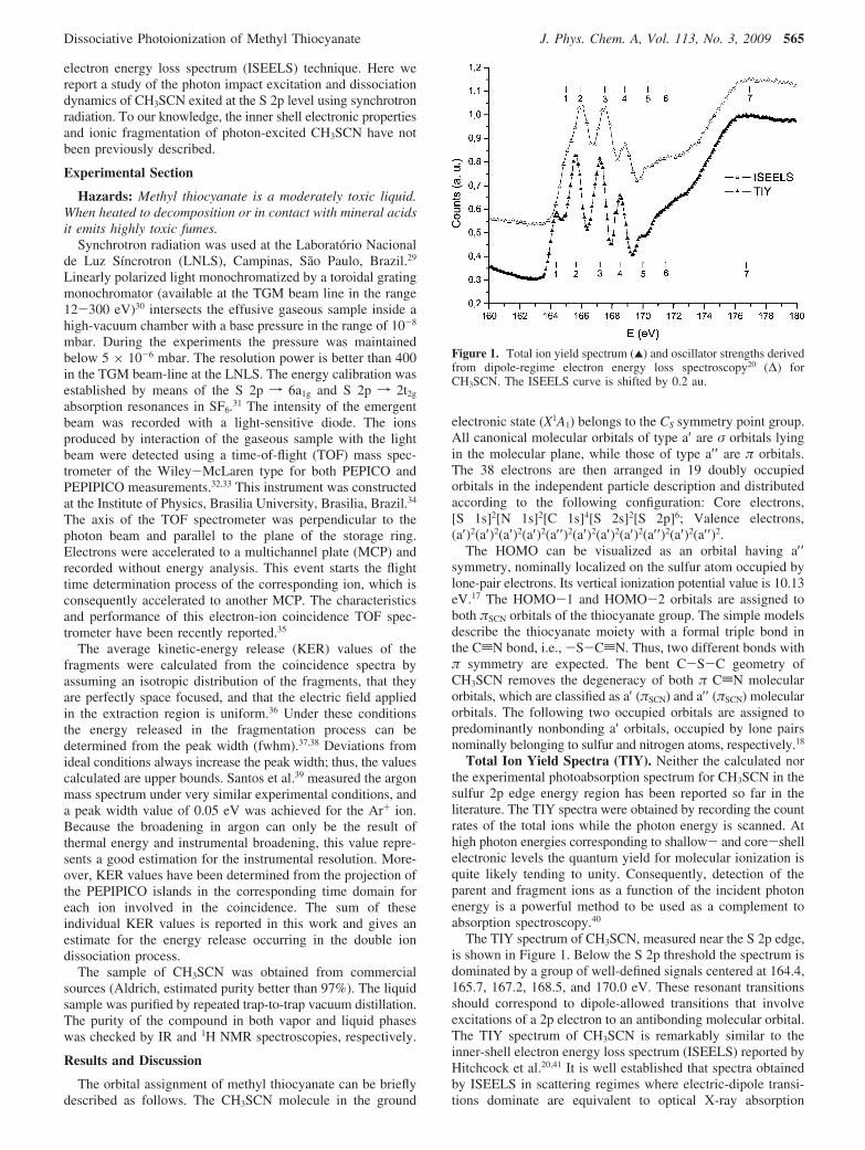
The well-resolved structures observed in the ISEELS and TIYspectra can be interpreted as originated by electronic transitionsinvolving the spin-orbit split of the 2p sulfur excited species(2p1/2 and 2p3/2 levels) to unoccupied antibonding orbitals,mainly the LUMO π*SCN (a′ and a′′ ) and σ*C-S orbitals.Quantum chemical calculations at the MP2/6-311++G(3df)level of approximation for neutral CH3SCN in its ground statepredict an unoccupied orbital arrangement which is in agreementwith this description (see Figure 2). It is worth mentioning thatthe σ*S-C (a′) antibonding MO in the thiocyanate group isslightly higher in energy than the corresponding σ*C-S (a′)antibonding one assigned to the H3C-S bond. These resultsare summarized in Table 1.
This description is in perfect agreement with the assignmentof the ISEELS spectra at the S 2p edge proposed by Hitchcocket al.20 These results are supported by experimental electrontransmission and electronic absorption spectra, which allow adescription of the vacant orbital of CH3SCN. Thus, the LUMOcorresponds to an antibonding π*SCN orbital. It was found thatboth the a′ (π*SCN) component lying in the C-S-C plane andthe perpendicular a′′ (π*SCN) component are stabilized byinteractions with S(3d) orbitals of appropriate symmetry. Theσ*C-S virtual molecular orbital (LUMO+2) associated with theH3C-S group is observed in the electron transmission spectraat relatively low energies, in agreement with similar reportsobserved for other sulfur-containing species.45 A considerablestabilization of the empty MO’s through mixing with the S(3d)and σ*C-S orbitals was postulated for CH3SCN.20
PEPICO Spectra. PEPICO spectra have been recorded bysetting the photon energy at the resonant values observed in
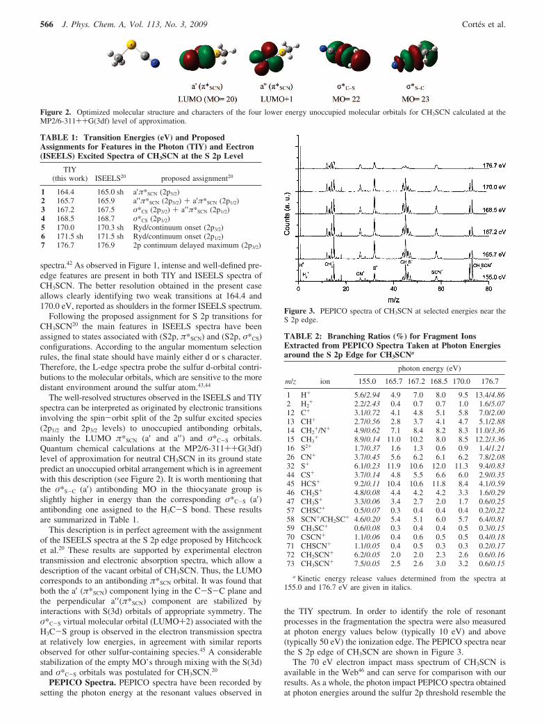
the TIY spectrum. In order to identify the role of resonantprocesses in the fragmentation the spectra were also measuredat photon energy values below (typically 10 eV) and above(typically 50 eV) the ionization edge. The PEPICO spectra nearthe S 2p edge of CH3SCN are shown in Figure 3.
The 70 eV electron impact mass spectrum of CH3SCN isavailable in the Web46 and can serve for comparison with ourresults. As a whole, the photon impact PEPICO spectra obtainedat photon energies around the sulfur 2p threshold resemble the
Figure 2. Optimized molecular structure and characters of the four lower energy unoccupied molecular orbitals for CH3SCN calculated at theMP2/6-311++G(3df) level of approximation.
TABLE 1: Transition Energies (eV) and ProposedAssignments for Features in the Photon (TIY) and Eectron(ISEELS) Excited Spectra of CH3SCN at the S 2p Level
TIY(this work) ISEELS20 proposed assignment20
1 164.4 165.0 sh a′π*SCN (2p3/2)2 165.7 165.9 a′′π*SCN (2p3/2) + a′π*SCN (2p1/2)3 167.2 167.5 σ*CS (2p3/2) + a′′π*SCN (2p1/2)4 168.5 168.7 σ*CS (2p1/2)5 170.0 170.3 sh Ryd/continuum onset (2p3/2)6 171.5 sh 171.5 sh Ryd/continuum onset (2p1/2)7 176.7 176.9 2p continuum delayed maximum (2p3/2)
Figure 3. PEPICO spectra of CH3SCN at selected energies near theS 2p edge.
TABLE 2: Branching Ratios (%) for Fragment IonsExtracted from PEPICO Spectra Taken at Photon Energiesaround the S 2p Edge for CH3SCNa
photon energy (eV)
m/z ion 155.0 165.7 167.2 168.5 170.0 176.7
1 H+ 5.6/2.94 4.9 7.0 8.0 9.5 13.4/4.862 H2
+ 2.2/2.43 0.4 0.7 0.7 1.0 1.6/5.0712 C+ 3.1/0.72 4.1 4.8 5.1 5.8 7.0/2.0013 CH+ 2.7/0.56 2.8 3.7 4.1 4.7 5.1/2.8814 CH2
+/N+ 4.9/0.62 7.1 8.4 8.2 8.3 11.0/3.3615 CH3
+ 8.9/0.14 11.0 10.2 8.0 8.5 12.2/3.3616 S2+ 1.7/0.37 1.6 1.3 0.6 0.9 1.4/1.2126 CN+ 3.7/0.45 5.6 6.2 6.1 6.2 7.8/2.0832 S+ 6.1/0.23 11.9 10.6 12.0 11.3 9.4/0.8344 CS+ 3.7/0.14 4.8 5.5 6.6 6.0 2.9/0.3545 HCS+ 9.2/0.11 10.4 10.6 11.8 8.4 4.1/0.5946 CH2S+ 4.8/0.08 4.4 4.2 4.2 3.3 1.6/0.2947 CH3S+ 3.3/0.06 3.4 2.7 2.0 1.7 0.6/0.2557 CHSC+ 0.5/0.07 0.3 0.4 0.4 0.4 0.2/0.2258 SCN+/CH2SC+ 4.6/0.20 5.4 5.1 6.0 5.7 6.4/0.8159 CH3SC+ 0.6/0.08 0.3 0.4 0.4 0.5 0.3/0.1570 CSCN+ 1.1/0.06 0.4 0.6 0.5 0.5 0.4/0.1871 CHSCN+ 1.1/0.05 0.4 0.5 0.3 0.3 0.2/0.1772 CH2SCN+ 6.2/0.05 2.0 2.0 2.3 2.6 0.6/0.1673 CH3SCN+ 7.5/0.05 2.5 2.6 3.0 3.2 0.6/0.15
a Kinetic energy release values determined from the spectra at155.0 and 176.7 eV are given in italics.
566 J. Phys. Chem. A, Vol. 113, No. 3, 2009 Cortes et al.
electron impact mass spectrum. The parent molecular ion ispredominantly formed together with CH3
+ and HCS+ fragments.Other ions, such as SCN+ and S+, are also observed, showingweaker signals in the spectrum. For instance, in the 155.0 eVspectrum the molecular ion signal at m/z ) 73 dominates themass spectra. The more intense peaks observed in the PEPICOspectra correspond to the S+, CH3
+, and HCS+ ions with relativeabundances of 6.1%, 8.9%, and 9.2%, respectively. The H+ ionis also present with an intensity of 5.6%. The next most abundantfragments derive from the SCN group: CN+ (m/z ) 26), SCN+
(m/z ) 58), and CS+ (m/z ) 44) with relative abundances near4%. The CHxSCN+, CHxSC+, CHxS+, and CHx
+ (x ) 3-0)series are also observed. The branching ratios determined fromthe PEPICO spectra of CH3SCN at photon energies around theS 2p edge are shown in Table 2.
The kinetic-energy release values have been determined foreach ion. In Table 2 the values obtained for two photon energies,i.e., 155.0 and 176.7 eV in the valence and S 2p continuum,respectively, are given. In the former spectrum the ions showKER values which are relatively low, while in the 176.7 eV
spectrum a broadening in their peak widths is clearly observed,and all ions show higher KER values. This effect becomesapparent in the PEPICO spectra shown in Figure 3. Therefore,it is possible to assume that the main contribution to the PEPICOspectra below the S 2p threshold comes from fragmentation ofsingle charged parent ion, which is formed by the one-photonionization process of valence electrons. On the other hand, whenthe incident photon energy is increased, S 2p electronicexcitation or ionization processes occur. It is well known thatthe decay of such an excited species normally leads to formationof the doubly charged CH3SCN2+ parent ion, for instance, ifnormal Auger processes take place. This fact is reflected in theKER values for the ions formed at 176.7 eV, which are muchbroader than the KER values determined for the previousenergies.36
Dynamics of Fragmentation Following Valence-ShellIonizations. The estimated double-ionization energy of CH3SCNis 26.8 eV, as calculated at the MP2/6-311++G** level ofapproximation. Consequently, 155.0 eV photons should beadequate to open most of the possible ionization channels
Figure 4. Partial ion yield (PIY) for selected series of ions following photon excitation of CH3SCN as a function of photon energy.
Figure 5. t1 and t2 projections of the PEPIPICO spectrum of CH3SCN recorded at 168.5 eV on the S 2p pre-edge resonance.
Dissociative Photoionization of Methyl Thiocyanate J. Phys. Chem. A, Vol. 113, No. 3, 2009 567
connected to the direct single and double ejections of valence-shell electrons. As observed in the TIY spectra, this energy isnot yet enough to ionize the core electrons. Thus, the coinci-dence spectrum taken at a 155.0 eV photon energy shouldprovide a good comparison with respect to the ionic fragmenta-tion pattern associated with core excitation or ionization.
The single charged molecular ion is observed in the wholerange of photon energies studied. A clear diminution of the peakintensity for CH3SCN+ ion is observed when resonant energiesare reached with typical abundance values of around 3%. WhenS 2p electrons are ionized, further diminution in the CH3SCN+
ion signal intensity becomes apparent, as observed in thePEPICO spectrum taken at 176.7 eV. The KER values deter-mined for this ion are close to the “thermal” value of 0.05 eV.39
The CHxSCN+ series of ions can be unambiguously charac-terized as formed from the parent ion by successive loss ofhydrogen atoms. The KER values, ca. 0.05 eV, are very similarto those determined for the CH3SCN+ ion. Indeed, it is expectedthat extrusion of a neutral hydrogen atom from chargedCHxSCN+ (x ) 3, 2, 1) has a little impact in the KER of theremaining ions. The partial ion yield spectra for the series ofCHxSCN+ (x ) 3-0) ions near the S 2p edge are displayed inFigure 4. When CH3SCN is irradiated with 155.0 eV photonsin the valence continuum region of the spectrum, CHxSCN+ (x) 3-0) ions are responsible for ca. 16% of single ionization.
On the other hand, processes that yield charged hydrogenatoms (H+) are evident from the intense signal at m/z ) 1 amu/q. Although an unambiguous description of these processes isnot possible at this point, it should be noted that the high KERvalue determined for the H+ ion could denote ejection of anenergetic ion. Similarly, high KER values are determined forthe H2
+ ion, which is a typical signature of processes involvingrearrangement reactions.47 It should be mentioned that studieson the decomposition of electronically excited CH3SCN haveconcluded that the main primary dissociation step forming SCNinvolves a rearrangement reaction to afford H2 (CH3SCN fCH + H2 + SCN).48
Rupture of both sulfur-carbon bonds in single chargedCH3SCN+ can also be observed. First, the H3C-S bond caneasily be broken to yield both the CH3
+ or SCN+ ions. Again,in the former case successive losses of hydrogen atoms lead tothe observation of the series of CHx
+ (x ) 3-0) ions. From thedetermined KER values, the following mechanisms can beproposed
CH3SCN+fCH3++ SCN (KER ≈ 0.14 eV)
CH3SCN+fCH3+SCN+ (KER ≈ 0.20 eV)
Moreover, it is quite interesting to note that fragments such asCHxS+ (x ) 3-0) or CN+ are also present in the PEPICOspectra depicted in Figure 3. These ions are associated withrupture of the carbon-sulfur bond in the thiocyanate group.Indeed, the series of CHxS+ (x ) 3-0) ions accounts for theca. 21% of ions detected in the 155.0 eV spectrum. The PIY
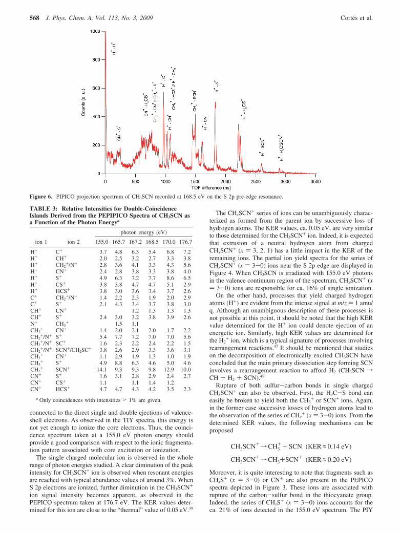
Figure 6. PIPICO projection spectrum of CH3SCN recorded at 168.5 eV on the S 2p pre-edge resonance.
TABLE 3: Relative Intensities for Double-CoincidenceIslands Derived from the PEPIPICO Spectra of CH3SCN asa Function of the Photon Energya
photon energy (eV)
ion 1 ion 2 155.0 165.7 167.2 168.5 170.0 176.7
H+ C+ 3.7 4.8 6.3 5.4 6.8 7.2H+ CH+ 2.0 2.5 3.2 2.7 3.3 3.8H+ CH2
+/N+ 2.8 3.6 4.1 3.3 4.3 5.6H+ CN+ 2.4 2.8 3.8 3.3 3.8 4.0H+ S+ 4.9 6.3 7.2 7.7 8.6 6.5H+ CS+ 3.8 3.8 4.7 4.7 5.1 2.9H+ HCS+ 3.8 3.0 3.6 3.4 3.7 2.6C+ CH2
+/N+ 1.4 2.2 2.3 1.9 2.0 2.9C+ S+ 2.1 4.3 3.4 3.7 3.8 3.0CH+ CN+ 1.2 1.3 1.3 1.3CH+ S+ 2.4 3.0 3.2 3.8 3.9 2.6N+ CH3
+ 1.5 1.1CH2
+ CN+ 1.4 2.0 2.1 2.0 1.7 2.2CH2
+/N+ S+ 5.4 7.7 7.2 7.0 7.0 5.6CH2
+/N+ SC+ 1.6 2.3 2.2 2.4 2.2 1.5CH2
+/N+ SCN+/CH2SC+ 3.8 2.6 2.9 3.3 3.6 3.1CH3
+ CN+ 1.1 2.9 1.9 1.3 1.0 1.9CH3
+ S+ 4.9 8.8 6.3 4.6 5.0 4.6CH3
+ SCN+ 14.1 9.3 9.3 9.8 12.9 10.0CN+ S+ 1.6 3.1 2.8 2.9 2.4 2.7CN+ CS+ 1.1 1.1 1.4 1.2CN+ HCS+ 4.7 4.7 4.3 4.2 3.5 2.3
a Only coincidences with intensities > 1% are given.
568 J. Phys. Chem. A, Vol. 113, No. 3, 2009 Cortes et al.
spectra for these ions are shown in Figure 4. When resonantenergies are reached an increment in the production of HCS+
and CS+ ion signals is observed with a maximum at 168.5 eV.Electronic transitions that populate the σ*S-C antibonding orbitalcould take place. The most abundant ion of this series corre-sponds to the HCS+ ion, which is known to possess a highthermodynamic stability.49,50 The KER values determined forthese ions are relatively low, denoting a minor impact in thekinetic-energy release when a neutral hydrogen atom is extrudedfrom the fragment. The following consecutive dissociationchannels are proposed
CH3SCN+fCH3S++CN (KER ≈ 0.06 eV)
CH3S+fCH2S
++H (KER ≈ 0.08 eV)
CH2S+fHCS++H (KER ≈ 0.11 eV)
As mentioned before, a common feature of the single chargedions is an impressive peak broadening observed on moving from155.0 to 176.7 eV through S 2p excitation, denoting theimportance of Auger-induced double-ionization and fragmenta-tion processes. Thus, multicoincidence spectra allowing fordetection of at least two ions are required in order to analyzethe dynamic of fragmentation of sulfur excited CH3SCN.
PEPIPICO Spectra. Two-dimensional PEPIPICO spectra forthe correlation between one electron and two positive ions wererecorded at each of the resonant energy values in the S 2p region.Projections of PEPIPICO spectra of CH3SCN on the t1 and t2
axes were obtained by integrating the signal intensities overthe corresponding time domains. These projections for thespectrum recorded at 168.5 eV are depicted in Figure 5. TheH+ ion signal strongly dominates the t1 domain followed inimportance by the CHx
+ (x ) 3-0) group of ions at TOF around1500 ns and those related to m/z values of 26 (CN+) and 32(S+) amu/q. A very weak signal observed for the M+ ion in thet1 domain at 3400 ns can be associated with false coincidences.
The t2 projection is dominated by the ion signal centered at2200 ns, corresponding to the m/z ion ratio of 32 amu/q, whileother signals with significant intensities are those related to CHx
+
(x ) 3-0), CN+, HxCS+ (3-0), and SCN+ ions. Identificationof the CHx
+ (x ) 2-0) group of ions in the t2 domain implies
that H+ is formed as the lighter ion (appearing in the t1 domain).Judging from the presence of the CH3
+ peak in the t2-projectedspectrum, coincidences with C+ or N+ ions from the SCN groupare expected. The heaviest fragment observed in the t2 domainis the M - 1 ion, CH2SCN+. The signal appearing at timescorresponding to the H+ ion in the t2 domain is associated withH+-H+ double coincidence, although the limited multihitresolution and possible discrimination effects against light ionscould affect this signal.
The PIPICO projections for the TOF difference (t2 minus t1)domain were also analyzed. Figure 6 shows the spectrumrecorded at 168.5 eV on the S 2p resonance including anassignment of the main peaks. A strong signal is evident forTOF differences close to 0 related with H+/H+ double coinci-dence. As suggested from the previous analysis of the PEPIPICOprojection, the progression of coincidences involving the H+
ion as the lighter fragment is clearly observed. Thus, H+/CHx+
(x ) 2-0), H+/CN+, H+/S+, H+/SCN+, and H+/CHxSCN+ (x) 2-0) double coincidences are easily identified as well-definedsignals. A broad unresolved signal is also observed at TOFdifferences between 500 and 800 ns, encompassing the CN+/CHxS+, CHx
+/CN+, and CHx+/S+ (x ) 3-0) double coinci-
dences. The CHx+/SCN+ gives rise to a clear defined signal at
a TOF difference of 1480 ns.Dynamics of Fragmentation for the CH3SCN2+ Ion. It is
well known that core excitation and core ionization lead toresonant and normal Auger processes, which are highly effectiveelectronic decay mechanisms in promoting dissociation ofmolecules. Analysis of the PEPIPICO spectra is useful foridentifying two-, three-,32 and four-body dissociation mecha-nisms which follow Auger decay mechanisms.51,52 As a firstapproximation, in the analysis of the PEPIPICO spectra thefollowing two aspects were taken into account. First, due tothe inherent limited resolution used in the experiments, forislands involving m/z values of 14 amu/q, the distinction betweenN+ and CH2
+ ions is not always possible. Second, the peakscorresponding to double coincidences involving m/z values of1, 12, 14, and 32 amu/q are the most intense signals, reflectingthe importance of the atomization processes in the dissociationmechanisms of CH3SCN. These processes may be originatedby several multibody dissociation events that reduce to the same
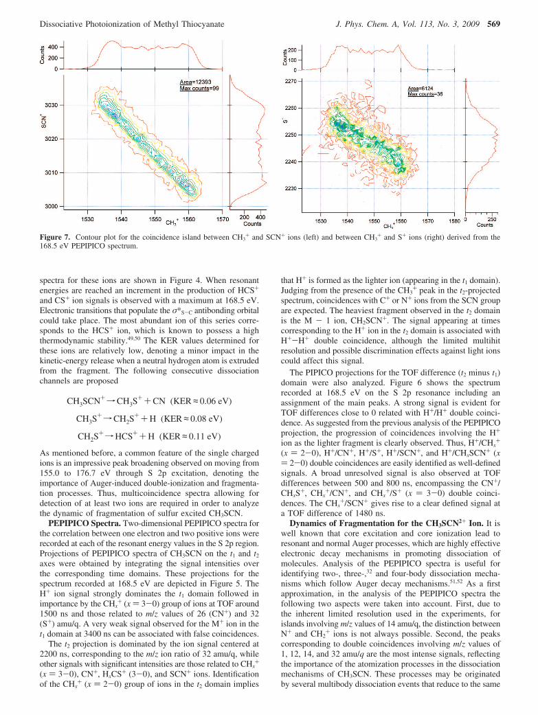
Figure 7. Contour plot for the coincidence island between CH3+ and SCN+ ions (left) and between CH3
+ and S+ ions (right) derived from the168.5 eV PEPIPICO spectrum.
Dissociative Photoionization of Methyl Thiocyanate J. Phys. Chem. A, Vol. 113, No. 3, 2009 569
final pair of atomic ions, making analysis of these coincidencesambiguous. Taking into consideration these facts, attention ispaid to selected pairs of ions for which both good statistics andwell-defined shapes are observed. In particular, ionic fragmentsoriginated by ruptures in the thiocyanate moiety will beconsidered.
The double coincidence branching ratios and KER values fordouble ion processes calculated from PEPIPICO spectra atseveral photon energies are given in Table 3. The experimentalslopes for coincidence islands were determined at both resonance
and off-resonance photon energies in the S 2p region, and nosignificant changes in the dissociation mechanism were ob-served. The following discussion will refer to slopes determinedfrom the PEPIPICO spectrum taken at a 168.5 eV photonenergy.
Fragmentation processes leading to formation of CH3+ and
SCN+ ions dominate the dissociation of CH3SCN excited atthe S 2p levels. The parallelogram-like shape of the island andthe observed slope close to -1.0 (Figure 7) can be explainedby a two-body mechanism
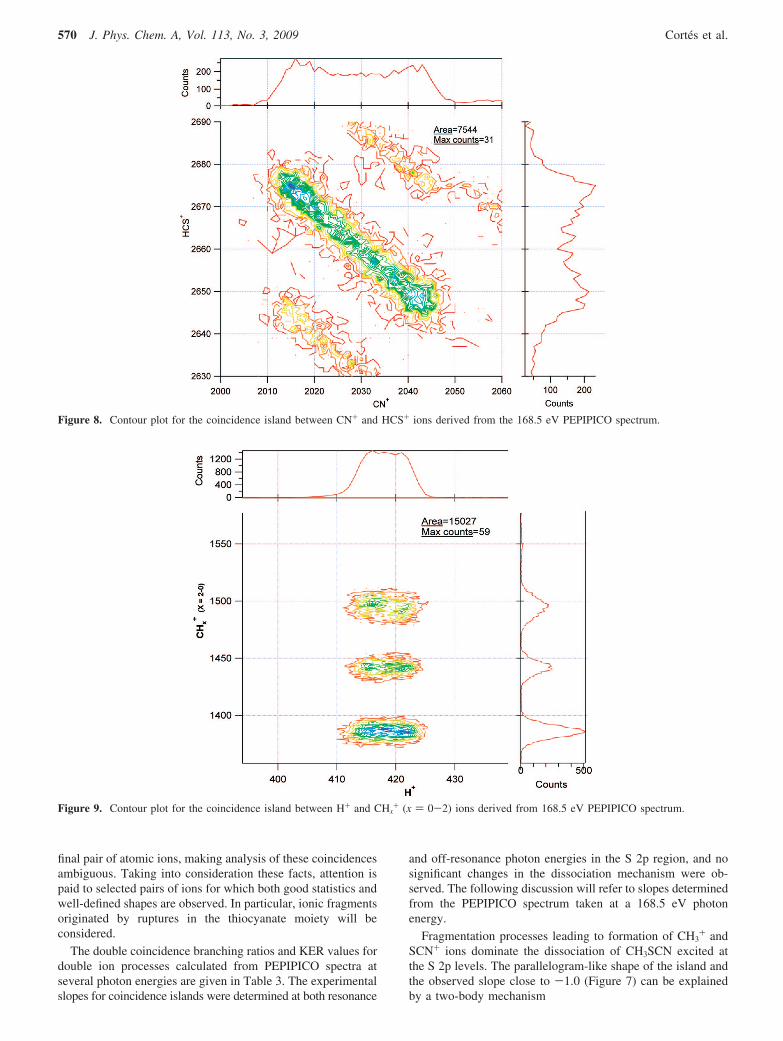
Figure 8. Contour plot for the coincidence island between CN+ and HCS+ ions derived from the 168.5 eV PEPIPICO spectrum.
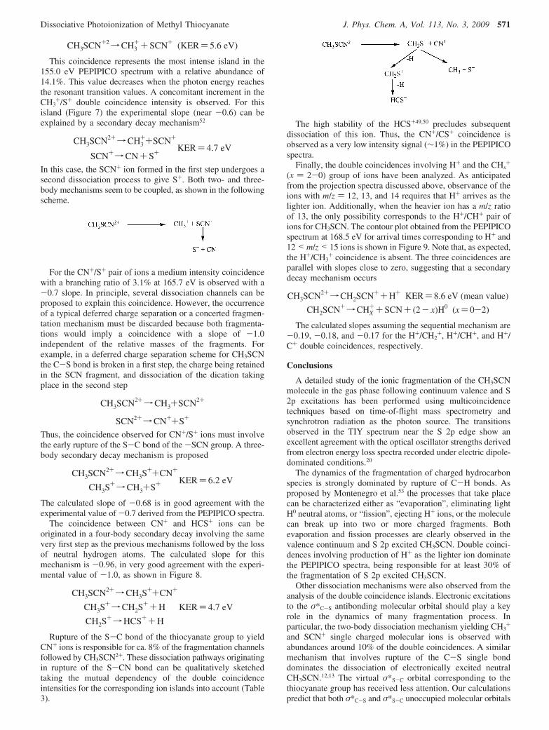
Figure 9. Contour plot for the coincidence island between H+ and CHx+ (x ) 0-2) ions derived from 168.5 eV PEPIPICO spectrum.
570 J. Phys. Chem. A, Vol. 113, No. 3, 2009 Cortes et al.
CH3SCN+2fCH3++ SCN+ (KER) 5.6 eV)
This coincidence represents the most intense island in the155.0 eV PEPIPICO spectrum with a relative abundance of14.1%. This value decreases when the photon energy reachesthe resonant transition values. A concomitant increment in theCH3
+/S+ double coincidence intensity is observed. For thisisland (Figure 7) the experimental slope (near -0.6) can beexplained by a secondary decay mechanism52
CH3SCN2+fCH3++SCN+
SCN+fCN+ S+ KER) 4.7 eV
In this case, the SCN+ ion formed in the first step undergoes asecond dissociation process to give S+. Both two- and three-body mechanisms seem to be coupled, as shown in the followingscheme.
For the CN+/S+ pair of ions a medium intensity coincidencewith a branching ratio of 3.1% at 165.7 eV is observed with a-0.7 slope. In principle, several dissociation channels can beproposed to explain this coincidence. However, the occurrenceof a typical deferred charge separation or a concerted fragmen-tation mechanism must be discarded because both fragmenta-tions would imply a coincidence with a slope of -1.0independent of the relative masses of the fragments. Forexample, in a deferred charge separation scheme for CH3SCNthe C-S bond is broken in a first step, the charge being retainedin the SCN fragment, and dissociation of the dication takingplace in the second step
CH3SCN2+fCH3+SCN2+
SCN2+fCN++S+
Thus, the coincidence observed for CN+/S+ ions must involvethe early rupture of the S-C bond of the -SCN group. A three-body secondary decay mechanism is proposed
CH3SCN2+fCH3S++CN+
CH3S+fCH3+S+ KER) 6.2 eV
The calculated slope of -0.68 is in good agreement with theexperimental value of -0.7 derived from the PEPIPICO spectra.
The coincidence between CN+ and HCS+ ions can beoriginated in a four-body secondary decay involving the samevery first step as the previous mechanisms followed by the lossof neutral hydrogen atoms. The calculated slope for thismechanism is -0.96, in very good agreement with the experi-mental value of -1.0, as shown in Figure 8.
CH3SCN2+fCH3S++CN+
CH3S+fCH2S
++H
CH2S+fHCS++H
KER) 4.7 eV
Rupture of the S-C bond of the thiocyanate group to yieldCN+ ions is responsible for ca. 8% of the fragmentation channelsfollowed by CH3SCN2+. These dissociation pathways originatingin rupture of the S-CN bond can be qualitatively sketchedtaking the mutual dependency of the double coincidenceintensities for the corresponding ion islands into account (Table3).
The high stability of the HCS+49,50 precludes subsequentdissociation of this ion. Thus, the CN+/CS+ coincidence isobserved as a very low intensity signal (∼1%) in the PEPIPICOspectra.
Finally, the double coincidences involving H+ and the CHx+
(x ) 2-0) group of ions have been analyzed. As anticipatedfrom the projection spectra discussed above, observance of theions with m/z ) 12, 13, and 14 requires that H+ arrives as thelighter ion. Additionally, when the heavier ion has a m/z ratioof 13, the only possibility corresponds to the H+/CH+ pair ofions for CH3SCN. The contour plot obtained from the PEPIPICOspectrum at 168.5 eV for arrival times corresponding to H+ and12 < m/z < 15 ions is shown in Figure 9. Note that, as expected,the H+/CH3
+ coincidence is absent. The three coincidences areparallel with slopes close to zero, suggesting that a secondarydecay mechanism occurs
CH3SCN2+fCH2SCN++H+ KER) 8.6 eV (mean value)
CH2SCN+fCHX++ SCN+ (2- x)H0 (x) 0-2)
The calculated slopes assuming the sequential mechanism are-0.19, -0.18, and -0.17 for the H+/CH2
+, H+/CH+, and H+/C+ double coincidences, respectively.
Conclusions
A detailed study of the ionic fragmentation of the CH3SCNmolecule in the gas phase following continuum valence and S2p excitations has been performed using multicoincidencetechniques based on time-of-flight mass spectrometry andsynchrotron radiation as the photon source. The transitionsobserved in the TIY spectrum near the S 2p edge show anexcellent agreement with the optical oscillator strengths derivedfrom electron energy loss spectra recorded under electric dipole-dominated conditions.20
The dynamics of the fragmentation of charged hydrocarbonspecies is strongly dominated by rupture of C-H bonds. Asproposed by Montenegro et al.53 the processes that take placecan be characterized either as “evaporation”, eliminating lightH0 neutral atoms, or “fission”, ejecting H+ ions, or the moleculecan break up into two or more charged fragments. Bothevaporation and fission processes are clearly observed in thevalence continuum and S 2p excited CH3SCN. Double coinci-dences involving production of H+ as the lighter ion dominatethe PEPIPICO spectra, being responsible for at least 30% ofthe fragmentation of S 2p excited CH3SCN.
Other dissociation mechanisms were also observed from theanalysis of the double coincidence islands. Electronic excitationsto the σ*C-S antibonding molecular orbital should play a keyrole in the dynamics of many fragmentation process. Inparticular, the two-body dissociation mechanism yielding CH3
+
and SCN+ single charged molecular ions is observed withabundances around 10% of the double coincidences. A similarmechanism that involves rupture of the C-S single bonddominates the dissociation of electronically excited neutralCH3SCN.12,13 The virtual σ*S-C orbital corresponding to thethiocyanate group has received less attention. Our calculationspredict that both σ*C-S and σ*S-C unoccupied molecular orbitals
Dissociative Photoionization of Methyl Thiocyanate J. Phys. Chem. A, Vol. 113, No. 3, 2009 571
should influence the electronic properties of CH3SCN. Frag-mentation of methyl thiocyanate excited at the S 2p edgeproduces a series of ions arising from rupture of the S-CNbond of the thiocyanate moiety. Thus, the group of doublecoincidences CN+/S+, CN+/HCS+, and CN+/CS+ accounts fornear 8% of the ion signals in the PEPIPICO spectra. This factrepresents indirect evidence that supports the significant roleof the σ*S-C MO in the dynamics of fragmentation of CH3SCN.
It is interesting to note, also, the significant enhancement inthe branching ratios for double coincidences involving the S+
ion at the S 2p excitation energies. The sum of doublecoincidences having S+ as the heavier ion is ca. 21% belowthe S 2p ionization edge. This value increases to 33% of thedouble coincidence production at the first resonance transition(165.7 eV). This could be an indication that state-specificfragmentation54,55 occurs in S 2p excited CH3SCN. These resultsinvite one to perform further studies on selective C 1s and/orN 1s core level photoionization.
Acknowledgment. This work has been largely supported bythe Brazilian Synchrotron Light Source (LNLS) under proposalD05A-TGM-6535. The authors wish to thank Arnaldo Navesde Brito and his research group for fruitful discussions andgenerous collaboration during their several stays in Campinasand the TGM beamline staff for their assistance throughout theexperiments. They are indebted to the Agencia Nacional dePromocion Cientıfica y Tecnologica (ANPCyT), Consejo Na-cional de Investigaciones Cientıficas y Tecnicas (CONICET),and the Comision de Investigaciones Cientıficas de la Provinciade Buenos Aires (CIC), Republica Argentina, for financialsupport. We also thank the Facultad de Ciencias Exactas,Universidad Nacional de La Plata, Republica Argentina, forfinancial support. C.O.D.V. especially acknowledges the DAAD,which generously sponsors the DAAD Regional Program ofChemistry for the Republica Argentina supporting Latin-American students to receive their Ph.D. in La Plata. Finally,we thank referee 2 for his/her time and effort in doing such aconstructive review. We highly appreciate his/her contributionsin improving the content, quality, and presentation of this work.E.C. and M.G. are doctoral fellows of CONICET. M.F.E.,C.O.D.V., and R.M.R. are members of the Carrera del Inves-tigador of CONICET.
References and Notes
(1) Mukerjee, A. K.; Ashare, R. Chem. ReV. 1991, 91, 1.(2) Erian, A. W.; Sherif, S. M. Tetrahedron 1999, 55, 7957.(3) Sharma, S. J. Sulfur Chem. 1989, 8, 327.(4) Moore, C. B.; Alvarez, R. A. Science 1994, 263, 205.(5) Dreizler, H.; Rudolph, H. D.; Schleser, H. Z. Naturforsch. 1970,
25A, 1643.(6) Jonathan, P.; Hamdan, M.; Brenton, A. G.; Willett, G. D. Chem.
Phys. 1988, 119, 159.(7) Babinec, P.; Leszczynski, J. J. Mol. Struct. (Theochem) 2000, 277,
501–502.(8) Fu, Z.; Pan, X.-M.; Li, Z.-S.; Sun, C.-C.; Wang, R.-S. Chem. Phys.
Lett. 2006, 430, 13.(9) Crowder, G. A. J. Mol. Spectrosc. 1967, 23, 108.
(10) Maienschein-Cline, M. G.; Londergan, C. H. J. Phys. Chem. A 2007,111, 10020.
(11) Li, P.; Ling Tan, Y.; Yip Fan, W. Chem. Phys. 2004, 302, 171.(12) Tokue, I.; Hiraya, A.; Shobatake, K. Chem. Phys. 1987, 117, 315.(13) Tokue, I.; Kobayashi, K.; Honda, T.; Ito, Y. J. Phys. Chem. 1990,
94, 3485.(14) Northrup, F. J.; Sears, T. J. J. Chem. Phys. 1990, 93, 2337.(15) Northrup, F. J.; Sears, T. J. J. Chem. Phys. 1990, 93, 2346.(16) Karpas, Z.; Stevens, W. J.; Buckley, T. J.; Metz, R. J. Phys. Chem.
1985, 89, 5274.(17) Neijzen, B. J. M.; De Lange, C. A. J. Electron Spectrosc. Relat.
Phenom. 1980, 18, 179.
(18) Pasinszki, T.; Veszpremi, T.; Feher, M.; Kovac, B.; Klasinc, L.;Mcglynn, S. P. Int. J. Quantum Chem., Quantum Chem. Symp. 1992, 26, 443.
(19) Pasinszki, T.; Yamakado, H.; Ohno, K. J. Phys. Chem. 1993, 97,12718.
(20) Hitchcock, A. P.; Tronc, M.; Modelli, A. J. Phys. Chem. 1989, 93,3068.
(21) Erben, M. F.; Romano, R. M.; Della Vedova, C. O. J. Phys. Chem.A 2004, 108, 3938.
(22) Gerones, M.; Erben, M. F.; Romano, R. M.; Della Vedova, C. O.J. Electron Spectrosc. Relat. Phenom. 2007, 155, 64.
(23) Erben, M. F.; Romano, R. M.; Della Vedova, C. O. J. Phys. Chem.A 2005, 109, 304.
(24) Erben, M. F.; Gerones, M.; Romano, R. M.; Della Vedova, C. O.J. Phys. Chem. A 2006, 110, 875.
(25) Erben, M. F.; Gerones, M.; Romano, R. M.; Della Vedova, C. O.J. Phys. Chem. A 2007, 111, 8062.
(26) Gerones, M.; Erben, M. F.; Romano, R. M.; Della Vedova, C. O.;Yao, L.; Ge, M. J. Phys. Chem. A 2008, 112, 2228.
(27) Erben, M. F.; Della Vedova, C. O. Inorg. Chem. 2002, 41, 3740.(28) Gerones, M.; Downs, A. J.; Erben, M. F.; Ge, M.; Romano, R. M.;
Yao, L.; Della Vedova, C. O. J. Phys. Chem. A 2008, 112, 5947.(29) Lira, A. C.; Rodrigues, A. R. D.; Rosa, A.; Goncalves da Silva,
C. E. T.; Pardine, C.; Scorzato, C.; Wisnivesky, D.; Rafael, F.; Franco,G. S.; Tosin, G.; Lin, L.; Jahnel, L.; Ferreira, M. J.; Tavares, P. F.; Farias,R. H. A.; Neuenschwander, R. T. First Year Operation of the BrazilianSinchrotron Light Source. European Particle Accelerator Conference,Stockholm, Sweden, 1998.
(30) de Fonseca, P. T.; Pacheco, J. G.; Samogin, E.; de Castro, A. R. B.ReV. Sci. Instrum. 1992, 63, 1256.
(31) Kivimaki, A.; Ruiz, J. A.; Erman, P.; Hatherly, P.; Garcia, E. M.;Rachlew, E.; Riu, J. R. i.; Stankiewicz, M. J. Phys. B: At. Mol. Opt. Phys.2003, 781.
(32) Frasinski, L. J.; Stankiewicz, M.; Randall, K. J.; Hatherly, P. A.;Codling, K. J. Phys. B: At. Mol. Phys 1986, 19, L819.
(33) Eland, J. H. D.; Wort, F. S.; Royds, R. N. J. Electron Spectrosc.Relat. Phenom. 1986, 41, 297.
(34) Naves de Brito, A.; Feifel, R.; Mocellin, A.; Machado, A. B.;Sundin, S.; Hjelte, I.; Sorensen, S. L.; Bjorneholm, O. Chem. Phys. Lett.1999, 309, 377.
(35) Burmeister, F.; Coutinho, L. H.; Marinho, R. R.; Homem, M. G. P.;de Morais, M. A. A.; Mocellin, A. ; Bjorneholm, O.; Sorensen, S. L. ; deFonseca, P. T.; Lindgren, A.; Naves de Brito, A. J. Electron Spectrosc.Relat. Phenom. 2008, in press; doi:10.1016/j.elspec.2006.05.007.
(36) Laskin, J.; Lifshitz, C. J. Mass Spectrom. 2001, 36, 459.(37) Hansen, D. L.; Arrasate, M. E.; Cotter, J.; Fisher, G. R.; Hemmers,
O.; Leung, K. T.; Levin, J. C.; Martin, R.; Neill, P.; Perera, R. C. C.; Sellin,I. A.; Simon, M.; Uehara, Y.; Vanderford, B.; Whitfield, S. B.; Lindle,D. W. Phys. ReV. A 1998, 58, 3757–3765.
(38) Simon, M.; LeBrun, T.; Morin, P.; Lavollee, M.; Marechal, J. L.Nucl. Instrum. Methods 1991, B62, 167.
(39) Santos, A. C. F.; Lucas, C. A.; de Souza, G. G. B. J. ElectronSpectrosc. Relat. Phenom. 2001, 115, 114–116.
(40) Nenner, I.; Beswick, J. A. Molecular Photodissociation andPhotoionization. In Handbook on Synchrotron Radiation; Marr, G. V., Ed.;Elsevier Science Publishers: Amsterdam, 1987; Vol. 2; p 355.
(41) Hitchcock, A. P. http://unicorn.mcmaster.ca/corex/cedb-title.html.(42) Hitchcock, A. P. J. Electron Spectrosc. Relat. Phenom. 1982, 25, 245.(43) Jalilehvand, F. Chem. Soc. ReV. 2006, 35, 1256.(44) Hudson, E.; Shirley, D. A.; Domke, M.; Remmers, G.; Puschmann,
A.; Mandel, T.; Xue, C.; Kaindl, G. Phys. ReV. A 1993, 47, 361.(45) Hitchcock, A. P.; Horsley, J. A.; Stohr, J. J. Chem. Phys. 1986,
85, 4835.(46) Stein, S. E. NIST Mass Spectroscopy Data Center, NIST Standard
Reference Database Number 69; Linstrom, P. J., Mallard, W. G., Eds.;National Institute of Standards and Technology: Gaithersburg, MD, March2003.
(47) Lago, A. F.; Santos, A. C. F.; de Souza, G. G. B. J. Chem. Phys.2004, 120, 9547.
(48) Nicholas, J. E.; Amodio, C. A. J. Chem. Soc., Faraday Trans. 11980, 76, 1669.
(49) Smith, D. Chem. ReV. 1992, 92, 1473.(50) Thaddeus, P.; Guelin, M.; Linke, R. A. Astrophys. J. 1981, 246, L41.(51) Eland, J. H. D. Mol. Phys. 1987, 61, 725.(52) Simon, M.; Lebrun, T.; Martins, R.; de Souza, G. G. B.; Nenner,
I.; Lavollee, M.; Morin, P. J. Phys. Chem. 1993, 97, 5228.(53) Montenegro, E. C.; Scully, S. W; Wyer, J. A.; Senthil, V.; Shah,
M. B. J. Electron Spectrosc. Relat. Phenom. 2007, 155, 81.(54) Eberhardt, W.; Sham, T. K.; Carr, R.; Krummacher, S.; Strongin,
M.; Weng, S. L.; Wesner, D. Phys. ReV. Lett. 1983, 50, 1038.(55) Miron, C.; Simon, M.; Leclercq, N.; Hansen, D. L.; Morin, P. Phys.
ReV. Lett. 1998, 81, 4104.
JP807230S
572 J. Phys. Chem. A, Vol. 113, No. 3, 2009 Cortes et al.


![VUV photoionization and dissociative photoionization ...schwell/Bellili_2015.pdfthat the Strecker reaction, i.e. the reaction between an aldehyde, HCN and NH 3 (see Refs. [5a,b] and](https://img.pdfslide.net/doc/110x75/612114431709f84c7835b7a3/vuv-photoionization-and-dissociative-photoionization-schwellbellili2015pdf.jpg)