Embed Size (px)
Citation preview
Strana 836 VOJNOSANITETSKI PREGLED Vojnosanit Pregl 2013; 70(9): 836–841.
Correspondence to: Saša Milenkovi , University Orthopaedic and Traumatology Clinic Niš, Bulevar dr Zorana Djindjica 48, 18 000 Niš,Serbia. E-mail: [email protected]
O R I G I N A L A R T I C L E UDC: 617.58::616.718.5-001.5-089DOI: 10.2298/VSP1309836M
Distal tibial pilon fractures (AO/OTA type B, and C) treated with theexternal skeletal and minimal internal fixation method
Zbrinjavanje preloma distalnog pilona tibije (AO/OTA tipa B, C) metodomspoljašnje skeletne i minimalne unutrašnje fiksacije
Saša Milenkovi *, Milorad Mitkovi *, Ivan Mici *, Desimir Mladenovi *,Stevo Najman†, Miroslav Trajanovi ‡, Miodrag Mani ‡, Milan Mitkovi *
*Orthopaedic and Traumatology Clinic, Clinical Center Niš, Faculty of Medicine,University of Niš, Niš, Serbia; Serbia; †Faculty of Medicine, University of Niš, Niš,
Serbia; ‡Faculty of Mechanical Engineering, University of Niš, Niš, Serbia
Abstract
Background/Aim. Distal tibial pilon fractures includeextra-articular fractures of the tibial metaphysis and themore severe intra-articular tibial pilon fractures. There isno universal method for treating distal tibial pilon frac-tures. These fractures are treated by means of open reduc-tion, internal fixation (ORIF) and external skeletal fixa-tion. The high rate of soft-tissue complications associatedwith primary ORIF of pilon fractures led to the use ofexternal skeletal fixation, with limited internal fixation asan alternative technique for definitive management. Theaim of this study was to estimate efficacy of distal tibialpilon fratures treatment using the external skeletal andminimal internal fixation method. Methods. We presenteda series of 31 operated patients with tibial pilon fractures.The patients were operated on using the method of exter-nal skeletal fixation with a minimal internal fixation. Ac-cording to the AO/OTA classification, 17 patients hadtype B fracture and 14 patients type C fractures. The rigidexternal skeletal fixation was transformed into a dynamicexternal skeletal fixation 6 weeks post-surgery. Results.
This retrospective study involved 31 patients with tibialpilon fractures, average age 41.81 (from 21 to 60) years.The average follow-up was 21.86 (from 12 to 48) months.The percentage of union was 90.32%, nonunion 3.22%and malunion 6.45%. The mean to fracture union was 14(range 12–20) weeks. There were 4 (12.19%) infectionsaround the pins of the external skeletal fixator and one(3.22%) deep infections. The ankle joint arthrosis as a latecomplication appeared in 4 (12.90%) patients. All arthro-ses appeared in patients who had type C fractures. The fi-nal functional results based on the AOFAS score were ex-cellent in 51.61%, good in 32.25%, average in 12.90% andbad in 3.22% of the patients. Conclusion. Externalskeletal fixation and minimal internal fixation of distaltibial pilon fractures is a good method for treating all typesof inta-articular pilon fractures. In fractures types B and Cdynamic external skeletal fixation allows early mobility inthe ankle joint.
Key words:tibial fractures; orthopedic procedures; externalfixators; internal fixators; treatment outocme.
Apstrakt
Uvod/Cilj. Prelomi distalnog pilona tibije podrazumevajuspoljašnje artikularne prelome metafize tibije i teže unutra-šnje artikularne prelome pilona tibije. Ne postoji univer-zalni metod za le enje preloma distalnog pilona tibije. Oviprelomi se le e metodom otvorene redukcije i stabilne fik-sacije (ORIF) i spoljašnjom skeletnom fiksacijom. Visokprocenat komplikacija na mekom tkivu udružen nakonprimarne ORIF preloma pilona, name e upotrebu metodespoljašnje skeletne fiksacije sa minimalnom unutrašnjomfiksacijom, kao alternativnu tehniku za kona no izle enje.Cilj rada bio je da se utvrdi efikasnost le enja distalnogpilona tibije primenom metode spoljašnje skeletne i mini-
malne unutrašnje fiksacije. Metode. Prikazali smo serijuod 31 operisanog bolesnika sa prelomima pilona tibije.Bolesnici su operisani metodom spoljašnje skeletne fiksa-cije sa minimalnom unutrašnjom fiksacijom. PremaAO/OTA klasifikaciji 17 bolesnika imalo je prelom tipa B,a 14 prelom tipa C. Kruta spoljašnja skeletna fiksacija jetransformisana u dinami ku spoljašnju skeletnu fiksacijušest nedelja posle operacije. Rezultati. Retrospektivnomstudijom analiziran je 31 bolesnik sa prelomima pilona ti-bije, prose ne starosti 41,81 (21–60) godina. Prose novreme pra enja iznosilo je 21,86 (12–48) meseci. Procenatzarastanja preloma iznosio je 90,32%, nezarastanja 3,22% ilošeg zarastanja 6,45%. Prose no trajanje zarastanja pre-loma iznosilo je 14 (12–20) nedelja. Bilo je 4 (12,19%) in-
Volumen 70, Broj 9 VOJNOSANITETSKI PREGLED Strana 837
Milenkovi S, et al. Vojnosanit Pregl 2013; 70(9): 836–841.
fekcija oko klinova spoljašnjeg skeletnog fiksatora i 1(3,22%) duboka infekcije. Artroza sko nog zgloba kao ka-sna komplikacija, pojavila se kod 4 (12,90%) bolesnika.Sve artroze su nastale kod bolesnika koji su imali prelomtipa C. Krajnji funkcionalni rezultati na osnovu AOFASskora bili su odli ni kod 51,61%, dobri kod 32,25%, ume-reni kod 12,90% i loši kod 3,22% bolesnika. Zaklju ak.Spoljašnja skeletna fiksacija i minimalna unutrašnja fiksa-
cija preloma distalnog pilona tibije dobra je metoda za le-enje svih tipova intraartikularnih preloma pilona. Kod
preloma tipa B i C, dinami ka spoljašnja skeletna fiksacijadozvoljava rane pokrete u sko nom zglobu.
Klju ne re i:tibija, prelomi; ortopedske procedure; fiksatori, spoljni;fiksatori, unutrašnji; le enje, ishod.
Introduction
In contrast to the rotational mechanisms that result in mal-leolar fractures and fracture-dislocations of the ankle, distaltibial pilon fractures typically result from high-energy axial-loading mechanisms. Distal tibial pilon fractures include extra-articular fractures of the tibial metaphysis and the more severeintraarticular tibial plafond or pilon fractures. The clinical mani-festation of this fractures difference is the generation of osteo-chondral fracturing, comminution and displacement of theweight-bearing articular portion of the tibial plafond and distaltibial metaphysis, as well as the development of marked swel-ling, blistering and devitalization of the surrounding soft-tissueenvelope typically identified in tibial pilon fractures. Thesefractures are estimated to comprise 3% to 10% of all tibia frac-tures and less than 1% of lower extremity fractures. These highenergy injuries, usually caused by falls from heights or motorvehicle accidens, are often open fractures and they are fre-quently associated with additional trauma in other areas of thebody 1–3. They are one of the most challenging injuries in ortho-paedic traumatology 4. Several treatment methods are recom-mended for the treatment of these injuries including externalskeletal fixation, intramedullary nailing, and plate fixation 5–8.The aim of this study was to estimate efficacy of a treatment of adistal tibial fracture (AO type B and C) using the method of ex-ternal skeletal fixation combined with minimal internal fixation.
The high rate of soft-tissue complications associatedwith primary open reduction, internal fixation (ORIF) ofdistal tibial pilon fractures led to use of external skeletalfixation, with limited internal fixation as an alternative tech-nique for definitive management. Our aim was to analyzeoriginal results of distal tibial pilon fractures treatmentusing the external skeletal and minimal internal fixationmethod.
Methods
The patients with distal tibial fracture were operatedon at the University Orthopedic and Traumatology Clinic,Niš. The patients with intra-articular fractures (AO/OTAtypes B and C) were operated on using the method of ex-ternal skeletal fixation and minimal internal fixation. Toperform minimal internal fixation, screws and K-wireswere used. In patients with types B and C fractures, rigidexternal skeletal fixation was transformed into dynamicexternal skeletal fixation 1.5 month later. All fractureswere classified according to the AO/OTA classification 9.The Gustilo-Anderson classification system was used for
all open fractures 10. To perform external skeletal fixation, aMitkovic’s unilateral external skeletal fixator was used. Toanalyze the final functional results, the AOFAS scoring systemwas used 11.
Results
This retrospective study involved 31 patients with distaltibial pilon fractures. According to the AO/OTA classifica-tion, 17 patients had fractures type B and 14 patients frac-tures type C (Figures 1–5 and 6–9 respectively). The averageage was 41.81 (21–60) years, and there were 20 male pa-tients and 11 female patients. Open fractures appeared in 11(35.48%) patients. A total of 10 (32.25%) fractures were ca-used by car accidents, 14 (45.16%) by falls from heights,whereas 7 (22.58%) fractures appeared under different cir-cumstances, such as in accidents at work, falls from stairs, oras a result of slip and fall accidents on an even surface. Theaverage follow-up of the patients was 21.86 (12–48) months.There were 28 (90.32%) unions, 1 (3.22%) nonunions and 2(6.45%) malunions. The mean to fracture union was 14(range 12–20) weeks. As regards complications, infectionaround the pins of the external skeletal fixator appeared in 4(12.19%) and deep infections appeared in 1 (3.22%) patients.Ankle joint arthrosis as late complication appeared in 4(12.90%) patients. All arthroses appeared in patients withfractures type C. According to AOFAS, the final functionalresults were excellent in 16 (51.61%) patients, good in 10(32.25%), average in 4 (12.90%) patietns and bad in 1(3.22%) cases. All the patients were operated on as urgentcases, immediately after hospitalization.
Fig. 1 (A and B) – Radiographs of distal tibial pilon fracture(AO/OTA type C) after the injury.
Strana 838 VOJNOSANITETSKI PREGLED Volumen 70, Broj 9
Milenkovi S, et al. Vojnosanit Pregl 2013; 70(9): 836–841.
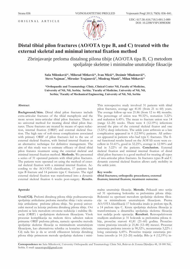
Fig. 2 (A and B) – Radioscopic views after external skeletal fixation and minimal internal K-wires fixation.
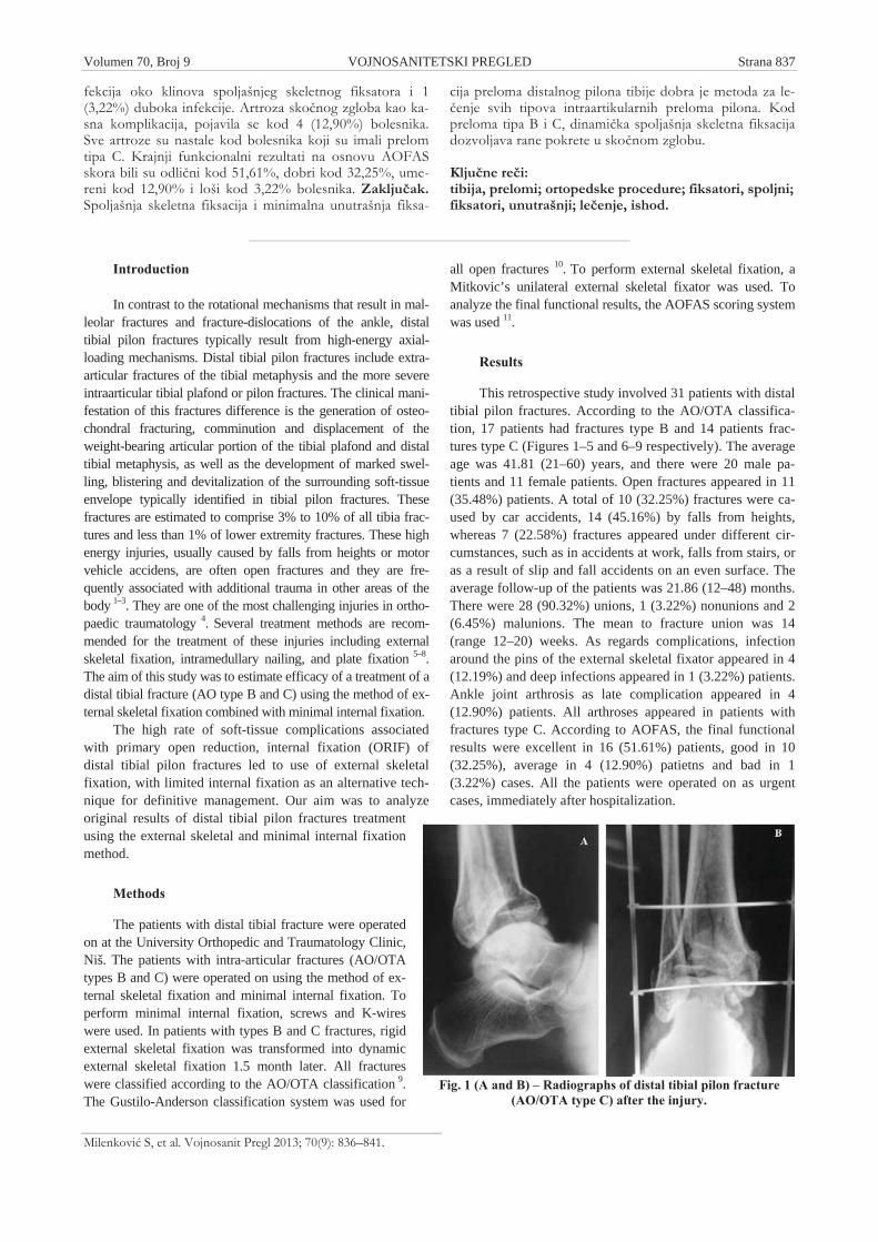
Fig. 3 – Radiopgraphs views after the surgery (A), and after 1 month (B).
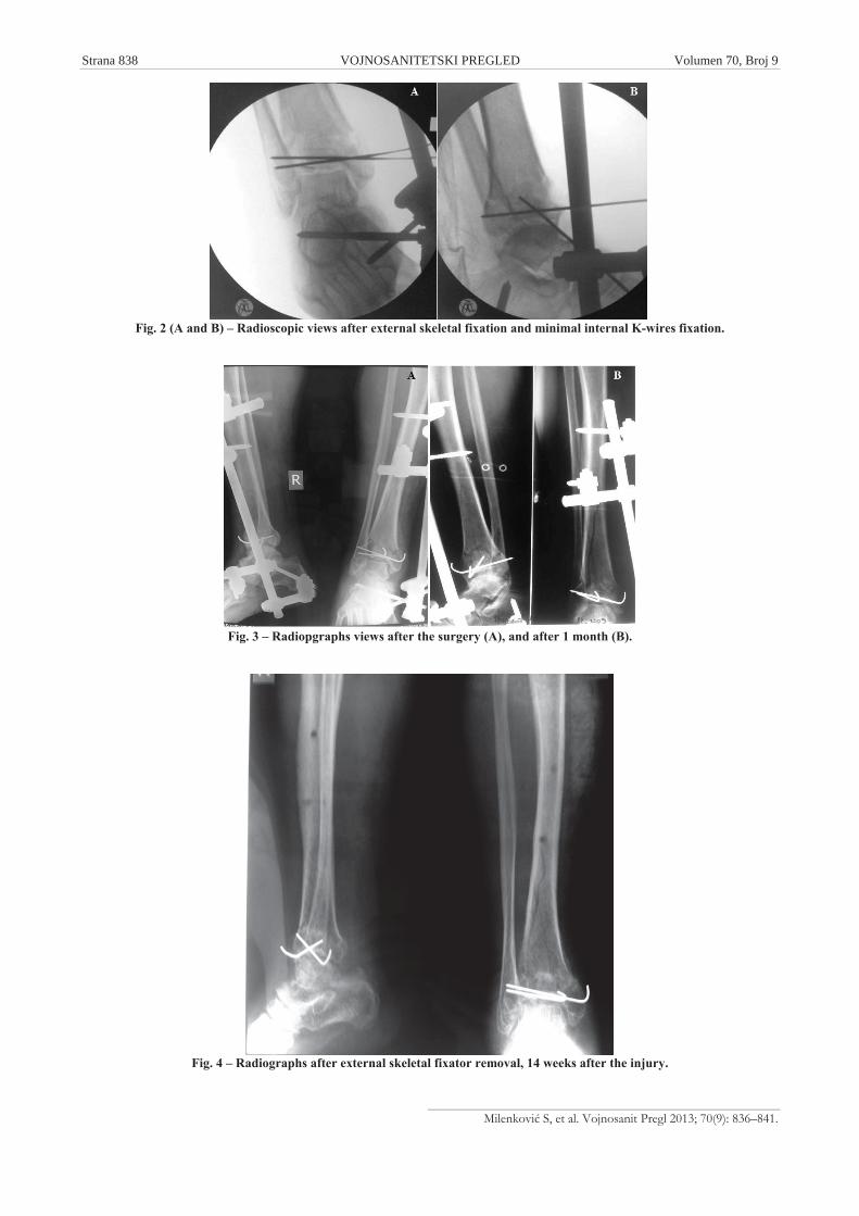
Fig. 4 – Radiographs after external skeletal fixator removal, 14 weeks after the injury.
Volumen 70, Broj 9 VOJNOSANITETSKI PREGLED Strana 839
Milenkovi S, et al. Vojnosanit Pregl 2013; 70(9): 836–841.
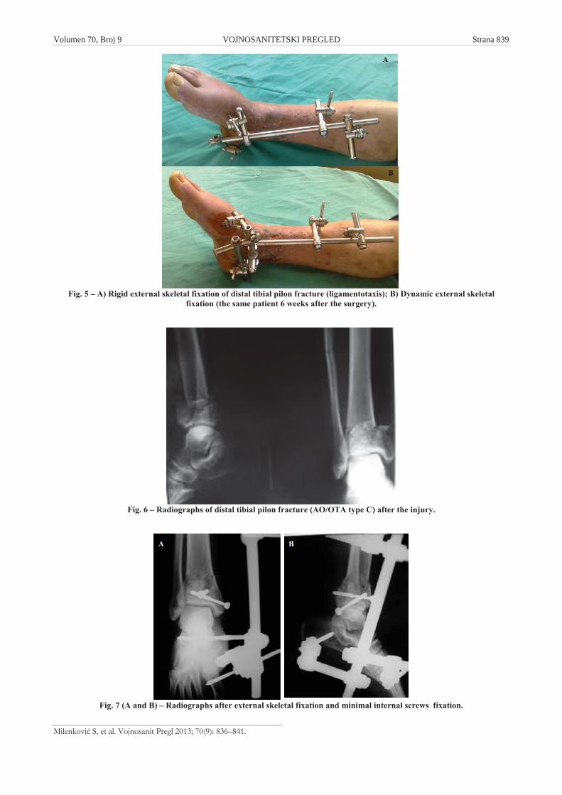
Fig. 5 – A) Rigid external skeletal fixation of distal tibial pilon fracture (ligamentotaxis); B) Dynamic external skeletalfixation (the same patient 6 weeks after the surgery).
Fig. 6 – Radiographs of distal tibial pilon fracture (AO/OTA type C) after the injury.
Fig. 7 (A and B) – Radiographs after external skeletal fixation and minimal internal screws fixation.
Strana 840 VOJNOSANITETSKI PREGLED Volumen 70, Broj 9
Milenkovi S, et al. Vojnosanit Pregl 2013; 70(9): 836–841.
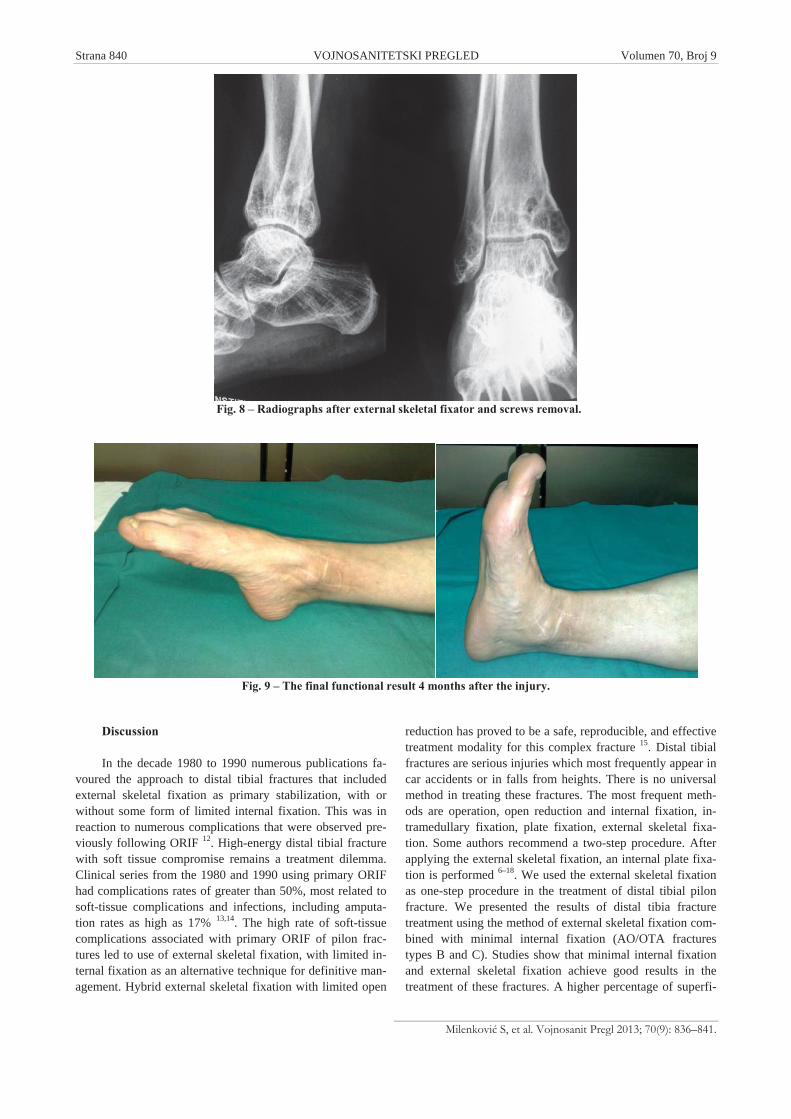
Fig. 8 – Radiographs after external skeletal fixator and screws removal.
Fig. 9 – The final functional result 4 months after the injury.
Discussion
In the decade 1980 to 1990 numerous publications fa-voured the approach to distal tibial fractures that includedexternal skeletal fixation as primary stabilization, with orwithout some form of limited internal fixation. This was inreaction to numerous complications that were observed pre-viously following ORIF 12. High-energy distal tibial fracturewith soft tissue compromise remains a treatment dilemma.Clinical series from the 1980 and 1990 using primary ORIFhad complications rates of greater than 50%, most related tosoft-tissue complications and infections, including amputa-tion rates as high as 17% 13,14. The high rate of soft-tissuecomplications associated with primary ORIF of pilon frac-tures led to use of external skeletal fixation, with limited in-ternal fixation as an alternative technique for definitive man-agement. Hybrid external skeletal fixation with limited open
reduction has proved to be a safe, reproducible, and effectivetreatment modality for this complex fracture 15. Distal tibialfractures are serious injuries which most frequently appear incar accidents or in falls from heights. There is no universalmethod in treating these fractures. The most frequent meth-ods are operation, open reduction and internal fixation, in-tramedullary fixation, plate fixation, external skeletal fixa-tion. Some authors recommend a two-step procedure. Afterapplying the external skeletal fixation, an internal plate fixa-tion is performed 6–18. We used the external skeletal fixationas one-step procedure in the treatment of distal tibial pilonfracture. We presented the results of distal tibia fracturetreatment using the method of external skeletal fixation com-bined with minimal internal fixation (AO/OTA fracturestypes B and C). Studies show that minimal internal fixationand external skeletal fixation achieve good results in thetreatment of these fractures. A higher percentage of superfi-
Volumen 70, Broj 9 VOJNOSANITETSKI PREGLED Strana 841
Milenkovi S, et al. Vojnosanit Pregl 2013; 70(9): 836–841.
cial infections around the pins does not affect the final out-come of the treatment 19. Bone 1 also describes satisfactoryresults in the application of this method. In fractures type Band C, it is necessary to achieve fracture reduction and ar-ticular tibial surface reconstruction. Fixation by means ofscrews and K-wires is open and minimal. External skeletalfixator pins are placed, 2 in the proximal fragment, and 2 inthe foot. One pin is placed in the calcaneus, the other in the Imetatarsal bone. After that, the external skeletal fixatorframe with clamps and carriers of the clamp placed. In thisway, rigid fracture fixation is achieved, and it transforms intodynamic fixation 6-week post-surgery, which allows earlyankle joint mobility 20. A dynamic external skeletal fixationis placed on an already existing external skeletal fixator con-struction with additional carriers of the clamp and clamps.This system for external skeletal fixation is suitable for addi-tional interventions, such as fracture position correctionwhile the apparatus is carried. Studies describe this methodof treatment as definitive or temporary method, after whichintramedullary or plate fixation of fracture will be per-
formed 21. Our experience in the treatment of these fracturesas definitive method and our results are very encouraging,giving us right to consider this method suitable for treatingall types of distal tibial pilon fractures. It is important to em-phasize that these fractures are considered as urgent, andthey should be treated urgently. Urgent surgical interventionreduces the possibility of complications.
Conclusion
External skeletal fixation of distal tibial and pilon frac-tures as one-step procedure is a good method for treating alltypes of fractures. In fractures types B and C, dynamic exter-nal skeletal fixation allows early mobility in the ankle joint.
Acknowledge
This work was supported by the Ministry of Education,Science and Technological Development of the Republic ofSerbia, project No III41017.
R E F E R E N C E S
1. Bone LB. Fractures of the tibial plafond. The pilon fracture.Orthop Clin North Am 1987; 18(1): 95 104.
2. Mandracchia VJ, Evans RD, Nelson SC, Smith KM. Pilon frac-tures of the distal tibia. Clin Podiatr Med Surg 1999; 16(4):743 67.
3. Burgess AR, Dischinger PC, O´Quinn TD, Schmidhauser CB. Lowerextremity injures in drivers of airbag-equipped automo-biles:clinical and crash reconstruction correlations. JTrauma1995; 38(4): 509 16.
4. Pollak AN, McCarthy ML, Bess RS, Agel J, Swiontkowski MF.Outcomes after treatment of high-energy tibial plafond frac-tures. J Bone Joint Surg Am 2003; 85-A(10): 1893 900.
5. Mosheiff R, Safran O, Segal D, Liebergall M. The unreamed tibialnail in the treatment of distal metaphyseal fractures. Injury1999; 30(2): 83 90.
6. Khoury A, Liebergall M, London E, Mosheiff R. Percutaneousplating of distal tibial fractures. Foot Ankle Int 2002; 23(9):818 24..
7. Anglen JO. Early outcome of hybrid external fixation for frac-ture of the distal tibia. J Orthop Trauma 1999; 13(2): 92 7.
8. Babis GC, Vayanos ED, Papaioannou N, Pantazopoulos T. Resultsof surgical treatment of tibial plafond fractures. Clin OrthopRelat Res 1997; (341): 99 105.
9. Ruedi T, Murphy WM. AO Principles of Fracture Management.Vol. 1. Stuttgart-New York: Thieme; 2000.
10. Gustilo RB, Anderson JT. Prevention of infection in the treat-ment of one thousand and twenty-five open fractures of longbones: retrospective and prospective analyses. J Bone JointSurg Am 1976; 58(4): 453 8.
11. Kitaoka HB, Patzer GL. Analysis of clinical grading scales forthe foot and ankle. Foot Ankle Int 1997; 18(7): 443 6.
12. Barbieri R, Schenk R, Koval K, Aurori K, Aurori B. Hybrid exter-nal fixation in the treatment of tibial plafond fractures. ClinOrthop Relat Res 1996; (332): 16 22.
13. McFerran MA, Smith SW, Boulas HJ, Schwartz HS. Complica-tions encountered in the treatment of pilon fractures. J OrthopTrauma 1992; 6(2): 195 200.
14. Wyrsch B, McFerran MA, McAndrew M, Limbird TJ, Harper MC,Johnson KD, et al. Operative treatment of fractures of the tibialplafond: A randomized, prospective study. J Bone Joint SurgAm 1996; 78(11): 1646 57.
15. French B, Tornetta P 3rd. Hybrid external fixation of tibial pilonfractures. Foot Ankle Clin 2000; 5(4): 853 71.
16. Blauth M, Bastian L, Krettek C, Knop C, Evans S. Surgical op-tions for the treatment of severe tibial pilon fractures: astudy of three techniques. J Orthop Trauma 2001; 15(3):153 60.
17. Dickson KF, Montgomery S, Field J. High energy plafond fracturestreated by a spanning external fixator initially and followed bya second stage open reduction internal fixation of the articularsurface-preliminary report. Injury 2001; 32(Suppl 4): SD92 8.
18. Patterson MJ, Cole JD. Two-staged delayed open reduction andinternal fixation of severe pilon fractures. J Orthop Trauma1999; 13(2): 85 91.
19. El-Shazly M, Dalby-Ball J, Burton M, Saleh M. The use of trans-articular and extra-articular external fixation for managementof distal tibial intra-articular fractures. Injury. 2001; 32(Suppl4): SD99 106.
20. Mitkovic M, Bumbasirevic M, Lesic A, Golubovic Z. Dynamic ex-ternal fixation of comminuted intra-articular fractures of thedistal tibia (type C pilon fractures). Acta Orthop Belg 2002;68(5): 508 14.
21. Hontzsch D, Karnatz N, Jansen T. One-or two-step management(with external fixator) of severe pilon-tibial fractures.AktuelleTraumatol 1990; 20(4): 199 204. (German)
Received on January 12, 2012.Revised on June 19, 2012.
Accepted on August 20, 2012.