Embed Size (px)
Citation preview
ORIGINAL RESEARCHpublished: 29 July 2016
doi: 10.3389/fmicb.2016.01175
Frontiers in Microbiology | www.frontiersin.org 1 July 2016 | Volume 7 | Article 1175
Edited by:
Dominique Sanglard,
University of Lausanne, Switzerland
Reviewed by:
Elvira Román,
Universidad Complutense de Madrid,
Spain
Jeanette Wagener,
University of Aberdeen, UK
*Correspondence:
Yuan Wu
Jinxing Lu
Specialty section:
This article was submitted to
Fungi and Their Interactions,
a section of the journal
Frontiers in Microbiology
Received: 23 May 2016
Accepted: 15 July 2016
Published: 29 July 2016
Citation:
Yu S, Li W, Liu X, Che J, Wu Y and
Lu J (2016) Distinct Expression Levels
of ALS, LIP, and SAP Genes in
Candida tropicalis with Diverse Virulent
Activities. Front. Microbiol. 7:1175.
doi: 10.3389/fmicb.2016.01175
Distinct Expression Levels of ALS,LIP, and SAP Genes in Candidatropicalis with Diverse VirulentActivitiesShuanbao Yu, Wenge Li, Xiaoshu Liu, Jie Che, Yuan Wu* and Jinxing Lu*
State Key Laboratory of Infectious Disease Prevention and Control, Collaborative Innovation Center for Diagnosis and
Treatment of Infectious Diseases, National Institute for Communicable Disease Control and Prevention, Chinese Center for
Disease Control and Prevention, Beijing, China
Candia tropicalis is an increasingly important human pathogen, causing nosocomial
fungemia among patients with neutropenia or malignancy. However, limited research has
been published concerning its pathogenicity. Based on the phenotypes of C. tropicalis
in our previous study, we selected nine representative strains with different activities
of virulence factors (adhesion, biofilm formation, secreted aspartic proteinases, and
hemolysins), and one reference strain, ATCC750. The present study aimed to investigate
the filamentation ability, the expression of virulence genes (ALST1-3, LIP1, LIP4, and
SAPT1-4) and the cell damage ofC. tropicalis strains with diverse virulences. C. tropicalis
exhibited strain-dependent filamentation ability, which was positively correlated with
biofilm formation. Reverse transcriptase PCR analysis showed that the ALST3 and
SAPT3 genes had the highest expression in their corresponding genes for most
C. tropicalis. The expressions of virulence genes, except ALST3 on polystyrene, were
upregulated compared with growth in the planktonic and on human urinary bladder
epithelial cell line (TCC-SUP) surface. Clustering analysis of virulence genes showed
that isolates had a high biofilm forming ability on polystyrene formed a group. Lactate
dehydrogenase assays showed that the cell damage induced by C. tropicalis markedly
increased with longer infection time (24 and 48 h). Strain FXCT01, isolated from blood,
caused the most serious cell damage; while ZRCT52, which had no filamentation ability,
caused the least cell damage. Correlation analysis demonstrated significant correlation
existed between adhesion on epithelial cells or the expression of ALST2-3 and cell
damage. Overall, our results supported the view that adhesion and filamentation may
play significant roles in the cell damage caused by C. tropicalis.
Keywords: Candida tropicalis, candidosis, virulence factor, virulence gene expression, cell damage
INTRODUCTION
Candida tropicalis, an emerging opportunistic pathogen, mainly causes superficial and invasiveinfections in human populations, especially among neutropenic patients and those withhematological malignancies (Guinea, 2014). However, less is known about the pathogenicity ofC. tropicalis compared with the more extensively studied C. albicans (Silva et al., 2011b). Severalfactors were reported to contribute to Candida pathogenicity, including adhesion to medicaldevices and host cells; biofilm formation (BF); filamentation ability; and secretion of hydrolytic
Yu et al. Candida tropicalis Virulence Activities
enzymes, including secreted aspartyl proteases (Saps), esterases,lipases, phospholipases, and hemolysins (Silva et al., 2011b;Lackey et al., 2013; Hirakawa et al., 2015).
Adherence to host cells is the first step in invasive infections by
Candida and in the BF, which plays a vital role in its pathogenicity
(Ramage et al., 2006; Silva-Dias et al., 2015). Filamentation is
also required for virulence-related processes, including invasion
of epithelial cell layers, and BF (Jayatilake et al., 2006; Lackey
et al., 2013). PCR using consensus primers identified at least threeAgglutinin-like sequences (ALST1-3) for adhesion of C. tropicalis(Hoyer et al., 2001). In addition, Buter et al. used a phylogenomicapproach to identify 16 genes in ALS gene family in C. tropicalis(Butler et al., 2009). In addition, hydrolytic enzymes might alsoplay an important role in infection by disrupting host mucosalmembranes, degrading immunological and structural defenseproteins, and providing nutrients (Buzzini and Martini, 2002;Silva et al., 2012; Rossoni et al., 2013). C. tropicalis possessesfour Saps-encoding genes (SAPT1-4), and five secreted lipase-encoding genes (Togni et al., 1991; Zaugg et al., 2001; Butler et al.,2009). The specific gene sequences of SAPT1-4, LIP1, and LIP4could be found in GenBank.
The virulence phenotypes (adhesion, BF, Saps,phospholipases, and hemolysins) of 68 C. tropicalis wereanalyzed in vitro in our previous study (Yu et al., 2015).However, limited studies have been performed on virulencefactors of C. tropicalis at the level of transcription. According tothe activities of virulence factors in C. tropicalis, we selected ninerepresentative strains and one reference strain, ATCC750, in thisstudy. The objective of this study was to determine the activitiesof the virulence factors (lipase, esterase, and filamentation ability)in vitro; to explore the expression profile of virulence genes,
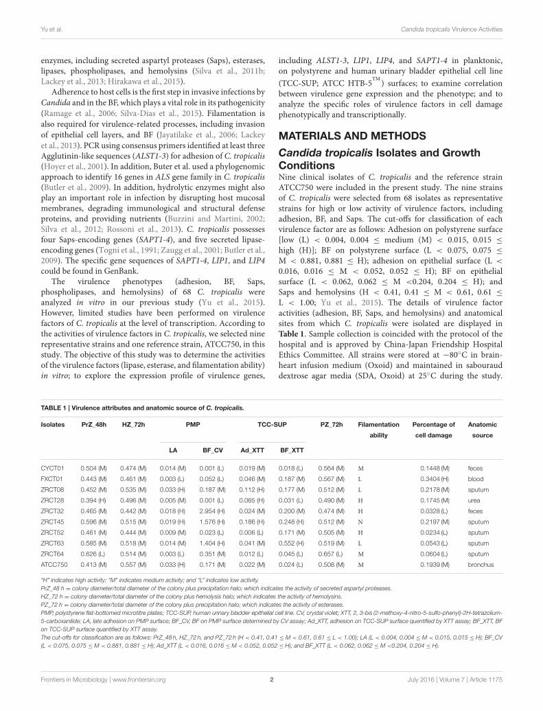
TABLE 1 | Virulence attributes and anatomic source of C. tropicalis.
Isolates PrZ_48h HZ_72h PMP TCC-SUP PZ_72h Filamentation Percentage of Anatomic
ability cell damage source
LA BF_CV Ad_XTT BF_XTT
CYCT01 0.504 (M) 0.474 (M) 0.014 (M) 0.001 (L) 0.019 (M) 0.018 (L) 0.564 (M) M 0.1448 (M) feces
FXCT01 0.443 (M) 0.461 (M) 0.003 (L) 0.052 (L) 0.046 (M) 0.187 (M) 0.567 (M) L 0.3404 (H) blood
ZRCT08 0.452 (M) 0.535 (M) 0.033 (H) 0.187 (M) 0.112 (H) 0.177 (M) 0.512 (M) L 0.2178 (M) sputum
ZRCT28 0.394 (H) 0.496 (M) 0.005 (M) 0.001 (L) 0.065 (H) 0.031 (L) 0.490 (M) H 0.1745 (M) urea
ZRCT32 0.465 (M) 0.442 (M) 0.018 (H) 2.954 (H) 0.024 (M) 0.200 (M) 0.474 (M) H 0.0328 (L) feces
ZRCT45 0.596 (M) 0.515 (M) 0.019 (H) 1.576 (H) 0.186 (H) 0.248 (H) 0.512 (M) N 0.2197 (M) sputum
ZRCT52 0.461 (M) 0.444 (M) 0.009 (M) 0.023 (L) 0.006 (L) 0.171 (M) 0.505 (M) H 0.0234 (L) sputum
ZRCT63 0.585 (M) 0.518 (M) 0.014 (M) 1.404 (H) 0.041 (M) 0.552 (H) 0.519 (M) L 0.0543 (L) sputum
ZRCT64 0.626 (L) 0.514 (M) 0.003 (L) 0.351 (M) 0.012 (L) 0.045 (L) 0.657 (L) M 0.0604 (L) sputum
ATCC750 0.413 (M) 0.557 (M) 0.033 (H) 0.171 (M) 0.022 (M) 0.024 (L) 0.508 (M) M 0.1939 (M) bronchus
“H” indicates high activity; “M” indicates medium activity; and “L” indicates low activity.
PrZ_48 h = colony diameter/total diameter of the colony plus precipitation halo; which indicates the activity of secreted aspartyl proteases.
HZ_72 h = colony diameter/total diameter of the colony plus hemolysis halo; which indicates the activity of hemolysins.
PZ_72 h = colony diameter/total diameter of the colony plus precipitation halo; which indicates the activity of esterases.
PMP, polystyrene flat-bottomed microtitre plates; TCC-SUP, human urinary bladder epithelial cell line. CV, crystal violet; XTT, 2, 3-bis (2-methoxy-4-nitro-5-sulfo-phenyl)-2H-tetrazolium-
5-carboxanilide; LA, late adhesion on PMP surface; BF_CV, BF on PMP surface determined by CV assay; Ad_XTT, adhesion on TCC-SUP surface quentified by XTT assay; BF_XTT, BF
on TCC-SUP surface quantified by XTT assay.
The cut-offs for classification are as follows: PrZ_48 h, HZ_72 h, and PZ_72 h (H < 0.41, 0.41 ≤ M < 0.61, 0.61 ≤ L < 1.00); LA (L < 0.004, 0.004 ≤ M < 0.015, 0.015 ≤ H); BF_CV
(L < 0.075, 0.075 ≤ M < 0.881, 0.881 ≤ H); Ad_XTT (L < 0.016, 0.016 ≤ M < 0.052, 0.052 ≤ H); and BF_XTT (L < 0.062, 0.062 ≤ M <0.204, 0.204 ≤ H).
including ALST1-3, LIP1, LIP4, and SAPT1-4 in planktonic,on polystyrene and human urinary bladder epithelial cell line
(TCC-SUP; ATCC HTB-5TM
) surfaces; to examine correlationbetween virulence gene expression and the phenotype; and toanalyze the specific roles of virulence factors in cell damagephenotypically and transcriptionally.
MATERIALS AND METHODS
Candida tropicalis Isolates and GrowthConditionsNine clinical isolates of C. tropicalis and the reference strainATCC750 were included in the present study. The nine strainsof C. tropicalis were selected from 68 isolates as representativestrains for high or low activity of virulence factors, includingadhesion, BF, and Saps. The cut-offs for classification of eachvirulence factor are as follows: Adhesion on polystyrene surface[low (L) < 0.004, 0.004 ≤ medium (M) < 0.015, 0.015 ≤
high (H)]; BF on polystyrene surface (L < 0.075, 0.075 ≤
M < 0.881, 0.881 ≤ H); adhesion on epithelial surface (L <
0.016, 0.016 ≤ M < 0.052, 0.052 ≤ H); BF on epithelialsurface (L < 0.062, 0.062 ≤ M <0.204, 0.204 ≤ H); andSaps and hemolysins (H < 0.41, 0.41 ≤ M < 0.61, 0.61 ≤
L < 1.00; Yu et al., 2015). The details of virulence factoractivities (adhesion, BF, Saps, and hemolysins) and anatomicalsites from which C. tropicalis were isolated are displayed inTable 1. Sample collection is coincided with the protocol of thehospital and is approved by China-Japan Friendship HospitalEthics Committee. All strains were stored at −80◦C in brain-heart infusion medium (Oxoid) and maintained in sabourauddextrose agar media (SDA, Oxoid) at 25◦C during the study.
Frontiers in Microbiology | www.frontiersin.org 2 July 2016 | Volume 7 | Article 1175
Yu et al. Candida tropicalis Virulence Activities
Yeast cells were inoculated in sabouraud dextrose broth (SDB,Oxoid) and incubated for 18 h at 120 rpm at 37◦C. Afterincubation, the cells were harvested by centrifugation at 5000 ×
g for 5 min and washed twice with phosphate buffered saline(PBS). The washed yeast cells were used in the subsequentassays.
Phenotypical Analysis of Virulence FactorsEsterase and Lipase ActivityEsterase and lipase activity were determined using Tween 80opacity test medium (Galan-Ladero et al., 2010) and tributyrinagar medium (Buzzini and Martini, 2002), respectively. TheTween 80 opacity medium was prepared with 5.0 g peptone,2.5 g NaCl, 0.05 g CaCl2, and 7.5 g agar in 497.5 ml distilledwater, adjusted to pH 6.8 and autoclaved. When the mediumcooled (50◦C), 2.5 ml of Tween 80 was added. The tributyrinagar medium was prepared with 2.5 g peptone, 1.5 g yeast extractand 7.5 g agar in 495 ml distilled water, adjusted to pH 6.0 andautoclaved. When the medium cooled (50◦C), 5.0 ml tributyrinwas added. Five microliters of yeast suspension (108 CFU ml−1)was dropped onto each test medium. The plates were incubatedfor 24, 48, and 72 h at 37◦C. The activity of esterase and lipasewas expressed according to the PZ index (colony diameter/totaldiameter of the colony plus the precipitation halo). The levelsof hydrolytic enzymes activity were established according to thefollowing range of PZ index: PZ < 0.41, high; 0.41 ≤ PZ < 0.61,medium; 0.61≤ PZ< 1.00, low; PZ= 1.00, none (Yu et al., 2015).
Filamentation AbilityThe filamentation ability of C. tropicalis was determinedmicroscopically according to Galan-Ladero et al., with somemodifications (Galan-Ladero et al., 2013). Briefly, C. tropicaliscolonies from SDA plates were incubated into yeast extract-peptone-dextrose (YEPD) broth (Lackey et al., 2013) andincubated at 120 rpm at 30◦C overnight. These cultures wereharvested by centrifugation at 5000 × g for 5 min, washed twicewith PBS and used to inoculate in non-inducing medium (YEPD,30◦C) and inducing medium (0.67% yeast nitrogen base+ 0.75%glucose + 50% fetal bovine serum, 37◦C; Lackey et al., 2013) atan initial optical density of 1.0 at 490 nm, and the cultures wereshaken at 200 rpm for 2 h. Finally, one drop of inoculum wasobserved at 40xmagnification to evaluate the hyphal morphologyunder an inverted light microscopy (Nikon Eclipse Ti-S). Allexperiments were repeated at least three times for each strain.
Analysis of Virulence Genes ExpressionEpithelial CellsHuman urinary bladder epithelial cell line (TCC-SUP) wasused in this study. Cells were cultured in Minimum EssentialMedium (Gibco) containing 10% fetal bovine serum (Gibco)supplemented with MEM Non-Essential Amino Acids Solution(Gibco) and Sodium Pyruvate (Gibco) in cell culture flasks at37◦C with 5% CO2. Cells were washed off using 0.25% trypsin-EDTA solution (Gibco). The cellular density was adjusted to1 × 105 cells ml−1 in fresh Minimum Essential Medium usinga Neubauer chamber. Two milliliter of the suspension was added
to a 12-well-plate and incubated for 24 h. Prior to inoculate withyeast suspension, the wells were washed twice with PBS.
RNA ExtractionThe washed yeast cells were suspended in minimum essentialmedium (Gibco), and the density of suspension was adjustedto 1 × 10⁷ cells ml−1. Four milliliters of yeast suspension ofeach C. tropicalis was then added to corresponding wells in theabsence of TCC-SUP or containing a confluent layer of TCC-SUP,respectively. After incubation at 37◦C with 5% CO2 for 24 h, theculture medium was analyzed for C. tropicalis-induced damageusing a lactate dehydrogenase (LDH) assay. The wells were thenrinsed twice with PBS to remove non-adherent Candida cells. C.tropicalis cells attached to polystyrene flat-bottomed microtiterplates (PMP, Corning) and TCC-SUP cells were scrappedinto 600µl Buffer RLT (Qiagen). Before, RNA extraction,glass beads (0.5 mm diameter, ∼500µl) were added and thetubes were homogenized twice for 30 s, using a TissueLyserLT (Qiagen). After disruption of the yeast cells, the RNeasyMini kit (Qiagen) was used to extract total RNA, accordingto the manufacturer’s recommended protocol. Potential DNAcontamination was removed by RNase-free DNase I (Qiagen)treatment. The purified RNA from all samples was confirmed asDNA-free by real time PCR using C. tropicalis ACT1 (actin) geneprimers. Additionally, RNA was also extracted, following thesame approach, from C. tropicalis planktonic cells (the washedyeast cells). To synthesize complementary DNA (cDNA), the
GoScriptTM
Reverse Transcription System (Promega) was usedaccording to the manufacturer’s instructions.
Primer DesignThe primers for SAPT3 and ACT1 used for real time PCRwere as described by Silva et al. (2011a). The other primers(for ALST1-3, LIP1, LIP4, SAPT1-2, and SAPT4) were designedusing the primer premier 5.0 software. Primers were checked forspecificity using Primer-BLAST (http://www.ncbi.nlm.nih.gov/tools/primer-blast). In addition, to verify the specificity of thenewly designed primer pairs for their corresponding target genes,PCR products were amplified fromATCC750 genomic DNA. Theprimer sequences are listed in Table 2.
Real Time PCRReal time PCR was used to determine the relative levels themRNA transcripts of virulence genes (ALST1-3, LIP1, LIP4, andSAPT1-4) in the RNA samples, with ACT1 used as a referencehousekeeping gene. Gene expression was assessed by the 1CT
method, using the control gene (ACT1) to normalize the data.Each reaction was performed in triplicate and mean values ofrelative expression were analyzed for each gene.
Epithelial Cell Damage AssayThe release of LDH by epithelial cells into the culturemedium was used as a measure of cell damage. The LDHconcentration in the medium was measured at 2, 6, 12,24, and 48 h using the CytoTox-ONE
TMkit (Promega), in
accordance with the manufacturer’s instructions. Two controlsfor LDH activity were prepared: (I) epithelial cells grown
Frontiers in Microbiology | www.frontiersin.org 3 July 2016 | Volume 7 | Article 1175
Yu et al. Candida tropicalis Virulence Activities
TABLE 2 | Candida tropicalis primers used for reverse transcriptase PCR
analysis of virulence and control gene expression.
Sequence (5′–3′) Primer Target PCR product Source
size (bp)
GGGCTCTGGTCGTGATGT Forward ALS1 164 This study
GTGAGGGAATGAGTCTTG Reverse
ACTCGTGCCTATACCTAC Forward ALS2 80 This study
TTGTTGCCGTAATGGTGG Reverse
AGGTGCTGTAGTTGTTCTT Forward ALS3 81 This study
AGCAGTCGGGTTGAAAGG Reverse
TGGGCAGCACCAATCAAAT Forward LIP1 194 This study
GGGTAGACAATCGGGACA Reverse
TTGACTGTGCTCCTTCCT Forward LIP4 138 This study
GCTTTGGACCTTCGTAAT Reverse
TATGACAATGTGCCAGTT Forward SAP1 150 This study
TAAAGCAGTCAAAGTCCC Reverse
GCTGGTTTCTGTGCTTTG Forward SAP2 82 This study
CCACGTAGGCATGTCTTA Reverse
ACTTGGATTTCCAGCGAAGA Forward SAP3 165 Silva et al.,
2011a
AGCCCTTCCAATGCCTAAAT Reverse
CTTCACCTCCTGGTTTCATTTC Forward SAP4 217 This study
TCAACTACCCATAAATCAGAGG Reverse
GACCGAAGCTCCAATGAATC Forward ACT1 181 Silva et al.,
2011a
AATTGGGACAACGTGGGTAA Reverse
in the absence of C. tropicalis; and (II) yeast cells as thesole culture. The LDH activity was analyzed according tothe method of Negri et al. (2011). In addition, colonizationand morphological characteristics of C. tropicalis on TCC-SUPsurface were confirmed by microscopic observation (NikonEclipse Ti-S, 40x magnification) of cells stained with periodicacid-Schiff stain (PAS, Sigma). All experiments were performedin triplicate.
Statistical AnalysisAnalyses were performed using the SPSS 22.0 program. TheWilcoxon signed-rank test was used for comparison differencesbetween two groups. The FriedmanM test was used to determinestatistically significant differences among groups. Spearman’srank correlation was used for correlation analysis. In addition,“heatmap” in Rwas used to illustrate the differential expression ofvirulence genes graphically. In brief, the heat map was generatedby a log transformation of the RT-PCR data, presented as valuesof relative expression. Clustering was performed by averagelinkage and Euclidean distances, used as a distances measure
for both dimensions. P < 0.05 was considered to be statisticalsignificance.
RESULTS
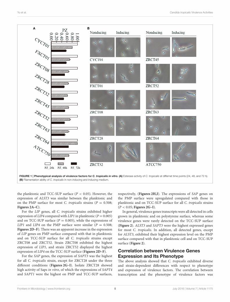
Phenotypical Analysis of Virulence FactorsIn vitroAnalysis of the adhesion, BF and hydrolytic enzymes (includingSaps, phospholipases, and hemolysins) for C. tropicalis indicatedthat all isolates could produce adhesion, BF, Saps, and hemolysinswith strain-dependent features, while no phospholipases weredetected (Yu et al., 2015). C. tropicalis did not display esteraseactivity at 24 h (Figure 1A). However, all 10 strains showed lowactivity of esterase, except for strain ZRCT64, at 48 h. At 72 h,strain ZRCT64 showed low esterase activity and the other isolatesexhibited medium esterase activity (Figure 1A and Table 1). Itindicated that the esterase activity would be strengthened withthe extension time. However, lipase activity was not detected at24, 48, 72 h or even longer (96 and 120 h) in any of the 10C. tropicalis isolates (data not shown).
The filamentation ability of C. tropicalis was evaluated underinducing condition in vitro. In non-inducing medium, strainsCYCT01, FXCT01, ZRCT08, ZRCT28, ZRCT52, ZRCT64, andATCC750 were mainly present as yeast cells, but for ZRCT32,ZRCT45, and ZRCT63, some pseudohyphae or hyphae appearedafter 2 h of incubation (Figure 1B). In inducing medium,ZRCT52 still presented as yeast cells; however, the other isolatesappeared as elongated yeast cells and filamentous cells to varyingdegrees (Figure 1B and Supplementary Material). Concerningto the form of C. tropicalis in non-inducing medium andinducing medium, the filamentation ability of ZRCT32, ZRCT45,and ZRCT63 was strong; FXCT01 and ATCC750 showedmedium filamentation ability; CYCT01, ZRCT08, ZRCT28, andZRCT64 had low filamentation ability; and ZRCT52 displayedno filamentation ability (Table 1). Table 1 shows the BF onPMP and TCC-SUP surfaces of the 10 strains. Interestingly, C.tropicalis strains with strong filamentation abilities also displayedhigh BF on PMP (ZRCT32, ZRCT45, and ZRCT63) and TCC-SUP (ZRCT45 and ZRCT63). Significant correlation was foundbetween the level of filamentation ability and BF on PMP (rs =0.737, P = 0.015), and TCC-SUP (rs = 0.674, P = 0.033).
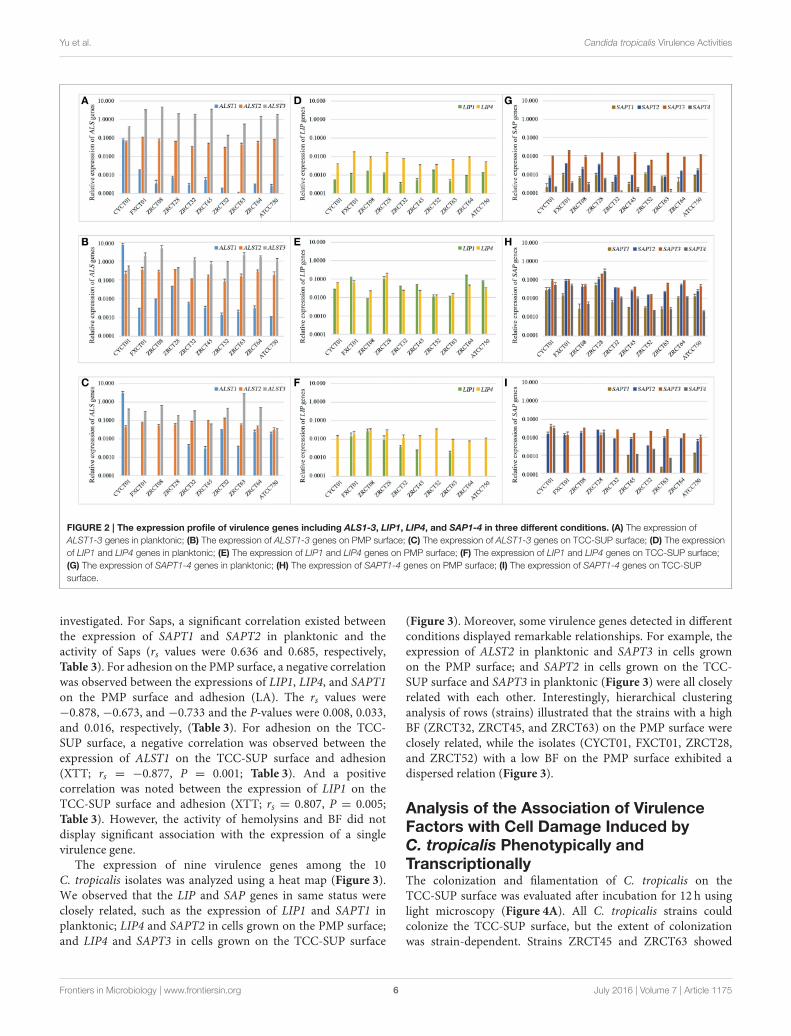
The Expression Profile of Virulence Genesin C. tropicalisReverse transcription PCR (RT-PCR) analysis revealed a widerange of expression profiles of virulence genes in the threeconditions examined (Figure 2). For the ALS genes, ALST3showed the highest expression in the three conditions for allC. tropicalis strains except CYCT01, ZRCT45, and ATCC750(Figures 2A–C). Strain CYCT01 showed the highest expressionlevel of ALST1 on PMP and TCC-SUP surfaces (7.46 and 2.93,respectively, Figures 2B,C). Meanwhile strains ZRCT45 andZRCT750 exhibited the highest expression level of ALST2 on theTCC-SUP surface. Comparing the expressions of the ALS genesin yeast grown on three different status, there was an increase inthe expression of ALST1-2 on the PMP surface compared with
Frontiers in Microbiology | www.frontiersin.org 4 July 2016 | Volume 7 | Article 1175
Yu et al. Candida tropicalis Virulence Activities
FIGURE 1 | Phenotypical analysis of virulence factors for C. tropicalis in vitro. (A) Esterase activity of C. tropicalis at differnet time points (24, 48, and 72 h).
(B) Filamentation ability of C. tropicalis in non-inducing and inducing medium.
the planktonic and TCC-SUP surface (P < 0.05). However, theexpression of ALST3 was similar between the planktonic andon the PMP surface for most C. tropicalis strains (P = 0.508;Figures 2A–C).
For the LIP genes, all C. tropicalis strains exhibited higherexpression of LIP4 compared with LIP1 in planktonic (P= 0.005)and on TCC-SUP surface (P = 0.005), while the expressions ofLIP1 and LIP4 on the PMP surface were similar (P = 0.508;Figures 2D–F). There was an apparent increase in the expressionof LIP genes on PMP surface compared with that in planktonicand on TCC-SUP surface for all C. tropicalis strains exceptZRCT08 and ZRCT52. Strain ZRCT08 exhibited the highestexpression of LIP1, and strain ZRCT52 displayed the highestexpression of LIP4 on the TCC-SUP surface (Figures 2D–F).
For the SAP genes, the expression of SAPT3 was the highest
for all C. tropicalis strain, except for ZRCT28 under the three
different conditions (Figures 2G–I). Isolate ZRCT28 showedhigh activity of Saps in vitro, of which the expressions of SAPT4and SAPT1 were the highest on PMP and TCC-SUP surfaces,
respectively, (Figures 2H,I). The expressions of SAP genes onthe PMP surface were upregulated compared with those inplanktonic and on TCC-SUP surface for all C. tropicalis strains(P < 0.05; Figures 2G–I).
In general, virulence genes transcripts were all detected in cells
grown in planktonic and on polystyrene surface, whereas some
virulence genes were rarely detected on the TCC-SUP surface
(Figure 2). ALST3 and SAPT3 were the highest expressed genesfor most C. tropicalis. In addition, all detected genes, exceptfor ALST3, exhibited their highest expression level on the PMPsurface compared with that in planktonic cell and on TCC-SUPsurface (Figure 2).
Correlation between Virulence GenesExpression and Its PhenotypeThe above analysis showed that C. tropicalis exhibited diverseand strain-dependent differences with respect to phenotypeand expression of virulence factors. The correlation betweentranscription and the phenotype of virulence factors was
Frontiers in Microbiology | www.frontiersin.org 5 July 2016 | Volume 7 | Article 1175
Yu et al. Candida tropicalis Virulence Activities
FIGURE 2 | The expression profile of virulence genes including ALS1-3, LIP1, LIP4, and SAP1-4 in three different conditions. (A) The expression of
ALST1-3 genes in planktonic; (B) The expression of ALST1-3 genes on PMP surface; (C) The expression of ALST1-3 genes on TCC-SUP surface; (D) The expression
of LIP1 and LIP4 genes in planktonic; (E) The expression of LIP1 and LIP4 genes on PMP surface; (F) The expression of LIP1 and LIP4 genes on TCC-SUP surface;
(G) The expression of SAPT1-4 genes in planktonic; (H) The expression of SAPT1-4 genes on PMP surface; (I) The expression of SAPT1-4 genes on TCC-SUP
surface.
investigated. For Saps, a significant correlation existed betweenthe expression of SAPT1 and SAPT2 in planktonic and theactivity of Saps (rs values were 0.636 and 0.685, respectively,Table 3). For adhesion on the PMP surface, a negative correlationwas observed between the expressions of LIP1, LIP4, and SAPT1on the PMP surface and adhesion (LA). The rs values were−0.878, −0.673, and −0.733 and the P-values were 0.008, 0.033,and 0.016, respectively, (Table 3). For adhesion on the TCC-SUP surface, a negative correlation was observed between theexpression of ALST1 on the TCC-SUP surface and adhesion(XTT; rs = −0.877, P = 0.001; Table 3). And a positivecorrelation was noted between the expression of LIP1 on theTCC-SUP surface and adhesion (XTT; rs = 0.807, P = 0.005;Table 3). However, the activity of hemolysins and BF did notdisplay significant association with the expression of a singlevirulence gene.
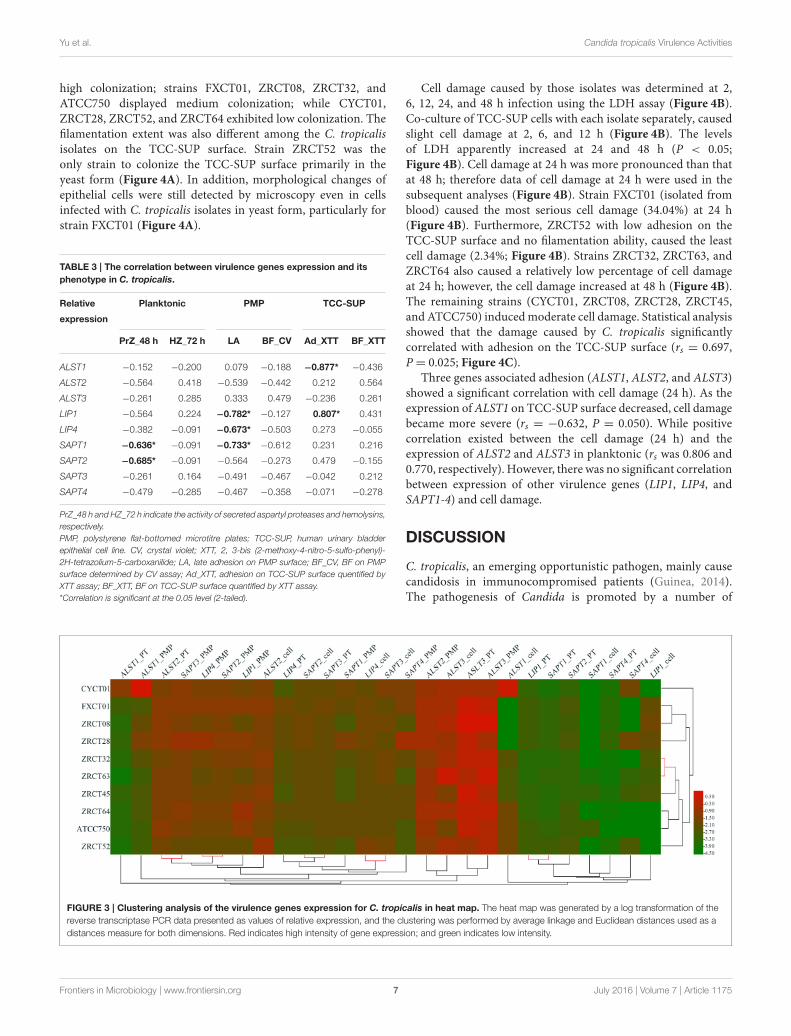
The expression of nine virulence genes among the 10C. tropicalis isolates was analyzed using a heat map (Figure 3).We observed that the LIP and SAP genes in same status wereclosely related, such as the expression of LIP1 and SAPT1 inplanktonic; LIP4 and SAPT2 in cells grown on the PMP surface;and LIP4 and SAPT3 in cells grown on the TCC-SUP surface
(Figure 3). Moreover, some virulence genes detected in differentconditions displayed remarkable relationships. For example, theexpression of ALST2 in planktonic and SAPT3 in cells grownon the PMP surface; and SAPT2 in cells grown on the TCC-SUP surface and SAPT3 in planktonic (Figure 3) were all closelyrelated with each other. Interestingly, hierarchical clusteringanalysis of rows (strains) illustrated that the strains with a highBF (ZRCT32, ZRCT45, and ZRCT63) on the PMP surface wereclosely related, while the isolates (CYCT01, FXCT01, ZRCT28,and ZRCT52) with a low BF on the PMP surface exhibited adispersed relation (Figure 3).
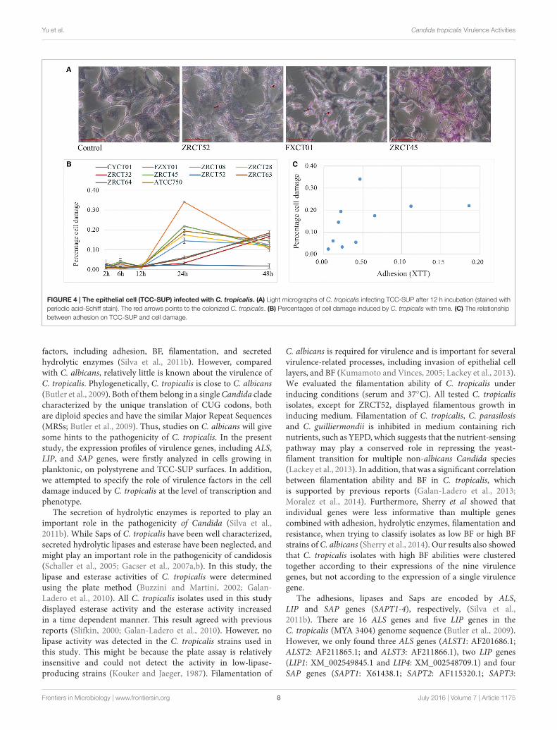
Analysis of the Association of VirulenceFactors with Cell Damage Induced byC. tropicalis Phenotypically andTranscriptionallyThe colonization and filamentation of C. tropicalis on theTCC-SUP surface was evaluated after incubation for 12 h usinglight microscopy (Figure 4A). All C. tropicalis strains couldcolonize the TCC-SUP surface, but the extent of colonizationwas strain-dependent. Strains ZRCT45 and ZRCT63 showed
Frontiers in Microbiology | www.frontiersin.org 6 July 2016 | Volume 7 | Article 1175
Yu et al. Candida tropicalis Virulence Activities
high colonization; strains FXCT01, ZRCT08, ZRCT32, andATCC750 displayed medium colonization; while CYCT01,ZRCT28, ZRCT52, and ZRCT64 exhibited low colonization. Thefilamentation extent was also different among the C. tropicalisisolates on the TCC-SUP surface. Strain ZRCT52 was theonly strain to colonize the TCC-SUP surface primarily in theyeast form (Figure 4A). In addition, morphological changes ofepithelial cells were still detected by microscopy even in cellsinfected with C. tropicalis isolates in yeast form, particularly forstrain FXCT01 (Figure 4A).
TABLE 3 | The correlation between virulence genes expression and its
phenotype in C. tropicalis.
Relative Planktonic PMP TCC-SUP
expression
PrZ_48 h HZ_72 h LA BF_CV Ad_XTT BF_XTT
ALST1 −0.152 −0.200 0.079 −0.188 −0.877* −0.436
ALST2 −0.564 0.418 −0.539 −0.442 0.212 0.564
ALST3 −0.261 0.285 0.333 0.479 −0.236 0.261
LIP1 −0.564 0.224 −0.782* −0.127 0.807* 0.431
LIP4 −0.382 −0.091 −0.673* −0.503 0.273 −0.055
SAPT1 −0.636* −0.091 −0.733* −0.612 0.231 0.216
SAPT2 −0.685* −0.091 −0.564 −0.273 0.479 −0.155
SAPT3 −0.261 0.164 −0.491 −0.467 −0.042 0.212
SAPT4 −0.479 −0.285 −0.467 −0.358 −0.071 −0.278
PrZ_48 h and HZ_72 h indicate the activity of secreted aspartyl proteases and hemolysins,
respectively.
PMP, polystyrene flat-bottomed microtitre plates; TCC-SUP, human urinary bladder
epithelial cell line. CV, crystal violet; XTT, 2, 3-bis (2-methoxy-4-nitro-5-sulfo-phenyl)-
2H-tetrazolium-5-carboxanilide; LA, late adhesion on PMP surface; BF_CV, BF on PMP
surface determined by CV assay; Ad_XTT, adhesion on TCC-SUP surface quentified by
XTT assay; BF_XTT, BF on TCC-SUP surface quantified by XTT assay.
*Correlation is significant at the 0.05 level (2-tailed).
Cell damage caused by those isolates was determined at 2,6, 12, 24, and 48 h infection using the LDH assay (Figure 4B).Co-culture of TCC-SUP cells with each isolate separately, causedslight cell damage at 2, 6, and 12 h (Figure 4B). The levelsof LDH apparently increased at 24 and 48 h (P < 0.05;Figure 4B). Cell damage at 24 h was more pronounced than thatat 48 h; therefore data of cell damage at 24 h were used in thesubsequent analyses (Figure 4B). Strain FXCT01 (isolated fromblood) caused the most serious cell damage (34.04%) at 24 h(Figure 4B). Furthermore, ZRCT52 with low adhesion on theTCC-SUP surface and no filamentation ability, caused the leastcell damage (2.34%; Figure 4B). Strains ZRCT32, ZRCT63, andZRCT64 also caused a relatively low percentage of cell damageat 24 h; however, the cell damage increased at 48 h (Figure 4B).The remaining strains (CYCT01, ZRCT08, ZRCT28, ZRCT45,and ATCC750) inducedmoderate cell damage. Statistical analysisshowed that the damage caused by C. tropicalis significantlycorrelated with adhesion on the TCC-SUP surface (rs = 0.697,P = 0.025; Figure 4C).
Three genes associated adhesion (ALST1, ALST2, and ALST3)showed a significant correlation with cell damage (24 h). As theexpression ofALST1 on TCC-SUP surface decreased, cell damagebecame more severe (rs = −0.632, P = 0.050). While positivecorrelation existed between the cell damage (24 h) and theexpression of ALST2 and ALST3 in planktonic (rs was 0.806 and0.770, respectively). However, there was no significant correlationbetween expression of other virulence genes (LIP1, LIP4, andSAPT1-4) and cell damage.
DISCUSSION
C. tropicalis, an emerging opportunistic pathogen, mainly causecandidosis in immunocompromised patients (Guinea, 2014).The pathogenesis of Candida is promoted by a number of
FIGURE 3 | Clustering analysis of the virulence genes expression for C. tropicalis in heat map. The heat map was generated by a log transformation of the
reverse transcriptase PCR data presented as values of relative expression, and the clustering was performed by average linkage and Euclidean distances used as a
distances measure for both dimensions. Red indicates high intensity of gene expression; and green indicates low intensity.
Frontiers in Microbiology | www.frontiersin.org 7 July 2016 | Volume 7 | Article 1175
Yu et al. Candida tropicalis Virulence Activities
FIGURE 4 | The epithelial cell (TCC-SUP) infected with C. tropicalis. (A) Light micrographs of C. tropicalis infecting TCC-SUP after 12 h incubation (stained with
periodic acid-Schiff stain). The red arrows points to the colonized C. tropicalis. (B) Percentages of cell damage induced by C. tropicalis with time. (C) The relationship
between adhesion on TCC-SUP and cell damage.
factors, including adhesion, BF, filamentation, and secretedhydrolytic enzymes (Silva et al., 2011b). However, comparedwith C. albicans, relatively little is known about the virulence ofC. tropicalis. Phylogenetically, C. tropicalis is close to C. albicans(Butler et al., 2009). Both of them belong in a singleCandida cladecharacterized by the unique translation of CUG codons, bothare diploid species and have the similar Major Repeat Sequences(MRSs; Butler et al., 2009). Thus, studies on C. albicans will givesome hints to the pathogenicity of C. tropicalis. In the presentstudy, the expression profiles of virulence genes, including ALS,LIP, and SAP genes, were firstly analyzed in cells growing inplanktonic, on polystyrene and TCC-SUP surfaces. In addition,we attempted to specify the role of virulence factors in the celldamage induced by C. tropicalis at the level of transcription andphenotype.
The secretion of hydrolytic enzymes is reported to play animportant role in the pathogenicity of Candida (Silva et al.,2011b). While Saps of C. tropicalis have been well characterized,secreted hydrolytic lipases and esterase have been neglected, andmight play an important role in the pathogenicity of candidosis(Schaller et al., 2005; Gacser et al., 2007a,b). In this study, thelipase and esterase activities of C. tropicalis were determinedusing the plate method (Buzzini and Martini, 2002; Galan-Ladero et al., 2010). All C. tropicalis isolates used in this studydisplayed esterase activity and the esterase activity increasedin a time dependent manner. This result agreed with previousreports (Slifkin, 2000; Galan-Ladero et al., 2010). However, nolipase activity was detected in the C. tropicalis strains used inthis study. This might be because the plate assay is relativelyinsensitive and could not detect the activity in low-lipase-producing strains (Kouker and Jaeger, 1987). Filamentation of
C. albicans is required for virulence and is important for severalvirulence-related processes, including invasion of epithelial celllayers, and BF (Kumamoto and Vinces, 2005; Lackey et al., 2013).We evaluated the filamentation ability of C. tropicalis underinducing conditions (serum and 37◦C). All tested C. tropicalisisolates, except for ZRCT52, displayed filamentous growth ininducing medium. Filamentation of C. tropicalis, C. parasilosisand C. guilliermondii is inhibited in medium containing richnutrients, such as YEPD, which suggests that the nutrient-sensingpathway may play a conserved role in repressing the yeast-filament transition for multiple non-albicans Candida species(Lackey et al., 2013). In addition, that was a significant correlationbetween filamentation ability and BF in C. tropicalis, whichis supported by previous reports (Galan-Ladero et al., 2013;Moralez et al., 2014). Furthermore, Sherry et al showed thatindividual genes were less informative than multiple genescombined with adhesion, hydrolytic enzymes, filamentation andresistance, when trying to classify isolates as low BF or high BFstrains of C. albicans (Sherry et al., 2014). Our results also showedthat C. tropicalis isolates with high BF abilities were clusteredtogether according to their expressions of the nine virulencegenes, but not according to the expression of a single virulencegene.
The adhesions, lipases and Saps are encoded by ALS,LIP and SAP genes (SAPT1-4), respectively, (Silva et al.,2011b). There are 16 ALS genes and five LIP genes in theC. tropicalis (MYA 3404) genome sequence (Butler et al., 2009).However, we only found three ALS genes (ALST1: AF201686.1;ALST2: AF211865.1; and ALST3: AF211866.1), two LIP genes(LIP1: XM_002549845.1 and LIP4: XM_002548709.1) and fourSAP genes (SAPT1: X61438.1; SAPT2: AF115320.1; SAPT3:
Frontiers in Microbiology | www.frontiersin.org 8 July 2016 | Volume 7 | Article 1175
Yu et al. Candida tropicalis Virulence Activities
AF115321.1; and SAPT4: AF115322.1) in GenBank. Therefore,we explored the expression profiles of ALS1-3, LIP1, LIP4, andSAPT1-4 genes in three different conditions. RT-PCR analysisrevealed that the expressions of ALST3 and SAPT3 were thehighest among the ALS and SAP genes, respectively. Thisobservation for SAPT3 expression was similar to publishedresults (Negri et al., 2011, 2012; Silva et al., 2011a). Additionally,Silva et al. found that there was an apparent increase in theexpression of most SAP genes when the cells were grown on aPMP surface (Silva et al., 2011a). Our study also showed thatC. tropicalis exhibited a higher expression level of virulencegenes not only SAP genes, but also ALST1-2, LIP1, and LIP4genes, on the PMP surface compared with growth on epithelialand planktonic cells. Other study demonstrated that sessileC. albicans cells on a biotic surface secreted more aspartylproteases than planktonic cells (Mendes et al., 2007). Theseresults suggested that biofilm formation on polystyrene couldpromote the expression of other virulence genes, but theunderlying mechanisms need to be further analyzed. In ourprevious study, a negative correlation was found between activityof Saps in vitro and adhesion (LA) on a PMP surface. At thesame time, the expressions of LIP1, LIP4 and SAPT1 negativelycorrelated with adhesion (LA) in the present study. Anotherstudy also revealed a significant negative correlation betweenSAPT3 and the biomass data in C. albicans (Sherry et al.,2014). Our study revealed that the activity of Saps in a mediumcontaining bovine serum albumin (BSA) as the sole source ofnitrogen was associated with the expression of SAPT1 and SAPT2in planktonic, which was in accordance with Zaugg’s results thatthe gene products (Sap1p) was the dominant product in BSAmedium (Zaugg et al., 2001). However, the majority of strainsdid not express SAPT1 on the TCC-SUP surface, suggestingits limited involvement in invasion and tissue damage (Silvaet al., 2011a). Additionally, no significant correlation was foundbetween the activity of Saps or the expression of SAP genes andcell damage. Other investigators also showed that for C. albicans(Lermann and Morschhauser, 2008; Naglik et al., 2008), and C.tropicalis (Okawa et al., 2008; Silva et al., 2011a), Saps are notrequired for invasion and damage to reconstituted human oralepithelium (RHOE). Based on the present and previous results,Saps probably have a limited role in epithelial cell or tissuedamage.
Our study showed that C. tropicalis was able to colonize anddevelop filamentous forms on epithelial cells (TCC-SUP) in astrain-dependent manner. The greatest cell damage was inducedby C. tropicalis at 24 h, and the most pronounced differenceswere found among isolates at this time point, which is similarto results reported previously (Silva et al., 2011a). This mightreflect a variation in the ability of distinct strains to continue togrow on epithelial cells and induce damage after initial invasion.The pathogenicity of C. albicans strains is well correlatedwith adherence to mucosal epithelial cells (Hela cells) andhydrophobicity (Okawa et al., 2007). Our results also showed thatadhesion on the TCC-SUP surface correlated positively with cell
damage. In addition, we observed significant correlation betweencell damage and the expression of ALST1-3 in C. tropicalis, whichfurther confirmed the hypothesis that adhesion on epithelial cellsplays an important role in subsequent cell damage. However,isolate FXCT01 from the blood is an exception: it exhibited thehighest percentages of cell damage with only moderate adhesionactivity. Importantly, one strain, ZRCT52, with no filamentationability, induced the least cell damage at 24 and 48 h. Thisobservation supported the hypothesis that filamentation plays arole in epithelial cell damage.
In conclusion, we analyzed the virulence factors ofC. tropicalisphenotypically and transcriptionally. C. tropicalis displayedstrain-dependent filamentation ability. Isolates with highfilamentation ability also displayed high BF on polystyrene.These results suggested that filamentation plays a critical rolein BF. Furthermore, ALST3 and SAPT3 were expressed at thehighest levels in most C. tropicalis strains. The expressionof virulence genes (ALST1-2, LIP1, LIP4, and SAPT1-4) wasupregulated on the PMP surface compared with planktonic andTCC-SUP counterparts. Isolates with high BF on polystyreneformed a distinct group in the hierarchical clustering analysisof virulence genes. Moreover, adhesion to epithelial cells andthe expression of ALST2-3 encoding an adhesins, correlatedpositively with cell damage, which suggested that adhesionon epithelial cells plays an important role in cell damage.One strain (ZRCT52) with no filamentation ability causedthe least cell damage, which might indicate that filamentationis involved in cell damage. Above all, C. tropicalis displayeddiverse and strain-dependent differences with respect tophenotype and transcription of virulence factors. Our studywill aid the further study of the invasive mechanisms ofC. tropicalis.
AUTHOR CONTRIBUTIONS
SY performed the experiments, analyzed the data, and drafted themanuscript. WL, XL, and JC helped in the related experiments.JL participated in the design of the experiment proposal andrevision of the paper. YW conceived the study, supervised theresearch, and revised the manuscript.
ACKNOWLEDGMENTS
This research was supported by the National Natural ScienceFoundation of China (Youth Project no. 81301409), theNational Key Technology Support Program (grant no.2012BAI11B05), and the National Sci-Tech Key Project(grant no. 2013ZX10004203-002).
SUPPLEMENTARY MATERIAL
The Supplementary Material for this article can be foundonline at: http://journal.frontiersin.org/article/10.3389/fmicb.2016.01175
Frontiers in Microbiology | www.frontiersin.org 9 July 2016 | Volume 7 | Article 1175
Yu et al. Candida tropicalis Virulence Activities
REFERENCES
Butler, G., Rasmussen, M. D., Lin, M. F., Santos, M. A., Sakthikumar, S., Munro,
C. A., et al. (2009). Evolution of pathogenicity and sexual reproduction in eight
Candida genomes. Nature 459, 657–662. doi: 10.1038/nature08064
Buzzini, P., andMartini, A. (2002). Extracellular enzymatic activity profiles in yeast
and yeast-like strains isolated from tropical environments. J. Appl. Microbiol.
93, 1020–1025. doi: 10.1046/j.1365-2672.2002.01783.x
Gacser, A., Schafer, W., Nosanchuk, J. S., Salomon, S., and Nosanchuk, J. D.
(2007a). Virulence ofCandida parapsilosis,Candida orthopsilosis, andCandida
metapsilosis in reconstituted human tissue models. Fungal Genet. Biol. 44,
1336–1341. doi: 10.1016/j.fgb.2007.02.002
Gacser, A., Trofa, D., Schafer, W., and Nosanchuk, J. D. (2007b). Targeted gene
deletion in Candida parapsilosis demonstrates the role of secreted lipase in
virulence. J. Clin. Invest. 117, 3049–3058. doi: 10.1172/JCI32294
Galan-Ladero, M. A., Blanco-Blanco, M. T., Hurtado, C., Perez-Giraldo, C.,
Blanco, M. T., and Gomez-Garcia, A. C. (2013). Determination of biofilm
production by Candida tropicalis isolated from hospitalized patients and its
relation to cellular surface hydrophobicity, plastic adherence and filamentation
ability. Yeast 30, 331–339. doi: 10.1002/yea.2965
Galan-Ladero, M. A., Blanco, M. T., Sacristan, B., Fernandez-Calderon, M. C.,
Perez-Giraldo, C., and Gomez-Garcia, A. C. (2010). Enzymatic activities of
Candida tropicalis isolated from hospitalized patients.Med.Mycol. 48, 207–210.
doi: 10.3109/13693780902801242
Guinea, J. (2014). Global trends in the distribution of Candida species causing
candidemia. Clin. Microbiol. Infect. 20(Suppl. 6), 5–10. doi: 10.1111/1469-
0691.12539
Hirakawa, M. P., Martinez, D. A., Sakthikumar, S., Anderson, M. Z., Berlin,
A., Gujja, S., et al. (2015). Genetic and phenotypic intra-species variation in
Candida albicans. Genome Res. 25, 413–425. doi: 10.1101/gr.174623.114
Hoyer, L. L., Fundyga, R., Hecht, J. E., Kapteyn, J. C., Klis, F. M., and Arnold, J.
(2001). Characterization of agglutinin-like sequence genes from non-albicans
Candida and phylogenetic analysis of the ALS family. Genetics 157, 1555–1567.
Available online at: http://www.genetics.org/search/Characterization%252
Bof%252Bagglutinin-like%252Bsequence%252Bgenes%252Bfrom%252Bnon-
albicans%252BCandida%252Band%252Bphylogenetic%252Banalysis%252Bof
%252Bthe%252B
Jayatilake, J. A., Samaranayake, Y. H., Cheung, L. K., and Samaranayake, L.
P. (2006). Quantitative evaluation of tissue invasion by wild type, hyphal
and SAP mutants of Candida albicans, and non-albicans Candida species in
reconstituted human oral epithelium. J. Oral Pathol. Med. 35, 484–491. doi:
10.1111/j.1600-0714.2006.00435.x
Kouker, G., and Jaeger, K. E. (1987). Specific and sensitive plate assay for bacterial
lipases. Appl. Environ. Microbiol. 53, 211–213.
Kumamoto, C. A., and Vinces, M. D. (2005). Contributions of hyphae and hypha-
co-regulated genes to Candida albicans virulence. Cell. Microbiol. 7, 1546–1554.
doi: 10.1111/j.1462-5822.2005.00616.x
Lackey, E., Vipulanandan, G., Childers, D. S., and Kadosh, D. (2013). Comparative
evolution of morphological regulatory functions inCandida species. Eukaryotic
Cell 12, 1356–1368. doi: 10.1128/EC.00164-13
Lermann, U., and Morschhauser, J. (2008). Secreted aspartic proteases are not
required for invasion of reconstituted human epithelia by Candida albicans.
Microbiology 154(Pt 11), 3281–3295. doi: 10.1099/mic.0.2008/022525-0
Mendes, A., Mores, A. U., Carvalho, A. P., Rosa, R. T., Samaranayake, L. P.,
and Rosa, E. A. (2007). Candida albicans biofilms produce more secreted
aspartyl protease than the planktonic cells. Biol. Pharm. Bull. 30, 1813–1815.
doi: 10.1248/bpb.30.1813
Moralez, A. T., Franca, E. J., Furlaneto-Maia, L., Quesada, R. M., and Furlaneto,
M. C. (2014). Phenotypic switching in Candida tropicalis: association with
modification of putative virulence attributes and antifungal drug sensitivity.
Med. Mycol. 52, 106–114. doi: 10.3109/13693786.2013.825822
Naglik, J. R., Moyes, D., Makwana, J., Kanzaria, P., Tsichlaki, E., Weindl, G.,
et al. (2008). Quantitative expression of the Candida albicans secreted aspartyl
proteinase gene family in human oral and vaginal candidiasis. Microbiology
154(Pt 11), 3266–3280. doi: 10.1099/mic.0.2008/022293-0
Negri, M., Botelho, C., Silva, S., Lopes, L. M., Henriques, M., Azeredo,
J., et al. (2011). An in vitro evaluation of Candida tropicalis infectivity
using human cell monolayers. J. Med. Microbiol. 60(Pt 9), 1270–1275. doi:
10.1099/jmm.0.031195-0
Negri, M., Silva, S., Breda, D., Henriques, M., Azeredo, J., and Oliveira, R. (2012).
Candida tropicalis biofilms: effect on urinary epithelial cells.Microb. Pathog. 53,
95–99. doi: 10.1016/j.micpath.2012.05.006
Okawa, Y., Miyauchi, M., and Kobayashi, H. (2008). Comparison of pathogenicity
of various Candida tropicalis strains. Biol. Pharm. Bull. 31, 1507–1510. doi:
10.1248/bpb.31.1507
Okawa, Y., Miyauchi, M., Takahashi, S., and Kobayashi, H. (2007). Comparison
of pathogenicity of various Candida albicans and C. stellatoidea strains. Biol.
Pharm. Bull. 30, 1870–1873. doi: 10.1248/bpb.30.1870
Ramage, G., Martinez, J. P., and Lopez-Ribot, J. L. (2006). Candida biofilms on
implanted biomaterials: a clinically significant problem. FEMS Yeast Res. 6,
979–986. doi: 10.1111/j.1567-1364.2006.00117.x
Rossoni, R. D., Barbosa, J. O., Vilela, S. F., Jorge, A. O., and Junqueira, J. C.
(2013). Comparison of the hemolytic activity between C. albicans and non-
albicans Candida species. Braz. Oral Res. 27, 484–489. doi: 10.1590/S1806-
83242013000600007
Schaller, M., Borelli, C., Korting, H. C., andHube, B. (2005). Hydrolytic enzymes as
virulence factors ofCandida albicans.Mycoses 48, 365–377. doi: 10.1111/j.1439-
0507.2005.01165.x
Sherry, L., Rajendran, R., Lappin, D. F., Borghi, E., Perdoni, F., Falleni, M.,
et al. (2014). Biofilms formed by Candida albicans bloodstream isolates
display phenotypic and transcriptional heterogeneity that are associated with
resistance and pathogenicity. BMC Microbiol. 14:182. doi: 10.1186/1471-2180-
14-182
Silva, S., Hooper, S. J., Henriques, M., Oliveira, R., Azeredo, J., and Williams,
D. W. (2011a). The role of secreted aspartyl proteinases in Candida tropicalis
invasion and damage of oral mucosa. Clin. Microbiol. Infect. 17, 264–272. doi:
10.1111/j.1469-0691.2010.03248.x
Silva, S., Negri, M., Henriques, M., Oliveira, R., Williams, D. W., and Azeredo,
J. (2011b). Adherence and biofilm formation of non-Candida albicans
Candida species. Trends Microbiol. 19, 241–247. doi: 10.1016/j.tim.2011.
02.003
Silva, S., Negri, M., Henriques, M., Oliveira, R., Williams, D. W., and Azeredo, J.
(2012). Candida glabrata, Candida parapsilosis and Candida tropicalis: biology,
epidemiology, pathogenicity and antifungal resistance. FEMS Microbiol. Rev.
36, 288–305. doi: 10.1111/j.1574-6976.2011.00278.x
Silva-Dias, A., Miranda, I. M., Branco, J., Monteiro-Soares, M., Pina-Vaz,
C., and Rodrigues, A. G. (2015). Adhesion, biofilm formation, cell
surface hydrophobicity, and antifungal planktonic susceptibility: relationship
among Candida spp. Front. Microbiol. 6:205. doi: 10.3389/fmicb.2015.
00205
Slifkin, M. (2000). Tween 80 opacity test responses of various Candida species. J.
Clin. Microbiol. 38, 4626–4628. Available online at: http://jcm.asm.org/content/
38/12/4626.abstract?sid=229a0cd6-2d33-4b30-8df9-575582afce39
Togni, G., Sanglard, D., Falchetto, R., and Monod, M. (1991). Isolation and
nucleotide sequence of the extracellular acid protease gene (ACP) from
the yeast Candida tropicalis. FEBS Lett. 286, 181–185. doi: 10.1016/0014-
5793(91)80969-A
Yu, S. B., Li, W. G., Che, J., Bian, F. N., Lu, J. X., and Wu, Y. (2015). Study on
virulence factors of Candida tropicalis isolated from clinical samples. Chin. J.
Epidemiol. 36, 1162–1166. doi: 10.3760/cma.j.issn.0254-6450.2015.10.027
Zaugg, C., Borg-Von Zepelin, M., Reichard, U., Sanglard, D., and Monod, M.
(2001). Secreted aspartic proteinase family ofCandida tropicalis. Infect. Immun.
69, 405–412. doi: 10.1128/IAI.69.1.405-412.2001
Conflict of Interest Statement: The authors declare that the research was
conducted in the absence of any commercial or financial relationships that could
be construed as a potential conflict of interest.
Copyright © 2016 Yu, Li, Liu, Che, Wu and Lu. This is an open-access article
distributed under the terms of the Creative Commons Attribution License (CC BY).
The use, distribution or reproduction in other forums is permitted, provided the
original author(s) or licensor are credited and that the original publication in this
journal is cited, in accordance with accepted academic practice. No use, distribution
or reproduction is permitted which does not comply with these terms.
Frontiers in Microbiology | www.frontiersin.org 10 July 2016 | Volume 7 | Article 1175